!"#$%&'%()'*+,-%&&.'+('*/%)+%,#+0' 12/.,'3%)+"4#%52.
|
|
|
- Juniper Lambert
- 5 years ago
- Views:
Transcription


1 !"#$%&'%()'*+,-%&&.'+('*/%)+%,#+0' 12/.,'3%)+"4#%52.!"#$%&'()$*+&,--#&$.//,0'1232'!-#0'45 *6 '7849!!"#$%&'"(&)*+),$-.*/*01) 2$34/&1)*+)5"-.3.(") 6%.(3")*+)7*(08/$)9(.:"%;.&1)) 1
")


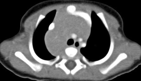
2 ,&*-'. /%0*'0$+*'#&#) 10*2#02*"1!"#$%&&'(")(*+,'#(-%."/0-%!+'! 340*&%1&#)0+)0$")(2%5!"#$%&#'() *++,+-!"#$%&#'()"**+* 67"("0'( 2
(\"*+)$*,')- Pneumothorax? Emphysema? Lung hypoplasia?")
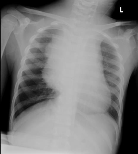
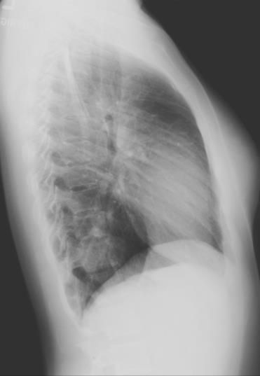
3 A 3 year old boy with acute dyspnea 1 day!"#$%&'($)("*+)$*,')- Pneumothorax? Emphysema? Lung hypoplasia? Foreign body obstruction? 3


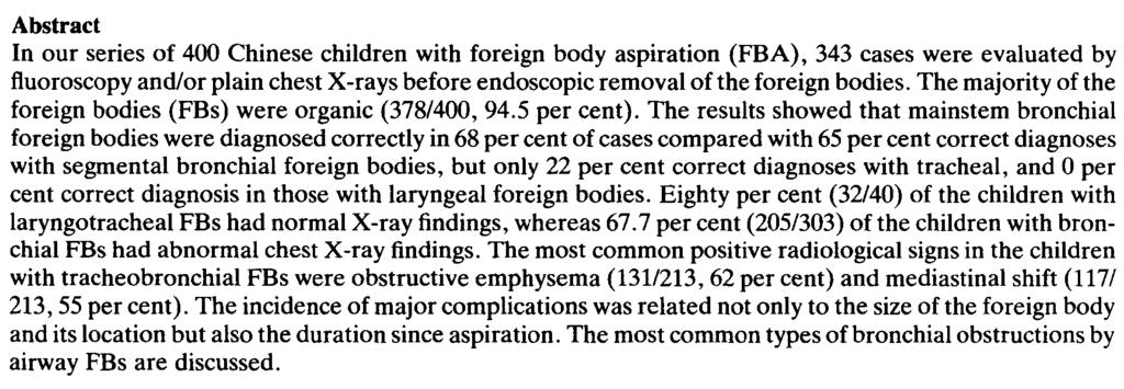
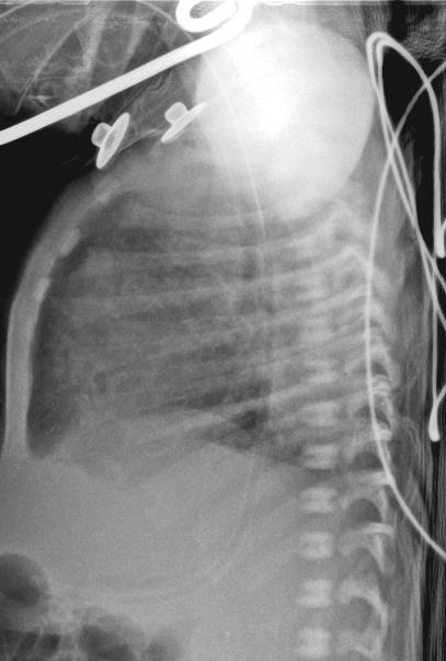
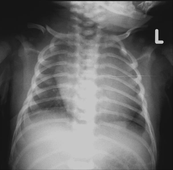
4 8+0'0&+% Symmetrical ribs and the medial border of the clavicles Longer posterior ribs 4


5 a 4 hour baby girl with meconium peritonitis a 3 month old boy Congenital lobar over-inflation of right lung 5


6 a 3 month old girl with wheezing Right lung hypoplasia 6

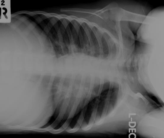
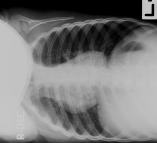
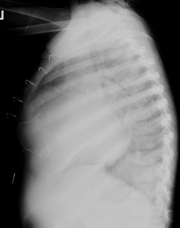
7 Left lateral decubitus 9":0);'&%)<*+%#$&'()+<10*2#0&+% A 2 year old boy dyspnea for 2days Right lateral decubitus 7

8 !"#$%&'()"*+(,-.%#/0%"' 8
9 =++*)/%1>&*'0&+% Interstitial pneumonia Pulmonary edema Cardiomegaly Mediastinal mass! 9


10 A 3 month old boy post operative CHD worsening pulmonary edema?? Curr Probl Diagn Radiol, May/June 1998 Tetralogy of Fallot? Courtesy of Edward Y Lee 10
11 Round cardiac apex RVH Prominent perihilar bronchovascular markings 9+*?+1&1 Horizontal or upturned anterior ribs.//0102)3&*,31$%3&*40$5 Assess cardiomegaly Determine location and confirm pulmonary nodule on frontal view Clarify number & position of FB Evaluate LN and pulmonary hilum 11
12 12


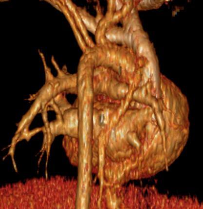
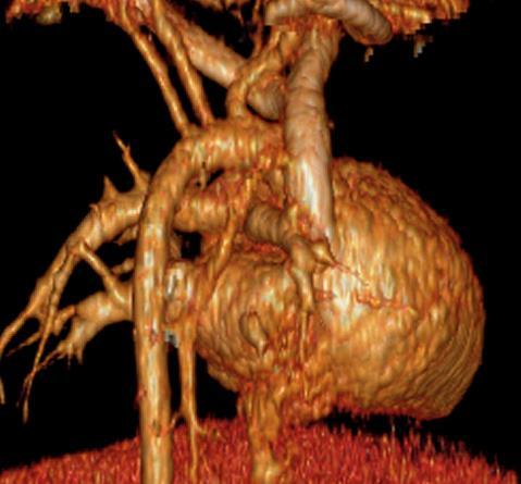
13 Pulmonary AVM.&+/'0-13

14 ,&*-'.1 Tracheal buckling simulating extrinsic compression by a mass Neck flexion simulating retropharyngeal soft tissue thickening! Tracheal buckling 14


15 Prevertebral soft tissue thickening? * * Curr Probl Diagn Radiol, May/June 1998 a 1- year old girl with neck stiffness * * Prevertebral soft tissue thickening? 15
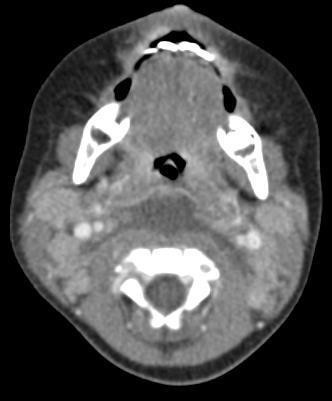
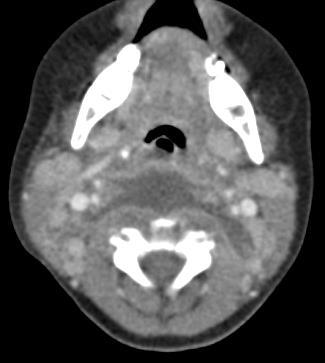
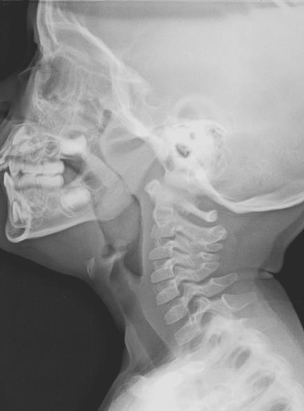
16 * * Retropharyngeal fluid/abscess * ()$*+&,--#&$./,'!0'-*'&:2';<-)=' >&?$/:'@7847A'4BC45BD4E9' a 3 - year boy with snoring 16
17 9'0"*'()A"#7 Prominent normal lymphoid tissue Over diagnosis of airway masses Significant enlargement airway obliteration Considered immune deficiency > 6 months! 1%2+'2$,+'#&#) 32+4#24+"- 17
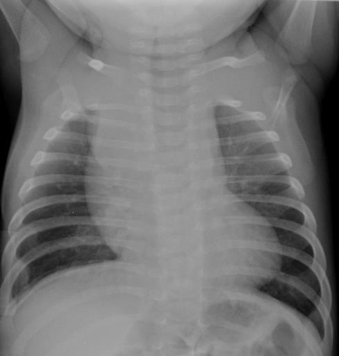
18 Mediastinal mass? Most frequent pitfall Located in prevascular space Large at birth!$.;21 Less prominent in older children Never displace mediastinum 18
?")
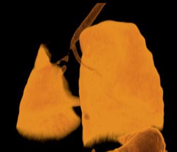
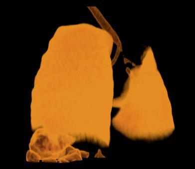
19 !$.;21 Unilateral / bilateral Wavy margin Cardio-thymic notch Thymic sail sign * -'0"*)?"%1&0. 19
1'&()1&5%")
20 -'B.)1&5%!$.;&#)1'&()1&5% 20


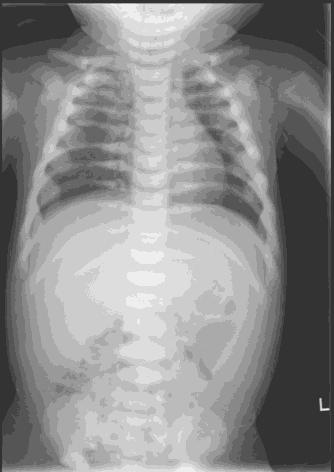
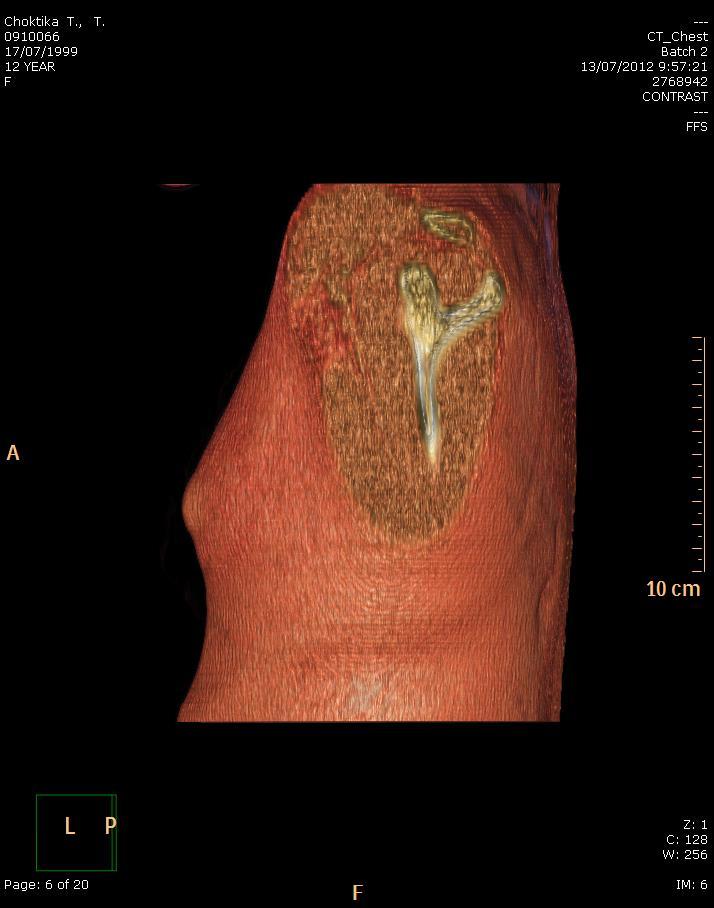
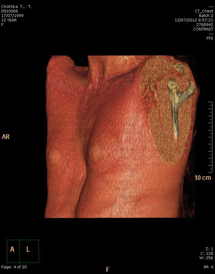
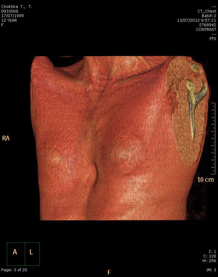
21 Rebound thymic hyperplasia Ectopic location: posterior mediastinum/ retrocaval position Above sternum during respiration / crying US or CT or MRI!$.;21 11/12/2009 A 7 year old boy NHL stage II complete treatment 17/03/ /7/
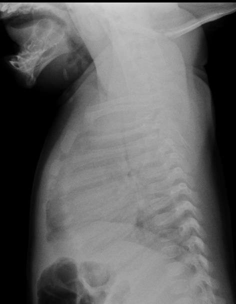
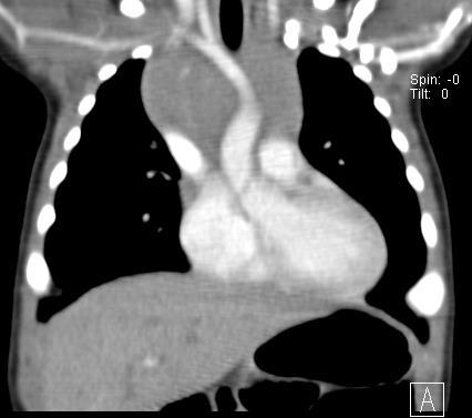
22 67"8'9*4:*;$/03910)3&*8399*<< 2-month-old girl with fever and coughing Courtesy of Edward Y Lee =291$%02%*;$/03910)3&*>?1$)902) Courtesy of Edward Y Lee Key Point!: contiguous with normal thymus 22
902) Courtesy of Edward Y Lee")

=2(;+%'*.")
23 @$%A0(3&*>?1$)902) Courtesy of Edward Y Lee Confluence of the pulmonary veins at right paraspinal region Prominent normal MPA in adolescent girls A+*;'()=2(;+%'*.) C'1#2('*)60*2#02*"1 Post stenotic dilatation in PS 23
 C'1#2('*)60*2#02*\"1 Confluence of the pulmonary veins at right paraspinal region Prominent normal")
24 a 10 year old boy contacted TB A+*;'()=2(;+%'*.) C'1#2('*)60*2#02*"1 Confluence of the pulmonary veins at right paraspinal region Prominent normal MPA in adolescent girls Post stenotic dilatation in PS 24


25 a - 18 year old girl check up Ductus bump A baby boy 2 nd DOL 25
2,))")
26 D2#021)E2;> Transient limited to newborn Prominent aortic knob Distended PA and DA The most prominent on 2 nd or 3 rd DOL *52+&%-&#)2,)) 2$")64%7-26


27 Pneumonia? Asymmetrical breast development Neonatal incubator access port 27


28 Right pleural effusion? a 1- month old girl - TGA Pneumothorax? 28
29 67&%)F+(? Performed portable in ICU Misinterpreted as pneumothorax Pleural line distinct, curvilinear parallel lateral chest wall Skin fold indistinct and random course 38"("2,% 29
67\"(\"0+% Simulating abnormalities to the")
30 /;;'02*")="?&'0*&#)67"("0+% Simulating abnormalities to the inexperienced observers! Mediastinal mass? 30

31 60"*%'()G11&:&#'0&+%)H"%0"*1 Not fused in infants and young children Slightly oblique Manubrial ossification center: mistaken for a mediastinal mass or foreign body! a 8 month old girl CHARGE association 31

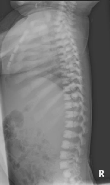
32 /%#+;>("0")G11&:&#'0&+% Caudal Cranial posterior spinal fusion Thoracic posterior spinal elements 1 st year Cervical spinal elements 2 nd -3 rd year Lack of clinical finding or pathologic widening of spinal canal 32


33 Enlarged cervical spinal canal 6-12 months 33
34 OSTEOPETROSIS? Bone in bone appearance OSTEOPETROSIS 34


35 E+%")&%)E+%"),>>"'*'%#" Younger than 2 months Will diminish with time Axially oriented cleft?? 35

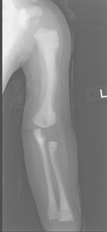
36 Young infants Noted in lateral projection Anterior notch sinusoid blood space Posterior notch penetrating arteries and veins avulsion fracture?? C"*0"<*'()A+0#$ 36


37 ='*0&'()G11&:#'0&+% Early ossified cartilaginous ring epiphysis at 12 years Mimic avulsion fracture Prominent intertubercular groove 37


38 =*+;&%"%0)/%0"*02<"*#2('*) I*++B" Tendon of long head of biceps tendon Simulate periosteal reaction or focal bony erosion Bilateral and symmetrical 38
39 J+&%0)C'#22; Immobilization Glenohumeral joint Intra-articular nitrogen 39


40 ="#021)34#'B'02; Narrow AP dimension Cardiomegaly Obscured right heart border Relatively vertical orientation of anterior ribs A 9 year old girl pectus excavatum 40
41 41
42 8&<1 Bulbuous cartilaginous anterior rib ends Misleading calcified mass or expansile rib!'7")k+;")=+&%01 Technical factors, un-cooperative patients, external factors Normal structures and immature skeleton Prevent misinterpretation Repeated radiographs, additional views or other advanced imaging 42
43 !"#$%&'()&*(+&'()+&#,,-$,.($ 43
Chest X rays and Case Studies. No disclosures. Outline 5/31/2018. Carlo Manalo, M.D. Department of Radiology Loma Linda University Children s Hospital
 Chest X rays and Case Studies Carlo Manalo, M.D. Department of Radiology Loma Linda University Children s Hospital No disclosures. Outline Importance of history Densities delineated on radiography An approach
Objectives. What is a Chest X Ray? CXR Workshop. Definition (diagnostic tool/internal PE) Types. Cost
 Types. Cost") Objectives CAPA 2011 Christy Wilson, PA C Georgia Lung Associates Identify the radiographic landmarks on a chest radiograph Recognize identifiers of poor quality on the chest radiograph Outline an approach
Objectives CAPA 2011 Christy Wilson, PA C Georgia Lung Associates Identify the radiographic landmarks on a chest radiograph Recognize identifiers of poor quality on the chest radiograph Outline an approach
Manage TB Dr. A. Chitrakumar Madras Medical College and RGGGH Institute of Thoracic Medicine, Chennai
 Manage TB Dr. A. Chitrakumar Madras Medical College and RGGGH Institute of Thoracic Medicine, Chennai Lecture 16 Radiology in diagnosis of Tuberculosis Session 01 So, welcome to the session Radiology in
Chapter 2 Cardiac Interpretation of Pediatric Chest X-Ray
 Chapter 2 Cardiac Interpretation of Pediatric Chest X-Ray Ra-id Abdulla and Douglas M. Luxenberg Key Facts The cardiac silhouette occupies 50 55% of the chest width on an anterior posterior chest X-ray
UERMMMC Department of Radiology. Basic Chest Radiology
 UERMMMC Department of Radiology Basic Chest Radiology PHYSICS DENSITIES BONE SOFT TISSUES WATER FAT AIR TELEROENTGENOGRAM Criteria for an Ideal Chest Radiograph 1. Upright 2. Posteroanterior View 3. Full
Pediatric TB Intensive Houston, Texas October 14, 2013
 Pediatric TB Intensive Houston, Texas October 14, 2013 Radiologic Presentation of Childhood TB Susan D. John, MD, FACR October 14, 2013 Disclosures I have no disclosures or conflicts of interest to report
Disclosure. Clinical Chest Radiography Interpretation Part I
 Clinical Chest Radiography Interpretation Part I Anthony M. Angelow, PhD(c), MSN, ACNPC, AGACNP-BC, CEN Associate Lecturer, Fitzgerald Health Education Associates Clinical practice Division of Trauma Surgery
Chest X-ray (CXR) Interpretation Brent Burbridge, MD, FRCPC
 Interpretation Brent Burbridge, MD, FRCPC") Chest X-ray (CXR) Interpretation Brent Burbridge, MD, FRCPC An approach to reviewing a chest x-ray will create a foundation that will facilitate the detection of abnormalities. You should create your own
Chest X-ray (CXR) Interpretation Brent Burbridge, MD, FRCPC An approach to reviewing a chest x-ray will create a foundation that will facilitate the detection of abnormalities. You should create your own
TB Radiology for Nurses Garold O. Minns, MD
 TB Nurse Case Management Salina, Kansas March 31-April 1, 2010 TB Radiology for Nurses Garold O. Minns, MD April 1, 2010 TB Radiology for Nurses Highway Patrol Training Center Salina, KS April 1, 2010
Approach to CXR. Terminology. 1.Identification. Greg Blecher SCH Respir Fellow. Correct patient Correct date and time Correct examination
 Approach to CXR Greg Blecher SCH Respir Fellow From Rob Posteraro http://home.earthlink.net/~rhpos/cxr_interpret.txt.html ; http://home.earthlink.net/~rhpos/cxr_main.txt.html) Approach to viewing Chest
Chest X-ray Interpretation
 Chest X-ray Interpretation Introduction Routinely obtained Pulmonary specialist consultation Inherent physical exam limitations Chest x-ray limitations Physical exam and chest x-ray provide compliment
Shedding Light on Neonatal X-rays. Objectives. Indications for X-Rays 5/14/2018
 Shedding Light on Neonatal X-rays Barbara C. Mordue, MSN, NNP-BC Neonatal Nurse Practitioner LLUH Children s Hospital, NICU Objectives Utilize a systematic approach to neonatal x-ray interpretation Identify
10/17/2016. Nuts and Bolts of Thoracic Radiology. Objectives. Techniques
 Nuts and Bolts of Thoracic Radiology October 20, 2016 Carleen Risaliti Objectives Understand the basics of chest radiograph Develop a system for interpreting chest radiographs Correctly identify thoracic
Radiological Anatomy of Thorax. Dr. Jamila Elmedany & Prof. Saeed Abuel Makarem
 Radiological Anatomy of Thorax Dr. Jamila Elmedany & Prof. Saeed Abuel Makarem Indications for Chest x - A chest x-ray may be used to diagnose and plan treatment for various conditions, including: Diseases/Fractures
Undergraduate Teaching
 Prof. James F Meaney Undergraduate Teaching Chest X-Ray Understanding the normal anatomical by reference to cross sectional imaging Radiology? It s FUN! Cryptic puzzle Sudoku (Minecraft?) It s completely
Pediatric TB Intensive Houston, Texas
 Pediatric TB Intensive Houston, Texas November 13, 2009 Radiographic Manifestations of Pediatric TB Susan D. John, MD, FACR November 13, 2009 Radiologic Presentation of Childhood TB Susan D. John, MD,
4/16/2017. Learning Objectives. Interpretation of the Chest Radiograph. Components. Production of the Radiograph. Density & Appearance
 Interpretation of the Arthur Jones, EdD, RRT Learning Objectives Identify technical defects in chest radiographs Identify common radiographic abnormalities This Presentation is Approved for 1 CRCE Credit
X-Rays. Kunal D Patel Research Fellow IMM
 X-Rays Kunal D Patel Research Fellow IMM The 12-Steps } 1: Name 2: Date 3: Old films 4: What type of view(s) 5: Penetration } Pre-read 6: Inspiration 7: Rotation Quality Control 8: Angulation 9: Soft tissues
Congenital Lung Malformations: Radiologic-Pathologic Correlation
 Acta Radiológica Portuguesa, Vol.XVIII, nº 70, pág. 51-60, Abr.-Jun., 2006 Congenital Lung Malformations: Radiologic-Pathologic Correlation Marilyn J. Siegel Mallinckrodt Institute of Radiology, Washington
Children are not small adults Children are Not Small Adults Anatomic considerations Pliable bony & cartilagenous structures - Significant thoracic inj
 PEDIATRIC CHEST TRAUMA Children are not small adults Role of imaging Spectrum of injury Children are not small adults Children are Not Small Adults Anatomic considerations Pliable bony & cartilagenous
Case 1. A 35-year-old male presented with fever, cough, and purulent sputum for one week. This was his CXR (Fig. 1.1). What is the diagnosis?
. What is the diagnosis?") 1 Interpreting Chest X-Rays CASE 1 Fig. 1.1 Case 1. A 35-year-old male presented with fever, cough, and purulent sputum for one week. This was his CXR (Fig. 1.1). What is the diagnosis? CASE 1 Interpreting
1 Interpreting Chest X-Rays CASE 1 Fig. 1.1 Case 1. A 35-year-old male presented with fever, cough, and purulent sputum for one week. This was his CXR (Fig. 1.1). What is the diagnosis? CASE 1 Interpreting
Learning Radiology: Recognizing the Basics. Text with Student Consult Online Access Code
 Learning Radiology: Recognizing the Basics. Text with Student Consult Online Access Code Herring, W ISBN-13: 9780323074445 Table of Contents 1. Recognizing Anything The "colorful" world of radiology A
Chest XRay interpretation INTERPRETATIONS Identifications: Name & Date Technical evaluation Basic Interpretations
 Chest XRay interpretation INTERPRETATIONS Identifications: Name & Date Technical evaluation Basic Interpretations TECHNICAL EVALUATION 1. Projection: AP/PA view To differentiate between AP & PA films,
Sectional Anatomy Quiz - III
 Sectional Anatomy - III Rashid Hashmi * Rural Clinical School, University of New South Wales (UNSW), Wagga Wagga, NSW, Australia A R T I C L E I N F O Article type: Article history: Received: 30 Jun 2018
Pediatric Trauma Karim Rafaat, MD
 Pediatric Trauma Karim Rafaat, MD Goals Time is short I m going to presume you know your basic ATLS (that s that whole ABCD thing, by the way) Discuss each general trauma susceptible region Focus on: Epidemiology
Interpreting thoracic x-ray of the supine immobile patient: Syllabus
 Interpreting thoracic x-ray of the supine immobile patient: Syllabus Johannes Godt Dep. of Radiology and Nuclear Medicine Oslo University Hospital Ullevål NORDTER 2017, Helsinki Content - Why bedside chest
Pitfalls of the Pediatric Chest and Abdomen SPR 2017
 Pitfalls of the Pediatric Chest and Abdomen SPR 2017 Richard I. Markowitz, MD, FACR Children s Hospital of Philadelphia Perelman School of Medicine University of Pennsylvania No Disclosures Cognitive Perceptual
Long Case Set 02. Dr Raviraj Uppoor. Dr Sameer Shamshuddin. Consultant Radiologist Cumberland Infirmary, Carlisle, UK
 Long Case Set 02 www.frcrtutorials.com Dr Raviraj Uppoor MBBS, DMRD, DNB, FRCR Consultant Radiologist Cumberland Infirmary, Carlisle, UK Dr Sameer Shamshuddin MBBS, DMRD, FRCR Consultant Radiologist Royal
Interactive Lecture. Lecture 7 - Interactive. Radiology of cardiorespiratory disease. Editing File. Done By. Color Coding Important Notes Extra
 Lecture 7 - Interactive 436 Teams Interactive Lecture Radiology of cardiorespiratory disease Done By Team Leaders: Khalid Alshehri Hanin Bashaikh Team Members: Ghaida Alsaeed Maha Alissa Nawwaf AlHarbi
The External Anatomy of the Lungs. Prof Oluwadiya KS
 The External Anatomy of the Lungs Prof Oluwadiya KS www.oluwadiya.com Introduction The lungs are the vital organs of respiration Their main function is to oxygenate the blood by bringing inspired air into
Imaging of Thoracic Trauma: Tips and Traps. Arun C. Nachiappan, MD Associate Professor of Clinical Radiology University of Pennsylvania
 Imaging of Thoracic Trauma: Tips and Traps Arun C. Nachiappan, MD Associate Professor of Clinical Radiology University of Pennsylvania None Disclosures Objectives Describe blunt and penetrating traumatic
24. An infant with recurrent pneumonia underwent a frontal chest radiograph (Fig 24-A) followed by
 followed by") 24. An infant with recurrent pneumonia underwent a frontal chest radiograph (Fig 24-A) followed by diagnosis? ndings, what is the most likely A. Pulmonary sequestration B. Congenital pulmonary airway malformation
24. An infant with recurrent pneumonia underwent a frontal chest radiograph (Fig 24-A) followed by diagnosis? ndings, what is the most likely A. Pulmonary sequestration B. Congenital pulmonary airway malformation
X-Rays. Prepared by Prof.Dr. Magda Hassab Allah Assist.lecturer Marwa Al Hady
 X-Rays Prepared by Prof.Dr. Magda Hassab Allah Assist.lecturer Marwa Al Hady CHEST X-RAYS Normal Chest X-ray Comments on chest X ray includes examination of 1- Bony cage (ribs,clavicles &vertebral column
Techniques of examination of the thorax and lungs. Dr. Szathmári Miklós Semmelweis University First Department of Medicine 24. Sept
 Techniques of examination of the thorax and lungs Dr. Szathmári Miklós Semmelweis University First Department of Medicine 24. Sept. 2013. Inspection of the thorax Observe: the shape of chest Deformities
An Introduction to Radiology for TB Nurses
 An Introduction to Radiology for TB Nurses Garold O. Minns, MD September 14, 2017 TB Nurse Case Management September 12 14, 2017 EXCELLENCE EXPERTISE INNOVATION Garold O. Minns, MD has the following disclosures
Concepts in Small Animal Thoracic Radiology Thoracic Radiology
 Concepts in Small Animal Thoracic Radiology + Radiology of the Pleural Space VMB 960 2/21/2011 Optimizing Image Quality Inherent subject contrast Thorax has high inherent subject contrast c/f abdomen Primarily
Signs in Chest Radiology
 Signs in Chest Radiology Jonathan H. Chung, MD Disclosures No pertinent disclosures Jonathan H. Chung, MD Assistant Professor Institute t of fadvanced d Biomedical Imaging National Jewish Health Denver,
Thorax Lecture 2 Thoracic cavity.
 Thorax Lecture 2 Thoracic cavity. Spring 2016 Dr. Maher Hadidi, University of Jordan 1 Enclosed by the thoracic wall. Extends between (thoracic inlet) & (thoracic outlet). Thoracic inlet At root of the
Chest Radiology Interpretation: Findings of Tuberculosis
 Chest Radiology Interpretation: Findings of Tuberculosis Get out your laptops, smart phones or other devices pollev.com/chestradiology Case #1 1 Plombage Pneumonia Cancer 2 Reading the TB CXR Be systematic!
Yara saddam & Dana Qatawneh. Razi kittaneh. Maher hadidi
 1 Yara saddam & Dana Qatawneh Razi kittaneh Maher hadidi LECTURE 10 THORAX The thorax extends from the root of the neck to the abdomen. The thorax has a Thoracic wall Thoracic cavity and it is divided
FUNDAMENTALS OF CXR INTERPRETATION THE BASICS
 FUNDAMENTALS OF CXR INTERPRETATION THE BASICS PART I QUALITY ASSESSMENT 1 PATIENT-DEPENDENT FACTORS 3 REVIEW OF IMPORTANT ANATOMY 7 LUNGS AND PLEURA 11 DIAPHRAGMS 13 BONES AND SOFT TISSUES 14 A BRIEF LOOK
Lab #3. Mohammad Hisham Al-Mohtaseb. Jumana Jihad. Ammar Ramadan. 0 P a g e
 Lab #3 Mohammad Hisham Al-Mohtaseb Jumana Jihad Ammar Ramadan 0 P a g e Last anatomy lab: Lungs and structure on the mediastinal surfs: 1-the right lung: How do we know it s the right lung??? -the 3 lobes
Lecturer: Ms DS Pillay ROOM 2P24 25 February 2013
 Lecturer: Ms DS Pillay ROOM 2P24 25 February 2013 Thoracic Wall Consists of thoracic cage Muscle Fascia Thoracic Cavity 3 Compartments of the Thorax (Great Vessels) (Heart) Superior thoracic aperture
Case 47 Clinical Presentation
 93 Case 47 C Clinical Presentation 45-year-old man presents with chest pain and new onset of a murmur. Echocardiography shows severe aortic insufficiency. 94 RadCases Cardiac Imaging Imaging Findings C
What s Your Diagnosis? Signalment: Species: Ferret, Mustela putorius furo Sex: Female Spayed Date of Birth: 03/01/02 History of Adrenal Disease
 What s Your Diagnosis? Signalment: Species: Ferret, Mustela putorius furo Sex: Female Spayed Date of Birth: 03/01/02 History of Adrenal Disease Presenting Complaint: Diarrhea; Acute Dyspnea. For a couple
Heart and Lungs. LUNG Coronal section demonstrates relationship of pulmonary parenchyma to heart and chest wall.
 Heart and Lungs Normal Sonographic Anatomy THORAX Axial and coronal sections demonstrate integrity of thorax, fetal breathing movements, and overall size and shape. LUNG Coronal section demonstrates relationship
Properties of Purdue. Anatomy. Positioning AXIAL SKELETAL RADIOLOGY FOR PRIVATE PRACTITIONERS 11/30/2018
 AXIAL SKELETAL RADIOLOGY FOR PRIVATE PRACTITIONERS Anatomy Complex Text book is needed Species Contrast Positioning Painful/ non cooperative Sedation General anesthesia Species Contrast 1 Slightly oblique
Initially for cardiac echo Subsequent studies non-cardiac applications
 No disclosures But Heavy accent Initially for cardiac echo Subsequent studies non-cardiac applications 1973: Goldberg et al in JCUS 30 mediastinal masses in pts. age 1-84 yrs. 1977: Kangarloo et al in
Cardiac Radiography. Jared D. Christensen, M.D.
 Cardiac Radiography Jared D. Christensen, M.D. Cardiac radiography Jared D. Christensen, M.D. Overview Basic Concepts Technique Normal anatomy Cases Technique 3 Standard Views Posterior-Anterior (PA) Anterior-Posterior
SUMPh N. Testemitanu Radiology and Medical imaging department PEDIATRIC IMAGING. M. Crivceanschii, assistant professor
 SUMPh N. Testemitanu Radiology and Medical imaging department PEDIATRIC IMAGING M. Crivceanschii, assistant professor GOALS AND OBJECTIVES to be aware of the role of modern diagnostic imaging modalities
Brain Atrophy. Brain Atrophy
 Aging Central Nervous System Processes Age related brain atrophy Non-age related brain atrophy Cerebrovascular disease Cerebral infarction Hypertensive hemorrhage Carotid artery stenosis and occlusion
Chest Radiology: A Systematic Approach. Objectives. Basic Principles 10/2/2014
 Chest Radiology: A Systematic Approach Brian Wetzel ACNP Senior Instructor OHSU School of Medicine Department of Emergency Medicine Objectives A systematic approach to evaluating CXRs Identifying common
Pulmonary Manifestations of Ankylosing Spondylitis
 Pulmonary Manifestations of Ankylosing Spondylitis PULMONARY MEDICINE. DR. R. ADITYAVADAN FINAL YEAR PG, DEPT. OF ETIOLOGY AS is a chronic multisystem disease characterized by inflammation of the spine,
Lung & Pleura. The Topics :
 Lung & Pleura The Topics : The Trachea. The Bronchi. The Brochopulmonary Segments. The Lungs. The Hilum. The Pleura. The Surface Anatomy Of The Lung & Pleura. The Root & Hilum. - first of all, the lung
Radiology of the respiratory/cardiac diseases (part 2)
") Cardiology Cycle - Lecture 6 436 Teams Radiology of the respiratory/cardiac diseases (part 2) Objectives Done By Team Leaders: Khalid Alshehri Hanin Bashaikh Team Members: Leena Alwakeel Aroob Alhuthail
Cardiology Cycle - Lecture 6 436 Teams Radiology of the respiratory/cardiac diseases (part 2) Objectives Done By Team Leaders: Khalid Alshehri Hanin Bashaikh Team Members: Leena Alwakeel Aroob Alhuthail
BIOE221. Session 5. Examination of Thorax- Respiratory system. Bioscience Department. Endeavour College of Natural Health endeavour.edu.
 BIOE221 Session 5 Examination of Thorax- Respiratory system Bioscience Department Session Objectives Understand the structure of the thorax and the organs contained in this cavity Understand the importance
B-I-2 CARDIAC AND VASCULAR RADIOLOGY
 (YEARS 1 3) CURRICULUM FOR RADIOLOGY 13 B-I-2 CARDIAC AND VASCULAR RADIOLOGY KNOWLEDGE To describe the normal anatomy of the heart and vessels including the lymphatic system as demonstrated by radiographs,
2. The vertebral arch is composed of pedicles (projecting from the body) and laminae (uniting arch posteriorly).
 and laminae (uniting arch posteriorly).") VERTEBRAL COLUMN 2018zillmusom I. VERTEBRAL COLUMN - functions to support weight of body and protect spinal cord while permitting movements of trunk and providing for muscle attachments. A. Typical vertebra
VERTEBRAL COLUMN 2018zillmusom I. VERTEBRAL COLUMN - functions to support weight of body and protect spinal cord while permitting movements of trunk and providing for muscle attachments. A. Typical vertebra
A Cardiologist s Approach to Thoracic Radiology. Outline. Technique. Technique. Principles of interpretation. Case Examples. Optimize image quality
 A Cardiologist s Approach to Thoracic Radiology Kacie Schmitt Felber, DVM, DACVIM Cardiology Thursday, May 17 th, 2018 Mid Atlantic States Veterinary Clinic Conference Outline Technique Principles of interpretation
CT of Acute Thoracic Aortic Syndromes Stuart S. Sagel, M.D.
 CT of Acute Thoracic Aortic Syndromes Stuart S. Sagel, M.D. Thoracic Aortic Aneurysms Atherosclerotic Dissection Penetrating ulcer Mycotic Inflammatory (vasculitis) Traumatic Aortic Imaging Options Catheter
Anatomy notes-thorax.
 Anatomy notes-thorax. Thorax: the part extending from the root of the neck to the abdomen. Parts of the thorax: - Thoracic cage (bones). - Thoracic wall. - Thoracic cavity. ** The thoracic cavity is covered
Mediastinal lines, stripes and interfaces on PA chest radiograph with CT correlations
 Mediastinal lines, stripes and interfaces on PA chest radiograph with CT correlations Award: Certificate of Merit Poster No.: C-0442 Congress: ECR 2013 Type: Educational Exhibit Authors: N. Bystrická,
CongHeartDis.doc. Андрій Миколайович Лобода
 CongHeartDis.doc Андрій Миколайович Лобода 2015 Зміст 3 Зміст Зміст 4 A child with tetralogy of Fallot is most likely to exhibit: -Increased pulmonary blood flow -Increased pressure in the right ventricle
ISUOG Basic Training. Assessing the Neck & Chest Gihad Chalouhi, Lebanon
 ISUOG Basic Training Assessing the Neck & Chest Gihad Chalouhi, Lebanon Learning objectives 9 & 10 At the end of the lecture you will be able to: recognise the differences between the normal & most common
Wheeze. Dr Jo Harrison
 Wheeze Dr Jo Harrison 9.9.14 Wheeze - Physiology a continuous musical sound that lasts longer than 250 msec. can be high-pitched or low-pitched, consist of single or multiple notes, and occur during inspiration
CHEST & ABDOMINAL X-RAYS MALIKA IBRAHIM CORE MEDICAL TRAINEE BLACKPOOL VICTORIA HOSPITAL DATA INTERPRETATION COURSE FEB 20, 2017
 CHEST & ABDOMINAL X-RAYS MALIKA IBRAHIM CORE MEDICAL TRAINEE BLACKPOOL VICTORIA HOSPITAL DATA INTERPRETATION COURSE FEB 20, 2017 1. Sample x-rays 2. Basic chest x-ray interpretation skills 3. Chest x-ray
SUBAXIAL CERVICAL SPINE TRAUMA- DIAGNOSIS AND MANAGEMENT
 SUBAXIAL CERVICAL SPINE TRAUMA- DIAGNOSIS AND MANAGEMENT 1 Anatomy 3 columns- Anterior, middle and Posterior Anterior- ALL, Anterior 2/3 rd body & disc. Middle- Posterior 1/3 rd of body & disc, PLL Posterior-
Do you want to be an excellent Radiologist? - Focus on the thoracic aorta on lateral chest image!!!
 The lateral chest radiograph: Challenging area around the thoracic aorta!!! Do you want to be an excellent Radiologist? - Focus on the thoracic aorta on lateral chest image!!! Dong Yoon Han 1, So Youn
Spinal infection. Outline ANATOMY 6/2/2017. Anatomy Pathogen
 Outline Spinal infection Pramot Tanutit, M.D. Department of Radiology, Songklanagarind Hospital Faculty of Medicine, Prince of Songkla University Anatomy Pathogen Pyogenic spondylodiscitis Tuberculous
Imaging of Cervical Spine Trauma Tudor H Hughes, M.D.
 Imaging of Cervical Spine Trauma Tudor H Hughes, M.D. General Considerations Most spinal fractures are due to a single episode of major trauma. Fatigue fractures of the spine are unusual except in the
PULMONARY TUBERCULOSIS RADIOLOGY
 PULMONARY TUBERCULOSIS RADIOLOGY RADIOLOGICAL MODALITIES Medical radiophotography Radiography Fluoroscopy Linear (conventional) tomography Computed tomography Pulmonary angiography, bronchography Ultrasonography,
Radiological conference. Left upper lobe collapse. Citation Hong Kong Practitioner, 1998, v. 20 n. 9, p
 Title Radiological conference. Left upper lobe collapse Author(s) Wong, LLS; Peh, WCG Citation Hong Kong Practitioner, 1998, v. 20 n. 9, p. 513-517 Issued Date 1998 URL http://hdl.handle.net/10722/44672
Lecture 2: Clinical anatomy of thoracic cage and cavity II
 Lecture 2: Clinical anatomy of thoracic cage and cavity II Dr. Rehan Asad At the end of this session, the student should be able to: Identify and discuss clinical anatomy of mediastinum such as its deflection,
Notes by Sandra Dankwa 2009 HF- Heart Failure DS- Down Syndrome IE- Infective Endocarditis ET- Exercise Tolerance. Small VSD Symptoms -asymptomatic
 Congenital Heart Disease: Notes. Condition Pathology PC Ix Rx Ventricular septal defect (VSD) L R shuntsdefect anywhere in the ventricle, usually perimembranous (next to the tricuspid valve) 30% 1)small
Intrathoracic tuberculous lymphadenopathy in children: a guide to chest radiography
 Pediatr Radiol (2017) 47:1277 1282 DOI 10.1007/s00247-017-3890-1 MINISYMPOSIUM: IMAGING OF CHILDHOOD TUBERCULOSIS Intrathoracic tuberculous lymphadenopathy in children: a guide to chest radiography Anthony
Anterior Mediastinal Masses: The 4 T s
 May 2001 Anterior Mediastinal Masses: The 4 T s Rachel Van Sambeek, Harvard Medical School, Year III 1 Mediastinal Compartments 3 arbitrary divisions that do not correlate with anatomic planes: Anterior
Pseudo Heart Disease: 1/5 Norman Bethune Faculty of Medicine, Jilin University, China
 http://www.medicine-on-line.com Pseudo Heart Disease: 1/5 Case 060: Pseudo Heart Disease Author: Affiliation: Zhang Shu Norman Bethune Faculty of Medicine, Jilin University, China A 17 year-old girl presented
Mediastinal Tumors: Imaging
 Mediastinal Tumors: Imaging References Imaging in Oncology, Husband and Reznek Computed Tomography and Magnetic Resonance of the thorax, Naidich, Zerhouni, Siegelman, Mediastinal compartments Anterior:
Dana Alrafaiah. - Moayyad Al-Shafei. -Mohammad H. Al-Mohtaseb. 1 P a g e
 - 6 - Dana Alrafaiah - Moayyad Al-Shafei -Mohammad H. Al-Mohtaseb 1 P a g e Quick recap: Both lungs have an apex, base, mediastinal and costal surfaces, anterior and posterior borders. The right lung,
4/28/2010. Fractures. Normal Bone and Normal Ossification Bone Terms. Epiphysis Epiphyseal Plate (physis) Metaphysis
 Metaphysis") Fractures Normal Bone and Normal Ossification Bone Terms Epiphysis Epiphyseal Plate (physis) Metaphysis Diaphysis 1 Fracture Classifications A. Longitudinal B. Transverse C. Oblique D. Spiral E. Incomplete
Fractures Normal Bone and Normal Ossification Bone Terms Epiphysis Epiphyseal Plate (physis) Metaphysis Diaphysis 1 Fracture Classifications A. Longitudinal B. Transverse C. Oblique D. Spiral E. Incomplete
Bony Thorax. Anatomy and Procedures of the Bony Thorax Edited by M. Rhodes
 Bony Thorax Anatomy and Procedures of the Bony Thorax 10-526-191 Edited by M. Rhodes Anatomy Review Bony Thorax Formed by Sternum 12 pairs of ribs 12 thoracic vertebrae Conical in shape Narrow at top Posterior
A STUDY OF MORPHOLOGY AND VARIATIONS OF LUNGS IN ADULTS AND FOETUS
 International Journal of Advancements in Research & Technology, Volume 3, Issue 4, April-2014 150 A STUDY OF MORPHOLOGY AND VARIATIONS OF LUNGS IN ADULTS AND FOETUS ZAREENA.SK (assistant professor of anatomy)
Copyright 2010 Pearson Education, Inc.
 E. VERTEBRAL COLUMN 1. The vertebral column extends from the skull to the pelvis and forms the vertical axis of the skeleton. 2. The vertebral column is composed of vertebrae that are separated by intervertebral
Thorax Review and Revitalize
 Outline Thorax Review and Revitalize Anatomy Sarah Tibbs, BVetMed, DACVR April 2016 I will try to share little Tibbits along the way (Tibbs Tidbits) Patterns Review like students Lungs Pleura Heart Other
Systemic lupus erythematosus (SLE): Pleuropulmonary Manifestations
: Pleuropulmonary Manifestations") 08/30/10 09/26/10 Systemic lupus erythematosus (SLE): Pleuropulmonary Manifestations Camila Downey S. Universidad de Chile, School of Medicine, Year VII Harvard University, School of Medicine Sept 17,
08/30/10 09/26/10 Systemic lupus erythematosus (SLE): Pleuropulmonary Manifestations Camila Downey S. Universidad de Chile, School of Medicine, Year VII Harvard University, School of Medicine Sept 17,
ESSENTIALS OF PLAIN FILM INTERPRETATION: SPINE DR ASIF SAIFUDDIN
 ESSENTIALS OF PLAIN FILM INTERPRETATION: SPINE DR ASIF SAIFUDDIN Consultant Musculoskeletal Radiologist Royal National Orthopaedic Hospital Stanmore,UK. INTRODUCTION 2 INTRODUCTION 3 INTRODUCTION Spinal
100 Chest X Rays for Study Group. by Dr. Suneet Khurana
 100 Chest X Rays for Study Group by Dr. Suneet Khurana Approach to - Chest X Ray (shadow of the viscera on a photographic plate) Gas appears Black Fat appears Dark Grey Water Appears as Light Grey Bone
P V S MEMORIAL HOSPITAL LTD.
 SHOULDER XRAYS Instability Series o True AP (Grashey s) o Axillary o Stryker Notch view o True AP in Internal rotation o Scapular Y view o West Point view for Bony Bankart ( looks like modif axillary view)
Chest and cardiovascular
 Module 1 Chest and cardiovascular A. Doss and M. J. Bull 1. Regarding the imaging modalities of the chest: High resolution computed tomography (HRCT) uses a slice thickness of 4 6 mm to identify mass lesions
October Paediatric Respiratory Workbook APCP RESPIRATORY COMMITTEE
 October 2017 Paediatric Respiratory Workbook APCP RESPIRATORY COMMITTEE This workbook is designed to introduce to you the difference between paediatric and adult anatomy and physiology. It will also give
Introduction to Chest Radiography
 Introduction to Chest Radiography RSTH 366: DIAGNOSTIC TECHNIQUES Alan Alipoon BS, RCP, RRT Instructor Department of Cardiopulmonary Sciences 1 Introduction Discovered in 1895 by Wilhelm Roentgen Terminology
Lecture 3. Inflammatory Processes
 Lecture 3 Inflammatory Processes Process: Increased vascular permeability Water and cellular infiltrations Results: Abscess, ulceration, cavitation Penetration, perforation and fistula formation Scarring,
Anatomy Lecture #19 AN INTRODUCTION TO THE THORAX April 3, 2012
 Page 1 بسم الله الرحمن الرحيم The Thoracic Wall Firstly, when we talk about thorax, we should begin with the thorax wall which means not only bones that construct the thorax but also the muscles which
Vertebral Column. Backbone consists of 26 vertebrae. Five vertebral regions. Cervical
 Vertebral Column Backbone consists of 26 vertebrae. Five vertebral regions Cervical vertebrae (7) in the neck. Thoracic vertebrae (12) in the thorax. Lumbar vertebrae (5) in the lower back. Sacrum (5,
PATIENT DATA EVALUATION AND RECOMMENDATION: IMAGING STUDIES
 PATIENT DATA EVALUATION AND RECOMMENDATION: IMAGING STUDIES Robert Harwood, MSA, RRT-NPS Objectives At the end of this presentation the student should be able to: Describe the indications of a chest radiograph.
TB Intensive Houston, Texas
 TB Intensive Houston, Texas October 15-17, 17 2013 Diagnosis of TB: Radiology Rosa M Estrada-Y-Martin, MD MSc FCCP October 16, 2013 Rosa M Estrada-Y-Martin, MD MSc FCCP, has the following disclosures to
Surface anatomy of Cardiovascular system
 Surface anatomy of Cardiovascular system Prof. Abdulameer Al-Nuaimi E-mail: a.al-nuaimi@sheffield.ac.uk E. mail: abdulameerh@yahoo.com The lines cover the front, side, and back of the thorax Midsternal
PLEURAE and PLEURAL RECESSES
 PLEURAE and PLEURAL RECESSES By Dr Farooq Aman Ullah Khan PMC 26 th April 2018 Introduction When sectioned transversely, it is apparent that the thoracic cavity is kidney shaped: a transversely ovoid space
Pediatric TB Radiology: It s Not Black and White Part 2
 Experiencing technical difficulties? Please call Adobe Connect for technical assistance at 1-800-422-3623 Pediatric TB Radiology: It s Not Black and White Part 2 June 18, 2018 A National Webinar June 18,