Manual Muscle Testing. Yasser Moh. Aneis, PhD, MSc., PT. Lecturer of Physical Therapy Basic Sciences Department
|
|
|
- Baldwin Golden
- 5 years ago
- Views:
Transcription
1 Manual Muscle Testing Yasser Moh. Aneis, PhD, MSc., PT. Lecturer of Physical Therapy Basic Sciences Department
2 Manual Muscle Testing Evaluation of the function and strength of individual muscles and muscles group based on effective performance of a movement.
3 Muscle Strength The maximal amount of tension or force that a muscle can voluntarily exert in one maximal effort.
4 Factors Affecting Strength 1. Age: A decrease in strength occurs with increasing age due to deterioration in muscle mass. 2. Sex: Males are generally stronger than females. 3. Type of Muscle Contraction : More tension can be developed during an eccentric contraction than during an isometric contraction.
5 4. Muscle Size: The larger the cross sectional area of a muscle, the greater the strength of the muscle. 5. Speed of Muscle Contraction: When a muscle contracts concentrically the force of the contraction decreases as the speed of the contraction increases. 6. Previous Training Effect: Strength performance depends up on the ability of the nervous system to activate the muscle mass.
6 7. Joint Position: Regardless of the type of muscle contraction, a muscle contracts with more force when it is stretched. 8. Fatigue: As the patient fatigue, muscle strength decrease. 9. Others: The patient s level of motivation, level of pain, occupation, and dominance are other factors that may affect strength.
7 Types of Muscle Contraction 1. Isometric (Static) Contraction: This occurs when there is tension developed in the muscle but the muscle length does not change. 2. Isotonic Contraction: The muscle develops constant tension against a load or resistance with change in length. i. Concentric Contraction: Tension is developed in the muscle and the origin and insertion of the muscle move closer together. The muscle shortens.
8 ii. Eccentric Contraction: Tension is developed in the muscle and the origin and insertion of the muscle move further apart, the muscle lengthens. A-B-C : Concentric Contraction of Biceps Brachii Muscle. E-F : Eccentric Contraction of Biceps Brachii Muscle.
9 Functional Classification of Muscle: Prime Mover or Agonist: A muscle or group of muscles that makes the major contribution to the movement at the joint. Antagonist: A muscle or group of muscles that has an opposite action to the prime mover's. the antagonist relaxes as the agonist moves the part through a ROM. Synergists: A muscle that contracts and works a long with the agonist to produce the desired movement.
10 Range of Muscle Work: The full range in which a muscle work is divided into parts, outer, inner and middle ranges. Outer Range: Is from a position where the muscle is on full stretch to a position half way through the full range of motion. Inner Range: Is from a position half way through the full range of motion to a position where the muscle is fully shortened.
11 Middle Range: It is the portion of the full range between the midpoint of the outer range and the midpoint of the inner range. The range of movement produced by contraction of brachialis(a) & Triceps (B)
12 Individual Versus Group Muscle Test Muscles with a common action or actions may be tested as a group or a muscle may be tested individually. For ex., flexor Carpi ulnaris and flexor Carpi radialis may be tested together as a group in the action of wrist flexion. Flexor carpi ulnaris may be tested more specifically in the action of wrist flexion with ulnar deviation.
13 Muscle Testing Assessment Procedures Explanation and Instruction: The therapist demonstrates and /or explains briefly the movement to be performed. Assessment of Normal Muscle Strength: Initially assess and record the strength of the uninvolved limb to determine the patient's normal strength. Patient Position: The patient is positioned to isolate the muscle or muscle group to be tested. Ensure the patient is comfortable and well supported.
14 Stabilization: Prevent substitutions and trick movements by making use of the following methods: 1. The patient normal muscles 2. The patient's body weight 3. The patient position 4. External forces Substitution and Trick Movement: When muscles are weak or paralyzed, other muscles may take over or gravity may be used to perform the movement.
15 Manual Muscle Testing Grades
16 Zero 0/5 The subject demonstrates no palpable muscle contraction. Trace 1/5 The subject's muscle contraction can be palpated, but there is no joint movement. Poor 2/5 The subject completes range of motion with gravity eliminated. Fair 3/5 The subject completes ROM against gravity without manual resistance
17 Good 4/5 The subject completes ROM against gravity with moderate resistance. Normal 5/5 The subject completes ROM against gravity with maximal resistance. Poor Minus 2-/5 The subject does not complete ROM in a gravity eliminated position. Poor Plus 2+/5 The subject is able to initiate movement against gravity.
18 Fair Minus 3-/5 The subject does not complete the range of motion against gravity, but does complete more than half of the range. Fair Plus 3+/5 The subject completes ROM against gravity with only minimal resistance. Good Minus 4-/5 The subject completes ROM against gravity with minimal-moderate resistance. Good Plus 4+/5 The subject completes ROM against gravity with moderate-maximal resistance.
19 Contraindications and Precautions of MMT Inflammation. Pain Cardiovascular problems. Abdominal surgery or herniation. Fatigue. Extreme debility.
20 Goniometry

21 Goniometry The technique of quantifying human joint active or passive range of motion.
22 Purpose of Goniometric assessment: To establish the existing range of motion available in a joint. To aid in diagnosis and determining the patient s joint function. To reassess the patient s status after treatment. To develop the patient s interest in and motivation for the treatment.
23 Defining Movement Passive (PROM) no effort by the patient Active (AROM) patient performs the movement themselves Assistive (AAROM) someone else helps the patient through the ROM Hypermobility excessive ROM Hypomobility less than normal ROM
24 Factors That Influence Range of Motion: Reliability: factors that will improve reliability include removal of tight and restrictive clothing, and measuring at the same time of day. Age: generally the younger the subject, the greater the range of motion. Sex: women tend to have greater ranges than men.

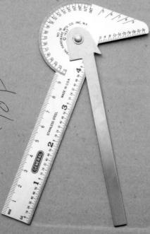
25 Joint structures: some persons, because of genetics or posture, normally have hypermobile or hypomobile joints. Types of goniometers Universal goniometers: plastic or metal protractor like device with moveable and stationary arms of varying lengths

26 Gravity Dependent Goniometers Inclinometers: report position of distal or proximal segment relative to the line of gravity

27 Electrogoniometers: potentiometer detects changes in position of two segments
 moving arm reading")
28 Parts of the Goniometer axis of rotation non-moving (Stationary arm) moving arm reading numbers
29 Procedures Patient position Locating joint axis Locating stationary arm Locating moving arm Reading goniometer Recording goniometer measures (the mean of 2 or 3 trials is more accurate)
30 Contraindications and Precautions for ROM Testing: Dislocation or unhealed fracture. Surgical procedures to tendons, ligaments, muscles, joint capsule or skin. Infections or inflammatory process. Marked osteoporosis. Hyper mobile or sublaxated joint. Painful conditions. Hematoma.

31 Shoulder flexion
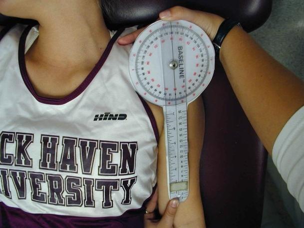
32 - Shoulder hyperextension - shoulder abduction


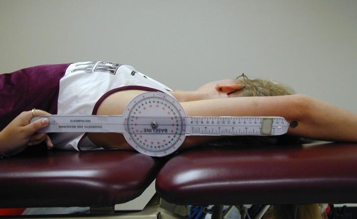
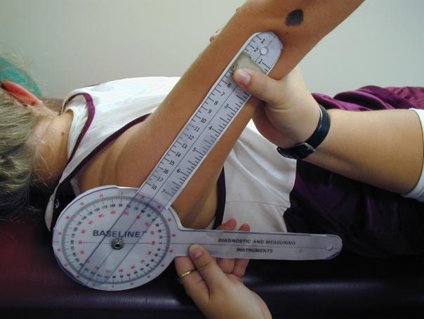
33 Shoulder external rotation Shoulder internal rotation


34 Elbow flexion and extension
35 END FEEL Nature of the motion barrier that characterizes the type of tissue limiting range. Normal or Physiologic END FEEL: Hard Soft Firm Capsular stretch
36 Hard (bony): An abrupt, hard stop to movement when bone contacts bone; for ex.: Passive elbow extension. the olecranon process contacts the olecranon fossa. Soft (soft tissue opposition): When two body surfaces come together a soft compression of tissue is felt, for ex.: in passive knee flexion, the posterior aspects of the calf and thigh come together.
37 Firm (soft tissue stretch): Firm or springy sensation that has some give when a muscle is stretched for ex.: passive ankle dorsiflexion performed with the knee in extension is stopped due to tension in the gastrocnemius muscle. Capsular stretch: Hard arrests to movement with some give when the joint capsule or ligaments are stretched. The feel is similar to stretching a piece of leather, for ex.: passive shoulder external rotation.
38 Long and Round Measurement Long measurement: Total: Measure the length of the upper limb from acromion to the radial styloid process. Segmental: - Measure the length of the humerus from acromion to the lateral epicondyle. - Measure the length of radius and ulna from lateral epicondyle of humerus to the radial styloid process.
39 Round measurement: Muscle atrophy: measure around the midpoint of the arm and around the upper third of the forearm. Joint edema: measure around the centre of the elbow joint (centre of the capital fossa) and 3cm & 6cm above and below.
40 Cervical & Lumbar Spine ROM

41 Spinal Column
 Construction of the")

42 Cervical Spine ROM Seven Cervical Vertebrae Forming The Cervical Spine, Including The First& Second Ones ( Atlas & Axis ) Construction of the Spinal Vertebra - Vertebral bodies: Weight bearing. - Neural arch: Protect Neural Elements. - Bony Processes: increase the efficiency of muscle action.

43 - The atlas (C1) and the axis (C2), have structures unlike those of any other vertebra. - The atlas has no vertebral body and is shaped like a ring with an anterior and a posterior arch. - Has no spinous process. Superiorly, it has a fovea, or dishlike depression, that holds the occiput of the skull. - The articulation of the atlas with the skull is called the atlantooccipital joint. At this joint, the head nods on the spine.
44 آ 10 The atlantooccipital joint allows approximately to آ 15 of flexion and extension and no lateral flexion or rotation. The weight of the head is transferred to the cervical spine via C2, the axis. The articulation with the atlas occurs via a pillar (odontoid process or dens ) projecting from the superior surface of the axis that fits into the atlas and locks the atlas into a pivoting joint. (the most mobile of the cervical joints). allowing approximately آ 10 of flexion and extension, accounts for 50% of the rotation in the cervical vertebrae and no lateral flexion.

45 Because of the short spinous processes, the shape of the discs, and the backward and downward orientation of the articulating facets, movement in the cervical region is greater than in any other region of the vertebral column. Forward flexion: 0 to 45 degrees. Extension: 0 to 45 degrees. Lateral Flexion: 0 to 45 degrees. Lateral Rotation: 0 to 80 degrees.


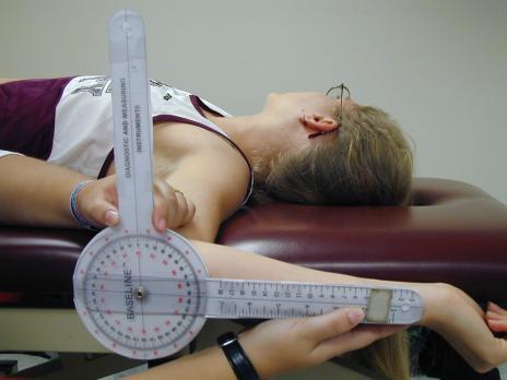
46 Methods to Measure the Range of Motion of the Cervical Spine Radiographs. inclinometers. Tape measures.


47 Forward flexion: 0 to 45 degrees. Extension: 0 to 45 degrees. Lateral Flexion: 0 to 45 degrees.

48 Lateral Rotation: 0 to 80 degrees.

49 Lumbar Spine ROM Five lumbar vertebrae and the sacrum making up the lumbar spine. The size of the vertebral body increases from L1 to L5, due to the increasing loads that each lower lumbar vertebral level has to absorb.
50 The collective range of motion in the lumbar region is: - Lumbar flexion (60 ) - Lumbar Extension (30 ) - Lumbar lateral flexion (30-40 ) - Lumbar rotation (45 ) The lumbosacral joint is the most mobile of the lumbar joints, accounting for a large proportion of the flexion and extension in the region. Of the flexion and extension in the lumbar vertebrae, 75% may occur at this joint, with 20% of the remaining flexion at L4-L5 and 5% at the other lumbar levels.

")
51 - Lumbar flexion (60 ) - Lumbar rotation (45 )

52 Clinical Methods to Assess Lumbar ROM Spinal flexion: - By measuring the degrees of forward inclination of the trunk in relation to the longitudinal axis of the body. The examiner should fix the pelvis with his hands.
53 - By indicating the level the fingertips reach along the patient leg. For instance, fingertips to the patella; or fingertips to mid-tibia. - By measuring the distance in inches or centimeters between the fingertips and the floor. - By the steel or plastic tape measuring method: o o The most accurate clinical method of measuring true motion of the spine in flexion. With the patient standing, the tape is held over the spinous process of C7, and the distal tape held over the spinous process of S1.
54 o As the patient bends forward, the spinous processes spread, this will be indicated by lengthening of the tape measure. o In the normal healthy adult, there is, on the average, an increase of 4 inches in forward flexion. If the patient bends forward with his back straight (as in rheumatoid arthritis), the tape will not record motion. o One is able to record motion of the thoracic spine by taping from the spinous process of C7 to T12. o Usually if the total spine in flexion is 4 inches the examiner will find that 1inch occurs in the dorsal spine, and 3 inches occurs in the lumbar spine.
55 Spinal lateral bending: While the patient is standing, the knee joint is used as a fixed point.record distance of finger tips from the knee joint on lateral bending.
56 Spinal extension: while the patient is standing,measure from C7 to L5 then ask the patient to extend his back and measure again the distance will decrease.

57 Spinal rotation: To estimate the degrees of rotation of the spine, the pelvis must be held firmly by the examiner s hands and the patient is instructed to rotate to the left side then to the right side then you have to compare between the degree of rotation on both sides.
EVALUATION AND MEASUREMENTS. I. Devreux
 EVALUATION AND MEASUREMENTS I. Devreux To determine the extent and degree of muscular weakness resulting from disease, injury or disuse. The records obtained from these tests provide a base for planning
Joint Range of Motion Assessment Techniques. Presentation Created by Ken Baldwin, M.Ed Copyright
 Joint Range of Motion Assessment Techniques Presentation Created by Ken Baldwin, M.Ed Copyright 2001-2006 Objectives Understand how joint range of motion & goniometric assessment is an important component
Basics of Soft- Tissue Examination
 Basics of Soft- Tissue Examination Basics of Soft Tissue Exam For practitioners who primarily use their hands to treat the human structure: Examination must include functional tests to determine the type
Goniometry. Wrist Flexion: Pt seated with forearm resting on table (use olecranon process & midline of ulna as reference for stationary arm)
") Goniometry Wrist Flexion: Pt seated with forearm resting on table (use olecranon process & midline of ulna as reference for stationary arm) Wrist Extension: Pt seated with forearm resting on table (Goniometer
Goniometry Wrist Flexion: Pt seated with forearm resting on table (use olecranon process & midline of ulna as reference for stationary arm) Wrist Extension: Pt seated with forearm resting on table (Goniometer
BLUE SKY SCHOOL OF PROFESSIONAL MASSAGE AND THERAPEUTIC BODYWORK. Musculoskeletal Anatomy & Kinesiology MUSCLES, MOVEMENTS & BIOMECHANICS
 BLUE SKY SCHOOL OF PROFESSIONAL MASSAGE AND THERAPEUTIC BODYWORK Musculoskeletal Anatomy & Kinesiology MUSCLES, MOVEMENTS & BIOMECHANICS MSAK101-I Session 7 Learning Objectives: 1. List the three types
CHAPTER 1: 1.1 Muscular skeletal system. Question - text book page 16. Question - text book page 20 QUESTIONS AND ANSWERS. Answers
 QUESTIONS AND ANSWERS CHAPTER 1: 1.1 Muscular skeletal system Question - text book page 16 Using the information on pages 12 to 14 above, complete the table below. joint joint type articulating bones associated
Dr.Israa H. Mohsen. Lecture 5. The vertebral column
 Anatomy Lecture 5 Dr.Israa H. Mohsen The vertebral column The vertebral column a flexible structure consisting of 33 vertebrae holds the head and torso upright, serves as an attachment point for the legs,
Certified Personal Trainer Re-Certification Manual
 Certified Personal Trainer Re-Certification Manual Section II 1 Anatomy & Physiology Terms Anatomy and physiology are closely related fields of study: anatomy is the study of form, and physiology is the
*Agonists are the main muscles responsible for the action. *Antagonists oppose the agonists and can help neutralize actions. Since many muscles have
 1 *Agonists are the main muscles responsible for the action. *Antagonists oppose the agonists and can help neutralize actions. Since many muscles have more than 1 action sometimes a muscle has to neutralize
Skeletal System. Std. VIII
 Skeletal System Std. VIII The skeleton in our body serves following functions : 1. Support and shape : The skeleton provides a support or framework to all the soft parts and gives the body and its parts
Figure 11-1: The lever-fulcrum principle is illustrated by flexion of the forearm.
 Chapter 11: The Muscular System Read pages 325 to 399 NAME Topic Outline And Objectives: A. How skeletal muscles produce movement, and naming muscles 1. Describe the relationship between bones and skeletal
PRELIMINARY HSC PDHPE. CQ1 How do the musculoskeletal and cardiorespiratory systems of the body influence and respond to movement?
 PRELIMINARY HSC PDHPE CQ1 How do the musculoskeletal and cardiorespiratory systems of the body influence and respond to movement? How do the musculoskeletal and cardiorespiratory systems of the body influence
What is Kinesiology? Basic Biomechanics. Mechanics
 What is Kinesiology? The study of movement, but this definition is too broad Brings together anatomy, physiology, physics, geometry and relates them to human movement Lippert pg 3 Basic Biomechanics the
Elbow & Forearm H O W V I T A L I S T H E E L B O W T O O U R D A I L Y L I V E S?
 Elbow & Forearm H O W V I T A L I S T H E E L B O W T O O U R D A I L Y L I V E S? Clarification of Terms The elbow includes: 3 bones (humerus, radius, and ulna) 2 joints (humeroulnar and humeroradial)
Lever system. Rigid bar. Fulcrum. Force (effort) Resistance (load)
 Resistance (load)") Lever system lever is any elongated, rigid (bar) object that move or rotates around a fixed point called the fulcrum when force is applied to overcome resistance. Force (effort) Resistance (load) R Rigid
Lever system lever is any elongated, rigid (bar) object that move or rotates around a fixed point called the fulcrum when force is applied to overcome resistance. Force (effort) Resistance (load) R Rigid
Muscle Testing of Knee Extensors. Yasser Moh. Aneis, PhD, MSc., PT. Lecturer of Physical Therapy Basic Sciences Department
 Muscle Testing of Knee Extensors Yasser Moh. Aneis, PhD, MSc., PT. Lecturer of Physical Therapy Basic Sciences Department Muscle Testing of Knee Extensors othe Primary muscle Quadriceps Femoris -Rectus
Human Anatomy - Problem Drill 06: The Skeletal System Axial Skeleton & Articualtions
 Human Anatomy - Problem Drill 06: The Skeletal System Axial Skeleton & Articualtions Question No. 1 of 10 Instructions: (1) Read the problem and answer choices carefully, (2) Work the problems on paper
Anatomy. Anatomy deals with the structure of the human body, and includes a precise language on body positions and relationships between body parts.
 Anatomy deals with the structure of the human body, and includes a precise language on body positions and relationships between body parts. Proper instruction on safe and efficient exercise technique requires
i;l Contents PART I INTRODUCTIOM TO GONIOMETRY, I ~haoter '1 Basic Conceots. 3 Chapter 2 Procedures, 19 Chapter 3 Validity and Reliability, 39
 w Contents i;l PART I INTRODUCTIOM TO GONIOMETRY, I ~haoter '1 Basic Conceots. 3 Goniometry, 3 Joint Motion, 4 Arthrokinematics, 4 Osteokinematics, 5 Planes and Axes, 5 Range of Motion, 6 Active Range
Traction. Process of drawing or pulling apart. May involve distraction and gliding. Pulling 2 articulating surfaces away from each other
 Traction Process of drawing or pulling apart May involve distraction and gliding Pulling 2 articulating surfaces away from each other Axis Traction in line with the long axis of a part Types of Traction
The Biomechanics of the Human Upper Extremity-The Elbow Joint C. Mirzanli Istanbul Gelisim University
 The Biomechanics of the Human Upper Extremity-The Elbow Joint C. Mirzanli Istanbul Gelisim University Structure of The Elbow Joint A simple hinge joint, actually categorized as a trochoginglymus joint
Physical Capability Exam Testing Protocol
 Test Duration: ~ min Physical Capability Exam Testing Protocol Pinch Gauge Grip Dynamometer Inclinometer Stop Watch Lift Box Table Weight Plates (5 lbs., lbs., lbs., 50 lbs., 0 lbs.) Physical Capability
Types of Body Movements
 Types of Body Movements Bởi: OpenStaxCollege Synovial joints allow the body a tremendous range of movements. Each movement at a synovial joint results from the contraction or relaxation of the muscles
Chapter 6 part 2. Skeletal Muscles of the Body
 Chapter 6 part 2 Skeletal Muscles of the Body Basic Principles 600 + muscles in the human body (you are required to learn 45, lucky kids)! Skeletal Muscles pull on bones Origin of a muscle = point of attachment
Clinical examination of the wrist, thumb and hand
 Clinical examination of the wrist, thumb and hand 20 CHAPTER CONTENTS Referred pain 319 History 319 Inspection 320 Functional examination 320 The distal radioulnar joint.............. 320 The wrist.......................
ACE s Essentials of Exercise Science for Fitness Professionals TRUNK
 ACE s Essentials of Exercise Science for Fitness Professionals TRUNK Posture and Balance Posture refers to the biomechanical alignment of the individual body parts and the orientation of the body to the
Pilates for Brachialis Tendonitis (Tennis Elbow)
") Pilates for Brachialis Tendonitis (Tennis Elbow) Sally Dunford September 2017 Wimbledon, UK Abstract Tennis Elbow is a term used to describe a painful condition in which the tendons of the elbow are overloaded
Pilates for Brachialis Tendonitis (Tennis Elbow) Sally Dunford September 2017 Wimbledon, UK Abstract Tennis Elbow is a term used to describe a painful condition in which the tendons of the elbow are overloaded
Hands PA; Obl. Lat.; Norgaard s Thumb AP; Lat. PA. PA; Lat.: Obls.; Elongated PA with ulnar deviation
 Projections Region Basic projections Additional / Modified projections Upper Limbs Hands PA; Obl. Lat.; Norgaard s Thumb ; Lat. PA Fingers PA; Lat. Wrist PA; Lat. Obls. Scaphoid Lunate Trapezium Triquetral
Anatomy of the Musculoskeletal System
 Anatomy of the Musculoskeletal System Kyle E. Rarey, Ph.D. Department of Anatomy & Cell Biology and Otolaryngology University of Florida College of Medicine Outline of Presentation Vertebral Column Upper
Elbow. Chapter 2 LISTEN. Mechanism of Injury (If Applicable) Pain
 Pain") Chapter 2 Elbow LISTEN Mechanism of Injury (If Applicable) Patient usually remembers their position at the time of injury Certain mechanisms of injury result in characteristic patterns Fall on outstretched
Chapter 2 Elbow LISTEN Mechanism of Injury (If Applicable) Patient usually remembers their position at the time of injury Certain mechanisms of injury result in characteristic patterns Fall on outstretched
1-Apley scratch test.
 1-Apley scratch test. The patient attempts to touch the opposite scapula to test range of motion of the shoulder. 1-Testing abduction and external rotation( +ve sign touch the opposite scapula, -ve sign
EXERCISE PRESCRIPTION PART 1
 EXERCISE PRESCRIPTION PART 1 Michael McMurray, PT, DPT, OCS, FAAOMPT Orthopaedic Manual Physical Therapy Series Charlottesville 2017-2018 What is MET? An active rehabilitation system based in the biopsychosocial
Physical Examination of the Shoulder
 General setup Patient will be examined in both the seated and supine position so exam table needed 360 degree access to patient Expose neck and both shoulders (for comparison); female in gown or sports
Muscle Tissue. Isometric Contraction. Isotonic Contractions 11/22/2016. Muscles. Anatomy Two Joints And Movements
 Muscles Anatomy Two Joints And Movements Structure of a Muscle Organ Copyright 2008 by Saunders Muscle Tissue Highly elastic and vascularized, produces movement through elongation and contraction Types
Osteoporosis Protocol
 PRODUCTS HELPING PEOPLE HELP THEMSELVES! Osteoporosis Protocol Rehabilitation using the Resistance Chair General Information Osteoporosis is a condition where bones gradually decrease in mass or density
Maximal isokinetic and isometric muscle strength of major muscle groups related to age, body weight, height, and sex in 178 healthy subjects
 Maximal isokinetic and isometric muscle strength of major muscle groups related to age, body weight, height, and sex in 178 healthy subjects Test protocol Muscle test procedures. Prior to each test participants
Traditional Thai Acupressure Points. The anterior aspect of the body THE ANATOMICAL ATLAS
 Traditional Thai Acupressure Points The anterior aspect of the body THE ANATOMICAL ATLAS lines of the SHOULDER BLADES AND POSTERIOR ARM Scapula Line This line runs through landmarks: 1. Above the midpoint
11/25/2012. Chapter 7 Part 2: Bones! Skeletal Organization. The Skull. Skull Bones to Know Cranium
 Chapter 7 Part 2: Bones! 5) Distinguish between the axial and appendicular skeletons and name the major parts of each 6) Locate and identify the bones and the major features of the bones that compose the
Chiropractic Glossary
 Chiropractic Glossary Anatomy Articulation: A joint formed where two or more bones in the body meet. Your foot bone, for example, forms an articulation with your leg bone. You call that articulation an
Connects arm to thorax 3 joints. Glenohumeral joint Acromioclavicular joint Sternoclavicular joint
 Connects arm to thorax 3 joints Glenohumeral joint Acromioclavicular joint Sternoclavicular joint Scapula Elevation Depression Protraction (abduction) Retraction (adduction) Downward Rotation Upward Rotation
BLUE SKY SCHOOL OF PROFESSIONAL MASSAGE AND THERAPEUTIC BODYWORK Musculoskeletal Anatomy & Kinesiology ROM & GONIOMETRY
 BLUE SKY SCHOOL OF PROFESSIONAL MASSAGE AND THERAPEUTIC BODYWORK Musculoskeletal Anatomy & Kinesiology & GONIOMETRY MSAK201-II Session 2 LEARNING OBJECTIVES: By the end of this session, the student will
Practical 2 Worksheet
 Practical 2 Worksheet Upper Extremity BONES 1. Which end of the clavicle is on the lateral side (acromial or sternal)? 2. Describe the difference in the appearance of the acromial and sternal ends of the
Yoga Anatomy & Physiology
 Yoga Anatomy & Physiology Anatomy & Physiology Anatomy- One of the basic essential sciences of medicine that studies the structure of an organism. Physiology- The biological study of the functions of living
MLT Muscle(s) Patient Position Therapist position Stabilization Limb Position Picture Put biceps on slack by bending elbow.
 Patient Position Therapist position Stabilization Limb Position Picture Put biceps on slack by bending elbow.") MLT Muscle(s) Patient Position Therapist position Stabilization Limb Position Picture Put biceps on slack by bending elbow. Pectoralis Minor Supine, arm at side, elbows extended, supinated Head of Table
MLT Muscle(s) Patient Position Therapist position Stabilization Limb Position Picture Put biceps on slack by bending elbow. Pectoralis Minor Supine, arm at side, elbows extended, supinated Head of Table
When a muscle contracts, it knows no direction it simply shortens. Lippert
 When a muscle contracts, it knows no direction it simply shortens. Lippert Muscles are attached to bones and to describe the relative points of attachment, we use the terms origin and insertion. Lippert,
The Skeletal System THE APPENDICULAR SKELETON
 The Skeletal System THE APPENDICULAR SKELETON The appendicular skeleton consists of the girdles and the skeleton of the limbs. The upper (anterior) limbs are attached to the pectoral (shoulder) girdle
The Elbow and Radioulnar Joints Kinesiology. Dr Cüneyt Mirzanli Istanbul Gelisim University
 The Elbow and Radioulnar Joints Kinesiology Dr Cüneyt Mirzanli Istanbul Gelisim University 1 The Elbow & Radioulnar Joints Most upper extremity movements involve the elbow & radioulnar joints. Usually
National Boards Part 4 Technique. Exam Format 5 stations (1 doctor and 1 patient). 2 setups per station (5 minutes) cervical
. 2 setups per station (5 minutes) cervical") 1 National Boards Part 4 Technique Exam Format 5 stations (1 doctor and 1 patient). 2 setups per station (5 minutes) cervical thoracic lumbar pelvic extremity Expect examiner interaction Graded on a Scantron
1 National Boards Part 4 Technique Exam Format 5 stations (1 doctor and 1 patient). 2 setups per station (5 minutes) cervical thoracic lumbar pelvic extremity Expect examiner interaction Graded on a Scantron
Chapter 8. The Pectoral Girdle & Upper Limb
 Chapter 8 The Pectoral Girdle & Upper Limb Pectoral Girdle pectoral girdle (shoulder girdle) supports the arm consists of two on each side of the body // clavicle (collarbone) and scapula (shoulder blade)
Musculoskeletal Examination Benchmarks
 Musculoskeletal Examination Benchmarks _ The approach to examining the musculoskeletal system is the same no matter what joint or limb is being examined. The affected and contralateral region should both
Osteology of the Elbow and Forearm Complex
 Osteology of the Elbow and Forearm Complex The ability to perform m any activities of daily living (ADL) d epends upon the elbow. Activities of Daily Living (ADL) Can you think of anything that you do
In which arm muscle are intramuscular injections most often given? (not in text)
") AP1 Lab 9 - Muscles of the Arms and Legs Locate the following muscles on the models and on yourself. Recall anatomical position. Directional terms such as anterior, posterior, lateral, etc. all assume
AP1 Lab 9 - Muscles of the Arms and Legs Locate the following muscles on the models and on yourself. Recall anatomical position. Directional terms such as anterior, posterior, lateral, etc. all assume
Module 7 - The Muscular System Muscles of the Arm and Trunk
 Module 7 - The Muscular System Muscles of the Arm and Trunk This Module will cover the muscle anatomy of the arms and trunk. We have already seen the muscles that move the humerus, so this module will
Prime movers provide the major force for producing a specific movement Antagonists oppose or reverse a particular movement Synergists
 Dr. Gary Mumaugh Prime movers provide the major force for producing a specific movement Antagonists oppose or reverse a particular movement Synergists Add force to a movement Reduce undesirable or unnecessary
Stiff Ribs can cause pain in the LEGS!
 Stiff Ribs can cause pain in the LEGS! Presenter: Kym Finch 2018 Massage and Myotherapy Australia Conference Gold Coast Outcomes Challenge Teach Develop Challenge your thought process Teach new skills
1 Pause and Practice: Facilitating Trunk and Shoulder Control with the Therapy Ball
 1 Pause and Practice: Facilitating Trunk and Shoulder Control with the Therapy Ball This is an example of Facilitating Combinations of Movements and Active Assist. Starting Position Have your patient sit
When a muscle contracts, it knows no direction; it simply shortens. Lippert
 When a muscle contracts, it knows no direction; it simply shortens. Lippert Muscle is the sole producer of active force in the body which makes it responsible for all active motions. Muscles also control
8 1 Basic Principles. Palpating Muscle Bellies. Palpating Bony Prominences
 8 1 Basic Principles Fig. 1.4 Locating the medial epicondyle. Tip: The shape of bony prominences can be visualized by looking at their morphology. However, variations are expected to be encountered quite
VCE PHYSICAL EDUCATION WORKBOOK UNIT 1 BODIES IN MOTION NAME:
 VCE PHYSICAL EDUCATION WORKBOOK UNIT 1 BODIES IN MOTION NAME: SKELETAL SYSTEM List the 5 functions of the skeletal system and complete the following table. FUNCTION DESCRIPTION Label the following features
10/15/2014. Wrist. Clarification of Terms. Clarification of Terms cont
 Wrist Clarification of Terms Palmar is synonymous with anterior aspect of the wrist and hand Ventral is also synonymous with anterior aspect of the wrist and hand Dorsal refers to the posterior aspect
Benefits of Weight bearing increased awareness of the involved side decreased fear improved symmetry regulation of muscle tone
 From the information we have gathered during our Evaluation, the Clinical Reasoning we used to identify key problem areas and the Goals Established with functional outcomes we now have enough information
The Muscular System PART C. PowerPoint Lecture Slide Presentation by Patty Bostwick-Taylor, Florence-Darlington Technical College
 PowerPoint Lecture Slide Presentation by Patty Bostwick-Taylor, Florence-Darlington Technical College The Muscular System 6 PART C Five Golden Rules of Skeletal Muscle Activity Table 6.2 Muscles and Body
The Role of Muscles in Movement
 The Role of Muscles in Movement Muscles can t push, they can only pull as they contract, so most often body movements are the result of the activity of pairs or teams of muscles acting together or against
Contraindicated and High-Risk Exercises
 Contraindicated and High-Risk Exercises Young sub Kwon, Ph.D. ACSM RCEP, NSCA CSCS,*D Exercise Physiology Laboratory The University of New Mexico Albuquerque, NM, USA Introduction Any activity selected
Location of Pain/Symptoms Where do you feel the pain/symptoms? Can you point with one finger to location of pain? Is the pain general or localized?
 Injury Evaluation History A complete and accurate medical history is one of the most important and useful parts of the clinical examination. A complete history consists of past history and a history of
VCE PHYSICAL EDUCATION WORKBOOK UNIT 1 BODIES IN MOTION NAME:
 VCE PHYSICAL EDUCATION WORKBOOK UNIT 1 BODIES IN MOTION NAME: SKELETAL SYSTEM List the 5 functions of the skeletal system and complete the following table. FUNCTION DESCRIPTION Label the following features
OBJECTIVES. Unit 7:5 PROPERTIES OR CHARACTERISTICS OF MUSCLES. Introduction. 3 Kinds of Muscles. 3 Kinds of Muscles 4/17/2018 MUSCULAR SYSTEM
 OBJECTIVES Unit 7:5 MUSCULAR SYSTEM Compare the three main kinds of muscles by describing the action of each Differentiate between voluntary and involuntary muscles List at least three functions of muscles
This presentation is the intellectual property of the author. Contact them for permission to reprint and/or distribute.
 The Stiff Hand: Manual Therapy Sylvia Dávila, PT, CHT San Antonio, Texas Orthopedic Manual Therapy Common Applications Passive stretch Tensile force to tissue to increase extensibility of length & ROM
30b Passive Stretches:! Technique Demo and Practice - Upper Body
 30b Passive Stretches:! Technique Demo and Practice - Upper Body 30b Passive Stretches:! Technique Demo and Practice - Upper Body! Class Outline" 5 minutes" "Attendance, Breath of Arrival, and Reminders
The Elbow and the cubital fossa. Prof Oluwadiya Kehinde
 The Elbow and the cubital fossa Prof Oluwadiya Kehinde www.oluwadiya.com Elbow and Forearm Anatomy The elbow joint is formed by the humerus, radius, and the ulna Bony anatomy of the elbow Distal Humerus
GENERAL EXERCISES ELBOW BMW MANUFACTURING CO. PZ-AM-G-US I July 2017
 GENERAL EXERCISES ELBOW BMW MANUFACTURING CO. PZ-AM-G-US I July 2017 Disclosure: The exercises, stretches, and mobilizations provided in this presentation are for educational purposes only are not to be
Lab Exercise #5 The Muscular System Student Performance Objectives
 Student Performance Objectives The material that you are required to learn in this exercise can be found in either the lecture text or the supplemental materials provided in lab. Prior to coming to class,
THE EXTREMITY SCREEN MANUAL: A Guide to the Subjective and Objective Outcomes Assessment of the Upper and Lower Extremity
 THE EXTREMITY SCREEN MANUAL: A Guide to the Subjective and Objective Outcomes Assessment of the Upper and Lower Extremity Steven G. Yeomans, DC, FACO INTRODUCTION: Objective screen for the extremities
3 Movements of the Trunk. Flexion Rotation Extension
 3 Movements of the Trunk Flexion Rotation Extension 1 TRUNK FLEXION 2 TRUNK FLEXION: Rectus Abdominalis O: Crest of Pubis & ligaments covering front of symphysis pubis. I: By «3 portions into cartilages
The Skeletal System. Dr. Naim Kittana. Faculty of Medicine & Health Sciences An-Najah National University
 The Skeletal System Dr. Naim Kittana Faculty of Medicine & Health Sciences An-Najah National University 1 Declaration The content and the figures of this seminar were directly adopted from the text book
The Skeletal System. Dr. Naim Kittana Dr. Suhaib Hattab. Faculty of Medicine & Health Sciences An-Najah National University
 The Skeletal System Dr. Naim Kittana Dr. Suhaib Hattab Faculty of Medicine & Health Sciences An-Najah National University 1 Declaration The content and the figures of this seminar were directly adopted
Multi-joint Mechanics Dr. Ted Milner (KIN 416)
") Multi-joint Mechanics Dr. Ted Milner (KIN 416) Muscle Function and Activation It is not a straightforward matter to predict the activation pattern of a set of muscles when these muscles act on multiple
Multi-joint Mechanics Dr. Ted Milner (KIN 416) Muscle Function and Activation It is not a straightforward matter to predict the activation pattern of a set of muscles when these muscles act on multiple
Regional Review of Musculoskeletal System: Head, Neck, and Cervical Spine Presented by Michael L. Fink, PT, DSc, SCS, OCS Pre- Chapter Case Study
 Regional Review of Musculoskeletal System: Presented by Michael L. Fink, PT, DSc, SCS, OCS (20 minutes CEU Time) Subjective A 43-year-old male, reported a sudden onset of left-sided neck and upper extremity
Reliability of Measuring Trunk Motions in Centimeters
 Reliability of Measuring Trunk Motions in Centimeters MARGARET ROST, SANDRA STUCKEY, LEE ANNE SMALLEY, and GLENDA DORMAN A method of measuring trunk motion and two related motions using a tape measure
Y12 Transition Pack Sports Leadership
 Y12 Transition Pack Sports Leadership Name: Base School: Section 1: The Muscular System Read through the following information on the Muscular System Cardiac muscle is unique to the heart. It never tires.
YOGA ANATOMY. Part Three - Bones. Yoga Teacher Training Robin Bennett 200 RYT
 YOGA ANATOMY Yoga Teacher Training Part Three - Bones 2015 Robin Bennett 200 RYT THE HUMAN SKELETON BONE COMPOSITION A femur head with a cortex of compact bone and medulla of trabecular (spongy) bone OSTEOBLASTS
Introduction to Biomechanical Analysis
 Introduction to Biomechanical Analysis LEARNING OBJECTIVES: At the end of this laboratory exercise the student will be able to: Identify forces used during activities Identify moments used during activities
Main Menu. Elbow and Radioulnar Joints click here. The Power is in Your Hands
 1 The Elbow and Radioulnar Joints click here Main Menu K.4 http://www.handsonlineeducation.com/classes//k4entry.htm[3/23/18, 1:29:53 PM] Bones Ulna is much larger proximally than radius Radius is much
Tennis Elbow Assessment & Treatment Workshop
 Tennis Elbow Assessment & Treatment Workshop Ian Gatt MSc, OMT, MAACP, MCSP, BSc (Hons) 9 th Southampton Hand Course 27 th June 2014, Chilworth Manor Hotel Assessment MacDermid et al., 2010 References
Ligaments of Elbow hinge: sagittal plane so need lateral and medial ligaments
 Ligaments of Elbow hinge: sagittal plane so need lateral and medial ligaments Ulnar Collateral ligament on medial side; arising from medial epicondyle and stops excess valgus movement (lateral movement)
Exercises to Correct Muscular Imbalances. presented by: Darrell Barnes, LAT, ATC, CSCS
 Exercises to Correct Muscular Imbalances presented by: Darrell Barnes, LAT, ATC, CSCS Objectives Review Functional Anatomy Identify physical imbalances that lead to injury and/or decrease performance
CHAPTER 4: The musculo-skeletal system. Practice questions - text book pages QUESTIONS AND ANSWERS. Answers
 CHAPTER 4: The musculo-skeletal system Practice questions - text book pages 64-66 1) A prime mover of hip flexion is the: a. rectus femoris. b. Iliopsoas. c. vastus muscles. d. gluteus maximus. b. Key
KNEE AND LEG EXERCISE PROGRAM
 KNEE AND LEG EXERCISE PROGRAM These exercises are specifically designed to rehabilitate the muscles of the hip and knee by increasing the strength and flexibility of the involved leg. This exercise program
Country Health SA Medical Imaging
 Country Health SA Medical Imaging REMOTE OPERATORS POSITIONING GUIDE Contents Image Evaluation Page 4 Positioning Guides Section 1 - THORAX 1.1 Chest Page 5 1.2 Bedside Chest Page 7 1.3 Ribs Page 8 Section
STRUCTURAL BASIS OF MEDICAL PRACTICE EXAMINATION 5. September 30, 2011
 STRUCTURAL BASIS OF MEDICAL PRACTICE EXAMINATION 5 September 30, 2011 PART l. Answer in the space provided. (12 pts) 1. Identify the structures. (2 pts) EXAM NUMBER A. Suprascapular nerve B. Axillary nerve
Muscles in the Shoulder, Chest, Arm, Stomach, and Back
 Muscles in the Shoulder, Chest, Arm, Stomach, and Back Shoulder Muscles Deltoid Supraspinatus Infraspinatus Teres Major Teres Minor Subscapularis Deltoid (Delts) Function: Raises the upper arm Origin:
Skeletal Muscles and Functions
 Skeletal Muscles and Functions Huei-Ming Chai, PT, Ph.D. School of Physical Therapy National Taiwan University Classification of Muscles striated muscles skeletal muscles: voluntary contraction cardiac
Spinal Biomechanics & Sitting Posture
 Spinal Biomechanics & Sitting Posture Sitting: weight of the body is transferred to a supporting area 1.Main Contact points (seat) Ischial tuberosities Soft tissues 2. Secondary contact points (other)
WTC II Term 3 Notes & Assessments
 Term 3 Notes & Assessments Planes of Motion/Axes The body moves in a number of various ways and directions. In the past you have learned about the terminology for movements at specific joints, for example,
External Obliques Abdominal muscles that attaches at the lower ribs, pelvis, and abdominal fascia.
 The Core The core is where most of the body s power is derived. It provides the foundation for all movements of the arms and legs. The core must be strong, have dynamic flexibility, and function synergistically
Lecture 9: Forearm bones and muscles
 Lecture 9: Forearm bones and muscles Remember, the region between the shoulder and the elbow = brachium/arm, between elbow and wrist = antebrachium/forearm. Forearm bones : Humerus (distal ends) Radius
Copyright 2010 Pearson Education, Inc.
 E. VERTEBRAL COLUMN 1. The vertebral column extends from the skull to the pelvis and forms the vertical axis of the skeleton. 2. The vertebral column is composed of vertebrae that are separated by intervertebral
ASSESSMENT OF STRENGTH IN CHILDREN WITH JUVENILE DERMATOMYOSITIS
 ASSESSMENT OF STRENGTH IN CHILDREN WITH JUVENILE DERMATOMYOSITIS CURE JM STANFORD SCHOOL OF MEDICINE OCTOBER 3, 2014 Minal Jain, PT, DSc, PCS Research Coordinator, Physical Therapy Section Rehabilitation