ADRENAL MR: PEARLS AND PITFALLS
|
|
|
- Norma Merritt
- 5 years ago
- Views:
Transcription

1 ADRENAL MR: PEARLS AND PITFALLS Frank Miller, M.D. Lee F. Rogers MD Professor of Medical Education Chief, Body Imaging Section and Fellowship Medical Director, MR Imaging Professor of Radiology Northwestern University, Feinberg School of Medicine Chicago IL
2 DISCLOSURES Research grant from Siemens Health Systems
3 OBJECTIVES Discuss pearls and pitfalls of adrenal MR Strengths and limitations of MR Problem-based approach with variety of interesting cases
4 ADRENAL MASS MR of adrenal gland used to characterize adrenal masses Incidental mass most frequently benign adenoma Malignant causes include adrenal cortical carcinoma and metastasis Other causes-pheochromocytoma, benign causes-cyst, hemorrhage, myelolipoma
5 ADRENAL PROTOCOL In and opposed phase T1-GRE-axial and coronal Most important sequence to assess for lipid seen in adenomas Consider subtraction imaging (in-opposed phase) HASTE (SSFSE) T2-axial and coronal T1 FS GRE Optional: Dynamic postgad T1FS
6 CHEMICAL SHIFT IMAGING Special sequences to assess for fat In and out of phase (opposed phase) images Based on fat and water protons precessing differently On in phase images, water and fat signal in same voxel are additive On opposed phase images, fat and water are out of phase and cancel and appear dark and therefore adenomas are dark
7 CHEMICAL SHIFT IMAGING At 1.5 T: Fat and water are out of phase at TE 2.1 (and odd multiples of 2.1) In phase at TE 4.2 (even multiples of 2.1) At 3T, occurs at multiples of 1.1 msec (out of phase odd multiples , 5.5 and in phase; in phase 2.2, 4.4, 6.6 msec) Want earliest possible echo times with opposed phase before the in phase Want both acquired in single breath hold to ensure accuracy
8 ADRENAL ADENOMA Incidence of 2-8% Challenging in oncology patient if metastasis or adenoma Also many patients have CECT not NCCT NCCT <10 HU 71% sensitive; 98% specific; want highest specificity Boland et al. AJR 1998; 171:
9


10 In Phase GRE T1 TE = 4.2 Opposed Phase GRE T1 TE = 2.1ms


11 IN PHASE GRE T1 TE = 4.2 OPPOSED PHASE GRE T1 TE = 2.1ms
12 T2
13 ADRENAL MASS Noninvasive characterization of adenoma is more important than diagnosis of metastases Critical that diagnosis of adenoma is very specific so patient with metastases do not have curative therapy of neoplasm Korobkin et al Radiology 1995

14

15 In Phase Opposed Phase

16 UTILITY OF SUBTRACTION IMAGE Subtraction In-Opposed Phase

17 In Phase Opposed Phase
18 ADRENAL IMAGING-DOGMA MR can distinguish adrenal adenomas from metastases Based on presence of lipid in adenomas and not in other lesions (same principle as unenhanced CT) Signal dropout on opposed-phase imaging is highly specific for adenoma
19 MY PERSPECTIVE Adenomas sometimes easier to diagnose on MR Drop in signal on opposed phase images may be easier to identify on MR than low density on CT Does not require radiation; more sensitive for lipid Patients may be more accepting of not having the same test again
20 ADRENAL ADENOMAS: CHEMICAL SHIFT MRI Quantitative assessment Adrenal signal intensity index ASII = Adrenal to spleen ratio ASR =


21 ADRENAL ADENOMAS: CHEMICAL SHIFT MRI In-phase Opposed-phase Cut-off values for diagnosing adenoma: ASII > 16.5% ASR < 0.71 ASII = 37% ASR = 0.58 adenoma
22 Adrenal Adenomas - MRI T1W & T2W sequences are not helpful in differentiating adrenal tumors generally higher T2W signal, rapid enhancement and delayed washout are consistent with malignant lesions, but considerable overlap so unreliable Diffusion-weighted imaging not reliable1 significant overlap between ADC values of adenomas and malignant tumors Chemical shift imaging sensitivity %, specificity % for diagnosing adenomas2 MRI is comparable to washout CT for hyperattenuating lesions measuring 10-30HU, with sensitivity of 89%2 76.2% of lesions that are lipid-poor on NCCT measuring 10-30HU are lipid-rich on chemical shift MR, but 66.6% of lesion >30HU remain indeterminate after MR3 Tsushima Y JMRI 2009; Miller FH AJR 2010; Sandrasegaran K AJR 2011 Halefoglu AM 2012 Haider MA,Radiology. 2004;231(3): Sebro R, Abdom Imaging


23

24

25 IN PHASE OPPOSED PHASE

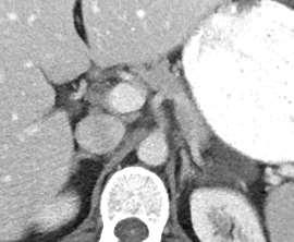
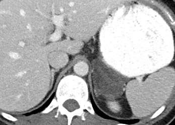
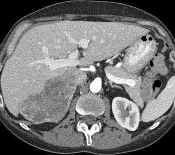
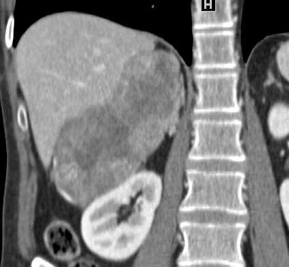
26 HX LUNG CANCER: ADRENAL HU 25

27 IN PHASE OPPOSED PHASE

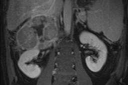
28 CORONAL T2
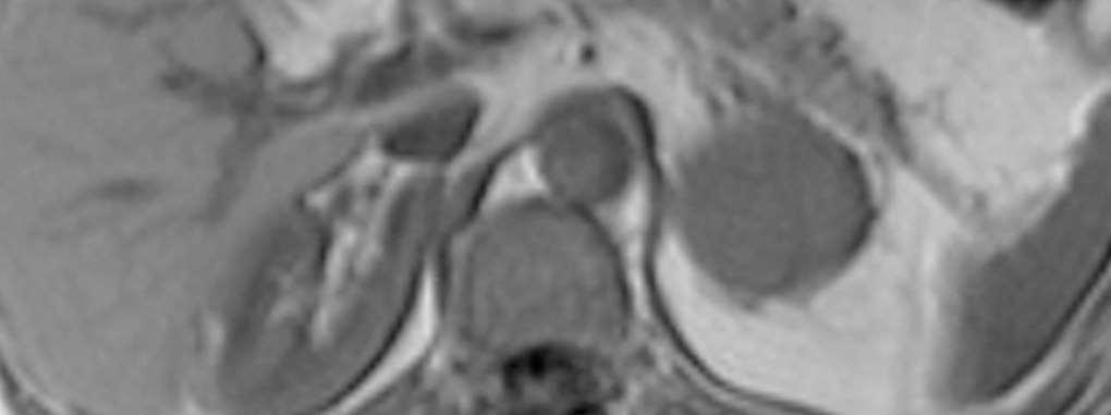
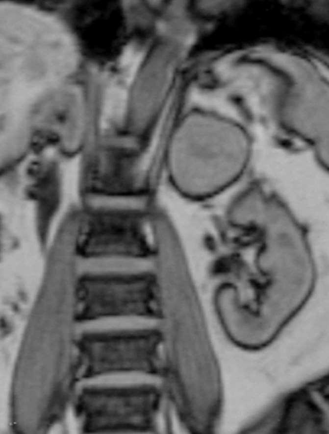
29 AXIAL IN PHASE AXIAL OPPOSED PHASE


30 IN PHASE T1 T1 FAT SUPPRESSED
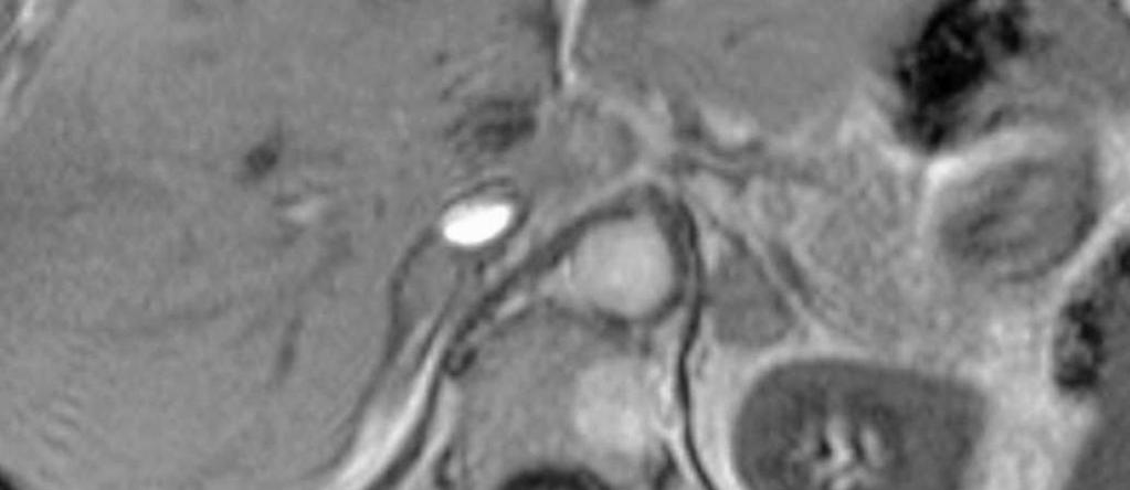
31 ADRENAL MYELOLIPOMA: MR Rare tumor with mature fat tissue and hematopoietic elementsbenign Adrenal myelolipoma-macroscopic fat-easy diagnosis on CT MR need to look at T1 in phase and frequency selective T1FS images not just in phase and opposed phase macroscopic fat-may not suppress on opposed phase (more sensitive for microscopic fat of adenomas) Look for India Ink artifact on opposed phase images


32 61 YEAR OLD WOMAN WITH LESION RIGHT ADRENAL GLAND IN PHASE OPPOSED PHASE

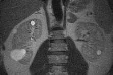
33 61 YEAR OLD WOMAN WITH LESION RIGHT ADRENAL GLAND POST CONTRAST FAT SUPPRESSED T1FS
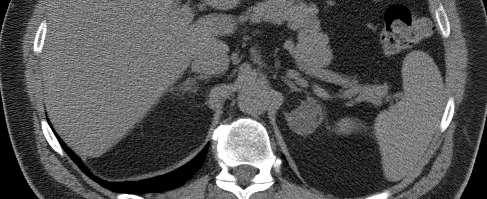
34 CT SCAN
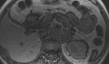
35 T2


36 IN PHASE India ink artifact with adrenal OPPOSED PHASE

37 T1 IN PHASE T1 FREQUENCY SELECTIVE FAT SUPPRESSION
38 ADRENAL MYELOLIPOMA In this patient, lesion may be collision tumor (adrenal adenoma and myelolipoma) or myelolipoma (lipid-rich and lipid-poor regions) No further work-up
39 NEXT STUDY


40


41 45 YEAR OLD FEMALE PRESENTED TO ER RIGHT LOWER QUADRANT PAIN
42 ANSWER


43 ADRENAL MYELOLIPOMA MIMICKER-LIPOSARCOMA


44 RECOMMENDED FOLLOWUP-FELT ADRENAL MYELOLIPOMA BUT CANNOT EXCLUDE LIPOSARCOMA- MR 6 MONTHS LATER- DOUBLED TO 8 CM
45 DISTINCTION OF MYELOLIPOMA FROM LIPOSARCOMA AND AML Large predominately fatty mass near adrenal may be difficult to identify if from adrenal or liposarcoma or AML Multiple planes help: if from kidney may see defect Liposarcoma may engulf or displace adrenal If normal adrenal excludes myelolipoma Davarpanah AH, Israel GM. RCNA 42 (214) 792.

46 CONTRAST ENHANCED CT

47 IN PHASE OPPOSED PHASE

48 IN PHASE OUT OF PHASE

49 T2 WEIGHTED IMAGE
50 ADRENAL CELL CARCINOMA BY BIOPSY Not typical for adenoma (lipid rich): fails to suppress on opposed phase imaging Differential diagnosis of adrenal mass (without liver mass): atypical (lipid poor) adenoma, adrenal cell carcinoma, metastasis, pheochromocytoma
51
52 PEARLS Chemical shift MR imaging is helpful in identifying adrenal adenomas Basis of distinction is the presence of abundant intracytoplasmic lipid in adenomas MR criterion for adenoma is COMPLETE suppression-subjective or quantitative
53 T1 WEIGHTED IN PHASE (TR/TE=130/4.1) OUT OF PHASE (TR/TE=130/2.2)

54 EXCEPTIONS Observation that some lesions demonstrate heterogeneous, incomplete suppression Appearance is not typical for an adenoma nor for a metastasis

 OUT OF")
55 T1 WEIGHTED IN PHASE (TR/TE=130/4.1) OUT OF PHASE (TR/TE=130/2.2)
56

57


58 IN PHASE OPPOSED PHASE * Lipid rich / * Lipid poor
59 ATYPICAL Adenomas 34 of 242 patients (14%) had adrenal lesions with heterogeneous suppression Combination of interwoven lipid rich cells and compact lipid poor cells Percentage of lipid rich cells varied from 20-80% Gabriel H, Pizzitola V, McComb EN, Wiley E, Miller FH. Adrenal Lesions with Heterogeneous Suppression on Chemical Shift Imaging: Clinical Implications. JMRI 2004; 19:
60 PATHOLOGY CORRELATION Amount and distribution of the cell types likely correlates with the MR imaging findings All patients with heterogeneous suppression were benign Patients with heterogeneous suppression and without history of malignancy may potentially be managed conservatively with follow up
61 RARE PITFALLS

62 54 YR OLD WOMAN CHEST CT FOR PULMONARY NODULES Contrast Enhanced CT T1 In Phase T1 Opposed Phase


63 6 YEARS LATER: CHEST CT R/O PE: SECOND ADRENAL MASS WITH PUNCTATE PERIPHERAL CALCIFICATIONS 6 Years Later


64 CHEMICAL SHIFT T1 In Phase T1 Opposed Phase
65 HASTE T2

66 GADOLINIUM-ENHANCED


67 BIOPSY SPECIMEN
68 ANSWER
69 ANSWER Collision tumor due to adrenal adenoma and hemangioma Siddiqi AJ, Miller FH, Kasuganti D, Nikolaidis P. Adrenal hemangioma-adenoma: an exceedingly rare adrenal collision tumor. J Magn Reson Imaging 2009;29(4):
70 COLLISION TUMORS Extremely rare; prevalence unknown Refer to 2 different types of adrenal lesions that coexist, may be benign or malignant Imaging appearance is combination of adenoma and hemangioma Suspect if 2 types of focal lesions, atypical adenoma grows, changes in appearance or somewhat large and heterogeneous with known malignancy
71 ADRENAL HEMANGIOMA-ADENOMA COLLISION TUMOR Adrenal hemangiomas rare with less than 50 reported cases Women yr old, usually asymptomatic and very large Similar to liver hemangiomas may be hypointense T1WI, hyperintense-t2wi, peripheral nodular enhancement less common due to prominent areas of necrosis and fibrosis
72 ANOTHER PITFALL


73 SIGNAL DROP IN ADRENAL AND LIVER


74 CORONAL IN-PHASE CORONAL OPPOSED PHASE ADRENAL ADENOMA? (Note absent left kidney) CORONAL CHEMICAL SHIFT SUBTRACTION (IN- OPPOSED PHASE)


75 4 MONTHS LATER 2.7 X 1.8 CM 3.5 X 2.3 CM


76 SIGNAL DROP-BIOPSY PROVEN CLEAR CELL CARCINOMA MET
77 PITFALL OF ADRENAL ADENOMAS-CLEAR CELL RENAL CELL CARCINOMA Clear cell and less commonly granular cell carcinomas can have loss of SI on out of phase MR due to microscopic fat In kidney mimics AML with microscopic fat or in case of adrenal lesion, adrenal adenoma Similar finding may be due to hepatocellular carcinoma metastasis to adrenal gland Outwater EK Radiology 1997; 205: Yoshimitsu K et al. Abdominal Imaging 2000; 25: , Sydow BD et al. AJR 2006; 187: W
78 Hypervascular metastases can show washout on adrenal protocol CT similar to adenomas (ie clear cell RCC, HCC-same issues as fat with MR) Choi YA. Radiology 2013: 266 (2)


79 45 YR OLD MAN W CLEAR CELL RCC; INDETERMINATE 3 CM MASS ON CT IN PHASE OPPOSED PHASE

80 SUBTRACTION IMAGE (IN-OPPOSED)
81 CLEAR CELL RENAL CELL CARCINOMA Need to recognize that dogma that lesions that contain fat are not all adenomas: exceptions include RCC and HCC metastases Patients with history of RCC or presence of mass suspicious for RCC, adrenal metastasis must be considered even if adrenal shows a drop in signal on opposed phase images General population without history of malignancy, mass with signal loss should be considered adenoma


82 60 YEAR OLD MALE IN PHASE OPPOSED PHASE

83 IN PHASE OPPOSED PHASE


84 39 YR OLD FEMALE WITH CUSHING IN PHASE OPPOSED PHASE SUBTRACTION IN-OPPOSED IN PHASE OPPOSED PHASE SUBTRACTION IN-OPPOSED
85 ADRENAL CORTICAL CANCER Can have fat as arise in adrenal cortext like adenoma Note that the mass however is large and generally heterogeneous Invades IVC and adjacent organs


86 IVC INVASION POSTGAD POSTGAD W IVC INVASION T2 POSTGAD

87


88 34 YR OLD ORTHOPEDIC SURGEON: ABDOMINAL PAIN
89 IN PHASE OPPOSED PHASE

90 T2 AXIAL TRUE FISP CORONAL TRUE FISP


91 HX - HEMATURIA AND L FLANK PAIN EARLY POST GAD DELAYED POST GAD CORONAL HASTE DELAYED POST GAD
92 ADRENAL CORTICAL CARCINOMA Extremely rare Usually large at diagnosis, majority >6cm Approximately 10% bilateral Usually present with pain and palpable mass, >50% with endocrinopathy Suspect if adrenal lesion with vascular invasion
93 ADRENAL CORTICAL CARCINOMA Often intermediate SI on T1WI, mixed intermediate and high SI on T2WI, larger lesions often have central necrosis Some may have small foci of lipid and lose signal on opposed phase images but generally they are larger lesions (>5 cm) than adenomas and more infiltrative Gadolinium helpful for vascular invasion


94 IN PHASE TE = 4.4 OPPOSED PHASE TE = 2.2

95 POST GAD T1 FS HASTE T2

96 ADRENAL MET FROM HCC
97 NEXT STUDY
98


99 T1 FS T2 True FISP


100


101 HEPATIC ARTERIAL BLOOD SUPPLY
102 IN PHASE OPPOSED PHASE


103 THOUGHT BE HEPATIC ADENOMA GIVEN FAT AND SURGERY PERFORMED IN PHASE OPPOSED PHASE
104 ANSWER
105 HART: HEPATIC ADRENAL REST TUMOR Originates from adrenal rest from aberrant adrenocortical tissue Round, well demarcated mass in posterior segment hepatic lobe in subcapsular region; has hepatic arterial blood supply but contiguous with adrenal gland Include in differential of solid hypervascular or fat-containing hepatic tumor in characteristic location; often not diagnosed preoperatively Mimic HCC, adenoma and angiomyolipoma Tajima JCAT : Shin Korean Journal of Hepatology 2010; 16:
106 HEPATIC ARTERIAL BLOOD SUPPY
107 DIFFUSION WEIGHTED MR Diffusion-weighted MR has been used to detect and characterize various abdominal lesions Dogma is malignant lesions have restricted diffusion and low apparent diffusion coefficients (ADC) while benign lesions do not
108 PURPOSE Evaluate diffusion MR ADC values to distinguish benign and malignant pathologies and see if correlation of signal intensity (SI) decrease on chemical shift sequences Miller FH, Wang Y, McCarthy RJ, et al. Utility of diffusion-weighted MR Imaging (DWI) in characterization of adrenal lesions. Am. J. Roentgenol 2010; 194: W179-W185
109 SUBJECTS AND METHODS Retrospectively reviewed 160 lesions 118 adenomas, 9 myelolipomas, 9 cysts, 4 adrenal hemorrhages, 1 angiolipoma 11 metastases, 4 adrenal cortical carcinomas, 3 pheochromocytomas, 1 neuroblastoma


110 ADRENAL CYSTS: ADC 3.0 T2 B500 Adrenal cysts had statistically higher ADC values compared to the remaining adrenal lesions due to free water
111 RESULTS ADC values: Median; (interquartile range) -same adrenal malignancies: 1.67 benign lesions: 1.61 Difference in the percentage SI decrease was greater in benign compared to malignant adrenal masses (P<0.05) The median (interquartile range) size of malignant lesions was 4.9 cm (2.8 to 8.5), which was greater than that of benign lesions 2.0 cm (1.6 to 2.6)
112 ADRENAL ADENOMA Diffusion-weighted MR could not distinguish lipid rich from lipid poor adenomas For adrenal adenomas, there was no relationship between ADCs and percentage SI decrease at chemical shift MR imaging (p>0.05)
113 RESULTS: ADCs of adrenal lesions ADC (x10-3 mm 2 /s) Myelolipoma Hemorrhage Adenoma Cyst Carcinoma Pheochromocytoma Metastasis
114 ADENOMA: ADC 2.78 B500 B0 ADC


115 ADENOMA: ADC 2.78 / SI DECREASE 91% IN PHASE OUT PHASE
116 ADRENAL ADENOMA For adrenal adenomas, there was no relationship between ADCs and percentage SI decrease at chemical shift MR imaging (p>0.05)


117 ADENOMA: ADC 0.64 / SI DECREASE 46% IN PHASE OUT PHASE B500 B0
118 CARCINOMA: ADC 0.99 B500 B0 ADC Mean of carcinomas was 1.47 without difference from other lesions except cysts being higher.
119 PHEOCHROMOCYTOMA: ADC 1.68 B500 B0 ADC
120 Sensitivity Signal Intensity (% decrease), AUC=0.93 Lesion size (cm), AUC = 0.82 ADC (x10-3 mm 2 /sec), AUC = Specificity
121 DIFFUSION Dogma that diffusion MR can distinguish benign from malignant lesions does not work in adrenal gland due to overlap Benign lesions such as adenomas had restricted diffusion; only less common adrenal cysts had higher values Chemical shift MR imaging still best Tsushima Y JMRI 2009; Miller FH AJR 2010; Sandrasegaran K AJR 2011 Halefoglu AM 2012


122 IN PHASE OPPOSED PHASE T2 IN PHASE: 6 MONTHS EARLIER
123 ANSWER
124 ANSWER Received parenteral IV iron therapy Iron deposition in adrenal and lymph nodes
125 ADRENAL SUMMARY MR can detect and characterize most adrenal masses Chemical shift imaging is sensitive for microscopic fat and can characterize lipid-rich adrenal adenomas Clear cell renal cell and HCC metastasis can have microscopic fat and mimic adrenal adenoma Myelolipoma diagnosed when macroscopic fat using T1 frequency selective fat suppression and chemical shift artifact
126 ADRENAL SUMMARY Adrenal cell carcinoma-suspect with large heterogeneous mass with vascular invasion Diffusion weighted MR-did not help distinguish lesions
127 CONCLUSION MR can be used as primary diagnostic test and not just problem solving Plays an integral role in evaluation of adrenal diseases Any questions:
ABDOMINAL DIFFUSION WEIGHTED MR
 ABDOMINAL DIFFUSION WEIGHTED MR Frank Miller, M.D. FACR Professor of Radiology Chief, Body Imaging Section Medical Director, MR Imaging Northwestern University Feinberg School of Medicine fmiller@northwestern.edu
ADRENAL LESIONS 10/09/2012. Adrenal + lesion. Introduction. Common causes. Anatomy. Financial disclosure. Dr. Boraiah Sreeharsha. Nothing to declare
 ADRENAL LESIONS Financial disclosure Nothing to declare Dr. Boraiah Sreeharsha MBBS;FRCR;FRCPSC Introduction Adrenal + lesion Adrenal lesions are common 9% of the population Increase in the detection rate
ESUR 2018, Sept. 13 th.-16 th., 2018 Barcelona, Spain
 ESUR 2018, Sept. 13 th.-16 th., 2018 Barcelona, Spain OUR APPROACH Incidental adrenal nodule/mass Isaac R Francis, M.B;B.S University of Michigan, Ann Arbor, Michigan Disclosures None (in memory) M Korobkin,
REVIEW. Distinguishing benign from malignant adrenal masses
 Cancer Imaging (2003) 3, 102 110 DOI: 10.1102/1470-7330.2003.0006 CI REVIEW Distinguishing benign from malignant adrenal masses Isaac R Francis Professor of Radiology, Department of Radiology, University
Liver MRI in 30 minutes
 X Liver MRI in 30 minutes SCBT/MR Annual Meeting Salt Lake City September 18, 2016 Scott B. Reeder, MD, PhD Department of Radiology University of Wisconsin Madison, WI Disclosures University of Wisconsin-Madison
Essentials of Clinical MR, 2 nd edition. 73. Urinary Bladder and Male Pelvis
 73. Urinary Bladder and Male Pelvis Urinary bladder carcinoma is best locally staged with MRI. It is important however to note that a thickened wall (> 5 mm) is a non-specific finding seen in an underfilled
Role of imaging in RCC. Ultrasonography. Solid lesion. Cystic RCC. Solid RCC 31/08/60. From Diagnosis to Treatment: the Radiologist Perspective
 Role of imaging in RCC From Diagnosis to Treatment: the Radiologist Perspective Diagnosis Staging Follow up Imaging modalities Limitations and pitfalls Duangkamon Prapruttam, MD Department of Therapeutic
SA CME Information SA CME INFORMATION. Target Audience
 SA CME INFORMATION SA CME Information Description Adrenal Imaging: A Three-category Approach To Managing The Adrenal "Incidentaloma" Imaging plays a critical role in the work-up and clinical management
Pediatric Retroperitoneal Masses Radiologic-Pathologic Correlation
 Acta Radiológica Portuguesa, Vol.XVIII, nº 70, pág. 61-70, Abr.-Jun., 2006 Pediatric Retroperitoneal Masses Radiologic-Pathologic Correlation Marilyn J. Siegel Mallinckrodt Institute of Radiology, Washington
STANDARDIZED MANAGEMENT RECOMMENDATIONS FOR ADRENAL NODULES: EVIDENCE-BASED CONSENSUS POWERSCRIBE MACROS FROM AN ACADEMIC/PRIVATE PRACTICE
 STANDARDIZED MANAGEMENT RECOMMENDATIONS FOR ADRENAL NODULES: EVIDENCE-BASED CONSENSUS POWERSCRIBE MACROS FROM AN ACADEMIC/PRIVATE PRACTICE COLLABORATIVE Pamela Johnson 1, Darcy Wolfman 2, Upma Rawal 3,
Contrast Enhanced Ultrasound of Parenchymal Masses in Children
 Contrast Enhanced Ultrasound of Parenchymal Masses in Children Sue C Kaste, DO On behalf of Beth McCarville, MD St. Jude Children s Research Hospital Memphis, TN Overview Share St. Jude experience with
CTA/MRA of Pediatric Hepatic Masses Radiology-Pathology Correlation
 Acta Radiológica Portuguesa, Vol.XVIII, nº70, pág. 41-50, Abr.-Jun., 2006 CTA/MRA of Pediatric Hepatic Masses Radiology-Pathology Correlation Marilyn J. Siegel Mallinckrodt Institute of Radiology, Washington
Abdominal MRI Techniques in Pediatric Oncology
 Abdominal MRI Techniques in Pediatric Oncology Jonathan R. Dillman, M.D. Assistant Professor Departments of Radiology & Urology Section of Pediatric Radiology C.S. Mott Children s Hospital Disclosures
1/25/13 Right partial nephrectomy followed by completion right radical nephrectomy.
 History and Physical Case Scenario 1 45 year old white male presents with complaints of nausea, weight loss, and back pain. A CT of the chest, abdomen and pelvis was done on 12/8/12 that revealed a 12
Malignant Focal Liver Lesions
 Malignant Focal Liver Lesions Other Than HCC Pablo R. Ros, MD, MPH, PhD Departments of Radiology and Pathology University Hospitals Cleveland Medical Center Case Western Reserve University Pablo.Ros@UHhospitals.org
CASE 1 11/1/2016 HEPATOBILIARY IMAGING CASE PRESENTATIONS DECLARATION. Dr. Chirag Patel ORGAN IMAGING yr old lady
 HEPATOBILIARY IMAGING CASE PRESENTATIONS DECLARATION No financial disclosures or affiliations with commercial organisations No discussion of investigational or off-label use of medical devices, products
CT & MRI of Benign Liver Neoplasms Srinivasa R Prasad
 CT & MRI of Benign Liver Neoplasms Srinivasa R Prasad No financial disclosures Acknowledgements Many thanks to Drs. Heiken, Narra & Menias (MIR) Dr. Sahani (MGH) for sharing images Benign Liver Tumors:
Traumatic and Non Traumatic Adrenal Emergencies
 Traumatic and Non Traumatic Adrenal Emergencies Michael N. Patlas, MD, FRCPC (1), Christine O. Menias, MD (2), Douglas S. Katz, MD, FACR (3), Ania Z. Kielar, MD, FRCPC (4), Alla M. Rozenblit, MD (5), Jorge
Diffusion Weighted Imaging in Prostate Cancer
 Diffusion Weighted Imaging in Prostate Cancer Disclosure Information Vikas Kundra, M.D, Ph.D. No financial relationships to disclose. Education Goals and Objectives To describe the utility of diffusion-weighted
Characterization of adrenal lesions on CT and MRI: all that a radiologist must know
 Characterization of adrenal lesions on CT and MRI: all that a radiologist must know Poster No.: C-2476 Congress: ECR 2013 Type: Educational Exhibit Authors: N. Benzina, S. MAJDOUB, C. H. ZARRAD, H. Zaghouani,
Daniela Faivovich K., MS VII Universidad de Chile Gillian Lieberman, MD Harvard Medical School
 Daniela Faivovich K., MS VII Universidad de Chile Gillian Lieberman, MD Harvard Medical School May 21st, 2010 56 year old male patient History of hypertension, hyperlipidemia and insulin-resistance 2009:
(2/3 PRCC!) (2/3 PRCC!)
 (2/3 PRCC!)") Approach to the Incidental Solid Renal Mass Stuart G. Silverman, MD, FACR Professor of Radiology Harvard ard Medical School Director, Abdominal Imaging and Intervention Brigham and Women s Hospital Boston,
Approach to the Incidental Solid Renal Mass Stuart G. Silverman, MD, FACR Professor of Radiology Harvard ard Medical School Director, Abdominal Imaging and Intervention Brigham and Women s Hospital Boston,
MRI OF FOCAL LESIONS IN
 Introduction MRI OF FOCAL LESIONS IN THE NON-CIRRHOTIC LIVER Ivan Pedrosa M.D. Ph.D. Associate Professor of Radiology and Advanced Imaging Research Center University of Texas Southwestern. Dallas, TX Incidental
Renal masses - the role of diagnostic imaging
 Renal masses - the role of diagnostic imaging Poster No.: C-2471 Congress: ECR 2015 Type: Educational Exhibit Authors: V. Rai#; Bjelovar/HR Keywords: Cysts, Cancer, Structured reporting, Ultrasound, MR,
Differentiation of osteoporosis from metastasis in the vertebral fracture using chemical shift and diffusion weighted imaging
 Differentiation of osteoporosis from metastasis in the vertebral fracture using chemical shift and diffusion weighted imaging Poster No.: C-0444 Congress: ECR 2012 Type: Educational Exhibit Authors: H.
Interesting Cases from Liver Tumor Board. Jeffrey C. Weinreb, M.D.,FACR Yale University School of Medicine
 Interesting Cases from Liver Tumor Board Jeffrey C. Weinreb, M.D.,FACR Yale University School of Medicine jeffrey.weinreb@yale.edu Common Liver Diseases Hemangioma Cyst FNH Focal Fat/Sparing THID Non-Cirrhotic
objectives Pitfalls and Pearls in PET/CT imaging Kevin Robinson, DO Assistant Professor Department of Radiology Michigan State University
 objectives Pitfalls and Pearls in PET/CT imaging Kevin Robinson, DO Assistant Professor Department of Radiology Michigan State University To determine the regions of physiologic activity To understand
Case Based Urology Learning Program
 Case Based Urology Learning Program Resident s Corner: UROLOGY Case Number 4 CBULP 2010 004 Case Based Urology Learning Program Editor: Associate Editors: Manager: Case Contributors: Steven C. Campbell,
LIVER IMAGING TIPS IN VARIOUS MODALITIES. M.Vlychou, MD, PhD Assoc. Professor of Radiology University of Thessaly
 LIVER IMAGING TIPS IN VARIOUS MODALITIES M.Vlychou, MD, PhD Assoc. Professor of Radiology University of Thessaly Hepatocellular carcinoma is a common malignancy for which prevention, screening, diagnosis,
Odise Cenaj, Harvard Medical School Year III. Gillian Lieberman, MD
 February 2012 Radiologic evaluation of adrenal masses and an atypical radiologic presentation of adrenocortical carcinoma in a patient with primary aldosteronism Odise Cenaj, Harvard Medical School Year
8/3/2016. Consultant for / research support from: Astellas Bayer Bracco GE Healthcare Guerbet Medrad Siemens Healthcare. Single Energy.
 U. Joseph Schoepf, MD Prof. (h.c.), FAHA, FSCBT-MR, FNASCI, FSCCT Professor of Radiology, Medicine, and Pediatrics Director, Division of Cardiovascular Imaging Consultant for / research support from: Astellas
Essentials of Clinical MR, 2 nd edition. 65. Benign Hepatic Masses
 65. Benign Hepatic Masses Pulse sequences acquired for abdominal MRI typically consist of fast acquisition schemes such as single-shot turbo spin echo (i.e. HASTE) and gradient echo schemes such as FLASH
ADRENAL MEDULLARY DISORDERS: PHAEOCHROMOCYTOMAS AND MORE
 ADRENAL MEDULLARY DISORDERS: PHAEOCHROMOCYTOMAS AND MORE DR ANJU SAHDEV READER AND CONSULTANT RADIOLOGIST QUEEN MARY UNIVERSITY AND ST BARTHOLOMEW S HOSPITAL BARTS HEALTH, LONDON, UK DISCLOSURE OF CONFLICT
Personal data. Age : 63 Gender : male
 Personal data Age : 63 Gender : male Chief complain No specific symptom or discomfort A hepatic mass, found by abdominal sonography of routine health exam on 88-12-08 Past history 1984-3-3 Old CVA with
Financial Disclosure
 Benign Liver Masses Adil Abdalla, MBBS Creighton University-CHI Health August 25, 2018 Financial Disclosure Nothing to disclose Financial Disclosure 1 Objectives To assess patients with benign liver tumors
Alice Fung, MD Oregon Health and Science University
 Alice Fung, MD Oregon Health and Science University Disclosure Comments The speaker Alice Fung, MD Has relevant financial relationships to disclose. Received honorarium from (Guerbet). This individual
Evaluation of Thyroid Nodules
 Evaluation of Thyroid Nodules Stephan Kowalyk, MD January 25 28, 2018 1 Primary goal Exclude malignancy Incidental thyroid nodules If found on CT, MRI, PET scan, carotid Doppler ULTRASOUND!! January 25
Evaluation of Incidental Lesions Discovered at Imaging
 Evaluation of Incidental Lesions Discovered at Imaging Radiology Associates of Indianapolis Richard L Scales MD Indeterminate Lesions Current Discussion Future Discussion Thyroid nodule Adrenal nodule
11/1/2014. Radiologic incidentalomas Ordering pitfalls Newer technology and applications
 Bilal Tahir, MD Gitasree Borthakur, MD Indiana University School of Medicine Department of Radiology & Imaging Sciences October 31, 2014 ACP 2014 Dr. V. Aaron Nuclear (vaaron@iupui.edu) Dr. S. Westphal
Innovations in HCC Imaging: MDCT/MRI
 Innovations in HCC Imaging: MDCT/MRI Anthony E. Cheng, M.D. Cardinal MRI Center Cardinal Santos Medical Center, Wilson Street, San Juan Innovations in HCC Imaging: Goals/Objectives MDCT/MRI Learn the diagnostic
Armed Forces Institute of Pathology.
 Armed Forces Institute of Pathology www.radpath.com Armed Forces Institute of Pathology Breast Disease www.radpath.org Armed Forces Institute of Pathology Interpretation of Breast MRI Leonard M. Glassman
Characterization of Adrenal Lesions at Chemical-Shift MRI: A Direct Intraindividual Comparison of In- and Opposed- Phase Imaging at 1.
 Genitourinary Imaging Original Research Ream et al. In- and Opposed-Phase Chemical-Shift 1.5 T and 3 T MRI of Adrenal Lesions Genitourinary Imaging Original Research Justin M. Ream 1 Byron Gaing 1 Thais
Common Occurrence of Benign Liver Lesions in Patients With Newly Diagnosed Breast Cancer Investigated by MRI for Suspected Liver Metastases
 JOURNAL OF MAGNETIC RESONANCE IMAGING 10:165 169 (1999) Original Research Common Occurrence of Benign Liver Lesions in Patients With Newly Diagnosed Breast Cancer Investigated by MRI for Suspected Liver
Pitfalls and Limitations of Breast MRI. Susan Orel Roth, MD Professor of Radiology University of Pennsylvania
 Pitfalls and Limitations of Breast MRI Susan Orel Roth, MD Professor of Radiology University of Pennsylvania Objectives Review the etiologies of false negative breast MRI examinations Discuss the limitations
Recommendations for cross-sectional imaging in cancer management, Second edition
 www.rcr.ac.uk Recommendations for cross-sectional imaging in cancer management, Second edition Renal and adrenal tumours Faculty of Clinical Radiology www.rcr.ac.uk Contents Renal cell carcinoma 3 Clinical
HEPATO-BILIARY IMAGING
 HEPATO-BILIARY IMAGING BY MAMDOUH MAHFOUZ MD PROF.OF RADIOLOGY CAIRO UNIVERSITY mamdouh.m5@gmail.com www.ssregypt.com CT ABDOMEN Indications Patient preparation Patient position Scanogram Fasting 4-6 hours
Acknowledgements. Update of Focal Liver Lesions Goals. Focal Liver Lesions. Imaging Choices For Liver Lesions. Focal Liver Lesions
 Acknowledgements Update of Focal Liver Lesions 2012 Giles Boland Massachusetts General Hospital Harvard Medical School No disclosures Dushyant Sahani Mukesh Harisinghani Goals Focal liver lesions Imaging
Evaluation of Liver Mass Lesions. American College of Gastroenterology 2013 Regional Postgraduate Course
 Evaluation of Liver Mass Lesions American College of Gastroenterology 2013 Regional Postgraduate Course Lewis R. Roberts, MB ChB, PhD Division of Gastroenterology and Hepatology Mayo Clinic College of
Patients with lung cancer are at risk for adrenal metastasis.
 MRI as an Alternative to CT-Guided Biopsy of Adrenal Masses in Patients With Lung Cancer Lawrence H. Schwartz, MD, Michelle S. Ginsberg, MD, Michael E. Burt, MD, PhD,* Karen T. Brown, MD, George I. Getrajdman,
Renal Masses in Patients with Known Extrarenal Primary Primary Cancer Primary Primary n Met Mets s RCC Beni L mphoma Lung Breast Others
 The Importance of Stuart G. Silverman, MD, FACR Professor of Radiology Harvard ard Medical School Director, Abdominal Imaging and Intervention Brigham and Women s Hospital Boston, MA The Importance of
Renal Mass Biopsy: Needed Now More than Ever
 Renal Mass Biopsy: Needed Now More than Ever Stuart G. Silverman, MD, FACR Professor of Radiology Harvard Medical School Director, Abdominal Imaging and Intervention Brigham and Women s Hospital Boston,
Prof. Dr. NAGUI M. ABDELWAHAB,M.D.; MARYSE Y. AWADALLAH, M.D. AYA M. BASSAM, Ms.C.
 Role of Whole-body Diffusion MR in Detection of Metastatic lesions Prof. Dr. NAGUI M. ABDELWAHAB,M.D.; MARYSE Y. AWADALLAH, M.D. AYA M. BASSAM, Ms.C. Cancer is a potentially life-threatening disease,
Case Report pissn J Korean Soc Radiol 2012;67(4): INTRODUCTION CASE REPORT
: INTRODUCTION CASE REPORT") Case Report pissn 1738-2637 Focal Fat Deposition Developed in the Segment IV of the Liver Following Gastrectomy Mimicking a Hepatic Metastasis: Two Case Reports 1 위절제술후에간의제 4 분절에서발생한간전이를닮은국소지방침윤 : 두증례보고
Case Report pissn 1738-2637 Focal Fat Deposition Developed in the Segment IV of the Liver Following Gastrectomy Mimicking a Hepatic Metastasis: Two Case Reports 1 위절제술후에간의제 4 분절에서발생한간전이를닮은국소지방침윤 : 두증례보고
State of the art imaging of typical and atypical adrenal adenomas.
 State of the art imaging of typical and atypical adrenal adenomas. Poster No.: C-0896 Congress: ECR 2012 Type: Educational Exhibit Authors: N. LAUNAY, S. Silvera, F. Tissier, L. Groussin, A. Oudjit, A.
Imaging in breast cancer. Mammography and Ultrasound Donya Farrokh.MD Radiologist Mashhad University of Medical Since
 Imaging in breast cancer Mammography and Ultrasound Donya Farrokh.MD Radiologist Mashhad University of Medical Since A mammogram report is a key component of the breast cancer diagnostic process. A mammogram
Sex: 女 Age: 51 Occupation: 無 Admission date:92/07/22
 Sex: 女 Age: 51 Occupation: 無 Admission date:92/07/22 Chief complaint Unknown fever for one month Hand tremor and left huge renal tumor was noted Present illness Suffered from fever for one month, hand
Case Scenario 1: Thyroid
 Case Scenario 1: Thyroid History and Physical Patient is an otherwise healthy 80 year old female with the complaint of a neck mass first noticed two weeks ago. The mass has increased in size and is palpable.
Evangelos Chartampilas Bioclinic Hospital Thessaloniki, Greece
 Evangelos Chartampilas Bioclinic Hospital Thessaloniki, Greece Hepatospecificcontrast agents Gadobenate dimeglumine (Multihance) Gadoxeticacid (Primovist) 3-5% liver uptake 50% liver uptake Hepatobiliary
Newcastle HPB MDM updated radiology imaging protocol recommendations. Author Dr John Scott. Consultant Radiologist Freeman Hospital
 Newcastle HPB MDM updated radiology imaging protocol recommendations Author Dr John Scott. Consultant Radiologist Freeman Hospital This document is intended as a guide to aid radiologists and clinicians
Pediatric Abdominal Masses. Andrew Phelps MD Assistant Professor of Pediatric Radiology UCSF Benioff Children's Hospital
 Pediatric Abdominal Masses Andrew Phelps MD Assistant Professor of Pediatric Radiology UCSF Benioff Children's Hospital No Disclosures Take Home Message All you need to remember are the 5 common masses
HEPATOCYTE SPECIFIC CONTRAST MEDIA: WHERE DO WE STAND?
 HEPATOCYTE SPECIFIC CONTRAST MEDIA: WHERE DO WE STAND? Andrew T. Trout, MD @AndrewTroutMD Disclosures No relevant disclosures Outline Review of hepatocyte specific contrast media Review of hepatocellular
Evaluation of adrenal masses using FIESTA-MRI sequence
 Evaluation of adrenal masses using FIESTA-MRI sequence 14.September.2018/Friday / Room 3 / 11.00-1230 GÖKHAN PEKİNDİL, FATMA CAN, CELAL BAYAR UNIVERSITY MEDICAL FACULTY DEP. OF RADIOLOGY MANİSA-TURKEY
ADRENAL INCIDENTALOMA. Jamii St. Julien
 ADRENAL INCIDENTALOMA Jamii St. Julien Outline Definition Differential Evaluation Treatment Follow up Questions Case Definition The phenomenon of detecting an otherwise unsuspected adrenal mass on radiologic
Update on RECIST and Staging of Common Pediatric Tumors Ethan A. Smith, MD
 Update on RECIST and Staging of Common Pediatric Tumors Ethan A. Smith, MD Section of Pediatric Radiology C.S. Mott Children s Hospital University of Michigan ethans@med.umich.edu Disclosures No relevant
Disclosure. Acknowledgement. What is the Best Workup for Rectal Cancer Staging: US/MRI/PET? Rectal cancer imaging. None
 What is the Best Workup for Rectal Cancer Staging: US/MRI/PET? Zhen Jane Wang, MD Assistant Professor in Residence UC SF Department of Radiology Disclosure None Acknowledgement Hueylan Chern, MD, Department
The Incidental Renal lesion
 The Incidental Renal lesion BACKGROUND Increase in abdominal CT/US in last 15 years Resulted in detection of many (small) renal lesions 50% > 50yrs has at least 1 lesion majority simple cysts Renal lesions
Characterization of Adrenal Masses With Diffusion-Weighted Imaging
 Genitourinary Imaging Original Research Sandrasegaran et al. Diffusion-Weighted Imaging of Adrenal Masses Genitourinary Imaging Original Research Kumaresan Sandrasegaran 1 Aashish A. Patel 1 Raja Ramaswamy
Endocrine MR. Jan 30, 2015 Michael LaFata, MD
 Endocrine MR Jan 30, 2015 Michael LaFata, MD Brief case 55-year-old female in ED PMH: HTN, DM2, HLD, GERD CC: Epigastric/LUQ abdominal pain, N/V x2 days AF, HR 103, BP 155/85, room air CMP: Na 133, K 3.6,
RECURRENT ADRENAL DISEASE. Megan Applewhite Endorama 2/19/2015 SR , SC
 RECURRENT ADRENAL DISEASE Megan Applewhite Endorama 2/19/2015 SR 2412318, SC 3421561 Category: Adrenal Attendings: Angelos & Grogan PATIENT #1 36yo woman with a hx of Cushing s Syndrome and right adrenalectomy
Jesse Civan, M.D. Medical Director, Jefferson Liver Tumor Center
 Liver Tumors Jesse Civan, M.D. Medical Director, Jefferson Liver Tumor Center Differential Diagnosis Malignant Metastatic from non-hepatic primary Hepatocellular carcinoma Cholangiocarcinoma Biliary cystcarcinoma
Adnexal Masses and Problem Solving Pelvic MRI
 28th Congress of the Hungarian Society of Radiologists RCR Session Budapest June 2016 Adnexal Masses and Problem Solving Pelvic MRI DrSarah Swift St James s University Hospital Leeds, UK Objectives Characterisation
Hyperechoic renal masses
 Hyperechoic renal masses Jean-Yves Meuwly, MD Department of Diagnostic and Interventional Radiology, University Hospital Lausanne, Switzerland Department of Diagnostic and Interventional Radiology Renal
Pre-operative Ultrasound of Lymph Nodes in Thyroid Cancer
 Pre-operative Ultrasound of Lymph Nodes in Thyroid Cancer AACE - Advances in Medical and Surgical Management of Thyroid Cancer - 2018 Robert A. Levine, MD, FACE, ECNU Thyroid Center of New Hampshire Geisel
Common and unusual CT and MRI manifestations of pancreatic adenocarcinoma: a pictorial review
 Review Article Common and unusual CT and MRI manifestations of pancreatic adenocarcinoma: a pictorial review Min-Jie Yang, Su Li, Yong-Guang Liu, Na Jiao, Jing-Shan Gong Department of Radiology, Shenzhen
Imaging Findings of Primary Angiomyolipoma of the Pancreas: A Case Report 췌장의원발성혈관근육지방종의영상소견 1 예 : 증례보고
 Case Report pissn 1738-2637 / eissn 2288-2928 https://doi.org/10.3348/jksr.2017.77.1.9 Imaging Findings of Primary Angiomyolipoma of the Pancreas: A Case Report 췌장의원발성혈관근육지방종의영상소견 1 예 : 증례보고 Hye Hee Kim,
Timothy L. Miao 1, Ania Z. Kielar 2,3, Rebecca M. Hibbert 2, Nicola Schieda 2,3
 DOES LESION T1 SIGNAL INTENSITY RELATIVE TO LIVER PARENCHYMA PREDICT VISIBILITY ON ULTRASOUND? A clinical tool to determine feasibility of ultrasound-guided percutaneous interventions Timothy L. Miao 1,
Gemstone Spectral Imaging quantifies lesion characteristics for a confident diagnosis
 GE Healthcare Gemstone Spectral Imaging quantifies lesion characteristics for a confident diagnosis CT clinical case study lesion characterization Desiree Morgan, MD Vice Chair of Clinical Research Professor
Pulmonary Nodules & Masses
 Pulmonary Nodules & Masses A Diagnostic Approach Heber MacMahon The University of Chicago Department of Radiology Disclosure Information Consultant for Riverain Technology Minor equity in Hologic Royalties
Adrenal Imaging. Isaac R. Francis and William W. Mayo-Smith. 9.1 Introduction Detection of Biochemically Active Adrenal Tumor
 Adrenal Imaging Isaac R. Francis and William W. Mayo-Smith 9 Learning Objectives To provide an overview as how to approach the evaluation of adrenal mass in various clinical scenarios To provide an understanding
JMSCR Vol 05 Issue 04 Page April 2017
 www.jmscr.igmpublication.org Impact Factor 5.84 Index Copernicus Value: 83.27 ISSN (e)-2347-176x ISSN (p) 2455-0450 DOI: https://dx.doi.org/10.18535/jmscr/v5i4.103 Imaging of Adrenal Tumors Using CT: Comparison
Imaging characterization of renal clear cell carcinoma
 Imaging characterization of renal clear cell carcinoma Poster No.: C-0327 Congress: ECR 2011 Type: Educational Exhibit Authors: S. Ballester 1, A. Gaser 2, M. Dotta 1, M. F. CAPPA 1, F. Hammar 1 ; 1 2
Hepatic Imaging: What Every Practitioner Should Know
 Hepatic Imaging: What Every Practitioner Should Know Shuchi K. Rodgers, MD Section Chief, Abdominal Imaging Director of Ultrasound Department of Radiology Einstein Medical Center rodgerss@einstein.edu
Los Angeles Radiological Society 62 nd Annual Midwinter Radiology Conference January 31, 2010
 Los Angeles Radiological Society 62 nd Annual Midwinter Radiology Conference January 31, 2010 Self Assessment Module on Nuclear Medicine and PET/CT Case Review FDG PET/CT IN LYMPHOMA AND MELANOMA Submitted
JMSCR Vol 05 Issue 06 Page June 2017
 www.jmscr.igmpublication.org Impact Factor 5.84 Index Copernicus Value: 83.27 ISSN (e)-2347-176x ISSN (p) 2455-0450 DOI: https://dx.doi.org/10.18535/jmscr/v5i6.29 MRI in Clinically Suspected Uterine and
Liver imaging takes a step forward with Ingenia
 Publication for the Philips MRI Community ISSUE 49 2013 / 2 Liver imaging takes a step forward with Ingenia Lyon South Hospital strives to move from several studies first CT, then MR or PET to using just
The Focal Hepatic Lesion: Radiologic Assessment
 The Focal Hepatic Lesion: Radiologic Assessment Kevin Kuo, Harvard Medical School Year III Our Patient: PS 67 y/o female w/ long history of alcohol use Drinking since age 18, up to one bottle of wine/day
INTERDISCIPLINARY DISCUSSIONS IN LOCALISED RCC DIAGNOSIS AND SURGICAL STRATEGIES FOR ATYPICAL RENAL CYSTIC LESIONS. Maria Cova
 INTERDISCIPLINARY DISCUSSIONS IN LOCALISED RCC DIAGNOSIS AND SURGICAL STRATEGIES FOR ATYPICAL RENAL CYSTIC LESIONS Maria Cova Radiology Department University of Trieste (IT) Eleventh European International
Contrast-enhanced Breast MRI RSSA 2013
 Contrast-enhanced Breast MRI RSSA 2013 Prof. dr. Maurice van den Bosch University Medical Center Utrecht, the Netherlands Index 1) Breast cancer 2) Why MRI of the breast 3) Technique 4) Interpretation
Imaging of Neuroendocrine Metastases
 Imaging of Neuroendocrine Metastases Aoife Kilcoyne, Shaunagh McDermott, Colin McCarthy,Manuel Patino, Dushyant Sahani, Michael Blake Abdominal Imaging Division Massachusetts General Hospital Disclosure
Disclosures. Diffusion and Perfusion Imaging in the Head and Neck. Learning objectives ???
 Disclosures No relevant financial disclosures Diffusion and Perfusion Imaging in the Head and Neck Ashok Srinivasan, MD Associate Professor Director of Neuroradiology University of Michigan Health System
Leonard M. Glassman MD
 BI-RADS The New BI-RADS Leonard M. Glassman MD FACR Former Chief of Breast Imaging American Institute for Radiologic Pathology Washington Radiology Associates, PC Breast Imaging Reporting and Data System
A pictorial essay depicting CT and MR characteristic of adrenal pathologies: Indian study
 A pictorial essay depicting CT and MR characteristic of adrenal pathologies: Indian study Poster No.: C-0703 Congress: ECR 2011 Type: Educational Exhibit Authors: A. J. B. Baxi, K. L. Tourani, N. R. Thanugonda,
Whole-tumor apparent diffusion coefficient measurements in nephroblastoma: Can it identify blastemal predominance? Abstract Purpose To explore the
 Whole-tumor apparent diffusion coefficient measurements in nephroblastoma: Can it identify blastemal predominance? Abstract Purpose To explore the potential relation between whole-tumor apparent diffusion
Ultrasound for Pre-operative Evaluation of Well Differentiated Thyroid Cancer
 Ultrasound for Pre-operative Evaluation of Well Differentiated Thyroid Cancer Its Not Just About the Nodes AACE Advances in Medical and Surgical Management of Thyroid Cancer - 2017 Robert A. Levine, MD,
MDCT signs differentiating retroperitoneal and intraperitoneal lesions- diagnostic pearls
 MDCT signs differentiating retroperitoneal and intraperitoneal lesions- diagnostic pearls Poster No.: C-0987 Congress: ECR 2015 Type: Educational Exhibit Authors: D. V. Bhargavi, R. Avantsa, P. Kala; Bangalore/IN
The Diagnosis of Hypovascular Hepatic Lesions Showing Hypo-intensity in the Hepatobiliary Phase of Gd-EOB- DTPA-enhanced MR Imaging in High-risk
 2013 67 4 239 244 The Diagnosis of Hypovascular Hepatic Lesions Showing Hypo-intensity in the Hepatobiliary Phase of Gd-EOB- DTPA-enhanced MR Imaging in High-risk Patients for Hepatocellular Carcinoma
ID data. Sex: female Age: 46y/o Birthday: 1955/10/13
 ID data Sex: female Age: 46y/o Birthday: 1955/10/13 Chief Complain Right upper quadrate abdominal tenderness for one month. Present illness (1) This 46 years old female patient was in a healthy condition
Hepatocellular carcinoma Cholangiocarcinoma. Jewels of hepatobiliary cancer imaging : what to look for? Imaging characteristics of HCC.
 Outline : Imaging Jewels Jewels of hepatobiliary cancer imaging : what to look for? Hepatocellular carcinoma Cholangiocarcinoma Surachate Siripongsakun, M.D. Chulabhorn Cancer Center Imaging characteristics
Kidney Case 1 SURGICAL PATHOLOGY REPORT
 Kidney Case 1 Surgical Pathology Report February 9, 2007 Clinical History: This 45 year old woman was found to have a left renal mass. CT urography with reconstruction revealed a 2 cm medial mass which