QEEG markers in stroke, ageing and cognitive decline
|
|
|
- Antony Gibson
- 5 years ago
- Views:
Transcription
1 QEEG markers in stroke, ageing and cognitive decline When is theta actually alpha? Simon Finnigan Senior Research Fellow UQ Centre for Clinical Research & Royal Brisbane Clinical Unit University of Queensland, Royal Brisbane & Women s Hospital campus finnigan.simon@gmail.com

2
3 Overview What does EEG measure? QEEG: Frequency-specific power measures QEEG in acute stroke QEEG in healthy aging & cognitive impairment QEEG & prediction of post-stroke cognitive impairment Implications for EEG neurofeedback training

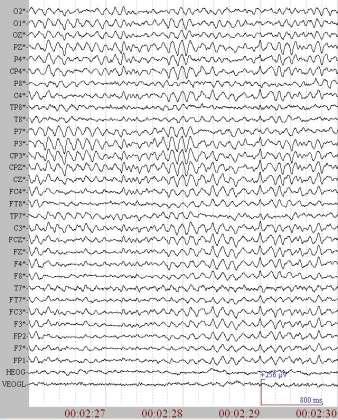


4 EEG - electroencephalography


5 What brain activity does EEG measure? Figures from M van Putten Essentials of Neurophysiology (2009)
6 What brain activity does EEG measure? Ionic potentials summed over large populations of synchronous, aligned neurons Primarily: Post-synaptic potentials of cortical pyramidal neurons A simple, take-home message: EEG directly measures cortical function

7 Alpha activity (~8-13Hz)
8 Alpha vs Delta Healthy Control (Alpha) Post-stroke (Delta) Voltage (microvolts) Time (ms)
9 Quantitative EEG qeeg/qeeg Computational analyses of raw digital EEG signals qeeg measures Objective measures - analogous to BP, HR, temp, etc. Can be readily compared or monitored over time Albeit may over-simplify complex EEG signals at Power is a commonly-used qeeg measure

10 Fourier / power spectrum analyses of EEG
11 Healthy Control (Alpha) Post-stroke (Delta) Voltage (microvolts) Time (ms) EEG power spectrum Healthy Control (Alpha) Post-stroke (Delta) Power EEG Frequency (Hz)
12 Interchangeable terms Power Amplitude Voltage Intensity Energy

13 EEG scalp topography
14 Power Relative power % of power in a given frequency band (e.g., alpha) As % of total power (0-30Hz: delta + theta + alpha + beta) Delta Theta Alpha Beta 0 Frequency band Relative delta power = 30 / ( ) = 30/100 = 30%

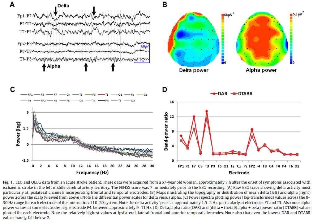
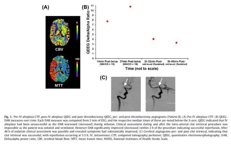
15 Ischaemic stroke Image source: The Bulletin, March 2004

16 Cerebral artery territories MCA = Middle Cerebral Artery ACA = Anterior Cerebral Artery PCA = Posterior Cerebral Artery
17 EEG: Delta (1-3Hz)
18

19 Delta & thalamo-cortical networks disconnection between cortical & thalamic regions thalamo-cortical dysrhythmia: disruption of top-down cortical modulation of thalamic neurons low-frequency bursting in thalamus, which is propagated to cortical regions delta activity van Wijngaarden et al. PLOS Computational Biology. 2016:10;12(8):e

20 Healthy Control
21 Power Delta/alpha ratio Delta Theta Alpha Beta Frequency band Delta/alpha ratio = 30/40 = 0.75
22 Post-stroke delta/alpha ratio (DAR) We have found DAR to: Correlate with post-stroke functional outcomes (2007) Be informative about success/failure of acute treatments (2013) Correlate with post-stroke cognitive outcomes (frontal DAR; 2014) Be 100% accurate for determining presence/absence of ischaemic stroke (confirmed by CT imaging; 2016) Normalise within minutes of successful clot retrieval treatment (2016)
 then Intra-arterial clot retrieval (")
23 Reperfusion treatments for acute ischaemic stroke If CT brain scan indicates large vessel occlusion, & Within 6 hours of symptom onset Inject clot-dissolving drug (intravenously) then Intra-arterial clot retrieval ( thrombectomy )
24 Delta/alpha ratio (DAR) normalises within minutes of cerebral blood flow being restored Cerebral angiograms (pre- & post-thrombectomy)
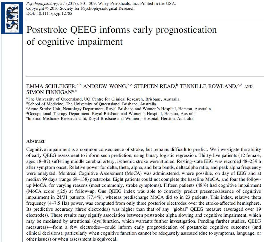
25 Post-stroke qeeg publications Schleiger E, et al. Post-stroke EEG informs early prognostication of cognitive impairment. Psychophysiology. 2017;54: Schleiger E, et al. Improved cerebral pathophysiology immediately following thrombectomy in acute ischaemic stroke: Monitoring via quantitative EEG. Clinical Neurophysiology. 2016;127: van Wijngaarden JBG, et al. The Impact of Cortical Lesions on Thalamo-Cortical Network Dynamics After Acute Ischaemic Stroke: A Combined Experimental and Theoretical Study. PLOS Computational Biology. 2016:10;12(8):e Finnigan S, Wong A, Read S. Defining abnormal slow EEG activity in acute ischaemic stroke: delta/alpha ratio as an optimal QEEG index. Clinical Neurophysiology. 2016;127: Schleiger E, et al. Frontal EEG delta/alpha ratio and screening for post-stroke cognitive deficits. International Journal of Psychophysiology. 2014;94: Sheikh N, et al. QEEG may uniquely inform and expedite decisions regarding intra-arterial clot retrieval in acute stroke. Clinical Neurophysiology. 2013;124: Finnigan S, van Putten MJAM. EEG in ischaemic stroke: Quantitative EEG can uniquely inform (sub-)acute prognoses and clinical management. Clinical Neurophysiology. 2013;124: Finnigan S, Rose SE, Chalk JB. Contralateral hemisphere delta EEG in acute stroke precedes worsening of symptoms and death. Clinical Neurophysiology. 2008;119: Finnigan S, et al. Quantitative EEG indices of sub-acute ischaemic stroke correlate with clinical outcomes. Clinical Neurophysiology. 2007;118: Finnigan S, Rose SE, Chalk JB. Rapid EEG changes indicate reperfusion after tissue plasminogen activator in acute stroke. Clinical Neurophysiology. 2006;117: Finnigan S, et al. Correlation of quantitative EEG in acute stroke with 30 day NIHSS score: Comparison with diffusion and perfusion MRI. Stroke. 2004;35:
26
27 Recap: Key points EEG directly measures activity of cortical neurons EEG power spectrum plots amplitudes associated with various frequencies Stroke increased delta & diminished alpha power Delta/alpha power ratio shows promise for various clinical applications (monitoring, prognoses)
 or to UQ St Lucia (via Milton & Regatta) (translink.")
28 Take a ride on a CityCat to New Farm / Bulimba / Teneriffe (via Riverside / City) or to UQ St Lucia (via Milton & Regatta) (translink.com.au)
29 qeeg studies of: Aging Mild cognitive impairment (MCI) Dementia Post-stroke cognitive impairment
30 Theta activity (~4-7Hz) & cognitive decline Several studies indicate that high, resting-state theta power in adults is associated with cognitive impairment, or subsequent cognitive decline increased theta power in early dementia (e.g., Coben et al 1985) theta power correlated with cognitive impairments; across controls, MCI & dementia (e.g., Prichep et al., 1994; Jelic et al., 1996) that higher baseline theta power is generally indicative of subsequent cognitive decline (e.g., progression to dementia; Jelic et al., 2000; Moretti et al., 2009; Prichep et al., 2006) However.
31 (Frontal) theta & cognition Theta especially at frontal midline is an EEG index of healthy cognitive function: Memory; working & episodic Spatial navigation Arithmetic Performance, practice, or meditation Non-specific, focused or sustained attention or concentration, or resistance to intrusion are common requirements Generated by networks including medial temporal, anterior cingulate & other cortical regions Review: Mitchell et al. Progress in Neurobiology 2008:86;
32
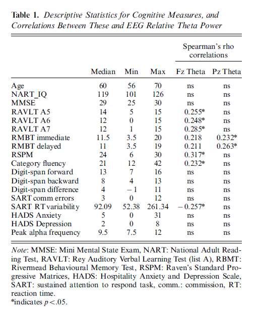
33 So is theta activity good or bad in relation to cognitive function?!?
34
 Cummins et al.")
35 Frontal midline theta: reduced in cognitive aging & MCI Frontal midline theta power: Young controls > Older controls > Mild cog impairment (MCI) Differences greatest in EEG during recognition memory task MCI categorised by consensus between two neuro-psychologists & neurologist (neuropsych assessment, medical history, clinical examination & MRI) Cummins et al. (Int J Psychophysiology 2007; 2008)

36

37
38
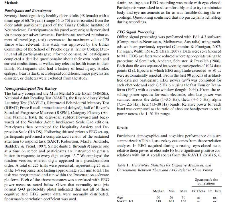
39 Frontal midline theta & cognition in healthy older adults N=73 cognitively healthy older adults (25 male); mean age 60.8 (range 56 to 70) Frontal midline theta power correlated significantly with cognitive performance: Rey Auditory Verbal Learning Test (RAVLT) delayed recall Raven s Standard Progressive Matrices Category Fluency (Animal Naming Test) Sustained Attention to Respond Task (SART; Robertson et al., 1997)
40
41 Slowing of the alpha rhythm Alpha slowing occurs: in aging with reduced brain metabolism & brain volume, in MCI and dementia (e.g. Coben et al, 1985; Buchan et al, 1997; Jelic et al, 2000) Peak alpha frequency can shift below 8Hz Some healthy older adults: 7.5 Hz (Finnigan & Robertson, 2011) We need to be careful & not confuse theta with slowed alpha activity
 did not correlate These results indicate the correlations involve frontal midline theta, not")
42 Frontal midline theta & cognition in healthy older adults Only frontal (not posterior) theta power correlated significantly with cognitive performance Alpha power & peak alpha frequency (Pz) did not correlate These results indicate the correlations involve frontal midline theta, not slowed alpha
43
44 Post-stroke cognitive impairment Cognitive impairment occurs in ~50% of stroke patients 53% (52/99) at 3 months (Srikanth et al, 2003) 46% (50/108) at 6 months (Kelly-Hayes et al, 2003) 58% (189/327) at 3 6 months (Dong et al, 2014) 48% (15/31) at 3 months (Schleiger et al, 2017) Difficult to assess or prognosticate in many cases Important for planning; e.g. required level of care; return to work Is early qeeg informative?
45
46 qeeg & post-stroke cognitive impairment Resting 2-5 days (median 3.5) post-stroke Montreal Cognitive Assessment (MoCA) on same day Follow-up cognitive days (median 99) Logistic regression modelling & ROC analyses to analyse predictive accuracies of early qeeg & MoCA
, whereas MoCA did so in 23 patients qeeg predictor: slowed alpha power @ occipital electrode on stroke-affected hemisphere This one electrode was more informative than")
47 Slowed alpha predicts post-stroke cognitive function qeeg correctly predicted presence/absence of cognitive impairment in 24/31 patients (77.4%), whereas MoCA did so in 23 patients qeeg predictor: slowed alpha occipital electrode on stroke-affected hemisphere This one electrode was more informative than all 19 Stroke-affected hemisphere
48 Alpha slowing Alpha slowing, to <8 Hz, has been reported in: Stroke patients (Yuasa et al, 2001) Vascular dementia patients (Moretti et al, 2004; Muresanu et al, 2008) Other neurological insults, e.g. traumatic brain injury (Angelakis et al, 2004; Nuwer et al, 2005; Hebb et al, 2007; Vespa et al, 2002) We found some stroke patients had peak alpha frequency as low as 6 Hz (Schleiger et al, 2017) Slowed alpha power reliably predicted cognitive outcomes, and peak alpha frequency is also informative (Schleiger et al, 2017)
49
50 Two distinct forms of 4-8 Hz activity It is important not to confuse these: Frontal midline theta Resting & task-state EEG; frontal focus Indicative of healthy neurocognitive function Slowed alpha Resting-state EEG; posterior focus In some healthy older adults, those with cognitive impairments or other conditions, e.g. stroke, dementia, TBI In these cases, adjusted frequency ranges seem appropriate for theta (e.g. 4-6 Hz) and alpha (e.g Hz)
51 When classifying EEG activity Frequency is only one part of the story It is also important to consider: The state/s during which EEG is recorded Resting / task Eyes open / eyes closed Awake / drowsy / asleep Scalp topography Other factors e.g. medications, (sub-clinical) conditions e.g. vascular issues, etc.
52 Implications for neurofeedback with stroke patients Cognitive impairment occurs in ~50% of stroke patients A study of > 200 stroke patients found attention to be the most commonly impaired cognitive domain (Hurford et al, 2013) Post-stroke sustained attention seems to be linked to degree of functional recovery (Robertson et al, 1997) If neurofeedback can increase alpha frequency, this may aid recovery of cognitive (& perhaps other?) functions Is inhibition of delta activity feasible?? If yes, this should also be beneficial

53 Neonatal studies
54 Thanks Funding acknowledgements Royal Brisbane & Women s Hospital Foundation National Stroke Foundation National Health & Medical Research Council
55
56 EEG: voltage oscillations over time
57 Alpha activity EEG oscillations at ~8-13 Hz Most posterior electrodes; resting, eyes closed Diminished ( blocked ) by: Opening eyes Intensive cognitive activity e.g. mental arithmetic The above findings are indicative of healthy brain function
58 Alpha activity Everyone has a characteristic individual alpha frequency e.g., 9.5 Hz, 12 Hz, etc. S l o w i n g of alpha can occur Common in older adults (e.g., 7-8 Hz) More pronounced in pathologies (e.g., 6-7 Hz in acute stroke) Irregularities can be reflected in: Similar alpha amplitudes between eyes closed & eyes open states Similar alpha amplitudes between posterior & frontal electrodes
59 Alpha is generated by thalamo-cortical networks Cortex Thalamus
60
61
62
63 Delta activity Abnormally slow EEG: ~1-3 Hz Often indicative of pathology (e.g., acute stroke) Evidently reflects pathophysiology in thalamo-cortical networks disconnection between cortical & thalamic regions (physical or functional) thalamo-cortical dysrhythmia: disruption of top-down cortical modulation of thalamic neurons low-frequency bursting in thalamus, which is propagated to cortical regions (van Wijngaarden et al. PLOS Computational Biology. 2016:10;12(8):e )
64
65 Theta activity EEG oscillations at ~4-7 Hz Linked with cognitive activity e.g. memory Often most obvious at frontal electrodes Generated by networks including medial temporal, anterior cingulate & other cortical regions However abnormally slow alpha can be within theta frequency range Important to consider state (e.g., resting eyes closed vs other), scalp location (e.g., frontal vs posterior), reactivity (blocking) (Finnigan & Robertson. Psychophysiology. 2011;48: )
66
67

68
69

70 Cortex Subplate Thalamus
71 Early history of EEG Late 1800s: animal studies in Britain & Europe 1924: first human EEG, by German psychiatrist Hans Berger Observed alpha activity (the Berger rhythm ) Berger published his first paper in 1929 technique for "recording the electrical activity of the human brain from the surface of the head"
72 EEG electrode positions: the system
73 EEG reporting, analyses & interpretations Can focus on various features such as: Frequency Scalp topography (location e.g., frontal) Voltage (e.g., inter-hemispheric symmetry) Waveform morphology (e.g., posterior sharp waves) Synchrony between electrodes (phase) Continuity vs discontinuity (e.g., burst suppression )
74 EEG reporting & analyses Conventional clinical EEG reporting: qualitative e.g., alpha activity has a posterior focus. There is also some periodic slow activity at temporal electrodes, more obvious over the left hemisphere Can be subjective Quantitative EEG (qeeg) analyses Computational analyses of raw digital EEG signals qeeg measures (analogous to BP, HR, temperature, etc.) Objective measures, albeit may over-simplify EEG at times More commonly used in research; starting to emerge in some clinical settings

75 Finnigan et al. Rapid EEG changes indicate reperfusion after tpa injection in acute ischaemic stroke. Clinical Neurophysiology. 2006;117:
76
Introduction to EEG del Campo. Introduction to EEG. J.C. Martin del Campo, MD, FRCP University Health Network Toronto, Canada
 Introduction to EEG J.C. Martin, MD, FRCP University Health Network Toronto, Canada What is EEG? A graphic representation of the difference in voltage between two different cerebral locations plotted over
Matrix Energetics Research Brainwaves and Heart waves Research on Matrix Energetics in Action
 Matrix Energetics Research Brainwaves and Heart waves Research on Matrix Energetics in Action QEEG (quantitative electroencephalography) and HRV (heart rate variability analysis) tests revealed Dr. Richard
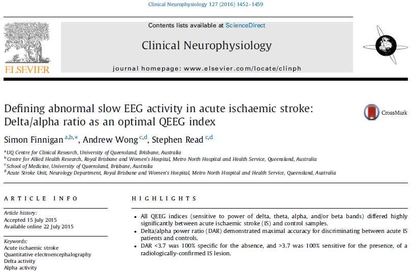
Accepted Manuscript. Defining abnormal slow EEG activity in acute ischaemic stroke: delta/alpha ratio as an optimal QEEG index
 Accepted Manuscript Defining abnormal slow EEG activity in acute ischaemic stroke: delta/alpha ratio as an optimal QEEG index Simon Finnigan, Andrew Wong, Stephen Read PII: S1388-2457(15)00723-3 DOI: http://dx.doi.org/10.1016/j.clinph.2015.07.014
Probing the Neuronal Status for Cerebrovascular Disease using EEG
 International Conference on Electrical, Computer and Communication Engineering (ECCE), February 6-8, 7, Cox s Bazar, Bangladesh Probing the Neuronal Status for Cerebrovascular Disease using EEG G. M. Mahmudur
Northeast Center for Special Care Grant Avenue Lake Katrine, NY
 300 Grant Avenue Lake Katrine, NY 12449 845-336-3500 Information Bulletin What is Brain Mapping? By Victor Zelek, Ph.D., Director of Neuropsychological Services Diplomate, National Registry of Neurofeedback
EEG IN FOCAL ENCEPHALOPATHIES: CEREBROVASCULAR DISEASE, NEOPLASMS, AND INFECTIONS
 246 Figure 8.7: FIRDA. The patient has a history of nonspecific cognitive decline and multiple small WM changes on imaging. oligodendrocytic tumors of the cerebral hemispheres (11,12). Electroencephalogram
EEG in the ICU: Part I
 EEG in the ICU: Part I Teneille E. Gofton July 2012 Objectives To outline the importance of EEG monitoring in the ICU To briefly review the neurophysiological basis of EEG To introduce formal EEG and subhairline
The Sonification of Human EEG and other Biomedical Data. Part 3
 The Sonification of Human EEG and other Biomedical Data Part 3 The Human EEG A data source for the sonification of cerebral dynamics The Human EEG - Outline Electric brain signals Continuous recording
The EEG Analysis of Auditory Emotional Stimuli Perception in TBI Patients with Different SCG Score
 Open Journal of Modern Neurosurgery, 2014, 4, 81-96 Published Online April 2014 in SciRes. http://www.scirp.org/journal/ojmn http://dx.doi.org/10.4236/ojmn.2014.42017 The EEG Analysis of Auditory Emotional
Neurophysiology & EEG
 Neurophysiology & EEG PG4 Core Curriculum Ian A. Cook, M.D. Associate Director, Laboratory of Brain, Behavior, & Pharmacology UCLA Department of Psychiatry & Biobehavioral Sciences Semel Institute for
EEG History. Where and why is EEG used? 8/2/2010
 EEG History Hans Berger 1873-1941 Edgar Douglas Adrian, an English physician, was one of the first scientists to record a single nerve fiber potential Although Adrian is credited with the discovery of
Neonatal EEG Maturation
 Neonatal EEG Maturation Cindy Jenkinson, R. EEG T., CLTM October 7, 2017 Fissure Development 3 http://www.hhmi.org/biointeractive/develop ment-human-embryonic-brain 4 WHAT IS IMPORTANT TO KNOW BEFORE I
Electroencephalography
 The electroencephalogram (EEG) is a measure of brain waves. It is a readily available test that provides evidence of how the brain functions over time. The EEG is used in the evaluation of brain disorders.
EEG- A Brief Introduction
 Fatemeh Hadaeghi EEG- A Brief Introduction Lecture Notes for BSP, Chapter 4 Master Program Data Engineering 1 4 Introduction Human brain, as the most complex living structure in the universe, has been
Neurotechnology for Special Needs Children
 ISSN 4-956 (Print) ISSN -849 (Online) Sep Dec 5 Neurotechnology for Special Needs Children Norsiah Fauzan Faculty of Cognitive Science and Human Development, Universiti Malaysia Sarawak Abstract This paper
Sleep stages. Awake Stage 1 Stage 2 Stage 3 Stage 4 Rapid eye movement sleep (REM) Slow wave sleep (NREM)
 Slow wave sleep (NREM)") Sleep stages Awake Stage 1 Stage 2 Stage 3 Stage 4 Rapid eye movement sleep (REM) Slow wave sleep (NREM) EEG waves EEG Electrode Placement Classifying EEG brain waves Frequency: the number of oscillations/waves
Sleep stages Awake Stage 1 Stage 2 Stage 3 Stage 4 Rapid eye movement sleep (REM) Slow wave sleep (NREM) EEG waves EEG Electrode Placement Classifying EEG brain waves Frequency: the number of oscillations/waves
states of brain activity sleep, brain waves DR. S. GOLABI PH.D. IN MEDICAL PHYSIOLOGY
 states of brain activity sleep, brain waves DR. S. GOLABI PH.D. IN MEDICAL PHYSIOLOGY introduction all of us are aware of the many different states of brain activity, including sleep, wakefulness, extreme
Exploratory Investigation into Mild Brain Injury and Discriminant Analysis with High Frequency Bands (32-64 Hz)
") Exploratory Investigation into Mild Brain Injury and Discriminant Analysis with High Frequency Bands (32-64 Hz), Brain Injury, August,1999, 477-488 Exploratory Investigation into Mild Brain Injury and
Exploratory Investigation into Mild Brain Injury and Discriminant Analysis with High Frequency Bands (32-64 Hz), Brain Injury, August,1999, 477-488 Exploratory Investigation into Mild Brain Injury and
Physiology Unit 2 CONSCIOUSNESS, THE BRAIN AND BEHAVIOR
 Physiology Unit 2 CONSCIOUSNESS, THE BRAIN AND BEHAVIOR In Physiology Today What the Brain Does The nervous system determines states of consciousness and produces complex behaviors Any given neuron may
Physiology Unit 2 CONSCIOUSNESS, THE BRAIN AND BEHAVIOR
 Physiology Unit 2 CONSCIOUSNESS, THE BRAIN AND BEHAVIOR What the Brain Does The nervous system determines states of consciousness and produces complex behaviors Any given neuron may have as many as 200,000
ACUTE STROKE IMAGING
 ACUTE STROKE IMAGING Mahesh V. Jayaraman M.D. Director, Inter ventional Neuroradiology Associate Professor Depar tments of Diagnostic Imaging and Neurosurger y Alper t Medical School at Brown University
Common EEG pattern in critical care
 Common EEG pattern in critical care พ.ญ.ส ธ ดา เย นจ นทร Causes Direct neuronal injury Cerebral dysfunction : encephalopathy Psychic problems EEG in critical care 1 October 2009, Pramongkutklao Hospital
Normal EEG of wakeful resting adults of years of age. Alpha rhythm. Alpha rhythm. Alpha rhythm. Normal EEG of the wakeful adult at rest
 Normal EEG of wakeful resting adults of 20-60 years of age Suthida Yenjun, M.D. Normal EEG of the wakeful adult at rest Alpha rhythm Beta rhythm Mu rhythm Vertex sharp transients Intermittent posterior
Electroencephalography & Neurofeedback
 Electroencephalography & Neurofeedback A Brief Introduction to the Science of Brainwaves Glyn Blackett YORK biofeedback CENTRE Introduction This article is a brief introduction to electroencephalography
Biomedical Research 2013; 24 (3): ISSN X
: ISSN X") Biomedical Research 2013; 24 (3): 359-364 ISSN 0970-938X http://www.biomedres.info Investigating relative strengths and positions of electrical activity in the left and right hemispheres of the human brain
Biomedical Research 2013; 24 (3): 359-364 ISSN 0970-938X http://www.biomedres.info Investigating relative strengths and positions of electrical activity in the left and right hemispheres of the human brain
Non epileptiform abnormality J U LY 2 7,
 Non epileptiform abnormality S U D A J I R A S A K U L D E J, M D. C H U L A L O N G KO R N C O M P R E H E N S I V E E P I L E P S Y C E N T E R J U LY 2 7, 2 0 1 6 Outline Slow pattern Focal slowing
To link to this article: PLEASE SCROLL DOWN FOR ARTICLE
 Journal of Neurotherapy: Investigations in Neuromodulation, Neurofeedback and Applied Neuroscience Clinical Corner D. Corydon Hammond PhD, Joel F. Lubar PhD & Marvin W. Sams ND Published online: 08 Sep
Spectral Analysis of EEG Patterns in Normal Adults
 Spectral Analysis of EEG Patterns in Normal Adults Kyoung Gyu Choi, M.D., Ph.D. Department of Neurology, Ewha Medical Research Center, Ewha Womans University Medical College, Background: Recently, the
Raw and Quantitative EEG for Identification of Ischemia
 Raw and Quantitative EEG for Identification of Ischemia Susan T. Herman, MD Assistant Professor of Neurology Beth Israel Deaconess Medical Center Harvard Medical School Boston, MA Disclosures None relevant
Beyond the Basics in EEG Interpretation: Throughout the Life Stages
 Beyond the Basics in EEG Interpretation: Throughout the Life Stages Steve S. Chung, MD, FAAN Chairman, Neuroscience Institute Director, Epilepsy Program Banner University Medical Center University of Arizona
Scope. EEG patterns in Encephalopathy. Diffuse encephalopathy. EEG in adult patients with. EEG in diffuse encephalopathy
 Scope EEG patterns in Encephalopathy Dr.Pasiri Sithinamsuwan Division of Neurology Department of Medicine Phramongkutklao Hospital Diffuse encephalopathy EEG in specific encephalopathies Encephalitides
EEG Electrode Placement
 EEG Electrode Placement Classifying EEG brain waves Frequency: the number of oscillations/waves per second, measured in Hertz (Hz) reflects the firing rate of neurons alpha, beta, theta, delta Amplitude:
Asian Epilepsy Academy (ASEPA) & ASEAN Neurological Association (ASNA) EEG Certification Examination
 & ASEAN Neurological Association (ASNA) EEG Certification Examination") Asian Epilepsy Academy (ASEPA) & ASEAN Neurological Association (ASNA) EEG Certification Examination EEG Certification Examination Aims To set and improve the standard of practice of Electroencephalography
Asian Epilepsy Academy (ASEPA) & ASEAN Neurological Association (ASNA) EEG Certification Examination EEG Certification Examination Aims To set and improve the standard of practice of Electroencephalography
Practical 3 Nervous System Physiology 2 nd year English Module. Dept. of Physiology, Carol Davila University of Medicine and Pharmacy
 Electroencephalography l h (EEG) Practical 3 Nervous System Physiology 2 nd year English Module Dept. of Physiology, Carol Davila University of Medicine and Pharmacy What is EEG EEG noninvasively records
EEG and some applications (seizures and sleep)
") EEG and some applications (seizures and sleep) EEG: stands for electroencephalography and is a graphed representation of the electrical activity of the brain. EEG is the recording of electrical activity
EEG and some applications (seizures and sleep) EEG: stands for electroencephalography and is a graphed representation of the electrical activity of the brain. EEG is the recording of electrical activity
PSD Analysis of Neural Spectrum During Transition from Awake Stage to Sleep Stage
 PSD Analysis of Neural Spectrum During Transition from Stage to Stage Chintan Joshi #1 ; Dipesh Kamdar #2 #1 Student,; #2 Research Guide, #1,#2 Electronics and Communication Department, Vyavasayi Vidya
The Nervous System. Neuron 01/12/2011. The Synapse: The Processor
 The Nervous System Neuron Nucleus Cell body Dendrites they are part of the cell body of a neuron that collect chemical and electrical signals from other neurons at synapses and convert them into electrical
Subhairline EEG Part II - Encephalopathy
 Subhairline EEG Part II - Encephalopathy Teneille Gofton September 2013 Objectives To review the subhairline EEG changes seen with encephalopathy To discuss specific EEG findings in encephalopathy To outline
Oscillations: From Neuron to MEG
 Oscillations: From Neuron to MEG Educational Symposium, MEG UK 2014, Nottingham, Jan 8th 2014 Krish Singh CUBRIC, School of Psychology Cardiff University What are we trying to achieve? Bridge the gap from
Developmental Changes Including Neonatal EEG. Gregory L. Holmes, MD
 Developmental Changes Including Neonatal EEG Gregory L. Holmes, MD A A + B =: B + A.Dravet Syndrome B.Menkes syndrome C.West syndrome D.Ohtahara shyndrome The Difficult Delivery 1 day old male transferred
EEG in Medical Practice
 EEG in Medical Practice Dr. Md. Mahmudur Rahman Siddiqui MBBS, FCPS, FACP, FCCP Associate Professor, Dept. of Medicine Anwer Khan Modern Medical College What is the EEG? The brain normally produces tiny
EEG Analysis on Brain.fm (Focus)
") EEG Analysis on Brain.fm (Focus) Introduction 17 subjects were tested to measure effects of a Brain.fm focus session on cognition. With 4 additional subjects, we recorded EEG data during baseline and while
EEG Analysis on Brain.fm (Focus) Introduction 17 subjects were tested to measure effects of a Brain.fm focus session on cognition. With 4 additional subjects, we recorded EEG data during baseline and while
Processed by HBI: Russia/Switzerland/USA
 1 CONTENTS I Personal and clinical data II Conclusion. III Recommendations for therapy IV Report. 1. Procedures of EEG recording and analysis 2. Search for paroxysms 3. Eyes Open background EEG rhythms
STRUCTURAL ORGANIZATION OF THE NERVOUS SYSTEM
 STRUCTURAL ORGANIZATION OF THE NERVOUS SYSTEM STRUCTURAL ORGANIZATION OF THE BRAIN The central nervous system (CNS), consisting of the brain and spinal cord, receives input from sensory neurons and directs
13 Electroencephalography
 13 Electroencephalography 13.1 INTRODUCTION The first recording of the electric field of the human brain was made by the German psychiatrist Hans Berger in 1924 in Jena. He gave this recording the name
Est-ce que l'eeg a toujours sa place en 2019?
 Est-ce que l'eeg a toujours sa place en 2019? Thomas Bast Epilepsy Center Kork, Germany Does EEG still play a role in 2019? What a question 7T-MRI, fmri, DTI, MEG, SISCOM, Of ieeg course! /HFO, Genetics
Electroencephalography (EEG) and Hemispheric Asymmetry
 and Hemispheric Asymmetry") Updated 12-22-03 BSL PRO Lesson H10: Electroencephalography (EEG) and Hemispheric Asymmetry Derived from original work created by Pr Jean-François Lambert and Nicole Chantrier, Institut d'enseignement
Updated 12-22-03 BSL PRO Lesson H10: Electroencephalography (EEG) and Hemispheric Asymmetry Derived from original work created by Pr Jean-François Lambert and Nicole Chantrier, Institut d'enseignement
Separation Of,, & Activities In EEG To Measure The Depth Of Sleep And Mental Status
 Separation Of,, & Activities In EEG To Measure The Depth Of Sleep And Mental Status Shah Aqueel Ahmed 1, Syed Abdul Sattar 2, D. Elizabath Rani 3 1. Royal Institute Of Technology And Science, R. R. Dist.,
Working Memory Impairments Limitations of Normal Children s in Visual Stimuli using Event-Related Potentials
 2015 6th International Conference on Intelligent Systems, Modelling and Simulation Working Memory Impairments Limitations of Normal Children s in Visual Stimuli using Event-Related Potentials S. Z. Mohd
Amy Kruse, Ph.D. Strategic Analysis, Inc. LCDR Dylan Schmorrow USN Defense Advanced Research Projects Agency
 What can modern neuroscience technologies offer the forward-looking applied military psychologist? Exploring the current and future use of EEG and NIR in personnel selection and training. Amy Kruse, Ph.D.
THE ACTIVITY RECORDED IN THE EEG
 Version 4. A Monthly Publication presented by Professor Yasser Metwally April 2008 THE ACTIVITY RECORDED IN THE EEG here is now considerable evidence from studies in experimental animals to suggest that
Clinical Validation of the NeuroGuide QEEG Normative Database. Phase Reset Duration Means. abs(out-phase) Cross Spectral Power. Burst Amplitude Means
 Cross Spectral Power. Burst Amplitude Means") 1.000 0.900 Clinical Validation of the NeuroGuide QEEG Normative Database Multiple R 0.800 Correlation Coefficient 0.700 0.600 0.500 0.400 0.300 0.200 0.100 0.000 PLATE 2.6 Phase Difference Coherence Phase
1.000 0.900 Clinical Validation of the NeuroGuide QEEG Normative Database Multiple R 0.800 Correlation Coefficient 0.700 0.600 0.500 0.400 0.300 0.200 0.100 0.000 PLATE 2.6 Phase Difference Coherence Phase
NEURORADIOLOGY DIL part 4
 NEURORADIOLOGY DIL part 4 Strokes and infarcts K. Agyem MD, G. Hall MD, D. Palathinkal MD, Alexandre Menard March/April 2015 OVERVIEW Introduction to Neuroimaging - DIL part 1 Basic Brain Anatomy - DIL
Electrophysiologic approaches to delirium. Overview
 Electrophysiologic approaches to delirium Alvaro Pascual Leone, MD, PhD Mouhsin Shafi, MD, PhD Overview Overview of the problem and techniques Electrophysiological studies in delirium Electrophysiological
Multiscale Evidence of Multiscale Brain Communication
 Multiscale Evidence of Multiscale Brain Communication Scott Makeig Swartz Center for Computational Neuroscience Institute for Neural Computation University of California San Diego La Jolla CA Talk given
Submitted report on Sufi recordings at AAPB 2013 in Portland. Not for general distribution. Thomas F. Collura, Ph.D. July, 2013
 Submitted report on Sufi recordings at AAPB 2013 in Portland Not for general distribution. Thomas F. Collura, Ph.D. July, 2013 Summary of EEG findings The intent of the EEG monitoring was to see which
True Epileptiform Patterns (and some others)
") True Epileptiform Patterns (and some others) a) What is epileptiform b) Some possible surprises c) Classification of generalized epileptiform patterns An epileptiform pattern Interpretative term based
True Epileptiform Patterns (and some others) a) What is epileptiform b) Some possible surprises c) Classification of generalized epileptiform patterns An epileptiform pattern Interpretative term based
AUXILIARIES AND NEUROPLASTICITY
 AUXILIARIES AND NEUROPLASTICITY Claudio Babiloni, Ph.D. Department of Biomedical Sciences, University of Foggia (UNIFG), Italy UNIFG structured personnel involved Prof. Claudio Babiloni (Coordinator),
Informationsverarbeitung im zerebralen Cortex
 Informationsverarbeitung im zerebralen Cortex Thomas Klausberger Dept. Cognitive Neurobiology, Center for Brain Research, Med. Uni. Vienna The hippocampus is a key brain circuit for certain forms of memory
Dysfunctional FINDINGS IN EEG. About 18 Cases were Seen in Consultation in our Hospital
 Cronicon OPEN ACCESS NEUROLOGY Clinical Images Angel Molina Leon* Dysfunctional FINDINGS IN EEG. About 18 Cases were Seen in Consultation in our Hospital Clinical Neurophysiology Service. Centro medico
DATA MANAGEMENT & TYPES OF ANALYSES OFTEN USED. Dennis L. Molfese University of Nebraska - Lincoln
 DATA MANAGEMENT & TYPES OF ANALYSES OFTEN USED Dennis L. Molfese University of Nebraska - Lincoln 1 DATA MANAGEMENT Backups Storage Identification Analyses 2 Data Analysis Pre-processing Statistical Analysis
The electrophysiological effects of a brain injury on auditory memory functioning The QEEG correlates of impaired memory
 Archives of Clinical Neuropsychology 18 (2003) 363 378 Abstract The electrophysiological effects of a brain injury on auditory memory functioning The QEEG correlates of impaired memory Kirtley Thornton
Introduction to Electrophysiology
 Introduction to Electrophysiology Dr. Kwangyeol Baek Martinos Center for Biomedical Imaging Massachusetts General Hospital Harvard Medical School 2018-05-31s Contents Principles in Electrophysiology Techniques
Brain and Cognition. Cognitive Neuroscience. If the brain were simple enough to understand, we would be too stupid to understand it
 Brain and Cognition Cognitive Neuroscience If the brain were simple enough to understand, we would be too stupid to understand it 1 The Chemical Synapse 2 Chemical Neurotransmission At rest, the synapse
The secrets of conventional EEG
 The secrets of conventional EEG The spike/sharp wave activity o Electro-clinical characteristics of Spike/Sharp wave The polymorphic delta activity o Electro-clinical characteristics of Polymorphic delta
Restoring Communication and Mobility
 Restoring Communication and Mobility What are they? Artificial devices connected to the body that substitute, restore or supplement a sensory, cognitive, or motive function of the nervous system that has
Source localisation in the clinical practice: spontaneous EEG examinations with LORETA. Ph.D. thesis. Márton Tamás Tóth M.D.
 Source localisation in the clinical practice: spontaneous EEG examinations with LORETA Ph.D. thesis Márton Tamás Tóth M.D. Department of Neurology, University of Pécs Leader of project:: Prof. István Kondákor,
A Study of Smartphone Game Users through EEG Signal Feature Analysis
 , pp. 409-418 http://dx.doi.org/10.14257/ijmue.2014.9.11.39 A Study of Smartphone Game Users through EEG Signal Feature Analysis Jung-Yoon Kim Graduate School of Advanced Imaging Science, Multimedia &
Neurofeedback for ASD AND ADHD
 Neurofeedback for ASD AND ADHD Thomas F. Collura, Ph.D., MSMHC, QEEG-D, BCN, LPC The Brain Enrichment Center and BrainMaster Technologies, Inc., Bedford, OH Association for Applied Psychophysiology and
Asian Epilepsy Academy (ASEPA) EEG Certification Examination
 EEG Certification Examination") Asian Epilepsy Academy (ASEPA) EEG Certification Examination EEG Certification Examination Aims To set and improve the standard of practice of Electroencephalography (EEG) in the Asian Oceanian region
Asian Epilepsy Academy (ASEPA) EEG Certification Examination EEG Certification Examination Aims To set and improve the standard of practice of Electroencephalography (EEG) in the Asian Oceanian region
Cognitive Enhancement Using 19-Electrode Z-Score Neurofeedback
 This article was downloaded by: [Lucas Koberda] On: 22 August 2012, At: 09:31 Publisher: Routledge Informa Ltd Registered in England and Wales Registered Number: 1072954 Registered office: Mortimer House,
Mental State Sensing and the Goal of Circuit-Synapse Synergy
 Mental State Sensing and the Goal of Circuit-Synapse Synergy Patrick L. Craven, Ph.D. Senior Member, Engineering Staff Advanced Technology Laboratories Cherry Hill, NJ Goals of Artificial Intelligence
EEG workshop. Epileptiform abnormalities. Definitions. Dr. Suthida Yenjun
 EEG workshop Epileptiform abnormalities Paroxysmal EEG activities ( focal or generalized) are often termed epileptiform activities EEG hallmark of epilepsy Dr. Suthida Yenjun Epileptiform abnormalities
Outline of Talk. Introduction to EEG and Event Related Potentials. Key points. My path to EEG
 Outline of Talk Introduction to EEG and Event Related Potentials Shafali Spurling Jeste Assistant Professor in Psychiatry and Neurology UCLA Center for Autism Research and Treatment Basic definitions and
The AASM Manual for the Scoring of Sleep and Associated Events
 The AASM Manual for the Scoring of Sleep and Associated Events Summary of Updates in Version 2.1 July 1, 2014 The American Academy of Sleep Medicine (AASM) is committed to ensuring that The AASM Manual
Nature Neuroscience: doi: /nn Supplementary Figure 1. Large-scale calcium imaging in vivo.
 Supplementary Figure 1 Large-scale calcium imaging in vivo. (a) Schematic illustration of the in vivo camera imaging set-up for large-scale calcium imaging. (b) High-magnification two-photon image from
Lesson 5 EEG 1 Electroencephalography: Brain Rhythms
 Physiology Lessons for use with the Biopac Science Lab MP40 PC running Windows XP or Mac OS X 10.3-10.4 Lesson 5 EEG 1 Electroencephalography: Brain Rhythms Lesson Revision 2.23.2006 BIOPAC Systems, Inc.
Modeling the Impact of Recurrent Collaterals In CA3 on Downstream CA1 Firing Frequency
 Modeling the Impact of Recurrent Collaterals In CA3 on Downstream CA1 Firing Frequency 1 2 3 4 5 6 7 Teryn Johnson Gladys Ornelas Srihita Rudraraju t8johnso@eng.ucsd.edu glornela@eng.ucsd.edu srrudrar@eng.ucsd.edu
QEEG Informed Protocol Recommendation
 Protocol ecommendation Psychopathology ating Substance Use AD(H)D (page 2) Depression (page 3) Anxiety Disorder OCD Autism Schizophrenia Memory Disorder Insomnia Stimulants Anti depressants Anxiolytics/Sedatives
EEG WORKSHOP Nonepileptiform Abnormalities
 EEG WORKSHOP Nonepileptiform Abnormalities Kamornwan Katanyuwong MD Chiangmai University Hospital EST: 20th July 2010 EEG reading Age Background Epileptiform Non epileptiform Activation procedure normal
Inside Your Patient s Brain Michelle Peterson, APRN, CNP Centracare Stroke and Vascular Neurology
 Inside Your Patient s Brain Michelle Peterson, APRN, CNP Centracare Stroke and Vascular Neurology Activity Everyone stand up, raise your right hand, tell your neighbors your name 1 What part of the brain
EEG ANALYSIS: ANN APPROACH
 EEG ANALYSIS: ANN APPROACH CHAPTER 5 EEG ANALYSIS: ANN APPROACH 5.1 INTRODUCTION The analysis of EEG signals using ANN deals with developing a network in order to establish a relation between input and
Quantitative EEG (QEEG) Consent Form
 Consent Form") Quantitative EEG (QEEG) Consent Form Quantitative EEG, sometimes referred to as brain mapping, is the measurement through digital technology of electrical patterns at the surface of the scalp which primarily
Quantitative EEG (QEEG) Consent Form Quantitative EEG, sometimes referred to as brain mapping, is the measurement through digital technology of electrical patterns at the surface of the scalp which primarily
GUIDELINES FOR THE EARLY MANAGEMENT OF PATIENTS WITH ACUTE ISCHEMIC STROKE
 2018 UPDATE QUICK SHEET 2018 American Heart Association GUIDELINES FOR THE EARLY MANAGEMENT OF PATIENTS WITH ACUTE ISCHEMIC STROKE A Summary for Healthcare Professionals from the American Heart Association/American
Altered Dynamic of EEG Oscillations in Fibromyalgia Patients at Rest
 Altered Dynamic of EEG Oscillations in Fibromyalgia Patients at Rest Ana M. González-Roldán, PhD Ignacio Cifre, PhD Carolina Sitges, PhDPedro Montoya, PhD Pain Medicine, Volume 17, Issue 6, 1 June 2016,
ACUTE ISCHEMIC STROKE. Current Treatment Approaches for Acute Ischemic Stroke
 ACUTE ISCHEMIC STROKE Current Treatment Approaches for Acute Ischemic Stroke EARLY MANAGEMENT OF ACUTE ISCHEMIC STROKE Rapid identification of a stroke Immediate EMS transport to nearest stroke center
Periodic and Rhythmic Patterns. Suzette M LaRoche, MD Mission Health Epilepsy Center Asheville, North Carolina
 Periodic and Rhythmic Patterns Suzette M LaRoche, MD Mission Health Epilepsy Center Asheville, North Carolina Continuum of EEG Activity Neuronal Injury LRDA GPDs SIRPIDs LPDs + NCS Burst-Suppression LPDs
Heads Temporal Lobe Disconnect Name: Amostra adulto Age: Trainer: Amostra Date: 10/26/2015. Results very high in range very low
 52 22 Name: Amostra adulto Age: 48 95 Histogram Overall EEG Shape F7 F3 Fz F4 F8 T3 C3 Cz C4 T4 T5 P3 Pz P4 T6 O1 Oz O2 Slow Percent EC 23% 34% 33% 36% 41% 26% 33% 30% 33% 31% 28% 30% 28% 29% 28% 22% 22%
Human Brain Institute Russia-Switzerland-USA
 1 Human Brain Institute Russia-Switzerland-USA CONTENTS I Personal and clinical data II Conclusion. III Recommendations for therapy IV Report. 1. Procedures of EEG recording and analysis 2. Search for
EEG Sleep Circadian rhythms Learning Objectives: 121, 122
 EEG Sleep Circadian rhythms Learning Objectives: 121, 122 Zoltán Lelkes Electroencenphalography Hans Berger pen time amplifier electrodes 1 The waves of the EEG gamma > 30 Hz beta: 13-30 Hz Mental activity:
BIOPAC Systems, Inc BIOPAC Inspiring people and enabling discovery about life
 BIOPAC Systems, Inc. 2016 BIOPAC Inspiring people and enabling discovery about life 1 BIOPAC s Guide to EEG for Research: Mobita Wireless EEG Housekeeping Attendees are on Mute Headset is Recommended!
Mechanical thrombectomy in Plymouth. Will Adams. Will Adams
 Mechanical thrombectomy in Plymouth Will Adams Will Adams History Intra-arterial intervention 1995 (NINDS) iv tpa improved clinical outcome in patients treated within 3 hours of ictus but limited recanalisation
Laurence M. Hirshberg, Sufen Chiu, and Jean A. Frazier
 EMERGING INTERVENTIONS Foreword Melvin Lewis xi Preface Laurence M. Hirshberg, Sufen Chiu, and Jean A. Frazier xiii Emerging Brain-Based Interventions for Children and Adolescents: Overview and Clinical
Continuous EEG monitoring of the premature infant in the NICU
 Continuous EEG monitoring of the premature infant in the NICU Tom Stiris Oslo University Hospital, NICU CIP, Paris 2011 Background A method that at a very early stage diagnose those babies which would
Comparison of Five Major Recent Endovascular Treatment Trials
 Comparison of Five Major Recent Endovascular Treatment Trials Sample size 500 # sites 70 (100 planned) 316 (500 planned) 196 (833 estimated) 206 (690 planned) 16 10 22 39 4 Treatment contrasts Baseline
Biomarkers in Schizophrenia
 Biomarkers in Schizophrenia David A. Lewis, MD Translational Neuroscience Program Department of Psychiatry NIMH Conte Center for the Neuroscience of Mental Disorders University of Pittsburgh Disease Process
RBWH ICU Journal Club February 2018 Adam Simpson
 RBWH ICU Journal Club February 2018 Adam Simpson 3 THROMBOLYSIS Reperfusion therapy has become the mainstay of therapy for ischaemic stroke. Thrombolysis is now well accepted within 4.5 hours. - Improved
Power-Based Connectivity. JL Sanguinetti
 Power-Based Connectivity JL Sanguinetti Power-based connectivity Correlating time-frequency power between two electrodes across time or over trials Gives you flexibility for analysis: Test specific hypotheses
SLEEP STAGING AND AROUSAL. Dr. Tripat Deep Singh (MBBS, MD, RPSGT, RST) International Sleep Specialist (World Sleep Federation program)
 International Sleep Specialist (World Sleep Federation program)") SLEEP STAGING AND AROUSAL Dr. Tripat Deep Singh (MBBS, MD, RPSGT, RST) International Sleep Specialist (World Sleep Federation program) Scoring of Sleep Stages in Adults A. Stages of Sleep Stage W Stage
SLEEP STAGING AND AROUSAL Dr. Tripat Deep Singh (MBBS, MD, RPSGT, RST) International Sleep Specialist (World Sleep Federation program) Scoring of Sleep Stages in Adults A. Stages of Sleep Stage W Stage