ABSTRACT PLASTICITY OF PERIPHERAL NEURONS FOLLOWING AXOTOMY OF THE SUPERIOR CERVICAL GANGLION. by Zheng Zhu
|
|
|
- Mervin Collins
- 6 years ago
- Views:
Transcription
1 ABSTRACT PLASTICITY OF PERIPHERAL NEURONS FOLLOWING AXOTOMY OF THE SUPERIOR CERVICAL GANGLION by Zheng Zhu The overall goal of this project was to understand the changes in peripheral neurons and their vascular targets following axotomy (transection) of sympathetic postganglionic axons arising from the superior cervical ganglion (SCG). Changes were documented at short- and long-term survival time points following the injury. At seven days following axotomy, the extracerebral blood vessels were denervated of sympathetic axons, yet sensory and parasympathetic perivascular axons were increased and decreased, respectively. Protein expression of vascular endothelial growth factor (VEGF) in the vasculature was unchanged, but VEGF was increased in the SCG as well as the trigeminal ganglion, which houses the parent cell bodies of the sensory perivascular axons. At 12 weeks, the sympathetic innervation showed some recovery, yet sensory and parasympathetic innervation remained altered, as did VEGF in the SCG and trigeminal ganglion. These results reveal the presence of long term plasticity in peripheral neurons following axotomy of the SCG.
2 Plasticity of Peripheral Neurons Following Axotomy of the Superior Cervical Ganglion A Thesis Submitted to the Faculty of Miami University In partial fulfillment of The requirements for the degree of Master of Science Department of Zoology by Zheng Zhu Miami University Oxford, Ohio 2011 Advisor Dr. Lori G. Isaacson Reader Dr. Paul A. Harding
3 TABLE OF CONTENTS 1. Introduction Sympathetic model and overall goal of this study 1 The extracerebral blood vessels: innervation by 1 Neurotrophin regulation of peripheral neurons 3 VEGF as a regulator of sympathetic neurons 5 Changes in peripheral neurons following injury 7 2. Rationale and Hypotheses 9 3. Specific Aims 9 4. Methods Animals and tissue processing 11 Surgical procedures and axotomy of the SCG 11 Immunofluorescence procedures 11 Analysis of axons of extracerebral blood vessels 12 Western blot analysis and semi-quantitative analysis Results Changes in sympathetic perivascular axons associated following axotomy 14 Changes in sensory and parasympathetic perivascular axons following axotomy 15 Changes in VEGF following axotomy of the SCG Discussion Plasticity of sympathetic innervations 49 Increased CGRP sensory innervations following aoxtomy of the SCG 50 Decreased VAChT parasympathetic innervation following aoxtomy of the SCG 51 Changes in VEGF changes and the role Conclusions References 58 ii
4 LIST OF TABLES Table 1. Summary of changes in 42 iii
5 LIST OF FIGURES Figure 1. The extracerebral blood vessels at the base of the brain 18 Figure 2. TH perivascular axons associated with the ACA following axotomy 20 Figure 3. Quantitative analysis of th 22 Figure 4. CGRP-ir and VAChT-ir perivascular axons show no colocalization 24 Figure 5. CGRP-ir perivascular axons associated with the ACA following axotomy 26 Figure 6. Quantitative analysis of the density of 28 Figure 7. CGRP-ir perivascular axons associated with the MCA following axotomy 30 Figure 8. Quantitative analysis of the density of 32 Figure 9. VAChT associated with the ACA following axotomy 34 Figure 10. Quantitative analysis of the density of 36 Figure 11. VAChT-ir axons associated with the MCA following axotomy 38 Figure 12. Quantitative analysis of the density of 40 Figure 13. VEGF western analysis of the extracerebral blood vessels 43 Figure 14. VEGF western analysis of the superior cervical ganglion (SCG) 45 Figure 15. VEGF western analysis of the trigeminal ganglion 47 iv
6 1. INTRODUCTION Sympathetic model and overall goal of this study. The sympathetic nervous system is part of the peripheral nervous system (PNS), which connects the brain and spinal cord to the limbs and target organs. The PNS can be divided into the somatic nervous system and the autonomic nervous system. One function of the somatic nervous system is to process sensory information and the neuronal cell bodies are located in sensory ganglia located just outside of the brain and spinal cord. The somatic nervous system also consists of cell bodies in brain and spinal cord that have axons that connect to skeletal muscles to regulate movement (Purves et al., 2008). The autonomic nervous system is comprised of the sympathetic nervous system and parasympathetic nervous systems. Primary functions of sympathetic and parasympathetic neurons are to regulate pupil dilation, heart rate and contraction, and urinary output (Wallin and Charkoudian, 2007; Charkoudian and Rabbitts, 2009). The cell bodies of autonomic neurons are located in ganglia in the periphery. The superior cervical ganglion (SCG) is the most rostral sympathetic ganglion and is located at the bifurcation of the common carotid artery and is comprised of a cluster of postganglionic neuronal cell bodies and glial cells. The cell bodies in the SCG project postganglionic axons through the external and internal carotid nerves to innervate the head and neck targets such as the irides, the submandibular gland, the pineal gland, and the extracerebral blood vessels, which are located at the base of the brain (Fig.1). The postganglionic axons that innervate the extracerebral blood vessels regulate lumen diameter and consequently monitor blood flow into the brain. The overall goal of the present study is to understand the regulation of these sympathetic neurons and their innervation of peripheral targets such as the extracerebral blood vessels. The extracerebral blood vessels: innervation by three different neuronal populations. The extracerebral blood vessels, located within the subarachnoid space at the base of the brain (Fig.1), provide arterial blood supply to the brain and originate from the vertebral and the internal carotid arteries. The internal carotid artery arises from the common carotid, passes through the neck to enter the cranial cavity, and then branches 1
7 into the anterior cerebral (ACA) and the middle cerebral (MCA) arteries (Fig.1) at the base of the brain. The representative extracerebral blood vessels for examination in this study are the MCA and ACA. The anterior extracerebral blood vessels, such as the MCA and ACA receive innervation from sympathetic axons that arise primarily from the SCG (Kajikawa 1968; Sato et al., 1980; Handa et al., 1990; Hamel, 2006), while the more caudal vessels such as the caudal basilar and vertebral, receive sympathetic innervation from stellate ganglion, located at the level of the first two thoracic vertebrae (Kajikawa, 1968; Hernández-Pérez and Stone, 1974). The main neurotransmitters released by sympathetic perivascular axons are norepinephrine and neuropeptide Y, which function to maintain vascular tone (Hernández-Pérez et al., 1975) and serve as vasoconstrictors (Kajikawa, 1968; Hernández-Pérez et al., 1975) to regulate blood flow into the brain. The sympathetic perivascular axons can be visualized using an antibody to tyrosine hydroxylase (TH), which serves as the rate-limiting enzyme in the biosynthesis of norepinephrine (noradrenaline) and epinephrine (adrenaline) (Nagatsu et al., 1964). In addition to sympathetic innervation, the extracerebral blood vessels also are innervated by two other neuronal populations. The sensory perivascular axons arise from the trigeminal ganglion (Ruskell and Simons, 1987; Saito and Moskowitz, 1989), which is located in the cranial vault. These sensory perivascular axons show robust immunoreactivity for calcitonin gene related peptide (CGRP), which is released from activated trigeminal sensory nerves to function as vasodilators of the extracerebral blood vessels (Hamel, 2006). The trigeminovascular pathway plays a protective role to restore vessel tone after vasocontractile stimuli (McCulloch et al., 1986). As a vasodilator, infusion of CGRP can also provoke migraine, a neurovascular disorder, and blockade of CGRP receptors may inhibit migraine (Villalón and Olesen, 2009). It is not known whether sensory perivascular axons are altered when sympathetic innervation is lost following axotomy. Documenting the changes in sensory innervation associated with the extracerebral blood vessels during sympathetic denervation and reinnervation following axotomy of the SCG is one goal of the present study. Increased CGRP levels were observed in rat parotid gland after axotomy of the SCG (Ekström and Ekman, 2005), which were thought to be the result of increased sensory innervation 2
8 following loss of sympathetic axons. In addition, CGRP was shown to be synthesized within damaged sensory axons following injury to the sensory axons of the sural nerve in rats and was an important neuropeptide for the interactions between axons and Schwann cells during peripheral nerve regrowth (Toth et al., 2009). Therefore, we hypothesize that sensory perivascular axons will increase following axotomy of the SCG and loss of sympathetic innervation. Parasympathetic axons also innervate the extracerebral blood vessels and arise mainly from the sphenopalatine and otic ganglia (Walters et al., 1986; Hardebo et al., 1991). The parasympathetic nerves release the neurotransmitter acetylcholine (ACh). Upon stimulation, the parasympathetic system works as a potent dilator of the extracerebral blood vessels (Hamel, 2006) and can increase cerebral blood flow (Seylaz et al., 1988). After axotomy of the SCG, other parasympathetic markers, such as vasointestinal peptide, were increased (Ekström and Ekman, 2005). Yet a different study showed loss of parasympathetic markers associated with perivascular axons when sympathetic innervation was lost (Hasan and Smith, 2009). Because parasympathetic perivascular axons release ACh, a useful marker for these axons is vesicular acetylcholine transporter (VAChT), a protein that is required for the storage and release of ACh (Eiden et al., 2004). VAChT has been used as a marker for cholinergic nerves in many studies (Giordano et al., 2004; Schütz et al., 2008; Murabayashi et al., 2009). We will use VAChT to determine whether the parasympathetic innervation of the extracerebral blood vessels is altered following axotomy of the SCG. We expect to see a decrease in parasympathetic axons following loss of sympathetic innervation. Neurotrophin regulation of peripheral neurons The activities of peripheral neurons are regulated by target-derived proteins called neurotrophins. Neurotrophins are synthesized by the target tissues, internalized by the innervating neurons, and retrogradely transported to the cell body (Fahnestock 1991; Campenot and Maclnnis, 2004) to carry out survival activities. All members of the neurotrophin family activate intracellular signaling pathways via a dual receptor system 3
9 utilizing trk, a tyrosine kinase receptor, and p75, a member of the tumor necrosis factor receptor super-family (Teng and Hempstead, 2004). While neurotrophins are critical to the survival of uninjured sympathetic neurons, they also play a survival role after injury of sympathetic neurons. One well-studied neurotrophin is nerve growth factor (NGF), a 13kDa protein, which binds the trka receptor to promote survival effects (Reichardt, 2006) and which has been show to be altered in the SCG following axotomy. For example, Shoemaker et al. (2006) observed a decrease of NGF protein levels in the SCG following axotomy and this was believed to be the result of disconnection of the SCG with its peripheral targets. Similarly, the 22-24kDa NGF species in the SCG was significantly reduced by 99% following 7 day axotomy (Walker et al., 2009). There is evidence that NGF might play a neurotrophic role after injury. Transection of sciatic nerve led to an increased expression of NGF by Schwann cells in the sciatic nerve (Heumann et al., 1987). In addition, NGF was also found to promote the neural survival and neurite outgrowth of SCG neurons in vitro (Ma et al., 2009). Another neurotrophin found in the periphery is brain derived neurotrophic factor (BDNF). The function of BDNF is mediated by TrkB receptor (Reichardt, 2006). BDNF/TrkB-signaling stimulated intracellular pathways that are important for neuronal survival, morphogenesis, and plasticity. Following axotomy of sciatic nerve, BDNF expression was found increased in large TrkB cells in dorsal root ganglion (Michael et al., 1999), suggesting a role for BDNF following injury to sensory neurons. In another study, BDNF was applied to regenerating tibial nerve and found to be a positive neurotrophic factor for regeneration and axonal growth (Gordon, 2010). Neurotrophin-4 (NT-4), also mediated by TrkB receptor (Barbacid, 1994), has been shown to regulate the survival of peripheral neurons. In NT-4 null mice, a loss of sensory mechanoreceptors in the sensory neurons was observed starting at 3 weeks after birth (Stucky et al., 1998). NT-4 was also found in distal muscle targets after sciatic nerve axotomy and this target-derived NT-4 was thought to play a role in axonal regeneration (Funakoshi et al., 1993). NT-4 was also found to promote sympathetic (Reis et al., 2002) as well as sensory (Stucky et al.,1998) neuronal survival. 4
10 Neurotrophin-3 (NT-3) is a neurotrophin that is also important for the regulation of peripheral neurons. NT-3 is thought to be produced by target tissues, internalized and retrogradely transported back to the cell body in the SCG (Zhou and Rush, 1996). Neuron loss in SCG was found in NT-3 deficient mice, and axonal outgrowth was also affected in vitro (Francis et al., 1999). NGF and NT-3 may work together to promote neuron survival in the SCG as administration of NT-3 in NGF-antiserum treated newborn rats inhibited neuronal loss in SCG (Tafreshi et al., 1998). VEGF as a regulator of sympathetic neurons Other factors regulate the survival as well as the regenerative properties of peripheral neurons. One factor is vascular endothelial growth factor (VEGF), a growth factor that was first found to be an angiogenic factor that regulates blood vessel formation (Ferrara et al., 1996), but more recently has been shown to have neuroprotective and neurotrophic roles in the nervous system (Sköld and Kanje, 2008; Ruiz de Almodovar et al., 2009). The human VEGF gene has eight exons, which are separated by seven introns, and different splicing sites result in different isoforms of VEGF (Tischer et al., 1991). VEGF-A was found to be expressed in all vascularized parts of the body during development, especially in heart, intestine and parts of the brain (Sköld and Kanje, 2008). VEGF-B is expressed in brain vessels and found to be important for homeostasis (Nag et al., 2002). The amino acid of VEGF-B167 is 44% identical to VEGF165, however, VEGF-B knockout mice are found to be healthy and fertile, indicating its redundancy in healthy conditions (Ruiz de Almodovar et al., 2009). VEGF-C and VEGF-D are found in the heart (Achen et al., 1998). VEGF-C is expressed in heart, placenta, and ovary during development but not in the CNS. And it was found to be of importance for lymphangiogenesis (Sköld and Kanje, 2008). VEGF-A promoted neurite outgrowth in postnatal rodent retinal neurons (Böcker- Meffert et al., 2002) and was also demonstrated to have neurotrophic effects by increasing survival and decreasing apoptotic proteins in cultured cortical neurons (Sanchez et al., 2010). In another experiment, Jin et al. (2002) showed that VEGF-A can stimulate neurogenesis both in vitro and in vivo. Following injury such as stroke, VEGF-A can increase the permeability of blood vessels to ensure sufficient blood 5
11 supply and may also work as a neuroprotective factor or an axon guidance molecule (Sköld and Kanje, 2008). VEGF-A injection into the lateral ventricle of rat brains after a transient middle cerebral artery occlusion reduced infarct volume and promoted cortical newborn neurons (Wang et al., 2009). VEGF-A may also serve to regulate sympathetic neurons, particularly those innervating vascular targets (Sköld and Kanje, 2008; Marko and Damon, 2008; Long et al., 2009). In vitro and in vivo experiments revealed that VEGF-A can promote sympathetic axon growth at sympathetic neurovascular junctions (Böcker-Meffert et al., 2002; Marko and Damon, 2008). Addition of VEGF to explanted cell cultures of sympathetic and sensory neurons promoted axonal outgrowth as well as Schwann cell survival and proliferation, which was inhibited when the mitogen-activated protein kinase (MAPK) pathway was blocked (Sondell et al., 1999b). In another experiment, Sondell et al. (2000) found that VEGF accumulated distal to the ligature of the sciatic nerve, which suggests that VEGF was produced at the targets and retrogradely transported back to the neurons. Other studies have concluded that VEGF is produced by vascular targets (Sondell et al., 1999b; Marko and Damon, 2008) and plays a role in sympathetic reinnervation of peripheral blood vessels following injury (Marko and Damon, 2008; Long et al., 2009). The function of VEGF in affecting sympathetic neuron function is mediated by three receptors, VEGF receptor 1 (VEGF1), VEGF receptor 2 (VEGFR2), and neuropilin1 (NRP1; Sköld and Kanje, 2008; Ruiz de Almodovar et al., 2009). VEGFR1 was the first VEGF receptor to be identified, though its exact role has not been elucidated (De Vries et al., 1992). VEGFR1has been shown to have weak tyrosine kinase activity but high affinity for VEGF (Ruiz de Almodovar et al., 2009). One study has shown that mice lacking the extracellular part of VEGFR1 has an increased number of endothelial progenitors which results in vascular disorganization (Fong et al., 1999), indicating VEGFR1 is not important for angiogenesis and rather it works as a regulator to prevent VEGF binding to VEGFR2 (Park et al., 1993). It has been suggested that VEGFR-1 may act as a mediator to prevent excessive VEGFR-2 activation (Olsson et al., 2006). 6
12 It has been proposed that VEGF stimulates axon outgrowth via the activation of VEGFR-1 (Marko and Damon, 2008). Yet Marko and Damon (2008) found that VEGF affects sympathetic growth cones by binding to VEGFR1. Other studies showed that VEGFR2 plays important roles in vascular permeability, endothelial cell survival, proliferation and migration (Ferrara et al., 2003). The mechanism for VEGF to stimulate axonal outgrowth of sympathetic and sensory neurons was thought to be mediated through VEGFR2 (Sondell et al., 1999b) and it has been shown that VEGFR2 is very important for angiogenesis since VEGFR2 knockout mice die at E8.5 because of disorganized blood vessels (Shalaby et al., 1995). VEGFR2 also mediated regeneration of motor neurons in mouse models for amyotrophic lateral sclerosis (ALS) (Lunn et al., 2009). Thus it is possible that the reinnervation is affected by both of the two VEGF receptors. The three VEGF receptors are all tyrosine kinases receptors (Sköld and Kanje, 2008). Among all the three receptors, VEGFR-2 is considered as the major mediator. It induces vascular permeability and angiogenesis. It also stimulates migration, proliferation, and survival of neural cells in the nervous system (Ruiz de Almodovar et al., 2009). Based on different phosphorylation sites, VEGFR can activate different downstream kinases. Phosphorylation of VEGFR-2 at Tyr1175 mediates activation of the mitogen-activated protein kinase (MAPK) and result in proliferation of endothelial cells; phosphorylation of Tyr951 regulates endothelial-cell migration. Also, phosphorylation at Tyr1175 (VEGFR-2) can also mediate endothelial cell survival (Olsson et al., 2006). Studies by Sondell et al. (2000) suggest that VEGF induces axon outgrowth via VEGFR-2. VEGFR-2 was found at the nerve cell bodies of SCG, and VEGF-induced axonal outgrowth was blocked by the VEGFR-2 inhibitor (Sondell et al., 1999a; Sondell et al., 2000). Changes in peripheral neurons following injury Our understanding of nerve regeneration in the PNS has increased significantly during the past several decades as a result of advances in cellular and molecular 7
13 biology. The peripheral neurons, such as those of the SCG, are different from neurons in the CNS in that they have the capacity of regeneration after injury (Langley, 1895; Ramon y Cajal, 1928; Butson, 1950; Purves and Thompson, 1979). The factors that contribute to this regeneration are uncertain and are currently under investigation. One means of promoting regeneration involves the production of retrograde signals at the injury site that are transported to the injured cell bodies where they activate transcription factors, adhesion molecules, growth-associated proteins and structural components that are needed for regeneration (Navarro et al., 2007). Proteins such as dyneins are necessary for this retrograde process to occur (Perry and Fainzilber, 2009). Axotomy of the post-ganglionic axons of SCG likely results in the activation of such retrograde signals, which can result in many changes in the SCG. Following axotomy, the injured sympathetic neurons of SCG showed decreased mrna (Koo et al., 1988; Sun and Zigmond, 1996) and protein levels of TH (Walker et al., 2009). Some have postulated that the majority of neurons in the SCG survive after axotomy and cell death is rare in adult peripheral neurons (Boeshore et al., 2004), but others have shown extensive cell death following crush injury (Purves, 1975). In one study by Boeshore et al. (2004), DNA microarray technology and real-time RT-PCR analysis were used to examine the gene changes after axotomy of SCG. The arg I gene was found to increase after the injury, which has anti-apoptotic effects in vitro (Esch et al., 1998). Following peripheral axon injury, nerve fibers distal to the lesion site undergo a process known as Wallerian degeneration, in which the axons undergo degeneration and removal (Fawcett and Keynes, 1990). Schwann cells and macrophages are thought to play a positive role in removing the degenerating debris. They can eliminate the degenerative debris and provide injured neurons a healthy environment for recovery and prepare the environment for regeneration (Fawcett and Keynes, 1990). Due to this elimination of fibers distal to the injury site, we expected that the extracerebral blood vessel targets of the SCG would be denervated following axotomy of the SCG. Yet the degree of reinnervation and the changes in other perivascular populations have not been studied and are one focus of this project. 8
14 2. RATIONALE AND HYPOTHESES Though numerous studies have addressed the changes in the SCG following axotomy, few studies have related these changes observed in the parent cell bodies with the changes in the peripheral targets following loss of sympathetic innervation, leading to the goals outlined in the present study. Work in our lab has focused on the innervation of the extracerebral blood vessels following transection (axotomy) of the sympathetic axons that arise from the SCG. We have documented that the sympathetic axons associated with the middle cerebral artery (MCA) are decreased after axotomy, and remain significantly decreased at 12 weeks (Zhu et al., 2010). However, changes in sympathetic innervation and the time course of sympathetic reinnervation of the ACA, a more anterior vessel in the arterial tree (Fig.1), were unknown. In Aim 1 the sympathetic innervation of the ACA following short term and long term axotomy was examined in order to understand the time course of sympathetic denervation and reinnervation of this vessel and to compare these processes with that associated with the MCA, which has already been documented in our laboratory. How the changes in sympathetic axons during the denervation and reinnervation processes affect the other, uninjured, perivascular axons is unknown. In Aim 2 of this study, we documented the response of the sensory and parasympathetic perivascular axons associated with the MCA and ACA after axotomy of the SCG and related these changes to the sympathetic denervation and reinnervation. The regulatory influences on cerebrovascular axons following injury were the focus of Aim 3. VEGF has been shown to promote the sympathetic reinnervation of the femoral artery (Marko and Damon, 2008), yet its role in regulating the cerebrovascular innervation following injury is unknown. Because VEGF appears to regulate both sympathetic and sensory neurons (Sondell et al., 2000), the third objective of this study is to examine the pattern of VEGF protein expression in sympathetic and sensory neurons as well as the blood vessel targets following axotomy of the SCG and during the sympathetic denervation and reinnervation processes. 3. SPECIFIC AIMS 9
15 Aim 1: To examine the time course of denervation and reinnervation of the ACA following short term (1 day, 7 days) and long term (8 weeks and 12 weeks) axotomy of the SCG. o TH immunohistochemical analysis of the ACA was carried out to assess changes in sympathetic innervation and to compare with previous analysis of the MCA as well as to relate to the changes in uninjured populations observed in Aim 2 of this study. Aim 2: To document any changes in the sensory and parasympathetic perivascular axons associated with the extracerebral blood vessels following short term and long term axotomy of the SCG. o Immunohistochemical analysis of the extracerebral blood vessels (ACA and MCA) to document changes in CGRP (sensory) and VAChT (parasympathetic) immunoreactive axons associated with these vessels was carried out. I hypothesized that the sensory innervation will increase while parasympathetic innervation will decrease following sympathetic denervation. Whether alterations in these populations are sustained at long term survival time points during the sympathetic reinnervation process was unknown. Aim 3: To assess any changes in VEGF regulation following axotomy of the SCG. o The pattern of VEGF protein expression in the extracerebral blood vessel targets as well as the SCG and trigeminal ganglion at short term and long term survival time points was examined. Because others have shown that VEGF plays a role in sympathetic reinnervation following injury, I hypothesized that VEGF would be altered in the blood vessels or the SCG. In addition, because uninjured sensory perivascular axons are expected to show plasticity following injury to the sympathetic population (via axotomy of the SCG), VEGF western analysis of the trigeminal ganglion was carried out to determine any changes in the parent sensory neurons. 4. METHODS 10
16 Animals and tissue processing. Young adult (3 months) female Sprague Dawley (Harlan) rats were housed in the Miami University Animal Facilities in a 12:12 light:dark environment at regulated temperature. Treatment groups for the experiment were: cont: received no treatment; sham: SCG was exposed but no transection was performed; axotomy: transection of SCG axons with a 1 day, 7 day, 8 week or 12 week survival period. All procedures were approved by the Miami University Institutional Animal Care and Use Committee. Surgical procedures and axotomy of the SCG. Young adult female Sprague Dawley rats ( gm; Harlan Labs) were anesthetized with the inhalant 2.5% isoflurane. A ventral incision approximately 3 cm in length was made in the neck region. For axotomy, the axons of the SCG were exposed and both the external carotid nerve and internal carotid nerve were gently separated from surrounding tissues. Both nerves were transected approximately 2 mm from their origin in the SCG with microdissecting scissors (Nagata et al., 1987; Sun et al., 1996). The procedure was repeated on the other side. Animals survived for 24 hours (control: n=6; axotomy: n=6), 7 days (control: n=6; axotomy: n=6), 8 weeks (control: n=6; axotomy: n=6), or 12 weeks (control: n=6; axotomy: n=6). The incision was closed using sutures and tissue glue (Nexaband, Phx, AZ). Success of the procedure was assessed by the extent of ptosis (eyelid droopiness), as well as post-surgical examination of the surgery sites. Immunofluorescence procedures. For immunohistochemistry, animals were sacrificed via transcardial perfusion with 4% paraformaldehyde in 0.1M phosphate buffer (PB). Brains (with attached vessels) were stored in 0.1M PB at 4 C. Vessels were clipped from the base of the brain and incubated for 24 hours at 4 C in a blocking solution of 0.3% Triton-X and 0.1% normal donkey serum (Jackson Research Labs) in 0.1M phosphate buffer saline (PBS). For assessing uninjured axonal populations, vessels were then incubated for 48 hours at 4 C in rabbit TH (1:200; Millipore; catalog number AB152; for sympathetic axons), rabbit anti-cgrp (1:500; Peninsula Laboratories; catalog number T-4032; for sensory 11
17 perivascular axons), goat anti-vacht (1:200; Abcam; catalog number ab43875; for parasympathetic axons). The vessels were then be rinsed 4 times for 5 minutes each with 0.1M PBS and then placed in secondary antibody (1:200; donkey anti-goat-488; catalog number A-11055; or 1:200 donkey anti-rabbit-594; catalog number A-21207; Molecular Probes) diluted in 0.1M PBS for 2 hours at room temperature. Following incubation, vessels were rinsed with 0.1M PBS 3 times for 5 minutes each followed by another 5 minute rinse in 0.1M PB. Vessels were mounted on glass microscope slides, and coverslipped with Vectashield mounting medium. Analysis of axons of extracerebral blood vessels. Vessels were viewed using a Zeiss 710 confocal laser scanning microscope (Center for Advanced Microscopy and Imaging) and analyzed using Image pro 6.0 software. Images were captured at 200X magnification and saved as TIFF files. For each blood vessel, six images (two from each region: proximal, middle, distal) were analyzed. To quantify the density of fluorescent perivascular axons, the vessel image was displayed on the computer monitor at 200X magnification and two 6 inch X12 inch rectangular grids were placed over the images on the computer monitor. The total number of axonal intersections with gridlines in the horizontal and vertical planes in each grid was recorded and a mean from the counts of the two rectangular grids was obtained as the axonal density in that image. A mean then was obtained for the two images of each region and that value was used in the statistical analyses. The total density for each vessel was obtained by adding the density values from each of the three regions. Because no significant differences were found between the controls from the different age groups, data from the control vessels were pooled to form one control group (n=12). Data were subjected to ANOVA with the post-hoc comparison test. Significance was reported at p<0.05. For simplicity, the results described below focused on two types of statistical comparisions. First, any changes in each of the survival time points compared with the control group were described. Second, any changes between the 7 day time point, in which almost total sympathetic denervation was observed, and the 12 week time point, 12
18 in which some recovery was noted, were described in order to understand how the changes in each of the different axonal populations might be related. Western blot analysis and semi-quantitative analysis For western analysis, animals were sacrificed using a Harvard guillotine apparatus. SCGs were removed and snap-frozen in liquid nitrogen and stored at -80 C until further processing. Total protein was extracted by sonicating tissue in 0.01M Tris- HCl buffer (ph 7.4) containing 1% sodium dodecyl phosphate and 1% protease inhibitor cocktail (Sigma). Protein concentration was determined from the supernatant by BCA protein assay (Pierce) and sample preparation was performed in accordance with the Laemmli method (Laemmli 1970). SDS-PAGE (5% stacking/12% resolving) were used to separate proteins, along with a Precision Plus protein standard, which was used to confirm molecular mass. Following transfer of 2400mA at 4 o C in transfer buffer (25mM Tris, 192 mm glycine, 10% v/v methanol) to PVDF membrane, blots were submerged in methanol, allowed to dry, cut so that the standard was processed separately, and rehydrated in methanol. The membrane containing samples were blocked for three hours in 4% nonfat dry milk in Tris-buffered saline with Tween20 (TBST) and incubated overnight at 4 o C in primary antibody rabbit anti-vegf-a (1:2000; SC-507; Santa Cruz Biotech) and the membrane containing the standard was incubated in TBST alone. Membranes were then washed four times for five minutes each in TBST, incubated in secondary antibody (goat anti-rabbit HRP IgG 1:100,000; Chemicon; catalog number AP132P; streptactin-hrp 1:500,000; Bio-Rad Labs for the standard; catalog number ) for two hours at room temperature. Membranes were washed four times for five minutes each in TBST, and submerged in Supersignal West Pico Chemiluminescent Substrate for five minutes. After placing membranes in an autoradiography cassette, protein was visualized with X-ray film. Blots were then stripped in IgG Elution Buffer, which eluted previously applied primary antibodies from the membrane, for one hour at room temperature, blocked, and re-probed for glyceraldehyde-3-phosphate dehydrogenase (GAPDH) by incubating overnight in mouse anti-gapdh (1:80,000 for SCG; 1:50,000 for blood vessel; Fitzgerald Industries 13
19 International; catalog number 10R-G109A) washing four times for five minutes each in TBST, incubating in secondary antibody goat anti-mouse HRP IgG (1:100,000 for SCG; 1:50,000 for blood vessel, Chemicon; catalog numberap124p) for two hours at room temperature, washing four times for five minutes each in TBST, submerging in Supersignal West Pico Chemiluminescent Substrate for five minutes, and developing as normal. The optical density of VEGF was normalized to GAPDH for each sample to produce a ratio. A mean of the ratios were obtained for the control cases and comparisons were then made to the ratios obtained from each of the treatment samples. Ratios for each tissue were collected from at least 3 animals per treatment and data were subjected to analysis using the S -test with statistical significance reported at p< RESULTS Changes in sympathetic perivascular axons associated with the ACA. Using TH as a marker for sympathetic axons, the sympathetic perivascular axons associated with the ACA were examined following axotomy of the SCG. The sympathetic innervation in the controls was robust with axons passing in both the longitudinal and circumferential planes (Fig. 2). The total sympathetic innervation of the ACA was significantly decreased (compared with controls) following axotomy of the SCG at all of the time points examined. As shown in Figures 2 and 3, the sympathetic innervation was decreased at 1 day and vessel was almost completely denervated by 7 days following axotomy of the SCG. By 12 weeks, the total sympathetic innervation of the ACA, though significantly decreased compared with the controls, was increased compared with the 7 day treatment group, indicating a potential recovery of the sympathetic innervation over time. Regional analysis of the ACA revealed a reduction in sympathetic innervation across all three regions of the vessel at every time point examined. By 12 weeks however, the regional sympathetic innervation of the ACA, though significantly decreased compared with the controls, was increased compared 14
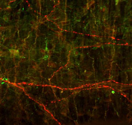
20 with the 7 day treatment group and indicated a potential recovery. These results are similar to that observed in the MCA in a previous study (Zhu et al., 2010). Changes in sensory and parasympathetic perivascular axons following axotomy. Changes in the sensory and parasympathetic populations were examined in the ACA and MCA following axotomy of the SCG at time points when the vessels were almost completely denervated of sympathetic axons (1 day and 7 days) and when sympathetic innervation appeared to be returning (8 weeks and 12 weeks). The distribution of sensory perivascular axons associated with the extracerebral blood vessels, which arise from the trigeminal ganglion (Wanaka et al., 1986; Tsai et al., 1988; Edvinsson, 1987; Suzuki et al., 1989), was observed by immunofluorescence for calcitonin gene related peptide (CGRP) (Ruskell and Simons, 1987; Saito and Moskowitz, 1989). The extracerebral blood vessels also receive parasympathetic innervation from the sphenopalatine and otic ganglia (Walters et al., 1986; Hardebo et al., 1991) and these parasympathetic axons can be labeled with an antibody directed toward VAChT (Eiden et al., 2004). First, co-localization studies were carried out to ensure that the markers used for sensory (CGRP) and parasympathetic (VAChT) axons did not co-localize. In Figure 4, a control MCA and one from a 12 week axotomy case are shown and no colocalization of these two markers was observed. Similarly, though not shown here, previous studies showed no colocalization of CGRP and TH and no colocalization of VAChT and TH in the perivascular axonal population in the controls or following axotomy of the SCG. Changes in sensory innervation. The normal appearance and distribution of CGRP-ir perivascular axons associated with the ACA and MCA are shown in Figures 5-8. As shown in Figures 5 and 7, the perivascular sensory axons appeared thinner and finer than the sympathetic axons and their overall density appeared less than the sympathetics, yet sensory axons showed a longitudinal and circumferential distribution pattern that was somewhat similar to sympathetic axons. The changes in sensory innervation of the ACA and MCA followed similar trends and generally increased following sympathetic denervation and reinnervation. While at 1 day following axotomy the density of sensory axons associated with both vessels was similar to controls, by 7 15
21 days, the density of sensory axons in both vessels was significantly increased compared to controls. A trend for an increase was observed at 8 weeks, and at 12 weeks following axotomy, the density of CGRP-ir axons was significantly increased and similar to the 7 day time point, indicating a sustained increase in sensory innervation at this time point. Regional analysis of the ACA (Fig. 6) indicated that the increase (compared with controls) was observed consistently in the middle and distal segments of the vessel at the 7 day and 12 week time points and that these time points generally were similar. However in the MCA (Fig. 8), the sensory innervation in the distal segment was significantly elevated compared with the 7 day time point. Changes in parasympathetic innervation. The typical appearance and distribution of the parasympathetic perivascular axons are shown in Figures As shown in Figures 9 and 11, the parasympathetic innervation of the ACA and MCA in all cases showed primarily a circumferential pattern. Following axotomy, the parasympathetic innervation of the two vessels followed similar trends across treatments and generally was decreased during the sympathetic denervation and reinnervation periods. In the control cases, VAChT-ir parasympathetic axons typically showed a circumferential distribution pattern with occasional axons in a longitudinal orientation (Fig. 9, 11). The total number of parasympathetic axons associated with the ACA was decreased at 1 day, 7 days and 8 weeks, but at 12 weeks following axotomy was similar to controls (Figs. 9, 10), revealing a potential recovery in the parasympathetic axons, similar to that observed with regard to the sympathetic axons. However, though not different from the control group, the parasympathetic density at 12 weeks was similar to the 7 day time point. The regional analysis of the ACA revealed decreases in parasympathetic innervation at both short and long term time points (Fig. 10), yet the 12 week time point was not significantly different from controls, even though in each region of the ACA, the 12 week cases were similar to the 7 day time point (Figs. 9,10). With respect to the MCA, similar decreases in the total number of parasympathetic axons were observed (Fig. 12). The decrease was not observed at 1 day, but at all other time points, including 12 weeks, a decrease in perivascular density compared to controls was observed (Figs. 11, 12). Regional analysis of the MCA 16
22 revealed decreases in some segments at both short and long term time points (Fig. 12). Again, the 7 day and 12 week cases were statistically similar in each regional segment. Changes in VEGF following axotomy of the SCG. Western analysis was used to assess the changes in VEGF in the extracerebral blood vessels, the SCG, and the trigeminal ganglion following following axotomy of the SCG. The 42 kda isoform was the primary form observed in the extracerebral blood vessels, and this isoform was similar to controls at each time point examined (Fig.13). In SCG, a 42kDa and 80 kda isoform were observed. Though unchanged at 1 day, the 42kDa VEGF isoform was significantly increased to 273% of controls at 7 days following axotomy (Fig.14) and was increased to 340% and 353% of control values at 8 weeks and 12 weeks axotomy, respectively. In contrast the 80kDa isoform was generally decreased and was 23%, 13%, and 2% of the control values at 7 days, 8 weeks, and 12 weeks, respectively (Fig. 14). VEGF protein also was altered in the trigeminal ganglion (Fig. 15). Though the 42kDa isoform was significantly decreased to 73% and 49% of control values at 1 day and 8 weeks following axotomy, this isoform was increased to 161% of controls at 12 weeks following axotomy. The 80kDa VEGF was significantly increased to 198% and 147% of control values at 7 days and 12 weeks, but was significantly decreased to 46% of control values at 8 weeks following axotomy (Fig. 15). 17

23 The extracerebral blood vessels at the base of the brain ACA MCA Circle of Willis Figure 1 18
24 Figure 1. Diagram of the extracerebral blood vessels located at the base of the brain. Western analyses were carried out on all vessels from one side that comprise the Circle of Willis. Immunohistochemical analysis was performed on the anterior cerebral artery (ACA) and the middle cerebral artery (MCA) as individual samples. Identification the proximal, middle, and distal portions of the ACA and MCA were based on the proximity of the vessels to the Circle, with the proximal segment located closest to the Circle and the distal segments farthest from the Circle. (Based on bloodsupply.html) 19


25 TH perivascular axons associated with the ACA following axotomy Proximal Middle Distal cont 1d 7d 8wk 12wk 100µm Figure 2 20
26 Figure 2. TH immunohistochemistry of the ACA following axotomy. ACA from control cases exhibited robust TH immunoreactivity (red). Axons passed in both the longitudinal and circumferential planes. TH immunoreactivity appeared reduced following a 1 day (1d) axotomy, especially at the middle and distal portion. TH immunoreactivity was virtually absent at all three portions of the ACA by 7 days (7d) after the axotomy. TH immunoreactivity remained decreased at 8 weeks (8wk) compared with controls, some longitudinal axons appeared in the ACA. By 12 weeks (12wk) following axotomy, the density appeared to increase compared with earlier time points. Scale bar, 100µm. 21
27 A. 700 Total number of TH perivascular axons: ACA 600 Density of perivascular axons # 0 cont 1d 7d 8wk 12wk B. Density of perivasulcar axons TH perivascular axons: regional analysis # Proximal Middle Distal # cont 1d 7d 8wk 12wk # Figure 3 22
28 Figure 3. Quantitative analysis of the density of TH-immunoreactive (-ir) sympathetic axons associated with the ACA. A. Total sympathetic innervation was significantly decreased (compared with controls) following axotomy of the SCG at all of the time points examined. Sympathetic innervation was decreased at 1 day (1d) and the vessel was almost completely denervated by 7 days (7d) following axotomy of the SCG. By 12 weeks (12wk), the total sympathetic innervation of the ACA, though significantly decreased compared with the controls, was increased compared with the 7 day treatment group, indicating a potential recovery of the sympathetic innervation over time. B. Regional analysis revealed a reduction in sympathetic innervation across all three regions of the vessel at every time point examined. By 12 weeks the regional innervation, though significantly decreased compared with the controls, was increased compared with the 7 day treatment group and indicated a potential recovery., significantly different from control, p<0.05; #, significantly different from 7 day treatment, p<0.05. Number of samples examined: cont (n=12), 1d (n=3), 7d (n=3), 8wk (n=3), 12wk (n=3). 23


29 CGRP-ir and VAChT-ir perivascular axons show no colocalization CGRP VAChT CGRP/VAChT cont CGRP VAChT CGRP/VAChT 12wk Figure 4 24
30 Figure 4. Immunohistochemical analysis of CGRP (red arrows, left panels) and VAChT (green arrows, middle panels) associated with the MCA from a 12 week control (upper) and from a rat receiving a 12 week axotomy (lower). At the 12 week time point after axotomy, CGRPimmunoreactive (-ir) axons were increased while VAChT-ir axons were decreased. Note in the merged images (right panels) the complete segregation of the two axonal populations and no colocalization of the two markers. Scale bar, 100µm. 25


31 CGRP-ir perivascular axons associated with the ACA following axotomy Proximal Middle Distal cont 1d 7d 8wk 12wk Figure 5 26
32 Figure 5. CGRP immunoreactive (-ir) axons associated with the ACA following axotomy of the SCG. The perivascular sensory axons appeared thinner and finer than the sympathetic axons, yet a longitudinal and circumferential distribution pattern was present. By 7 days (7d) following axotomy, the density of sensory axons appeared increased at proximal, middle and distal portions of the ACA compared to controls. At 12 weeks (12wk) following axotomy, the density of CGRP-ir axons was significantly increased compared with controls and statistically similar to the 7 day time point, indicating a sustained increase in sensory innervation at this time point. Scale bar, 100µm. 27
33 A. Density of perivascular axons Total number of CGRP perivascular axons: ACA 0 cont 1d 7d 8wk 12wk B. Density of perivascular axons CGRP perivascular axons: regional analysis cont 1d 7d 8wk 12wk 0 Proximal Middle Distal Figure 6 28
34 Figure 6. Quantitative analysis of the density of CGRP-immunoreactive (-ir) sensory axons associated with the ACA. A. The density of CGRP-ir axons was increased at 7 days (7d) following axotomy, and at 12 weeks (12wk), the density was significantly increased compared with controls but statistically similar to the 7 day time point, indicating a sustained increase in sensory innervation. B. Regional analysis of the ACA revealed a significant increase (compared with controls) in the middle and distal segments of the vessel at the 7 day (7d) and 12 week (12 wk) time points and that these time points generally were similar., significantly different from control, p<0.05. Number of samples examined: cont (n=12), 1d (n=3), 7d (n=3), 8wk (n=3), 12wk (n=3). 29


35 CGRP-ir perivascular axons associated with the MCA following axotomy Proximal Middle Distal cont 1d 7d 8wk 12wk Figure 7 30
36 Figure 7. CGRP-immunoreactive(-ir) perivascular axons associated with the MCA following axotomy of the SCG. The MCA from control cases exhibited a typical distribution of longitudinal and circumferential CGRP-ir sensory axons. By 7 days (7d) following axotomy, the density of sensory axons appeared increased compared to controls, the increase was more prominent at proximal portion of the MCA. The increase continued at 12 weeks (12wk) following axotomy. Scale bar, 100µm. 31
37 A. Density of perivascular axons Total number of CGRP perivascular axons: MCA cont 1d 7d 8wk 12wk B. Density of perivascular axons CGRP perivascular axons: MCA regional analysis cont 1d 7d 8wk 12wk 0 Proximal Middle Distal Figure 8 32
38 Figure 8. Quantitative analysis of the density of CGRP-immunoreactive(-ir) sensory axons associated with the MCA. A. Analysis of the total density of the CGRP-ir axons on the MCA revealed a significant increase in CGRP-ir axons at 7 days (7d) and 12 weeks (12wk) following axotomy of the SCG. The density in the 12 week treatment was similar to the 7 day cases. B. Regional analysis of the density of CGRP-ir axons associated with the MCA revealed significant increase at 7 days after axotomy at the proximal portion of the MCA. By 12 weeks following axotomy, a significant increase in CGRP-ir axons was observed at all three regions of the MCA., significantly different from control, p<0.05. Number of samples examined: cont (n=12), 1d (n=3), 7d (n=3), 8wk (n=3), 12wk (n=3). 33


39 VAChT associated with the ACA following axotomy Proximal Middle Distal cont 1d 7d 8wk 12wk Figure 9 34
40 Figure 9. VAChT-immunoreactive(-ir) perivascular axons associated with the ACA following axotomy. VAChT-ir axons showing a circumferential pattern were typically observed in the control cases. Abundant circumferential VAChT-ir axons were observed in the proximal, middle, and distal portion of the ACA in the control group. These axons appeared reduced in the 1 day (1d), especially at middle and distal portion. The decrease was also observed at 7 day (7d) and 8 week (8wk) cases, which dramatic decrease was observed at all three portions of the ACA. By 12-week axotomy, the density of VAChT-ir parasympathetic axons appeared to return to control values. Scale bar, 100µm. 35
41 A. 600 Total number of VAChT perivascular axons: ACA Density of perivascular axons cont 1d 7d 8wk 12wk B. Density of perivascular axons VAChT perivascular axons: regional analysis cont 1d 7d 8wk 12wk 0 Proximal Middle Distal Figure 10 36
42 Figure 10. Quantitative analysis of the density of VAChT-immunoreactive(-ir) parasympathetic axons associated with the ACA. A. The total number of parasympathetic axons associated with the ACA was decreased at 1 day, 7 days and 8 weeks following axotomy, but at 12 weeks was similar to controls, revealing a potential recovery over time in these axons. However, though not different from the control group, the parasympathetic density at 12 weeks was similar to the 7 day time point. B. The regional analysis of the ACA revealed decreases in parasympathetic innervation at both short and long term time points, yet the 12 week time point was not significantly different from controls although in each region of the ACA, the 12 week cases were similar to the 7 day time point., significantly different from controls, p<0.05. Number of samples examined: cont (n=12), 1d (n=3), 7d (n=3), 8wk (n=3), 12wk (n=3). 37


43 VAChT-ir axons associated with the MCA following axotomy Proximal Middle Distal cont 1d 7d 8wk 12wk Figure 11 38
44 Figure 11. VAChT-immunoreactive(-ir) perivascular axons associated with the MCA following axotomy. VAChT-ir axons showing a circumferential pattern were typically observed in the control cases. These axons appeared to be reduced in the 7 day (7d) and 8 week (8wk) and 12 week (12wk) survival time points following axotomy. Scale bar, 100µm. 39
45 A. 400 Total number of VAChT perivascular axons: MCA Density of perivascular axons cont 1d 7d 8wk 12wk B. VAChT perivascular axons: regional analysis Density of perivascular axons cont 1d 7d 8wk 12wk 0 Proximal Middle Distal Figure 12 40
46 Figure 12. Quantitative analysis of the density of VAChT-immunoreactive(-ir) parasympathetic axons associated with the MCA. A. No change was observed at 1 day (1d) following axotomy, but at all other time points, including 12 weeks (12wk), a decrease in VAChT-ir perivascular density compared to controls was observed. B. Regional analysis of the MCA revealed decreases in some segments at both short and long term time points. Again, the 7 day and 12 week cases were statistically similar in each regional segment. Number of samples examined: cont (n=12), 1d (n=3), 7d (n=3), 8wk (n=3), 12wk (n=3). 41
47 Table 1. Summary of changes in perivascular axonal populations associated with the ACA and MCA. ACA MCA Short term Long term Short term Long term Sympathetic axons Sensory axons Parasympathetic axons 42


48 VEGF in the extracerebral blood vessels following axotomy A. VEGF 42 kda 1d cont 1d axotomy 7d cont 7d axotomy 36 kda GAPDH VEGF 42 kda 8wk cont 8wk axotomy 12wk cont 12wk axotomy 36 kda GAPDH B. VEGF in the extracerebral blood vessels 140 1d 120 7d 8wk wk %Control Figure 13 43
ABSTRACT PLASTICITY OF ADULT SYMPATHETIC NEURONS FOLLOWING INJURY. by Ryan George Walker
 ABSTRACT PLASTICITY OF ADULT SYMPATHETIC NEURONS FOLLOWING INJURY by Ryan George Walker The goal of the present study was to characterize the response by neurons and glial cells in the superior cervical
The Nervous System: Autonomic Nervous System Pearson Education, Inc.
 17 The Nervous System: Autonomic Nervous System Introduction The autonomic nervous system: Functions outside of our conscious awareness Makes routine adjustments in our body s systems The autonomic nervous
ABSTRACT PLASTICITY IN THE INTERMEDIOLATERAL CELL COLUMN OF THE SPINAL CORD FOLLOWING INJURY TO SYMPATHETIC POSTGANGLIONIC AXONS
 ABSTRACT PLASTICITY IN THE INTERMEDIOLATERAL CELL COLUMN OF THE SPINAL CORD FOLLOWING INJURY TO SYMPATHETIC POSTGANGLIONIC AXONS by Sean Michael Gannon The effects of transection of sympathetic postganglionic
ABSTRACT ALTERED NEUROTROPHIN EXPRESSION IN AGED PERIPHERAL NEURONS AND TARGETS. by Michael Alexander Bierl
 ABSTRACT ALTERED NEUROTROPHIN EXPRESSION IN AGED PERIPHERAL NEURONS AND TARGETS by Michael Alexander Bierl Target-derived neurotrophic factors such as nerve growth factor (NGF) and neurotrophin-3 (NT-3)
The Nervous System: Autonomic Nervous System
 17 The Nervous System: Autonomic Nervous System PowerPoint Lecture Presentations prepared by Steven Bassett Southeast Community College Lincoln, Nebraska Introduction The autonomic nervous system functions
I. Neural Control of Involuntary Effectors. Chapter 9. Autonomic Motor Nerves. Autonomic Neurons. Autonomic Ganglia. Autonomic Neurons 9/19/11
 Chapter 9 I. Neural Control of Involuntary Effectors The Autonomic Nervous System Lecture PowerPoint Copyright The McGraw-Hill Companies, Inc. Permission required for reproduction or display. Autonomic
Chapter 15: The Autonomic Nervous System. Copyright 2009, John Wiley & Sons, Inc.
 Chapter 15: The Autonomic Nervous System Comparison of Somatic and Autonomic Nervous Systems Comparison of Somatic and Autonomic Nervous Systems Anatomy of Autonomic Motor Pathways Preganglionic neuron
Human Anatomy. Autonomic Nervous System
 Human Anatomy Autonomic Nervous System 1 Autonomic Nervous System ANS complex system of nerves controls involuntary actions. Works with the somatic nervous system (SNS) regulates body organs maintains
The cells of the nervous system
 The cells of the nervous system LESSON N.9 - PSYCHOBIOLOGY because of the location and volume as compared to our body, the brain has always been a matter of conjecture about its fundamental role in the
Autonomic Nervous System. Lanny Shulman, O.D., Ph.D. University of Houston College of Optometry
 Autonomic Nervous System Lanny Shulman, O.D., Ph.D. University of Houston College of Optometry Peripheral Nervous System A. Sensory Somatic Nervous System B. Autonomic Nervous System 1. Sympathetic Nervous
AUTONOMIC NERVOUS SYSTEM PART I: SPINAL CORD
 AUTONOMIC NERVOUS SYSTEM PART I: SPINAL CORD How is the organization of the autonomic nervous system different from that of the somatic nervous system? Peripheral Nervous System Divisions Somatic Nervous
Chp. 16: AUTONOMIC N.S. (In Review: Peripheral N. S.)
") Chp. 16: AUTONOMIC N.S. (In Review: Peripheral N. S.) Peripheral nerves contain both motor and sensory neurons Among the motor neurons, some of these are somatic and innervate skeletal muscles while some
Chp. 16: AUTONOMIC N.S. (In Review: Peripheral N. S.) Peripheral nerves contain both motor and sensory neurons Among the motor neurons, some of these are somatic and innervate skeletal muscles while some
Chapter 14 The Autonomic Nervous System Chapter Outline
 Chapter 14 The Autonomic Nervous System Chapter Outline Module 14.1 Overview of the Autonomic Nervous System (Figures 14.1 14.3) A. The autonomic nervous system (ANS) is the involuntary arm of the peripheral
The Nervous System PART D. PowerPoint Lecture Slide Presentation by Patty Bostwick-Taylor, Florence-Darlington Technical College
 PowerPoint Lecture Slide Presentation by Patty Bostwick-Taylor, Florence-Darlington Technical College The Nervous System 7 PART D PNS: Spinal Nerves There is a pair of spinal nerves at the level of each
Principles of Anatomy and Physiology
 Principles of Anatomy and Physiology 14 th Edition CHAPTER 15 The Autonomic Nervous System Comparison of Somatic and Autonomic Nervous Systems The somatic nervous system includes both sensory and motor
Nervous Systems: Diversity & Functional Organization
 Nervous Systems: Diversity & Functional Organization Diversity of Neural Signaling The diversity of neuron structure and function allows neurons to play many roles. 3 basic function of all neurons: Receive
Chapter 16. APR Enhanced Lecture Slides
 Chapter 16 APR Enhanced Lecture Slides See separate PowerPoint slides for all figures and tables pre-inserted into PowerPoint without notes and animations. Copyright The McGraw-Hill Companies, Inc. Permission
Autonomic Nervous System Dr. Ali Ebneshahidi
 Autonomic Nervous System Dr. Ali Ebneshahidi Nervous System Divisions of the nervous system The human nervous system consists of the central nervous System (CNS) and the Peripheral Nervous System (PNS).
HST-583 Cerebrovascular anatomy and neural regulation of CNS blood flow
 HST.583: Functional Magnetic Resonance Imaging: Data Acquisition and Analysis Harvard-MIT Division of Health Sciences and Technology Dr. Randy Gollub HST-583 Cerebrovascular anatomy and neural regulation
Human Anatomy & Physiology
 PowerPoint Lecture Slides prepared by Barbara Heard, Atlantic Cape Community College Ninth Edition Human Anatomy & Physiology C H A P T E R 14 Annie Leibovitz/Contact Press Images 2013 Pearson Education,
Human Anatomy and Physiology - Problem Drill 15: The Autonomic Nervous System
 Human Anatomy and Physiology - Problem Drill 15: The Autonomic Nervous System Question No. 1 of 10 Which of the following statements is correct about the component of the autonomic nervous system identified
ANATOMY & PHYSIOLOGY - CLUTCH CH THE AUTONOMIC NERVOUS SYSTEM.
 !! www.clutchprep.com ANATOMY & PHYSIOLOGY - CLUTCH CONCEPT: THE AUTONOMIC NERVOUS SYSTEM: DIVISIONS AND STRUCTURE The Autonomic Nervous System and its Divisions: Autonomic Nervous System (ANS) controls
Composed by Natalia Leonidovna Svintsitskaya, Associate professor of the Chair of Human Anatomy, Candidate of Medicine
 Theoretical background to the study of the autonomic nervous system. Sympathetic and parasympathetic divisions of the autonomic nervous system. Features of the structure, function Composed by Natalia Leonidovna
NERVOUS SYSTEM ANATOMY
 INTRODUCTION to NERVOUS SYSTEM ANATOMY M1 - Gross and Developmental Anatomy Dr. Milton M. Sholley Professor of Anatomy and Neurobiology and Dr. Michael H. Peters Professor of Chemical and Life Science
The Nervous System. Functions of the Nervous System input gathering To monitor occurring inside and outside the body Changes =
 The Nervous System Functions of the Nervous System input gathering To monitor occurring inside and outside the body Changes = To process and sensory input and decide if is needed output A response to integrated
Sympathetic Nervous System
 Sympathetic Nervous System Lecture Objectives Review the subdivisions of the nervous system. Review the general arrangement and compare the sympathetic and parasympathetic parts. Describe the following
CHAPTER 15 LECTURE OUTLINE
 CHAPTER 15 LECTURE OUTLINE I. INTRODUCTION A. The autonomic nervous system (ANS) regulates the activity of smooth muscle, cardiac muscle, and certain glands. B. Operation of the ANS to maintain homeostasis,
Autonomic Division of NS
 Autonomic Division of NS Compare and contrast the structures of the sympathetic and the parasympathetic divisions, including functions and neurotransmitters. Show the levels of integration in the ANS,
NERVOUS SYSTEM ANATOMY
 NTRODUCTON to NERVOUS SYSTEM ANATOMY M1 - Gross and Developmental Anatomy Dr. Milton M. Sholley Professor of Anatomy and Neurobiology and Dr. Michael H. Peters Professor of Chemical and Life Science Engineering
Lesson 33. Objectives: References: Chapter 16: Reading for Next Lesson: Chapter 16:
 Lesson 33 Lesson Outline: Nervous System Structure and Function Neuronal Tissue Supporting Cells Neurons Nerves Functional Classification of Neuronal Tissue Organization of the Nervous System Peripheral
The Nervous System PART A
 7 The Nervous System PART A PowerPoint Lecture Slide Presentation by Jerry L. Cook, Sam Houston University ESSENTIALS OF HUMAN ANATOMY & PHYSIOLOGY EIGHTH EDITION ELAINE N. MARIEB Structural Classification
Do Now pg What is the fight or flight response? 2. Give an example of when this response would kick in.
 Do Now pg 81 1. What is the fight or flight response? 2. Give an example of when this response would kick in. Autonomic Nervous System The portion of the PNS that functions independently (autonomously)
Neuropsychiatry Block
 Neuropsychiatry Block Physiology of the Autonomic Nervous System By Laiche Djouhri, PhD Dept. of Physiology Email: ldjouhri@ksu.edu.sa Ext:71044 References The Autonomic Nervous System and the Adrenal
Systems Neuroscience November 21, 2017 The autonomic nervous system
 Systems Neuroscience November 21, 2017 The autonomic nervous system Daniel C. Kiper kiper@ini.phys.ethz.ch http: www.ini.unizh.ch/~kiper/system_neurosci.html How is the organization of the autonomic nervous
Ch 9. The Autonomic Nervous System
 Ch 9 The Autonomic Nervous System SLOs Review the organization of the ANS Describe how neural regulation of smooth and cardiac muscles differs from that of skeletal muscles Describe the structure and innervation
Autonomic Nervous System. Autonomic (Visceral) Nervous System. Visual Anatomy & Physiology First Edition. Martini & Ober
 Nervous System. Visual Anatomy & Physiology First Edition. Martini & Ober") Visual Anatomy & Physiology First Edition Martini & Ober Chapter 14 Autonomic Nervous System Lecture 21 1 Autonomic (Visceral) Nervous System CNS PNS 2 Autonomic Nervous System functions without conscious
Visual Anatomy & Physiology First Edition Martini & Ober Chapter 14 Autonomic Nervous System Lecture 21 1 Autonomic (Visceral) Nervous System CNS PNS 2 Autonomic Nervous System functions without conscious
The Autonomic Nervous
 Autonomic Nervous System The Autonomic Nervous Assess Prof. Fawzia Al-Rouq System Department of Physiology College of Medicine King Saud University LECTUR (1) Functional Anatomy & Physiology of Autonomic
NERVOUS SYSTEM. Academic Resource Center. Forskellen mellem oscillator og krystal
 NERVOUS SYSTEM Academic Resource Center Forskellen mellem oscillator og krystal Overview of the Nervous System Peripheral nervous system-pns cranial nerves spinal nerves ganglia peripheral nerves enteric
Biology 218 Human Anatomy
 Chapter 20 Adapted form Tortora 10 th ed. LECTURE OUTLINE A. Introduction (p. 632) 1. The autonomic nervous system (ANS) regulates the activity of smooth muscle, cardiac muscle, and certain glands. 2.
Cerebral hemisphere. Parietal Frontal Occipital Temporal
 Cerebral hemisphere Sulcus / Fissure Central Precental gyrus Postcentral gyrus Lateral (cerebral) Parieto-occipital Cerebral cortex Frontal lobe Parietal lobe Temporal lobe Insula Amygdala Hippocampus
10.1: Introduction. Cell types in neural tissue: Neurons Neuroglial cells (also known as neuroglia, glia, and glial cells) Dendrites.
 Dendrites.") 10.1: Introduction Copyright The McGraw-Hill Companies, Inc. Permission required for reproduction or display. Cell types in neural tissue: Neurons Neuroglial cells (also known as neuroglia, glia, and glial
10.1: Introduction Copyright The McGraw-Hill Companies, Inc. Permission required for reproduction or display. Cell types in neural tissue: Neurons Neuroglial cells (also known as neuroglia, glia, and glial
NERVOUS SYSTEM C H A P T E R 2 8
 NERVOUS SYSTEM C H A P T E R 2 8 CAN AN INJURED SPINAL CORD BE FIXED? Injuries to the spinal cord disrupt communication between the central nervous system (brain and spinal cord) and the rest of the body
The Nervous System. Autonomic Division. C h a p t e r. PowerPoint Lecture Slides prepared by Jason LaPres North Harris College Houston, Texas
 C h a p t e r 17 The Nervous System Autonomic Division PowerPoint Lecture Slides prepared by Jason LaPres North Harris College Houston, Texas Copyright 2009 Pearson Education, Inc., publishing as Pearson
Cranial Nerves. Steven McLoon Department of Neuroscience University of Minnesota
 Cranial Nerves Steven McLoon Department of Neuroscience University of Minnesota 1 Course News Change in Lab Sequence Week of Oct 2 Lab 5 Week of Oct 9 Lab 4 2 Sensory and Motor Systems Sensory Systems:
! BIOL 2401! Week 5. Nervous System. Nervous System
 Collin County Community College! BIOL 2401! Week 5 Nervous System 1 Nervous System The process of homeostasis makes sure that the activities that occur in the body are maintained within normal physiological
Human Anatomy - Problem Drill 11: The Spinal Cord and Spinal Nerves
 Human Anatomy - Problem Drill 11: The Spinal Cord and Spinal Nerves Question No. 1 of 10 Instructions: (1) Read the problem statement and answer choices carefully, (2) Work the problems on paper as needed,
Reaction to Injury & Regeneration. Steven McLoon Department of Neuroscience University of Minnesota
 Reaction to Injury & Regeneration Steven McLoon Department of Neuroscience University of Minnesota 1 Course News Dec 4 (Mon) Dec 6 (Wed) adult neurogenesis injury & regeneration Dec 8 (Fri) research paper
ParasymPathetic Nervous system. Done by : Zaid Al-Ghnaneem
 ParasymPathetic Nervous system Done by : Zaid Al-Ghnaneem In this lecture we are going to discuss Parasympathetic, in the last lecture we took sympathetic and one of the objectives of last lecture was
Nervous tissue, charachteristics, neurons, glial cells
 Nervous tissue, charachteristics, neurons, glial cells Functional Organization of Nervous Tissue The Nervous System Components Brain, spinal cord, nerves, sensory receptors Responsible for Sensory perceptions,
Functional Organization of Nervous Tissue. Nervous tissue, charachteristics, neurons, glial cells. The Nervous System. The Nervous System 21/12/2010
 Nervous tissue, charachteristics, neurons, glial cells Functional Organization of Nervous Tissue The Nervous System Components Brain, spinal cord, nerves, sensory receptors Responsible for Sensory perceptions,
Chapter 17 Nervous System
 Chapter 17 Nervous System 1 The Nervous System Two Anatomical Divisions Central Nervous System (CNS) Brain and Spinal Cord Peripheral Nervous System (PNS) Two Types of Cells Neurons Transmit nerve impulses
Neural Integration II: The Autonomic Nervous System and Higher-Order Functions
 16 Neural Integration II: The Autonomic Nervous System and Higher-Order Functions PowerPoint Lecture Presentations prepared by Jason LaPres Lone Star College North Harris Figure 16-1 An Overview of Neural
Brain Development III
 Brain Development III Neural Development In the developing nervous system there must be: 1. The formation of different regions of the brain. 2. The ability of a neuron to differentiate. 3. The ability
The Autonomic Nervous System Outline of class lecture for Physiology
 The Autonomic Nervous System Outline of class lecture for Physiology 1 After studying the endocrine system you should be able to: 1. Describe the organization of the nervous system. 2. Compare and contrast
Week 7 and 8 Master Worksheet
 The Nervous System Week 7 and 8 Master Worksheet 1. Complete the chart regarding the 3 functions of the nervous system: Sensory input What does it do? Integration Motor output 2. Complete the chart: Component
The Nervous System. Nervous System Functions 1. gather sensory input 2. integration- process and interpret sensory input 3. cause motor output
 The Nervous System Nervous System Functions 1. gather sensory input 2. integration- process and interpret sensory input 3. cause motor output The Nervous System 2 Parts of the Nervous System 1. central
Chapter 17. Nervous System Nervous systems receive sensory input, interpret it, and send out appropriate commands. !
 Chapter 17 Sensory receptor Sensory input Integration Nervous System Motor output Brain and spinal cord Effector cells Peripheral nervous system (PNS) Central nervous system (CNS) 28.1 Nervous systems
Human Anatomy. Spinal Cord and Spinal Nerves
 Human Anatomy Spinal Cord and Spinal Nerves 1 The Spinal Cord Link between the brain and the body. Exhibits some functional independence from the brain. The spinal cord and spinal nerves serve two functions:
VEGFR2-Mediated Vascular Dilation as a Mechanism of VEGF-Induced Anemia and Bone Marrow Cell Mobilization
 Cell Reports, Volume 9 Supplemental Information VEGFR2-Mediated Vascular Dilation as a Mechanism of VEGF-Induced Anemia and Bone Marrow Cell Mobilization Sharon Lim, Yin Zhang, Danfang Zhang, Fang Chen,
I. Autonomic Nervous System (ANS) A. Dual Innervation B. Autonomic Motor Pathway 1. Preganglionic Neuron a. Preganglionic Fibers (Axons) (1)
 A. Dual Innervation B. Autonomic Motor Pathway 1. Preganglionic Neuron a. Preganglionic Fibers (Axons) (1)") I. Autonomic Nervous System (ANS) A. Dual Innervation B. Autonomic Motor Pathway 1. Preganglionic Neuron a. Preganglionic Fibers (Axons) (1) Acetylcholine - ACh 2. Ganglion (Ganglia) 3. Ganglionic Neuron
I. Autonomic Nervous System (ANS) A. Dual Innervation B. Autonomic Motor Pathway 1. Preganglionic Neuron a. Preganglionic Fibers (Axons) (1) Acetylcholine - ACh 2. Ganglion (Ganglia) 3. Ganglionic Neuron
Tymaa Al-zaben & Amin Al-ajalouni
 Done by: Tymaa Al-zaben & Amin Al-ajalouni ** Hello SERTONIN! SLIDE 3 note:: the slide included within the sheet but make sure back to slide for pictures The Autonomic Nervous System Function : Regulate
Drugs Affecting The Autonomic Nervous System(ANS)
") Drugs Affecting The Autonomic Nervous System(ANS) ANS Pharmacology Lecture 1 Dr. Hiwa K. Saaed College of Pharmacy, University of Sulaimani 2018-2019 AUTOMATIC NERVOUS SYSTEM (ANS) The ANS is the major
Drugs Affecting The Autonomic Nervous System(ANS) ANS Pharmacology Lecture 1 Dr. Hiwa K. Saaed College of Pharmacy, University of Sulaimani 2018-2019 AUTOMATIC NERVOUS SYSTEM (ANS) The ANS is the major
Peripheral Nervous System Dr. Gary Mumaugh
 Peripheral Nervous System Dr. Gary Mumaugh Spinal Nerves Overview Thirty-one pairs of spinal nerves are connected to the spinal cord No special names; numbered by level of vertebral column at which they
Chapter 8 Nervous System
 Chapter 8 Nervous System Two message centers: Functions of these systems: 1. * 2. * Overview of the Nervous System Parts: General Functions: Functions Sensory input: Sensation via nerves Integration: interpretation
Nervous System. Master controlling and communicating system of the body. Secrete chemicals called neurotransmitters
 Nervous System Master controlling and communicating system of the body Interacts with the endocrine system to control and coordinate the body s responses to changes in its environment, as well as growth,
The Autonomic Nervous System
 The Autonomic Nervous System Responsible for control of visceral effectors and visceral reflexes: smooth muscle, glands, the heart. e.g. blood pressure, cardiac output, plasma glucose The autonomic system
Brain and spinal nerve. By: shirin Kashfi
 Brain and spinal nerve By: shirin Kashfi Nervous system: central nervous system (CNS) peripheral nervous system (PNS) Brain (cranial) nerves Spinal nerves Ganglions (dorsal root ganglions, sympathetic
Chapter 9. Nervous System
 Chapter 9 Nervous System Central Nervous System (CNS) vs. Peripheral Nervous System(PNS) CNS Brain Spinal cord PNS Peripheral nerves connecting CNS to the body Cranial nerves Spinal nerves Neurons transmit
Introduction to Autonomic
 Part 2 Autonomic Pharmacology 3 Introduction to Autonomic Pharmacology FUNCTIONS OF THE AUTONOMIC NERVOUS SYSTEM The autonomic nervous system (Figure 3 1) is composed of the sympathetic and parasympathetic
PNS and ANS Flashcards
 1. Name several SOMATIC SENSES Light touch (being touched by a feather), heat, cold, vibration, pressure, pain are SOMATIC SENSES. 2. What are proprioceptors; and how is proprioception tested? PROPRIOCEPTORS
Laboratory Manual for Comparative Anatomy and Physiology Figure 15.1 Transparency Master 114
 Neuron Capillary Astrocyte Microglial cell Neuron Fluid-filled cavity Process of oligodendrocyte Ependymal cells Brain or spinal cord tissue Myelin sheath Nerve fibers Figure 15.1 Transparency Master 114
Autonomic Nervous System
 Autonomic Nervous System Autonomic nervous system organization Sympathetic Nervous System division of the autonomic nervous system that arouses the body, mobilizing its energy in stressful situations
Chapter 9 The Nervous System: The Spinal Cord and Spinal Nerves
 Chapter 9 The Nervous System: The Spinal Cord and Spinal Nerves Copyright 2015 Wolters Kluwer Health Lippincott Williams & Wilkins Overview Key Terms acetylcholine motor presynaptic action potential nerve
Principles Arteries & Veins of the CNS LO14
 Principles Arteries & Veins of the CNS LO14 14. Identify (on cadaver specimens, models and diagrams) and name the principal arteries and veins of the CNS: Why is it important to understand blood supply
The Nervous System An overview
 Nervous System The Nervous System An overview Includes Nerve tissue Sense organs Functions to Sense environment Process information it receives Respond to information 1 Copyright 2009 Pearson Education,
Organization of The Nervous System PROF. SAEED ABUEL MAKAREM
 Organization of The Nervous System PROF. SAEED ABUEL MAKAREM Objectives By the end of the lecture, you should be able to: List the parts of the nervous system. List the function of the nervous system.
Chapter 13 PNS and reflex activity
 Chapter 13 PNS and reflex activity I. Peripheral nervous system A. PNS links CNS to the body B. Sensory: the afferent division C. Motor: the efferent division D. Ganglia: collections of cell bodies in
Human Anatomy and Physiology - Problem Drill 11: Neural Tissue & The Nervous System
 Human Anatomy and Physiology - Problem Drill 11: Neural Tissue & The Nervous System Question No. 1 of 10 The human body contains different types of tissue. The tissue is formed into organs and organ systems.
Autonomic nervous system
 Autonomic nervous system Key notes Autonomic: an independent system that runs on its own The ANS is a visceral and involuntary sensory and motor system The visceral motor fibers in the autonomic nerves
number Done by Corrected by Doctor
 number 13 Done by Tamara Wahbeh Corrected by Doctor Omar Shaheen In this sheet the following concepts will be covered: 1. Divisions of the nervous system 2. Anatomy of the ANS. 3. ANS innervations. 4.
BIOH111. o Cell Module o Tissue Module o Skeletal system o Muscle system o Nervous system o Endocrine system o Integumentary system
 BIOH111 o Cell Module o Tissue Module o Skeletal system o Muscle system o Nervous system o Endocrine system o Integumentary system Endeavour College of Natural Health endeavour.edu.au 1 Textbook and required/recommended
1. Materials and Methods 1.1 Animals experiments process The experiments were approved by the Institution Animal Ethics Committee of Jilin University
 1. Materials and Methods 1.1 Animals experiments process The experiments were approved by the Institution Animal Ethics Committee of Jilin University (Reference NO. 2015-003). 96 Kunming (KM) mice (8 weeks;
Collin County Community College BIOL Week 5. Nervous System. Nervous System
 Collin County Community College BIOL 2401 Week 5 Nervous System 1 Nervous System The process of homeostasis makes sure that the activities that occur in the body are maintained within normal physiological
4/8/2015. Autonomic Nervous System (ANS) Learn and Understand: Divisions of the ANS. Sympathetic division Parasympathetic division Dual innervation
 Learn and Understand: Divisions of the ANS. Sympathetic division Parasympathetic division Dual innervation") Autonomic Nervous System (ANS) Learn and Understand: Divisions of the ANS Sympathetic division Parasympathetic division Dual innervation ~ All visceral organs served by both divisions, usually cause opposite
Autonomic Nervous System (ANS) Learn and Understand: Divisions of the ANS Sympathetic division Parasympathetic division Dual innervation ~ All visceral organs served by both divisions, usually cause opposite
Human Anatomy and Physiology I Laboratory
 Human Anatomy and Physiology I Laboratory Histology of Nervous Tissue and The Spinal Cord This lab involves two laboratory exercises: 1) Histology of Nervous Tissue, and 2) Spinal Cord, Spinal Nerves,
Autonomic Nervous System
 Autonomic Nervous System Objectives 1. Describe the CNS components of the ANS 2. Understand the peripheral pathways that connect the ANS with targets in the body. 3. Understand the classes of disorders
Outline. Animals: Nervous system. Neuron and connection of neurons. Key Concepts:
 Animals: Nervous system Neuron and connection of neurons Outline 1. Key concepts 2. An Overview and Evolution 3. Human Nervous System 4. The Neurons 5. The Electrical Signals 6. Communication between Neurons
Divisions of ANS. Divisions of ANS 2 Divisions dualing innervate most organs. Autonomic Nervous System (Chapter 9)
") Autonomic Nervous System (Chapter 9) Autonomic Nervous System (ANS) general properties anatomy Autonomic Effects on Target Organs Subs of Nervous System Central nervous system Brain Spinal cord Peripheral
Autonomic Nervous System (Chapter 9) Autonomic Nervous System (ANS) general properties anatomy Autonomic Effects on Target Organs Subs of Nervous System Central nervous system Brain Spinal cord Peripheral
4/9/2019. Autonomic Nervous System (ANS)
") Autonomic Nervous System (ANS) Learn and Understand: What appears to be two separate systems actually work together to maintain homeostasis under varying conditions. Receptors on the membranes of ANS effectors
Autonomic Nervous System (ANS) Learn and Understand: What appears to be two separate systems actually work together to maintain homeostasis under varying conditions. Receptors on the membranes of ANS effectors
Biological Bases of Behavior. 3: Structure of the Nervous System
 Biological Bases of Behavior 3: Structure of the Nervous System Neuroanatomy Terms The neuraxis is an imaginary line drawn through the spinal cord up to the front of the brain Anatomical directions are
Classification of the nervous system. Prof. Dr. Nikolai Lazarov 2
 1 1. Formation and general organization 2. Spinal ganglia 3. Zonal and segmental innervation 4. Dorsal rami of the spinal nerves 5. Ventral rami of the spinal nerves 6. Cervical plexus Classification of
T. Laitinen Departments of Physiology and Clinical Physiology, University of Kuopio and Kuopio University Hospital, Kuopio, Finland
 AUTONOMOUS NEURAL REGULATION T. Laitinen Departments of Physiology and Clinical Physiology, University of Kuopio and Kuopio University Hospital, Kuopio, Finland Keywords: Autonomic nervous system, sympathetic
2.4 Autonomic Nervous System
 2.4 Autonomic Nervous System The ANS regulates visceral activities normally outside the realm of consciousness and voluntary control: Circulation. Digestion. Sweating. Pupillary size. The ANS consists
Derived copy of Divisions of the Autonomic Nervous System *
 OpenStax-CNX module: m56161 1 Derived copy of Divisions of the Autonomic Nervous System * Stephanie Fretham Based on Divisions of the Autonomic Nervous System by OpenStax This work is produced by OpenStax-CNX
Fundamentals of the Nervous System and Nervous Tissue: Part A
 PowerPoint Lecture Slides prepared by Janice Meeking, Mount Royal College C H A P T E R 11 Fundamentals of the Nervous System and Nervous Tissue: Part A This is Your Brain on Music Assignment 1 With your
Blood Supply. Allen Chung, class of 2013
 Blood Supply Allen Chung, class of 2013 Objectives Understand the importance of the cerebral circulation. Understand stroke and the types of vascular problems that cause it. Understand ischemic penumbra
ACTIVITY2.15 Text:Campbell,v.8,chapter48 DATE HOUR NERVOUS SYSTEMS NEURON
 AP BIOLOGY ACTIVITY2.15 Text:Campbell,v.8,chapter48 NAME DATE HOUR NERVOUS SYSTEMS NEURON SIMPLE REFLEX RESTING POTENTIAL ACTION POTENTIAL ACTION POTENTIAL GRAPH TRANSMISSION ACROSS A SYNAPSE QUESTIONS:
Bone marrow-derived mesenchymal stem cells improve diabetes-induced cognitive impairment by
 Nakano et al. Supplementary information 1. Supplementary Figure 2. Methods 3. References Bone marrow-derived mesenchymal stem cells improve diabetes-induced cognitive impairment by exosome transfer into
Decreased Acetylcholine in the Basal Forebrain: Insight to the Neurocognitive Deficits in the Subarachnoid Hemorrhage Patient
 Decreased Acetylcholine in the Basal Forebrain: Insight to the Neurocognitive Deficits in the Subarachnoid Hemorrhage Patient Erol Veznedaroglu, MD Department of Neurosurgery/Division of Cerebrovascular