Classification of Cutaneous T cell Lymphomas (CTCLs) Hernani Cualing, MD
|
|
|
- Rosamond Allison
- 5 years ago
- Views:
Transcription
1 Classification of Cutaneous T cell Lymphomas (CTCLs) Hernani Cualing, MD Pathology and Cell Biology, USF IFLOW, Inc.
 was proposed at an international workshop sponsored by the National Cancer Institute and as coined by the Lutzner")

2 CTCL, MF, and Sézary syndrome In 1806, mycosis fungoides (MF) was first described 1 Alibert, a French dermatologist, described a severe disorder in which large necrotic tumors resembling mushrooms presented on a patient's skin In 1979, the term cutaneous T-cell lymphoma (CTCL) was proposed at an international workshop sponsored by the National Cancer Institute and as coined by the Lutzner group in ,3 CTCL was used to describe a heterogenous group of malignant T-cell lymphomas with primary manifestations in the skin MF is the most common type of CTCL Sézary syndrome (SS) is a variant of MF, occurring in about 5% of all cases of MF 1 Alibert JL. Description des Maladies de la Peau: Observees l'hospital St. Louis et Exposition des Meilleurs Methods Suiview pour leur Traitement. Paris. In: Barrois l'aine et Fils, Lamberg SI, Bunn PA. Cancer Treat Rep. 1979;63:561 and Willemze R et al. Blood 1997:90: Lutzner, Edelson et al Cutaneous T cell lymphomas: The Sezary Syndrome, MF and related disorders, Ann Int Med
3 Epidemiology of MF Frequency US: approximately 1900 new cases of MF occur per year (ie, 0.64 cases per 100,000 population)** Weinstock MA et al. Arch Dermatol. 2007;143: The continued rise in CTCL is substantial, and the cause of this increase is unknown More common in men and blacks (2:1 for both) Majority of patients are aged years Mortality/morbidity The overall mortality rate is per 100,000 persons; however, the mortality rate widely varies depending on stage of disease at diagnosis. 3
4 New WHO-EORTC Classification Facilitate more uniformity in diagnosis, management, and treatment of cutaneous lymphomas Provides a useful distinction between indolent and more aggressive types of primary cutaneous lymphoma This will prevent patients receiving high-grade treatment for low-grade biological disease Willemze R et al. Blood. 2005;105: WHO = World Health Organization; EORTC = European Organisation for Research and Treatment of Cancer. 4
5 WHO/EORTC Classification of CTCL CTCL, NK-cell Lymphomas MF/MF variants and subtypes Folliculotropic MF Pagetoid reticulosis Granulomatous slack skin Sézary syndrome Adult T-cell leukemia/lymphoma Primary cutaneous CD30+ lymphoproliferative disorders Primary cutaneous anaplastic large cell lymphoma Lymphomatoid papulosis Subcutaneous panniculitis-like T-cell lymphoma Extranodal NK/T-cell lymphoma, nasal type Primary cutaneous peripheral T-cell lymphoma, unspecified Primary cutaneous aggressive epidemiotropic CD8+ T-cell lymphoma (provisional) Cutaneous gamma/delta T-cell lymphoma (provisional) Primary cutaneous CD4+ small/medium-sized pleomorphic T-cell lymphoma (provisional) Precursor Neoplasm- Blastic NK or Plasmacytoid Dendritic Hematodermic Neoplasm Willemze R, et al. Blood. 2005;105:
6 Cutaneous Lymphomas: Making the Diagnosis: Issues 1. Neoplastic vs atypical vs inflammatory 2. B-cell vs T-cell 3. Subtype 4. Primary cutaneous or systemic 6


7 Basis: accurate morphology Malignant cerebriform cell Good Histology is key to classifying the atypical cells = Cerebriform morphology shown by thin well stained section of epidermis 7


 +")

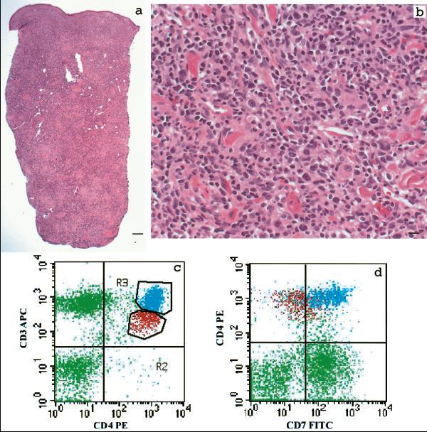
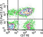
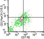
8 Immunophenotyping Classic MF Malignant skin-homing T-cells: CD45RO +, CLA + CLA = cutaneous lymphoid antigen. CD3 +, CD4 +, CD5 +/- CD7 +/-, CD26 +/- Usually T-cell receptor (TCR) + Cytokine profile may change with disease progression (switch from Th1 to Th2 in advanced disease) CD3 CD4 CD8 CD7 8
9 Immunophenotyping CD4 and CD8 immunophenotypic can be very useful Cutaneous T cell infiltrate of small cells CD8 > CD4 CD4=CD8 CD4>CD8 Juxtaepidermal CD8 MF reactive MF/Sezary Syndrome Aggressive CD8 CTCL 9
10 Flow Cytometry: Requires fresh tissue VIRTUAL FLOW CYTOMETRY does not require fresh tissue- formalin fixed tissue is ok ROLE IN DIAGNOSIS, STAGING, AND TARGETED THERAPY 10
11 ROLE IN DIAGNOSIS: SKIN BIOPSY MF Determine markers: Immunopathologic Criteria 1. CD2, 3 or 5 < 50% of T-cells 2. CD7 < 10% of T-cells 3. Epidermal discordance from expression of CD2,3,5, or 7 on dermal T-cells Pimpinelli N, et al. JAAD 2005: 11
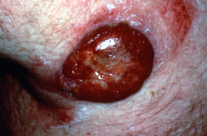
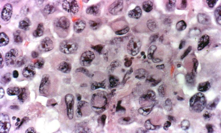
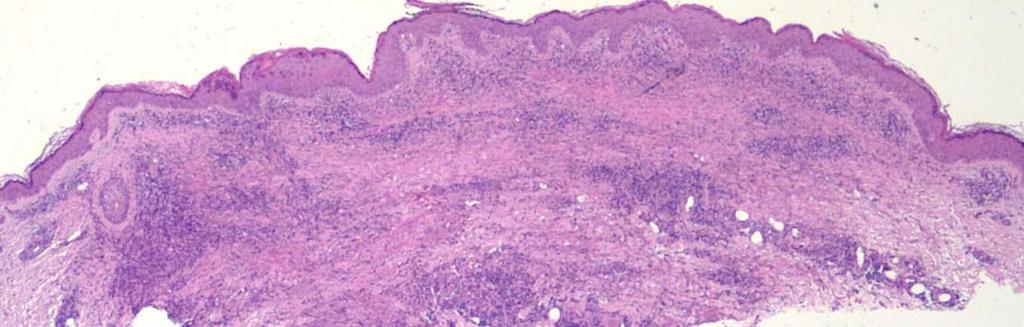
12 MF and transformation 12
13 Diagnostic Criteria: Blood In Sézary Syndrome, flow cytometry and molecular evaluation of the blood are needed to establish the diagnosis: Demonstration of a T-cell gene rearrangement in the blood and either 1.0 K/µL or more Sézary cells OR one of the 2 criteria outlined by the International Cutaneous Lymphoma Society (1) increased CD4 + or CD3 + cells with CD4/CD8 of 10 or more by flow cytometery or (2) increase in CD4 + cells with an abnormal phenotype ( 40% CD4 + /CD7 or 30% CD4 + /CD26] 13
14 CAVEATS Peripheral blood flow cytometry More objective than Sézary prep May indicate abnormality (T cell antigen aberrancy) although Molecular PCR test remain THE definitive test for clonality for T cells 14

15 ROLE IN STAGING MF Olsen et al. Revision to the staging and classification.. Blood

16 ROLE IN STAGING NON MF CUTANEOUS LYMPHOMAS Youn H. Kim, Rein Willemze, Nicola Pimpinelli, Sean Whittaker, Elise A. Olsen, Annamari Ranki, Reinhard Dummer, and Richard T. Hoppe, for the ISCL and the EORTC TNM classification system for primary cutaneous lymphomas other than mycosis fungoides and Sézary syndrome: a proposal of the International Society for Cutaneous Lymphomas (ISCL) and the Cutaneous Lymphoma Task Force of the European Organization of Research and Treatment of Cancer (EORTC) 16
17 ROLE IN THERAPY CYTOKINE PROFILE: Decreased cellmediated immunity with a dominant Th2 cytokine profile is frequently observed in patients with advanced stages of mycosis fungoides or Sézary syndrome. TARGETED surface molecules CD52-Alemtuzumab(Campath) (IL-2R alpha chain, CD25 antigen) RITUXAN IN B CELL LYMPHOMAS 17
18 Most Important Prognostic Factors In multivariate analyses, the most important prognostic factors are related to stage of disease: Presence of visceral disease 1,2 Type of skin involvement 1,2 Lymph node involvement 3 Blood involvement 3 1 Bunn PA, Lamberg SI. Cancer Treatment Reports 1979;63: Schechter GP, et al. Blood 1987;69: Klemke et al. Br J Dermatol. 2005;153:

19 LYMPH NODE GRADING-LN GRADING AIDED BY FLOW CYTOMETRY 19
20 FOLLICULAR LYMPHOMA Lesional skin Useful in cutaneous lymphomas other than MF ( B cell) B cell lymphomas express monotypic light chains Punch Biopsy sensitivity: Flow cytometry detected clonality in 88% (15 of 17) of cutaneous primary or secondary B cell lymphomas, compared to 37% (three of eight) by immunohistochemistry - J Invest Dermatol 121: ,


21 Flow cytometry and immunohistochemistry are complementary tools J Invest Dermatol 121: ,
22 FOLLICULAR LYMPHOMA CD20+, CD79a+, may show monotypic light chain expression Bcl-6+, CD10 (+ in follicular, - in diffuse), CD5-, bcl-2- or faint+, Mum-1 -, 22

, bcl-6+/-, CD10-,")
23 DLBC, LEG TYPE CD20+, CD79a+, monotypic light chain expression Bcl-2+ (strong), bcl-6+/-, CD10-, mum-1+ 23


24 Differential DX : cutaneous lymphoid hyperplasia or cutaneous pseudolymphoma 24
25 Cutaneous CD30+ LPD May be useful in CD30 lymphomas Represents a biologic and histologic spectrum with lymphomatoid papulosis (a benign disorder with spontaneous regression) at one end and primary cutaneous anaplastic large cell lymphoma (C-ALCL, an indolent CD30+ lymphoma usually treated with local therapy) at the other end. The classification of CD30+ LPD is predominantly based of the number and size of lesions, number of large CD30+ cells, and the clinical evolution of the lesion (progression versus regression). It is extremely important to distinguish CALCL from secondary involvement of the skin by systemic ALCL, an aggressive disease that requires multiagent, systemic chemotherapy. 25

26 Ki-1+ Anaplastic LCL (ALCL) 26

27 Lymphomatoid Papulosis(LyP) 27


28 CLA and TRAF-1 are useful in differentiating Lymphomatoid Papulosis, cutaneous ALCL and systemic ALCL. LyP cutaneousalcl Systemic ALCL H AND E TRAF-1 CLA H Lee Moffitt Cancer Center and Research Institute, Department Of Pathology and The University of South Florida, College of Medicine.Dorna Rezania, MD, Elizabeth Sagatys, MD, Marshall E. Kadin, M.D, Frank Glass, MD, Hernani Cualing MD. 28


 vs pcalcl(b) (p<0.")
 A B C Column 55 50 45 40 35 30 25 20 15")
, and between pcalcl(b) vs salcl (p")
29 VIRTUAL FLOW CYTOMETRY OF IMMUNOSTAINED TISSUE IMAGES TRAF-1 11 % 13% 1 % 34 % 18% 1 % CLA LyP cutaneousalcl Systemic ALCL CLA expression Mean and Standard Deviation LyP(A) vs pcalcl(b) (p<0.05) LyP vs salcl(c) (p<0.001) A B C Column TRAF-1 expression Mean and Standard Deviation A B C Column LyP(A) vs salcl (p<0.01), and between pcalcl(b) vs salcl (p <0.001) 29


30 Flow cytometry results may be helpful in differentiating from the similar monocytic leukemia DIFFERENTIATING CD4+/CD56+ HEMATODERMIC NEOPLASM FROM CD4+/CD56+ ACUTE MONOBLASTIC LEUKEMIA. USCAP Denver, 2008, IT Yu, D Rezania, LC Moscinski, JD Messina, Sokol L, Morgan M, HD Cualing, Moffitt Cancer Cancer & University of South Florida, Florida, USA 30
31 Summary Flow cytometry/immunohistochemistry have important roles in diagnosis, staging and targeted therapy of Cutaneous Lymphomas including MF. Non MF peripheral T cell lymphomas and B cell lymphomas of the skin and CD30 lymphomas workup should include immunophenotyping modalities Fresh tissue is amenable to flow cytometry and paraffin embedded tissue may benefit from quantitative tissue virtual flow cytometry non MF PTCL, CD30 lymphomas and CBCL are unique lymphoproliferative process with clinical course and outcome very different from histopathologically similar nodal lymphomas Cutaneous lymphoproliferative tumors: regard as lymphomas and send fresh tissue enabling use of ancillary tests 31
Sezary Syndrome(SS) and other malignancies. Hernani Cualing MD Hematopathologist IHCFLOW Lab
 and other malignancies. Hernani Cualing MD Hematopathologist IHCFLOW Lab") Sezary Syndrome(SS) and other malignancies Hernani Cualing MD Hematopathologist IHCFLOW Lab Disclosures IHCFLOW Laboratory:consultant and director NEOGENOMICS: contract consultant USF: contract reviewer
Sezary Syndrome(SS) and other malignancies Hernani Cualing MD Hematopathologist IHCFLOW Lab Disclosures IHCFLOW Laboratory:consultant and director NEOGENOMICS: contract consultant USF: contract reviewer
Michi Shinohara MD Associate Professor University of Washington/Seattle Cancer Care Alliance Dermatology, Dermatopathology
 Michi Shinohara MD Associate Professor University of Washington/Seattle Cancer Care Alliance Dermatology, Dermatopathology Agenda Overview of cutaneous T and B- cell lymphomas Diagnosis, Staging, Prognosis
Overview of Cutaneous Lymphomas: Diagnosis and Staging. Lauren C. Pinter-Brown MD, FACP Health Sciences Professor of Medicine and Dermatology
 Overview of Cutaneous Lymphomas: Diagnosis and Staging Lauren C. Pinter-Brown MD, FACP Health Sciences Professor of Medicine and Dermatology Definition of Lymphoma A cancer or malignancy that comes from
Lymphoma and Pseudolymphoma
 Lymphoma and Pseudolymphoma Laura B. Pincus, MD Co-Director, Cutaneous Lymphoma Clinic Associate Professor Dermatology and Pathology University of California, San Francisco I HAVE NO RELEVANT RELATIONSHIPS
Primary Cutaneous CD30-Positive T-cell Lymphoproliferative Disorders
 Primary Cutaneous CD30-Positive T-cell Lymphoproliferative Disorders Definition A spectrum of related conditions originating from transformed or activated CD30-positive T-lymphocytes May coexist in individual
SH/EAHP Workshop 2011 Los Angeles, California, USA
 SH/EAHP Workshop 2011 Los Angeles, California, USA October 27-29, 2011 Session 3 Non-Mycosis Fungoides CTCL Patty Jansen & Rein Willemze Introduction Submitted: 101 cases + 7 cases group 1: 108 Deactivated
Disclosures. Advisory Board. Consultant. Investigator. MiRagen, Actelion, Celgene, Therakos. Mindera
 Cutaneous Lymphomas Christiane Querfeld, MD, PhD Director, Cutaneous Lymphoma Program City of Hope ~ How the Experts Treat Hematologic Malignancies Symposium March 10 13, 2017 Disclosures Advisory Board
New Haven, Connecticut
 New Haven, Connecticut Yale University Main Campus Yale mascot: Handsome Dan Cutaneous Lymphomas Tony Subtil, MD, MBA Associate Professor Yale University Cutaneous Lymphomas: 1. Intro 2. CTCL/NK 3. CBCL
ISPUB.COM. Primary Cutaneous Anaplastic Large Cell Lymphoma Long-term Management with Low Dose Methotrexate. S Parker INTRODUCTION
 ISPUB.COM The Internet Journal of Dermatology Volume 7 Number 3 Primary Cutaneous Anaplastic Large Cell Lymphoma Long-term Management with Low Dose S Parker Citation S Parker.. The Internet Journal of
clinical recommendations
 19 (Supplement 2): ii72 ii76, 2008 doi:10.1093/annonc/mdn095 Primary cutaneous lymphoma: ESMO Clinical Recommendations for diagnosis, treatment and follow-up R. Dummer 1 & M. Dreyling 2 On behalf of the
See Important Reminder at the end of this policy for important regulatory and legal information.
 Clinical Policy: Vorinostat (Zolinza) Reference Number: CP.PHAR.83 Effective Date: 10.01.18 Last Review Date: 07.13.18 Line of Business: Oregon Health Plan Revision Log See Important Reminder at the end
88-year-old Female with Lymphadenopathy. Faizi Ali, MD
 88-year-old Female with Lymphadenopathy Faizi Ali, MD Clinical History A 88-year-old caucasian female presented to our hospital with the complaints of nausea, vomiting,diarrhea, shortness of breath and
Cover Page. The handle holds various files of this Leiden University dissertation.
 Cover Page The handle http://hdl.handle.net/1887/2010 holds various files of this Leiden University dissertation. Author: Benner, Marchina Frederika Title: Cutaneous CD30-positive lymphoproliferations
Review Article. Cutaneous lymphoproliferative disorders. NJ Trendell-Smith
 Hong Kong J. Dermatol. Venereol. (2010) 18, 190-201 Review Article Cutaneous lymphoproliferative disorders NJ Trendell-Smith Cutaneous lymphoproliferative disorders (CLD) include reactive lymphoid hyperplasias,
See Important Reminder at the end of this policy for important regulatory and legal information.
 Clinical Policy: (Istodax) Reference Number: CP.PHAR.314 Effective Date: 01.01.17 Last Review Date: 11.18 Line of Business: Medicaid, HIM-Medical Benefit Coding Implications Revision Log See Important
Commentary on the 2008 WHO classification of mature T- and NK-cell neoplasms
 J Hematopathol (2009) 2:65 73 DOI 10.1007/s12308-009-0034-z COMMENT Commentary on the 2008 WHO classification of mature T- and NK-cell neoplasms Megan S. Lim & Laurence de Leval & Leticia Quintanilla-Martinez
Cutaneous Lymphoid Proliferations: A Comprehensive Textbook of Lymphocytic Infiltrates of the Skin
 Cutaneous Lymphoid Proliferations: A Comprehensive Textbook of Lymphocytic Infiltrates of the Skin Magro, Cynthia M., MD ISBN-13: 9780471695981 Table of Contents Chapter One: Introduction to the Classification
Clinical Policy: Vorinostat (Zolinza) Reference Number: CP.PHAR.83 Effective Date: 10/11
 Reference Number: CP.PHAR.83 Effective Date: 10/11") Clinical Policy: (Zolinza) Reference Number: CP.PHAR.83 Effective Date: 10/11 Last Review Date: 12/16 Revision Log See Important Reminder at the end of this policy for important regulatory and legal information.
Clinical Policy: (Zolinza) Reference Number: CP.PHAR.83 Effective Date: 10/11 Last Review Date: 12/16 Revision Log See Important Reminder at the end of this policy for important regulatory and legal information.
Immunopathology of Lymphoma
 Immunopathology of Lymphoma Noraidah Masir MBBCh, M.Med (Pathology), D.Phil. Department of Pathology Faculty of Medicine Universiti Kebangsaan Malaysia Lymphoma classification has been challenging to pathologists.
Primer of Immunohistochemistry (Leukocytic)
") Primer of Immunohistochemistry (Leukocytic) Paul K. Shitabata, M.D. Dermatopathology Institute Torrance, CA BENIGN LYMPHOID SKIN LESIONS CAPABLE OF SIMULATING LYMPHOMA -Jessner s lymphoid infiltrate -Dermal-subcutaneous
Primer of Immunohistochemistry (Leukocytic) Paul K. Shitabata, M.D. Dermatopathology Institute Torrance, CA BENIGN LYMPHOID SKIN LESIONS CAPABLE OF SIMULATING LYMPHOMA -Jessner s lymphoid infiltrate -Dermal-subcutaneous
CASE 15 Patient: A 41-year-old Thai female Chief Compliant: Generalized papulovesicular rash for 1 month Present Illness: She presented with a 1-week
 CASE 15 Patient: A 41-year-old Thai female Chief Compliant: Generalized papulovesicular rash for 1 month Present Illness: She presented with a 1-week history of the generalized asymptomatic erythematous
Hepatic Lymphoma Diagnosis An Algorithmic Approach
 Hepatic Lymphoma Diagnosis An Algorithmic Approach Ryan M. Gill, M.D., Ph.D. University of California, San Francisco PLEASE TURN OFF YOUR CELL PHONES Disclosure of Relevant Financial Relationships USCAP
Differential diagnosis of hematolymphoid tumors composed of medium-sized cells. Brian Skinnider B.C. Cancer Agency, Vancouver General Hospital
 Differential diagnosis of hematolymphoid tumors composed of medium-sized cells Brian Skinnider B.C. Cancer Agency, Vancouver General Hospital Lymphoma classification Lymphoma diagnosis starts with morphologic
CUTANEOUS T-CELL LYMPHOMA PROFORMA
 STATE OF KUWAIT MINISTRY OF HEALTH AS AD ALHAMAD DERMATOLOGY CENTER ALSABAH HOSPITAL دولة الكویت وزارة الصحة مرآز أسعد الحمد للا مراض الجلدیة مستشفى الصباح بسم االله الرحمن الرحيم CUTANEOUS TCELL LYMPHOMA
Contents. vii. Preface... Acknowledgments... v xiii
 Contents Preface... Acknowledgments... v xiii SECTION I 1. Introduction... 3 Knowledge-Based Diagnosis... 4 Systematic Examination of the Lymph Node... 7 Cell Type Identification... 9 Cell Size and Cellularity...
ISPUB.COM. M Duvic, E Olsen PURPOSE OF REVISION
 ISPUB.COM The Internet Journal of Dermatology Volume 7 Number 3 International Society of Cutaneous Lymphoma (ISCL) and the European Organization of Research and Treatment of Cancer (EORTC) revisions to
Non-Hodgkin s Lymphomas Version
 NCCN Clinical Practice Guidelines in Oncology (NCCN Guidelines ) Non-Hodgkin s Lymphomas Version 2.2015 NCCN.org Continue Use of Immunophenotyping/ Genetic Testing in Differential Diagnosis of Mature B-Cell
Bone Marrow. Procedures Blood Film Aspirate, Cell Block Trephine Biopsy, Touch Imprint
 Bone Marrow Protocol applies to acute leukemias, myelodysplastic syndromes, myeloproliferative disorders, chronic lymphoproliferative disorders, malignant lymphomas, plasma cell dyscrasias, histiocytic
Mycosis Fungoides, then and now Have we travelled?
 USCAP 103 rd Annual Meeting 2014 American Society of Dermatopathology Companion Meeting Mycosis Fungoides, then and now Have we travelled? Vijaya B. Reddy, MD, MBA Professor of Pathology Rush University
U006 Primary Cutaneous Lymphomas: Diagnosis, Staging and When to Refer M. Yadira Hurley, MD
 U006 Primary Cutaneous Lymphomas: Diagnosis, Staging and When to Refer M. Yadira Hurley, MD hurleyy@slu.edu DISCLOSURE OF RELATIONSHIPS WITH INDUSTRY Actelion: Speaker Honoraria, Investigator Grants, Consultant
Lymphoma: What You Need to Know. Richard van der Jagt MD, FRCPC
 Lymphoma: What You Need to Know Richard van der Jagt MD, FRCPC Overview Concepts, classification, biology Epidemiology Clinical presentation Diagnosis Staging Three important types of lymphoma Conceptualizing
TOX expression in cutaneous B-cell lymphomas
 Arch Dermatol Res (2016) 308:423 427 DOI 10.1007/s00403-016-1654-7 ORIGINAL PAPER TOX expression in cutaneous B-cell lymphomas Anne M. R. Schrader 1 Patty M. Jansen 1 Rein Willemze 2 Received: 20 January
Therapeutic Management of Early Cutaneous Mycosis Fungoides
 Therapeutic Management of Early Cutaneous Mycosis Fungoides L Frank Glass, MD Cutaneous Lymphoma Programs H Lee Moffitt Cancer Center and Research Institute George Washington University Dermatology and
Lymphoma/CLL 101: Know your Subtype. Dr. David Macdonald Hematologist, The Ottawa Hospital
 Lymphoma/CLL 101: Know your Subtype Dr. David Macdonald Hematologist, The Ottawa Hospital Function of the Lymph System Lymph Node Lymphocytes B-cells develop in the bone marrow and influence the immune
A middle-aged man with self-healing papulonecrotic lesions over the trunk and proximal limbs
 Hong Kong J. Dermatol. Venereol. (2011) 19, 30-34 Case Report A middle-aged man with self-healing papulonecrotic lesions over the trunk and proximal limbs JC Chan, N Trendell-Smith, CK Yeung Lymphomatoid
Granulomatous Slack Skin with an unusually aggressive course due to the subsequent development of a CD30-positive Large Cell Lymphoma
 Granulomatous Slack Skin with an unusually aggressive course due to the subsequent development of a CD30-positive Large Cell Lymphoma Alexandra Papoudou-Bai 1, Eleni Kapsali 2, Ioannis Kostas-Agnantis
WHO Classification. B-cell chronic lymphocytic leukemia/small T-cell granular lymphocytic leukemia
 Blood Malignancies-II Prof. Dr. Herman Hariman, a Ph.D, SpPK (KH). Prof. Dr. Adikoesoema Aman, SpPK (KH) Dept. of Clinical Pathology, School of Medicine, University of North Sumatra WHO classification
The spectrum of flow cytometry of the bone marrow
 The spectrum of flow cytometry of the bone marrow Anna Porwit Lund University Faculty of Medicine Dept. of Clinical Sciences Div. Oncology and Pathology anna.porwit@med.lu.se Disclosure of speaker s interests
Non-Hodgkin Lymphoma. Protocol applies to non-hodgkin lymphoma involving any organ system except the gastrointestinal tract.
 Non-Hodgkin Lymphoma Protocol applies to non-hodgkin lymphoma involving any organ system except the gastrointestinal tract. Protocol revision date: January 2005 No AJCC/UICC staging system Procedures Cytology
Integrated Hematopathology. Morphology and FCI with IHC
 Integrated Hematopathology Morphology and FCI with IHC FrontMatter.indd i 9/6/2009 9:30:12 PM FrontMatter.indd ii 9/6/2009 9:30:18 PM Integrated Hematopathology Morphology and FCI with IHC Cherie H Dunphy,
Case Report A Severe Case of Lymphomatoid Papulosis Type E Successfully Treated with Interferon-Alfa 2a
 Hindawi Case Reports in Dermatological Medicine Volume 2017, Article ID 3194738, 5 pages https://doi.org/10.1155/2017/3194738 Case Report A Severe Case of Lymphomatoid Papulosis Type E Successfully Treated
2. Sézary syndrome (SS)
") Go Back to the Top To Order, Visit the Purchasing Page for Details Clinical images are available in hardcopy only. Clinical images are available in Clinical images are available in d e f g h i j Fig..36-2
Go Back to the Top To Order, Visit the Purchasing Page for Details Clinical images are available in hardcopy only. Clinical images are available in Clinical images are available in d e f g h i j Fig..36-2
Pearls and pitfalls in interpretation of lymphoid lesions in needle biopsies
 Pearls and pitfalls in interpretation of lymphoid lesions in needle biopsies Megan S. Lim MD PhD University of Pennsylvania October 8, 2018 Objectives To understand how the trend toward less invasive lymph
184 Cutaneous Lymphomas: Morphology, Immunohistochemistry and Molecular Testing. David Cassarino MD, PhD Aaron Auerbach MD
 184 Cutaneous Lymphomas: Morphology, Immunohistochemistry and Molecular Testing David Cassarino MD, PhD Aaron Auerbach MD 2011 Annual Meeting Las Vegas, NV AMERICAN SOCIETY FOR CLINICAL PATHOLOGY 33 W.
Fig. 3.1 Fig Past history: She was previously healthy and not taking any medication.
 Case 3 A 41-year-old Thai female from Bangkok Chief compliant: Erythematous patch at left thigh for 2 months Present illness: The patient presented with a 10- year history of erythematous patch on her
Session Summary session 6. Reactive Lymphoproliferations of the skin. Session 6 - case 211
 SH/EAHP Workshop 2011 Los Angeles, California, USA October 27-29, 2011 Session 6 Reactive Lymphoproliferations of the skin Rein Willemze Summary session 6 Atypical T-cell infiltrates (lymphomatoid; pseudo-t-cell
Important Decisions in Dermatopathology: The Clinico- Pathologic Correlation. Dermatopathology Specialists Needed. Changing Trends
 Important Decisions in Dermatopathology: The Clinico- Pathologic Correlation Uma Sundram, MD, PhD Departments of Pathology and Dermatology Stanford University May 29, 2008 Dermatopathology Specialists
CD30+ Lymphoproliferative Disorders Associated with Longstanding Mycosis Fungoides
 Case Report DOI: 10.6003/jtad.16102c5 CD30+ Lymphoproliferative Disorders Associated with Longstanding Mycosis Fungoides Esra Adışen, 1 MD, Özlem Erdem, 2 MD, Mehmet Ali Gürer, 1 MD Address: 1 Gazi University
ISPUB.COM. Management of Co-existing Mycosis Fungoides and Lymphomatoid Papulosis. E Kim PHYSICAL FINDINGS INTRODUCTION INITIAL PRESENTATION
 ISPUB.COM The Internet Journal of Dermatology Volume 7 Number 3 Management of Co-existing Mycosis Fungoides and Lymphomatoid Papulosis E Kim Citation E Kim. Management of Co-existing Mycosis Fungoides
Clusterin Expression Correlates With Stage and Presence of Large Cells in Mycosis Fungoides
 Anatomic Pathology / Clusterin Expression in Mycosis Fungoides Clusterin Expression Correlates With Stage and Presence of Large Cells in Mycosis Fungoides Pranil Chandra, DO, 1 Jose A. Plaza, MD, 2,4 Zhuang
Primary cutaneous lymphomas: ESMO Clinical Practice Guidelines for diagnosis, treatment and follow-up
 Annals of Oncology 24 (Supplement 6): vi149 vi154, 2013 doi:10.1093/annonc/mdt242 Published online 17 July 2013 Primary cutaneous lymphomas: ESMO Clinical Practice Guidelines for diagnosis, treatment and
Malignant Lymphomas Decision Making and Problem Solving
 Malignant Lymphomas Decision Making and Problem Solving Cutaneous T-cell lymphomas (including rare subtypes). Current concepts. II. [haematologica] 2004;89:1372-1388 MARCO PAULLI EMILIO BERTI A B S T R
Primary cutaneous large cell lymphoma CD30+: a case-based review
 Case-based review Primary cutaneous large cell lymphoma CD30+: a case-based review Anna Campanati 1 Katia Giuliodori 1 Emanuela Martina 1 Luca Conocchiari 1 Giulia Ganzetti 1 Gaia Goteri 2 Annamaria Offidani
From Morphology to Molecular Pathology: A Practical Approach for Cytopathologists Part 1-Cytomorphology. Songlin Zhang, MD, PhD LSUHSC-Shreveport
 From Morphology to Molecular Pathology: A Practical Approach for Cytopathologists Part 1-Cytomorphology Songlin Zhang, MD, PhD LSUHSC-Shreveport I have no Conflict of Interest. FNA on Lymphoproliferative
Research Article Characteristics of Primary Cutaneous T-Cell Lymphoma in Iran: A 10-Year Retrospective Study
 International Scholarly Research Notices, Article ID 820921, 5 pages http://dx.doi.org/10.1155/2014/820921 Research Article Characteristics of Primary Cutaneous T-Cell Lymphoma in Iran: A 10-Year Retrospective
Unusual cutaneous presentation of a T-cell lymphoproliferation
 Department of Pathology and Cytology University Hospital Centre Zagreb, Croatia Unusual cutaneous presentation of a T-cell lymphoproliferation Snjezana Dotlic, Stefan Dojcinov, Leticia Quintanilla-Fend
Cover Page. The handle holds various files of this Leiden University dissertation.
 Cover Page The handle http://hdl.handle.net/1887/39089 holds various files of this Leiden University dissertation. Author: Cetinozman, F. Title: PD-1 Expression in primary cutaneous lymphoma Issue Date:
Classification of Hematologic Malignancies. Patricia Aoun MD MPH
 Classification of Hematologic Malignancies Patricia Aoun MD MPH Objectives Know the basic principles of the current classification system for hematopoietic and lymphoid malignancies Understand the differences
Peripheral T-cell lymphomas
 XXXVI REUNIÓN ANUAL Peripheral T-cell lymphomas Dr. Antonio Martinez Hospital Clinic, University ofbarcelona antonmar@clinic.ub.es Madrid, February 8th, 2013 T-NHL vs B-NHL: the T-cell paradigm lambda
How I treat patients whose biopsies are reported descriptively Youn H Kim, MD
 How I treat patients whose biopsies are reported descriptively Youn H Kim, MD Director, Multidisciplinary Cutaneous Lymphoma Group Stanford Cancer institute & School of Medicine NCCN NHL Panel Member Disclosure
Methods used to diagnose lymphomas
 Institut für Pathologie Institut für Pathologie Methods used to diagnose lymphomas Prof. Dr.Med. Leticia Quintanilla-Fend Molecular techniques NGS histology Cytology AS-PCR Sanger seq. MYC Immunohistochemistry
Change Summary - Form 2018 (R3) 1 of 12
 1 of 12") Summary - Form 2018 (R3) 1 of 12 Form Question Number (r3) Type Description New Text Previous Text Today's date was removed 2018 N/A Today's Date Removed from Key Fields 2018 N/A HCT Type 2018 N/A Product
Summary - Form 2018 (R3) 1 of 12 Form Question Number (r3) Type Description New Text Previous Text Today's date was removed 2018 N/A Today's Date Removed from Key Fields 2018 N/A HCT Type 2018 N/A Product
Cutaneous T-cell Lymphoma: An Entry From Thomson Gale's Gale Encyclopedia Of Cancer, 2nd Ed. [HTML] [Digital]
![Cutaneous T-cell Lymphoma: An Entry From Thomson Gale's Gale Encyclopedia Of Cancer, 2nd Ed. [HTML] [Digital]](/thumbs/80/80529096.jpg "Cutaneous T-cell Lymphoma: An Entry From Thomson Gale's Gale Encyclopedia Of Cancer, 2nd Ed. [HTML] [Digital]") Cutaneous T-cell Lymphoma: An Entry From Thomson Gale's Gale Encyclopedia Of Cancer, 2nd Ed. [HTML] [Digital] By Michelle, M.S., J.D. Johnson;Rebecca, PhD Frey READ ONLINE If searched for a ebook Cutaneous
Cutaneous T-cell Lymphoma: An Entry From Thomson Gale's Gale Encyclopedia Of Cancer, 2nd Ed. [HTML] [Digital] By Michelle, M.S., J.D. Johnson;Rebecca, PhD Frey READ ONLINE If searched for a ebook Cutaneous
Renaissance of Low-Dose Radiotherapy Concepts for Cutaneous Lymphomas
 Review Article DOI: 10.1159/000470845 Received: February 06, 2017 Accepted: March 13, 2017 Published online: April 6, 2017 Renaissance of Low-Dose Radiotherapy Concepts for Cutaneous Lymphomas Bouthaina
Lymphoma Update: Lymphoma Update: What s Likely to be New in the New WHO. Patrick Treseler, MD, PhD University of California San Francisco
 Lymphoma Update: What s Likely to be New in the New WHO Blood 127:2375; 2016 Patrick Treseler, MD, PhD University of California San Francisco Lymphoma Update: What IS New in the New WHO! Patrick Treseler,
HODGKIN LYMPHOMA DR. ALEJANDRA ZARATE OSORNO HOSPITAL ESPAÑOL DE MEXICO
 HODGKIN LYMPHOMA DR. ALEJANDRA ZARATE OSORNO HOSPITAL ESPAÑOL DE MEXICO HODGKIN LYMPHOMA CLASSIFICATION Lukes & Butler Rye WHO-2016 Linphocytic and/or histiocytic Nodular & diffuse Nodular Sclerosis Lymphocyte
Cluster of differentiation (CD) markers in erythrodermic patients: A case series study
 markers in erythrodermic patients: A case series study") Original Article Cluster of differentiation (CD) markers in erythrodermic patients: A case series study Fariba Ghalamkarpour, MD Faranak Niknafs, MD Shima Younespour, PhD Skin Research Center, Shahid Beheshti
Original Article Cluster of differentiation (CD) markers in erythrodermic patients: A case series study Fariba Ghalamkarpour, MD Faranak Niknafs, MD Shima Younespour, PhD Skin Research Center, Shahid Beheshti
Burkitt lymphoma. Sporadic Endemic in Africa associated with EBV Translocations involving MYC gene on chromosome 8
 Heme 8 Burkitt lymphoma Sporadic Endemic in Africa associated with EBV Translocations involving MYC gene on chromosome 8 Most common is t(8;14) Believed to be the fastest growing tumor in humans!!!! Morphology
The skin is the second most common extranodal site for
 CAP Laboratory Improvement Programs Best Practices in Diagnostic Immunohistochemistry Workup of Cutaneous Lymphoid Lesions in the Diagnosis of Primary Cutaneous Lymphoma Rajan Dewar, MD, PhD; Aleodor Alexandru
Mycosis Fungoides and Variants
 Mycosis Fungoides and Variants Jennifer Madison McNiff, M.D. Associate Professor, Dermatology and Pathology Yale University School of Medicine Classic mycosis fungoides The most common cutaneous lymphoma
T cell lymphoma diagnostics and differential diagnosis to Hodgkin lymphoma
 T cell lymphoma diagnostics and differential diagnosis to Hodgkin lymphoma Sylvia Hartmann Dr. Senckenberg Institute of Pathology Goethe University Frankfurt Overview Borderline ALCL classical HL Borderline
Cutaneous Mycosis Fungoides (MF), the subtype of cutaneous
, the subtype of cutaneous") DOI 10. 5001/omj.2012.28 The Histological Spectrum of Early Mycosis Fungoides: A Study of 58 Saudi Arab patients Maha Arafah, Shaesta Naseem Zaidi, Hala Kassouf Kfoury, Ammar Al Rikabi, Khalid Al Ghamdi
DOI 10. 5001/omj.2012.28 The Histological Spectrum of Early Mycosis Fungoides: A Study of 58 Saudi Arab patients Maha Arafah, Shaesta Naseem Zaidi, Hala Kassouf Kfoury, Ammar Al Rikabi, Khalid Al Ghamdi
2010 Hematopoietic and Lymphoid ICD-O Codes - Alphabetical List THIS TABLE REPLACES ALL ICD-O-3 Codes
 Acute basophilic leukemia 9870/3 Acute biphenotypic leukemia [OBS] 9805/3 Acute erythroid leukemia 9840/3 Acute megakaryoblastic leukemia 9910/3 Acute monoblastic and monocytic leukemia 9891/3 Acute myeloid
2012 Hematopoietic and Lymphoid ICD-O Codes - Numerical List THIS TABLE REPLACES ALL ICD-O-3 Codes
 Malignant lymphoma, NOS 9590/3 Non-Hodgkin lymphoma, NOS 9591/3 B-cell lymphoma, unclassifiable, with features intermediate between diffuse large B-cell lymphoma and classical Hodgkin lymphoma 9596/3 Primary
3.1 Introduction. It is emphasised that not all tests are necessarily required in every case. 3.2 Taxonomic structure
 CHAPTER 3 CLASSIFICATION 3.1 Introduction Accurate diagnosis underpins lymphoma management. Historically, competing lymphoma classifications have been a source of frustration to pathologists, clinicians
Subject Index. Dry desquamation, see Skin reactions, radiotherapy
 Subject Index Actinic keratosis disseminated disease 42 surgical excision 42 AIDS, see Kaposi s sarcoma Amifostine, skin reaction prophylaxis 111 Basal cell carcinoma, superficial X-ray therapy Bowen s
During past decades, because of the lack of knowledge
 Staging and Classification of Lymphoma Ping Lu, MD In 2004, new cases of non-hodgkin s in the United States were estimated at 54,370, representing 4% of all cancers and resulting 4% of all cancer deaths,
T CELL LYMPHOMA ANALYSIS
 T CELL LYMPHOMA ANALYSIS Charles Goolsby, Ph.D. Floyd E. Patterson Research Professor of Pathology Northwestern Feinberg School of Medicine c-goolsby@northwestern.edu 1 T CELL LYMPHOMA ANALYSIS Diverse
Combinations of morphology codes of haematological malignancies (HM) referring to the same tumour or to a potential transformation
 referring to the same tumour or to a potential transformation") Major subgroups according to the World Health Organisation (WHO) Classification Myeloproliferative neoplasms (MPN) Myeloid and lymphoid neoplasms with eosinophilia and abnormalities of PDGFRA, PDGFRB or
Major subgroups according to the World Health Organisation (WHO) Classification Myeloproliferative neoplasms (MPN) Myeloid and lymphoid neoplasms with eosinophilia and abnormalities of PDGFRA, PDGFRB or
Lymphocytoma Cutis. Cynthia M. Magro MD. Director of Dermatopathology Weill Medical College of Cornell University New York, New York
 Lymphocytoma Cutis Cynthia M. Magro MD Professor of Pathology Director of Dermatopathology Weill Medical College of Cornell University New York, New York Lymphocytoma Cutis Falls under other designations
CD4-Positive T-Cell Large Granular Lymphocytosis Mimicking Sézary Syndrome in a Patient With Mycosis Fungoides
 Case Report CD4-Positive T-Cell Large Granular Lymphocytosis Mimicking Sézary Syndrome in a Patient With Mycosis Fungoides Ling Zhang, MD, Magali Van den Bergh, MD, and Lubomir Sokol, MD, PhD Summary:
Guidelines for the management of cutaneous lymphomas (2011): A consensus statement by the Japanese Skin Cancer Society Lymphoma Study Group
: A consensus statement by the Japanese Skin Cancer Society Lymphoma Study Group") doi: 10.1111/j.1346-8138.2012.01639.x Journal of Dermatology 2013; 40: 2 14 GUIDELINE Guidelines for the management of cutaneous lymphomas (2011): A consensus statement by the Japanese Skin Cancer Society
doi: 10.1111/j.1346-8138.2012.01639.x Journal of Dermatology 2013; 40: 2 14 GUIDELINE Guidelines for the management of cutaneous lymphomas (2011): A consensus statement by the Japanese Skin Cancer Society
A rare clinical presentation of non Hodgkin Lymphoma as multiple neurofibromas with review of literature
 Original article A rare clinical presentation of non Hodgkin Lymphoma as multiple neurofibromas with review of literature Dr.C.R.Sirajunnisa Begum, Professor 1, Dr. Ira Bharadhwaj, Professor 1, Dr. Lavanya,
Lymphoma: The Basics. Dr. Douglas Stewart
 Lymphoma: The Basics Dr. Douglas Stewart Objectives What is lymphoma? How common is it? Why does it occur? How do you diagnose it? How do you manage it? How do you follow patients after treatment? What
Immunohistochemical classification of haematolymphoid tumours. Stephen Hamilton-Dutoit Institute of Pathology Aarhus University Hospital
 Immunohistochemical classification of haematolymphoid tumours Stephen Hamilton-Dutoit Institute of Pathology Aarhus University Hospital Malignant lymphoproliferative diseases What are they? Haematolymphoid
What s new on the horizon in T-cell lymphoma Elaine S Jaffe National Cancer Institute, Bethesda MD
 What s new on the horizon in T-cell lymphoma Elaine S Jaffe National Cancer Institute, Bethesda MD WHO classification: where are we today? Of 12 monographs planned for 4 th Edition Bluebook series, only
Exploring the Borderlands between Diffuse Large B-cell Lymphoma and Classical Hodgkin s Lymphoma
 Exploring the Borderlands between Diffuse Large B-cell Lymphoma and Classical Hodgkin s Lymphoma Elaine S. Jaffe National Cancer Institute Bethesda, MD, USA On the Pathological Changes In Hodgkin s Disease
Given that mycosis fungoides (MF) is the most common
 is the most common") Histopathologic approach to epidermotropic lymphocytic infiltrates Shyam S Raghavan, MD 1 and Jinah Kim, MD, PhD 2 Abstract Mycosis fungoides is the most common and therefore quintessential cutaneous lymphoma
Histopathologic approach to epidermotropic lymphocytic infiltrates Shyam S Raghavan, MD 1 and Jinah Kim, MD, PhD 2 Abstract Mycosis fungoides is the most common and therefore quintessential cutaneous lymphoma
Immunocytochemistry and the diagnosis of cutaneous lymphoma
 Histopathology 2010, 56, 71 90. DOI: 10.1111/j.1365-2559.2009.03457.x REVIEW Immunocytochemistry and the diagnosis of cutaneous lymphoma Alistair Robson Department of Dermatopathology, St John s Institute
Follicular Lymphoma: the WHO
 Follicular Lymphoma: the WHO and the WHERE? Yuri Fedoriw, MD Associate Professor of Pathology and Laboratory Medicine Director of Hematopathology University of North Carolina Chapel Hill, NC Disclosure
1) Title: Cutaneous lymphoma in Japan: a nationwide study of 1,733 patients.
 Title: Cutaneous lymphoma in Japan: a nationwide study of 1,733 patients.") 1) Title: Cutaneous lymphoma in Japan: a nationwide study of 1,733 patients. 2) The short running title: Cutaneous lymphoma in Japan. 3) Toshihisa Hamada and Keiji Iwatsuki 4) Departments of Dermatology,
1) Title: Cutaneous lymphoma in Japan: a nationwide study of 1,733 patients. 2) The short running title: Cutaneous lymphoma in Japan. 3) Toshihisa Hamada and Keiji Iwatsuki 4) Departments of Dermatology,
Angioinvasive Lymphomatoid Papulosis: A new variant simulating aggressive lymphomas
 Zurich Open Repository and Archive University of Zurich Main Library Strickhofstrasse 39 CH-8057 Zurich www.zora.uzh.ch Year: 2013 Angioinvasive Lymphomatoid Papulosis: A new variant simulating aggressive
Molecular Pathology of Lymphoma (Part 1) Rex K.H. Au-Yeung Department of Pathology, HKU
 Rex K.H. Au-Yeung Department of Pathology, HKU") Molecular Pathology of Lymphoma (Part 1) Rex K.H. Au-Yeung Department of Pathology, HKU Lecture outline Time 10:00 11:00 11:15 12:10 12:20 13:15 Content Introduction to lymphoma Review of lymphocyte biology
Molecular Pathology of Lymphoma (Part 1) Rex K.H. Au-Yeung Department of Pathology, HKU Lecture outline Time 10:00 11:00 11:15 12:10 12:20 13:15 Content Introduction to lymphoma Review of lymphocyte biology
Many of the hematolymphoid disorders are derived
 REVIEW ARTICLE Practical Immunohistochemistry in Hematopathology: A Review of Useful Antibodies for Diagnosis Ji Lu, MD and Karen L. Chang, MD Abstract: This review article offers some useful panels of
Interhospital Dermatology Conference 2013
 Case 21 A 50-year-old woman from Chonburi. Chief complaint: Asymptomatic rash on neck, trunk, both upper and lower extremities for 2 years. Present illness: The patient has developed asymptomatic scaly
Hematopathology Specialty Conference Case #1
 Hematopathology Specialty Conference Case #1 Robert (Bob) Ohgami, MD, PhD Assistant Professor Stanford University Disclosure of Relevant Financial Relationships Disclosure of Relevant Financial Relationships
Large cell immunoblastic Diffuse histiocytic (DHL) Lymphoblastic lymphoma Diffuse lymphoblastic Small non cleaved cell Burkitt s Non- Burkitt s
 Lymphoblastic lymphoma Diffuse lymphoblastic Small non cleaved cell Burkitt s Non- Burkitt s") Non Hodgkin s Lymphoma Introduction 6th most common cause of cancer death in United States. Increasing in incidence and mortality. Since 1970, the incidence of has almost doubled. Overview The types of
Non Hodgkin s Lymphoma Introduction 6th most common cause of cancer death in United States. Increasing in incidence and mortality. Since 1970, the incidence of has almost doubled. Overview The types of