Important Decisions in Dermatopathology: The Clinico- Pathologic Correlation. Dermatopathology Specialists Needed. Changing Trends
|
|
|
- Christina Spencer
- 5 years ago
- Views:
Transcription
1 Important Decisions in Dermatopathology: The Clinico- Pathologic Correlation Uma Sundram, MD, PhD Departments of Pathology and Dermatology Stanford University May 29, 2008 Dermatopathology Specialists Needed Skin and Allergy News Sep 2007: Interview of Dr. Clay Cockerell Changing trends in dermatopathology Increasing numbers of primary care physicians evaluate skin disorders on a routine basis Many of these biopsies are routed to general pathologists Changing Trends Cockerell: Only 35% of skin biopsies come from dermatologists Economic pressures may also force PCP s to keep and work up their difficult dermatology cases, rather than refer Dermatopathologists not being trained in numbers to meet demand 1

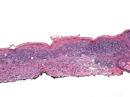
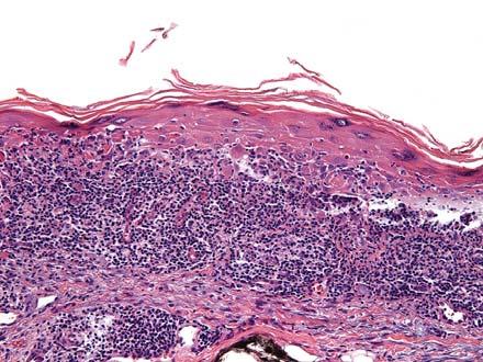
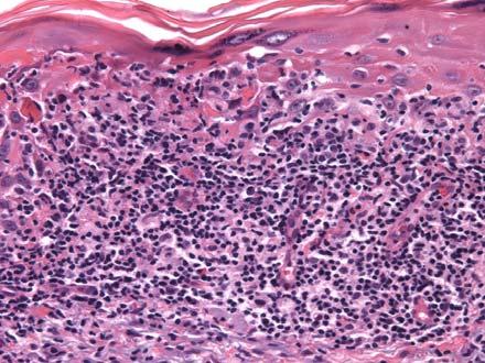
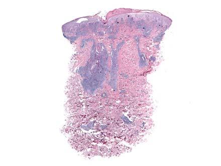
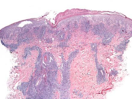
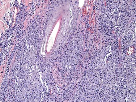
2 Diagnostic Errors Due to lack of understanding of the dermatologic clinical scenario, overinterpretation or under-interpretation of biopsies take place Purpose of this talk: To stress the importance of clinico-pathologic correlation and assessing the clinical scenario in conjunction with slide interpretation Case I 45 year old female with persistent red patch on the left arm. 2
3 3
4 What is your diagnosis? 1. Mycosis fungoides 2. Drug eruption 3. Lichen planus 4. Lichenoid keratosis 0% 0% 0% 0% Mycosis fungoides Drug eruption Lichen planus Lichenoid keratosis What is your next step? 1. Call the clinician 2. Initiate immunohistochemical studies 3. Initiate PCR analysis for TCR gene rearrangements Call the clinician 0% 0% 0% Initiate immuno-hist... Initiate PCR analysis f.. If you called the clinician, what would you ask about? 1. Size and number of lesions 2. Appearance of lesions (patches/plaques or papules) 3. Distribution of lesions (sun spared or sun exposed sites) 4. Duration of lesions 5. Drug history 6. All of the above Size and number of l... Appearance of lesion... 0% 0% 0% 0% 0% 0% Distribution of lesions... Duration of lesions Drug history All of the above 4
5 If ordering immunohistochemical studies, what would you order? 1. CD20 and CD3 2. CD3, CD4, and CD8 3. CD4 and CD8 only 4. CD20, CD3, and CD30 5. None CD20 and CD3 0% 0% 0% 0% 0% CD3, CD4, and CD8 CD4 and CD8 only CD20, CD3, and CD30 None Would you order PCR analysis? 1. Yes 2. No 0% 0% Yes No Clinical Clues Mycosis fungoides: Tend to be patches/plaques in sun protected sites, persistent, no temporal connection to drugs Lichenoid keratosis: Solitary small lesions 5
6 Clinical Clues Lichen planus: Purple polygonal papules, sometimes related to drugs Drug eruption: Have a temporal relationship to drug exposure We called the clinician and The lesion is solitary, limited to the arm, persistent patch, with no significant drug history Our approach Given the solitary nature of the lesion and lack of clinical concern for mycosis fungoides, our final diagnosis was benign (so called lymphomatoid ) lichenoid keratosis. The solitary nature of the lesion made the diagnoses of mycosis fungoides, lichen planus, and drug eruption less likely 6
7 Our approach We elected not to perform any further ancillary studies, knowing that reactive conditions can be CD4 predominant 1 and can be clonal 2 1. Harvell JD et al. An immunohistochemical study of CD4, CD8, TIA-1 and CD56 subsets in inflammatory skin disease. J Cutan Pathol 2003 Feb; 30(2): Plaza JA et al. Assessment of TCR-beta clonality in a diverse group of cutaneous T cell infiltrates. J Cutan Pathol 2008 Apr; 35(4): Lichenoid Keratosis Common cutaneous entity, also known as lichen planus-like keratosis Clinically, these are solitary small lesions The clinical differential diagnosis usually includes basal cell carcinoma, squamous cell carcinoma, verruca, actinic keratosis, and atypical nevi Lichenoid Keratosis On histology, typically characterized by a dense, relatively superficial lymphocytic infiltrate, numerous necrotic keratinocytes at the dermal-epidermal junction, and epidermal hyperplasia If the lesion is captured in its entirety, it is not unusual to see flanking areas of typical solar lentigo or seborrheic keratosis 7
8 Other Histologic Variants These include 3,4 : Lupus erythematosus-like Bullous-type Early/interface type Late regressed/atrophic type Mycosis fungoides-like (so called lymphomatoid lichenoid keratosis) 3. Morgan MB et al. Benign lichenoid keratosis. A clinical and pathologic reappraisal of 1040 cases. Am J Dermatopathol 2005 Oct; 27 (5): Al-Hoqail IA et al. Benign lichenoid keratosis with histologic features of mycosis fungoides: clinicopathologic description of a clinically significant histologic pattern. J Cutan Pathol : Lichenoid Keratosis and Mycosis Fungoides In a study of 15 cases of LK, these features, typical of MF, were found 3 : Pautrier s microabscesses (93%) Epidermotropism (80%) Lymphocytes populating the lower half of the epidermis (93%) Cytologic atypia (47%) 3. Morgan MB et al. Benign lichenoid keratosis. A clinical and pathologic reappraisal of 1040 cases. Am J Dermatopathol 2005 Oct; 27 (5): Unilesional Mycosis Fungoides Separate from localized pagetoid reticulosis Rare and controversial 5 Some documented association with drug exposure Truncal location, similar to lichenoid keratosis Ultimate separation from lymphomatoid lichenoid keratosis requires clinical correlation 5. Cerroni L et al. Solitary skin lesions with histopathologic features of early mycosis fungoides. Am J Dermatopathol 1999; 21:518. 8
9 What favors lichenoid keratosis? Histologic features that favor benign lichenoid keratosis, in the context of a small (<2 cm), solitary lesion: Necrotic keratinocytes Flanking findings of solar lentigo or seborrheic keratosis Pointed rather than rounded rete pegs in areas of inflammation Ancillary Studies: Are they needed? Specific comparative ancillary studies have not been done Positive clonality studies have been documented in benign lichenoid keratosis 5 5. Arai E et al. Lymphomatoid keratosis: an epidermotropic type of cutaneous lymphoid hyperplasia-clinicopathologic, immunohistochemical, and molecular biological study of 6 cases. Arch Dermatol 2007; 143: Case II 35 year old female with a few scattered itchy and tender papules that have developed a hemorrhagic crust. 9
10 10
 3. Arthropod bite reaction 4.")


11 What is your diagnosis? 1. Malignant lymphoma 2. Pityriasis lichenoides et varioliformis acuta (PLEVA) 3. Arthropod bite reaction 4. Herpetic dermatitis 5. Lupus erythematosus 6. Lymphomatoid papulosis Malignant lymphoma Pityriasis lichenoides... 0% 0% 0% 0% 0% 0% Arthropod bite reaction Herpetic dermatitis Lupus erythematosus Lymphomatoid papu... What would be your next step? 1. Call the clinician 2. Order immunohistochemical studies 3. Perform PCR analysis 0% 0% 0% Call the clinician Order immunohist... Perform PCR analysis 11
12 If you contact the clinician, what would you ask? 1. Distribution of lesions 2. Duration of lesions 3. Clinical course of the lesions (do they wax/wane or are they persistent) 4. Relationship to the sun 5. Systemic symptoms 6. All of the above Distribution of lesions Duration of lesions 0% 0% 0% 0% 0% 0% Clinical course of the... Relationship to the sun Systemic symptoms All of the above If you ordered immunohistochemical stains, what would they be? 1. None 2. CD20 and CD3 3. CD30 4. CD3, CD4, and CD8 5. CD56 0% 0% 0% 0% 0% None CD20 and CD3 CD30 CD3, CD4, and CD8 CD56 Would you order PCR? 1. Yes, for IgH 2. Yes, for TCR 3. Yes, for both IgH and TCR 4. No 0% 0% 0% 0% Yes, for IgH Yes, for TCR Yes, for both IgH an... No 12

, lupus would enter into the differential 13")
13 CD30 CD30 Clinical Clues If the lesions are grouped, the differential diagnosis would include arthropod bites and herpes If the lesions are in a sun-related distribution (i.e., face), lupus would enter into the differential 13
14 Clinical Clues If the lesions wax and wane, and resolve with scarring, the differential would include lymphomatoid papulosis If the lesions are persistent, and the patient has systemic symptoms, lymphoma is a consideration We spoke to the clinician and... The lesions are on the back, somewhat grouped, and not in a sun-related distribution The patient does not have systemic symptoms The lesions are persistent and do not heal with scarring; there is no wax/wane course 1. Yes 2. No Could this be malignant lymphoma? 0% 0% Yes No 14
15 Our approach Given the nature of the clinical lesions and the clinician s lack of concern for a lymphoma, we favored a reactive process The papillary dermal edema, wedge shaped infiltrate and numerous deeply placed eosinophils suggested an arthropod bite reaction Clinical follow-up supported this diagnosis over lymphomatoid papulosis (the leading entity in the differential) CD30+ Cells in Lymphocytic Infiltrates CD30 is a member of the TNF/NGFR superfamily Recognized by the monoclonal antibody Ki-1 raised on Hodgkin cells Known to be present on activated, but not resting, B and T cells CD30+ Cells in Lymphomas CD30 is expressed in the following entities: Reed Sternberg cells of Hodgkin lymphoma Lymphomatoid papulosis Anaplastic large cell lymphoma Transformed mycosis fungoides Pagetoid reticulosis 15
16 Inflammatory Diseases Can Also Have CD30+ Cells Herpetic dermatitis 6 Arthropod bite reaction (scabies 7, spider bites 8, tick bites 9, other insect bites) 6. Leinweber B et al. Histopathologic features of cutaneous herpes virus infections (herpes simplex, herpes varicella/zoster): A broad spectrum of presentations with common pseudolymphomatous aspects). Am J Surg Pathol 2006; 30: Gallardo F et al. CD30 antigen expression in cutaneous inflammatory infiltrates of scabies: a dynamic immunophenotypic pattern that should be distinguished from lymphomatoid papulosis. J Cutan Pathol 2002; 29: Cepeda LT et al. CD30 positive atypical lymphoid cells in common non neoplastic cutaneous infiltrates rich in neutrophils and eosinophils. Am J Surg Pathol 2003; 27(7): Hwong H et al. Persistent atypical lymphocytic hyperplasia following tick bite in a child: report of a case and review of the literature. Ped Dermatol 2001; 18 (6): Inflammatory Diseases Can Also Have CD30+ Cells Tuberculosis 10 Atopic dermatitis 7 Drug eruption (carbamazepine, cefuroxime) Massi D et al. Atypical CD30+ cutaneous lymphoid proliferation in a patient with tuberculosis infection. Am J Dermatopathol 2004; 26(3): CD30 Expression in Scabies Gallardo et al. described 11 skin biopsies of patients with known active lesions or persistent nodules post treatment 7 All had dense lymphocytic/eosinophilic infiltrates with varying degrees of CD30 expression Some active lesions had Sarcoptes scabiei mites All lesions were CD4 predominant Lesions of less than 2 month duration were less likely to have CD30+ cells 7. Gallardo F et al. CD30 antigen expression in cutaneous inflammatory infiltrates of scabies: a dynamic immunophenotypic pattern that should be distinguished from lymphomatoid papulosis. J Cutan Pathol 2002; 29:
17 CD30 Expression in Other Arthropod Bite Reactions Cepeda et al. documented CD30 expression in 7 cases of arthropod bites, including 2 spider bite cases 8 CD30 expression was common in neutrophil-rich and eosinophil-rich inflammatory conditions Gene rearrangement studies were negative for a T cell clone, but some showed B cell oligoclonality 8. Cepeda LT et al. CD30 positive atypical lymphoid cells in common non neoplastic cutaneous infiltrates rich in neutrophils and eosinophils. Am J Surg Pathol 2003; 27(7): CD30 Expression in Herpes Leinweber et al examined biopsies from 65 patients with known diagnoses of HSV1/2 and VZV 6 Nearly all demonstrated viropathic changes on histology, some subtle Atypical lymphocytes were present in a majority of cases (67%) CD30 expression also present in a majority (80%) 6. Leinweber B et al. Histopathologic features of cutaneous herpes virus infections (herpes simplex, herpes varicella/zoster): A broad spectrum of presentations with common pseudolymphomatous aspects). Am J Surg Pathol 2006; 30: CD30 Expression in Herpes About 5 cases had dense lymphocytic infiltrates and numerous atypical cells with CD30 expression Two of these had clusters of atypical CD30+ cells; these were also positive for a T cell clone BUT: classic histologic findings of herpes were present and PCR confirmed presence of herpetic DNA 17
18 Dr. Cockerell s Cases 65 year old woman sees a primary care physician with a solitary lesion Clinical diagnosis: basal cell carcinoma The pathologist notes epidermotropism of atypical lymphocytes and renders diagnosis of probable mycosis fungoides The patient seeks a second opinion from Dr. Cockerell His diagnosis: benign lichenoid keratosis Dr. Cockerell s Cases 36 year old woman who visits her PCP with complaints of a chronic rash The pathologist notes numerous atypical cells, and renders a diagnosis of T cell lymphoma The patient gets two cycles of chemotherapy which resolve the lesions but they reappear after the therapy has concluded Dr. Cockerell s Cases The oncologist asks for a second opinion Dr. Cockerell s diagnosis: lymphomatoid papulosis, which does not require conventional chemotherapy for treatment 18
19 Summary Benign dermatologic entities can mimic malignant ones on histopathology ( clinically benign, histologically malignant ) Conversation with the clinician is very important in arriving at an appropriate diagnosis Proper clinicopathologic correlation can avoid inaccurate diagnosis, avoid delay in correct diagnosis, and save money 19
Lymphoma and Pseudolymphoma
 Lymphoma and Pseudolymphoma Laura B. Pincus, MD Co-Director, Cutaneous Lymphoma Clinic Associate Professor Dermatology and Pathology University of California, San Francisco I HAVE NO RELEVANT RELATIONSHIPS
A middle-aged man with self-healing papulonecrotic lesions over the trunk and proximal limbs
 Hong Kong J. Dermatol. Venereol. (2011) 19, 30-34 Case Report A middle-aged man with self-healing papulonecrotic lesions over the trunk and proximal limbs JC Chan, N Trendell-Smith, CK Yeung Lymphomatoid
Primary Cutaneous CD30-Positive T-cell Lymphoproliferative Disorders
 Primary Cutaneous CD30-Positive T-cell Lymphoproliferative Disorders Definition A spectrum of related conditions originating from transformed or activated CD30-positive T-lymphocytes May coexist in individual
Overview of Cutaneous Lymphomas: Diagnosis and Staging. Lauren C. Pinter-Brown MD, FACP Health Sciences Professor of Medicine and Dermatology
 Overview of Cutaneous Lymphomas: Diagnosis and Staging Lauren C. Pinter-Brown MD, FACP Health Sciences Professor of Medicine and Dermatology Definition of Lymphoma A cancer or malignancy that comes from
Michi Shinohara MD Associate Professor University of Washington/Seattle Cancer Care Alliance Dermatology, Dermatopathology
 Michi Shinohara MD Associate Professor University of Washington/Seattle Cancer Care Alliance Dermatology, Dermatopathology Agenda Overview of cutaneous T and B- cell lymphomas Diagnosis, Staging, Prognosis
2016 AAD SUMMER MEETING SYMPOSIUM S011: CLINICOPATHOLOGIC SELF-ASSESSMENT 7/30/16 2:00 PM. Tom Helm, MD
 2016 AAD SUMMER MEETING SYMPOSIUM S011: CLINICOPATHOLOGIC SELF-ASSESSMENT 7/30/16 2:00 PM Tom Helm, MD Case 1 Diagnosis: Pseudoepitheliomatous hyperplasia associated with CD30+ lymphoproliferative disease
1/14/2018. Objectives
 2018 Pathology CME Cutaneous Hematopathology Maui, HI Jan 18 th 26 th Pseudolymphomas Alejandro A. Gru, M.D. Assistant Professor of Pathology & Dermatology Dermatopathology Division and Fellowship Director
Lymphomatoid Papulosis 3 Case Reports
 IOSR Journal of Dental and Medical Sciences (IOSR-JDMS) e-issn: 2279-0853, p-issn: 2279-0861.Volume 14, Issue 7 Ver. III (July. 2015), PP 31-35 www.iosrjournals.org Lymphomatoid Papulosis 3 Case Reports
Lichenoid Tissue Reaction in Malignant Melanoma A Potential Diagnostic Pitfall
 natomic Pathology / LICHENOID TISSUE RECTION IN MLIGNNT MELNOM Lichenoid Tissue Reaction in Malignant Melanoma Potential Diagnostic Pitfall CPT Scott R. Dalton, MC, US, 1,3 Capt Matt. aptista, USF, MC,
Viral Infections. Chicken Pox 5/21/2018
 Napa Valley Dermatopathology Meeting 2018 - Select Infections & Infestations Whitney A. High, MD, JD, MEng whitney.high@ucdenver.edu Professor of Dermatology & Pathology Vice-Chairman, Dermatology Director
Basal cell carcinoma 5/28/2011
 Goal of this Presentation A practical approach to the diagnosis of cutaneous carcinomas and their mimics Thaddeus Mully, MD University of California San Francisco To review common non-melanoma skin cancers
Mycosis Fungoides and Variants
 Mycosis Fungoides and Variants Jennifer Madison McNiff, M.D. Associate Professor, Dermatology and Pathology Yale University School of Medicine Classic mycosis fungoides The most common cutaneous lymphoma
Dermatopathology: The tumor is composed of keratinocytes which show atypia, increase mitoses and abnormal mitoses.
 Squamous cell carcinoma (SCC): A common malignant tumor of keratinocytes arising in the epidermis, usually from a precancerous condition: 1- UV induced actinic keratosis, usually of low grade malignancy.
Primer of Immunohistochemistry (Leukocytic)
") Primer of Immunohistochemistry (Leukocytic) Paul K. Shitabata, M.D. Dermatopathology Institute Torrance, CA BENIGN LYMPHOID SKIN LESIONS CAPABLE OF SIMULATING LYMPHOMA -Jessner s lymphoid infiltrate -Dermal-subcutaneous
Primer of Immunohistochemistry (Leukocytic) Paul K. Shitabata, M.D. Dermatopathology Institute Torrance, CA BENIGN LYMPHOID SKIN LESIONS CAPABLE OF SIMULATING LYMPHOMA -Jessner s lymphoid infiltrate -Dermal-subcutaneous
Session Summary session 6. Reactive Lymphoproliferations of the skin. Session 6 - case 211
 SH/EAHP Workshop 2011 Los Angeles, California, USA October 27-29, 2011 Session 6 Reactive Lymphoproliferations of the skin Rein Willemze Summary session 6 Atypical T-cell infiltrates (lymphomatoid; pseudo-t-cell
Microscopic study of the normal skin in cases of mycosis fungoides
 Microscopic study of the normal skin in cases of mycosis fungoides M. El Darouti, S. Marzook, M. Bosseila, O. Abu Zeid, O. El- Safouri, A. Zayed, A. El-Ramly Egyptian Dermatology Online Journal 2 (1):
CD30 + cells in benign inflammatory infiltrate of some dermatological diseases. Abstract. Latef M. El Balshy. Benha University-Benha, Egypt.
 CD30 + cells in benign inflammatory infiltrate of some dermatological diseases 1 Asmaa M. El Refaeie, 1 Osama H. Abdel Salam, 1 Sherine H.Abd EL-Rahman and 2 Abdel Latef M. El Balshy. 1 Dermatology & Andrology
ISPUB.COM. Management of Co-existing Mycosis Fungoides and Lymphomatoid Papulosis. E Kim PHYSICAL FINDINGS INTRODUCTION INITIAL PRESENTATION
 ISPUB.COM The Internet Journal of Dermatology Volume 7 Number 3 Management of Co-existing Mycosis Fungoides and Lymphomatoid Papulosis E Kim Citation E Kim. Management of Co-existing Mycosis Fungoides
Review Article. Cutaneous lymphoproliferative disorders. NJ Trendell-Smith
 Hong Kong J. Dermatol. Venereol. (2010) 18, 190-201 Review Article Cutaneous lymphoproliferative disorders NJ Trendell-Smith Cutaneous lymphoproliferative disorders (CLD) include reactive lymphoid hyperplasias,
CUTIS. Do Not Copy. Pityriasis lichenoides is a T cell mediated papular. Pityriasis Lichenoides Chronica in Black Patients. Pediatric Dermatology
 Series Editor: Camila K. Janniger, MD Pityriasis Lichenoides Chronica in Black Patients Tanda N. Lane, MD; Sareeta S. Parker, MD Pityriasis lichenoides chronica (PLC) is a cutaneous disease of unknown
New Haven, Connecticut
 New Haven, Connecticut Yale University Main Campus Yale mascot: Handsome Dan Cutaneous Lymphomas Tony Subtil, MD, MBA Associate Professor Yale University Cutaneous Lymphomas: 1. Intro 2. CTCL/NK 3. CBCL
CASE 15 Patient: A 41-year-old Thai female Chief Compliant: Generalized papulovesicular rash for 1 month Present Illness: She presented with a 1-week
 CASE 15 Patient: A 41-year-old Thai female Chief Compliant: Generalized papulovesicular rash for 1 month Present Illness: She presented with a 1-week history of the generalized asymptomatic erythematous
Cutaneous Lymphoid Proliferations: A Comprehensive Textbook of Lymphocytic Infiltrates of the Skin
 Cutaneous Lymphoid Proliferations: A Comprehensive Textbook of Lymphocytic Infiltrates of the Skin Magro, Cynthia M., MD ISBN-13: 9780471695981 Table of Contents Chapter One: Introduction to the Classification
International Board Certification in Dermatopathology: A worldwide effort to rise standards in dermatopathology
 International Board Certification in Dermatopathology: A worldwide effort to rise standards in dermatopathology Helmut Kerl, Lorenzo Cerroni, Günter Burg, Rino Cerio, Harald Gollnick, Heinz Kutzner, Omar
CD30-Positive Atypical Lymphoid Cells in Common Non-Neoplastic Cutaneous Infiltrates Rich in Neutrophils and Eosinophils
 -----Original Message----- From: Rob Howes [mailto:robhowes@cox.net] Sent: Thursday, 17 March 2005 11:05 PM To: Rob Howes Subject: 2003 CD30-Positive Atypical Lymphoid Cells in Common Non-Neoplastic Cutaneous
Case Report A Severe Case of Lymphomatoid Papulosis Type E Successfully Treated with Interferon-Alfa 2a
 Hindawi Case Reports in Dermatological Medicine Volume 2017, Article ID 3194738, 5 pages https://doi.org/10.1155/2017/3194738 Case Report A Severe Case of Lymphomatoid Papulosis Type E Successfully Treated
Cutaneous Mycosis Fungoides (MF), the subtype of cutaneous
, the subtype of cutaneous") DOI 10. 5001/omj.2012.28 The Histological Spectrum of Early Mycosis Fungoides: A Study of 58 Saudi Arab patients Maha Arafah, Shaesta Naseem Zaidi, Hala Kassouf Kfoury, Ammar Al Rikabi, Khalid Al Ghamdi
DOI 10. 5001/omj.2012.28 The Histological Spectrum of Early Mycosis Fungoides: A Study of 58 Saudi Arab patients Maha Arafah, Shaesta Naseem Zaidi, Hala Kassouf Kfoury, Ammar Al Rikabi, Khalid Al Ghamdi
Actinic keratosis (AK): Dr Sarma s simple guide
: Dr Sarma s simple guide") Actinic keratosis (AK): Dr Sarma s simple guide Actinic keratosis is a very common lesion that you will see in your day-to-day practice. First, let me explain the name Actinic keratosis. It means keratosis
Actinic keratosis (AK): Dr Sarma s simple guide Actinic keratosis is a very common lesion that you will see in your day-to-day practice. First, let me explain the name Actinic keratosis. It means keratosis
2. Sézary syndrome (SS)
") Go Back to the Top To Order, Visit the Purchasing Page for Details Clinical images are available in hardcopy only. Clinical images are available in Clinical images are available in d e f g h i j Fig..36-2
Go Back to the Top To Order, Visit the Purchasing Page for Details Clinical images are available in hardcopy only. Clinical images are available in Clinical images are available in d e f g h i j Fig..36-2
Oral Manifestations of Dermatologic Disease: A Focus on Lichenoid Lesions. Proceedings of the NASHNP Companion Meeting, March, 2011, San Antonio, TX
 1 Oral Manifestations of Dermatologic Disease: A Focus on Lichenoid Lesions Proceedings of the NASHNP Companion Meeting, March, 2011, San Antonio, TX Susan Müller, DMD, MS Professor Department of Pathology
Histopathology: skin pathology
 Histopathology: skin pathology These presentations are to help you identify, and to test yourself on identifying, basic histopathological features. They do not contain the additional factual information
Lymphocytoma Cutis. Cynthia M. Magro MD. Director of Dermatopathology Weill Medical College of Cornell University New York, New York
 Lymphocytoma Cutis Cynthia M. Magro MD Professor of Pathology Director of Dermatopathology Weill Medical College of Cornell University New York, New York Lymphocytoma Cutis Falls under other designations
CPC. Chutika Srisuttiyakorn, M.D. Kobkul Aunhachoke, M.D. Phramongkutklao Hospital Bangkok, Thailand
 CPC Chutika Srisuttiyakorn, M.D. Kobkul Aunhachoke, M.D. Phramongkutklao Hospital Bangkok, Thailand A 53 year-old woman with fever, facial swelling and rashes on face, trunk and upper extremities for 3
DESCRIPTIONS FOR MED 3 ROTATIONS Dermatology A3S
 Regardless of your future field of practice, you will be exposed to a considerable amount of dermatology and this rotation provides you the chance to see a range of skin diseases. You will have the opportunity
Supplementary Online Content
 Supplementary Online Content Ross NA, Chung H-J, Li Q, Andrews JP, Keller MS, Uitto J. Pityriasis rubra pilaris: a case series of patients. Published online March 9, 26. JAMA Dermatol. doi:./jamadermatol.26.9.
Benign and malignant epithelial lesions: Seborrheic keratosis: A common benign pigmented epidermal tumor occur in middle-aged or older persons more
 Benign and malignant epithelial lesions: Seborrheic keratosis: A common benign pigmented epidermal tumor occur in middle-aged or older persons more common on the trunk; but extremities, head and neck are
Given that mycosis fungoides (MF) is the most common
 is the most common") Histopathologic approach to epidermotropic lymphocytic infiltrates Shyam S Raghavan, MD 1 and Jinah Kim, MD, PhD 2 Abstract Mycosis fungoides is the most common and therefore quintessential cutaneous lymphoma
Histopathologic approach to epidermotropic lymphocytic infiltrates Shyam S Raghavan, MD 1 and Jinah Kim, MD, PhD 2 Abstract Mycosis fungoides is the most common and therefore quintessential cutaneous lymphoma
Clinical and Histopathological Spectrum of Mycosis Fungoides
 Awad Hasan Al-Tarawneh, MD, Jordan Board* Background: Early diagnosis of mycosis fungoides is essential but difficult and can be easily missed because it mimics many inflammatory skin diseases both clinically
Simulators of melanoma
 Simulators of melanoma Philip E. LeBoit, M.D. Depts. of Pathology and Dermatology University of California, San Francisco Simulators of melanoma Simulators of melanoma in situ Melanocytic Non-melanocytic
Dermatologists and subspecialty dermatopathologists,
 Practical Strategies to Improve the Clinical Utility of the Dermatopathology Report Martin J. Trotter, MD, PhD, FRCPC; Sheila Au, MD, FRCPC; Karen A. Naert, MD, FRCPC Context. Dermatologists and subspecialty
THERE IS A GROUP OF PAtients. Defining Urticarial Dermatitis. A Subset of Dermal Hypersensitivity Reaction Pattern
 STUDY Defining Urticarial Dermatitis A Subset of Dermal Hypersensitivity Reaction Pattern Steven Kossard, FACD; Ian Hamann, FACD; Barbara Wilkinson, BSc Background: Urticarial dermatitis may represent
Int J Clin Exp Pathol 2013;6(4): /ISSN: /IJCEP Tadashi Terada
: /ISSN: /IJCEP Tadashi Terada") Int J Clin Exp Pathol 2013;6(4):749-756 www.ijcep.com /ISSN:1936-2625/IJCEP1301060 Case Report Mycosis fungoides in plaque stage with pronounced eosinophilic infiltration, folliculotropism, and concomitant
Int J Clin Exp Pathol 2013;6(4):749-756 www.ijcep.com /ISSN:1936-2625/IJCEP1301060 Case Report Mycosis fungoides in plaque stage with pronounced eosinophilic infiltration, folliculotropism, and concomitant
04/09/2018. Squamous Cell Neoplasia and Precursor Lesions. Agenda. Squamous Dysplasia. Squamo-proliferative lesions. Architectural features
 Squamous Cell Neoplasia and Precursor Lesions Jennifer L. Hunt, MD, MEd Aubrey J. Hough Jr, MD, Endowed Professor of Pathology Chair of Pathology and Laboratory Medicine University of Arkansas for Medical
Citation The Journal of Dermatology, 37(8), available at
, available at") NAOSITE: Nagasaki University's Ac Title Two cases of blaschkitis with promi Author(s) Utani, Atsushi Citation The Journal of Dermatology, 37(8), Issue Date 2010-08 URL Right http://hdl.handle.net/10069/25634
NAOSITE: Nagasaki University's Ac Title Two cases of blaschkitis with promi Author(s) Utani, Atsushi Citation The Journal of Dermatology, 37(8), Issue Date 2010-08 URL Right http://hdl.handle.net/10069/25634
Dermatopathology. Dr. Rafael Botella Estrada. Hospital La Fe de Valencia
 Dermatopathology Dr. Rafael Botella Estrada. Hospital La Fe de Valencia Melanoma and mimics Dr. Martin Mihm Malignant lesions result from the accumulation of mutations Class I lesions (benign) Class II
4. Pityriasis lichenoides
 Go Back to the Top To Order, Visit the Purchasing Page for Details usually more than 5 cm in diameter and accompanied by poikiloderma. Some but not all patients may develop mycosis fungoides (Fig. 22.35).
Classification of Cutaneous T cell Lymphomas (CTCLs) Hernani Cualing, MD
 Hernani Cualing, MD") Classification of Cutaneous T cell Lymphomas (CTCLs) Hernani Cualing, MD Pathology and Cell Biology, USF IFLOW, Inc. CTCL, MF, and Sézary syndrome In 1806, mycosis fungoides (MF) was first described 1
Classification of Cutaneous T cell Lymphomas (CTCLs) Hernani Cualing, MD Pathology and Cell Biology, USF IFLOW, Inc. CTCL, MF, and Sézary syndrome In 1806, mycosis fungoides (MF) was first described 1
Pathology of the skin. Dr Fónyad László, 1sz. Patológiai és Kísérleti Rákkutató Intézet, SE
 Pathology of the skin Dr Fónyad László, 1sz. Patológiai és Kísérleti Rákkutató Intézet, SE The skin Biggest organ Kb. 1.8 nm Kb. 10 kg Most frequent site for tumor development (BCC) Pathology of the skin
Case 18. M75. Excision of mass on scalp. Clinically SCC. The best diagnosis is:
 Case 18 M75. Excision of mass on scalp. Clinically SCC. The best diagnosis is: A. Pilomatrical carcinoma B. Adnexal carcinoma NOS C. Metastatic squamous cell carcinoma D.Primary squamous cell carcinoma
21/07/2017. Hobnail endothelial cells are not the same as epithelioid endothelial cells
 UPDATE IN CUTANEOUS VASCULAR S DERMATOPATHOLOGY SESSION BELFAST PATHOLOGY JUNE 21/2017 Dr E Calonje St John s Institute of Dermatology, London, United Kingdom THE FAMILY OF VASCULAR S WITH EPITHELIOID
Clusterin Expression Correlates With Stage and Presence of Large Cells in Mycosis Fungoides
 Anatomic Pathology / Clusterin Expression in Mycosis Fungoides Clusterin Expression Correlates With Stage and Presence of Large Cells in Mycosis Fungoides Pranil Chandra, DO, 1 Jose A. Plaza, MD, 2,4 Zhuang
ISPUB.COM. Primary Cutaneous Anaplastic Large Cell Lymphoma Long-term Management with Low Dose Methotrexate. S Parker INTRODUCTION
 ISPUB.COM The Internet Journal of Dermatology Volume 7 Number 3 Primary Cutaneous Anaplastic Large Cell Lymphoma Long-term Management with Low Dose S Parker Citation S Parker.. The Internet Journal of
المركب النموذج--- سبيتز وحمة = Type Spitz's Nevus, Compound SPITZ NEVUS 1 / 7
 SPITZ NEVUS 1 / 7 Epidemiology An annual incidence rate of 1.4 cases of Spitz nevus per 100,000 individuals has been estimated in Australia, compared with 25.4 per 100,000 individuals for cutaneous melanoma
Female 18. Deeply pigmented lesion on trunk.?warty naevus?seborrhoeic keratosis?malignant melanoma. The best diagnosis is:
 Female 18. Deeply pigmented lesion on trunk.?warty naevus?seborrhoeic keratosis?malignant melanoma. The best diagnosis is: A. deep penetrating naevus B. naevoid malignant melanoma C. pigment synthesising
Sezary Syndrome(SS) and other malignancies. Hernani Cualing MD Hematopathologist IHCFLOW Lab
 and other malignancies. Hernani Cualing MD Hematopathologist IHCFLOW Lab") Sezary Syndrome(SS) and other malignancies Hernani Cualing MD Hematopathologist IHCFLOW Lab Disclosures IHCFLOW Laboratory:consultant and director NEOGENOMICS: contract consultant USF: contract reviewer
Sezary Syndrome(SS) and other malignancies Hernani Cualing MD Hematopathologist IHCFLOW Lab Disclosures IHCFLOW Laboratory:consultant and director NEOGENOMICS: contract consultant USF: contract reviewer
In routine practice, the experienced clinician is accustomed to receiving
 Maximizing the clinical utility of descriptive lymphoid pathology reporting Timothy H McCalmont, MD Abstract Dermatopathology reporting can be both exact and inexact. Exact reporting represents the use
A PRACTICAL APPROACH TO ATYPICAL MELANOCYTIC LESIONS BIJAN HAGHIGHI M.D, DIRECTOR OF DERMATOPATHOLOGY, ST. JOSEPH HOSPITAL
 A PRACTICAL APPROACH TO ATYPICAL MELANOCYTIC LESIONS BIJAN HAGHIGHI M.D, DIRECTOR OF DERMATOPATHOLOGY, ST. JOSEPH HOSPITAL OBJECTIVES Discuss current trends and changing concepts in our understanding of
Maligna Melanoma and Atypical Fibroxanthoma: An Unusual Collision Tumour G Türkcü 1, A Keleş 1, U Alabalık 1, D Uçmak 2, H Büyükbayram 1 ABSTRACT
 Maligna Melanoma and Atypical Fibroxanthoma: An Unusual Collision Tumour G Türkcü 1, A Keleş 1, U Alabalık 1, D Uçmak 2, H Büyükbayram 1 ABSTRACT Two different neoplasia in the same biopsy material called
NON-MELANOCYTIC MELANOMA LOOK-ALIKES. Hideko Kamino, M.D. Dermatopathology Section New York University School of Medicine
 NON-MELANOCYTIC MELANOMA LOOK-ALIKES Hideko Kamino, M.D. Dermatopathology Section New York University School of Medicine There is a diagnostically important group of entities called non-melanocytic melanoma
SH/EAHP Workshop 2011 Los Angeles, California, USA
 SH/EAHP Workshop 2011 Los Angeles, California, USA October 27-29, 2011 Session 3 Non-Mycosis Fungoides CTCL Patty Jansen & Rein Willemze Introduction Submitted: 101 cases + 7 cases group 1: 108 Deactivated
Granulomatous mycosis fungoides (GMF) is
 is") CASE REPORT Granulomatous mycosis fungoides, a rare subtype of cutaneous T-cell lymphoma Marta Kogut, MD, a Eva Hadaschik, MD, a Stephan Grabbe, MD, b Mindaugas Andrulis, MD, c Alexander Enk, MD, a and
CASE REPORT Granulomatous mycosis fungoides, a rare subtype of cutaneous T-cell lymphoma Marta Kogut, MD, a Eva Hadaschik, MD, a Stephan Grabbe, MD, b Mindaugas Andrulis, MD, c Alexander Enk, MD, a and
Cluster of differentiation (CD) markers in erythrodermic patients: A case series study
 markers in erythrodermic patients: A case series study") Original Article Cluster of differentiation (CD) markers in erythrodermic patients: A case series study Fariba Ghalamkarpour, MD Faranak Niknafs, MD Shima Younespour, PhD Skin Research Center, Shahid Beheshti
Original Article Cluster of differentiation (CD) markers in erythrodermic patients: A case series study Fariba Ghalamkarpour, MD Faranak Niknafs, MD Shima Younespour, PhD Skin Research Center, Shahid Beheshti
Interstitial mycosis fungoides with lichen sclerosus-like clinical and histopathological features
 Zurich Open Repository and Archive University of Zurich Main Library Strickhofstrasse 39 CH-8057 Zurich www.zora.uzh.ch Year: 2016 Interstitial mycosis fungoides with lichen sclerosus-like clinical and
Clinicopathologic Self- Assessment S003 AAD 2017
 Clinicopathologic Self- Assessment S003 AAD 2017 Clay J. Cockerell, M.D. Director, Cockerell Dermatopathology Director, Division of Dermatopathology UT Southwestern Medical Center July 2017 No relevant
David B. Troxel, MD. Common Medicolegal Situations: Misdiagnosis of Melanoma
 Common Medicolegal Situations: Misdiagnosis of Melanoma David B. Troxel, MD Medical Director, The Doctors Company, Napa, California Clinical Professor Emeritus, University of California at Berkeley Past
Benign Lichenoid Keratosis
 Benign Lichenoid Keratosis ALAN F. FRIGY, M.D. AND PHILIP H. COOPER, M.D. The microscopic spectrum of benign lichenoid keratosis (BLK) was studied by examination of 30 examples. BLK consists of a segment
Cover Page. The handle holds various files of this Leiden University dissertation.
 Cover Page The handle http://hdl.handle.net/1887/2010 holds various files of this Leiden University dissertation. Author: Benner, Marchina Frederika Title: Cutaneous CD30-positive lymphoproliferations
EARLY ONLINE RELEASE
 EARLY ONLINE RELEASE Note: This article was posted on the Archives Web site as an Early Online Release. Early Online Release articles have been peer reviewed, copyedited, and reviewed by the authors. Additional
MECHANISMS OF HUMAN DISEASE: LABORATORY SESSION PATHOLOGY OF THE SKIN LAB. Friday, February 12, :30 am 11:00 am
 MECHANISMS OF HUMAN DISEASE: LABORATORY SESSION PATHOLOGY OF THE SKIN LAB Friday, February 12, 2012 9:30 am 11:00 am FACULTY COPY GOALS: Describe the basic clinical and morphologic features of various
BSD Self Assessment Workshop 7 th July 2013 CASE 27 RAC6123
 BSD Self Assessment Workshop 7 th July 2013 CASE 27 RAC6123 M55. 4/7 tender lesions on knee, legs and arms. Also iritis/ weight loss/headache, synovitis.?vasculitis. Sarcoidosis. Biopsy from left elbow
Disclosures. Advisory Board. Consultant. Investigator. MiRagen, Actelion, Celgene, Therakos. Mindera
 Cutaneous Lymphomas Christiane Querfeld, MD, PhD Director, Cutaneous Lymphoma Program City of Hope ~ How the Experts Treat Hematologic Malignancies Symposium March 10 13, 2017 Disclosures Advisory Board
Primary cutaneous large cell lymphoma CD30+: a case-based review
 Case-based review Primary cutaneous large cell lymphoma CD30+: a case-based review Anna Campanati 1 Katia Giuliodori 1 Emanuela Martina 1 Luca Conocchiari 1 Giulia Ganzetti 1 Gaia Goteri 2 Annamaria Offidani
How I treat patients whose biopsies are reported descriptively Youn H Kim, MD
 How I treat patients whose biopsies are reported descriptively Youn H Kim, MD Director, Multidisciplinary Cutaneous Lymphoma Group Stanford Cancer institute & School of Medicine NCCN NHL Panel Member Disclosure
Classification of Hematologic Malignancies. Patricia Aoun MD MPH
 Classification of Hematologic Malignancies Patricia Aoun MD MPH Objectives Know the basic principles of the current classification system for hematopoietic and lymphoid malignancies Understand the differences
Desmoplastic Melanoma R/O BCC. Clinical Information. 74 y.o. man with lesion on left side of neck r/o BCC
 R/O BCC Sabine Kohler, M.D. Professor of Pathology and Dermatology Dermatopathology Service Stanford University School of Medicine Clinical Information 74 y.o. man with lesion on left side of neck r/o
Title: Erythema annulare centrifugum associated with chronic lymphocytic leukaemia. Authors: Helbling I, Walewska R, Dyer MJS, Bamford M, Harman KE
 Title: Erythema annulare centrifugum associated with chronic lymphocytic leukaemia Authors: Helbling I, Walewska R, Dyer MJS, Bamford M, Harman KE Sir, A wide range of conditions have been described as
Self assesment Case 21
 17-18 MAY 2018 London Dermatopathology Symposium 2018 Self assesment Case 21 MARC HASPESLAGH CASE 21 1802-50585 48 year old lady with eczematous lesions at ear helix and red patch on nose bridge since
Melanocytic Lesions: Use of Immunohistochemistry and Special Studies Napa Valley 2018
 Melanocytic Lesions: Use of Immunohistochemistry and Special Studies Napa Valley 2018 Victor G. Prieto, MD, PhD Professor Depts. of Pathology and Dermatology University of Texas - MD Anderson Cancer Center
, , 2011 HODGKIN LYMPHOMA
 European Federation of Cytology Societies 4tu Annual Tutorial in Cytopathology Trieste, June 6-10, 2011 HODGKIN LYMPHOMA Classification The World Health Organization Classification of Lymphomas (2001)
Unusual cutaneous presentation of a T-cell lymphoproliferation
 Department of Pathology and Cytology University Hospital Centre Zagreb, Croatia Unusual cutaneous presentation of a T-cell lymphoproliferation Snjezana Dotlic, Stefan Dojcinov, Leticia Quintanilla-Fend
Pathology of the skin. 2nd Department of Pathology, Semmelweis University
 Pathology of the skin 2nd Department of Pathology, Semmelweis University Histology of the skin Epidermis: Stratum corneum Stratum granulosum Stratum spinosum Stratum basale Dermis: papillary and reticular
CD30+ Lymphoproliferative Disorders Associated with Longstanding Mycosis Fungoides
 Case Report DOI: 10.6003/jtad.16102c5 CD30+ Lymphoproliferative Disorders Associated with Longstanding Mycosis Fungoides Esra Adışen, 1 MD, Özlem Erdem, 2 MD, Mehmet Ali Gürer, 1 MD Address: 1 Gazi University
CASE 35 CLINICAL HISTORY
 Female, 24 Painful ulcerated lesion Left buttock Developed over a few weeks?abscess Excision CASE 35 CLINICAL HISTORY Two months later developed a similar lesion on right buttock CD30 CD3 CD4
Squamous Cell Neoplasia and Precursor Lesions
 Squamous Cell Neoplasia and Precursor Lesions Jennifer L. Hunt, MD, MEd Aubrey J. Hough Jr, MD, Endowed Professor of Pathology Chair of Pathology and Laboratory Medicine University of Arkansas for Medical
Immunocytochemistry and the diagnosis of cutaneous lymphoma
 Histopathology 2010, 56, 71 90. DOI: 10.1111/j.1365-2559.2009.03457.x REVIEW Immunocytochemistry and the diagnosis of cutaneous lymphoma Alistair Robson Department of Dermatopathology, St John s Institute
Chapter 6 Squamous Cell Carcinoma: Variants and Challenges
 Chapter 6 Squamous Cell Carcinoma: Variants and Challenges Michael B. Morgan EPIDEMIOLOGY: Second most common skin cancer, rare in the dark-skinned races. ETIOLOGY: Ultraviolet light, HPV infection. PATHOGENESIS:
Fig. 3.1 Fig Past history: She was previously healthy and not taking any medication.
 Case 3 A 41-year-old Thai female from Bangkok Chief compliant: Erythematous patch at left thigh for 2 months Present illness: The patient presented with a 10- year history of erythematous patch on her
Histopathology of Melanoma
 THE YALE JOURNAL OF BIOLOGY AND MEDICINE 48, 409-416 (1975) Histopathology of Melanoma G. J. WALKER SMITH Department ofpathology, Yale University School ofmedicine, 333 Cedar Street, New Haven, Connecticut
=ﻰﻤاﻤﺤﻠا ﺔﻴﻘﻠﺤﻠا ﺔذﺒاﻨﻠا
 1 / 15 Erythema Annulare Centrifugum and Other Figurate Erythemas The figurate erythemas include a variety of eruptions characterized by annular and polycyclic lesions. Classification of this group has
88-year-old Female with Lymphadenopathy. Faizi Ali, MD
 88-year-old Female with Lymphadenopathy Faizi Ali, MD Clinical History A 88-year-old caucasian female presented to our hospital with the complaints of nausea, vomiting,diarrhea, shortness of breath and
Rash Decisions Approach to the patient with a skin condition
 National Conference for Nurse Practitioners April 25, 2014 Rash Decisions Approach to the patient with a skin condition Margaret A. Bobonich, DNP, FNP C, DCNP, FAANP Assistant Professor, Case Western Reserve
ISPUB.COM. Seborrheic Keratosis: A Pictorial Review of the Histopathologic Variations. D Sarma, S Repertinger
 ISPUB.COM The Internet Journal of Dermatology Volume 7 Number 2 Seborrheic Keratosis: A Pictorial Review of the Histopathologic Variations D Sarma, S Repertinger Citation D Sarma, S Repertinger.. The Internet
STUDY. with a benign clinical course but a malignant appearance
 Lymphomatoid Papulosis STUDY Reappraisal of Clinicopathologic Presentation and Classification Into Subtypes A,, and C Laila El Shabrawi-Caelen, MD; Helmut Kerl, MD; Lorenzo Cerroni, MD Objectives: To analyze
Challenging Cases in Dermatopathology. Rosalie Elenitsas, M.D. Professor of Dermatology Director, Dermatopathology University of Pennsylvania
 Challenging Cases in Dermatopathology Rosalie Elenitsas, M.D. Professor of Dermatology Director, Dermatopathology University of Pennsylvania DISCLOSURE OF RELATIONSHIPS WITH INDUSTRY Rosalie Elenitsas
MECHANISMS OF HUMAN DISEASE: LABORATORY SESSION PATHOLOGY OF THE SKIN LAB. Friday, February 13, :30 am 11:00 am
 MECHANISMS OF HUMAN DISEASE: LABORATORY SESSION PATHOLOGY OF THE SKIN LAB Friday, February 13, 2009 9:30 am 11:00 am FACULTY COPY GOALS: Describe the basic clinical and morphologic features of various
Granulomatous Slack Skin with an unusually aggressive course due to the subsequent development of a CD30-positive Large Cell Lymphoma
 Granulomatous Slack Skin with an unusually aggressive course due to the subsequent development of a CD30-positive Large Cell Lymphoma Alexandra Papoudou-Bai 1, Eleni Kapsali 2, Ioannis Kostas-Agnantis
Cellular Neurothekeoma
 Cellular Neurothekeoma Scott W Binder, MD Pritzker Professor of Pathology & Dermatology Sr. Vice Chair Director, Pathology Clinical Services Chief, Dermatopathology Geffen/UCLA School of Medicine Clinical
44 year-old male. Follicular Hyperkeratosis 3/4/2019. Clinical: Erythematous scaling papules symmetrically on the forearms, abdomen and lower back
 DISCLOSURE OF RELATIONSHIPS WITH INDUSTRY Dermatopathology Case Challenge: Recognizing Mimics and Masqueraders Tammie Ferringer, MD Section Head and Fellowship Director of Dermatopathology Depts of Dermatology
ISPUB.COM. Advanced Stage CTCL, PTCL with Cutaneous Involvement. J Messenger, P Porcu INTRODUCTION INITIAL PRESENTATION
 ISPUB.COM The Internet Journal of Dermatology Volume 7 Number 3 Advanced Stage CTCL, PTCL with Cutaneous Involvement J Messenger, P Porcu Citation J Messenger, P Porcu. Advanced Stage CTCL, PTCL with Cutaneous
Regression 2/3/18. Histologically regression is characterized: melanosis fibrosis combination of both. Distribution: partial or focal!
 Regression Margaret Oliviero MSN, ARNP Harold S. Rabinovitz MD Histologically regression is characterized: melanosis fibrosis combination of both Distribution: partial or focal! Dermatoscopic terminology