Hematopathology Specialty Conference Case #1
|
|
|
- Hillary Barnett
- 5 years ago
- Views:
Transcription
 in a position to influence or control the content of CME disclose any relevant financial")


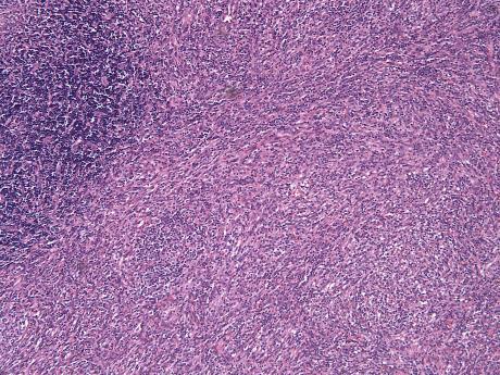
1 Hematopathology Specialty Conference Case #1 Robert (Bob) Ohgami, MD, PhD Assistant Professor Stanford University Disclosure of Relevant Financial Relationships Disclosure of Relevant Financial Relationships USCAP requires that all planners (Education Committee) in a position to influence or control the content of CME disclose any relevant financial relationship WITH COMMERCIAL INTERESTS which they or their spouse/partner have, or have had, within the past 12 months, which relates to the content of this educational activity and creates a conflict of interest. USCAP requires that all faculty in a position to influence or control the content of CME disclose any relevant financial relationship WITH COMMERCIAL INTERESTS which they or their spouse/partner have, or have had, within the past 12 months, which relates to the content of this educational activity and creates a conflict of interest. Dr. Ohgami declares he has no conflicts of interest to disclose. Case history Case history A 32 year old man presents with a 4 month history of isolated left inguinal lymphadenopathy. A physical exam demonstrates no other abnormal findings. The patient has no personal or family history of malignancy. Further workup including CBC, peripheral blood smear and bone marrow biopsy were unremarkable. CBC and Differential WBC: 6.2 K/μL HGB: 14.3 g/dl PLT: 210 K/μL Neutrophils: 65% Lymphocytes: 30% Monocytes: 3% 1


2 Case history A 32 year old man presents with a 4 month history of isolated left inguinal lymphadenopathy. A physical exam demonstrates no other abnormal findings. The patient has no personal or family history of malignancy. Further workup including CBC, peripheral blood smear and bone marrow biopsy were unremarkable. A LN biopsy was performed 2


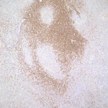
3 CD3 CD4 CD8 CD99 3


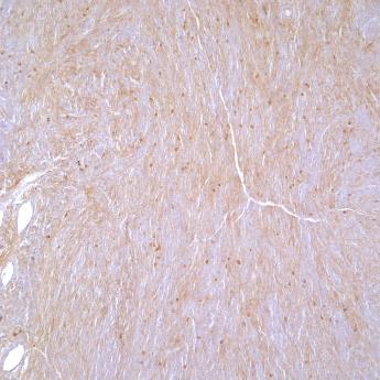


4 What about the atypical spindled cells? CD3+/CD4+/CD8+/CD99+/+ D240 CD123 CD21 Keratin HHV8 4
![Cytogenetic and molecular studies Normal male karyotype: 46,XY[20]. Molecular studies: Negative for T-cell and B- cell clonality.](/docs-images/84/89694264/images/5-2.jpg "Summary Clinical Healthy male Prolonged isolated inguinal adenopathy Morphology Infiltrate of immature blastic lymphoid cells in sheets and clusters Atrophic lollipop follicles with onion skinning")
5 Cytogenetic and molecular studies Normal male karyotype: 46,XY[20]. Molecular studies: Negative for T-cell and B- cell clonality. Summary Clinical Healthy male Prolonged isolated inguinal adenopathy Morphology Infiltrate of immature blastic lymphoid cells in sheets and clusters Atrophic lollipop follicles with onion skinning mantle zones Effacement by a proliferation of atypical spindled cells, some binucleate Immunophenotype Lymphoid infiltrate: CD3+, CD4+, CD8+, CD99+, + Spindled cells: D240+, CD123+, focal dim CD21 Cytogenetics/Molecular Non-clonal T-cells Normal cytogenetics Differential Diagnosis Thymoma T-lymphoblastic lymphoma Kaposi Sarcoma Castleman disease Follicular dendritic cell sarcoma Indolent T-lymphoblastic proliferation Differential Diagnosis No Keratin positive cells Thymoma T-lymphoblastic lymphoma Kaposi Sarcoma Castleman disease Follicular dendritic cell sarcoma Indolent T-lymphoblastic proliferation Differential Diagnosis Differential Diagnosis Thymoma T-lymphoblastic lymphoma No HHV8 positive staining Kaposi Sarcoma Castleman disease Follicular dendritic cell sarcoma Indolent T-lymphoblastic proliferation Thymoma T-lymphoblastic lymphoma Indolent, non-clonal, no marrow involvement Kaposi Sarcoma Castleman disease Follicular dendritic cell sarcoma Indolent T-lymphoblastic proliferation 5

6 Differential Diagnosis Thymoma T-lymphoblastic lymphoma Kaposi Sarcoma Castleman disease Follicular dendritic cell sarcoma Indolent T-lymphoblastic proliferation Final Diagnosis Indolent T-lymphoblastic proliferation Castleman disease, hyaline vascular type Follicular dendritic cell sarcoma Final Diagnosis Indolent T-lymphoblastic proliferation Castleman disease, hyaline vascular type Follicular dendritic cell sarcoma Final Diagnosis Indolent T-lymphoblastic proliferation Castleman disease, hyaline vascular type Follicular dendritic cell sarcoma Castleman disease HIV Castleman disease HHV8 Morphology Clinicopathologic Hyaline Vascular Intermediate Plasma cell Unicentric Multicentric 6
7 Follicular dendritic cell sarcomas Castleman disease Follicular dendritic cell sarcoma Spindled proliferation Plump, sometimes binucleate cells Positive for follicular dendritic cell markers (CD21, CD35, D240, CD23) Lost CD21 Express CD123 Rarely happens But I m here to talk about CD3+/CD4+/CD8+/CD99+/+ perhaps What they ^didn t teach you in fellowship Indolent T-lymphoblastic proliferation perhaps ^ What they didn t teach you in fellowship Indolent T-lymphoblastic proliferations Rules /Dogma can be broken Robert (Bob) Ohgami, MD, PhD Stanford University 7
8 What they didn t teach you in fellowship Indolent T-lymphoblastic Proliferations Indolent T-lymphoblastic proliferations Rules /Dogma can be broken Initial case in 1999 Milind Velankar, Larry Weiss et al. Described a 33 year old patient with a 16 year history of an upper aerodigestive tract proliferation of indolent non-clonal +/CD3+ T-cells H&E TDT Velankar et al., AJSP 1999 Indolent T-lymphoblastic Proliferation Indolent T-lymphoblastic Proliferation First case in 1999: Healthy patient Proliferation of blastic lymphoid cells CD3+/+ T-cells Extra thymic expansion Clinical indolence Non-clonal Similar to Malignant T-lymphoblastic lymphoma First case in 1999: Healthy patient Proliferation of blastic lymphoid cells CD3+/+ T-cells Extra thymic expansion Clinical indolence Non-clonal EXCEPT Velankar et al., AJSP 1999 Velankar et al., AJSP 1999 Since then 13 other cases in the literature Summary of cases of indolent T-lymphoblastic proliferations Age: ~40 (10-70) Male:Female 1:1 Morphology: small-medium sized cells, blastic chromatin Immunophenotype: 100% +/CD4+/CD8+/CD3+ T-cells Molecular: 100% non-clonal Velankar et al., AJSP 1999 Eun et al., J Kor Med Sci 2010 Ohgami et al., AJSP 2012 Qian et al., Leuk Lymph 2009 Strauchen et al., AJSP 2001 Wang et al., Leuk Lymph 2006 Kim et al., Hum Path 2010 Ohgami et al, AJSP 2014 You et al., AJCP 2015 Yang et al., IJCEP 2014 Kansal et al., Hum Path 2015 Woo et a., JPTM 2015 Ohgami et al., Adv Anat Pathol
9 Prethymic CD2/CD5/CD7 CD3 CD34 CD4 CD8 CD1a T-cell development Cytoplasmic Thymus Cortex Double positive Double positive Thymus Medulla Surface Single positive Single positive Summary of cases of indolent T-lymphoblastic proliferations thus far Age: ~40 (10-70) Male:Female 1:1 Morphology: small-medium sized cells, blastic chromatin Immunophenotype: 100% +/CD3+ T-cells Molecular: 100% non-clonal Velankar et al., AJSP 1999 Eun et al., J Kor Med Sci 2010 Ohgami et al., AJSP 2012 Qian et al., Leuk Lymph 2009 Strauchen et al., AJSP 2001 Wang et al., Leuk Lymph 2006 Kim et al., Hum Path 2010 Ohgami et al, AJSP 2014 You et al., AJCP 2015 Yang et al., IJCEP 2014 Kansal et al., Hum Path 2015 Woo et a., JPTM 2015 Ohgami et al., Adv Anat Pathol 2013 Summary of cases of indolent T-lymphoblastic proliferations thus far Age: ~40 (10-70) Male:Female 1:1 Morphology: small-medium sized cells, blastic chromatin Immunophenotype: 100% +/CD3+ T-cells Molecular: 100% non-clonal Associated diseases: Castleman disease, Follicular dendritic cell sarcoma Velankar et al., AJSP 1999 Eun et al., J Kor Med Sci 2010 Ohgami et al., AJSP 2012 Qian et al., Leuk Lymph 2009 Strauchen et al., AJSP 2001 Wang et al., Leuk Lymph 2006 Kim et al., Hum Path 2010 Ohgami et al, AJSP 2014 You et al., AJCP 2015 Yang et al., IJCEP 2014 Kansal et al., Hum Path 2015 Woo et a., JPTM 2015 Ohgami et al., Adv Anat Pathol 2013 Summary of cases of indolent T-lymphoblastic proliferations thus far Age: ~40 (10-70) Male:Female 1:1 Morphology: small-medium sized cells, blastic chromatin Immunophenotype: 100% +/CD3+ T-cells Molecular: 100% non-clonal Associated diseases: Castleman disease, Follicular dendritic cell sarcoma Velankar et al., AJSP 1999 Eun et al., J Kor Med Sci 2010 Ohgami et al., AJSP 2012 Qian et al., Leuk Lymph 2009 Strauchen et al., AJSP 2001 Wang et al., Leuk Lymph 2006 Kim et al., Hum Path 2010 Ohgami et al, AJSP 2014 You et al., AJCP 2015 Yang et al., IJCEP 2014 Kansal et al., Hum Path 2015 Woo et a., JPTM 2015 Ohgami et al., Adv Anat Pathol T-lymphoblastic cells are increased in Castleman disease and FDCS/FDCT + T-lymphoblastic cells are increased in Castleman disease and FDCS/FDCT /CD3+ cells per HPF +/CD3+ cells per HPF Ohgami et al., AJSP 2012 Ohgami et al., AJSP
 1000 +/CD3+ cells per HPF Ohgami et al., AJSP 2012 Ohgami et al.")

10 + T-lymphoblastic cells are increased in Castleman disease and FDCS/FDCT + T-lymphoblastic cells are increased in angioimmunoblastic T-cell lymphoma (AITL) /CD3+ cells per HPF Ohgami et al., AJSP 2012 Ohgami et al., AJSP 2012 Association with CD, FDCS, AITL In 2013 Some have speculated interleukine/cytokines are responsible for cells Il-6 Still not understood Diagnostic Criteria Why are diagnostic criteria needed? Diagnosis Reactive Indolent T-lymphoblastic proliferation Malignant T-lymphoblastic lymphoma 10
 No associated thymic epithelium Clinical")


11 Diagnostic Criteria: it-lbp Major Criteria + T cells in sheets/dense clusters primarily in interfollicular region Preservation of general follicular lymphoid architecture Small-medium sized T cells without significant morphologic atypia No aberrant antigen expression Non-clonal (TCR) No associated thymic epithelium Clinical evidence of indolence Diseases associated with Castleman disease and/or follicular dendritic cell sarcomas/tumors Concurrent angioimmunoblastic T-cell lymphoma (AITL) or history of AITL What they didn t teach you in fellowship Indolent T-lymphoblastic proliferations Rules /Dogma can be broken Ohgami et al., Adv Anat Path 2013 What they didn t teach you in fellowship Case B H&E H&E Indolent T-lymphoblastic proliferations Rules /Dogma can be broken 49 year woman Diffuse lymphadenopathy Healthy otherwise H&E H&E H&E CD3 Ki67 Ohgami et al., AJSP 2014 Expression of other antigens, including CD33 CD33 expression odd H&E H&E CD3 CD1a CD4 CD8 CD33 Ki67 TCR-b TCR-g Ohgami et al., AJSP

12 CD33 expression odd = lymphoma CD33 expression odd = lymphoma? CD33 expression odd Expression can be seen on reactive and highly activated T-cells 4 years later patient is still alive without treatment Diagnosis here: Indolent T-lymphoblastic proliferation with CD33 expression Unusual disseminated disease Since that report a second case with CD33 has been described H&E CD33 Woo et a., JPTM 2015 That s interesting But we re not done yet 12

13 A very recent case Healthy 24 year old male Isolated inguinal adenopathy Non-clonal CD8 CD56 Additional clinical information Clinical indolence: 3 months without progression No marrow or peripheral blood involvement Radiology: SUV of lymph node was very low, minimally increased. What they didn t teach you in fellowship Indolent T-lymphoblastic proliferations Rules /Dogma can be broken What they didn t teach you in fellowship Indolent T-lymphoblastic proliferations Non-clonal Rules /Dogma can be broken CD8 CD56 13


14 Is this malignant? Is this reactive? Non-clonal Prethymic T-cell development Thymus Cortex Thymus Medulla CD2/CD5/CD7 CD3 Cytoplasmic Surface CD8 CD34 CD4 CD8 CD1a Double positive Double positive Single positive Single positive CD56??CD56?? CD56 expression odd CD56 expression odd =?lymphoma? NK/T cell development NK/T cell development Stem cell NK/T cell precursor Committed NK/T cell Stem cell NK/T cell precursor Committed NK/T cell CD2 CD2 CD3 Cytoplasmic CD3 Cytoplasmic CD34 CD34 CD4 CD8 Double positive Double positive Single positive or double negative Single positive or double negative CD4 CD8 Double positive Double positive Single positive or double negative Single positive or double negative CD1a CD1a CD56 Positive or Negative CD56 Positive or Negative 14
15 More information on the case: Followup Many more months, patient is healthy, no disease, no treatment Diagnosis here? Castleman disease, hyaline vascular type Unusual CD56+ lymphoblastic proliferation Koo and Cloetingh et al., in preparation Indolent NK/T-lymphoblastic proliferation Stay tuned Summary Indolent T-lymphoblastic proliferations In the context of science and reason Rules /Dogma can be broken^ Koo and Cloetingh et al., in preparation Questions remain What is driving these proliferations? Do these indolent immature T-cells mature into functional T-cells? Why are these associated with Castleman disease? Follicular dendritic cell sarcomas? Angioimmumnoblastic T-cell lymphoma? Returning to Case 1 What did our panelists think? 15
16 Case 1 - Panelists Diagnoses Acknowledgements Indolent T-lymphoblastic proliferation in the setting of Castleman s Disease TDT+ T lymphoblastic proliferation in Castleman's disease Indolent T lymphoblastic proliferation, Castleman disease and follicular dendritic cell proliferation (recommend more stains to better define follicular dendritic cell proliferation). Stanford University Roger Warnke Yaso Natkunam Michaela Liedtke Susan Atwater Brent Tan Dita Gratzinger Jason Kurzer Colleagues Beyond Mark Fleming, CHB/Harvard Dan Arber, U Chicago Tracy George, UNM Milind Velankar, Loyola U Larry Weiss, Neogenomics 16
3/24/2017 DENDRITIC CELL NEOPLASMS: HISTOLOGY, IMMUNOHISTOCHEMISTRY, AND MOLECULAR GENETICS. Disclosure of Relevant Financial Relationships
 DENDRITIC CELL NEOPLASMS: HISTOLOGY, IMMUNOHISTOCHEMISTRY, AND MOLECULAR GENETICS Jason L. Hornick, M.D., Ph.D. Director of Surgical Pathology and Immunohistochemistry Brigham and Women s Hospital Professor
ACCME/Disclosures 4/13/2016. Clinical History
 ACCME/Disclosures The USCAP requires that anyone in a position to influence or control the content of CME disclose any relevant financial relationship WITH COMMERCIAL INTERESTS which they or their spouse/partner
88-year-old Female with Lymphadenopathy. Faizi Ali, MD
 88-year-old Female with Lymphadenopathy Faizi Ali, MD Clinical History A 88-year-old caucasian female presented to our hospital with the complaints of nausea, vomiting,diarrhea, shortness of breath and
Simon Haefliger. Pathologie. Fallvorstellung
 Simon Haefliger Pathologie Fallvorstellung Castleman s disease Historical perspective Classification Epidemiology and clinical aspects Microscopic caracteristics Pathogenesis Historical perspective First
Follicular Lymphoma: the WHO
 Follicular Lymphoma: the WHO and the WHERE? Yuri Fedoriw, MD Associate Professor of Pathology and Laboratory Medicine Director of Hematopathology University of North Carolina Chapel Hill, NC Disclosure
The spectrum of flow cytometry of the bone marrow
 The spectrum of flow cytometry of the bone marrow Anna Porwit Lund University Faculty of Medicine Dept. of Clinical Sciences Div. Oncology and Pathology anna.porwit@med.lu.se Disclosure of speaker s interests
7 Omar Abu Reesh. Dr. Ahmad Mansour Dr. Ahmad Mansour
 7 Omar Abu Reesh Dr. Ahmad Mansour Dr. Ahmad Mansour -Leukemia: neoplastic leukocytes circulating in the peripheral bloodstream. -Lymphoma: a neoplastic process in the lymph nodes, spleen or other lymphatic
Case Report A case of EBV positive diffuse large B-cell lymphoma of the adolescent
 Int J Clin Exp Med 2014;7(1):307-311 www.ijcem.com /ISSN:1940-5901/IJCEM1311029 Case Report A case of EBV positive diffuse large B-cell lymphoma of the adolescent Qilin Ao 2, Ying Wang 1, Sanpeng Xu 2,
DETERMINATION OF A LYMPHOID PROCESS
 Chapter 2 Applications of Touch Preparation Cytology to Intraoperative Consultations: Lymph Nodes and Extranodal Tissues for Evaluation of Hematolymphoid Disorders INTRODUCTION As discussed in Chap. 1,
Extramedullary precursor T-lymphoblastic transformation of CML at presentation
 Extramedullary precursor T-lymphoblastic transformation of CML at presentation Neerja Vajpayee, Constance Stein, Bernard Poeisz & Robert E. Hutchison Clinical History 30 year old man presented to the emergency
Pathology #07. Hussein Al-Sa di. Dr. Sohaib Al-Khatib. Mature B-Cell Neoplasm. 0 P a g e
 Pathology #07 Mature B-Cell Neoplasm Hussein Al-Sa di Dr. Sohaib Al-Khatib 0 P a g e Thursday 18/2/2016 Our lecture today (with the next 2 lectures) will be about lymphoid tumors This is a little bit long
Hepatic Lymphoma Diagnosis An Algorithmic Approach
 Hepatic Lymphoma Diagnosis An Algorithmic Approach Ryan M. Gill, M.D., Ph.D. University of California, San Francisco PLEASE TURN OFF YOUR CELL PHONES Disclosure of Relevant Financial Relationships USCAP
Case Workshop of Society for Hematopathology and European Association for Haematopathology
 Case 148 2007 Workshop of Society for Hematopathology and European Association for Haematopathology Robert P Hasserjian Department of Pathology Massachusetts General Hospital Boston, MA Clinical history
Pearls and pitfalls in interpretation of lymphoid lesions in needle biopsies
 Pearls and pitfalls in interpretation of lymphoid lesions in needle biopsies Megan S. Lim MD PhD University of Pennsylvania October 8, 2018 Objectives To understand how the trend toward less invasive lymph
Differential diagnosis of hematolymphoid tumors composed of medium-sized cells. Brian Skinnider B.C. Cancer Agency, Vancouver General Hospital
 Differential diagnosis of hematolymphoid tumors composed of medium-sized cells Brian Skinnider B.C. Cancer Agency, Vancouver General Hospital Lymphoma classification Lymphoma diagnosis starts with morphologic
Hematopathology Service Memorial Sloan Kettering Cancer Center, New York
 SH2017-0334 t(14;18) Negative Follicular Lymphoma with 1p36 abnormality associated with In Situ Follicular Neoplasia with t(14;18) translocation Pallavi Khattar MD, Jennifer Maerki MD, Alexander Chan MD,
Mimics of Lymphoma in Routine Biopsies. I have nothing to disclose regarding the information to be reported in this talk.
 Mimics of Lymphoma in Routine Biopsies Patrick Treseler, MD, PhD Professor of Pathology University of California San Francisco I have nothing to disclose regarding the information to be reported in this
Pathology of Hematopoietic and Lymphoid tissue
 Pathology of Hematopoietic and Lymphoid tissue Peerayut Sitthichaiyakul, M.D. Department of Pathology and Forensic Medicine Faculty of Medicine, Naresuan University CONTENTS White blood cells and lymph
Evening specialty conference: Liver
 Evening specialty conference: Liver Joseph Misdraji, M.D. Disclosure of Relevant Financial Relationships Disclosure of Relevant Financial Relationships USCAP requires that all planners (Education Committee)
Immunopathology of Lymphoma
 Immunopathology of Lymphoma Noraidah Masir MBBCh, M.Med (Pathology), D.Phil. Department of Pathology Faculty of Medicine Universiti Kebangsaan Malaysia Lymphoma classification has been challenging to pathologists.
Case Presentation. Maha Akkawi, MD, Fatima Obeidat, MD, Tariq Aladily, MD. Department of Pathology Jordan University Hospital Amman, Jordan
 Case Presentation Maha Akkawi, MD, Fatima Obeidat, MD, Tariq Aladily, MD Department of Pathology Jordan University Hospital Amman, Jordan The 25th Annual Congress of the ADIAP The 8/11/2013 1 5th International
77R (REPEAT) Flow Cytometry: Basic Principles and Case Analysis. Charles Goolsby PhD Kristy Wolniak MD, PhD
 Flow Cytometry: Basic Principles and Case Analysis. Charles Goolsby PhD Kristy Wolniak MD, PhD") 77R (REPEAT) Flow Cytometry: Basic Principles and Case Analysis Charles Goolsby PhD Kristy Wolniak MD, PhD 011 Annual Meeting Las Vegas, NV AMERICAN SOCIETY FOR CLINICAL PATHOLOGY 33 W. Monroe, Ste. 1600
77R (REPEAT) Flow Cytometry: Basic Principles and Case Analysis Charles Goolsby PhD Kristy Wolniak MD, PhD 011 Annual Meeting Las Vegas, NV AMERICAN SOCIETY FOR CLINICAL PATHOLOGY 33 W. Monroe, Ste. 1600
Overview B cell development T cell development
 Topics Overview B cell development T cell development Lymphocyte development overview (Cont) Receptor diversity is produced by gene rearrangement and is random Includes specificities that will bind to
Integrated Diagnostic Approach to the Classification of Myeloid Neoplasms. Daniel A. Arber, MD Stanford University
 Integrated Diagnostic Approach to the Classification of Myeloid Neoplasms Daniel A. Arber, MD Stanford University What is an integrated approach? What is an integrated approach? Incorporating all diagnostic
Classification of Hematologic Malignancies. Patricia Aoun MD MPH
 Classification of Hematologic Malignancies Patricia Aoun MD MPH Objectives Know the basic principles of the current classification system for hematopoietic and lymphoid malignancies Understand the differences
FOLLICULARITY in LYMPHOMA
 FOLLICULARITY in LYMPHOMA Reactive Follicular Hyperplasia Follicular Hyperplasia irregular follicles Follicular Hyperplasia dark and light zones Light Zone Dark Zone Follicular hyperplasia MIB1 Follicular
Contents. vii. Preface... Acknowledgments... v xiii
 Contents Preface... Acknowledgments... v xiii SECTION I 1. Introduction... 3 Knowledge-Based Diagnosis... 4 Systematic Examination of the Lymph Node... 7 Cell Type Identification... 9 Cell Size and Cellularity...
Lymphoma and Pseudolymphoma
 Lymphoma and Pseudolymphoma Laura B. Pincus, MD Co-Director, Cutaneous Lymphoma Clinic Associate Professor Dermatology and Pathology University of California, San Francisco I HAVE NO RELEVANT RELATIONSHIPS
Presentation material is for education purposes only. All rights reserved URMC Radiology Page 1 of 98
 Presentation material is for education purposes only. All rights reserved. 2011 URMC Radiology Page 1 of 98 Radiology / Pathology Conference February 2011 Brooke Koltz, Cytopathology Resident Presentation
PRECURSOR LYMHPOID NEOPLASMS. B lymphoblastic leukaemia/lymphoma T lymphoblastic leukaemia/lymphoma
 PRECURSOR LYMHPOID NEOPLASMS B lymphoblastic leukaemia/lymphoma T lymphoblastic leukaemia/lymphoma B lymphoblastic leukaemia/lymphoma Definition: B lymphoblastic leukaemia/lymphoma is a neoplasm of precursor
3/27/2017. Pulmonary Pathology Specialty Conference. Disclosure of Relevant Financial Relationships. Clinical History:
 Pulmonary Pathology Specialty Conference Saul Suster, M.D. Medical College of Wisconsin Disclosure of Relevant Financial Relationships USCAP requires that all planners (Education Committee) in a position
DiagnosticUtility of Interleukin-6 Expression by Immunohistochemistry in Differentiating Castleman Disease Subtypes and Reactive Lymphadenopathies
 474 Available online at www.annclinlabsci.org Annals of Clinical & Laboratory Science, vol. 46, no. 5, 2016 DiagnosticUtility of Interleukin-6 Expression by Immunohistochemistry in Differentiating Castleman
2007 Workshop of Society for Hematopathology & European Association for Hematopathology Indianapolis, IN, USA Case # 228
 2007 Workshop of Society for Hematopathology & European Association for Hematopathology Indianapolis, IN, USA Case # 228 Vishnu V. B Reddy, MD University of Alabama at Birmingham Birmingham, AL USA 11/03/07
Molecular Pathology of Lymphoma (Part 1) Rex K.H. Au-Yeung Department of Pathology, HKU
 Rex K.H. Au-Yeung Department of Pathology, HKU") Molecular Pathology of Lymphoma (Part 1) Rex K.H. Au-Yeung Department of Pathology, HKU Lecture outline Time 10:00 11:00 11:15 12:10 12:20 13:15 Content Introduction to lymphoma Review of lymphocyte biology
Molecular Pathology of Lymphoma (Part 1) Rex K.H. Au-Yeung Department of Pathology, HKU Lecture outline Time 10:00 11:00 11:15 12:10 12:20 13:15 Content Introduction to lymphoma Review of lymphocyte biology
Mantle Cell Lymphoma
 HEMATOPATHOLOGY Original Article Mantle Cell Lymphoma Morphologic Findings in Bone Marrow Involvement JAY WASMAN, MD, 1 NANCY S. ROSENTHAL, MD,' AND DIANE C. FARHI, MD 2 Although mantle cell lymphoma (MCL),
Reac%ve and Benign Flow Cytometry findings
 Reac%ve and Benign Flow Cytometry findings Lymph nodes and other /ssues Sindhu Cherian, MD University of Washington, Sea
Case 3. Ann T. Moriarty,MD
 Case 3 Ann T. Moriarty,MD Case 3 59 year old male with asymptomatic cervical lymphadenopathy. These images are from a fine needle biopsy of a left cervical lymph node. Image 1 Papanicolaou Stained smear,100x.
From Morphology to Molecular Pathology: A Practical Approach for Cytopathologists Part 1-Cytomorphology. Songlin Zhang, MD, PhD LSUHSC-Shreveport
 From Morphology to Molecular Pathology: A Practical Approach for Cytopathologists Part 1-Cytomorphology Songlin Zhang, MD, PhD LSUHSC-Shreveport I have no Conflict of Interest. FNA on Lymphoproliferative
Many of the hematolymphoid disorders are derived
 REVIEW ARTICLE Practical Immunohistochemistry in Hematopathology: A Review of Useful Antibodies for Diagnosis Ji Lu, MD and Karen L. Chang, MD Abstract: This review article offers some useful panels of
Development of B and T lymphocytes
 Development of B and T lymphocytes What will we discuss today? B-cell development T-cell development B- cell development overview Stem cell In periphery Pro-B cell Pre-B cell Immature B cell Mature B cell
Small B-cell (Histologically Low Grade) Lymphoma
 Lymphoma") Frequency of Lymphoid Neoplasms Small B-cell (Histologically Low Grade) Lymphoma Stephen Hamilton-Dutoit Institute of Pathology Aarhus University Hospital B-cell neoplasms 88% Diffuse large B-cell lymphoma
Frequency of Lymphoid Neoplasms Small B-cell (Histologically Low Grade) Lymphoma Stephen Hamilton-Dutoit Institute of Pathology Aarhus University Hospital B-cell neoplasms 88% Diffuse large B-cell lymphoma
11/8/2018 DISCLOSURES. I have NO Conflicts of Interest to Disclose. UTILTY OF DETECTING PATTERNS
 Bharat N. Nathwani, M.D. City of Hope Medical Center Professor, Director of Pathology Consultation Services, 1500 East Duarte Road, Duarte, California, 91010 DISCLOSURES -------------------------------------------------------
ECP meeting, Lisbon, september 2012 Slide seminar New and old challenges in the diagnosis of peripheral T-cell lymphomas
 ECP meeting, Lisbon, september 2012 Slide seminar New and old challenges in the diagnosis of peripheral T-cell lymphomas Philippe Gaulard, Dept of Pathology, INSERM U955, Hôpital Henri Mondor, 94010 -
Ordering Physician CLIENT,CLIENT. Collected REVISED REPORT
 HPWET Hematopathology Consultation, MML Embed Client Hematopathology Consult REVISED INAL DIAGNOSIS Interpretation Peripheral blood, bone marrow aspirate and biopsies, bilateral iliac crests: 1. Normocellular
Bone Marrow. Procedures Blood Film Aspirate, Cell Block Trephine Biopsy, Touch Imprint
 Bone Marrow Protocol applies to acute leukemias, myelodysplastic syndromes, myeloproliferative disorders, chronic lymphoproliferative disorders, malignant lymphomas, plasma cell dyscrasias, histiocytic
Mimics of Lymphoma in Routine Biopsies. Mixed follicular and paracortical hyperplasia. Types of Lymphoid Hyperplasia
 Mimics of Lymphoma in Routine Biopsies Patrick Treseler, MD, PhD Professor of Pathology University of California San Francisco Types of Lymphoid Hyperplasia Follicular hyperplasia (B-cells) Paracortical
NON HODGKINS LYMPHOMA: INDOLENT Updated June 2015 by Dr. Manna (PGY-5 Medical Oncology Resident, University of Calgary)
") NON HODGKINS LYMPHOMA: INDOLENT Updated June 2015 by Dr. Manna (PGY-5 Medical Oncology Resident, University of Calgary) Reviewed by Dr. Michelle Geddes (Staff Hematologist, University of Calgary) and Dr.
NON HODGKINS LYMPHOMA: INDOLENT Updated June 2015 by Dr. Manna (PGY-5 Medical Oncology Resident, University of Calgary) Reviewed by Dr. Michelle Geddes (Staff Hematologist, University of Calgary) and Dr.
ADx Bone Marrow Report. Patient Information Referring Physician Specimen Information
 ADx Bone Marrow Report Patient Information Referring Physician Specimen Information Patient Name: Specimen: Bone Marrow Site: Left iliac Physician: Accession #: ID#: Reported: 08/19/2014 - CHRONIC MYELOGENOUS
Osteosclerotic Myeloma (POEMS Syndrome)
") Osteosclerotic Myeloma (POEMS Syndrome) Osteosclerotic Myeloma (POEMS Syndrome) Synonyms Crow-Fukase syndrome Multicentric Castleman disease Takatsuki syndrome Acronym coined by Bardwick POEMS Scheinker,
Osteosclerotic Myeloma (POEMS Syndrome) Osteosclerotic Myeloma (POEMS Syndrome) Synonyms Crow-Fukase syndrome Multicentric Castleman disease Takatsuki syndrome Acronym coined by Bardwick POEMS Scheinker,
Myelodysplastic Syndrome Case 158
 Myelodysplastic Syndrome Case 158 Dong Chen MD PhD Division of Hematopathology Mayo Clinic Clinical History 86 year old man Persistent borderline anemia and thrombocytopenia. His past medical history was
The development of clonality testing for lymphomas in the Bristol Genetics Laboratory. Dr Paula Waits Bristol Genetics Laboratory
 The development of clonality testing for lymphomas in the Bristol Genetics Laboratory Dr Paula Waits Bristol Genetics Laboratory Introduction The majority of lymphoid malignancies belong to the B cell
Lymphoma: What You Need to Know. Richard van der Jagt MD, FRCPC
 Lymphoma: What You Need to Know Richard van der Jagt MD, FRCPC Overview Concepts, classification, biology Epidemiology Clinical presentation Diagnosis Staging Three important types of lymphoma Conceptualizing
T cell lymphoma diagnostics and differential diagnosis to Hodgkin lymphoma
 T cell lymphoma diagnostics and differential diagnosis to Hodgkin lymphoma Sylvia Hartmann Dr. Senckenberg Institute of Pathology Goethe University Frankfurt Overview Borderline ALCL classical HL Borderline
Persistent lymphocytosis. Persistent lymphocytosis: are there prognostic indicators? Problem. Questions. Basic markers used to identify lymphocytes
 Persistent lymphocytosis Persistent lymphocytosis: are there prognostic indicators? Paul R. Avery VMD, PhD, DACVP Marjorie Williams, DVM Anne C. Avery VMD, PhD Clinical Immunology Laboratory Colorado State
5000 International Clinical Cytometry Society: Practical Flow Cytometry in Hematopathology A Case-Based Approach
 5000 International Clinical Cytometry Society: Practical Flow Cytometry in Hematopathology A Case-Based Approach Joseph A DiGiuseppe, MD, PhD Hartford Hospital Disclosures In the past 12 months, I have
WBCs Disorders 1. Dr. Nabila Hamdi MD, PhD
 WBCs Disorders 1 Dr. Nabila Hamdi MD, PhD ILOs Compare and contrast ALL, AML, CLL, CML in terms of age distribution, cytogenetics, morphology, immunophenotyping, laboratory diagnosis clinical features
Case Report Angioimmunoblastic T-Cell Lymphoma: A Questionable Association with Follicular Dendritic Cell Sarcoma
 Hindawi Case Reports in Hematology Volume 2017, Article ID 9601094, 4 pages https://doi.org/10.1155/2017/9601094 Case Report Angioimmunoblastic T-Cell Lymphoma: A Questionable Association with Follicular
Lymphoma Read with the experts
 Lymphoma Read with the experts Marc Seltzer, MD Associate Professor of Radiology Geisel School of Medicine at Dartmouth Director, PET-CT Course American College of Radiology Learning Objectives Recognize
The patient had a mild splenomegaly but no obvious lymph node enlargement. The consensus phenotype obtained from part one of the exercise was:
 Case History An 86 year old male was admitted to hospital with chest infection. Haematological examination subsequently revealed the following: Hb- 11.0 g/dl; WBC- 67.1 x 10^9/l; PLT- 99 x10^9/l; RBC-
Pathology. #11 Acute Leukemias. Farah Banyhany. Dr. Sohaib Al- Khatib 23/2/16
 35 Pathology #11 Acute Leukemias Farah Banyhany Dr. Sohaib Al- Khatib 23/2/16 1 Salam First of all, this tafreegh is NOT as long as you may think. If you just focus while studying this, everything will
Hematology Unit Lab 2 Review Material
 Objectives Hematology Unit Lab 2 Review Material - 2018 Laboratory Instructors: 1. Assist students during lab session Students: 1. Review the introductory material 2. Study the case histories provided
T CELL LYMPHOMA ANALYSIS
 T CELL LYMPHOMA ANALYSIS Charles Goolsby, Ph.D. Floyd E. Patterson Research Professor of Pathology Northwestern Feinberg School of Medicine c-goolsby@northwestern.edu 1 T CELL LYMPHOMA ANALYSIS Diverse
3/23/2017. Disclosure of Relevant Financial Relationships. Pitfalls in Immunohistochemistry in Hematopathology: CD20 and CD3 Can Let Me Down?!
 Pitfalls in Immunohistochemistry in Hematopathology: CD20 and CD3 Can Let Me Down?! Judith A. Ferry Massachusetts General Hospital Disclosure of Relevant Financial Relationships USCAP requires that all
Integrated Hematopathology. Morphology and FCI with IHC
 Integrated Hematopathology Morphology and FCI with IHC FrontMatter.indd i 9/6/2009 9:30:12 PM FrontMatter.indd ii 9/6/2009 9:30:18 PM Integrated Hematopathology Morphology and FCI with IHC Cherie H Dunphy,
V. Acute leukemia. Flow cytometry in evaluation of hematopoietic neoplasms: A case-based approach
 V. Acute leukemia Evaluating a sample for an acute leukemia Acute leukemia is a neoplasm of immature myeloid or lymphoid cells characterized by a block in maturation, usually at the stage of an early progenitor
Classifications of lymphomas
 Classifications of lymphomas Lukes and Collins Kiel classification Working formulation REAL classification (1994) WHO classification (2000) WHO CLASSIFICATIONF OF NEOPLASMS HAEMATOPETIC AND LYMPHOID TISSUES
Case year old male with abdominal lymphadenopathy Treated with 8 cycles of R-CHOP One year later B-symptoms and progressive disease
 Codirectors Tsieh Sun, M.D., FASCP Francisco Vega, M.D., Ph.D. Department of Hematopathology UT MD Anderson Cancer Center Houston Texas There is no conflict of interest involved in the content and presentation
Combinations of morphology codes of haematological malignancies (HM) referring to the same tumour or to a potential transformation
 referring to the same tumour or to a potential transformation") Major subgroups according to the World Health Organisation (WHO) Classification Myeloproliferative neoplasms (MPN) Myeloid and lymphoid neoplasms with eosinophilia and abnormalities of PDGFRA, PDGFRB or
Major subgroups according to the World Health Organisation (WHO) Classification Myeloproliferative neoplasms (MPN) Myeloid and lymphoid neoplasms with eosinophilia and abnormalities of PDGFRA, PDGFRB or
Gray Zones and Double Hits Distinguishing True Burkitt Lymphoma from Other High-Grade B-NHLs Burkitt Lymphoma Burkitt-Like Lymphoma DLBCL Patrick Tres
 Gray Zones and Double Hits Distinguishing True Burkitt Lymphoma from Other High-Grade B-NHLs Burkitt Lymphoma Burkitt-Like Lymphoma DLBCL Patrick Treseler, MD, PhD University of California San Francisco
Lymphoma Update: Lymphoma Update: What s Likely to be New in the New WHO. Patrick Treseler, MD, PhD University of California San Francisco
 Lymphoma Update: What s Likely to be New in the New WHO Blood 127:2375; 2016 Patrick Treseler, MD, PhD University of California San Francisco Lymphoma Update: What IS New in the New WHO! Patrick Treseler,
Beyond the CBC Report: Extended Laboratory Testing in the Evaluation for Hematologic Neoplasia Disclosure
 Beyond the CBC Report: Extended Laboratory Testing in the Evaluation for Hematologic Neoplasia Disclosure I am receiving an honorarium from Sysmex for today s presentation. 1 Determining the Etiology for
HENATOLYMPHOID SYSTEM THIRD YEAR MEDICAL STUDENTS- UNIVERSITY OF JORDAN AHMAD T. MANSOUR, MD. Parts 2 and 3
 HENATOLYMPHOID SYSTEM THIRD YEAR MEDICAL STUDENTS- UNIVERSITY OF JORDAN AHMAD T. MANSOUR, MD Parts 2 and 3 NEOPLASTIC LYMPHOID DISEASES Introduction o The bone marrow is the source of all cells in the
3/22/2017. Disclosure of Relevant Financial Relationships. Disclosure of Relevant Financial Relationships. Grading G1. Grading. Ki67 index V.
 Disclosure of Relevant Financial Relationships USCAP requires that all planners (Education Committee) in a position to influence or control the content of CME disclose any relevant financial relationship
A Practical Guide To Diagnose B-Cell Lymphomas on FNAs. Nancy P. Caraway, M.D.
 A Practical Guide To Diagnose B-Cell Lymphomas on FNAs Nancy P. Caraway, M.D. Major Factors Impacting Dx Lymphomas on Small Bxs Classification systems Immunophenotyping by multiprobe flow cytometry and
Leukocytosis - Some Learning Points
 Leukocytosis - Some Learning Points Koh Liang Piu Department of Hematology-Oncology National University Cancer Institute National University Health System Objectives of this talk: 1. To provide some useful
Done By : WESSEN ADNAN BUTHAINAH AL-MASAEED
 Done By : WESSEN ADNAN BUTHAINAH AL-MASAEED Acute Myeloid Leukemia Firstly we ll start with this introduction then enter the title of the lecture, so be ready and let s begin by the name of Allah : We
HEMATOPATHOLOGY (SHANDS HOSPITAL AT THE UNIVERSITY OF FLORIDA): Rotation Director: Ying Li, M.D., Ph.D., Assistant Professor
: Rotation Director: Ying Li, M.D., Ph.D., Assistant Professor") HEMATOPATHOLOGY (SHANDS HOSPITAL AT THE UNIVERSITY OF FLORIDA): Rotation Director: Ying Li, M.D., Ph.D., Assistant Professor I. Description of the rotation: During this rotation, the resident will gain
HEMATOPATHOLOGY (SHANDS HOSPITAL AT THE UNIVERSITY OF FLORIDA): Rotation Director: Ying Li, M.D., Ph.D., Assistant Professor I. Description of the rotation: During this rotation, the resident will gain
2013 AAIM Pathology Workshop
 2013 AAIM Pathology Workshop John Schmieg, M.D., Ph.D. None Disclosures 1 Pathology Workshop Objectives Define the general philosophy of reviewing pathology reports Review the various components of Bone
Reactive and Neoplastic Lymphocytosis
 Reactive and Neoplastic Lymphocytosis Koranda A. Walsh, VMD, BS Assistant Professor, Clinical Pathobiology University of Pennsylvania School of Veterinary Medicine PLEASE NOTE: These notes are meant as
Immunohistochemical classification of haematolymphoid tumours. Stephen Hamilton-Dutoit Institute of Pathology Aarhus University Hospital
 Immunohistochemical classification of haematolymphoid tumours Stephen Hamilton-Dutoit Institute of Pathology Aarhus University Hospital Malignant lymphoproliferative diseases What are they? Haematolymphoid
Thomas Hodgkin and Hodgkin lymphoma
 J Hematopathol (2014) 7:123 138 DOI 10.1007/s12308-014-0214-3 REVIEW ARTICLE Thomas Hodgkin and Hodgkin lymphoma Judith A. Ferry Received: 26 June 2014 /Accepted: 31 July 2014 /Published online: 12 August
Conjunctival CD5+ MALT lymphoma and review of literatures
 ISPUB.COM The Internet Journal of Pathology Volume 8 Number 2 Conjunctival CD5+ MALT lymphoma and review of literatures M Fard Citation M Fard. Conjunctival CD5+ MALT lymphoma and review of literatures.
MECHANISMS OF HUMAN DISEASE: LABORATORY SESSIONS LYMPHOMA. April 16, 2008
 MECHANISMS OF HUMAN DISEASE: LABORATORY SESSIONS LYMPHOMA April 16, 2008 FACULTY COPY GOAL: Learn the appearance of normal peripheral blood elements and lymph nodes. Recognize abnormal peripheral blood
2010 Hematopoietic and Lymphoid ICD-O Codes - Alphabetical List THIS TABLE REPLACES ALL ICD-O-3 Codes
 Acute basophilic leukemia 9870/3 Acute biphenotypic leukemia [OBS] 9805/3 Acute erythroid leukemia 9840/3 Acute megakaryoblastic leukemia 9910/3 Acute monoblastic and monocytic leukemia 9891/3 Acute myeloid
2012 Hematopoietic and Lymphoid ICD-O Codes - Numerical List THIS TABLE REPLACES ALL ICD-O-3 Codes
 Malignant lymphoma, NOS 9590/3 Non-Hodgkin lymphoma, NOS 9591/3 B-cell lymphoma, unclassifiable, with features intermediate between diffuse large B-cell lymphoma and classical Hodgkin lymphoma 9596/3 Primary
Case Workshop of Society for Hematopathology and European Association for Haematopathology
 Case 24 2007 Workshop of Society for Hematopathology and European Association for Haematopathology Aliyah Rahemtullah 1, Martin K Selig 1, Paola Dal Cin 2 and Robert P Hasserjian 1 Departments of Pathology,
Participants Identification No. % Evaluation
 BMD-02 Cell Identification Participants Identification No. % Evaluation Erythrocyte precursor, normal 228 95.8 Educational Erythrocyte, normal 7 3.0 Educational Erythrocyte precursor with megaloblastic
Chronic Lymphocytic Leukemia FISH Panel. Impact on Diagnosis
 Hematopathology / CLL, FISH, AND 14Q32 TRANSLOCATIONS Chronic Lymphocytic Leukemia FISH Panel Impact on Diagnosis Beverly P. Nelson, MD, 1 Rohit Gupta, MD, 1 Gordon W. Dewald, PhD, 2 Sarah F. Paternoster,
Disclosure of Relevant Financial Relationships
 Squamous entities of the thyroid: Reactive to Neoplastic Michelle D. Williams Associate Professor Dept of Pathology, Head & Neck Section University of Texas MD Anderson Cancer Center Disclosure of Relevant
Disclosure of Relevant Financial Relationships
 Evening Specialty Conference - Genitourinary Pathology Case 2 Disclosure of Relevant Financial Relationships Sean R Williamson, MD Henry Ford Health System, Detroit, MI @Williamson_SR USCAP requires that
WBCs Disorders. Dr. Nabila Hamdi MD, PhD
 WBCs Disorders Dr. Nabila Hamdi MD, PhD ILOs Compare and contrast ALL, AML, CLL, CML in terms of age distribution, cytogenetics, morphology, immunophenotyping, laboratory diagnosis clinical features and
Lymphocytoma Cutis. Cynthia M. Magro MD. Director of Dermatopathology Weill Medical College of Cornell University New York, New York
 Lymphocytoma Cutis Cynthia M. Magro MD Professor of Pathology Director of Dermatopathology Weill Medical College of Cornell University New York, New York Lymphocytoma Cutis Falls under other designations
Adaptive immune responses: T cell-mediated immunity
 MICR2209 Adaptive immune responses: T cell-mediated immunity Dr Allison Imrie allison.imrie@uwa.edu.au 1 Synopsis: In this lecture we will discuss the T-cell mediated immune response, how it is activated,
3/28/2017. Disclosure of Relevant Financial Relationships. GU Evening Subspecialty Case Conference. Differential Diagnosis:
 GU Evening Subspecialty Case Conference Rajal B. Shah, M.D. VP, Medical Director, Urologic Pathology Miraca Life Sciences, Irving, Texas Clinical Associate Professor of Pathology Baylor College of Medicine,
Hematopoiesis. Hematopoiesis. Hematopoiesis
 Chapter. Cells and Organs of the Immune System Hematopoiesis Hematopoiesis- formation and development of WBC and RBC bone marrow. Hematopoietic stem cell- give rise to any blood cells (constant number,
Large cell immunoblastic Diffuse histiocytic (DHL) Lymphoblastic lymphoma Diffuse lymphoblastic Small non cleaved cell Burkitt s Non- Burkitt s
 Lymphoblastic lymphoma Diffuse lymphoblastic Small non cleaved cell Burkitt s Non- Burkitt s") Non Hodgkin s Lymphoma Introduction 6th most common cause of cancer death in United States. Increasing in incidence and mortality. Since 1970, the incidence of has almost doubled. Overview The types of
Non Hodgkin s Lymphoma Introduction 6th most common cause of cancer death in United States. Increasing in incidence and mortality. Since 1970, the incidence of has almost doubled. Overview The types of
Classification of Cutaneous T cell Lymphomas (CTCLs) Hernani Cualing, MD
 Hernani Cualing, MD") Classification of Cutaneous T cell Lymphomas (CTCLs) Hernani Cualing, MD Pathology and Cell Biology, USF IFLOW, Inc. CTCL, MF, and Sézary syndrome In 1806, mycosis fungoides (MF) was first described 1
Classification of Cutaneous T cell Lymphomas (CTCLs) Hernani Cualing, MD Pathology and Cell Biology, USF IFLOW, Inc. CTCL, MF, and Sézary syndrome In 1806, mycosis fungoides (MF) was first described 1
SH/EAHP WORKSHOP 2017 CASE 210 PRESENTATION
 SH/EAHP WORKSHOP 2017 CASE 210 PRESENTATION Jonathon H Gralewski DO, MS, Ginell R Post MD, PhD, Youzhong Yuan MD September 9, 2017 Clinical History 60 year old male with history of c-maf high-risk IgG