THE ROLE OF SEMINAL VESICLE SECRETION (SVS) 7 PROTEIN IN BONE HOMEOSTASIS
|
|
|
- Jayson Walton
- 5 years ago
- Views:
Transcription
1 THE ROLE OF SEMINAL VESICLE SECRETION (SVS) 7 PROTEIN IN BONE HOMEOSTASIS WILLIAM CUNDAWAN, B. Sc B. MedSci (Hons) This thesis is submitted to fulfil the requirements of the University of Western Australia for the degree of DOCTOR OF PHILOSOPHY 2013 The work presented in this thesis was performed at the University of Western Australia, School of Pathology and Laboratory Medicine and School of Surgery, Centre for Orthopaedic Research, QEII Medical Centre, Nedlands, Western Australia
2 Table of Contents Declaration 1 Scientific Abstract 2 Abstracts and Award 6 Acknowledgements 7 List of Figures 9 Abbreviations 12 CHAPTER 1: BONE BIOLOGY Introduction to Bone Biology Osteoclast Osteoclast phenotype Osteoclast differentiation and function Osteoblast Osteoblast phenotype Osteoblast differentiation and function Molecular Communication Between Osteoclasts and 37 Osteoblasts Osteoblast regulation of the osteoclast Osteoclast regulation of the osteoblast Bidirectional signalling between osteoclasts and 44 osteoblasts 1.4 Bone Diseases Osteoporosis Osteopetrosis Paget s disease of bone (PDB) 48 CHAPTER 2: SEMINAL VESICLE SECRETION (SVS) 7 PROTEIN AND 50 LY-6 PROTEIN FAMILY 2.1 Introduction Seminal vesicle secretion (Svs) Svs7 as a secreted member of Ly-6 protein family and the 52 biological importance of other Ly-6 proteins 2.4 Identification of Ly-6 family member, Svs7, as a putative 57 osteoclast-derived coupling factor CHAPTER 3: HYPOTHESIS AND AIMS Hypothesis 62
3 3.2 Aims 62 CHAPTER 4: MATERIALS AND METHODS Materials Cell lines Bacterial strain Chemical reagents Tissue culture reagents Cytokines Enzymes Antibodies Vectors Purchased kits and systems Oligonucleotide primers Other materials Software Equipments Centrifugation Buffers and Solutions General solutions Media and agar Animals Svs7 transgenic (TG) mice generation Svs7 knockout (KO) mice generation Mouse tail tip DNA genotyping MicroCT analysis Histology on hindlimbs and prostate of Svs7 TG and KO 81 mice Fixation, decalcification (for hindlimbs), tissue 81 processing, tissue embedding, and sectioning Dewaxing and H&E staining Dewaxing and TRAP staining Processing, embedding, and sectioning of non- 83 decalcified bone samples Goldner trichrome staining for the non- 84 decalcified bone samples
4 4.3.6 Double calcein labelling of Svs7 KO mice General Methods Isolation and culture of primary cells Isolation and culture of bone marrow cells Isolation and culture of spleen cells Isolation and culture of calvarial osteoblasts Cell culture of COS-7 cell line Cryopreservation of cell lines for long-term storage RNA extraction RNA concentration measurement RT reactions Polymerase chain reaction (PCR) and quantitative 89 PCR (qpcr) DNA agarose gel electrophoresis Cell Biology In vitro RANKL-induced osteoclastogenesis, TRAP 93 staining, and osteoclasts count Analysis of osteoclast nuclei and area (osteoclast fusion 93 mechanism) In vitro osteoclast bone resorption assay Preparation of bone slices Mature osteoclasts culture on bone slices SEM and analysis of osteoclast resorption pits Osteoclast bone resorption assays using Corning 95 Osteo Assay plates CFU alkaline phosphatase (ALP) staining for osteoblast 96 differentiation assay In vitro bone marrow stromal cells-derived osteoblasts bone 96 nodule formation (mineralization) assay MTS proliferation assay on Svs7 KO BMM Cloning Method Restriction enzyme digestion of DNA DNA extraction and purification from agarose gels Cloning strategy and ligation to construct 98 pgem-t Easy Svs7
5 4.6.4 Cloning strategy and ligation to construct 98 pcdna3.1 Svs7-V5 His Transformation and plating of the transformed cells Bacterial culture and maintenance Plasmid extraction and isolation Experimental Methods Cell transfection and collection of conditioned medium 99 expressing Svs7 protein Luciferase assay Flow cytometry analysis on fixed and stained Svs7 KO BM 100 cells Fluo-4AM intracellular calcium assay on Svs7 KO osteoclast Cell lysis procedure for Western Blot (WB) protein analysis Velocity density gradient centrifugation Measuring protein concentration Western Blot (WB) SDS-PAGE system Preparation of the SDS-PAGE system Protein transfer to nitrocellulose membrane WB reaction Stripping the membranes Statistical Analysis 106 CHAPTER 5: CHARACTERISATION OF SVS7 TRANSGENIC MICE Introduction Results Tissue distribution of Svs7 gene Generation of the Svs7 transgenic (TG) mice Histological phenotyping of Svs7 TG mice Body weight and radiographic assessment of Svs7 TG mice MicroCT analysis of tibia, femur, and lumbar vertebrae of 112 Svs7 TG mice Svs7 TG mice exhibit a normal bone mass Histological assessment of Svs7 TG bones Svs7 TG mice exhibit an unaffected osteoclast differentiation 121 in vitro 5.3 Discussion 121
6 CHAPTER 6: CHARACTERISATION OF SVS7 KNOCKOUT MICE Introduction Results Genotyping and characterisation of Svs7 KO mice Slow mice breeding process and gross body phenotypic 131 examination of Svs7 KO mice Gross tissues phenotypic (macro vs. microscopic) 131 assessment of Svs7 KO mice Svs7 KO mice exhibit a reduced bone mass phenotype Svs7 KO bone histology Bone formation activity in vivo is unaffected in Svs7 KO 139 mice 6.3 Discussion 139 CHAPTER 7: INVESTIGATION OF INTRINSIC CELLULAR 144 MECHANISM IN SVS7 KNOCKOUT MICE (OSTEOCLAST AND OSTEOBLAST) 7.1 Introduction Results Part 1: Intrinsic cellular mechanism in osteoclasts 146 derived from Svs7 KO mice Svs7 KO BMM cultures have increased osteoclastogenesis 146 in vitro Svs7 KO osteoclasts are larger and have more nuclei than 146 WT WT and Svs7 KO mice have comparable hematopoietic 149 stem cells (HSCs) and osteoclast progenitor cells Svs7 KO mice exhibit unaltered expression levels of key 152 osteoclast marker genes Osteoclast bone resorption activity is not affected in Svs7 152 KO mice Svs7 KO osteoclasts are more sensitive to Ca 2+ influx in 157 response to RANKL and extracellular Ca 2+ stimulation Svs7 KO mice exhibit elevated NFATc1 and ATP6v0d2 160 proteins expression during RANKL-induced osteoclastogenesis Svs7 protein treatment inhibits the activation of NFAT 164 promoter luciferase activity
7 7.3 Results Part 2: Osteoblasts from Svs7 KO mice are 166 phenotypically comparable to WT cells Svs7 KO primary calvarial osteoblasts exhibit an 166 unaffected proliferation rate in vitro No defect is observed in Svs7 KO alkaline phosphatase 166 (ALP) osteoblast differentiation in vitro Svs7 KO osteoblasts exhibit an unaffected bone formation 169 activity in vitro Analysis of osteoblast marker genes expression levels in 169 Svs7 KO mice 7.4 Discussion 172 CHAPTER 8: GENERAL SUMMARY AND FUTURE DIRECTIONS Overview of thesis and general discussion Future directions The role of Svs7 in bone homeostasis The role of Svs7 in osteoclast and osteoblast differentiation 185 and activation CHAPTER 9: BIBLIOGRAPHY 188 APPENDIX 223
8 Declaration This is to certify that all the work contained herein was performed by myself, except where indicated otherwise. William Cundawan W/Prof Jiake Xu Assoc Prof Nathan Pavlos W/Prof Minghao Zheng
9 Scientific Abstract Bone, a living tissue that forms our skeletal frame, is continuously remodelled throughout life by the complementary activities of bone-resorbing osteoclasts and boneforming osteoblasts. The cell-to-cell interaction between osteoclasts and osteoblasts is tightly regulated to achieve bone homeostasis and is governed by the production and exchange of various coupling factors. However, the nature and identity of these factors are still largely obscure. In search for novel osteoclast-derived coupling factors, we have previously employed subtractive hybridization-based differential screening to probe for differentially expressed genes between osteoclasts and their mononuclear progenitors. Using this approach, we have successfully identified seminal vesicle secretion (Svs) 7, known as calcium (Ca 2+ ) transport inhibitor (Caltrin), as a candidate gene that was robustly upregulated during receptor activator of NF-κB ligand (RANKL)-induced bone marrow macrophages (BMM) differentiation into mature osteoclasts (Davey 2008; Phan 2004). Svs7 is exclusively expressed by osteoclasts in bone and liberated as a secreted factor. Svs7 is a secreted member of Lymphocyte antigen (Ly)-6 protein family, sharing the common three fingered proteins (TFP)/Ly-6/urokinase-type plasminogen activator receptor (upar) motif, has been shown to be involved in several cellular signalling pathways. Furthermore, previous observations by Davey 2008 describe the protective effects of recombinant purified Svs7 protein (500μg/kg daily injection) against the loss of trabecular bone volume, trabecular thickness, and trabecular number at the distal femoral metaphysis of ovariectomised mice (oestrogen deficiency-induced bone loss mouse model). From here, it was hypothesized that Svs7 might be an important regulator of bone homeostasis. To address this, a two-armed approach was employed using genetically modified mouse models. The first approach involved the generation and characterisation of Svs7 transgenic (TG) mice. Following the confirmation that Svs7 transgene was successfully incorporated and expressed at both the transcriptional and protein levels, histological assessment of tissues, known to abundantly express Svs7 (i.e. prostate), from the TG mice was performed. Despite being successfully expressed, no obvious morphological difference was observed in these prostate tissues between WT and Svs7 TG mice. To further explore the potential role of Svs7 in bone, X-ray analysis was performed. The result showed that there was no striking difference in skeletons between WT and Svs7 TG mice. More precise Svs7 TG bone phenotypic examination was conducted by performing microct and histology analysis on 2
10 calvariae, proximal tibia, distal femur, and vertebral body of the lumbar bone. MicroCT analysis together with histological assessment revealed that all major bone parameters (trabecular bone volume, trabecular separation, trabecular thickness, and trabecular number) were comparable between WT and Svs7 TG mice. Moreover, tartrate-resistant acid phosphatase (TRAP) histology sections and histomorphometry data further confirmed that there was no significant difference in the number of TRAP-positive osteoclasts in the primary epiphysis between WT and Svs7 TG hindlimbs, implying that Svs7 TG mice exhibit normal bone mass and structure. As Svs7 is exclusively expressed in osteoclasts in bone, osteoclast formation was assessed by performing in vitro time course of RANKL-induced osteoclastogenesis. The results indicated that osteoclast formation in Svs7 TG mice was comparable to that of WT littermates. Thus, Svs7 overexpression in Svs7 TG mice was insufficient to determine the physiological role of Svs7 in bone. Another approach to investigate the potential role of Svs7 in bone involved the generation and characterisation of Svs7 global knockout (KO) mice. The deletion of exon 2 and 3 of Svs7 gene, which encode for the TFP/Ly-6/uPAR domain, led to the complete absence of Svs7 gene and protein in Svs7 KO mice. Despite an unexpectedly slow mice breeding process, Svs7 KO mice displayed no overt gross body phenotype. In addition, no obvious difference was observed in the phenotype of major organs and tissues between WT and Svs7 KO mice, indicating that Svs7 is not a requirement for organogenesis. To assess the potential bone phenotype of Svs7 KO mice, microct analysis on WT and Svs7 KO femurs was performed. The results showed that there were significant reductions in trabecular bone volume and trabecular number in Svs7 KO mice compared to WT, suggesting that Svs7 KO mice exhibit a phenotype of osteopenia/osteoporosis. Furthermore, this reduced bone mass phenotype was also confirmed histologically in sections of Svs7 KO hindlimbs. TRAP histology sections and histomorphometry data revealed that there was a trend of increase in osteoclast number near growth plate region in Svs7 KO hindlimbs compared to WT, while no significant difference in bone formation activity in vivo was observed between WT and KO mice. Together, these results indicate that Svs7 is a novel regulator of bone homeostasis, possibly owing to a defect in osteoclast differentiation observed in Svs7 KO mice. 3
11 To elucidate the intrinsic cellular mechanism underlying the osteoporotic phenotype of Svs7 KO mice, a series of in vitro assays for osteoclast and osteoblast differentiation and activity were performed. In vitro time course of RANKL-induced osteoclastogenesis showed that there was a significant increase in osteoclast number in Svs7 KO BMM cultures at day 3 and 5 of osteoclastogenesis compared to WT. Interestingly, relative to WT, Svs7 KO-derived osteoclasts were significantly larger and possessed more nuclei (i.e. 11) than WT at day 5 of osteoclastogenesis, while no detectable defects in Svs7 KO early osteoclast progenitor and hematopoietic stem cell populations were observed. Despite the increase in osteoclast number and size, there was no significant difference in osteoclast bone resorption activity or the expression of known osteoclast marker genes between WT and Svs7 KO mice, indicating that Svs7 has a potential intrinsic role in osteoclast differentiation but not in osteoclast activity. By performing Fluo-4AM intracellular Ca 2+ assays on WT and Svs7 KO osteoclasts, it was found that KO osteoclasts were more sensitive to Ca 2+ influx than WT in response to RANKL and extracellular Ca 2+ stimulation, implying that, together with the observed larger osteoclast size in Svs7 KO BMM cultures, Svs7 may function as a regulator of osteoclast fusion. Consistent with this position, western blot analysis on cell lysates of time course RANKL-induced WT and Svs7 KO BMM differentiation into osteoclasts showed a significant elevation in nuclear factor-activated T cell cytoplasmic 1 (NFATc1) (at day 3 of osteoclast differentiation) and a trend of increase in d2 subunit of v0 domain of V-H + ATPase complex (ATP6v0d2) fusogenic protein expression in KO osteoclasts compared to WT, indicating that Svs7 may regulate osteoclast fusion through the Ca 2+ -NFATc1 signaling pathway, which has its main effect on ATP6v0d2 protein expression. On the other hand, no significant differences were observed in osteoblast differentiation and mineralization activity in vitro between WT and Svs7 KO mice. In conclusion, these studies constitute the first comprehensive genetic approach to study the involvement of Svs7 in bone. Moreover, these studies demonstrate that Svs7 KO mice exhibit an osteoporotic phenotype, with a significant increase in osteoclast number and size and no observable defects in osteoblast differentiation and activity in vitro and in vivo. Through the Ca 2+ -NFATc1 signaling pathway, Svs7 has shown to regulate osteoclast differentiation and fusion but not osteoclast activity. Therefore, Svs7 plays an important regulatory role in the maintenance of bone homeostasis, via an autocrine 4
12 action on osteoclasts, and thus might represent a potential target for the management and treatment of osteoclast-related bone diseases. 5
13 Abstracts and Award 1. Cundawan W, Tickner J, Abel T, Chim SM, Kular J, Zheng MH, Pavlos N, and Xu J (2012) Seminal Vesicle Secretion (SVS) VII, upregulated by RANKL in Mature Osteoclasts, Regulates Osteoclast Precursor Proliferation, Differentiation, and Bone Homeostasis. Australian and Scientific Medical Research (ASMR) Week Symposium, Perth, Western Australia. (Oral Presentation) 2. Cundawan W, Tickner J, Abel T, Chim SM, Kular J, Zheng MH, Pavlos N, and Xu J (2012) Seminal Vesicle Secretion (SVS) VII is expressed by mature osteoclasts and acts to regulate osteoclast precursor proliferation, differentiation, and bone homeostasis Annual Australian and New Zealand Orthopaedic Research Symposium (ANZORS), Perth, Western Australia. (Oral Presentation) 3. Cundawan W, Tickner J, Abel T, Chim SM, Kular J, Zheng MH, Pavlos N, and Xu J (2012) Seminal Vesicle Secretion (SVS) VII is expressed by mature osteoclasts and acts to regulate osteoclast precursor proliferation, differentiation, and bone homeostasis. 22 nd Annual Australian and New Zealand Bone and Mineral Society (ANZBMS) Research Meeting, Perth, Western Australia. (Oral Presentation) 4. Cundawan W, Tickner J, Abel T, Chim SM, Kular J, Zheng MH, Pavlos N, and Xu J (2012) Seminal Vesicle Secretion (SVS) VII is expressed by mature osteoclasts and acts to regulate osteoclast precursor proliferation, differentiation, and bone homeostasis. Sir Charles Gairdner Hospital Medical Research Month Symposium, Perth, Western Australia. (Oral Presentation) 2012 Scientific Young Investigator Award 5. Cundawan W, Tickner J, Abel T, Chim SM, Kular J, Zheng MH, Pavlos N, and Xu J (2013) Seminal Vesicle Secretion (Svs) 7 Protein is expressed by Mature Osteoclasts and Acts to Regulate Osteoclast Fusion. Combined Biological Science Meeting (CBSM), Perth, Western Australia. (Poster Presentation) 6
14 Acknowledgements I would like to thank my coordinating supervisor, W/Prof Jiake Xu (UWA School of Pathology and Laboratory Medicine) for giving me the chance to do PhD in medical research and for your excellent supervision and care. My sincere warm appreciation and thanks go to my co-supervisor, Assoc Prof Nathan Pavlos (UWA Centre for Orthopaedic Research, School of Surgery) for his patience, guidance, and an excellent supervision throughout my PhD years. Your unbelievable positive attitude in medical research and your excellent example of the integrity in life has always been there to support me to complete my study and to learn to become a better person each day. I would also like to thank warmly W/Prof Minghao Zheng (UWA Centre for Orthopaedic Research, School of Surgery) for his support and advices throughout my candidature years. My special thanks and huge appreciation go to Dr Tamara Abel (UWA CMCA) for your tremendous help, technical support, and advices that have contributed a lot to this study. My special thanks also go to Dr Jennifer Tickner and Dr Shek Man Chim (both from UWA School of Pathology and Laboratory Medicine) and Dr Taksum Cheng (UWA Centre for Orthopaedic Research, School of Surgery) for the friendship, help, technical support, and advices during my study. For my special friends, Dr Ee Cheng Khor, Dr Jasreen Kular, Dickson Phiri, and Chulwon Chang. Thank you and I cannot express it enough to you all for the true friendship, support, and advice. Even though very few in number, your friendship holds a special place in my life and has helped me tremendously during my study and will always be during my life time here in Australia. I am so sorry I haven t been able to catch up with you guys for sometimes but I promise I will soon. To Euphemie, Ying Hua, and Li (UWA Centre for Orthopaedic Research, School of Surgery) and Jacob (UWA School of Pathology and Laboratory Medicine), thank you so much for your expertise and assistance in histology. I am really happy to have had experiences working with you guys during my study. Thank you Jay (UWA School of Medicine and Pharmacology) for your assistance in using BMG machine during my 7
15 study and thank you Dr Kathy Heel (UWA School of Pathology and Laboratory Medicine) for your help in my flow cytometry experiments. To James Burgess, thank you so much for helping me to get connected to the school network and printers, with me being the frequent office mover (3 different offices during 3-years study in the School of Pathology and Laboratory Medicine). Thank you. To all my lab colleagues from UWA School of Pathology and Laboratory Medicine: Bay Sie, Vincent, Ben, Dian, Lin, Ryan, and Gaurav, and from UWA Centre for Orthopaedic Research, School of Surgery: Pei Ying, Su, Zhu Xiang, Qin An, Lin Zhen, Louis, and Colin, thank you for your friendship, help, supports, and laugh/fun that u guys have provided during my study. To my office colleagues, Laura, Li and Dagwin, thank you for your friendship and support. My loneliness and emptiness in the office during my thesis writing was slowly disappeared after you guys came in. For my family, especially Ama, Mum and Dad, and my brothers, thank you so much for the love, opportunity, supports, and trusts you have given me to complete my study in Australia. Through your unconditional love, I remember and hold God blessing in my life every day. To my dearest love, Audrey Chan, my whole heart fills with gratitude of thankful and love for you every day, as from nowhere earlier years in my life, now I can picture and know who my self really is with your extremely strong perseverance and unconditional love and trust for me. We had been tested really hard through times but we come on top and God has really answered our prayers. With no doubt at all, my heart and soul want to dedicate this thesis for you. Without you, who have filled my time with unconditional love, companion, support, and prayers, I would not able to do it. I Love You So Much and I will never give up. 8
16 List of Figures Page Figure 1.1 The four important domains of polarized, multinucleated 21 osteoclasts Figure 1.2 Cell-to-cell contact between osteoclast and osteoblast 25 Figure 1.3 Signalling pathways in osteoclast differentiation 26 Figure 1.4 ARF cycle in bone remodelling process 31 Figure 1.5 Osteoblast differentiation from mesenchymal stem cells 33 Figure 1.6 The Wnt/β-catenin signalling pathway in osteoblast 38 Figure 2.1 Consensus cysteine residues and a C-terminal consensus 53 sequence motif of CCXXXXCN (TFP/Ly-6/uPAR domain) of Ly-6 proteins family Figure 2.2 Identification of Svs7 gene using subtractive hybridization- 58 based differential screening in RAW cells culture with RANKL stimulation Figure 2.3 Svs7 is the most highly expressed Ly-6 gene by mature 59 osteoclasts, shown by the microarray heatmap analysis Figure 2.4 Svs7 gene expression in osteoclast 60 Figure 4.1 Svs7 transgenic (TG) mice generation 79 Figure 4.2 Svs7 knockout (KO) mice generation 80 Figure 5.1 Tissue distribution of Svs7 mrna expression 110 Figure 5.2 Generation of the Svs7 TG mice 111 Figure 5.3 Histological examination of prostate of Svs7 TG mice 113 Figure 5.4 Body weight and radiographic assessment of Svs7 TG mice 114 Figure 5.5 2D and 3D microct images of proximal tibia, distal femurs, 116 and vertebral body of the lumbar bone of Svs7 TG mice Figure 5.6 Svs7 TG mice exhibit a normal bone mass 118 Figure 5.7 Histological examination on H&E-stained decalcified Svs7 119 TG bone samples Figure 5.8 Histological examination on goldner trichrome and TRAP- 122 stained non-decalcified femur sections of Svs7 TG mice Figure 5.9 Bone histomorphometry analysis on NOc/BS (N/mm) of 124 TRAP-stained non-decalcified femur sections of Svs7 TG mice Figure 5.10 Svs7 TG mice exhibit an unaffected osteoclastogenesis 125 9
17 in vitro Figure 6.1 Svs7 gene and protein were absent in Svs7 KO mice 130 Figure 6.2 Slow mice breeding process and gross body phenotypic 132 examination of Svs7 KO mice Figure 6.3 Gross tissues phenotypic assessment of Svs7 KO mice 133 Figure 6.4 Svs7 KO mice exhibit a reduced bone mass phenotype 135 Figure 6.5 Svs7 KO bone histology 137 Figure 6.6 Bone histomorphometry analysis on TRAP-stained 140 decalcified femur sections of Svs7 KO mice Figure 6.7 Svs7 KO mice exhibit an unaffected bone formation 141 activity in vivo Figure 7.1 In vitro time course RANKL-induced Svs7 KO BMM 147 differentiation into mature osteoclasts Figure 7.2 Svs7 KO osteoclasts are larger than WT 148 Figure 7.3 Svs7 KO osteoclasts have more nuclei than WT 150 Figure 7.4 Svs7 KO mice exhibit unaltered population of HSCs and 151 early osteoclast precursor Figure 7.5 The deletion of the Svs7 gene does not affect MAPK and 153 NF-κB signalling pathways in Svs7 KO osteoclast precursor Figure 7.6 Key osteoclast marker genes expression levels are not 154 affected in Svs7 KO mice Figure 7.7 SEM analysis (on bone slices) for Svs7 KO osteoclast bone 155 resorption activity Figure 7.8 Corning osteo plate analysis for Svs7 KO osteoclast bone 156 resorption activity Figure 7.9 Svs7 KO osteoclasts are more sensitive to Ca 2+ influx in 158 response to RANKL and extracellular Ca 2+ stimulation Figure 7.10 Svs7 KO osteoclasts have a significant increase in 161 NFATc1 protein expression during RANKLinduced osteoclastogenesis Figure 7.11 Svs7 KO osteoclasts have a trend of increase in 162 ATP6v0d2 protein expression during RANKLinduced osteoclastogenesis Figure 7.12 DCSTAMP protein expression appears to be unaffected
18 in Svs7 KO osteoclasts throughout the time course of RANKL-induced osteoclastogenesis Figure 7.13 Svs7 protein treatment inhibits NFAT promoter luciferase 165 activity Figure 7.14 Svs7 KO primary calvarial osteoblasts exhibit an unaffected 167 proliferation rate in vitro Figure 7.15 No defect is observed in Svs7 KO ALP osteoblast 168 differentiation in vitro Figure 7.16 Svs7 KO osteoblasts exhibit an unaffected bone formation 170 activity in vitro Figure 7.17 Analysis of osteoblast marker genes expression levels in 171 Svs7 KO mice Figure 8.1 Hypothetical model of the role of Svs7 in the regulation of 182 osteoclast fusion through the Ca 2+ -NFATc1 signalling pathway Supplementary Figure 1 Serum biochemistry measurement on calcium 224 and phosphate levels in Svs7 TG mice Supplementary Figure 2 Svs7 protein detection at the secretory vesicles 225 fraction of osteoclasts 11
19 Abbreviations ALP Alkaline phosphatase α-mem Alpha minimum essential medium AP-1 Activator Protein 1 ARF Activation (Bone resorption) reversal and formation ARO Autosomal recessive osteopetrosis ATF4 Activating transcription factor 4 ATP Adenosine Triphosphate BFR Bone formation rate BM Bone marrow BMC Bone mineral content BMD Bone mineral density BMM Bone marrow macrophages BMP Bone morphogenetic protein BMSC Bone mesenchymal stem cell BMU Basic multicellular unit BV/TV Bone volume per tissue volume Ca 2+ CA-II Caltrin CaMKIV camp CCD cdna C/EBP CFU ClCN CLS CREB Csk CT-1 CTP CTR Calcium Carbonic anhydrase-ii Calcium transport inhibitor Calcium/Calmodulin-dependent kinase IV Cyclic adenosine monophosphate Cleidocranial dysplasia Complementary deoxyribonucleic acid CCAAT/enhancer binding protein Colony forming unit Chloride channel Coffin-Lowry syndrome camp response element-binding protein C-terminus Src family kinase Cardiotrophin-1 Cytidine triphosphate Calcitonin receptor DAP12 DNAX-activating protein 12 12
20 DCSTAMP DHT Dkk-1 DNA DTT DEXA ECM EDTA ELISA EMA ERK FAK FBS FcRγ FDA FGF GPCR GPI H&E HSC HSCT IBSP IGF IκB IKK IL-1 IP 3 IPTG ITAM JNK KO LDL LEF LRP Ly-6 Dendritic cell-specific transmembrane protein dihydroxytestosterone Dickoff-1 Deoxyribonucleic acid Dithiothreitol Dual energy X-ray absorptiometry Extracellular matrix Ethylene diamine tetracetic acid Enzyme-linked immunosorbent assay European medicines agency Extracellular signal-regulated kinase Focal adhesion kinase Fetal bovine serum Fc receptor common gamma chain United States Food and drug administration Fibroblast growth factor G-protein coupled receptor Glycosylphosphatidylinositol Haematoxylin and eosin Hematopoietic stem cell Hematopoietic stem cell therapy Integrin-binding sialoprotein Insulin-like growth factor Inhibitor of kappa B Inhibitor of kappa B kinase Interleukin-1 Inositol-1, 4, 5-triphosphate Isoprophyl-β-D-thiogalactopyranoside Immunoreceptor tyrosine-based activation motif c-jun N-terminal kinase Knockout Low density lipoprotein Lymphoid-enhancer factor LDL receptor-related protein Lymphocyte antigen-6 13
21 M-CSF Macrophage colony stimulating factor MAPK Mitogen-activated protein kinase MAR Mineral apposition rate MicroCT Microcomputed tomography MITF Microphthalmia-associated transcription factor MMP Matrix metalloproteinase MRF Myogenic regulatory factor mrna Messenger ribonucleic acid MSC Mesenchymal stem cell nachrs Nicotinic acetylcholine receptors NADH Nicotinamide adenine dinucleotide NADPH Nicotinamide adenine dinucleotide phosphate NBF Neutral Buffered Formalin NF-κB Nuclear factor kappa b NFATc1 Nuclear factor-activated T-cell cytoplasmic 1 OB Osteoblast OC Osteoclast OCN Osteocalcin OPG Osteoprotegerin OPN Osteopontin OPPG Osteoporosis-pseudoglioma syndrome OSCAR Osteoclast-associated receptor OSE Osteocalcin-specific element Osx Osterix OVX Ovariectomised PAGE Polyacrylamide gel electrophoresis PATE Prostate and testes-expressed protein PBS Phosphate buffered saline PCR Polymerase chair reaction PDB Paget s disease of bone PIP 2 Phosphatidylinositol-4, 5-bisphosphate PLC Phospholipase C PMSF Phenylmethylsulphonyl fluoride PPAR Peroxisome proliferator-activated receptor PTH Parathyroid hormone 14
22 Pyk2 Proline-rich tyrosine kinase 2 RANKL Receptor activator of NF-κB ligand RGD Arginine-glycine-aspartic acid RNA Ribonucleic acid Rpm Revolutions per minute RT Reverse transcription S1P Sphingosine 1-phosphate Sca-1 Stem cell antigen-1 SDS Sodium dodecyl sulphate SEM Scanning electron microscopy SEM (Statistics) Standard error mean sfrp Secreted frizzled receptor protein SLURP Secreted Ly-6/uPAR-related protein SOCE Store-operated calcium entry SOST Sclerostin Sox Sex determining region Y-box SPARC Secreted protein acidic and rich in cysteine SQSTM1 Sequestosome 1 STAT Signal transducer and activator of transcription SVC Seminal vesicle content Svs7 Seminal vesicle secretion 7 TAE Tris-acetate EDTA Tb.Sp Trabecular separation Tb.Th Trabecular thickness Tb.N Trabecular number TBS Tris buffered saline TCF T-cell factor TCIRG1 T cell immune regulatory 1 TFP Three-fingered protein TG Transgenic TGF Transforming growth factor TNF Tumour necrosis factor TRAF Tumour necrosis factor receptor-associated factor TRAP Tartrate-resistant acid phosphatase TREM2 Triggering receptor-expressed on myeloid cells 2 15
23 TRPV4 Transient receptor potential vanilloid 4 upar Urokinase-type plasminogen activator receptor V-H + ATPase Vacuolar- H + ATPase WB Western blot WT Wild type X-gal 5-bromo-4-chloro-3-indolyl-β-D-galactoside 16
24 CHAPTER 1 BONE BIOLOGY 17
25 Chapter One Bone Biology 1. Introduction to Bone Biology Bone is a living tissue whose function is to provide a skeletal frame in mammals and an important organ for the formation of hematopoietic cells and the maintenance of a normal level of blood calcium (Dvorak-Ewell et al. 2011; Takayanagi 2007). Bone is continuously remodelled throughout life by two major resident cells, bone-resorbing osteoclasts and bone-forming osteoblasts. Bone remodelling is an important metabolic process regulating bone growth, structure and function during adult life (Boyle, Simonet & Lacey 2003; Teitelbaum 2000a). A highly regulated cell-to-cell interaction between osteoclasts and osteoblasts is governed by local and systemic factors, such as growth factors, cytokines, and hormones (Karsenty & Wagner 2002; Zaidi 2007). A balance in osteoclast and osteoblast activity is critical to maintain healthy bone mass (bone homeostasis), otherwise perturbations in skeletal structure and function associated with skeletal diseases may occur (Zaidi 2007). An uncoupled bone formation by the osteoblast can result in osteopetrosis (an extreme high bone mass), while an excessive bone resorption activity by osteoclasts underlies skeletal diseases such as osteoporosis (an extreme low bone mass) and Paget s disease of bone (PDB) (He et al. 2011; Raisz 2005b). Osteoclasts are large terminally differentiated multinucleated cells, capable of resorbing bones (Teitelbaum 2000a; Vaananen et al. 2000). Osteoclasts originate from monocytes/macrophages lineage of hematopoietic stem cell, in which multinucleated osteoclasts are formed by the fusion of mononuclear macrophage precursors (Teitelbaum 2000a). On the other hand, osteoblasts are the only bone cells with the ability to form bone (Harada & Rodan 2003). Osteoblasts are derived from multipotent mesenchymal stem cells, which later express specific bone matrix proteins such as osteopontin (OPN), osteocalcin (OCN), and type I collagen to be incorporated in newly formed bones (Aubin et al. 2008; Ducy et al. 1997; Yoshida 2012). Fully differentiated osteoblasts lay down bone matrix proteins in the osteoclasts resorbed compartment, which are then mineralized to become calcified bone to complete bone remodelling process. The balance in activity between osteoclasts and osteoblasts is essential to the maintenance of bone homeostasis. Disruption to this balanced system will greatly contribute to the development of many skeletal diseases including osteoporosis, osteopetrosis, PDB, and rheumatoid arthritis. Osteoporosis is a common skeletal disease 18
26 Chapter One Bone Biology occurring worldwide, in which there is excessive bone resorption activity by osteoclasts, causing a decrease in bone mass and strength and microarchitectural deterioration of the skeleton and hence having a higher risk of bone fragility fractures (Raisz 2005b). Bone mineral density (BMD) measurement is used to diagnose osteoporosis and osteopenia before fractures occur (Raisz 2005a). The current and still most acceptable treatment for osteoporosis is using anti-resorptive agents such as nitrogen-containing bisphosphonates, which include alendronate, risedronate, ibrandonate, and zoledronic acid (Epstein 2006; Lewiecki 2011). These agents bind to the bone surface, causing inactivation and programmed cell death when they are taken up by the osteoclasts. Although this treatment is the current frontline therapy, it is unable to reverse the structural damage caused by osteoporosis (Delmas 2002). More recently, clinical trials have focused on the use of denosumab, a fully human immunoglobulin G2 monoclonal antibody specific to Receptor Activator of NF-κB ligand (RANKL) (Kostenuik et al. 2009; Rizzoli, Yasothan & Kirkpatrick 2010). This drug is designed to block the interaction between RANKL, expressed by osteoblasts, and its receptor RANK, expressed by osteoclasts and their precursors, hence inhibiting osteoclast differentiation and activation and causing a net increase in bone formation. The European Commission and US Food and Drug Administration (FDA) have both approved Denosumab for clinical use and to date, it shows promising results in treating postmenopausal osteoporosis and bone loss in men associated with hormone ablation and prostate cancer (EMA ; FDA ; Rizzoli, Yasothan & Kirkpatrick 2010). On the other hand, defects in osteoclast differentiation (osteoclast-poor) and function (osteoclast-rich) are the common cause of osteopetrosis, characterized by an abnormal high bone mass and a defect in bone-marrow formation (Frattini et al. 2000; Guerrini et al. 2008; Sobacchi et al. 2007; Takayanagi 2007). Together with PDB, these three skeletal diseases become a huge burden in our society and they will be discussed later in this chapter. Basic research examining the regulation of cross-talk between osteoclasts and osteoblasts is fundamental to the discovery of novel genes/proteins, which regulate intercellular communication, and may help to uncover new therapies for the management and treatment of many pathological bone diseases. Hence, this project will focus on a novel osteoclast-secreted protein, Svs7, which has shown to have a role in regulating bone homeostasis. 19
27 Chapter One Bone Biology 1.1 Osteoclast Osteoclasts are multinucleated, giant, bone resorbing cells that are terminally differentiated and formed by the fusion of mononuclear progenitors of the monocyte/macrophage lineage of hematopoietic stem cells (Suda et al. 1995; Takayanagi 2007; Teitelbaum 2000a; Vaananen et al. 2000). Osteoclasts are located on both endosteal and periosteal surfaces of bone (Li, Kong & Qi 2006). Activated osteoclasts form resorption pits on bone and dentine slices by acid decalcification and proteolytic degradation (Boyle, Simonet & Lacey 2003; MacDonald & Gowen 1993; Suda et al. 1995; Takayanagi 2007) to produce irregular scalloped cavities (Howship lacunae) on trabecular bone surfaces, or cylindrical Haversian canals in cortical bone (Raisz 1999) Osteoclast Phenotype Osteoclasts exist in two functional states, the motile and the resorptive (active) states. Motile osteoclasts are flattened, non-polarised cells, having membrane protrusions, called lamellipodia, at their leading edge and podosome complexes in a row behind the leading edge to help them to move from bone marrow to their resorptive sites. When reaching their resorptive sites, osteoclasts become polarized and active by cytoskeletal reorganization (Li, Kong & Qi 2006). This cytoskeletal reorganization results in the formation of four important plasma membrane domains: a ruffled border, sealing zone, functional secretory domain, and basolateral membrane, as shown in Figure 1.1. The ruffled border, located inside the sealing zone, presents finger-shaped projections of the osteoclast plasma membrane juxtaposed to the bone matrix, which contain all acidification resorbing machinery organelles (Li, Kong & Qi 2006). The sealing zone, which is composed of actin ring structure, separates the resorptive lacuna from the rest of the cell; hence it is an osteoclast-specific structure (Lakkakorpi et al. 1999; Li, Kong & Qi 2006). Osteoclastic bone resorption activity takes place within the sealing zone. 20
28 Chapter One Bone Biology Functional Secretory Domain Basolateral Domain Ruffled Border Legend BONE Resorptive Pit Sealing Zone H + Cathepsin K V-H + ATPase Nucleus Figure 1.1 The four important domains of polarized, multinucleated osteoclasts. Activated osteoclasts, by having cytoskeletal reorganization, have four crucial domains when they attach on to bone surface; ruffled border, sealing zone, basolateral domain, and functional secretory domain. H + proton and proteases, such as Cathepsin K, are secreted by the acidification machinery organelles, V-H + ATPase complex, located in the ruffled border of the osteoclast to degrade bone matrix. 21
29 Chapter One Bone Biology Because of this sealed resorptive compartment, acidity maintenance, which is an essential condition for bone degradation to occur, can be achieved. Osteoclasts endocytose by-products of resorption through their basolateral domain and excrete them through the functional secretory domain. During its bone resorption activity, osteoclasts are dome-shaped and do not display lamellipodias. Osteoclast precursors and mature osteoclasts express the tyrosine kinase receptor c-fms, which is the receptor for macrophage-colony stimulating factor (M-CSF), and RANK, a member of TNF receptor family, which is the receptor for RANKL (Suda et al. 1999; Takayanagi 2007; Yasuda et al. 1998). Osteoclasts express tartrate-resistant acid phosphatase (TRAP) (Hayman et al. 1996; Suda et al. 1997), which is widely used as a specific histochemical marker of osteoclasts. Mice lacking TRAP activity exhibit a mild osteopetrosis condition due to an intrinsic defect in osteoclast activity (Hayman et al. 1996). Osteoclasts also highly express the specific lysosomal cysteine protease, Cathepsin K, which is the major bone matrix-degrading enzyme at low ph environment (Drake et al. 1996; Gelb et al. 1996; Raisz 1999; Takayanagi 2007; Wilson et al. 2009). Cathepsin K is responsible for the degradation of type I collagen during osteoclast bone resorption activity. Hence because of its importance during osteoclast activity, Cathepsin K-deficient mice show severe osteopetrosis, while in human, mutation in Cathepsin K gene has been shown to result in pycnodysostosis, a condition characterized by osteosclerosis and short stature (Gelb et al. 1996; Rachner, Khosla & Hofbauer 2011; Saftig et al. 2000). Another specific osteoclast marker is matrix metalloproteinase (MMP), MMP-9 (Nyman et al. 2011; Teti 2012). It belongs to the gelatinase subfamily of the MMP and hence its substrate is gelatin or denatured collagen (Ram, Sherer & Shoenfeld 2006). Together with cathepsin K, MMP-9 degrades the organic matrix of bone. Earlier, Haibo, Z et al., 1997 found that the expression of MMP-9 gene was higher in osteoporotic bone tissues compared to normal ones, indicating that MMP-9 plays a crucial role in the development of osteoporosis. In many autoimmune diseases, such as multiple sclerosis and rheumatoid arthritis, the level of MMP-9 expression is also found to be higher than normal controls (Gruber et al. 1996; Leppert et al. 1998). The above mentioned proteolytic enzymes are secreted at the ruffled border of activated osteoclasts (Figure 1.1), which contains the osteoclast primary acidification machinery, the vacuolar H + -adenosine triphosphatase (V-H + ATPase) complex (Vaananen et al. 22
30 Chapter One Bone Biology 2000). This V-H + ATPase pump facilitates the secretion of hydrochloric acid (HCl)/H + protons into resorption lacunae to create an acidic environment necessary for osteoclasts to demineralize the hydroxyapatite of bone (Baron et al. 1985; Blair et al. 1989; Kawasaki-Nishi, Nishi & Forgac 2003; Li, Kong & Qi 2006), followed by the secretion of the proteolytic enzymes to degrade the organic bone matrix (Vaananen et al. 2000). Hence, an enhanced bone resorption activity by osteoclasts parallels with a marked expansion in ruffled border surface. Osteoclast mononuclear precursors are able to fuse to become multinucleated cells through an as yet unknown mechanism, which involves the fusogenic genes, Dendritic cell-specific transmembrane protein (DCSTAMP) (Yagi 2005) and d2 subunit of V- H + ATPase complex (ATP6v0d2) (Lee et al. 2006). The expression of both of these fusogenic genes are upregulated during RANKL-induced osteoclastogenesis and are transcriptionally regulated by Nuclear Factor-Activated T-cell cytoplasmic 1 (NFATc1) transcription factor, the master regulator of osteoclast differentiation (Feng et al. 2008; Kim et al. 2008). Osteoclast differentiation and fusion are completely disrupted in both DCSTAMP and ATP6v0d2-deficient mice despite normal gene expression of other osteoclast markers, resulting in an osteopetrotic condition (Lee et al. 2006; Yagi et al. 2005). Another important marker of a mature osteoclast is the high expression of calcitonin receptor (CTR). Calcitonin has been shown to inhibit osteoclast bone resorption activity (Wallach et al. 1999; Li, Kong & Qi 2006) through its binding to CTR. Davey, R. A et al has shown that global CTR KO mice had a mild increase in bone formation and had a normal serum calcium level, as well as unaffected osteoclast surface and activity indicating a modest role of CTR in the regulation of basal state of bone turnover and calcium homeostasis (Davey et al. 2008) Osteoclast differentiation and function Osteoclasts are differentiated from the monocyte/macrophage lineage of the hematopoietic stem cells (Suda et al. 1995; Teitelbaum 2000a). Osteoclast differentiation is induced by cell-to-cell contact between osteoclast precursors and osteoblasts (Kong et al. 1999; Nakashima & Takayanagi 2011; Suda, Takahashi & Martin 1992; Takayanagi 2007; Udagawa et al. 1990). M-CSF, secreted by osteoblasts, 23
31 Chapter One Bone Biology through binding to its receptor c-fms (Stanley et al. 1997) as shown in Figure 1.2, promotes proliferation, differentiation, and survival of osteoclast progenitors, as shown in Figure 1.3 (Suda et al. 1995; Takayanagi 2007), through the activation of extracellular-signal-regulated-kinase (ERK) 1/2 (p42/44) and phosphoinositide 3-kinase (PI3K)/AKT signalling pathways (Glantschnig et al. 2003; Oikawa & Yamada 2003; Ross & Teitelbaum 2005; Sugatani 2005; Tanaka et al. 2003). Downstream signalling pathway of ERK results in the activation of PU.1, a family member of E-twenty six (ETS)-domain transcription factors, which then regulates the initial developmental stages of macrophages and osteoclasts by controlling the expression of c-fms (Takayanagi 2007; Tondravi et al. 1997). PU.1-deficient mice exhibit osteopetrosis (Tondravi et al. 1997). Mice having non-functional M-CSF (M-CSF deficiency) have an osteopetrotic condition and the administration of recombinant human M-CSF to these mice has been shown to rescue the phenotype (Felix, Cecchini & Fleisch 1990; Yoshida et al. 1990). The osteoclast differentiation process is driven primarily by the binding of RANKL, expressed as a membrane-bound cytokine on osteoblasts, to RANK on the osteoclast surface (Figure 1.2), which leads to the activation (fusion, polarization and cytoskeletal rearrangement, as described above in Section 1.1.1) of mature osteoclasts on the bone surface (Burgess et al. 1999; Kong et al. 1999; Nakashima et al. 2011; Nakashima & Takayanagi 2011). RANKL activates mature osteoclasts in a dose-dependent manner in vitro and can also induce rapidly pre-existing osteoclasts to resorb bone in vivo (Boyle, Simonet & Lacey 2003; Burgess et al. 1999; Fuller et al. 2002). A number of osteoclast transcriptional factors have been shown to be involved in the regulation of RANKL-induced osteoclastogenesis, in which one of them is a Nuclear Factor of Activated T cell cytoplasmic 1 (NFATc1) (Takayanagi et al. 2002). The NFAT transcription factor family was originally discovered in T cells and has been shown to be involved in many biological systems (Crabtree & Olson 2002; Hogan et al. 2003). 24
32 Chapter One Bone Biology Precursor cells Fusion Mechanism Osteoclast Legend M-CSF c-fms OSTEOCLAST DIFFERENTIATION RANKL RANK OPG OSTEOCLAST OSTEOBLAST Figure 1.2 Cell-to-cell contact between osteoclast and osteoblast. Osteoblasts express RANKL (membrane-bound cytokine) and secrete M-CSF to induce osteoclastogenesis. Osteoprotegerin (OPG), decoy receptor for RANKL, is also secreted by osteoblasts to inhibit osteoclastogenesis and hence it is one of the negative regulators of osteoclastogenesis. 25
33 Chapter One Bone Biology RANKL Pathway ITAM Co-Stimulatory Pathway Plasma Membrane Cytosol c-fms M-CSF RANK TRAF6 RANKL DAP12 / FcRγ ITAM P TREM2 / OSCAR Proliferation Cytoskeletal Reorganization Survival NFκB IκB IκB P Ubiquitination and degradation Ca 2+ PLCγ c-fos CaMKIV AP-1 Creb Calcineurin Auto-amplification NFATc1 Anti-Osteoclastic Genes IRF-8, MafB, Bcl6 etc. Nucleus AP-1 Creb MITF NFATc1 PU.1 Transcription Osteoclastic Genes Blimp1 DC-STAMP, Atp6v0d2, OSCAR, β3integrin, TRAP, Calcitonin receptor, Cathepsin K etc. Figure 1.3 Signalling pathways in osteoclast differentiation. The binding of M-CSF to its receptor c-fms promotes proliferation, survival, and cytoskeletal organization of osteoclast precursors and mature osteoclasts. The RANKL-RANK interaction activates TRAF6, which leads to phosphorylation (degradation) of IκBα and activation of NFκB through AP1 transcription factor. The co-activation of TREM2/OSCAR, which contains DAP12/FcRγ domain, by calcium influx into the cell during RANKL signalling causes the activation of Ca 2+ signalling pathway. This pathway signals to PLCγ, which then results in phosphorylation of calcineurin by calcium-dependent calmodulin kinase IV (CaMKIV) and subsequent dephosphorylation (activation) of the master regulator of osteoclastogenesis, NFATc1 by calcineurin. NFATc1 autoamplifies its gene to get a robust induction, regulating the expression of many osteoclast genes such as DCSTAMP, ATP6v0d2, Cathepsin K, and OSCAR. NFATc1 has also been shown to regulate the expression of Blimp1 gene, which in turn regulates the expression of antiosteoclastogenic factors, such as MafB and Bcl6. (Figure adapted from Nakashima & Takayanagi 2011; Kular et al. 2012) 26
34 Chapter One Bone Biology RANKL stimulation results in the induction of Ca 2+ oscillation through the activation of co-stimulatory signal of the immunoglobulin-like receptors, such as osteoclast associated receptor (OSCAR) and triggering receptor-expressed on myeloid cells (TREM) 2, and the subsequent association of immunoreceptor tyrosine-based activation motif (ITAM)-containing molecules (DNAX-activating protein (DAP) 12 and Fc receptor common gamma chain (FcRγ), as shown in Figure 1.3 (Koga et al. 2004; Kular et al. 2012; Nakashima & Takayanagi 2011). This signalling pathway then leads to the activation of NFATc1 via a calcium-calmodulin-calcineurin-dependent pathway (Takayanagi et al. 2002). The NFATc1 promoter contains NFAT binding sites and NFATc1 specifically auto-regulates (auto-amplifies) its own promoter during osteoclastogenesis generating a robust induction (Figure 1.3). Activator protein 1 (AP1)-containing c-fos and the continuous calcium signalling are essential for this auto-amplification (Matsuo et al. 2004; Takayanagi et al. 2002; Takayanagi 2007). Common cytokines, such as tumour necrosis factor (TNF), have been shown to induce Ca 2+ oscillations in human macrophages, which subsequently leads to the induction and activation of NFATc1 transcription factor (Yarilina et al. 2011). NFATc1-deficient embryonic cells fail to differentiate into mature osteoclasts under RANKL stimulation and with overexpression of NFATc1, osteoclast precursors are able to differentiate into active osteoclasts without the need for RANKL stimulation (Takayanagi et al. 2002). NFATc1 has also been shown to directly regulate the gene expression of fusionmediating molecules, which are DCSTAMP and ATP6v0d2, as well as Cathepsin K, TRAP, CTR, and OSCAR in cooperation with other transcription factors such as AP1, PU.1, and microphthalmia-associated transcription factor (MITF), as shown in Figure 1.3 (Crotti et al. 2006; Kim et al. 2005; Kim et al. 2008; Lee et al. 2006; Matsumoto et al. 2004; Nakashima & Takayanagi 2011; Takayanagi et al. 2002; Takayanagi 2007; Yagi et al. 2005). NFATc1 regulation of these osteoclast genes has also been shown to require the cooperation between camp response element-binding protein (CREB), which is activated by Ca 2+ /Calmodulin-dependent kinases IV (CaMKIV), with NFATc1 (Figure 1.3) (Sato et al. 2006; Takayanagi 2007). NF-κB is another essential transcription factor signalling pathway in osteoclastogeneis. There are 5 NF-κB subunits, which are crel (Rel), RelA (p65), RelB, NF-κB1 (p50), and NF-κB2 (p52) (Karin, Yamamoto & Wang 2004; Takayanagi 2007; Xu et al. 2009). Upon RANKL binding to its receptor RANK, the trimerization of RANK and TNF receptor-associated factor (TRAF) 6 begins, which leads to the activation of the NF-κB 27
35 Chapter One Bone Biology signalling pathway (Figure 1.3) (Kobayashi et al. 2001). Its classical activation involves the inhibitor κb (IκB) kinase (IKK) phosphorylation, followed by phosphorylationinduced proteasomal degradation of IκB and subsequent NF-κB (RelA-p50 dimers) translocation and activation in the nucleus to regulate a vast number of target genes (Karin, Yamamoto & Wang 2004; Xu et al. 2009). On the other hand, the alternative pathway depends on the homodimerization of IKK-α, followed by phosphorylation and the proteasomal processing of p100 to p52, and the nuclear translocation of RelB-p52 dimers (Franzoso et al. 1997; Karin, Yamamoto & Wang 2004; Xu et al. 2009). Double knockout (KO) mice for NF-κB subunit p50 and p52 exhibit severe osteopetrosis due to the defect in osteoclastogenesis (Iotsova et al. 1997). The AP-1 transcription factor complex is also essential for osteoclastogenesis (Wagner & Eferl 2005). The family consists of Fos-related (FosB, Fra-1, Fra-2) and Jun-related (c-jun, JunB, JunD) genes (Grigoriadis et al. 1994). RANK-RANKL binding induces AP-1 activation through the induction of its component, c-fos, which is activated by CaMKIV/CREB signalling pathway, as shown in Figure 1.3 (Sato et al. 2006; Nakashima & Takayanagi 2011). Mice with c-fos deficiency develop osteopetrosis due to a defect in hematopoietic stem/precursor cells differentiation into osteoclasts (Grigoriadis et al. 1994). A mutation in c-jun gene has been shown to inhibit NFATc1- induced osteoclastogenesis in vitro (Ikeda et al. 2004). The proto-oncogene c-src, highly expressed by osteoclasts and concentrated on the ruffled border membranes (Tanaka et al. 1992), is a non-receptor tyrosine kinase, which regulates osteoclast polarization and activation. C-src-deficient mice show defects in integrin-mediated intracellular signalling and ruffled border formation and hence develop osteopetrosis (Boyce et al. 1992; Nakamura et al. 1998; Nakamura et al. 2001). The c-src tyrosine kinase activity is regulated by phosphorylation and dephosphorylation of the tyrosine residue located close to the C-terminus region (Tanaka et al. 2003). Dephosphorylation of c-src induces its activation and C-terminus Src family kinase (Csk) has been shown to be the negative regulator of c-src kinase activity by inducing its phosphorylation state (Okada et al. 1991). Downstream signalling pathway of c-src begins with the activation of αvβ3 integrin, which induces Pyk2 (another non-receptor tyrosine kinase of the focal adhesion kinase (FAK) family) tyrosine phosphorylation and subsequent its association with c-src via its SH2 domain (Duong et al. 1998; Nakamura et al. 2012). This complex is then completed with the 28
36 Chapter One Bone Biology binding of p130 Cas (Cas, Crk- associated substrate) to Pyk2 via its SH3 domain to regulate the formation of actin rings (sealing zones), an essential step in osteoclast activation (Lakkakorpi et al. 1999). Both Pyk2 and p130 Cas are localised to the sealing zones in resorbing osteoclasts and c-src-deficient mice have been shown to have a decrease in activity of both molecules and subsequent defective ruffled border formation (resulting in an osteopetrotic phenotype), indicating that both molecules are downstream modulators of c-src (Boyce et al. 1992; Duong et al. 1998; Lakkakorpi et al. 1999; Soriano et al. 1991). Upon attachment to the bone surface, a multinucleated osteoclast polarizes to form 4 important domains; the sealing zone, basolateral domain, functional secretory domain, and the ruffled border, as described before in Section This process results in the creation of an isolated extracellular microenvironment, which will then be acidified to create a low ph environment. This acidification process is mediated by the secretion of HCL/H + protons into the resorptive microenvironment by the V-ATPases complex, resulting in a ph of ~4.5 (Baron 1985; Teitelbaum 2000b). This first process degrades the hydroxyapatite of bone, which is then followed by the osteoclast secretion of Cathepsin K and MMPs to resorb the organic bone matrix (comprises primarily of type I collagen) (Delaissé et al. 2003; Drake et al. 1996). Bone resorption by-products are endocytosed through the basolateral domain, transported and excreted to the external environment through the functional secretory domain of osteoclasts (Vaananen et al. 2000). Following the completion of bone resorption, osteoclasts undergo apoptosis. 29
37 Chapter One Bone Biology 1.2 Osteoblast After osteoclasts undergo apoptosis, bone remodelling process continues with the reversal phase, at which bone resorption activity stops, followed by new bone formation activity by osteoblasts to lay down new mineralized bone matrix at the resorption lacunae. Hence, this bone remodelling process is tightly regulated to ensure that there is an equal amount of bone resorbed and formed (bone homeostasis). Many other cells, apart from osteoclast and osteoblast, have shown to be involved in bone remodelling process, such as osteocytes, which are located within the bone matrix, and bone lining cells, as shown in Figure 1.4 (MacDonald & Gowen 1993; N. A. Sims & J. H. Gooi 2008; Xiong et al. 2011). Together, these cells are commonly known as the basic multicellular unit (BMU) of bone. This section will focus on the bone forming osteoblast Osteoblast Phenotype Osteoblasts are cuboidal mononuclear cells (Figure 1.2 and 1.4), which form and deposit bone matrix (Long 2012). Osteoblasts are usually found in a single layer adherent to periosteal or endosteal surfaces of bone (sites of active bone formation) (Baker et al. 1992; MacDonald & Gowen 1993). As discussed before and shown in Figure 1.2, osteoblasts express RANKL as its membrane-bound osteoclast differentiation factor (Kong et al. 1999; Yasuda et al. 1998). Osteoblasts are stained positive for alkaline phosphatase (ALP) and express a number of bone matrix proteins, such as type I collagen (the major component of the organic bone matrix providing strength and elasticity to the bone (Mundlos & Olsen 1997)), as well as osteonectin (SPARC), OCN, OPN, and bone sialoprotein (IBSP) (Aubin & Triffitt 2002; Koga et al. 2005; Termine et al. 1984; Yoshida et al. 2012). These latter non-collagenous proteins and ALP expression by osteoblasts are essential in the regulation of mineral deposition and turnover and bone-cell activity (Clarke 2008; Kular et al. 2012). ALP knock out mice have impaired growth and die before weaning as a consequence of defective bone mineral deposition and abnormal morphology of osteoblasts (Narisawa, Fröhlander & Millán 1997). Osteoblasts also express parathyroid hormone (PTH) receptor (PTH1R) at which PTH exerts its role in regulating bone mass (Goltzman 2008). PTH1R is a seven trans-membrane spanning receptor, which belongs to the B subfamily of G- protein-coupled receptors (GPCRs) (Juppner et al. 1991). Mice having mutated PTH1R 30
38 Chapter One Bone Biology Bone lining cells Osteoclast Osteoblast BONE Osteoid Osteocyte Resorption Reversal Formation Mineralization Figure 1.4 Osteoclast bone resorption activation, reversal, and new bone matrix formation and mineralization by the osteoblast, known as ARF cycle, in bone remodelling process. After a regulated bone resorption activity by osteoclasts, there is then a reversal phase, where there are presumably osteoblast precursors and other cells modifying the resorbed surface to attract osteoblasts. Then osteoblasts start to secrete bone matrix protein (unmineralized bone matrix or osteoid), which is then mineralized. 31
39 Chapter One Bone Biology gene result in perinatal death with an abnormal skeleton (Soegiarto et al. 2001). PTH has both anabolic and catabolic effects on bone, which will be discussed further in osteoblast differentiation and function section Osteoblast differentiation and function Osteoblasts, together with chondrocytes, adipocytes, and myoblasts, are derived from undifferentiated mesenchymal stem cells (MSCs) (Dennis et al. 1999; Pittenger et al. 1999). This cellular differentiation is highly regulated by specific transcription factors and growth factors, governing the MSCs to differentiate into each different lineage. Sex determining region Y-box (Sox) 5, 6, and 9 are essential transcription factors regulating the MSCs differentiation into chondrocytes (Akiyama et al. 2002; Harada & Rodan 2003), while the induction of CCAAT/enhancer binding proteins (C/EBP) α and the nuclear hormone receptor peroxisome proliferator-activated receptor γ (PPAR)γ results in the MSCs differentiation into adipocytes (Rosen et al. 2002). Myogenic regulatory factors (MRFs), namely MyoD, Myf5, myogenin, and MRF4/Myf6, are required for myogenic differentiation, as shown in Figure 1.5 (Parker et al. 2012). The importance of the runt-related transcription factor 2 (Runx2) in osteoblasts differentiation, as shown in Figure 1.5, has been shown by knockout mice having a complete absence of osteoblasts/ossification (cartilaginous skeleton) and die shortly after birth without breathing (Ducy et al. 1997; Komori et al. 1997; Otto et al. 1997). The complete failure of osteoblast differentiation, shown by the absence of alkaline phosphatase activity, is accompanied by failure of the bone marrow vascularization in these mice. Heterozygous mice have a defect in intramembranous ossification, corresponding to the human condition known as cleidocranial dysplasia (CCD defect in the closure of the cranial fontanelles) (Mundlos et al. 1997; Otto et al. 1997). Runx2 regulates osteoblast-specific bone matrix genes expression such as type I collagen, OPN, OCN, and IBSP, hence it is required for osteoblast differentiation and activity (Ducy et al. 1999). Runx2 has also been shown to be important for chondrocytes maturation as there is delayed chondrocytes maturation in Runx2-deficient mice, and the overexpression of Runx2 causes ectopic hypertrophic chondrocytes differentiation and endochondral ossification (Inada et al. 1999; Ueta et al. 2001; Takeda et al. 2001). These findings indicate that Runx2 regulates the differentiation of both hypertrophic chondrocytes and osteoblasts 32
40 Chapter One Bone Biology MESENCHYMAL STEM CELL MRFs (MyoD) Sox9 CHONDROCYTE MYOCYTE BMP Osterix Runx2 C/EBPα PPARγ ADIPOCYTE OSTEOBLAST PRECURSOR BMP Osterix Runx2 PTH LINING CELL MATURE OSTEOBLAST Apoptosis BMP PTH OSTEOCYTE Figure 1.5 Osteoblast differentiation from mesenchymal stem cells (MSCs). With specific transcription factors, mesenchymal stem cells can differentiate into myocytes, chondrocytes, adipocytes, and osteoblasts. Specific transcription factors, Runx2 and Osterix together with growth factors (BMP) and hormones (PTH) drive this MSCs differentiation into osteoblast lineage. After new bone formation activity finishes, osteoblasts can have one of three fates; they undergo apoptosis, become embedded in the bone matrix as osteocytes, or become bone-lining cells. (Figure modified from Raisz 1999). 33
41 Chapter One Bone Biology (Inada et al. 1999; Ueta et al. 2001). Interestingly, it has been revealed that co-culture system of ST2 bone marrow-derived mesenchymal pluripotent cell line (expressing Runx2) with murine splenocytes results in the induction of osteoclast genes expression and the formation of multinucleated osteoclasts (Baniwal 2012). This result has successfully correlated with the previous study by Geoffroy et al. 2002, who showed that overexpression of Runx2 in osteoblasts, under type I collagen promoter, could cause an impairment in osteoblast maturation and an increase in osteoclastic bone resorption, resulting in an osteopenic phenotype. Hence, together these studies support the notion that temporal control of Runx2 expression is necessary for healthy bone development. The dual role of Runx2 has suggested that there must be another crucial transcription factor that specifically drives MSCs differentiation into osteogenesis pathways, which is now known as osterix (Osx) (Figure 1.5). Osx is a novel zinc finger containing protein that was originally identified in C2C12 skeletal muscle stimulated with BMP (a potent stimulator of bone formation) (Harada & Rodan 2003; Nakashima et al. 2002). Osxhomozygous null mice have a normal cartilaginous skeleton but lack osteoblast activity, hence no mineralized bone matrix, and die shortly after birth (Nakashima 2002). In these mice, chondrocytes are fully differentiated, hinting the specific role of Osx in osteoblast differentiation (Nakashima et al. 2002). Runx2 expression is normal in these mice, while Osx is absent in Runx2-deficient mice, indicating that Osx works downstream of Runx2 signalling pathways to specifically induce osteoblast differentiation. In this Runx2 downstream signalling pathway, further study on the inhibition of bone formation activity in NFATc1-deficient osteoblasts has shown that Osx forms a complex with NFATc1 transcription factor and this interaction regulates Osx-dependent transcriptional activation of type I collagen in osteoblasts (Koga et al. 2005). Later, Baek, W. Y et al. (2009) showed an osteopenic phenotype with an increase of trabecular bone but a decrease in cortical bone in Osx conditional KO mice under the control of a 2.3-kb type I collagen promoter. In these mice, both bone formation rate (BFR) and mineral apposition rate (MAR) were significantly reduced as well as the expression of OCN bone matrix protein, indicating that Osx plays an essential role in regulating osteoblast differentiation and bone formation during early bone development (Baek et al. 2009). Recently, however it has been revealed that the overexpression of Osx in primary osteoblasts inhibits the late stage of osteoblast differentiation as shown by reduced expression of alkaline phosphatase and bone matrix 34
42 Chapter One Bone Biology proteins (type I collagen and OCN) and a decrease in mineralization, hence causing osteopenic phenotype (Yoshida et al. 2012). Together these findings suggest that Osx is essential for osteoblast differentiation at an early stage (in embryo and postnatal bone development), but its level of expression at the late stage of osteoblast differentiation requires a tight regulation for a healthy bone formation and maintenance. From the study of Coffin-Lowry Syndrome (CLS) (X-linked mental retardation associated with skeletal abnormalities) by Yang et al. 2004, it is then established that activating transcription factor 4 (ATF4) is important in the maturation process of osteoblast cell. ATF4 is critical substrate of RSK2, a gene mutated in CLS. ATF4 has been shown to directly regulate the expression of osteocalcin in osteoblasts by binding directly to osteocalcin-specific element (OSE) 1 of the osteocalcin gene promoter region, hence ATF4 KO mice display delayed bone formation during embryonic development and low bone mass during postnatal life (Long 2012; Yang et al. 2004). Furthermore, as Runx2 has shown to bind to OSE2 (Ducy et al. 1997), further study has illustrated that in the presence of C/EBP, ATF4 could form a complex and synergize with Runx2 to induce osteocalcin expression (Tominaga et al. 2008). Cytokines, growth factors, and hormones are also important regulators in osteoblast differentiation and bone formation. Transforming growth factor (TGF)-β has been shown to be one of the important regulators for osteoblast differentiation (Janssens et al. 2005; Zuo et al. 2012). This family has three isoforms; TGF-β1, 2, and 3, in which TGF-β1 is the most abundant isoform in skeletal tissue and has its main role in postnatal skeletal development and remodelling (Geiser et al. 1998; Janssens et al. 2005; Zuo et al. 2012). Compared to wild-type (WT) mice, TGF-β1-null mice had significantly decreased bone mineral content (BMC) and shorter tibia (Geiser et al. 1998). A research on the role of TGF-β1 in osteoblast regulation shows that TGF-β1 induces early osteoblast proliferation and bone matrix formation (Hock, Canalis & Centrella 1990). Furthermore, a recent study has illustrated that TGF-β signal in promoting osteoprogenitor proliferation, differentiation and to drive the commitment of MSCs to the osteoblast lineage is through the activation of mitogen-activated protein kinases (MAPKs), which are ERK1/2, c-jun N-terminal kinase (JNK), and p38, and Smad2/3 signalling (Matsunobu et al. 2009). BMPs, which belong to TGF-β superfamily, have also major roles in the skeletal development. BMPs signalling is initiated through specific type I and II serine/threonine kinase receptors with a specific role in osteoblast 35
43 Chapter One Bone Biology differentiation, shown by type IB BMPR (Zhao et al. 2002). Mice overexpressing truncated dominant-negative BMPR-IB targeted to osteoblasts have been shown to have severely reduced bone mineral density, bone volume, and bone formation rates as there were impairment of postnatal bone formation and inhibition of BMP2-induced osteoblast differentiation (Zhao et al. 2002). Loss of both BMP-2 and -4 in developing limbs has been shown to result in a severe impairment of osteogenesis (Bandyopadhyay et al. 2006). Following the binding of BMPs to its receptor, Smad proteins (1, 5 and 8) are phosphorylated and together with Smad4 then translocate to nucleus to interact with Runx2 transcription factor to induce osteoblast differentiation (Chen, Zhao & Mundy 2004; Zuo et al. 2012). A recent study has shown that TGF-β1 acts synergistically to enhance bone formation induced by BMP-2 (Tachi et al. 2011). PTH has been well-known as a factor that can induce bone remodelling (N. A. Sims & J. H. Gooi 2008). PTH binds and exerts its signalling through PTH1R, expressed by osteoblasts (Goltzman 2008). Intermittent PTH treatment in rats has been shown to increase osteoblast differentiation (increased cellular ALP activity) and hence resulted in an increase in bone mineral density (Nishida et al. 1994). Kramer, I et al have shown that this PTH bone anabolism action is achieved by the suppression of bone formation inhibitor, Sclerostin (SOST), which is secreted by osteocytes and embedded deep in bone matrix. Interestingly, PTH (Keller & Kneissel 2005) and mechanical stimulation (Robling, Bellido & Turner 2006) have been shown to regulate the expression of sclerostin. Hence, both environmental and metabolic activities influence the expression of sclerostin by osteocytes to regulate the anabolic effect of PTH (Kramer et al. 2010). Interesting studies by Qiu et al have shown that TGF-β type II receptor (TβRII) directly phosphorylates PTH1R to facilitate the endocytosis process of the TβRII-PTH1R complex, which ultimately leads to the downregulation of PTH signal. This study indicates that there is an intimate cross talk between growth factors, hormones, and/cytokines to regulate osteoblast differentiation and activation. Osteoblast bone formation activity comes into two different processes, endochondral and intramembranous ossification (Olsen, Reginato & Wang 2000). Endochondral ossification involves the process of removal of terminally differentiated (hypertrophic) chondrocytes, the resorption of cartilage matrix, the invasion by both vascular and hematopoietic cells, and the synthesis of osteoid by migrating osteoblasts. This ossification process is important for the formation of long bones, such as limbs and ribs 36
44 Chapter One Bone Biology (Long 2012; Olsen, Reginato & Wang 2000). On the other hand, intramembranous ossification describes direct synthesis of bone matrix proteins by osteoblasts. Flat bones, such as the skull and clavicle bones, are formed by the intramembranous ossification process (Olsen, Reginato & Wang 2000). The canonical signalling pathway governing this osteoblast bone formation activity is Wnt/β-catenin signalling pathway (Kular et al. 2012; Zuo et al. 2012), as shown in Figure 1.6, in which TGF-β, BMP, and PTH share the same pathway (Lin & Hankenson 2011). Wnt signalling begins with the ligands binding to a receptor complex, composing low density lipoprotein (LDL) receptor-related protein (LRP) 5/6 and frizzled receptor. This Wnt ligand binding causes the activation of Dishevelled, which in turn block phosphorylation-induced ubiquitination of β-catenin by glycogen synthase kinase (GSK)-3β. β-catenin is then able to translocate into the nucleus to activate lymphoid enhancer factor (LEF)/T-cell factor (TCF)-mediated osteoblast genes transcription (Kubota, Michigami & Ozono 2009; Kular et al. 2012). Various secreted inhibitors for Wnt/β-catenin are known, which include Sclerostin (Li et al. 2005) and Dickoff-1 (Dkk-1) binding to LRP5/6 (Bafico et al. 2001) and secreted frizzled receptor protein (sfrp)-1 binding to Wnt ligands, as shown in Figure 1.6 (Bodine et al. 2004; Kubota, Michigami & Ozono 2009; Kular et al. 2012). The importance of Wnt/β-catenin signalling pathway in osteoblast activity has been shown by a study of loss-of-function mutation in the LRP5 that results in the development of a rare autosomal recessive osteoporosis-pseudoglioma syndrome (OPPG) (Gong et al. 2001). The patients of this disease are blind and have early onset of osteoporosis. Conversely, gain-of-function mutation of LRP5 associates with autosomal dominant high bone mass (Boyden et al. 2002; Little et al. 2002; Van Wesenbeeck et al. 2003). Besides forming a new bone matrix, osteoblast also functions to regulate osteoclast differentiation and activation, through its osteoclastogenic cytokine expression of RANKL, M-CSF, and OPG, which has been discussed previously in Section Molecular Communication Between Osteoclasts and Osteoblasts Intercellular communication between osteoclasts and osteoblasts is critically regulated to achieve a healthy bone mass (bone homeostasis), as pertubations in this system will result in the development of many pathological bone diseases (Zaidi 2007), as discussed 37
45 Chapter One Bone Biology sfrp Wnt Wnt DKK Plasma Membrane Cytosol LRP5/6 Frizzled SOST Disheveled Disheveled GSK-3 GSK-3 Axin APC Axin APC P β-catenin β-catenin Proteosomal degradation Nucleus TCF/LEF β-catenin Target genes Figure 1.6 The Wnt/β-catenin signalling pathway in osteoblast. The binding of Wnt to a receptor complex, LRP5/6 and Frizzled, causes the activation of Dishelved, which in turn inhibits the phosphorylation-induced ubiquitination of β-catenin by GSK-3 activity. β-catenin is then free to translocate into the nucleus, where it activates lymphoid enhancer factor (LEF)/T-cell factor (TCF)-mediated osteoblast genes transcription. Dkk-1 and Sclerostin (SOST) have been shown to inhibit this signalling by binding to LRP5/6, while sfrp-1 binds to Wnt, hence blocking its binding to Frizzled receptor. (Figure adapted from Kular et al. 2012) 38
46 Chapter One Bone Biology later in this chapter. The communication can occur through a direct cell-to-cell contact between osteoclasts and osteoblasts with RANKL-RANK signalling as the classical pathway of osteoblast regulation of the osteoclast (discussed in more details in Section 1.3.1), secondly through diffusible paracrine factors, such as cytokines, chemokines, growth factors, and proteins secreted by either cell acting on the other, and lastly through factors that are deposited during osteoblast bone formation activity and liberated during osteoclast bone resorption with latent and active form of TGF-β being the classical example for this (Matsuo & Irie 2008) Osteoblast regulation of the osteoclast Osteoblasts secrete M-CSF and express RANKL, which are two critical factors driving osteoclast differentiation and activation. M-CSF promotes osteoclast precursors (monocytes/macrophages) and mature osteoclasts proliferation, survival, differentiation, and cytoskeletal organization leading for activation (Glantschnig et al. 2003; Kodama et al. 1991; Lagasse & Weissman 1997; Nakamura et al. 2001; Oikawa & Yamada 2003; Ross & Teitelbaum 2005; Suda et al. 1995; Sugatani & Hruska 2005; Takahashi, Udagawa & Suda 1999; Takayanagi 2007; Tanaka et al. 2003; Yoshida et al. 1990; Zou et al. 2007). M-CSF signalling has been described previously in Section Injection of rhm-csf two or three times a day for 14 days into osteopetrosis (Op/Op) mice has been shown to result in an increase in the number of monocytes in the peripheral blood and hence an increase in osteoclast number seen in the bone trabeculae in metaphyseal and diaphyseal regions compared to the uninjected mice control (Kodama et al. 1991). This M-CSF alone however is not sufficient to induce osteoclast differentiation from its precursors. A physical contact between osteoblast/stromal cells and monocyte/macrophages is required to drive this osteoclast differentiation (Suda, Takahashi & Martin 1992). This is achieved by the interaction of RANKL, expressed as a membrane-bound form on osteoblasts, and its receptor RANK, expressed on osteoclast precursor cells (Takahashi, Udagawa & Suda 1999; Theill, Boyle & Penninger 2002). RANKL KO mice have severe osteopetrosis and defective tooth eruption as osteoclastogenesis is stopped at the pre-osteoclast stage (TRAP-negative osteoclast precursors) (Kong et al. 1999). The RANKL-RANK interaction subsequently induces trimerization of RANK and the recruitment of RANK main adaptor molecule, TRAF6, for the activation of intracellular signalling as described in Section and shown in Figure 1.3, which is critical for differentiation and activation of osteoclasts 39
47 Chapter One Bone Biology (Kobayashi et al. 2001; Lomaga et al. 1999; Wong et al. 1999). RANKL expression has been shown to be upregulated by osteoclastogenic factors, such as vitamin D 3, prostaglandin E 2, PTH, IL-1, IL-6, IL-11, IL-17, and TNF-α (Horwood et al. 1998; Kong et al. 1999; Lee & Lorenzo 1999; Nakamura et al. 2012; Theill, Boyle & Penninger 2002; Yasuda et al. 1998). This RANKL-RANK system is closely regulated by OPG, secereted by osteoblasts, which acts as a decoy receptor for RANKL (Yasuda et al. 1998) (Figure 1.2). Mice lacking OPG have been shown to have severe osteoporosis caused by enhanced osteoclast activity (Bucay et al. 1998; Nakamura et al. 2003). TNF-α expression and secretion by osteoblasts (Bu et al. 2003; Gowen et al. 1990) have shown to induce osteoclast differentiation and activity in the presence of M-CSF, independent of RANKL signalling (Fuller et al. 2002). This signalling mechanism has been confirmed by the robust induction of osteoclast differentiation and activity by TNF-α in the presence of OPG. Fuller et al., 2002 has also shown that TNF-α enhances the RANKL-induced osteoclast differentiation and activation through the formation of actin ring. Further research by Yarilina, A et al has shown the mechanism that TNF induces a sustained calcium oscillation and subsequently NFATc1 activity in macrophages to enhance osteoclastogenesis in the presence of RANKL. These results indicate that TNF-α and RANKL work synergistically to drive osteoclast differentiation and activation through both NF-κB and NFATc1 (through RANK co-stimulatory pathway, OSCAR/TREM2, as shown in Figure 1.3) signalling pathways. TGF-β1, as described previously in Section 1.2.2, is the most abundant TGF-β isoform, expressed and secreted by osteoblasts during bone formation and stored in bone matrix in its latent form (Hering et al. 2001; Horner et al. 1998; Huang et al. 2007; Pelton et al. 1991). Osteoclast bone resorption activity, through its lysosomal enzymes as described in Section 1.1.2, has been shown to release and activate TGF-β1 (Oursler 1994; Pfeilschifter & Mundy 1987; Zuo et al. 2012), which in turn at its high level TGF-β1 could attenuate or inhibit osteoclast differentiation (Janssens et al. 2005; Takai et al. 1998). The mechanism behind its inhibition in osteoclast differentiation, particularly in in vivo bone remodelling context and in co-culture system, is through the suppression of RANKL and enhancement of OPG expression by osteoblasts (Takai et al. 1998). Besides this consistent result on how TGF-β could inhibit osteoclast differentiation, many other studies have shown that co-stimulation of RANKL/M-CSF on the M-CSF- 40
48 Chapter One Bone Biology stimulated bone marrow cells or extremely pure osteoclast precursor population with TGF-β could stimulate osteoclastogenesis through the activation of NF-κB pathway (Kaneda et al. 2000; Koseki et al. 2002). Kim et al., 2005 however have also shown that apart from RANKL-RANK-TRAF6 signalling pathway, osteoclastogenesis can be induced through a co-stimulation of TNF-α and TGF-β, indicating that TGF-β indeed has a regulatory role in osteoclast differentiation and in a big picture, bone remodelling process, by being involved in the number of important osteoclastogenesis signalling pathways and its interaction with a wide range of other growth factors, cytokines, and hormones in bone microenvironment, which needs to be further elucidated. Osteoblast has also shown to express and secrete BMP2 (Huang et al. 2007), which, together with RANKL and M-CSF, can induce osteoclast differentiation and survival (Itoh et al. 2001), although the mechanism behind this is still unknown. Recently, it has been discovered that conditioned medium of OPG-deficient calvarial osteoblast could inhibit osteoclast formation and at the same time promoted osteoblast bone formation activity (Hayashi et al. 2012). Through anion-exchange chromatography and subsequent SDS-PAGE analysis on the protein fractions, the anti-osteoclastogenic molecule has been found to be the axon guidance molecule, Semaphorin 3A (Sema3A). It has been found that Sema3A inhibition of osteoclastogenesis before RANKL stimulation is through its complex with a receptor complex of the ligand-binding subunit Nrp1, which then binds to the class A plexins 1 (PlxnA1) to suppress its signalling pathway of PlxnA1-TREM2-DAP12 (Hayashi et al. 2012). This results in the severely impaired osteoclastic gene expression of Cathepsin K, TRAP, and NFATc1, and hence the inhibition of osteoclast differentiation in the Sema3A-treated bone marrow macrophages (BMMs) (Hayashi et al. 2012). In addition, Hayashi et al also showed that the migration of BMMs were impaired in Sema3A-treated cells as the RhoA activation, which is important in actin cytoskeletal rearrangement (Kruger, Aurandt & Guan 2005), in response to M-CSF was abolished. On the other hand, Sema3A KO mice have been shown to have a massive decrease in alkaline phosphatase activity and bone nodule formation, which was also reflected in the suppression of osteoblastic genes expression, such as Runx2, Osx, and osteocalcin, while at the same time induced adipocyte differentiation (Hayashi et al. 2012). The mechanism behind this phenotype was then revealed to be the significant reduction in genes expression of most of the transcriptional target and nuclear accumulation of β-catenin (Hayashi et al. 2012), which is a critical factor in the canonical Wnt signalling pathway, as shown in 41
49 Chapter One Bone Biology Figure 1.6. The activation of small G protein Rac1, which has been shown to be important in promoting β-catenin nuclear translocation (Wu et al. 2008), was also reduced in Sema3A KO mice (Hayashi et al. 2012). Hence, Sema3A, a novel osteoblastsecreted factor, has a major role in maintaining bone homeostasis by regulating both osteoclast and osteoblast differentiation and activation. Research on how osteoblast controls osteoclast activity still needs to be done. There is an indication that osteoblast might control osteoclast activity by controlling the secretion of bone matrix proteins, such as osteopontin (OPN) and bone sialoprotein (IBSP) (containing arginine-glycine-aspartic acid (RGD) sequences), which are needed by the osteoclast to attach to bone surface via its αvβ3 integrin (Murphy et al. 2005; Ross & Teitelbaum 2005; Schaffner & Dard 2003; Yamamoto et al. 1998). Furthermore, a recent study has shown that OPN can induce Ca 2+ oscillation in osteoclast, hence resulting in an enhanced NFATc1 activation (Tanabe et al. 2011). However, the mechanism behind this signalling pathway together with other potential bone matrix proteins in regulating osteoclastogenesis is still unknown and hence, further study in this area is necessary Osteoclast regulation of the osteoblast As the mechanism on how osteoblasts regulate osteoclast differentiation has been well studied, the other way of communication, in which osteoclasts express factors to regulate osteoblastogenesis, is still largely undiscovered. After osteoclast stops its bone resorption activity, the reversal (quiescent) and new bone formation phases begin, when osteoblasts lay new bone matrix proteins in regulated manner in the resorbed compartments (Everts et al. 2002; MacDonald & Gowen 1993; Natalie A. Sims & Jonathan H. Gooi 2008; Zuo et al. 2012). Studies on nanotopography have shown that osteoclasts might regulate osteoblast bone formation activity by using the size and shape of its resorbed area (N. A. Sims & J. H. Gooi 2008). Many cytokines and growth factors are released and activated during osteoclast bone resorption activity and might stimulate osteoblast activity. One of the classical factors is the insulin-like growth factor (IGF) I and II (Hayden, Mohan & Baylink 1995), with IGF-II been the major growth factor stored in bone. IGFs, released in active form from bone matrix during osteoclast bone resorption, stimulate osteoblast activity to initiate site-specific bone formation in proportion to the resorbed bone volume (Hayden, Mohan 42
50 Chapter One Bone Biology & Baylink 1995). Furthermore, Zhang, M et al has shown that OB-specific deletion of type I IGF receptor (Igf1r) results in a significant decrease of trabecular bone volume, trabecular number and connectivity, and hence an increase in trabecular spacing. These mice show an osteoporotic phenotype. In vitro data showed that there was a striking decrease in bone mineralization activity by the osteoblast of these mice (Zhang et al. 2002). Study from Power, R. A et al showed that IGF-I gene expression was upregulated in ovariectomised (OVX) mice treated with fibroblast growth factor (FGF), which mediated at least in part the increase in trabecular bone volume in these mice compared to the vehicle treated OVX group. These results show the importance of osteoclast activity in the release and activation of cytokines and/or growth factors from bone matrix to initiate and regulate bone formation activity by the osteoblast. The other well-known factor that regulates the migration of bone mesenchymal stem cells (BMSCs) to the resorptive sites to couple bone resorption with formation is TGFβ1 (Hughes, Aubin & Heersche 1992; Lucas 1989; Oreffo et al. 1989; Tang et al. 2009). High level of active TGF-β1 has been shown to be present in conditioned medium from osteoclast cultured on bone slices, hence bone resorption is critical for the release and activation of TGF-β1 (Tang et al. 2009). By using a mouse model lacking TGF-β1 gene and the injection of the osteogenic BMSCs with GFP tag into these mice, it was then proved that in reference to WT TGF-β1 null mice had its transplanted BMSCs dispersed in the bone marrow and only very few were detected at the bone surface, again showing the importance of TGF-β1 for the migration of BMSCs to the bone surface (Tang et al. 2009). Furthermore, SMAD3 and 4 have been found to be the main signalling pathway involved in TGF-β1-induced migration of BMSCs to bone surface (Tang et al. 2009). Another factor that is secreted by osteoclasts and acts on osteoblasts is sphingosine 1- phosphate (S1P). S1P enhances RANKL expression, as well as migration and survival of osteoblast cells (Ryu et al. 2006). Recently, it has been discovered that S1P also has an autocrine effect, where it regulates the migration of osteoclast precursors to and from bone surface, hence it is holding a major role in a healthy bone remodelling process (Ishii et al. 2009). This result has been supported by the osteoporotic phenotype of type 1 S1P receptor (S1PR1) KO mice, as there was an enhanced osteoclast attachment on the bone surface and poor recirculation of S1P-driven osteoclast precursor to systemic blood flow (Ishii et al. 2009). 43
51 Chapter One Bone Biology A recent study has found an osteoclast-expressed factor that regulates osteoblast activity, which is now known as semaphorin 4D (Sema4D) (Negishi-Koga et al. 2011). The gene expression of Sema4D is up regulated during RANKL-induced osteoclastogenesis and a low expression is detected in osteoblasts (Negishi-Koga et al. 2011). The interaction between Sema4D, as an osteoclast membrane-bound factor, and its receptor Plexin-B1 on osteoblasts causes the activation of the small GTPase RhoA. This RhoA activation in turn, through Rho-associated protein kinase (ROCK) pathway, suppresses IGF-I signalling and induces osteoblast motility, thereby suppressing osteoblast differentiation and its bone formation activity (Negishi-Koga et al. 2011). With regards to osteoblast motility in Sema4D KO mice model, they also showed that there were few quiescent bone surfaces and a cluster of osteoblasts in close distance to osteoclasts, indicating that Sema4D is required for regulating osteoblast localization, through IGF-I signalling pathway, and hence the maintenance of quiescent surfaces between osteoblasts and osteoclasts (Negishi-Koga et al. 2011). Mature osteoclasts have also been found to express another cytokine called cardiotrophin-1 (CT-1), which is able to regulate osteoblast differentiation and activity (Walker et al. 2008). CT-1 treatment on murine stromal Kusa cells had resulted in dosedependently elevated ALP activity and bone nodules formation as well as an increase in both STAT3 and ERK phosphorylation, resulting in an enhanced osteoblast activity (Walker et al. 2008). This enhanced osteoblast activity has also been supported by the finding that CT-1 induced 2-fold increase in Runx2 gene transcription activation by using 6xOSE2 osteocalcin reporter assay. Lastly, by using CT-1 null mice model, in vitro and in vivo experiments showed that not only there was a reduced osteoblast activity, but also bigger osteoclasts (an increase in DCSTAMP gene expression) and impaired bone resorption were also observed in reference to WT (Walker et al. 2008). Hence CT-1 is another osteoclast-derived coupling factor that regulates both osteoclast and osteoblast differentiation and activity (Walker et al. 2008) Bidirectional signalling between osteoclasts and osteoblasts Recent and promising studies in the coupling system between osteoclast and osteoblast have shown that cell-to-cell contact between the two cells can directly regulate the differentiation and activity of both cells. Eph receptors are tyrosine kinase receptors and together with its ligand, Ephrin, are classified into A and B subgroups (mainly based on 44
52 Chapter One Bone Biology their structural homologies). The Eph-Ephrin interaction provides a bidirectional signalling, producing forward (Eph) and reverse (Ephrin) signals (Irie et al. 2009; Matsuo 2010; Zuo et al. 2012). EphrinB2, a membrane-bound ligand expressed by osteoclasts, has shown to bind to its receptor EphB4, expressed by osteoblasts (Zhao et al. 2006; Martin et al. 2010). This binding has been shown to result in the suppression of osteoclastogenesis by inhibiting the NFATc1 signalling pathway (as reverse signalling) and at the same time promotes osteoblast differentiation via the inhibition of RhoA pathway as well as an enhanced ERK1/2 phosphorylation (as forward signalling) in mouse (Zhao et al. 2006). Further research has shown that PTH can induce the gene expression of EphrinB2 in osteoblasts (Allan et al. 2008), suggesting that this EphrinB2-EphB4 interaction might contribute to the anabolic effect of PTH on osteoblasts (Irie et al. 2009). Hence this cell-to-cell contact dependence communication of EphrinB2-EphB4 has been proposed to be a crucial control for the transition from resorption/osteoclast activation phase to renewal/bone formation phase (Irie et al. 2009). Other Ephrin-Eph bidirectional signalling has been shown by the interaction of EphrinA2, expressed by osteoclast precursors, with its receptor EphA2, expressed also by osteoclast precursors (autocrine signalling) and osteoblasts (Irie et al. 2009). Overexpression of both EphrinA2 and EphA2 in osteoclast precursors, producing both forward and reverse signalling, has been shown to result in an enhanced osteoclast differentiation through the phospholipase Cγ2 signalling pathway (Irie et al. 2009). On the other end for osteoblasts, the forward signalling of EphA2 has shown to inhibit osteoblast differentiation through the regulation of RhoA activity (Irie et al. 2009). To finally conclude, by fine tuning the EphrinB2-EphB4 and EphrinA2-EphA2 bidirectional signalling pathways to inhibit osteoclast bone resorption activity and to increase osteoblast bone formation activity, the bone remodelling process in postmenopausal women could be corrected (Zuo et al. 2012). 1.4 Bone Diseases Osteoclasts and osteoblasts, being the two main players in the bone environment, coordinate with other bone cells (as shown in Figure 1.4) to achieve a healthy bone mass that can avoid any structural defects and fractures in our skeleton (Dempster et al. 2012; Uthgenannt et al. 2007). As described earlier, bone remodelling process starts 45
53 Chapter One Bone Biology when bone-matrix is resorbed by osteoclasts and osteoblasts then lay down new organic bone matrix, called osteoid, in the resorbed compartments in a balanced manner, which eventually gets mineralized to form strong and healthy bone. Disruption in this system will result in a number of pathological bone diseases, such as osteoporosis, osteopetrosis, and Paget s disease of bone (PDB) Osteoporosis Osteoporosis is characterised by a net bone loss and a decrease in bone strength, caused generally by an excessive osteoclastic bone resorption (Kular et al. 2012; Riggs & Melton 1983). As bone becomes weaker, susceptibility of fractures increases and usually the signs show up after the initial fractures. The lifetime fracture risk for osteoporotic patients could be as high as 40% (Burge et al. 2007; Melton et al. 1992; Rachner, Khosla & Hofbauer 2011; Ray et al. 1997). Early diagnostic strategy for osteoporotic condition includes the measurement of bone mineral density (BMD) of the osteoporotic patients, which can be assessed by using dual x-ray absorptiometry (DXA), and osteoporotic condition has been defined as a condition having a T score of less than 2.5, which is well more than 2.5 standard deviations below the average of a young adult (Rachner 2011). The two most important factors in osteoporosis are the loss of gonadal function, seen mostly in postmenopausal women (Riggs & Melton 1983), and aging (Manolagas & Jilka 1995). In postmenopausal period, there is an altered (upregulated) regulation of many cytokines, such as IL-1, TNF-α, IL-6, which are potent inducers for osteoclast differentiation and bone resorption activity (Horowitz 1993). As research progresses, it is then known that the mediator of this increase in osteoclast activity is the upregulated RANKL expression by osteoblasts and other cells (Eghbali-Fatourechi et al. 2003). On the other hand, there is a continuous decline of osteoblast bone formation activity in aged people, hence less bone formed in proportion to the demand for them (Manolagas & Jilka 1995; Riggs & Melton 1983). Treatment of osteoporosis mainly focuses on the prevention of further fractures. Firstly, nutrition, life style (exercise), and dietary intake (calcium and Vitamin D) are all important factors that can maximise our bone mass and can also slow down the rate of bone loss and reduce fracture risk throughout life (Delmas 2002; Raisz 2005b). Anti- 46
54 Chapter One Bone Biology resorptive therapy, such as bisphosphonate (alendronate, risedronate, and ibandronate), has been shown to reduce the incidence of fractures in both vertebral and non-vertebral and in hip region (Rosen 2005). Osteoclasts take up bisphosphonate, which binds onto bone surface, and undergo apoptosis, hence this treatment slows down the bone remodelling process (Delmas 2002; Fleisch 2000). The anabolic agent, PTH, treatment is only approved by FDA in 2002 and it has been shown that daily injection of PTH (Nterminal portion) could increase bone formation, hence an increase in bone mass, while only affected little on osteoclast bone resorption activity (Neer et al. 2001). Current therapeutic treatments for osteoporosis have mainly focused on the bone remodelling process, in which Denosumab, a novel recombinant human monoclonal RANKLspecific antibody has been shown to suppress bone resorption, hence effectively reduces vertebral, non-vertebral, and hip fractures (Dempster et al. 2012) in postmenopausal women. Studies have been planned to look at the efficacy and safety profile of Denosumab for the treatment of male osteoporosis and glucocorticoid-induced osteoporosis as well as its long-term use out of 10 years with a bigger participant (Rizzoli, Yasothan & Kirkpatrick 2010). Other novel treatments include the use of Cathepsin K-specific inhibitor (another anti-resorptive therapy), odanacatib, and antibodies against the inhibitors of bone formation (anabolic therapies), sclerostin and Dkk-1, which all of them have shown promising pre-clinical and clinical results (Bone et al. 2010; Gauthier et al. 2008; Heath et al. 2009; Ominsky et al. 2010; Rachner, Khosla & Hofbauer 2011; Stoch et al. 2009) Osteopetrosis In contrast to osteoporosis, osteopetrosis is an autosomal recessive disorder characterised by an abnormal high bone density. This stone bone disease varies across suffering individuals, with the most severe cases causing optical nerve compression and death in childhood (Quarello et al. 2004). Several mutations have been identified in human, including in RANKL and RANK genes, which cause osteoclast-poor autosomal recessive osteopetrosis (ARO) (Guerrini et al. 2008; Sobacchi et al. 2007). On the other hand, the defect in osteoclast bone resorption activity, particularly connected to the V-H + ATPase acidification machinery of osteoclasts, contribute to the infantile malignant osteoclast-rich ARO, which counts for about 70% of cases (Villa et al. 2009). In 2000, Frattini, A et al has shown that 50% 47
55 Chapter One Bone Biology of osteoclast dysfunction cases in ARO was found to have mutation in TCIRG1 (encoding a3 subunit of V0 domain of V-H + ATPase proton pump), which resulted in the formation of osteoclasts that were unable to resorb bone. Mutation in ClCN7 chloride channel has also been shown to be another cause of osteoclast-rich ARO (Frattini et al. 2003; Kornak et al. 2001), although there is also a strong link between the ClCN7 mutation and neurodegeneration (hinting the involvement of ClCN7 in the nervous system) (Kasper et al. 2005). The less common cause of osteoclast-rich ARO is carbonic anhydrase (CA)-II mutation, which is easily diagnosed as it is accompanied by renal tubular acidosis and cerebral calcification (Sly et al. 1983). Specifically in osteoclasts, loss of CA-II function causes osteoclasts unable to acidify (generating H + ) the intended resorbed bone compartment, hence preventing its bone resorption activity (Whyte 1993). The current curative treatment for ARO is total bone marrow transplant (allogeneic hematopoietic stem cell therapy (HSCT)) (Eapen et al. 1998) and it has shown to cure for RANK, TCIRG1, and CA-II mutations-caused osteopetrosis (Frattini et al. 2000; Guerrini et al. 2008; McMahon et al. 2001). Meanwhile, mesenchymal cells (MSCs) transplant and/or administration of exogenous RANKL administration are still on clinical trials for the treatment of RANKL mutation-induced ARO (Villa et al. 2009) Paget s disease of bone (PDB) Paget s disease of bone (PDB) is characterised by a local increase in bone turnover and usually has no symptoms (Selby 2005). It is a common disease with age, coming second after osteoporosis. In this PDB pathological condition, there is an accelerated bone remodelling activity in the affected areas of bone, which includes the presence of giant and active multinucleated osteoclasts (Kanis 1991). The bone formation is also increased but in a disorganised manner. These conditions then eventually lead to the formation of woven bone, which can develop to form bone deformity and increases the risk of fractures (Kanis 1991; Langston et al. 2007; Ralston & Layfield 2012). The abnormality in osteoclast is the hallmark of PDB. They have an abnormal increase in number and size, hence having more nuclei than normal controls (Roodman & Windle 2005). The osteoclasts of PDB also have unique nuclear inclusions, which are made up of microcylinders in paracrystalline arrays similar to nucleocapsids of 48
56 Chapter One Bone Biology paramyxovirus (Mills, Yabe & Singer 1988; Rebel et al. 1981). Further investigations into osteoclast biology have shown that in in vitro osteoclastogenesis monocytes from Paget s patients were hyper-responsive to RANKL and 1,25-(OH) 2 vitamin D 3, hence generating significantly more osteoclasts and having an increase in bone resorption, compared to normal controls (Neale et al. 2000). This result is in line with the finding that there was an upregulation of RANKL gene expression in pagetic-lesion-developed stromal cells and in Paget s patient bone samples compared to healthy controls (Menaa 2000). To date, the original cause of PDB is still unknown. However mutation in the SQSTM1 gene has been identified to be the common and familial (being autosomal dominanttransmitted disease) linkage to the development of PDB (Hocking et al. 2001; Laurin et al. 2001). The exact recurrent mutation of this gene affects primarily on p62 protein (Laurin et al. 2002), which binds to the deubiquitinating enzyme CYLD and plays an important role in NF-κB signalling pathway (Jin et al. 2008). The binding of p62 to CYLD inhibits RANKL-induced osteoclastogenesis by deubiquitinating TRAF6 (Jin et al. 2008). Hence, the point mutation at p62 causes CYLD unable to bind to p62, which eventually leads to TRAF6 ubiquitination and NF-κB signalling pathway activation. In conclusion, SQSTM1 is the gene most frequently affected in PDB, besides numerous evidences of involvement of other mutations in genes related to osteoclast differentiation, such as CSF-1 (M-CSF-coding gene) and TNFRSF11A (RANK-coding gene) (Albagha et al. 2010). The current management for PDB is by using bisphosphonate (Kular et al. 2012; Langston et al. 2007; Ralston & Layfield 2012; Reid et al. 2005). 49
57 CHAPTER 2 SEMINAL VESICLE SECRETION (SVS) 7 PROTEIN AND LY-6 PROTEIN FAMILY 50
58 Chapter Two Seminal Vesicle Secretion (Svs) 7 Protein and Ly-6 Protein Family 2.1 Introduction Highly regulated cell-to-cell communication between osteoclasts and osteoblasts is a prerequisite for sustaining a healthy bone mass (Karsenty & Wagner 2002; Zaidi 2007). As mentioned in Chapter 1, one of the methods to achieve this cell-to-cell communication is through secretion of diffusible paracrine factors, which can be cytokines, chemokines, growth factors, or proteins, by either cell acting on the other (Matsuo & Irie 2008). As osteoblast regulation of the osteoclast differentiation and activation has been well studied, research on osteoclast-expressed novel factors or proteins regulating osteoclast (autocrine) and/or osteoblast (paracrine) differentiation and function remains limited. The identification of novel intercellular coupling factors will enhance our understanding on this basic regulation of osteoclast and osteoblast in bone microenviroment and help uncover new targets and/or therapies for the treatment of pathological bone diseases. 2.2 Seminal vesicle secretion (Svs) 7 Svs7 was originally identified in bovine seminal plasma (Rufo et al. 1982), and later in reproductive tract fluid and seminal vesicle content (SVC) of guinea pig (Coronel, San Agustin & Lardy 1988), rat and mouse (Coronel et al. 1992). Svs7 gene has been identified to be at chromosome 9 in mouse and chromosome 11 in human (Margalit et al. 2012; Turunen et al. 2011). Further study on the protein composition and functionality shows that Svs7, a small 8538Da (99 amino acids) protein, is a secreted protein, harbouring N-terminal sequence, which contains signal peptides, and lacking a carboxyl-terminal glycosylphosphatidylinositol (GPI) moiety (Adermann et al. 1999; Levitin et al. 2008; Luo, Lin & Chen 2001; Ploug et al. 1991; Turunen et al. 2011). By binding to post-acrosomal region of head, midpiece, and tail region of the sperm (Luo, Lin & Chen 2001; Margalit et al. 2012) and having cysteine residues (Figure 2.1) to form disulphide bonds (Luo, Lin & Chen 2001), functionally mouse Svs7 has been shown to inhibit Ca 2+ influx across the membrane of the sperm cells during their transit from vagina to oviduct in order to prevent premature acrosomal reaction and spermatozoa hyperactivation (Coronel et al. 1992). Hence, Svs7 is also known as calcium transport inhibitor (caltrin) (Coronel et al. 1992; Rufo et al. 1982). Any biological modifications (reduction and/or alkylation) on these cysteine residues have 51
59 Chapter Two Seminal Vesicle Secretion (Svs) 7 Protein and Ly-6 Protein Family been shown to abolish this Ca 2+ transport inhibitory activity of Svs7 (Coronel et al. 1992). The nature of the binding partners/sites of Svs7 protein has been shown to be lipids, which are reported to be phosphotidylethanolamine and phosphotidylserine (Luo, Lin & Chen 2001). This specific binding of Svs7 to the phospholipid liposomes of the sperm cells has been shown to enhance sperm motility. However there is still no further study showing the significances and possible downstream signalling pathways coming from this binding/interaction. Additionally, Svs7 is also known as Prostate and testes-expressed protein (PATE)-B, whose gene has been shown to be highly expressed in prostate and interestingly lower expression was detected in testes (Turunen et al. 2011). Not surprisingly, Svs7 expression has been shown to be regulated by androgen (Levitin et al. 2008). Castration enhances the prostate gene expression of PATE-B, while the administration of dihydroxytestosterone (DHT) has been shown to ablate this induction (Levitin et al. 2008). With the consensus cysteine residues domain, Svs7 /PATE-B has shared this distinctive pattern with lymphocyte antigen (Ly)-6 family (Levitin et al. 2008; Turunen et al. 2011). Both protein families share the characteristic of three-fingered proteins (TFP)/Ly-6/urokinase-type plasminogen activator receptor (upar) consensus domain, which is discussed in Section 2.3 below (Levitin et al. 2008; Turunen et al. 2011). 2.3 Svs7 as a secreted member of Ly-6 protein family and the biological importance of other Ly-6 proteins Svs7 is a secreted member of Ly-6 protein family, which shares the TFP/Ly-6/uPAR common motif consisting of a conserved cysteine (C) residues (8-10 residues forming disulphide bonds), and a carboxyl (C)-terminal consensus sequence motif of CCXXXXCN, with X represents any amino acid residue, as shown in Figure 2.1 (Coronel et al. 1992; Southan et al. 2002; Turunen et al. 2011; Ushizawa et al. 2009). This family also includes membrane-linked proteins, which are anchored to the plasma membrane through a post-translationally added C-terminal GPI attachment (Margalit et al. 2012; McKenzie et al. 1977; Ploug et al. 1991). 52
 of Ly-6 proteins family.")
60 Chapter Two Seminal Vesicle Secretion (Svs) 7 Protein and Ly-6 Protein Family Svs7 SLURP-1 SLURP-2 Ly6a (Sca-1) UPAR Svs7 SLURP-1 SLURP-2 Ly6a (Sca-1) UPAR Figure 2.1 Consensus cysteine residues and a C-terminal consensus sequence motif of CCXXXXCN (TFP/Ly-6/uPAR domain) of Ly-6 proteins family. Amino acids alignment of Svs7 and other Ly-6 proteins (2 secreted proteins, SLURP-1 and -2 and 2 membrane-bound, Ly-6a (Sca-1) and upar) showing a consensus cysteine residues (forming disulphide bonds) and a C-terminal consensus sequence motif of CCXXXXCN (both features are shown in red-coloured letters), where X represents any amino acid residue and the N stands for asparagine. These two features represent the TFP/Ly-6/uPAR consensus domain of Ly-6 protein family. 53
61 Chapter Two Seminal Vesicle Secretion (Svs) 7 Protein and Ly-6 Protein Family For the purpose of this chapter, only two other secreted Ly-6 proteins named secreted Ly-6/uPAR-related protein (SLURP)-1 (Adermann et al. 1999) and -2 (Tsuji et al. 2003), and two GPI-anchored proteins, Stem cell antigen (Sca)-1 (Bonyadi et al. 2003) and upar (Ohkura et al. 1994), will be discussed with regard to their known biological roles in order to reveal the current understanding of the significances of this protein family in the human biological system, especially in bone biology. After its identification as the first secreted Ly-6 protein (Adermann et al. 1999), further research has shown that SLURP mrnas (both 1 and 2) are expressed in variety of tissues and organs, including spleen, thymus, and brain (Moriwaki et al. 2007; Moriwaki et al. 2009). The study of SLURP-1 becomes more and more important since the finding that mutation in this gene causes an autosomal recessive inflammatory palmoplantar keratodema, Mal de Meleda (MDM) (Fischer et al. 2001), showing its important expression in skin and keratinocytes (Arredondo et al. 2005; Chimienti et al. 2003) These two pioneering studies have shown a crucial role of SLURP-1 as an α7 subunit of nicotinic acetylcholine receptors (nachrs) ligand to regulate keratinocytes differentiation and apoptosis. SLURP-2, which shares substantial protein homology with SLURP-1, is also expressed in skin and keratinocytes (Tsuji et al. 2003). It has been reported that SLURP-2 binds to the α-3 nachrs, which is important in preventing keratinocytes apoptosis (Arredondo et al. 2006) and hence its gene expression was shown to be upregulated by 3-fold in psoriatic lesional skin compared to healthy controls (Tsuji et al. 2003). These results suggest the importance of SLURP-1 and -2 in maintaining epidermal and keratinocytes homeostasis through their fine-tune regulation of nachrs signalling pathway. Because of structural homology within Ly-6 protein family (shown in Figure 2.1), not surprisingly Svs7 has also been shown to regulate α7 nachr and has no effect on other nachr (Levitin et al. 2008). Even though there is common functionality between these ly-6 proteins in regulating nachrs, further investigation, particularly for Svs7 in this signalling, is still required, as only one published study is currently available in this topic. Furthermore, the detection of SLURP-1 and -2 expression in T and B cells, bone marrow-derived dendritic cells, and macrophages prompts future studies to elucidate the possible role of these two proteins in immune system and possibly in bone homeostasis (Moriwaki et al. 2007). 54
62 Chapter Two Seminal Vesicle Secretion (Svs) 7 Protein and Ly-6 Protein Family Sca-1 (Ly-6A), an 18kDa GPI-linked Ly-6 protein, has been shown to be an important factor in bone microenvironment as it is expressed by many different kinds of stem cells, such as hematopoietic stem cells (HSCs), skeletal muscle stem cells, and mammary epithelial stem cells, a subpopulation of bone marrow (BM) stromal cells including osteoblasts, and Sca-1 KO mice exhibit age-dependent osteoporotic phenotype as a result of defects in stem cell renewal capacity (Bonyadi et al. 2003; Gussoni et al. 1999; Ito et al. 2003; Trevisan & Iscove 1995). Sca-1 KO mice have healthy bone during adulthood, but with age they develop a massive decrease in bone mass by having defects in self-renewal capacity of mesenchymal stem cells (MSCs) specifically (Bonyadi et al. 2003). Consistent data has shown a significant decrease in osteoprogenitor population (starting from 7-month old mice and onward) and markedly reduced bone formation activity in in vitro colony-forming unit (CFU)-alkaline phosphatase (ALP) and bone nodule formation assay of both Sca-1 KO primary murine calvarial and BM stromal cell-derived osteoblasts, hence Sca-1 KO mice have a significantly lower whole body bone mineral density (BMD) than controls (Bonyadi et al. 2003; Holmes et al. 2007). As MSCs make a key component in HSCs niche (Ito et al. 2003; Mansour et al. 2012), Sca-1 (an important antigen marker for both MSCs and HSCs) holds a central role in maintaining the self-renewal capacity of stem cells, hence it is one of crucial factors in maintaining homeostasis of bone microenvironment. Its importance in bone homeostasis is further supported by its expression in mature osteoclasts (>2-fold increase), shown in Figure 2.3, hence encouraging further research of this gene in bone environment. Lastly, upar is another GPI-linked Ly-6 protein (Ohkura et al. 1994). upar has been well studied as the regulator of the plasminogen activation system by binding to its ligand upa and pro-upa (Smith & Marshall 2010), showing its crucial role in the degradation and clearance of fibrin clots (fibrinolysis) and the activation of MMP9 and 13 (Carmeliet et al. 1997). Besides its function in this proteolysis system, upar has also been shown to be involved in many signalling pathways, governing cell motility, invasion, proliferation, and survival (Smith & Marshall 2010), such as in mitogenactivated protein kinases (MAPK) (Aguirre Ghiso 2002; Kjoller & Hall 2001; Liu et al. 2002; Vial, Sahai & Marshall 2003) and signal transducer and activator of transcription (STAT) pathways (Degryse et al. 2005; Koshelnick et al. 1997; Lester et al. 2007). With this involvement of upar in many biological systems, extensive studies have shown that its expression is elevated during inflammation, tissue remodelling, and in many 55
63 Chapter Two Seminal Vesicle Secretion (Svs) 7 Protein and Ly-6 Protein Family human cancers (Del Rosso et al. 2008; Guerrini et al. 2008; Jacobsen & Ploug 2008). Being GPI-anchored and therefore lacking transmembrane and intracellular domains, upar must interact with transmembrane receptors to carry its role in regulating intracellular signalling pathways (Smith & Marshall 2010). Many studies have shown evidences that integrins are upar crucial co-receptor in signalling pathways (Madsen et al. 2007; Smith & Marshall 2010; Zhang et al. 2003). Amongst many studies elucidating the interaction of integrins isoforms and upar, αvβ3 integrin, which is highly expressed by osteoclasts (Duong et al. 1998; Nakamura et al. 2012), has been shown to interact with upar (Schiller et al. 2009; Smith, Marra & Marshall 2008; Wei 2008) and this interaction regulates subsequent signalling pathways, notably governing cell proliferation and survival (MAPK pathway) (Woods et al. 2001) and cell motility through the activation of Rho family small GTPase Rac (Kjoller & Hall 2001; Lester et al. 2007; Smith, Marra & Marshall 2008; Vial, Sahai & Marshall 2003). This activation of Rac has been shown to regulate downstream signalling pathway through p130 Cas, which is important for the formation of F-actin ring (sealing zone), a crucial step for osteoclast bone resorption activity, as discussed in Chapter 1 Section (Lakkakorpi et al. 1999; Smith, Marra & Marshall 2008). With the binding of integrin to extracellular matrix (ECM) proteins, such as vitronection (Lakkakorpi et al. 1999; Nakamura et al. 2012), to induce its downstream signalling pathway, further research is needed to confirm the role of upar in its interaction with integrins and ECM proteins and many molecular details of upar signalling pathways, which still remain unclear (Smith & Marshall 2010). 56
64 Chapter Two Seminal Vesicle Secretion (Svs) 7 Protein and Ly-6 Protein Family 2.4 Identification of Ly-6 family member, Svs7, as a putative osteoclast-derived coupling factor Current research in our laboratory has focussed on elucidating novel factors, secreted by osteoclast and/or osteoblast, which have potential roles in regulating either or both cells, hence is crucial in maintaining bone homeostasis (Kular et al. 2012). Using subtractive hybridization-based differential screening method on RAW cells with (mature osteoclasts) and without (osteoclast precursors) RANKL stimulation, Phan 2004 identified Svs7 gene, only expressed by mature osteoclasts and not by osteoclast precursors (Davey 2008), as shown in Figure 2.2. Furthermore, microarray heatmap analysis on Svs7 and other Ly-6 genes expressions in mature osteoclasts (relative to osteoclast precursors) shows that Svs7 is the most highly expressed gene by mature osteoclasts amongst other Ly-6 genes (more than 10-fold increase in Svs7 gene expression by mature osteoclasts) (Figure 2.3). The initial study of this Ly-6 family member, Svs7, was then continued by investigating its gene expression in other key bone cells. It is revealed that Svs7 is only expressed by osteoclasts but not by osteoblasts or other key bone cells (Figure 2.4A) and its gene expression is upregulated at day 3 and 5 during RANKL-induced differentiation of bone marrow macrophages (BMM) into mature osteoclasts, in parallel with other known osteoclast marker genes, such as TRAP, CTR, and NFATc1 (Figure 2.4B). Hence, Svs7 is a novel osteoclast-expressed factor. In conclusion, with many Ly-6 genes have shown to be involved in many cellular signalling pathways, such as upar in αvβ3 integrin signalling pathway, which is crucial for osteoclast bone resorption activity, and Sca-1, being the central player for stem cell renewal capacity, it was then of interest to elucidate the biological role of Svs7, a novel osteoclast-expressed factor and shown to be the most highly expressed Ly-6 gene by mature osteoclasts, in regulating osteoclast-osteoblast differentiation and activation, hence in maintaining bone homoeostasis. 57

 RAW 264.")
 messenger RNA (mrna) was extracted from osteoclasts and osteoclast precursor RAW 264.")

65 Chapter Two Seminal Vesicle Secretion (Svs) 7 Protein and Ly-6 Protein Family Driver: RAW RAW RANKL Driver Forward subtraction Svs7 Figure 2.2 Identification of Svs7 gene using subtractive hybridization-based differential screening in RAW cells culture with RANKL stimulation. (Top panel) RAW cells were stimulated with RANKL to form multinucleated osteoclasts. (Bottom panel) messenger RNA (mrna) was extracted from osteoclasts and osteoclast precursor RAW cells and was hybridized. Forward and reverse screening was done to remove false positives and cdna probes, labelled with 32 p-dctp (deoxycytidine triphosphate), were used to isolate the clones. Among the isolated and sequenced clones, Svs7 was identified, only expressed by mature osteoclasts and not by osteoclast precursors. (Figure adapted from Davey, 2008) 58
 0.85 2.07 0.96 0.79 2.36 1.21 1.06 1.12 0.99 1.81 1.07 0.85 0.92 1.02 1.23 1.25 10.49-1 0 1 Figure 2.")
66 Chapter Two Seminal Vesicle Secretion (Svs) 7 Protein and Ly-6 Protein Family Osteoclast Precursor (P) Mature (M) Acrv1 Ly6a Ly6c Ly6d Ly6e Ly6f Ly6g5b Ly6g5c Ly6g6c Ly6g6d Ly6g6e Ly6i Ly6k Lynx1 Psca Slurp1 Svs7 Ratio (M/P) Figure 2.3 Svs7 is the most highly expressed Ly-6 gene by mature osteoclasts, shown by the microarray heatmap analysis. RNA of osteoclast precursors (day 0 of RANKL stimulation) and mature osteoclasts (day 5 of RANKL stimulation) were reverse transcribed and hybridised for the microarray genes expression profile. The relative (mature/precursor) gene expression analysis (expressed as fold increase/decrease), particularly on Svs7 and some other Ly-6 genes expressions, was carried out and it is clear that Svs7 is the most highly expressed gene by mature osteoclast amongst other Ly-6 genes (more than 10-fold increase in Svs7 gene expression by mature osteoclasts). 59
 RANKL-stimulated WT BMM day 0 1 3 5 Svs7 CTR TRAP NFATc1 DCSTAMP ATP6v0d2 β-actin Figure 2.4 Svs7 gene expression in osteoclast.")
67 Chapter Two Seminal Vesicle Secretion (Svs) 7 Protein and Ly-6 Protein Family (A) C2C12 OB BMM OC RAW RAW RANKL Svs7 β-actin (B) RANKL-stimulated WT BMM day Svs7 CTR TRAP NFATc1 DCSTAMP ATP6v0d2 β-actin Figure 2.4 Svs7 gene expression in osteoclast. (A) RNA from myoblasts (C2C12), murine stromal cells-derived osteoblasts culture (OB), BMM, day 5 RANKL-stimulated BMM (osteoclasts (OC)), osteoclast precursor RAW cells, and RANKL-stimulated RAW cells were subjected to reverse transcriptase-pcr (RT-PCR) reactions using specific primers for Svs7 and β-actin as a house-keeping gene. Svs7 gene is only expressed by osteoclasts but not by osteoblasts or other key bone cells. (B) RNA from time course RANKL-induced differentiation of BMM into mature osteoclasts was subjected to RT-PCR reactions using specific primers for Svs7 and other common osteoclast marker genes. Svs7 gene expression is upregulated at day 3 and 5 during RANKL-induced osteoclastogenesis (Svs7 is exclusively expressed by mature osteoclasts). β-actin was used as a loading control. 60
68 CHAPTER 3 HYPOTHESIS AND AIMS 61
69 Chapter Three Hypothesis and Aims 3.1 Hypothesis Bone is continuously remodelled throughout life via the interplay between key bone resident cells; namely the bone-resorbing osteoclast, bone-forming osteoblast, and mechano-sensing osteocytes. Bone homeostasis is thus maintained by the coordinated activities between these cells. Disruption in the balance between bone resorption and formation will result in the development of many pathological conditions, including osteoporosis, osteopetrosis, and PDB, as described previously in Chapter 1 Section 1.4. However, the complement of factors, crucially regulating this intimate intercellular communication coupling between osteoclasts and osteoblasts, remain largely unknown. Svs7, a secreted Ly-6 family protein (Southan et al. 2002), was previously identified in our laboratory as a candidate osteoclast-secreted couling factor (Davey 2008; Phan 2004). Svs7 gene expression is upregulated during RANKL-induced osteoclastogenesis, indicating that Svs7 is exclusively expressed by mature osteoclasts in bone. Furthermore, studies by Davey 2008 found that recombinant purified Svs7 protein treatment could protect against loss of trabecular bone volume, trabecular thickness, and trabecular number in ovariectomised mice (oestrogen deficiency-induced bone loss mouse model (Aitken, Armstrong & Anderson 1972; Kalu 1991; Saville 1969)). When considering these preliminary observations, together with the established importance of Ly-6 family members in many intracellular signalling pathways (e.g. Sca-1, which is also expressed by osteoclasts) and their crucial role in maintaining self-renewal capacity of stem cells (Bonyadi et al. 2003; Holmes et al. 2007), it is conceivable that Svs7 acts to similarly regulate osteoclast and/or osteoblast differentiation and activity and thus may play an important role in the maintenance of bone homeostasis. Therefore, the central hypothesis under investigation in this thesis is that Svs7 is an important regulator of bone homeostasis via the modulation of osteoclast and/or osteoblast differentiation and activation. 3.2 Aims To address this, the specific aims of this thesis are: 1. To investigate the physiological importance of Svs7 in bone homeostasis through the generation and characterisation of: (i) Mice overexpressing Svs7 (Transgenic) (ii) Mice lacking Svs7 (Knockout) 62
70 Chapter Three Hypothesis and Aims 2. To identify potential intrinsic cellular defects in osteoclasts and/or osteoblasts derived from Svs7 mouse models Collectively, these studies will shed light on the as yet unknown role of Svs7 in bone cells function and uncover its precise role in the maintenance of bone homeostasis. 63
71 CHAPTER 4 MATERIALS AND METHODS 64
72 4.1 Materials Chapter Four Materials and Methods Cell lines Cell line Description Supplier RAW (Subclone C4) Murine monocyte cell line Dr. A. I Cassady, Centre for Molecular and Cellular Biology, Department of Biochemistry, University of Queensland Bacterial Strain Strain Description Supplier DH5α cdna insert cloning host, propagation of plasmid DNA Invitrogen, Australia Chemical reagents Description Acridine Orange Acrylamide 30% Agar Powder Agarose Alizarin red Aluminium Sulphate Ammonium persulfate Ampicillin (L) Ascorbic acid Bacto Yeast Extract Bis-acrylamide Bovine Serum Albumin (BSA) Chloroform Complete Protease Inhibitor Cocktail Tablets Dexamethasone Supplier Sigma-Aldrich, USA Bio-Rad Laboratories, USA Oxoid, UK Promega Corp, USA Raymond A Lamb, UK Sigma-Aldrich, USA Bio-Rad Laboratories, USA Sigma-Aldrich, USA Sigma-Aldrich, USA Oxoid, UK Bio-Rad Laboratories, USA Hyclone, USA Sigma-Aldrich, USA Roche, Germany Sigma-Aldrich, USA 65
73 Chapter Four Materials and Methods Dimethysulfoxide (DMSO) Sigma-Aldrich, USA Ethanol Thermo Fisher Scientific, USA Ethylene Diamine Tetra-acetic acid Acros, USA (EDTA) Glacial Acetic Acid Thermo Fisher Scientific, USA Glycerol Thermo Fisher Scientific, USA Glycine Sigma-Aldrich, USA Glutaraldehyde Thermo Fisher Scientific, USA Haematoxylin Sigma-Aldrich, USA Hydrochloric acid (HCl) VWR International, Australia Isopropanol (Propan-2-ol) Thermo Fisher Scientific, USA Isopropyl-β-D-thiogalactopyranoside Promega Corp, USA (IPTG) β-mercaptoethanol Sigma-Aldrich, USA Methanol Thermo Fisher Scientific, USA 5x MuLV Reverse Transcriptase Buffer Promega Corp, USA Phenylmethylsulfonyl Fluoride (PMSF) Boehringer Mannheim Corp, USA Probenecid Sigma-Aldrich, USA Skim Milk Powder Standard market brand Sodium Acetate (NaCH 3 COO) Merck, Australia Sodium Chloride (NaCl) Thermo Fisher Scientific, USA Sodium Dodecyl Sulphate (SDS) AppliChem, Germany Sodium Hydroxide (NaOH) Sigma-Aldrich, USA N, N, N, N -tetra-methylethylenediamine Bio-Rad Laboratories, USA (TEMED) Trisma Base Thermo Fisher Scientific, USA Trisma Hydrochloride Thermo Fisher Scientific, USA Triton X-100 Thermo Fisher Scientific, USA Trizol Sigma-Aldrich, USA Trypsin Gibco, Australia Tryptone Oxoid, UK Tween-20 Acros, USA Xylene Thermo Fisher Scientific, USA 66
74 Chapter Four Materials and Methods Tissue culture reagents Description Supplier α-mem (α-modification of Eagle s Gibco, Australia Medium) D-MEM (Dulbecco s-modification of Gibco, Australia Eagle s Medium)-GlutaMax TM -1 Foetal Bovine Serum (FBS) Gibco, Australia Hanks buffer (for Fluo-4AM calcium Gibco, Australia assay) Opti-MEM Reduced Serum Medium Gibco, Australia Penicillin-Streptomycin Gibco, Australia Trypsin-EDTA (ph 7.0) Gibco, Australia Cytokines Description M-CSF RANKL-GST Supplier Produced in the laboratory in the form of conditioned media of CMG cells Produced in the laboratory as described previously (Xu et al. 2000) Enzymes Description Collagenase DNase I 2xGoTaq Green Master Mix DNA polymerase M-MuLV Reverse Transcriptase Restriction Enzymes HindIII, XhoI, SfuI RNase inhibitor SYBR Green Master Mix DNA polymerase T4 ligase Supplier Sigma-Aldrich, USA Sigma-Aldrich, USA Promega Corp, USA Promega Corp, USA Promega Corp, USA Promega Corp, USA Promega Corp, USA Promega Corp, USA 67
75 Chapter Four Materials and Methods Antibodies Description Supplier Dilution Anti-mouse (β) actin Calbiochem 1:5000 (WB 1 Ab) (JLA-20 clone) Anti-DCSTAMP (1A2 Millipore, USA 1:500 (WB 1 Ab) clone) Anti-ERK1/2 (V114a) Promega Corp, USA 1:5000 (WB 1 Ab) Anti-p-ERK (E-4) Santa Cruz, USA 1:1000 (WB 1 Ab) Anti-IκB-α (C-21) Santa Cruz, USA 1:1000 (WB 1 Ab) Anti-SAPK/JNK (#9252) Cell Signalling, USA 1:1000 (WB 1 Ab) Anti-phospho-SAPK/JNK Cell Signalling, USA 1:1000 (WB 1 Ab) (#9251) Anti-NFATc1 BD Pharmingen, USA 1:1000 (WB 1 Ab) Anti-p38 (#9212) Cell Signalling, USA 1:1000 (WB 1 Ab) Anti-phospho-p38 (#9211) Cell Signalling, USA 1:1000 (WB 1 Ab) Anti-Svs7 Institute of Medical and 1:500 (WB 1 Ab) Veterinary Science (IMVS), South Australia Anti-mouse IgG Sigma-Aldrich, USA 1:5000 (WB 2 Ab) Peroxidase Conjugate (A9917) Anti-rabbit IgG Peroxidase Sigma-Aldrich, USA 1:5000 (WB 2 Ab) Conjugate (A0545) PE Cy.7-conjugated anti- BD Pharmingen, USA 1:1000 (Flow cytometry) CD3e FITC-conjugated CD45R BD Pharmingen, USA 1:1000 (Flow cytometry) V450-conjugated CD11b BD Pharmingen, USA 1:1000 (Flow cytometry) PE-conjugated ckit BD Pharmingen, USA 1:1000 (Flow cytometry) APC-conjugated Sca-1 R&D systems, USA 1:20 (Flow cytometry) Vectors Description pcdna3.1/v5-his pgem-t Easy Vector Invitrogen, Australia Supplier Promega Corp, USA 68
76 Chapter Four Materials and Methods pnfat-luc2p (Luc2P/NFAT- Promega Corp, USA RE/Hygro) Purchased kits and systems Description Alkaline phosphatase (ALP) staining kit CellTiter 96 Aqueous One Solution Cell Proliferation Assay (MTS) Fluo-4 AM calcium molecular probe (F14201) Lipofectamine-2000 Transfection Reagent Live/Dead Fixable Aqua Dead cell stain Luciferase assay system (Substrate) QIAEXII Gel Extraction Kit (500) Qiagen Plasmid plus midi kit QIAprep spin miniprep RNeasy RNA Extraction Kit Western Lightening Ultra (Detection kitchemiluminescence substrate) Supplier Sigma-Aldrich, USA Promega Corp, USA Invitrogen, Australia Invitrogen, Australia Invitrogen, Australia Promega Corp, USA QIAGEN, Australia QIAGEN, Australia QIAGEN, Australia QIAGEN, Australia Perkin Elmer, USA Oligonucleotide primers Oligonucleotide primers for use in PCR reaction were purchased from GeneWorks, Australia. Upon receipt, the oligos were resuspended in the recommended amount of double-distilled water (ddh 2 O) to make up 100mM stock concentration. An aliquot of 20μl of each stock primer was transferred to a new 0.5ml tube and was diluted with 80μl of ddh 2 O (1 in 5 dilution) to make a working concentration of 20mM. Both stock and aliquot primers were stored at -20 C until use Other materials Description 100bp DNA ladder (250μl) 1kb DNA ladder (250μl) 6xDNA Blue/Orange Loading Dye Supplier Promega Corp, USA Promega Corp, USA Promega Corp, USA 69
77 Chapter Four Materials and Methods Baxter ddh 2 O Baxter, Australia Biocoat collagen 1-coated 6-well plate Becton Dickinson, UK Carbon steel surgical blade sterile Swann Morton, UK Cell Culture Flask T 25, 75 Nunc, Denmark Cell Culture Plates: 6-, 12-, 24-, 48-, and Nunc, Denmark 96-wells Cell Dissociation Solution (1x Nonenzymatic) Sigma-Aldrich, USA Cell Scraper Sarstedt, Germany Cell Strainer 100μm BD Biosciences, USA Centrifuge Tubes 5, 15 and 15ml Sarstedt, Germany Corning Osteo Assay Plates Corning, USA Cryogenic Vials (2ml) Simport, USA DAKO aqueous mounting media Invitrogen, Australia DePeX mounting media BDH Laboratory, UK Double-sided carbon tape (12mm wide) ProSciTech, Australia Filter paper Whatmans, GE Healthcare, USA Hybond TM C Nitrocellulose Membrane Amersham Biosciences, USA Microcentrifuge tubes 0.5, 1.5, and 2ml Sarstedt, Germany Neubauer Haemocytometer Optik Labor, USA Optical Bottom Plate Polymer (black Nunc, Denmark base) Parafilm American National, USA PCR tubes 0.2ml Sarstedt, Germany Petri dishes (90mm) Nunc, Denmark Pin Type SEM mount (12.6mm diameter) ProSciTech, Australia Pipette tips Sarstedt, Germany Precision Plus Protein Standard Bio-Rad Laboratories, USA Protein Assay Dye Reagent concentrate Bio-Rad Laboratories, USA SEM paper storage box ProSciTech, Australia Serological Pipettes 5 and 10ml Sarstedt, Germany SYBR safe DNA stain Invitrogen, Australia Needle 23 Gx1.25 (0.65x32mm) BD Biosciences, USA Syringe 5cc/ml BD Biosciences, USA 70
78 Chapter Four Materials and Methods Transfer pipette 1ml Sarstedt, Germany MicroAmp optical adhesive film A&B Applied Biosystems, USA Software Description CT analyser Version Dataviewer Version FlowJo ImageJ 1.45s Microsoft Excel 2011 Microsoft Word 2011 NIS-Elements Basic Research NRecon Version ScanScope Supplier Skyscan, Belgium Skyscan, Belgium Tree Star, USA Wayne Rasband, NIH, USA Microsoft Corp, USA Microsoft Corp, USA Nikon, USA Skyscan, Belgium Aperio, USA Equipment Description A&D HT-500 balance Agarose Gel Comb Beckman J-6B centrifuge BH120 Gelman Sciences cell culture hood Binder 37 C incubator (not for cell culture) Bio-Rad Gel Tray Bio-Rad power pack HC for Western Blot Bio-Rad Spectrophotometer Plus Canon Camera Class II biological safety cabinet Clemco fume hood Clifton Cerastir (magnetic stirrer) Diamond wafering blade Eppendorf Biophotometer plus Supplier A&D Corp, Korea Bio-Rad Laboratories, USA Beckman Instruments, USA Gelman, Singapore VWR international, Australia Bio-Rad Laboratories, USA Bio-Rad Laboratories, USA Bio-Rad Laboratories, USA Canon, Japan Gelaire, Australia Oliphant Pty Ltd, Australia Lomb Scientific, Australia Buehler, USA Eppendorf, Germany 71
79 Chapter Four Materials and Methods Eppendorf 5430R centrifuge (including Eppendorf, Germany for 4 C centrifugation) Eppendorf 5810R centrifuge (for cell Eppendorf, Germany culture) Eppendorf MiniSpin Plus Eppendorf, Germany Microcentrifuge Eppendorf Pipettes: 0.2-2μl, 2-20μl, 20- Eppendorf, Germany 200μl, μl, multichannel Eppendorf Thermocycler Eppendorf, Germany FACS Canto BD Pharmingen, USA Forma Steri-cycle CO 2 incubator Thermo Fisher Scientific, USA FujiFilm ImageQuant LAS-3000 FujiFilm, Japan FujiFilm ImageQuant LAS-4000 FujiFilm, Japan Gelman Biological Safety Cabinet Class II Gelman, Singapore Grant W14 water bath Selby Scientific and Medical, USA Invitrogen Safe Imager 2.0 Invitrogen, Australia Isomet low speed saw Buehler, USA LabServ Incubator Biolab, Australia Liquid nitrogen bucket Nulgene, USA MaxQ4450 shaking incubator Thermo Fisher Scientific, USA MilliQ Advantage A10 Millipore, USA Mini-PROTEAN III Cell Electrophoresis Bio-Rad Laboratories, USA Mini Trans-Blot Electrophoretic Transfer Bio-Rad Laboratories, USA Cell Model 680 Microplate Reader Bio-Rad Laboratories, USA Nikon Eclipse 50i Nikon, Japan Nikon Eclipse Ti Nikon, Japan PV-1 benchtop vortex Grant-bio Quantum Scientific, USA Ratek Orbital Mixer Incubator Ratek Instruments, Australia Ratek Rocking Platform Mixer Ratek Instruments, Australia Skyscan 1072 microct scanner Skyscan, Belgium Skyscan 1174 microct scanner Skyscan, Belgium Tomy MicroOne mini centrifuge Tomy Tech, USA TPS digital ph meter TPS, Australia 72
80 UV Bench-top Transilluminator Chapter Four Materials and Methods UVP, USA Centrifugation Centrifugation of 0.2ml PCR tubes at room temperature was performed using Tomy MicroOne mini centrifuge. Other microcentrifuge tubes (0.5, 1.5, and 2ml) were centrifuged using Eppendorf MiniSpin Plus Microcentrifuge. Centrifugation at 4 C was carried out using Eppendorf 5430R centrifuge. 4.2 Buffers and Solutions All solutions were prepared using highly purified ddh 2 O and where applicable, were sterilized either by autoclaving or filter sterilization. All chemicals were weighed using A&D HT-500 balance (A&D Corp). The adjustment of ph was carried out using TPS digital ph meter, calibrated with appropriate ph standards General solutions Solutions Composition and Preparation 10% Ammonium persulphate 10% (w/v) ammonium persulphate dissolved in ddh 2 O. Stored at 4 C. Ampicilin 100mg/ml stock, dissolved in ddh 2 O, and aliquoted to 1ml. Stored at -20 C. 0.2% BSA/PBS 0.2% (w/v) BSA dissolved in 1xPBS. Stored at 4 C. dntps 100mM stock solution for each dntp (dctp, datp, dttp, dgtp). 25% (v/v) of each constituent was mixed together to yield a 25mM combined stock of dntps, which was then diluted to 5mM with ddh 2 O and stored as 200μl aliquots at - 20 C. 14% EDTA ph % (w/v) EDTA dissolved in ddh 2 O, brought to ph 7.4 with NaOH. Autoclaved and stored at room temperature. Eosin (containing phloxine) Stock solution: 1% (w/v) of eosin Y 73
81 Chapter Four Materials and Methods dissolved in ddh 2 O. Phloxine stock solution: 1% (w/v) phloxine B dissolved in ddh 2 O. Working stock staining (per litre) composed of 100ml of Eosin stock solution, 10ml of phloxine stock solution, 780ml 95% ethanol and 4ml glacial acetic acid. Flow Cytometry Wash Buffer 50ml stock solution in 1xPBS; composed of 1% FBS (500μl) and 0.1% (w/v) sodium azide (0.05g). Stored at 4 C. Fluo4-AM 1mM concentration made by dissolving 50μg of the dye in 45.58μl of 20% pluronic acid. Fluo4-AM/Hanks solution 4μl of Fluo4-AM/ml of Hanks/probenecid required to make 4μM concentration. Haematoxylin (Gill s) 1L stock solution made: 250ml of ethylene glycol dissolved in 750ml of ddh 2 O, added 6g of haematoxylin, 0.6g of sodium iodate, 80g of aluminium sulphate and 20ml of glacial acetic acid to the mixture and stirred for an hour at room temperature. Working concentration of 1 in 5 of stock solution used. Filtered and stored at room temperature. Hanks/Probenecid buffer Prepared 2mM probenecid in Hanks buffer. IPTG/X-gal 0.2M IPTG, 0.1M X-gal in dimethylformamide. Stored at -20 C in the dark. Luciferase Lysis Buffer 500ml stock made: 1.51g of Tris-HCl (ph 7.8), 0.37g of EDTA, 50ml of 74
82 Chapter Four Materials and Methods glycerol, and 5ml of Triton X-100 dissolved in ddh 2 O. Stored at 4 C. 10% Neutral Buffered Formalin (NBF) A mixture of 10% (v/v) formaldehyde (100ml), 900 ml of ddh 2 O, 33mM NaH 2 PO 4 (4g), and 45mM Na 2 HPO 4 (6.5g). Autoclaved and stored at room temperature 4% Paraformaldehyde (PFA) 4% (w/v) in 1xPBS. Stored as 10ml aliquots at -20 C. 1xPBS ph ml stock solution mixed with 900ml of ddh 2 O to give 1x concentration, filter sterilised and stored at room temperature. PMSF 17.6mg/ml stock was prepared in isopropanol. Stored as 1ml aliquots at - 20 C. Pluronic acid (20%) Dissolving 0.2g of the pluronic in 1ml of DMSO. Red Blood Cell Lysis Buffer (Flow 500ml stock solution: 4.15g of cytometry) ammonium chloride (NH 4 Cl), 0.788g Tris-HCl, 0.9ml 5% EDTA ph 8, mixed with 450ml of ddh 2 O, ph adjusted to 7.4, and the final volume was adjusted to 500ml with ddh 2 O. Filter sterilized and stored at 4 C. RipaRipa Lysis Buffer A mixture of 50mM Tris-HCl ph 7.5 (25ml), 150mM NaCl (15ml), 1% Nonidet P (NP)-40 (5ml), 0.5% sodium deoxycholate (2.5g), 0.1% SDS (0.5g), adjusted to 500ml with ddh 2 O. Autoclaved and stored at 4 C. At the time of cell lysis: 100μg/ml of PMSF, 1x cocktail protease inhibitor, 1mM Sodium orthovanadate, and 500μg/ml of DNaseI is freshly added to 75
83 Chapter Four Materials and Methods the required amount of the lysis buffer 10% SDS 10% (w/v) SDS dissolved in ddh 2 O. Stored at room temperature. 4x SDS-PAGE Loading Buffer 4x stock solution composed of 240mM Tris-HCl (2.4ml), ph adjusted to 6.8, 8% (w/v) SDS (0.8g), 40% glycerol (4ml), 0.04% bromophenol blue (4mg), and 5% β-mercaptoethanol (0.5ml), adjusted to 10ml with ddh 2 O. Stored at 4 C. 10x SDS-PAGE Running Buffer 10x stock solution; a mixture of 25mM Trizma base (30.28g), 1.92M Glycine (144.13g), and 1% (v/v) of 10% SDS (100ml), adjusted to 1L with ddh 2 O. Stored at room temperature. SDS-PAGE Separating Gel Buffer A mixture of 1.5M Trizma base, 0.4% SDS, prepared in ddh 2 O. ph adjusted to 8.8 (before addition of SDS). Stored at room temperature. SDS-PAGE Stacking Gel Buffer A mixture of 1M Trizma base, 0.4% SDS, prepared in ddh 2 O. ph adjusted to 6.8 (before addition of SDS). Stored at room temperature. Solution A (Goldner s trichrome stain) Prepared by mixing 0.075g of Ponceau 2R, 0.025g of acid fuchsin, 0.01g of azophloxine, and 0.2ml of acetic acid with 100ml of ddh 2 O. Solution B (Goldner s trichrome stain) Prepared by mixing 2g of orange G and 4g of phosphomolybdic acid with 100ml of ddh 2 O. The solution was left to stand at room temperature for a couple of days for any precipitate to settle out and then filtered before use. Solution C (Goldner s trichrome stain) Prepared by mixing 0.2g of light green and 0.2ml of acetic acid with 100ml of 76
84 Chapter Four Materials and Methods ddh 2 O. 50x Tris-acetate EDTA (TAE) 50x stock solution made of 2M Trizma base (242g), 5.71% (v/v) glacial acetic acid (57.1ml), and 500mM EDTA (100ml), adjusted to 1L with ddh 2 O. Stored at room temperature. (1x TAE was used by doing dilution of 50x stock solution) 10x TBS 10x stock solution made of 0.5M Trizma base (60.57g), 1.5M NaCl (87.66g) dissolved in ddh 2 O, and ph adjusted to 7.4 with concentrated HCl and volume adjusted to 1L. Stored at room temperature. 1x TBS/Tween 1xTBS with additional of 0.1% (v/v) Tween-20. Stored at room temperature. TRAP Stain Solution A A mixture of 100mM sodium acetate trihydrate (6.8g), 50mM sodium tartrate dihydrate (5.8g), 0.22% glacial acetic acid (1.1ml), ph adjusted to 5, and volume adjusted to 500ml with ddh 2 O. TRAP Stain (Complete) 5mg of AS-MX dissolved in 250μl of 2- Ethoxyethanol (EGME) and 30mg of Fast Red-Violet-LB salt dissolved in 50ml of TRAP stain solution A. Stored as 10ml aliquots at -20 C. Filtered before use. 0.1% Triton X-100/PBS 0.1% (v/v) stock solution (50μl of 100% Triton X-100) in 50ml 1xPBS. Filter sterilized and stored at 4 C. Weigert s iron haematoxylin Solution A prepared by mixing 1g of haematoxylin (CI75290) with 100ml of 96% ethanol; Solution B prepared by mixing 4ml of 30% Iron chloride, 1ml of 37% HCl with 100ml of ddh 2 O. Equal 77
85 Chapter Four Materials and Methods part of solution A and B were mixed to make a working solution. Western Blot Stripping Buffer A mixture of 62.5mM Tris-HCl (3.79g), 2% SDS (10g), ph adjusted to 6.7, and then 100mM β-mercaptoethanol (3.5ml) was then added. Volume adjusted to 500ml with ddh 2 O. Autoclaved and stored at room temperature. Western Blot Transfer Buffer A mixture of 25mM Trizma base (6.06g), 192mM glycine (28.8g), and 10% methanol (200ml). Volume adjusted to 2L with ddh 2 O and stored at room temperature Media and agar Media and agar Luria Bertoni (LB) Broth LB/Ampicillin (Amp) LB/Amp Agar Plates LB/Amp/IPTG/X-gal Agar Plates Composition and preparation A mixture of 10g/L Bacto-Tryptone, 5g/L yeast extract, and 5g/L NaCl, dissolved in ddh 2 O. Autoclaved and stored at room temperature. After autoclaving, LB was cooled at room temperature until the temperature drop to <70 C and then supplemented with 1ml (100μg/ml) ampicillin. Stock LB plus 15g/L agar. Autoclaved, cooled at room temperature to <70 C, supplemented with 1ml (100μg/ml) ampicillin, and poured into petri dishes. Stored at 4 C until use. IPTG (0.012%) and X-gal (40μg/ml) were spread onto LB/Amp plates and allowed to dry prior to use. 78
86 Chapter Four Materials and Methods 4.3 Animals Svs7 transgenic (TG) mice generation Svs7 gene was cloned into pcdna3.1 V5-His (described in more details in cloning method Section 4.6). After SfuI restriction enzyme digestion (as depicted in Figure 4.1), the linearized DNA fragment, containing CMV promoter region, Svs7 coding sequence, and BGH polyadenylation signal, was purified and used in blastocysts microinjection to create TG mice. 3,597bp CMV promoter Svs7 coding sequence V5 6xHis BGH polyadenylation signal 811bp 654bp 500bp 442bp SfuI Svs7 transgene (6,000bp) SfuI Figure 4.1 Svs7 TG mice generation Svs7 knockout (KO) mice generation Briefly, a targeting vector and Cre-LoxP system (in SpeI and KpnI sites) were used to excise exon 2 and 3 of Svs7 gene with PGK Neo cassette in 129/Sv embryonic stem cells (ES) (Hayman et al. 1996; Mansour, Thomas & Capecchi 1988) (Figure 4.2). The Svs7 KO ES cells were then injected into C57BL/6 blastocysts, which were then transferred to the uterus of a recipient mouse (acting as a surrogate mother). The male chimeras were then mated with female C57BL/6 mice and the resulted heterozygous were then inter-crossed to yield Svs7 KO homozygous mice Mouse tail tip DNA genotyping Mouse tail tip DNA genotyping was performed according to Jackson laboratory s DNA isolation protocol. Briefly, each mouse tail tip was transferred to a sterile PCR tube, followed by the addition of 75μl of 25mM NaOH/0.2mM EDTA. The sample was then placed in Eppendorf thermocycler at 98 C for an hour incubation, which was subsequently followed by 15 C incubation until ready for the next step. Each sample was then mixed with 75μl of 40mM Tris-HCl (ph 5.5) and centrifuged at 4,000rpm for 3 minutes at room temperature. Two μl of each DNA sample (supernatant) was then used as a template in PCR DNA amplification and the rest of the samples were stored at -20 C. 79
87 Chapter Four Materials and Methods Figure 4.2 Svs7 KO mice generation PCR reaction was carried out with three primers detailed below. Primers Sequence (5-3 ) 769 WT Rev GAC ATG TAG GTT AGA TCT GGG T GTG TGT CTG AGT ACA CTC TTG GAT G 769 M 5 TGG ATG CAA GTC AAG ACC TCC TGT G Svs7 transgene forward ATGATTCAGTGACGAAAT Svs7 transgene reverse GAAGCTATTACACAAGTTTT Each PCR genotyping reaction tube contained 12.5μl of 2xGoTaq DNA polymerase Green Reaction Mix, 0.5μl of each primer, 9μl of ddh 2 O, and 2μl of tail tip DNA sample to yield a final reaction volume of 25μl. The reaction tubes were mixed by centrifugation using Tomy MicroOne centrifuge and then placed in the Eppendorf 80
88 Chapter Four Materials and Methods thermocycler for initial denaturation at 95 C for 5 minutes, followed by 35 cycles of 95 C for a minute, 58 C for 40 seconds, and 72 C for a minute. The DNA amplification ended with the extension period at 72 C for 5 minutes ΜicroCT analysis MicroCT analysis was carried out using Skyscan 1174 microct scanner (Skyscan, Belgium) by Dr Tamara Davey. Tibia, femur, and lumbar bones were imaged using an X-ray tube with settings: 50kV, 800μA, with a 0.5mm aluminium filter, and 4000ms exposure time. The scanning angular rotation was 180 and the angular increment step was 0.4. The images were reconstructed using NRecon skyscan software (settings: level 2 smoothing, 32% beam hardening, and 12 rings artefact) and trabecular bone distal to the proximal growth plate was selected for analysis. A region of 0.3mm from the growth plate and 1mm in height was analysed. The parameters included trabecular bone volume (bone volume over tissue volume (BV/TV)), trabecular number (Tb.N), trabecular separation (Tb.Sp), and trabecular thickness (Tb.Th) Histology on hindlimbs and prostate of Svs7 TG and KO mice Fixation, decalcification (for hindlimbs), tissue processing, tissue embedding, and sectioning Hindlimbs from WT and Svs7 KO mice were dissected out and fixed in 10% NBF for 24 hours. The bones were then washed twice with 1xPBS and were kept in PBS overnight to completely wash off the fixation solution. The bones were then transferred into 70% ethanol and stored at 4 C until ready for decalcification and tissue processing. Prior to decalcification, all non-bone tissues were removed and tibia and femur were separated gently. The bones were washed once with PBS and then kept wet in PBS overnight. The decalcification process began by resuspending the bones in excess of 14% EDTA (ph 7.4) and placed them in 37 C incubator for 5 days (with fresh EDTA media change every day). When the bones became soft and flexible, they were ready for tissue processing using the Leica TP1020 tissue processor (as shown in table 4.1 below). Before the tissue processing step, the bones were washed with PBS then 70% ethanol. Table 4.1 Tissue processing (Dehydration) Station Reagent Time (minutes) 1 70% Ethanol % Ethanol 30 81
89 Chapter Four Materials and Methods 3 95% Ethanol % Ethanol % Ethanol % Ethanol 60 7 Ethanol/Xylene 60 8 Xylene 90 9 Xylene Paraffin wax Paraffin wax Paraffin wax 120 The processed bones were then paraffin-embedded and cut into 5μm thick sections, performed by Mr Jacob Kenny from The School of Pathology and Laboratory Medicine. Sections were adhered to microscopy glass slides and dried overnight at 37 C Dewaxing and H&E staining Sections were dewaxed by bringing them into 3 changes of fresh xylene for 2 minutes each. Sections were then rehydrated in 3 changes of 100% ethanol for a minute each, followed by 95% and 70% ethanol also for a minute each. Sections were then washed in running deionised ddh 2 O for about a minute. Sections were then removed from ddh 2 O and immersed in Gill s haematoxylin stain for 3 minutes. Excess stain was washed off by rinsing the sections in ddh 2 O, followed by a minute wash in Scott s tap water to balance ph. The sections were then briefly washed in running deionised ddh 2 O before bringing them into 2 changes of ethanol (in the order of 70% then 95% ethanol) for a minute each. One % eosin staining was used to counterstain the section for a minute, followed by dehydration in 3 changes of 100% ethanol for a minute each. Sections were then immersed in 50% ethanol/50% xylene for 2 minutes first before going through 3 changes of xylene for 2 minutes each. Sections were mounted with DPX mounting medium. The images were then taken using 4x objective magnification of Nikon Eclipse Ti microscope Dewaxing and TRAP staining Sections were dewaxed and rehydrated as described above. After washing in ddh 2 O, excess liquid was removed from the sections (without causing drying to the section) and 82
90 Chapter Four Materials and Methods filtered TRAP stain was then added drop wise to the sections. The staining process was then carried out at 37 C incubation in the dark for about 1-2 hours. After TRAP stain was fully developed, the sections were rinsed in ddh 2 O before counterstaining them with Gill s haematoxylin for about 10 seconds. The sections were then rinsed in ddh 2 O for a minute to remove excess of the counterstain, followed by a minute wash in Scott s tap water to balance ph. When the counterstain colour was too strong, 1% acid alcohol (for 3 seconds on the sections) was used to wash off some of the counterstain, which was then further adjusted by bringing them again into the Scott s tap water for about 30 seconds. The sections were then briefly washed in running deionised ddh 2 O before bringing them into 2 changes of ethanol (in the order of 70% then 95% ethanol) for a minute each. After a brief wash in running deionised ddh 2 O, the TRAP-stained sections were now ready to be mounted in DAKO aqueous mounting medium and DPX for longterm storage. The images were then taken using 10x objective magnification of Nikon Eclipse Ti microscope Processing, embedding, and sectioning of non-decalcified bone samples After the process of extraction, fixation, and PBS wash of hindlimbs (as described in Section ) of WT and Svs7 TG mice, the undecalcified samples were then ready for tissue processing using the Leica TP1020 tissue processor (as shown in table 4.2 below). Table 4.2 Undecalcified tissue processing (Dehydration) Station Reagent Time (minutes) 1 50% Ethanol % Ethanol % Ethanol % Ethanol % Ethanol % Ethanol Xylene 60 8 Xylene 720 The samples were then washed twice and immersed in the infiltration reagent (89g of methylmethacrylate (MMA), 10g of plasticiser, 1g of perkadox 16, and 0.01g of tinogard tt (scavenger)) for 3-4 days at 4 C in vacuum conditions. Finally, the samples were embedded in the infiltration reagent and immersed in 29 C water bath for 3 days 83
91 Chapter Four Materials and Methods to complete the polymerization process. Samples were then sectioned at the thickness of 5μm using the Leica RM2255 Automated Microtome. The processing, embedding, and sectioning of undecalcified femur samples of Svs7 TG mice was performed by Miss Euphemie Landao (Centre for Orthopaedic research, School of Surgery, UWA) Goldner trichrome staining for the undecalcified bone samples Prior to staining, the MMA resin was removed from the sections by incubating the samples in 3 changes of methoxyethyl acetate (MEA) solution for 20 minutes each, followed by 2 changes of xylene for 10 minutes each. The samples were then ready for rehydration process by rinsing them briefly (about 10 to 15 seconds for each change) in a decreasing row of ethanol (2 changes of 100%, 96%, 80%, 70%, and finally in 50% ethanol). To complete the rehydration process, the samples were then rinsed briefly in 2 changes of ddh 2 O. The sections were then stained for nuclei using Weigert s iron haematoxylin for 20 minutes, followed by washing in ddh 2 O for a minute. The staining was differentiated in acid alcohol for no more than 5 seconds, which was followed by gentle wash in ddh 2 O. The sections were then incubated in solution A stain for 5 minutes and rinsed with 1% aqueous acetic acid after. The sections were treated with solution B for approximately 3 minutes until collagen is decolourized, followed by a brief rinse in 1% aqueous acetic acid. The sections were then finally stained in solution C for 5 minutes and treated with 1% aqueous acetic acid for 5 minutes. The stained sections were then dehydrated by a brief rinse in 100% ethanol and were cleared and mounted. The mineralised bone is stained green, orange/red colour for osteoid, blue/grey for nuclei, and purple for cartilage. The images were then taken using 10x objective magnification of Nikon Eclipse Ti microscope Double calcein labelling of Svs7 KO mice To analyse bone formation activity in vivo, 3 pairs of 3 month-old WT and Svs7 KO mice were injected with calcium-binding fluorochrome dye, calcein, intraperitoneally, twice (double labelling) seven days apart. The mice were sacrificed 2 days after the second injection. The hindlimbs were extracted from mice and fixed in 10% NBF followed by plastic embedding. The processing and sectioning of the plastic-embedded tissues was carried out by our collaborator, Dr Jerry Feng, in China. Briefly, the samples were first dehydrated at room temperature in gradual increase of ethanol content; 50%, 70%, 80%, 96%, and lastly 2 changes of 100% ethanol for 5 hours each step. Two changes of xylene, for 5 and 25 hours respectively, at room temperature were 84
92 Chapter Four Materials and Methods then used to finalise the dehydration process. The samples were then immersed in infiltration reagent (89g of methylmethacrylate, 10g of plasticiser, 1g of perkadox 16, and 0.01g of Tinogard TT (scavenger)) for 3-4 days. Finally, the samples were embedded in the infiltration reagent and immersed in 29 C water bath to complete the polymerization process. Sections were then cut at the thickness of 5μm. The image of double calcein labelling was taken using confocal microscopy and the distance between 2 labels was measured at 3 points along cortical bone using ImageJ software, which was also performed by Dr Jerry Feng. 4.4 General Methods Isolation and culture of primary cells Isolation and culture of bone marrow cells Bone marrow cells were extracted from tibia, femur, and humerus of WT and Svs7 KO mice. First, the long bones were dissected out from the mice and placed in the 5ml tube containing complete α-mem medium (α-mem medium supplemented with 10% FBS, 100U/ml Penicillin, and 100μg/ml Streptomycin). After all the samples have been collected on the sterile bench, the isolation of bone marrow cells was achieved by flushing the bone marrow cavity with the complete α-mem medium using a 23G needle. Bone marrow cells, which was suspended in α-mem medium, was filtered through a 100μm cell strainer on top of the 50ml tube. The bone marrow cell suspension was then centrifuged at 1,500rpm for 5 minutes to pellet the cells. The cell pellet was then resuspended in fresh complete α-mem medium. For osteoclast culture, bone marrow macrophages/monocytes (BMM) were resuspended in complete α-mem supplemented with 10ng/ml M-CSF in a T 75 flask until reaching confluency. The cells were then trypsinised (using 1xtrypsin for minutes incubation at 37 C 5% CO 2 incubator), seeded at the required cell density into culture plates (shown in table below), and cultured in the same medium as above with additional stimulation of 100ng/ml RANKL. Culture medium was fully changed every two days. Cell culture plate for osteoclast culture Cell density 6-well (RNA and protein extraction, cell 5x10 4 cells/2ml/well transfection) 48-well (Fluo4-AM calcium assay) 1.6x10 4 cells/500μl/well 96-well (Time-course RANKL-induced 6x10 3 cells/100μl/well 85
93 Chapter Four Materials and Methods osteoclastogenesis) For osteoblast culture, bone marrow cells were counted and seeded directly into culture plates and cultured in complete α-mem medium. When they reached confluence, the culture medium was then supplemented with osteoblast differentiation/mineralization factors (10nM dexamethasone, 2mM β-glycerophosphate, and 50μg/ml ascorbate). Full medium change was carried out every 3-4 days. Cell culture plate for osteoblast culture Cell density 24-well 1x10 6 cells/ml/well Isolation and culture of spleen cells Spleen was dissected out from WT and Svs7 KO mice and placed into a 5 ml tube containing complete α-mem medium. Using the back of syringe, spleen was then crushed against a cell strainer, which was on top of a 50ml tube, to release the cells. Complete α-mem medium was then pipetted into the cell strainer to wash the cells through. The cell suspension was then centrifuged at 1,500rpm for 5 minutes to pellet the cells. The cell pellet was then resuspended in complete α-mem medium supplemented with 10ng/ml M-CSF and transferred to a T 75 culture flask until reaching confluency and used for experimental purposes. All cell cultures were grown and maintained in a 37 C cell culture incubator with 5% CO Isolation and culture of calvarial osteoblasts Calvariae from WT and Svs7 KO neonatal mice were dissected out from the head. After the removal of skin and duramater, calvariae were subjected to four sequential 15 minutes digestions in an enzyme mixture containing 0.05% trypsin and 1.5U/ml collagenase at 37 C incubation. Cell fractions of 2 to 4 were pooled and enzyme activity was stopped by the addition of DMEM complete medium. The calvarial osteoblasts were then plated at a density of 1x10 4 cells/ml/well in a 12-well plate for MTS proliferation assay Cell culture of COS-7 cell line The monkey kidney fibroblast-like cell line, COS-7 cells, were cultured in complete DMEM-Glutamax-1 medium in a 37 C 5% CO 2 cell incubator. Culture medium was fully replenished every two days and cells were passaged upon reaching confluence. 86
94 Chapter Four Materials and Methods Cryopreservation of cell lines for long-term storage When the cells were not needed for a while and they had reached confluence, cryopreservation procedure of the cells was conducted. After culture medium had been discarded, the cells in T 75 flask were washed once with sterile 1xPBS and trypsinised (for COS-7 cells to detach usually takes 3-5 minutes incubation at 37 C 5% CO 2 incubator). To deactivate trypsin, about 5ml of fresh complete culture medium was added to the flask and the cells in suspension was transferred to a sterile 50ml tube. To pellet the cells, the tube was centrifuged at 1,500rpm for 5 minutes at room temperature. Supernatant was then removed and the cells were resuspended in 92% FBS and 8% DMSO and aliquoted into 1ml per sterile cryovial. Cryovials were then stored at -80 C in ethanol/isopropanol-equilibrated Mr. Frosty freezing container. Cryovials can later be stored in liquid nitrogen tank to virtually stop all biological activity of the cells and hence for long-term storage RNA extraction RNA extraction (for osteoclast cells grown in monolayer and for tissues/organs) was carried out using Trizol RNA isolation protocol. Briefly, culture medium was aspirated and cells grown in monolayer were washed once with 1xPBS. Cells were then lysed by directly adding 1ml of trizol reagent/3.5cm diameter dish into the cultured plates, followed by scraping the cells with the pipette tip. The sample suspension in trizol was then pipetted up and down to completely homogenize the sample. The complete homogenized sample was then transferred to a 1.5 ml tube and incubated for 5 minutes at room temperature to allow complete dissociation of nucleoprotein complexes. The sample was then centrifuged at 12,000rpm 4 C for 10 minutes to pellet cell debris. The supernatant was then transferred to a new 1.5ml tube, followed by the additional of 0.2ml of chloroform/1ml of trizol reagent used. The sample was vigorously shaken for 15 seconds and then vortexed for another 15 seconds. The sample was incubated at room temperature for 2-3 minutes and then centrifuged at 12,000rpm 4 C for 15 minutes to allow RNA release into a colourless upper aqueous phase. The aqueous RNA was then transferred to a new 1.5ml tube and precipitated by mixing it with 0.5ml of isopropyl alcohol/1ml of trizol reagent used. The sample was incubated at room temperature for 10 minutes and centrifuged at 12,000rpm 4 C for 10 minutes to pellet RNA. The supernatant was then completely discarded and the RNA pellet was washed with 1ml of 75% ethanol/1ml of trizol reagent used, vortexed, and centrifuged at 7,500rpm 4 C for 5 minutes. The leftover ethanol was then removed and the washing 87
95 Chapter Four Materials and Methods procedure was repeated once more. The RNA pellet was air-dried for 10 minutes at room temperature and 30μl of nuclease-free water was then added to the tube to dissolve and resuspend the RNA pellet. RNA samples were then stored at -80 C until required for reverse transcription (RT) reactions. For the isolation and RNA extraction from tissues/organs, a pair of WT and Svs7 KO mice was sacrificed and organs, such as bone, seminal vesicle, brain, heart, thymus, lung, liver, spleen, stomach, kidney, intestine, skin, fat, and ovary, were extracted and each of them was then placed in an individual sterile mortar. One full scoop of liquid nitrogen was then added to the mortar and the frozen organ was subsequently crushed by grinding it using a pestle. After uniform powder-form of the organ was observed in the mortar, 1ml trizol solution was then added and the grinding procedure continued to get a homogenous trizol mixture. The sample (in trizol) was then ready to be processed as described above RNA concentration measurement RNA concentration was measured before RT reaction was carried out. Eppendorf Biophotometer Plus was used to measure RNA concentration. RNA samples were taken out from -80 C storage and put on ice. Firstly, the spectrophotometer was blanked using 1μl of nuclease-free water, followed by 1μl of each RNA sample in concentration unit of ng/μl. After measurement, the samples were returned to -80 C storage until ready for RT reactions RT reactions RT reaction was carried out using Promega enzymes. Each RNA concentration was standardised to 1μg and hence the required volume for each RNA was adjusted to 2μl with nuclease-free water. For the first reaction, each PCR tube contained 2μl of dntp mix (5mM), 0.25μl of Oligo(dT) first strand primer (100μM), 10.75μl of nuclease-free water, and 2μl of RNA sample (1μg) for a total reaction volume of 15μl. The samples were then mixed by a short spin (5 seconds) using Tomy MicroOne centrifuge and then heated in Eppendorf thermocycler at 75 C for 3 minutes to disrupt the formation of secondary structures. Immediately after, the tubes were centrifuged for another 5 seconds using Tomy MicroOne centrifuge and placed back on ice to prevent the reformation of secondary structures. The second reaction was then begun by adding a reaction mix of 4μl of 5xRT buffer (Promega Corp), 0.5μl of RNase inhibitor (Promega 88
96 Chapter Four Materials and Methods Corp), and 0.5μl of M-MuLV RT (Promega Corp) to each reaction tube to yield the final reaction volume of 20μl. The samples were then spined briefly for 5 seconds and then placed in Eppendorf thermocycler for the 42 C incubation for an hour. Finally, the samples were heated at 92 C for 10 minutes to inactivate the RT. All cdnas were stored at -20 C Polymerase Chain Reaction (PCR) and quantitative PCR (qpcr) Briefly, each PCR reaction tube contained 7μl of 2x GoTaq green reaction mix, 0.5μl of each 20μM primer (forward and reverse), 5μl of nuclease-free water, and 1μl of each cdna sample to yield a final reaction volume of 14μl. While for qpcr, 10μl total reaction composed of 5μl of 2x SYBR green reaction mix (Promega), 0.2μl of a mixture of 20μM forward and reverse primers (for each gene), 1μl of 1 in 3 diluted cdna, and 3.8μl of nuclease-free water. Each gene at one time point had triplicate wells to get average Ct value. After normalization of Ct values of test genes against the housekeeping gene to get ΔCt, the fold induction of each gene (particularly for time course RANKL-induced osteoclastogenesis) was calculated using the Livak relative gene quantification method (as shown in the equation below), where the ΔCt of the test gene for WT and Svs7 KO mice at each time point was normalized to the ΔCt of the test gene for WT at day 0 of RANKL stimulation. -(ΔCt- ΔCt (of WT day 0)) = 2 = 2 -ΔΔCT The standard PCR/qPCR cycle was run consisting of initial denaturation at 94 C for 5 minutes, cycled at 94 C for 40 seconds, an annealing temperature (varied depends on primers), and extension at 72 C for 40 seconds for 30 cycles. The reactions were subjected to final extension step at 72 C for 7 minutes. Table 4.3 Osteoclast and osteoblast marker genes primers sequences Name Primer Sequence PCR Conditions (Anneling β-actin (For) (House-keeping gene) β-actin (Rev) CTG TCG AGT CGC GTC CAC CC CCA CCA TCA temperature and number of cycle) 60 C 40 seconds, 30 cycles Size of Amplicon (bp)
97 (House-keeping gene) Svs7 (For) Svs7 (Rev) TRAP (For) TRAP (Rev) CTR (For) CTR (Rev) NFATc1 (For) NFATc1 (Rev) DCSTAMP (For) DCSTAMP (Rev) ATP6v0d2 (For) Chapter Four Materials and Methods CAC CCT GGT GCC GCA GAT GCG 60 C 40 seconds, TTG CAA AAC 30 cycles CTG GTG TCC TTG CAC TGA GGC GAG CAC CCC AAT GAT 60 C 40 seconds, GCA CTT CTG 30 cycles CCC CAG CAG CAC CAC CCA TGA ATC CTA CCT G CAA GTC AGG 60 C 40 seconds, AGA GCC AGC 30 cycles CGC GGG ATG CCA ACG GCA GGG TG TGA GGC TGG 60 C 40 seconds, TCT TCC GAG 30 cycles TT CGC TGG GAA CAC TCG ATA GG CTT GCA ACC 60 C 40 seconds, TAT GGG CAA 30 cycles AG TCA ACA GCT CTG TCG TGA CC GTG TCC CAT 60 C 40 seconds, TCT TGA GTT 30 cycles
98 ATP6v0d2 (Rev) Runx2 (For) Runx2 (Rev) Osterix (For) Osterix (Rev) Alkaline phosphatase / ALP (For) Alkaline phosphatase / ALP (Rev) Osteopontin / OPN (For) Osteopontin / OPN (Rev) Osteocalcin / OCN (For) Osteocalcin / OCN (Rev) Chapter Four Materials and Methods TGA GGC CG CCG CAG GTG GGG AAG AGG GT CGC ATT CCT 60 C 40 seconds, CAT CCC AGT 30 cycles AT TGT AGG TAA AGG TGG CTG GG TTC CCT CAC 60 C 40 seconds, TCA TTT CCT 30 cycles GG GTA GGG AGC TGG GTT AAG GG AAC TGC TGG 60 C 40 seconds, CCC TTG ACC 30 cycles CCT TCC TGC CTC CTT CCA CCA GCA TCT GAT GAG 60 C 40 seconds, ACC GTC ACT 30 cycles GC TCT CCT GGC TCT CTT TGG AA GCG CTC TGT 60 C 40 seconds, CTC TCT GAC 30 cycles CT ATA GAT GCG TTT GTA GGC GG
99 M-CSF (For) M-CSF (Rev) RANKL (For) RANKL (Rev) OPG (For) OPG (Rev) Chapter Four Materials and Methods AGC CGA GAT 60 C 40 seconds, GTG GTG ACC 30 cycles AAG CC AAG CCA GCC AGA GGG GCC AT TCT GTT CCT 60 C 40 seconds, GTA CTT TCG 30 cycles AGC GCA G TGT TGC AGT TCC TTC TGC ACG GC GCC GTG CAG 60 C 40 seconds, AAG GAA CTG 30 cycles CAA C GGT GTG CAA ATG GCT GGG CCT DNA agarose gel electrophoresis 1.5% (w/v) agarose gel was used for PCR product gel electrophoresis in this study. Briefly, 1.5g of agarose powder (Promega Corp) was melted in 100ml 1xTAE buffer, followed by the addition of 5μl of SYBR safe DNA stain (after cooling period of melted agar in room temperature for about 3 minutes). The melted agar was then poured into the Bio-Rad mini- or wide-sub cell apparatus and allowed to set at room temperature (15-20 minutes). DNA samples together with 4μl of 100bp or 1kb DNA ladder were loaded onto the gel and the electrophoresis was run for 30 minutes at 90V. DNA bands were visualized using FujiFilm LAS The images were edited by inverting the original colour. 4.5 Cell Biology This section looks at the methods for in vitro RANKL-induced BMM osteoclastogenesis, TRAP staining, osteoclasts count, and analysis of osteoclast nuclei and area to analyse Svs7 KO osteoclast differentiation and fusion mechanism in reference to WT, and bone resorption assay for osteoclast activity. For osteoblast 92
100 Chapter Four Materials and Methods differentiation and activity, CFU alkaline phosphatase and alizarin red staining would be detailed In vitro RANKL-induced osteoclastogenesis, TRAP staining, and osteoclast count WT and KO BMM were cultured in complete α-mem supplemented with M-CSF and RANKL, as described previously in Section , in 96-well plates for 5-7 days with full medium change every 2 days. When osteoclasts had been observed at day 5/7 cultured wells, the culture medium was then discarded and the cells were washed twice with 1xPBS. The cells were then fixed in 4% PFA for 20 minutes at room temperature. The fixed cells were then washed twice with PBS and TRAP stained. The staining was carried out for minutes incubation at 37 C incubator in order to induce TRAP activity. The TRAP staining development was then observed under Nikon Eclipse Ti microscope. When the TRAP stain colour was fully developed, the staining solution was discarded and the cells were washed twice with PBS. The stained cells were then kept wet in PBS for long-term storage. Osteoclast count was carried out using Nikon Eclipse Ti under 10x objective magnification and only cells having 3 or more nuclei were counted as osteoclasts Analysis of osteoclast nuclei and area (osteoclast fusion mechanism) The analysis of osteoclast nuclei and area for osteoclast fusion mechanism was carried out on osteoclasts at day 3 and 5 of RANKL-induced osteoclastogenesis. For osteoclast nuclei analysis, 3 categories system had been chosen (Fujita et al. 2012). The system consists of a group of osteoclasts having 3 to 5 nuclei, followed by 6 to 10, and 11 or more nuclei groups. After osteoclasts had been TRAP stained, osteoclast pictures at day 3 and 5 (each time point had at least 3 wells, consisting of at least 6 images per well, for each WT and KO pair) were taken using 10x objective magnification of Nikon Eclipse Ti microscope integrated with NIS element software. The analysis of osteoclast nuclei was done using ImageJ software and the average number of osteoclasts falling into each category was presented as a ratio to total number of TRAP+ osteoclasts/well and the final result was a representative of 3 biological repeats experiments. Osteoclast area analysis was also carried out using ImageJ software, where each osteoclast outline, from at least 3 wells, consisting of at least 6 images of 10x objective magnification per well, for each time point (day 3 and 5) for each WT and KO pair, was drawn and its area in μm 2 was measured and analysed. The final result was presented as an average OC area 93
101 Chapter Four Materials and Methods (μm 2 ) for day 3 and 5 of RANKL-induced osteoclastogenesis and from 3 biological repeats In vitro osteoclast bone resorption assay Preparation of bone slices A small segment of shaft from bovine femur or tibia was used as a source for bone slices for in vitro osteoclast bone resorption assay. The cortical bone was cut into slices, having only 0.5mm in depth, using the Isomet low speed saw (Buehler) at a speed setting of 4 with a diamond wafering blade (Buehler). The bone slices were cut from those cortical bone slices, having only 5-6mm in diameter, using a hole-punch. The bone slices were then sterilised and stored in 70% ethanol. Before use, the bone slices were washed twice with sterile 1xPBS and left overnight in α-mem medium at 4 C with gentle shake. The following day the bone slices were ready to be seeded onto 96- well plates for bone resorption assay Mature osteoclast culture on bone slices RANKL-induced WT and Svs7 KO BMM osteoclastogenesis was carried out using Biocoat collagen 1-coated 6-well plates for 5 days or until osteoclasts were formed. Culture medium was discarded and osteoclasts were then washed once with sterile 1xPBS and detached from the plates using 500μl of non-enzymatic cell dissociation solution (Sigma Aldrich)/well at 37 C 5% CO 2 incubation (15-20 minutes). The cells were then gently scraped off the bottom of the plate with a cell scraper and the cell suspension was then transferred to a new 2ml tube for centrifugation (1,500rpm for 5 minutes) to pellet the cells. The cells were then resuspended in a 1.5ml tube containing complete α-mem medium supplemented with M-CSF and RANKL (same concentration as mentioned in Section ). One hundred μl of the cell suspension was then added to each required well of 96-well plates, which already contained sterilized bone slices. The culture plates were then incubated further for 2 days. After incubation, the cells on bone slices were washed twice with PBS and fixed in 4% PFA, followed by TRAP staining as described before at Section Osteoclast pictures on bone slices (at least 3 bone slices for each WT and KO) were taken for each WT and KO pair for osteoclast count. To visualise bone resorption pits, osteoclasts were gently scraped off bone slices using a toothbrush and ddh 2 O and bone slices were numbered accordingly at the non-cell surface and gold coated to get ready for scanning electron microscopy (SEM) analysis. 94
102 Chapter Four Materials and Methods SEM and analysis of osteoclast resorption pits After coating, bone slices, which were attached on pins using double-sided carbon tape (both from ProSciTech), were ready for Philip XL30 SEM analysis at the centre for microscopy, characterization, and analysis (CMCA), The University of Western Australia. The setting was: set for back-scattered electron (BSE), 200x magnification, 15kV electron speed, 14-15mm working distance, and 6mm spot size. After resizing, SEM images were saved as TIFF files and ready for pit area measurement using ImageJ software. The resorption pits were outlined and the area was expressed in μm 2. The average resorbed area was then divided by the average number of osteoclasts on the bone slice to give the final result as a resorption pit area/osteoclast for WT versus Svs7 KO mice Osteoclast bone resorption assays using Corning Osteo Assay plates Another method used in this study for analysis of WT and Svs7 KO osteoclast bone resorption activity was Corning Osteo Assay plates (Corning). The wells of these plates are coated with hydroxyapatite mineral surface to mimic the actual bone mineral surface. Briefly, WT and KO BMM were cultured first in Biocoat collagen 1-coated 6- well plates in the presence of RANKL and M-CSF for 5 days or until osteoclasts were formed, as described above in Section Then, instead of using sterilised bone slices in 96-well plates, each 100μl of WT and KO osteoclast suspensions was seeded onto Corning Osteo Assay wells (4 wells for TRAP staining and osteoclasts count and the other 4 (a total of 8 wells or 1 strip for each WT and KO) for resorption pits visualization) and cultured for additional 6 days with full media change (with same concentration of M-CSF and RANKL as mentioned in Section ) every 2 days. The media was then aspirated and for TRAP-stained wells the procedure was the same as described in Section In order to visualise the resorbed pits, the culture wells were first incubated with 100μl/well of 10% household bleach solution for 5 minutes at room temperature to remove osteoclasts. The bleach solution was then removed and the wells were washed twice with 150μl/well of ddh 2 O, followed by air-drying for 3-5 hours at room temperature. The osteoclast count was carried out on TRAP-stained osteoclasts images and the pits were visualized under 10x and 20x objective magnification of Nikon Eclipse Ti microscope. The plates were then stored with 200μl of ddh 2 O in each well at 4 C for long-term storage. 95
103 Chapter Four Materials and Methods CFU alkaline phosphatase (ALP) staining for osteoblast differentiation assay The ALP staining was purchased from Sigma Aldrich and the experiment was carried out according to the protocol provided. Briefly for making 5ml ALP master mix staining solution, 100μl of FBB alkaline solution was mixed with 100μl of sodium nitrite solution and the mix was incubated for 2 minutes at room temperature. Four and half ml of ddh 2 O was then added to the mixture, followed by the addition of 100μl of Naphthol ASBI alkaline solution (kept in dark). The ALP staining was then added dropwise to the fixed bone-marrow stromal cell-derived osteoblasts and the plate was incubated in the dark for minutes (until blue/purple colour developed) at room temperature. The positive colonies were then analysed and counted In vitro bone marrow stromal cell-derived osteoblast bone nodule formation (mineralization) assay WT and Svs7 KO BM cells were cultured in mineralization/differentiation medium (complete α-mem medium supplemented with 10nM dexamethasone, 2mM β- glycerophosphate, and 50μg/ml ascorbate), as described previously in Section , for 2-3 weeks until calcium depositions were observed. The medium was then discarded and the cells were washed with 1xPBS. The cells were then fixed in 2.5% glutaraldehyde for 15 minutes at room temperature. After fixation, the cells were washed three times with PBS, followed by three times 70% ethanol washes and left to dry completely at room temperature. One % alizarin red staining was then added to the cells for 2 minutes, followed by three times 50% ethanol washes. The plates were then allowed to dry completely at room temperature. To measure the total mineralized area, the plates were scanned at 600dpi on Epson 3490 Photo scanner. The scanned images (red stain for calcium deposition) were then converted to black (for the red-stained calcium) and white colour and thresholded. With binary process on Image J software, the total mineralized area was measured MTS proliferation assay on WT and Svs7 KO BMM Proliferation assay on WT and Svs7 KO BMM was carried out using CellTiter 96 Aqueous One Solution Cell Proliferation Assay (MTS) (Promega). MTS compound is bioreduced by metabolically active cells into coloured formazan (soluble in culture medium), by presumably involving NADPH or NADH produced by dehydrogenase enzymes. The assay was done by measuring the absorbance of formazan-incorporated culture medium at 490nm using Bio-Rad spectrophotometer plus. The quantity of 96
104 Chapter Four Materials and Methods formazan (measured by the absorbance at 490nm), is directly proportional to the number of living cells in culture wells. After reaching confluence in T 75 culture flasks, WT and Svs7 KO BMM were seeded with cell density of 6x10 3 cells/100μl/well on 96- well plates and cultured in complete α-mem medium supplemented with M-CSF. Three individual 96-well plates were needed for each WT and KO BMM pair. The first plate was used for standard curve (starting from 0, 6, to 96x10 3 cells density with an exponential rise), with the absorbance read the next day after cell seeding procedure and after the addition of 20μl/well of the MTS reagent, followed by incubation at 37 C 5% CO 2 in the dark for about 2-3 hours. The next 2 plates, which were for 24 and 48 hours time points (each having quadruplicate wells for each WT and KO BMM), were read on the next two days accordingly after the standard curve reading. The absorbance for 24 and 48 hours time points were then related back to the standard curve equation to get an estimated cell number for each WT and KO for each time point. 4.6 Cloning Method Restriction enzyme digestion of DNA Restriction enzymes cut double-stranded DNA at the restriction site (specific sequence bases) to release plasmid DNA, which can be used to check for a correct DNA insert, for DNA sequence analysis, or for an intermediate step to release DNA insert for the subsequent ligation reactions with other vectors. Digestion reaction contained 5μl (for miniprep concentration) or 2μl (for midiprep concentration) of plamid DNA, 1μl of 10x restriction enzyme buffer B, 0.5μl of each restriction enzyme (HindIII and XhoI), and ddh 2 O to make up a final volume of 10μl. The reaction mix was incubated overnight at 37 C water bath. The following day, the reaction mix was mixed with 2μl of 6x loading dye and electrophoresed together with 1kb and 100bp DNA ladder, and visualized on a UV transilluminator. The gel was examined for the plasmid DNA insert (295bp for Svs7 DNA insert, which was amplified by PCR using sense 5 ATGATTCAGTGACGAAAT 3 and antisense 5 GAAGCTATTACACAAGTTTT 3 primers constructed by Dr Tony Phan PhD) DNA extraction and purification from agarose gels The DNA insert was extracted from agarose gels using QIAEX II Gel purification kit (Qiagen). The DNA band was excised from the gel using a sterile surgical blade and transferred into a clean 1.5ml tube. Gel fragment was weighed and solubilised 97
105 Chapter Four Materials and Methods according to the manufacturer s instructions. All DNA was eluted using sterile ddh 2 O and used immediately or stored at -20 C until required Cloning strategy and ligation to construct pgem-t Easy Svs7 Briefly, 0.5μl of pgem-t Easy vector (Promega), 9μl of 2x rapid ligation buffer (Promega), and 1μl of T4 ligase (Promega) were added to a sterile 0.5ml tube, containing 7.5μl of the eluted Svs7 DNA insert in a total reaction volume of 18μl. The reaction mix was incubated at 16 C overnight. Following ligation process, the DNA was transformed into DH5α competent cells the next day Cloning strategy and ligation to construct pcdna3.1 Svs7-V5 His Svs7 DNA insert was digested from the T Easy vector using HindIII/XhoI restriction enzymes and ligated into HindIII/XhoI site of the pcdna3.1 V5 His mammalian expression vector (Invitrogen). Briefly, 2μl of pcdna3.1 vector, 1μl of 10x T4 ligase buffer (Promega), and 1μl of T4 ligase (Promega) were transferred into a 0.5 ml tube, containing 6μl of the purified Svs7 DNA, for a final reaction volume of 10μl. The final mixture was incubated at 16 C overnight and transformed into DH5α competent cells the next day Transformation and plating of the transformed cells The ligation product was transferred to a 2ml tube, containing 100μl aliquot of E. Coli DH5α competent cells, and mixed gently. The cells were incubated on ice for 30 minutes and then heat shocked at 42 C water bath for 90 seconds. The tube was immediately cooled on ice for 2 minutes. One ml of LB broth was added to the cells and incubated at 37 C 225rpm for an hour. The cells were then pelleted by centrifugation at 13,400rpm room temperature for a minute. The supernatant was then discarded and the cells were resuspended in 100μl of fresh LB. Under aseptic conditions, the cells were spread on LB/Amp/IPTG/Xgal (for pgem-t Easy-Svs7 construct) or on LB/Amp plates (for pcdna3.1 Svs7-V5 His construct). The plate was dried, inverted, and incubated overnight at 37 C Bacterial culture and maintenance Briefly, 3 clones (for Svs7-V5 His-expressing clones) were picked from the plate using a pipette tip per clone and dropped directly into a 10ml tube, containing 4ml LB and 4μl (100mg/ml) of ampicillin. The cultures were incubated at 37 C at 225rpm for 8 hours 98
106 Chapter Four Materials and Methods and then transferred to a culture flask, containing 50ml LB/Amp (at the same concentration as mentioned above), for every clone, followed by incubation overnight at 37 C at 225rpm in order to yield midiprep plasmids Plasmid extraction and isolation Plasmid DNA was extracted from small-scale DH5α cultures (4ml LB/Amp culture overnight) using QIAprep spin miniprep (Qiagen). The overnight bacterial culture was transferred to a 1.5ml tube, cells were pelleted, and the plasmid DNA was extracted according to the manufacturer s protocol. Purified plasmid DNA was eluted from the mini column using 100μl of sterile ddh 2 O and was stored at -20 C until ready for use. Isolation of plasmid DNA from 50ml overnight cultures was performed using Qiagen plasmid plus midiprep (Qiagen) and done according to the manufacturer s protocol. For long-term storage of cloned bacterial cultures, 3.2ml of cloned bacterial suspension was mixed with 800μl of glycerol (20%) and stored at -80 C. The midiprep purified plasmid was subjected to restriction enzyme digestion reactions to confirm the correct DNA insert for Svs7 in the HindIII/XhoI site of the pcdna3.1 V5 His mammalian expression vector, prior to the transfection experiment into COS-7 cells. 4.7 Experimental Methods Cell transfection and collection of conditioned medium expressing Svs7 protein COS-7 cells, grown in a T 75 flask until reaching confluency, were transfected with either empty pcdna3.1 plasmid (as a control) or pcdna3.1 Svs7-V5 His (one flask for each plasmid in order to get mass production of conditioned medium expressing Svs7) using Lipofectamine-2000 transfection reagent (Invitrogen) in accordance with the manufacturer s protocol. A total of 24μg of each plasmid DNA was diluted in 1.5ml of Opti-MEM reduced serum medium and mixed gently. Sixty μl of Lipofectamine reagent was also diluted in 1.5ml of the Opti-MEM medium, mixed, and incubated at room temperature for 5 minutes. The diluted Lipofectamine reagent was then added to each diluted plasmid DNA to make a total volume of 3ml and the complex was then incubated further for 20 minutes at room temperature. Each 3ml DNA-Lipofectamine complex was then added drop-wisely to the corresponding flask using a transfer pipette and mixed gently by rocking the plate back and forth. After 6 hours incubation at 37 C 5% CO 2 cell culture incubator, the transfection medium was discarded from each flask and the cells were washed twice with fresh Opti-MEM medium to wash away the transfection reagent. Cells were then incubated in 10ml of fresh Opti-MEM medium and 99
107 Chapter Four Materials and Methods the collection of conditioned medium expressing Svs7 protein and control began the next day (day 1) and performed daily for up to 6 days to generate plenty of conditioned medium required for luciferase assay experiments. At the day of collection, each conditioned medium (control and Svs7 plasmid) was collected and transferred to a clean 50ml tube, ready to be centrifuged for 10 minutes at 3,000rpm to pellet any dead cells. The clean conditioned medium was then transferred to a clean 15ml tube and a 30μl aliquot of each medium was transferred to a 1.5ml tube for western blot (WB) protein analysis to test for the expression of Svs7 protein in the medium Luciferase assay RAW cells, stably expressing NFAT promoter (stable transfection of RAW cells with a GL4.30 (luc2p/nfat-re/hygro) (Promega) (Singh et al. 2012)), were seeded overnight on a 48-well plate with a cell density of 1.5x10 5 cells/500μl/well. With the confirmation from WB analysis that Svs7 protein was successfully expressed in the medium, the following day some wells were then pre-treated with 50% Svs7 conditioned medium and the rest with the control medium for an hour, followed by 100ng/ml RANKL stimulation overnight. The cells were then lysed using 100μl/well of luciferase lysis buffer with the addition of 2mM DTT. The lysates were then collected into 1.5ml tubes accordingly and centrifuged at 14,000rpm at 4 C for 20 minutes to clear cellular debris. The clear lysates were then transferred to fresh 1.5ml tubes. Fifty μl of each sample was then put into a clean white-bottom 96-well plate and mixed with 50μl of luciferase assay substrate (Promega). The luminescence reading was performed using the BMG Polar Star Optima luminescence reader at UWA school of Pharmacology and the NFAT luciferase activity was expressed as arbitrary unit (this experiment had been assisted by Mingli Yang, a PhD candidate at Jiake Xu cell signalling group, UWA School of Pathology and Laboratory Medicine) Flow cytometry analysis on fixed and stained WT and Svs7 KO BM cells BM cells were extracted from WT and Svs7 KO long bones as described in Section The cell suspensions were then centrifuged at 1,500rpm for 5 minutes to pellet the cells. One ml of red blood cell (RBC) lysis buffer was then added to the cells for a minute, which was then followed by the addition of 9ml of 1xPBS to inactivate the buffer activity. The cells were then pelleted and the supernatant was then replaced with 1ml of 1xPBS. The cells were then counted using Optik Labor haemocytometer for 10 6 cells in 50μl 1xPBS suspension for each WT and KO and also for controls (5 antibodies 100
108 Chapter Four Materials and Methods minus 1), unstained, and live/dead (L/D) cells, which were prepared from WT cells. Antibodies master mix was then prepared for WT and KO test samples by diluting the antibodies (FITC-CD45R, V450-CD11b, PE Cy.7-CD3e, PE-c-Kit, and APC-Sca-1) to 0.5μg in 50μl cold flow cytometry wash buffer. The diluted antibodies master mix was then added to the 50μl of 10 6 cells suspension for a total volume of 100μl and mixed gently by tapping. The mixture was then incubated in the dark on ice for 45 minutes. The samples were washed with 500μl of wash buffer to dilute the antibodies and the cells were then pelleted at 350 g centrifugation 4 C for 5 minutes. After the removal of supernatant, the samples were washed twice with 200μl of cold wash buffer at 350 g centrifugation 4 C for 5 minutes. The cells were then resuspended in 500μl wash buffer. To prepare for L/D control, 250μl of cells suspension was aliquoted out and snap-freezed in liquid nitrogen and mixed them again with the other 250μl of cells suspension. One μl of L/D Aqua stain was then added into the 500μl cell suspension and mixed well by gentle tapping. The L/D control sample was then incubated on ice in the dark for 30 minutes. All the test and control samples were then spined down and the cells were then washed once with 500μl of wash buffer. The cells were then resuspended in 300μl of 1xBD Stabilizing Fixative and stored at 4 C in the dark overnight for the next day analysis. For preparation of bead samples for compensation, 100μl of wash buffer was added into each labelled tube (1 tube for 1 antibody). One full drop of each negative and positive beads was then added to the tube, followed by the addition of the appropriate antibody (with the same concentration as mentioned above). The 5 samples were then incubated in the dark at room temperature for 30 minutes. Following the incubation, 1ml of wash buffer was then added to each tube and the beads were then pelleted by 200 g centrifugation for 10 minutes. The supernatant was discarded and the pellet was resuspended in 300μl of 1xBD stabilizing fixative and stored at 4 C overnight together with the rest of samples for analysis the next day. Flow cytometry analysis was performed using FACS Canto at CMCA, UWA. Lineage negative (Lin - ) population, which is regarded as early osteoclast precursors and characterised as CD3e - CD45R - CD11b low/-, was gated first, followed by the analysis of Sca-1 + c-kit + markers from Lin - population to deduce hematopoietic stem/progenitor cells (HSCs) niche in the bone marrow of WT and KO mice Fluo-4AM intracellular calcium assay on WT and Svs7 KO osteoclasts Hanks wash buffer for the assay was purchased from Gibco, Australia, while Fluo-4AM calcium-binding dye kit was from Invitrogen, Australia. First, 20% pluronic acid was 101
109 Chapter Four Materials and Methods made by dissolving 0.2g of the pluronic in 1ml of DMSO. Subsequently, 1mM Fluo- 4AM was made by dissolving 50μg of the dye (provided in the kit) in 45.58μl of 20% pluronic acid. The wash buffer in this assay was prepared by having 2mM probenecid (stock of 1M) in Hanks buffer solution. Fluo-4AM working solution (Fluo-4AM/Hanks) was then made by mixing 4μl of Fluo-4AM stock/ml (4μM) of Hanks/Probenecid required for loading 250μl per well. Forty eight-well plates were used for this assay. After aspirating culture medium from the wells upon the observation of osteoclasts on coverslips, the cells were washed twice with 250μl of Hanks/Probenecid wash buffer/well, followed by loading and incubating the cells with 250μl of Fluo- 4AM/Hanks/well for 45 minutes at room temperature in the dark. After incubation, the cells were then washed once with the Hanks/Probenecid wash buffer and the cells were incubated further for 20 mins in 200μl of the wash buffer at room temperature in the dark. The cells were then washed 3 times with Hanks/Probenecid wash buffer and kept in 200μl of the wash buffer for the initial baseline reading of fluorescence intensity (calcium oscillation). Fluo-4 emits at green fluorescence channel (488nm excitation). Nikon Eclipse Ti microscope with fluorescent light and incorporated NIS-Element Basic research software were used in this calcium assay. The baseline reading (calcium oscillation) was observed and recorded for 2 minutes using timelapse tool of the NIS software. The assay then incorporated the addition of extracellular calcium (using calcium chloride (CaCl)) of 5, 10, and 20mM concentrations and each fluorescence intensity reading was recorded for 3 minutes following the initial baseline reading Cell lysis procedure for Western Blot (WB) protein analysis Briefly, culture medium was aspirated and the cells were washed once with 1xPBS. The cells were then lysed with 200μl/well (6-well plate) of RIPA lysis buffer, followed by incubation on ice for 20 minutes. The cells were scrapped off the bottom of the plate using a pipette tip and the cell lysates were collected into 1.5ml tubes and centrifuged at 12,000rpm at 4 C to pellet cellular debris. The clear cell lysates were then transferred to fresh 1.5ml tubes and stored at -20 C until ready for protein concentration measurement and WB analysis Velocity density gradient centrifugation Velocity density gradient centrifugation was performed as previously described (Ng et al. 2013; Pavlos et al. 2011). Briefly, ~10 7 osteoclasts were washed with ice-cold 1xPBS and scraped with 500ml of lysis buffer (50mM HEPES-KOH ph 7.2, 150mM 102
110 Chapter Four Materials and Methods NaCl, and 1mM MgCl 2 ) supplemented with protease inhibitors: 1mM PMSF, 1mg/ml aprotinin, 1mg/ml leupeptin, and 0.7mg/ml pepstatin (all from Sigma Aldrich). The cell lysate was then homogenized by passing cells through a 25-gauge syringe (at least 10 strokes). Proteins were cleared by centrifugation at 3,000rpm for 15 minutes at 4 C. Five hundreds ml of clarified supernatant was layered on top of 12ml of 5-20% linear sucrose density gradient, which was prepared in the lysis buffer. Centrifugation was then performed at 150,000g for 18 hours in a SW40 rotor (Beckman Coulter), followed by the collection of 1ml protein fractions. An equal volume of fractions were analysed in western blot system using rabbit polyclonal anti-svs7 antibody. α-tubulin was used as a loading control Measuring protein concentration The Bio-Rad protein assay is a simple colorimetric assay for determining total protein concentration. The Bio-Rad protein dye reagent was diluted 1 in 5 using ddh 2 O, while protein samples were diluted 1 in 10 using 1xPBS. Two hundreds μl of the dye reagent was then added per well to a 96-well plate. Ten μl of each protein standard (BSA/PBS with concentration of 0, 0.05, 0.1 to 0.5mg/ml (an increment of 0.1mg/ml)), PBS control, RIPA lysis buffer control, and protein samples (assayed in duplicate) was then added to the dye, mixed gently, followed by incubation at room temperature for 5 minutes for colour development. The absorbance was read at 595nm wavelength using the Bio-Rad Model 680 microplate reader Western Blot (WB) SDS-PAGE system SDS-PAGE system is the most commonly used system to separate and analyze proteins according to their size. To do that, sodium dodecyl sulfate (SDS), a detergent, denatures and covers the protein primary structures with many negative charges by its negatively charged sulfate group. Hence all proteins will migrate towards the positive pole (anode) in an electric field. Finally polyacrylamide gel, with running electricity, will separate proteins based on their size (Da). Hence, by using standard known protein samples, relative molecular weight of unknown proteins and their distribution can be estimated and examined using this SDS-PAGE system Preparation of the SDS-PAGE system There are two different gels in this SDS-PAGE system, a stacking gel, where the protein 103
111 Chapter Four Materials and Methods samples are loaded onto, and a separating gel underneath, where the proteins run and get separated based on their size. Electrophoresis of the SDS-PAGE system was carried out using the Mini-Protean III Cell Electrophoresis System (Bio-Rad) casting apparatus. 10% and 17.5% separating gels were prepared as follows: Separating Gel Components 10% 17.5% MilliQ H 2 O 2.97ml 1.095ml 1.5M Tris-HCl, ph ml 1.875ml 10% SDS 75μl 75μl 30% Acrylamide/Bis 2.5ml 4.375ml 10% Ammonium persulfate (APS) 75μl 75μl TEMED 6μl 6μl Total Volume 7.5ml 7.5ml The separating gel final mixture was then carefully poured into a glass-gel sandwich to about 1cm below the level of the well comb. Twenty % ethanol was slowly and evenly overlaid on top of the separating mixture to remove any bubbles and to avoid oxidation of the gel. The gel was then allowed to set at room temperature for minutes. A 5% stacking gel was then prepared as follows: 5% Stacking Gel Components Volume MilliQ H 2 O 2.062ml 1M Tris-HCl, ph μl 10% SDS 30μl 30% Acrylamide/Bis 500μl 10% APS 30μl TEMED 3μl Total Volume 3ml As the separating gel set, a clear line was formed between the gel and the 20% ethanol. The ethanol was then removed from the cast and the stacking gel mixture was poured carefully into the sandwich on top of the separating gel. A 10 or 15-well comb was immediately inserted into the stacking gel mixture and the gel was allowed to set at room temperature for minutes. After the gel polymerised, the comb was carefully 104
112 Chapter Four Materials and Methods removed and the glass-gel sandwich was then transferred into the SDS-PAGE tank system, which was filled with 1xSDS-PAGE running buffer. Protein samples were then prepared by adding right amount of 4xSDS-PAGE loading dye, containing 5% ß- mercaptoethanol (which acts to break down disulphide bonds within and between protein molecules), and subsequently boiled at 99 C using Eppendorf Thermomixer Comfort for 5 minutes to denature the proteins. Each protein sample was then loaded onto the stacking gel. A 10μl volume of Precision Plus protein standard (Bio-Rad) was also loaded and run together with the protein samples for molecular weight analysis. The electrophoresis was run at 100V for 120 minutes Protein transfer to nitrocellulose membrane The Mini Trans-Blot Electrophoresis Transfer Cell (Bio-Rad) apparatus was set up according to manufacturer's instructions. Two scotch pads and 6 Whatman 3MM papers were pre-soaked in WB transfer buffer. Following electrophoresis, the glass-gel sandwich was removed from the tank. One soaked scotch pad was placed onto the clear side of the gel holder cassette, which was then overlaid by 3 pieces of the soaked Whatman papers and a nitrocellulose membrane. The gel was slowly released from the inside of the sandwich and the stacking gel was carefully separated and discarded away from the separating gel. Finally, the separating gel was then carefully placed on top of the nitrocellulose membrane, followed by another 3 soaked Whatman papers and the second soaked scotch pad (respectively from the middle to the top). The gel holder cassette was then closed and placed in the WB transfer tank filled with WB transfer buffer and an ice block, making sure that the gel is facing the black terminal (cathode) and the membrane is facing the red terminal (anode). The protein standard and samples were transferred to the nitrocellulose membrane at a current of 0.03A overnight WB reaction The next day following WB transfer, the gel cassette was removed from the WB transfer tank and the membrane was carefully released from the separating gel, placed on a square dish, and incubated with 10ml of 5% skim milk in 1xTBS-Tween (TBST) solution for an hour at room temperature on the RATEX rocking platform mixer as a blocking step in order to reduce non-specific antibody binding (background). After an hour in blocking step, the skim milk was discarded and the membrane was then washed once with TBST for 5 minutes at room temperature, followed by overnight incubation in 10ml 1:1000 or 1:500 dilution of primary antibody (listed in Section
113 Chapter Four Materials and Methods Antibodies) in 1% skim milk/tbst at 4 C. The following day the membrane was washed 3 times with TBST for 5 minutes/wash at room temperature, followed by an hour room temperature incubation with 10ml 1:5000 dilution of goat peroxidaseconjugated secondary antibody in 1% skim milk/tbst on the RATEX rocking platform mixer. After incubation and removal of the secondary antibody, the membrane was washed twice with TBST for 5 minutes/wash, followed by twice TBS washes each for 5 minutes. The detection of proteins by immuno-reaction was achieved using Western Lightening Ultra Extra Sensitivity (Perkin Elmer) kit. Solution A and B (from the Detection System) were pre-warmed at room temperature and 1:1 ratio mixture of these 2 solutions was then made in a 1.5ml microcentrifuge tube. The detection reagent mixture was then incubated with the washed membrane for a minute at room temperature in the dark. After the removal of excess detection reagent from the membrane, chemiluminescence detection was performed using the FujiFilm LAS Stripping the membranes Briefly, the stripping buffer was pre-warmed to 55 C, while the used membranes were washed once with TBST for 5 minutes. The wash solution was discarded and the stripping buffer was then added to the membranes, followed by incubation at 55 C with a gentle shake for minutes. The membranes were washed twice with TBST for 5 minutes each. Finally, the membranes were ready for the next WB reaction starting at the blocking step. 4.8 Statistical Analysis Experimental data are presented as mean ± SEM of 3 independent samples. Statistics were performed using two-tailed Student t-test for each biological pair (at least 3 biological repeats), where p values < 0.05 or < 0.01 were considered significant. 106
114 CHAPTER 5 CHARACTERISATION OF SVS7 TRANSGENIC MICE 107
115 Chapter Five Characterisation of Svs7 Transgenic Mice 5.1 Introduction Aforementioned, healthy bone mass is achieved largely by balanced yet complementary activity between bone-resorbing osteoclasts and bone-forming osteoblasts. This bone remodelling process is governed by several direct cell-to-cell interaction systems, including between osteoclasts and osteoblasts, as exemplified by the binding of EphrinB2, an osteoclast-expressed membrane-bound ligand, to its receptor EphB4, expressed by osteoblasts (Martin et al. 2010; Zhao et al. 2006), and through the secretion of growth factors, cytokines, and proteins by both cells (Zaidi 2007). Bone diseases, such as osteoporosis and osteopetrosis, may therefore occur as a consequence of the loss of coupling and communication between osteoclasts and osteoblasts (Guerrini et al. 2008; Riggs & Melton 1983; Sobacchi et al. 2007; Villa et al. 2009). Osteoporosis, by far the most common bone disease (Lerner 2006), is characterized by a net bone loss, causing bones to become weaker and have a greater susceptibility of fractures (Riggs & Melton 1983). Excessive osteoclast bone resorption (at the level where more bone resorbed than formed) accompanies this pathological bone disease, which is primarily seen in postmenopausal women (triggered by hormonal change) (Riggs & Melton 1983) and in the elderly (Manolagas & Jilka 1995). As the cost for management and treatment of osteoporosis and other bone diseases increases each year (approximately to be $14 billions/year for 1.3 millions fractures in US (Lerner 2006; Ray et al. 1997)), research has been focused on identifying novel factors, expressed by osteoclasts and/or osteoblasts, that influence their differentiation and activation and hence may be utilised as therapeutic targets for skeletal diseases. In search of candidate coupling factors from osteoclasts, we have previously employed subtractive hybridization-based differential screening method and identified Svs7 as a novel osteoclast-expressed factor. Furthermore, studies from Davey 2008 suggested that recombinant purified Svs7 protein treatment could enhance trabecular bone/tissue volume, trabecular thickness, and trabecular number in ovariectomised mice (oestrogen deficiency-induced bone loss mouse model (Aitken, Armstrong & Anderson 1972; Kalu 1991; Saville 1969)), implying that the administration of recombinant Svs7 protein might be a potential anabolic factor and therefore be beneficial to the treatment of osteoporosis. Hence, based on these initial data, the major aim of this thesis was to explore the physiological role of Svs7 in bone homeostasis. To address this, a twoarmed approach was employed: 1. To generate transgenic (TG) mice overexpressing 108
116 Chapter Five Characterisation of Svs7 Transgenic Mice Svs7 and 2. To genetically ablate Svs7 globally in mice. The generation and characterisation of mice overexpressing Svs7 are presented herein. 5.2 Results Tissue distribution of Svs7 gene As an initial step towards characterising the influence of Svs7 overexpression in mice, the global expression pattern of Svs7 was first established in all major mouse tissues, with osteoclast as a control. For this, organs and tissues were extracted from 12-week old WT mice, snap frozen, and total RNA was extracted using Trizol method (as described in Chapter 4 Section 4.4.4). Semi-quantitative reverse transcription PCR (RT- PCR) reaction was then conducted across each tissue sample using specific primers for Svs7 and densitometry analysis was performed to quantify the relative ratio of Svs7 expression in each tissue over β-actin, which served as a loading control. As expected, Svs7 was most abundantly expressed in seminal vesicles (SV), which included prostate tissue, with little to no expression observed in other tissues examined, including brain, thymus, lung, liver, and stomach (Figure 5.1). In addition, Svs7 was detectable in osteoclasts but not in total bone, possibly owing to low level of resident osteoclasts in vivo (Figure 5.1). Thus, these data indicate that Svs7 is largely restricted to SV tissue and osteoclasts Generation of the Svs7 transgenic (TG) mice Having established the expression pattern of Svs7 in osteoclast and other peripheral tissues, we proceeded with the generation of Svs7 TG mice. This was achieved by microinjecting a linearized and purified pcdna3.1 Svs7 V5-His transgene (confirmed by agarose gel electrophoresis (lane 2) and shown in Figure 5.2A) into blastocysts, as detailed in Chapter 4 Section Figure 4.1. Following breeding, genotyping was performed for the first six litters using specific primers for Svs7. The expression of the Svs7 coding sequence (295bp) in litter number 4 and 5 was confirmed by gel electrophoresis (Figure 5.2B). In order to confirm the transgenic expression of Svs7 at the protein level, livers were extracted from 9-week old WT and Svs7 TG mice, snap frozen, and subsequently crushed using sterile mortars and pestles. Protein extraction was performed using Ripa lysis buffer. Western blot (WB) analysis on cleared liver lysates from WT and Svs7 TG mice was conducted using rabbit polyclonal anti-svs7 antibody. Unexpectedly, the result showed that although the expression of Svs7 transgene was successful, the TG mice only showed a modest increase in Svs7 protein 109
117 Chapter Five Characterisation of Svs7 Transgenic Mice OC Bone Seminal Vesicle Brain Heart Thymus Lung Liver Spleen Stomach Kidney Intestine Skin Fat Ovary Svs7 β-actin Ovary Fat Skin Intestine Kidney Stomach Spleen Liver Lung Thymus Heart Brain Seminal vesicle Bone OC Ratio to β-actin Figure 5.1 Tissue distribution of Svs7 mrna expression. Selected organs and tissues were extracted from 12-week old WT mice, snap frozen with liquid nitrogen, and RNA extraction from these frozen tissues was performed using Trizol method. RT-PCR reaction on these tissues samples was then performed using specific primers for Svs7. Osteoclast RNA sample was used as a positive control. Densitometry analysis was carried out using ImageJ software and the Svs7 gene expression in each tissue was presented as a ratio to β-actin, which served as a loading control. The result showed that Svs7 mrna was abundantly expressed in SV (including prostate tissue) but undetectable in other tissues, including bone, brain, thymus, lung, liver, and stomach. 110
 WT TG Svs7 β-actin 0.3 Svs7 protein expression in liver (ratio to β-actin) 0.25 0.2 0.15 0.1 0.05 0 WT TG Figure 5.2 Generation of the Svs7 TG mice.")

118 Chapter Five Characterisation of Svs7 Transgenic Mice (A) DNA Ladder 1 2 (B) 500bp Marker bp 6000bp pcdna3.1 Svs7-V5 His linearised with SfuI 3000bp pcdna3.1 Svs7-V5 His (C) WT TG Svs7 β-actin 0.3 Svs7 protein expression in liver (ratio to β-actin) WT TG Figure 5.2 Generation of the Svs7 TG mice. (A) SfuI restriction enzyme digestion on pcdna3.1 Svs7 V5-His was performed to get the linearized form of the transgene plasmid and the product was subsequently run and checked on agarose gel electrophoresis, together with the undigested plasmid. The result showed a successful generation of the linearized Svs7 transgene in lane 2 with size of about 6,000bp. The linearized Svs7 transgene plasmid was then purified and microinjected into blastocysts for the generation of Svs7 TG mice. (B) Genotyping for the first six litters using specific primers for Svs7 and gel electrophoresis for the DNA genotyping products showed that litter number 4 and 5 had successfully expressed Svs7 coding sequence (295bp). (C) Livers were extracted from 9-week old WT and Svs7 TG mice and after being snap-frozen with liquid nitrogen and crushed using sterile mortars and pestles, the protein extraction was performed using Ripa lysis buffer. (Top panel) WB analysis on these liver lysates using rabbit polyclonal anti-svs7 antibody and (Bottom panel) densitometry measurement using ImageJ software (at which Svs7 expression was expressed as a ratio to β-actin that served as a loading control) revealed the modest increase in Svs7 protein expression (about 2.5 fold increase) in Svs7 TG mice, relative to the very low expression of Svs7 in WT. 111
119 Chapter Five Characterisation of Svs7 Transgenic Mice expression (about 2.5 fold increase, shown by densitometry measurement using ImageJ software (Figure 5.2C)), which was relative to the very low expression of Svs7 in WT liver lysate. β-actin served as a loading control. Together, these results reveal the successful albeit modest expression of the Svs7 transgene in Svs7 TG mice Histological phenotyping of Svs7 TG mice To begin to characterise the effect of Svs7 overexpression in TG mice, prostate tissues were dissected from 9-week old (sex-matched) WT and Svs7 TG mice, fixed, processed, embedded, sectioned, and Haematoxylin and eosin (H&E) stained for histological examination. Prostate tissue was selected for closer phenotypic assessment as it has the highest Svs7 gene expression amongst other organs. As shown in Figure 5.3, the histological assessment revealed no distinguishable differences in the size or morphology of the prostate between WT and Svs7 TG mice, indicating that overexpression of Svs7 does not influence prostate development or morphogenesis Body weight and radiographic assessment of Svs7 TG mice To further assess the phenotype of Svs7 TG mice, the total body weight was next assessed. Body weight measurements of 9-week old WT and Svs7 TG mice were performed. Mice were sacrificed and each body weight was measured. The result showed that there was no significance difference in body weight between WT and Svs7 TG mice (Figure 5.4A). Subsequently, the whole body of WT and Svs7 TG mice were taken for X-ray analysis. The result showed that there was no striking difference in skeletons between WT and Svs7 TG mice (Figure 5.4B Left panel). To further confirm this result, the vertebrae were dissected from the pairs and taken for X-ray. Again, the result showed that there was no difference in the vertebrae between WT and Svs7 TG mice (Figure 5.4B Right panel). Together, these results indicate that there is no obvious change in skeletons of Svs7 TG mice MicroCT analysis of tibia, femur, and lumbar vertebrae of Svs7 TG mice To further elucidate the bone phenotype of Svs7 TG mice, the trabecular bone architecture of the proximal tibia, distal femur, and vertebral body of the lumbar bone of TG mice was investigated by microct. For this, 9-week old WT and Svs7 TG mice were sacrificed and tibia, femur, and lumbar vertebral bones were dissected from both, fixed, and analysed by microct. Representative 2D and 3D microct images showed no apparent difference in trabecular bone architecture at the proximal tibia, distal femur, 112


120 Chapter Five Characterisation of Svs7 Transgenic Mice WT TG Prostate Figure 5.3 Histological examination of prostate of Svs7 TG mice. Prostates were dissected from 9-week old WT and Svs7 TG mice, fixed, processed, embedded, sectioned, and H&E stained for histological assessment. The result showed no obvious difference in the size or morphology of the prostate between WT and Svs7 TG mice. Hence, the Svs7 transgene expression does not appear to affect prostate development or morphogenesis. Scale bars, 500μm. 113
 WT TG WT TG Figure 5.4 Body weight and radiographic assessment of Svs7 TG mice.")

 (Left panel) The whole body of the 9-week old WT and Svs7 TG mice were taken for X-ray analysis.")
 To further confirm this result, the vertebrae were dissected from these mice and taken for")
121 Chapter Five Characterisation of Svs7 Transgenic Mice (A) Body Weight (g) WT TG ± ± 0.20 (B) WT TG WT TG Figure 5.4 Body weight and radiographic assessment of Svs7 TG mice. (A) Nineweek old WT and Svs7 TG mice were sacrificed and their body weights measurements were conducted using the A&D HT-500 balance. The result revealed that there was no significant difference in body weight between WT and Svs7 TG mice. (B) (Left panel) The whole body of the 9-week old WT and Svs7 TG mice were taken for X-ray analysis. The result showed that there was no obvious difference in skeletons between WT and Svs7 TG mice. (Right panel) To further confirm this result, the vertebrae were dissected from these mice and taken for X-ray. The result confirmed that there was no obvious change in skeletons of Svs7 TG mice. Data are presented as mean ± SEM. 114
122 Chapter Five Characterisation of Svs7 Transgenic Mice and at the vertebral body of the lumbar bone between WT and Svs7 TG mice (Figure 5.5), further confirming that there is no obvious change in overall bone architecture of Svs7 TG mice Svs7 TG mice exhibit a normal bone mass In addition to radiographic and microct assessments of the bones of Svs7 TG mice, the solid quantitative bone parameters were also assessed. Bone parameters, including trabecular bone/tissue volume (BV/TV) (%), trabecular separation (Tb.Sp) (mm), trabecular thickness (Tb.Th) (mm), and trabecular number (Tb.N) (1/mm), were assessed from femurs of 9-week old WT and Svs7 TG mice, based on data obtained from microct analysis. The results revealed that there were no significant differences in all bone parameters between WT and Svs7 TG mice (Figure 5.6). Hence, the bone mass of Svs7 TG mice is comparable to their WT littermates Histological assessment of Svs7 TG bones As a standard procedure for bone phenotype characterisation, histological examination on both paraffin-embedded decalcified and plastic-embedded non-decalcified bone samples from 9-week old WT and Svs7 TG mice was performed. First, calvarial, tibia, femur, and lumbar bones were extracted from both WT and TG mice, fixed, decalcified, processed, embedded, sectioned, and H&E stained for histological assessment, as described in Chapter 4 Section The other femurs from the same WT and TG pairs of mice described above were not decalcified and processed for plastic embedding, as described in Chapter 4 Section starting at point The non-decalcified sections were then stained with goldner trichrome (bone stained green) and/or TRAP (an osteoclast marker; red precipitates). By using goldner trichrome stain on nondecalcified sections, the osteoid, which is the unmineralised organic component of bone and stained orange/red, can be observed and used in the histomorphometry analysis. The H&E-stained sections showed that there was no obvious difference in bone volume architecture and periosteal thickness of calvarial bone between WT and Svs7 TG mice (Figure 5.7A and B). Furthermore, there were also no apparent changes in trabecular bone architecture and cortical bone at the metaphyseal region of tibia (Figure 5.7C and D), femur (Figure 5.7E and F), and lumbar (Figure 5.7G and H) bones of Svs7 TG mice relative to that of WT littermates. In support of these results, the goldner trichrome nondecalcified femurs sections depicted normal and healthy appearances of trabecular bone architecture and cortical thickness of Svs7 TG hindlimbs in comparison 115


123 Chapter Five Characterisation of Svs7 Transgenic Mice WT TG Tibia Femur Lumbar 116
124 Chapter Five Characterisation of Svs7 Transgenic Mice Figure 5.5 2D and 3D microct images of proximal tibia, distal femurs, and vertebral body of the lumbar bone of Svs7 TG mice. Tibia, femur, and lumbar vertebral bones were extracted from 9-week old WT and Svs7 TG mice, fixed, and analysed using the Skyscan 1072 microct system. The representative 2D and 3D microct images showed that there was no obvious difference in trabecular bone architecture at the proximal tibia, distal femur, and at the vertebral body of the lumbar bone between WT and Svs7 TG mice. These results further confirmed that there was no obvious change in overall bone architecture of Svs7 TG mice. Scale bars, 500μm. 117
 5 4.5 4 3.5 3 2.5 2 1.5 1 0.5 0 WT n.s. TG Figure 5.6 Svs7 TG mice exhibit a normal bone mass.")
, Tb.Sp (mm), Tb.Th (mm), and Tb.N (1/mm)) between WT and Svs7 TG mice.")
125 Chapter Five Characterisation of Svs7 Transgenic Mice 30 WT TG n.s. BV/TV (%) WT TG Tb.Sp (mm) WT n.s. TG Tb.Th (mm) WT n.s. TG Tb.N (1/mm) WT n.s. TG Figure 5.6 Svs7 TG mice exhibit a normal bone mass. Nine-week old WT and Svs7 TG mice were sacrificed and femurs were extracted from both, fixed, and analysed using the Skyscan 1072 microct system. The results showed that there were no significant differences in all bone parameters (BV/TV (%), Tb.Sp (mm), Tb.Th (mm), and Tb.N (1/mm)) between WT and Svs7 TG mice. Hence, Svs7 TG mice exhibit a normal bone mass. Data are presented as mean ± SEM. Number of mice (n) = 2-3/group. n.s. = no significance against WT. Scale bars, 500μm. 118


126 Chapter Five Characterisation of Svs7 Transgenic Mice WT TG (A) (B) Calvarial (C) (D) Tibia (E) (F) Femur (G) (H) Lumbar 119
127 Chapter Five Characterisation of Svs7 Transgenic Mice Figure 5.7 Histological examination on H&E-stained decalcified Svs7 TG bone samples. (A and B) Calvarial, (C and D) tibia, (E and F) femur, and (G and H) lumbar vertebral bones were extracted from 9-week old WT and Svs7 TG mice, fixed, decalcified, processed, embedded, sectioned, and H&E stained for histological examination. The representative images were taken with 10x objective magnification. The results showed that there were no obvious differences in bone volume architecture and periosteal thickness of the calvarial bone between WT (A) and Svs7 TG mice (B). Furthermore, there were also no apparent changes in trabecular bone architecture and cortical bone at the metaphyseal region of tibia (C and D), femur (E and F), and lumbar (G and H) bones of Svs7 TG mice relative to that of WT littermates. Scale bars, 100μm. 120
128 Chapter Five Characterisation of Svs7 Transgenic Mice to that of WT (Figure 5.8A). Moreover, TRAP-stained sections and bone histomorphometry analysis on number of osteoclasts/bone surface (NOc/BS) parameter further revealed a comparable number of TRAP-positive osteoclasts near the growth plate region between WT and Svs7 TG hindlimbs (Figure 5.8B and Figure 5.9, respectively), which indicate that Svs7 TG mice exhibit a characteristic of healthy bone remodelling process Svs7 TG mice exhibit an unaffected osteoclast differentiation in vitro Having established that the bone architecture of Svs7 TG mice appeared phenotypically to be comparable to WT littermates and as a final step towards characterising any potential cell-specific influence of Svs7 overexpression in TG mice, the capacity of bone marrow (BM) progenitor cells to differentiate into osteoclasts was assessed. For this, 9-week old WT and Svs7 TG mice were sacrificed, which was followed by isolation and culture of bone marrow macrophages (BMM) from long bones (tibia and femur), as described in Chapter 4 Section Upon confluency, the cells were seeded into 96-well plates, ready for time course RANKL and M-CSF-induced osteoclastogenesis. The result showed that there was no significant difference in osteoclastogenesis between WT and Svs7 TG mice (Figure 5.10). Hence, this result further supported the findings in the TRAP sections histological assessment and histomorphometry data described above that Svs7 TG mice exhibited an unaffected osteoclastogenesis in vitro and in vivo. 5.3 Discussion Bone homeostasis is maintained by balanced yet complementary activity between boneresorbing osteoclast and bone-forming osteoblast, in which many growth factors, cytokines, and proteins, secreted by either or both cells, have been shown to have regulatory roles in maintaining bone homeostasis (Kular et al. 2012; Martin et al. 2010; Walker et al. 2008; Zaidi 2007; Zhao et al. 2006). Based on previous observations in our laboratory, Svs7 has been identified to be exclusively expressed in mature osteoclasts in bone and its gene expression is upregulated during RANKL-induced osteoclastogenesis (as discussed in Chapter 2 Section 2.4). In addition, studies from Davey 2008 suggested that recombinant purified Svs7 protein treatment could enhance trabecular bone/tissues volume, trabecular thickness, and trabecular number in ovariectomised mice (oestrogen deficiency-induced bone loss mouse model (Aitken, Armstrong & Anderson 1972; Kalu 1991)). 121


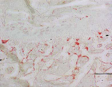


129 Chapter Five Characterisation of Svs7 Transgenic Mice (A) WT TG Goldner trichrome (B) 100μm 100μm 500μm 500μm TRAP (10x obj.) TRAP (4x obj.) 100μm 100μm 122
130 Chapter Five Characterisation of Svs7 Transgenic Mice Figure 5.8 Histological examination on goldner trichrome (A) and TRAP (B) stained non-decalcified femur sections of Svs7 TG mice. The other femurs from the same pairs of WT and Svs7 TG mice described above were not decalcified and processed for plastic embedding procedure. The non-decalcified sections were then goldner trichrome (bone stained green) and TRAP (osteoclast stained red)-stained. In support of the H&E sections results, (A) the goldner trichrome-stained sections depicted normal and healthy appearances of trabecular bone architecture and cortical thickness of Svs7 TG hindlimbs in comparison to that of WT littermates. (B) Accordingly, from TRAP sections, it showed a comparable number of TRAP-positive osteoclasts near the growth plate region between WT and Svs7 TG hindlimbs, indicating that Svs7 TG mice exhibit a characteristic of healthy bone remodelling process. Together, these results suggested normal bone phenotype and TRAP-positive osteoclasts number in Svs7 TG hindlimbs. Scale bars for 4x obj. magnification images, 500μm; for 10x, 100μm. 123
131 Chapter Five Characterisation of Svs7 Transgenic Mice NOc/BS (N/mm) WT TG Figure 5.9 Bone histomorphometry analysis on NOc/BS (N/mm) of TRAP-stained non-decalcified femur sections of Svs7 TG mice. Histomorphometry analysis was performed on TRAP sections of non-decalcified femurs from WT and Svs7 TG mice described above. No difference was observed in NOc/BS parameter between WT and Svs7 TG mice. Data are presented as mean ± SEM. Number of mice (n) = 2/group. 124


132 Chapter Five Characterisation of Svs7 Transgenic Mice Number of TRAP+ multinucleated cells ( 3 nuclei)/well n.s. n.s. WT TG 0 RANKL stimulation Day 0 Day 1 Day 3 Day 5 WT TG Figure 5.10 Svs7 TG mice exhibit an unaffected osteoclastogenesis in vitro. BM cells were isolated from long bones of 9-week old WT and Svs7 TG mice and cultured in M-CSF supplemented complete medium until reaching confluency. The cells were then seeded into 96-well plates and cultured for 5-day time course RANKL and M-CSF stimulation with a fresh media change every 2 days. The result showed that there was no significant difference in osteoclastogenesis between WT and Svs7 TG mice. Hence, Svs7 TG mice exhibited an unaffected osteoclastogenesis in vitro. Data are presented as mean ± SEM and are representative of three independent experiments. n.s. = no significance against WT. 125
133 Chapter Five Characterisation of Svs7 Transgenic Mice Hence, these results have indicated that Svs7 might have a potential role in the maintenance of bone homeostasis. In the present study, global tissue distribution of Svs7 mrna expression revealed that Svs7 was almost exclusively restricted to SV (including prostate tissue) and a lesser degree in osteoclasts. In an effort to address this, mice overexpressing the Svs7 transgene were generated by introducing the purified and linearized pcdna3.1 Svs7 V5-His transgene into WT mice by blastocysts microinjection. Svs7 TG mice generation was confirmed by DNA tail tip genotyping and agarose gel electrophoresis as well as WB analysis on cleared liver lysates from 9- week old WT and TG mice to confirm the transgenic protein expression of Svs7. Unfortunately, immunoblot assessment confirmed that there was only a modest increase in Svs7 protein expression in Svs7 TG mice (about 2.5 fold increase in liver) relative to WT. Although several factors, including post transcriptional and translational regulations as well as hormonal change, could affect the relationship between the gene and protein expression level (King et al. 2012; Levitin et al. 2008), these observations suggested that global overexpression of Svs7 protein in Svs7 TG mice was only mildly overexpressed above the endogenous level, which might have excluded the detection of a clear tissue/bone phenotype in these TG mice. Despite the modest increase in Svs7 expression in Svs7 TG mice, the assessment of the gross tissue morphology and the bone phenotype of these mice were persevered. First, the tissue histology of the prostate (including SV), which has the highest Svs7 gene expression, was assessed. The result showed that Svs7 TG prostate appeared to have a normal phenotype relative to that of WT. Hence, from this global tissue phenotype characterisation study, the Svs7 transgene expression does not appear to affect organs and tissues phenotype. Furthermore, the normal organ and tissue phenotype observed in Svs7 TG mice correlated well with their comparable body weight with WT littermates. To assess the potential bone phenotype of Svs7 TG mice, X-ray analysis on the whole body and spine of WT and TG mice were performed. The result revealed that there was no obvious change in skeletons of Svs7 TG mice. These findings were further supported by the microct analysis on trabecular bone architecture at proximal tibia, distal femur, and vertebral body of the lumbar bone of Svs7 TG mice that showed no striking change in the bone volume architecture at all three sites in these TG mice in reference to WT counterparts. Moreover, the quantitative analysis on trabecular bone in the secondary spongiosa region of distal femurs (Lee et al. 2006; Nishikawa et al. 2010) from WT and 126
134 Chapter Five Characterisation of Svs7 Transgenic Mice Svs7 TG mice clearly indicated that TG mice had a normal bone volume architecture, by showing comparable statistics on all bone parameters (BV/TV (%), Tb.Sp (mm), Tb.Th (mm), and Tb.N (1/mm)) to WT littermates. As a second standard procedure in the bone phenotype characterisation, histological examination on both paraffinembedded decalcified (H&E stained) and plastic-embedded non-decalcified (goldner trichrome and TRAP stained) bone samples of WT and Svs7 TG mice was performed. Both H&E and goldner trichrome-stained sections demonstrated that there were no marked differences in bone volume and periosteal thickness of the calvarial bone between WT and Svs7 TG mice, together with no observable change in trabecular bone architecture and cortical bone in the metaphyseal region of tibia, femur, and lumbar bones of the TG mice relative to that of WT littermates. In line with the normal bone mass and architecture observed in Svs7 TG mice, the TRAP sections and bone histomorphometry data on number of osteoclasts/bone surface parameter showed that there was no significant difference in the number of TRAP-positive osteoclasts near the growth plate region between WT and Svs7 TG hindlimbs. These results, along with the observed findings of unaltered calcium and phosphate serum levels (Supplementary Figure 1), indicate the presence of healthy bone remodelling process in Svs7 TG mice. Finally, in order to probe for potential intrinsic cell-specific influences in Svs7 TG mice, in vitro RANKL-induced osteoclastogenesis assays on WT and TG BMM was performed. The result revealed that, in reference to WT, indeed Svs7 TG mice exhibited an unaffected osteoclastogenesis in vitro. Hence in conclusion, Svs7 TG mice showed no obvious defects in organs and tissues (especially bone) phenotype. Through microct analysis and histological examination, Svs7 TG mice showed to have normal bone volume and architecture. The indication of normal bone remodelling process in Svs7 TG mice to explain the observed phenotype had been illustrated by the histological TRAP-stained hindlimbs sections, histomorphometry data, unaltered calcium and phosphate serum levels, and in vitro osteoclastogenesis results that showed an unaffected osteoclastogenesis in the TG mice. Thus, the Svs7 transgenic strategy did not allow an accurate assessment of the potential role of Svs7 in bone homeostasis, likely owing to the modest transgene protein expression of Svs7 in Svs7 TG mice. Therefore, an alternative knockout strategy was required to gain better insights into the potential role of Svs7 in the maintenance of bone homeostasis. 127
135 CHAPTER 6 CHARACTERISATION OF SVS7 KNOCKOUT MICE 128
136 Chapter Six Characterisation of Svs7 Knockout Mice 6.1 Introduction Despite the naturally low gene expression of Svs7 in bone, as shown in Chapter 5 Figure 5.1, and with the initial studies that indicate Svs7 gene is upregulated during RANKL-induced osteoclastogenesis together with other common osteoclast marker genes (Chapter 2 Figure 2.4), it would be plausible to hold the hypothesis that Svs7 is an important regulator of osteoclasts and/or osteoblasts differentiation and activation. By considering that Svs7 is a secreted member of Ly-6 protein family, sharing common TFP/Ly-6/uPAR motif (Coronel et al. 1992; Levitin et al. 2008; Southan et al. 2002; Turunen et al. 2011; Ushizawa et al. 2009), and the importance of other Ly-6 members in many cellular signalling pathways, such as shown by upar in αvβ3 integrin signalling pathway (Schiller et al. 2009; Smith, Marra & Marshall 2008; Wei et al. 2008) and Sca-1, also expressed by osteoclasts (Chapter 2 Figure 2.3), in stem cell renewal capacity (Bonyadi et al. 2003; Ito et al. 2003), the initial approach and investigation using Svs7 TG mice was conducted but it had failed to provide accurate insights into the potential role of Svs7 in the maintenance of bone homeostasis. Svs7 TG mice showed no obvious change in organs and tissues, especially in bone, phenotype. Normal bone mass and architecture, with the addition of an unaffected osteoclastogenesis in vitro, were observed in Svs7 TG mice. This might be partly due to a weak level of Svs7 transgene protein expression in Svs7 TG mice. Therefore, as an alternative approach to investigate the potential role of Svs7 in regulating bone homeostasis, Svs7 global knockout (KO) mouse model was generated. Hence, this chapter focuses on the characterisation of Svs7 global KO mice, with a particular emphasis on the bone phenotype. 6.2 Results Genotyping and characterization of Svs7 KO mice DNA isolated from mouse tail tips was genotyped according to Jackson laboratory s DNA isolation protocol, as discussed in Chapter 4 Section 4.3.3, and DNA products were PCR amplified using specific primers for Svs7 and run on a 1.5% agarose gel. Figure 6.1A illustrates the 99 amino acids composition of Svs7 protein (Amino (N) to carboxyl (C) terminal), encoded by 3 exons (each exon is separated by red dot), and the importance of exon 2 and 3, which together encode for the common TFP/Ly-6/uPAR domain, consisting of conserved cysteine residues and the consensus C-terminal sequence motif of CCXXXXCN ( X represents any amino acid and N for asparagine 129
 TFP/Ly-6/uPAR domain (B) WT Svs7 KO Het NEG (C) WT KO Svs7 1 2 3 4 β-actin Figure 6.")
 (Top panel) Svs7 amino acids composition, encoded by 3 exons (each exon is separated by red rot), showing consensus cysteine (red highlighted letter C ) residues and the consensus C- terminal")
.")
137 Chapter Six Characterisation of Svs7 Knockout Mice (A) Svs7 protein (99 aa): MNSVTKISTLLIVILSFLCFVE.GLICNSCEKSRDSRCTMSQSRCVAKPGESCSTVS HFV.GTKHVYSKQMCSPQCKEKQLNTGKKLIYIMCCEKNLCNSF Exon 1 Exon 2 Exon 3 SP C-C-C-C-C C-C-C-C-CN Poly (A) TFP/Ly-6/uPAR domain (B) WT Svs7 KO Het NEG (C) WT KO Svs β-actin Figure 6.1 Svs7 gene and protein were absent in Svs7 KO mice. (A) (Top panel) Svs7 amino acids composition, encoded by 3 exons (each exon is separated by red rot), showing consensus cysteine (red highlighted letter C ) residues and the consensus C- terminal sequence motif CCXXXXCN ( X for any amino acid and N for asparagine), and (Bottom panel) the structure of Svs7 gene with exon 2 and 3 encoding for TFP/Ly- 6/uPAR domain (The structure of Svs7 gene was illustrated by Levitin et al. 2008). (B) Agarose gel electrophoresis of mouse tail tip DNA genotyping product showing that the deletion of exon 2 and 3 of Svs7 gene in Svs7 KO mice (lane 2) had produced a smaller DNA product compared to WT (lane 1). (C) WB protein analysis on WT and Svs7 KO seminal vesicle lysates showed that there was no Svs7 protein detected in the KO lysate. β-actin served as a loading control. 130
138 Chapter Six Characterisation of Svs7 Knockout Mice residue) (Levitin et al. 2008). Deletion of these exons was confirmed by DNA analysis using agarose gel electrophoresis. As shown in Figure 6.1B, Svs7 KO (lane 2) produced a smaller DNA product compared to WT (lane 1), indicating that the exons had been successfully deleted. To confirm Svs7 deletion at the protein level, western blot (WB) analysis on seminal vesicle (SV) lysates from WT and Svs7 KO mice was performed. As anticipated, the loss of exon 2 and 3 within Svs7 gene resulted in the complete absence of Svs7 protein in Svs7 KO lysate, as shown in Figure 6.1C. β-actin served as a loading control. Together, these results validate the deletion of Svs7 in Svs7 KO mice Slow mice breeding process and gross body phenotypic examination of Svs7 KO mice Following the generation of heterozygous (Het) mice, the breeding process was continued by setting up Het x Het crossing to produce WT and Svs7 KO littermates. As shown in Figure 6.2A, amongst 71 newborn pups (39 for male and 32 for female), the ratio of getting male WT and female Svs7 KO mice were only 12% and 16% respectively, which are significantly lower than the expected 25% (Mendelian ratio), suggesting that there was a slow breeding process and hence difficulty in getting adequate WT and KO pairs littermates for both male and female pups. The possible outcome of this slow mice breeding process was then investigated by first examining the gross body phenotype of age- and sex-matched WT and Svs7 KO pairs littermates. To achieve this, 12-week old WT and Svs7 KO mice were sacrificed and the two bodies were laid side-by-side for examination and the representative images were taken. The result depicted that there was no apparent defect in the gross body phenotype of Svs7 KO mice compared to WT in the same litter (Figure 6.2B). Collectively, although there was a slow mice breeding process, Svs7 KO mice appeared to be similar in body size as WT littermates Gross tissues phenotypic (macro vs. microscopic) assessment of Svs7 KO mice To continue with the gross organs and tissues phenotype characterization of Svs7 KO mice, 12-week old WT and KO pair littermates were sacrificed and general organs and tissues were dissected from both mice and placed side-by-side for careful macroscopic examination. The result showed that, in relative to WT, Svs7 KO organs and tissues were comparable (Figure 6.3A). Furthermore, histology-prepared prostate (including seminal vesicle) sections from WT and Svs7 KO mice were H&E stained for microscopic assessment. No obvious difference in prostate tissue phenotype was 131

139 Chapter Six Characterisation of Svs7 Knockout Mice (A) Percentage of male pups WT Het KO Percentage of female pups WT Het KO (B) WT KO Figure 6.2 Slow mice breeding process and gross body phenotypic examination of Svs7 KO mice. (A) Amongst 71 newborn pups (39 for male and 32 for female), the ratio of getting male WT and female Svs7 KO mice were 12% and 16% respectively, which are well below than the expected 25%, suggesting that there was a slow mice breeding process and subsequent difficulty in getting adequate WT and KO pairs littermates for both male and female mice for experiments. (B) Twelve-week old WT and Svs7 KO mice were sacrificed and laid side-by-side for gross body phenotypic examination. The result showed that Svs7 KO mice appeared to be similar in body size as WT littermates. 132
 WT KO 500μm 500μm Figure 6.")
 Organs and tissues were extracted from 12-week old WT and Svs7 KO mice and placed side-byside for macroscopic examination.")
 Prostates (including seminal vesicle) were extracted from WT and Svs7 KO mice, fixed, processed, embedded, sectioned, and H&E stained for histological")
140 Chapter Six Characterisation of Svs7 Knockout Mice (A) Tibia SV (prostate) Testis Brain Heart Thymus WT KO WT KO WT KO WT KO WT KO WT KO Lung Liver Spleen Stomach Kidney WT KO WT KO WT KO WT KO WT KO Intestine Fat Ovary WT KO WT KO WT KO (B) WT KO 500μm 500μm Figure 6.3 Gross tissues phenotypic assessment of Svs7 KO mice. (A) Organs and tissues were extracted from 12-week old WT and Svs7 KO mice and placed side-byside for macroscopic examination. The result showed that Svs7 KO organs and tissues were comparable to that of WT. (B) Prostates (including seminal vesicle) were extracted from WT and Svs7 KO mice, fixed, processed, embedded, sectioned, and H&E stained for histological assessment. The result depicted that there was no obvious difference in prostate tissue phenotype between WT and Svs7 KO mice. Hence, the loss of the Svs7 gene did not appear to affect the organogenesis in Svs7 KO mice. Scale bars, 500μm. 133
141 Chapter Six Characterisation of Svs7 Knockout Mice observed between WT and Svs7 KO mice (Figure 6.3B). Together, these results suggested that the deletion of the Svs7 gene did not appear to affect the organogenesis in Svs7 KO mice Svs7 KO mice exhibit a reduced bone mass phenotype As an initial step toward characterising the potential bone phenotype of Svs7 KO mice, femurs were extracted from 15- and 30-week old WT and Svs7 KO mice, fixed, and processed for microct analysis, as detailed in Chapter 4 Section Initially, no striking phenotype was observed in Svs7 KO mice. Mice grew at lower ratio than the expected without gross abnormality was observed. However, Svs7 KO mice exhibited a progressive decline in bone mass with age as compared to WT littermates. The 2D and 3D microct images of distal femurs from WT and Svs7 KO mice showed that Svs7 KO mice appeared to have less trabecular bone volume compared to WT littermates (Figure 6.4A). Indeed, through the quantitative analysis on trabecular bone in secondary spongiosa region (Lee et al. 2006; Nishikawa et al. 2010) of both WT and Svs7 KO femurs, it was then revealed that KO bones had significantly lower bone/tissue volume (BV/TV (%)) in both age (15- and 30-week old) groups and a significantly less trabecular number (Tb.N (1/mm)) in the 30-week old group compared to WT (Figure 6.4B). However, there were no significant differences in trabecular separation (Tb.Sp (mm)) and trabecular thickness (Tb.Th (mm)) in both age groups between WT and Svs7 KO mice (Figure 6.4B). Together, these results imply that Svs7 KO mice exhibit a phenotype of osteopenia/osteoporosis Svs7 KO bone histology The reduced bone volume in Svs7 KO mice prompted us to conduct a close examination of the bone phenotype using histology and histomorphometry analysis. For this, histology sections of decalcified femurs from 12-week old WT and Svs7 KO mice were H&E and TRAP (as an osteoclast marker; red precipitates) stained. The H&E-stained sections showed that, indeed, Svs7 KO femurs seemed to have less trabecular bone volume in the secondary spongiosa region than WT (Figure 6.5A). In accordance with this result, TRAP-stained sections depicted that Svs7 KO hindlimbs appeared to have a trend of increase in the number of TRAP-positive osteoclasts near the growth plate region compared to WT (Figure 6.5B). The bone histomorphometry analysis on TRAPstained femurs sections from WT and Svs7 KO mice for the number of 134

 WT KO (B) BV/TV")
 0.3 0.")


 4 3 2")
142 Chapter Six Characterisation of Svs7 Knockout Mice (A) WT KO (B) BV/TV (%) WT KO Tb Sp (mm) n.s. n.s wk 30wk 15wk 30wk 0.06 n.s. n.s. 5 n.s. Tb.Th (mm) Tb N (1/mm) wk 30wk 15wk 30wk 135
143 Chapter Six Characterisation of Svs7 Knockout Mice Figure 6.4 Svs7 KO mice exhibit a reduced bone mass phenotype. (A) 2D and 3D (longitudinal and cross sections) microct images of distal femurs from 15- and 30- week old WT and Svs7 KO mice showed that the KO mice appeared to have less trabecular bone volume compared to WT. (B) The quantitative microct analysis on bone parameters of femurs from 15- and 30-week old WT and Svs7 KO mice revealed significantly reduced BV/TV (in both age groups) and trabecular number (Tb.N) (only in the 30-week old mice) in Svs7 KO mice compared to WT. There were no significant differences in trabecular separation (Tb.Sp) and trabecular thickness (Tb.Th) between WT and Svs7 KO mice in both age groups. Hence, Svs7 KO mice exhibit a phenotype of osteopenia/osteoporosis. Data are presented as mean ± SEM. * p < 0.05, n.s. = no significance against WT. Scale bars, 500μm. 136


144 Chapter Six Characterisation of Svs7 Knockout Mice (A) WT KO TRAP (20x obj.) TRAP (10x obj.) H&E (B) 137
145 Chapter Six Characterisation of Svs7 Knockout Mice Figure 6.5 Svs7 KO bone histology. (A) Fixed and decalcified femurs sections from 12-week old WT and Svs7 KO mice were H&E stained. The result showed that Svs7 KO hindlimbs appeared to have less trabecular bone volume in the secondary spongiosa region compared to WT. (B) (Top panel) 10x and (Bottom panel) 20x objective magnifications of representative images of TRAP (osteoclasts stained red)-stained WT and Svs7 KO femurs sections showed a trend of increase in the number of TRAPpositive osteoclasts near the growth plate region in KO hindlimbs relative to WT. Scale bars for 4x obj. H&E sections, 500μm; for 10x and 20x obj. TRAP sections, 100μm. 138
146 Chapter Six Characterisation of Svs7 Knockout Mice osteoclasts/bone surface (NOc/BS (N/mm)) similarly indicated that there was a trend of increase in TRAP-positive osteoclasts number in the KO hindlimbs compared to WT (Figure 6.6). Therefore, the bone histology sections of Svs7 KO mice supported the MicroCT data that there was a reduced bone volume and a trend of increase in the number of TRAP-positive osteoclasts in Svs7 KO hindlimbs Bone formation activity in vivo is unaffected in Svs7 KO mice To further characterize the Svs7 KO bone phenotype, double calcein labelling was performed. For this purpose, 12-week old WT and Svs7 KO mice were subjected to the calcium-binding fluorescent dye, calcein, injection. Calcein is incorporated into the newly mineralized tissues and hence by giving the mice two injections (two labels) seven days apart and measuring the distance between two labels using ImageJ software (after images were taken using confocal microscopy), the difference in bone formation activity (mineral apposition rate (MAR)) in vivo between WT and Svs7 KO mice can be deduced. Analysis of plastic-embedded non-decalcified tibia sections from WT and Svs7 KO mice by confocal microscopy (Figure 6.7A) and MAR measurement (Figure 6.7B) showed that there was no significant difference in bone formation activity in vivo between WT and KO mice. Hence, Svs7 KO mice exhibit an unaffected bone formation activity in vivo. 6.3 Discussion This study builds upon our previous observations that Svs7 is exclusively expressed by mature osteoclasts but not by osteoblasts or other key bone cells and its gene expression is upregulated during RANKL-induced osteoclastogenesis (as discussed in Chapter 2 Section 2.4 Figure 2.4). Here to directly address the possible role of Svs7 in bone homeostasis, which had failed to be shown in our first approach and study using Svs7 TG mice, we generated Svs7-deficient mice. Svs7 KO mice generation was achieved by the deletion of exon 2 and 3 of Svs7 gene. This deletion was then confirmed by DNA tail tip genotyping and immunoblotting for Svs7 protein in Svs7 KO seminal vesicle lysate, which demonstrated that Svs7 protein expression was lost in these KO mice. Assessment of Svs7 KO mice breeding revealed that the ratio of male WT and female Svs7 KO mice were well below 25% (12% and 16% respectively), which consequently limited both the sample size and number of assays available for assessment. Although 139
147 Chapter Six Characterisation of Svs7 Knockout Mice 12 n.s. 10 NOc/BS (N/mm) WT KO Figure 6.6 Bone histomorphometry analysis on TRAP-stained decalcified femur sections of Svs7 KO mice. Fixed and decalcified femurs sections from 12-week old WT and Svs7 KO mice were TRAP stained and number of osteoclasts/bone surface (NOc/BS (N/mm)) parameter was analysed. The result revealed that there was a trend of increase in the number of TRAP-positive osteoclasts in Svs7 KO hindlimbs compared to WT. Data are presented as mean ± SEM. Number of mice (n) = 3/group. n.s. = no significance against WT. 140


148 Chapter Six Characterisation of Svs7 Knockout Mice (A) WT KO (B) MAR (μm/day) WT n.s. KO Figure 6.7 Svs7 KO mice exhibit an unaffected bone formation activity in vivo. (A) Calcein double labelling confocal microscopy images of plastic-embedded nondecalcified tibia sections from 12-week old WT and Svs7 KO mice showed that there was no striking difference in bone formation activity between WT and KO mice. (B) MAR analysis by measuring the distance between the two calcein labels (in each section) revealed that there was no significant difference in bone formation activity in vivo between WT and Svs7 KO mice. Hence, Svs7 KO mice exhibit an unaffected bone formation activity in vivo. Data are presented as mean ± SEM and are representative of three independent experiments. n.s. = no significance against WT. Scale bars, 10μm. 141
149 Chapter Six Characterisation of Svs7 Knockout Mice the precise reason for this is unclear, it may imply an important role of Svs7 in seminal vesicle and fertility, which has been previously suggested by Coronel et al that Svs7 inhibits calcium (Ca 2+ ) influx across the membrane of sperm cells during fertilization to prevent premature acrosomal reaction and spermatozoa hyperactivation. Despite this breeding limitation, the examination of the gross phenotype of body, organs, and tissues, including histological assessment on prostate (the tissue which has the highest Svs7 gene expression, as shown in Chapter 5 Figure 5.1), of Svs7 KO mice relative to WT littermates (age- and sex-matched pairs) was successfully achieved. The results showed that there were no apparent differences in body, organs, and tissues gross phenotype between WT and Svs7 KO mice. Hence, the loss of the Svs7 gene does not appear to affect the gross phenotype (macro- and microscopic) of body, organs, and tissues of Svs7 KO mice. Moreover, microct analysis on trabecular bone volume and architecture in the secondary spongiosa region of distal femurs from 15- and 30-week old WT and Svs7 KO mice (Lee et al. 2006; Nishikawa et al. 2010) revealed that the KO mice had significantly reduced trabecular bone/tissue volume (BV/TV) at both age groups compared to WT littermates. Consistently, there was a significant reduction in trabecular number (Tb.N) in Svs7 KO hindlimbs, even though only in the 30-week old mice group. The possibility that Svs7 KO mice exhibit a progressive decline in bone mass with age, as compared to WT littermates, could play a significant role in this microct analysis. In line with these microct results, H&E-stained sections of decalcified femurs from 12-week old WT and Svs7 KO mice showed that there was an obvious decrease in trabecular bone volume in the secondary spongiosa region of KO hindlimbs relative to that of WT littermates. Subsequently, TRAP-stained sections and bone histomorphometry analysis on the number of osteoclasts/bone surface (NOc/BS) indicated that there was a trend of increase in the number of TRAP-positive osteoclasts near the growth plate region of Svs7 KO hindlimbs compared to WT littermates. Collectively, these results suggest that Svs7 KO mice exhibit a phenotype of reduced bone mass (osteopenia/osteoporosis) and a trend of increase in osteoclasts number in vivo, which could strongly indicate an alteration in osteoclast differentiation and/or activation in the KO mice. To further explore the possible cause of the reduced bone mass phenotype in Svs7 KO mice, osteoblastic bone formation activity in vivo was investigated by performing 142
150 Chapter Six Characterisation of Svs7 Knockout Mice double calcein labelling (Hayashi et al. 2012) in 12-week old WT and Svs7 KO mice and measuring the distance between two labels on the images of calcein-labelled plastic embedded non-decalcified tibia sections to calculate mineral apposition rate (MAR). The result showed that there was no significant difference in MAR between WT and Svs7 KO mice. Hence, this finding indicates that Svs7 KO mice exhibit an unaffected bone formation activity in vivo and that the alteration in osteoclast differentiation and/or activation might be the major contributor to the osteoporotic phenotype observed in Svs7 KO mice. In conclusion, Svs7, a novel osteoclast-secreted factor (Supplementary Figure 2), appears to be a candidate regulator of bone homeostasis. Svs7 KO mice exhibit an osteopenic/osteoporotic phenotype and an unaffected bone formation activity in vivo, but an elevated osteoclast number in KO hindlimbs suggests that Svs7 might influence bone homeostasis via the regulation of osteoclast differentiation and/or activity. 143
151 CHAPTER 7 INVESTIGATION OF INTRINSIC CELLULAR MECHANISM IN SVS7 KNOCKOUT MICE (OSTEOCLAST AND OSTEOBLAST) 144
152 Chapter Seven Investigation of Intrinsic Cellular Mechanism in Svs7 Knockout Mice (Osteoclast and Osteoblast) 7.1 Introduction Bone homeostasis is achieved by balanced activities between bone-resorbing osteoclast and bone-forming osteoblast. Various cytokines, proteins, and growth factors are expressed and/or secreted by both osteoclasts and osteoblasts in a regulated manner to create intercellular communication and regulation, crucial for the differentiation and activation of each cell. RANKL and M-CSF, produced by osteoblasts, are two essential cytokines for driving osteoclast differentiation and activation (Kodama et al. 1991; Takahashi, Udagawa & Suda 1999). On the other hand, osteoclasts release the active form of IGF-II from bone matrix during its bone resorption activity to stimulate and initiate site-specific bone formation by osteoblasts (Hayden, Mohan & Baylink 1995). Thus, an imbalance in this coupling system, which could come from defects in either or both osteoclasts and osteoblasts, may lead to the development of many pathological bone diseases, such as osteoporosis (reduced bone mass) and osteopetrosis (increased bone mass). The results presented in Chapter 6 demonstrate that Svs7 KO mice exhibit a reduced bone mass phenotype, consistent with osteopenia/osteoporosis. Since osteoporosis is attributed to an excessive bone resorption by osteoclasts (Riggs & Melton 1983) and/or a decline in bone formation activity by osteoblasts (Manolagas & Jilka 1995), these observations imply that the changes in osteoclasts and/or osteoblasts number and/or activity might account for the reduced bone mass phenotype observed in Svs7 KO mice. To address this, the present chapter focuses on the in vitro characterization of osteoclasts (including their bone marrow macrophages (BMM) progenitors) and osteoblasts from Svs7 KO mice. Parameters explored include Svs7 KO osteoclasts and osteoblasts differentiation and activity and osteoclast fusion mechanism. In addition, it has been well documented that calcium (Ca 2+ ) signalling is essential in the activation of NFATc1, the master regulator of osteoclastogenesis (Takayanagi et al. 2002). Amongst other osteoclast genes that are known to be transcriptionally regulated by NFATc1, such as TRAP, cathepsin K, c-src, and β3 integrin (Crotti et al. 2006; Ikeda et al. 2006; Matsumoto et al. 2004; Takayanagi et al. 2002), it has been shown that NFATc1 also crucially regulates fusion-mediating genes activation in osteoclasts, which are DCSTAMP and ATP6v0d2 genes (Kim et al. 2008; Lee et al. 2006; Yagi et al. 2005). In general, mononuclear preosteoclasts fuse together to become multinucleated cells, which takes place around day 3 of RANKL-induced osteoclast 145
153 Chapter Seven Investigation of Intrinsic Cellular Mechanism in Svs7 Knockout Mice (Osteoclast and Osteoblast) differentiation (osteoclastogenesis) and upon further RANKL stimulation they become active and functional bone resorbing cells (Fujita et al. 2012; Kim et al. 2008). NFATc1 has been shown to bind to the promoter region of both fusogenic genes to induce their expression. The inactivation of NFATc1 decreases the expression of these fusogenes and attenuates osteoclast fusion (Kim et al. 2008). Hence, the overexpression of ATP6v0d2 and DCSTAMP genes has been shown to rescue the NFATc1-deficient osteoclast fusion defect (Kim et al. 2008). As Svs7 is known to regulate Ca 2+ influx across the membrane of sperm cells during fertilization (Coronel et al. 1992), it was then plausible to investigate the potential role of Svs7 in Ca 2+ -NFATc1 signalling pathway, particularly in the regulation of osteoclast fusion mechanism. Through a series of in vitro experiments, the findings in this chapter indicate that Svs7 plays a role in regulating osteoclast fusion through the NFATc1 signalling pathway. 7.2 Results Part 1: Intrinsic cellular mechanism in osteoclasts derived from Svs7 KO mice Svs7 KO BMM cultures have increased osteoclastogenesis in vitro First, the histomorphometry results obtained from Svs7 KO hindlimbs indicated that there was a trend of increase in osteoclast number in vivo. Based on this observation, we speculated that the reduced bone mass phenotype observed in Svs7 KO mice might be due to an alteration in osteoclast differentiation and/or activation. To assess osteoclast formation in Svs7 KO mice, in vitro 5-day time course (day 0, 1, 3, and 5) RANKL and M-CSF-induced osteoclastogenesis assays on WT and KO bone marrow (BM) cells were performed. At the end of the time course, the cells were fixed, TRAP stained and osteoclasts (TRAP-positive cells having 3 nuclei) were counted. The result showed that at day 3 and 5 of osteoclastogenesis, there were significantly more TRAP-positive osteoclasts in Svs7 KO cultures compared to WT (Figure 7.1). Hence, in vitro osteoclastogenesis was increased in Svs7 KO mice Svs7 KO osteoclasts are larger and have more nuclei than WT Further investigation in in vitro osteoclastogenesis assay results showed that Svs7 KO osteoclasts exhibited significantly larger spread area than WT at day 5 of RANKLinduced osteoclastogenesis (Figure 7.2). Although there was no significant difference at day 3 between WT and Svs7 KO osteoclasts (Figure 7.2), these results indicate that the KO cells have an accelerated fusion process during differentiation. To further assess this, osteoclasts were separated into three groups based on the number of nuclei per 146

/well 160 140")


154 Chapter Seven Investigation of Intrinsic Cellular Mechanism in Svs7 Knockout Mice (Osteoclast and Osteoblast) 180 Number of TRAP+ multinucleated cells ( 3 nuclei)/well RANKL stimulation Day 0 Day 1 Day 3 Day 5 WT KO WT KO Figure 7.1 In vitro time course RANKL-induced Svs7 KO BMM differentiation into mature osteoclasts. BM cells from 12-week old WT and KO mice were cultured for 5-day time course in the presence of M-CSF and RANKL with a fresh media change every 2 days. Upon formation of osteoclasts, TRAP stain and osteoclast count were performed. Svs7 KO cultures showed a significant increase in osteoclastogenesis compared to WT at day 3 and 5 of RANKL-induced osteoclastogenesis. Data are presented as mean ± SEM and are representative of three independent experiments. ** p < 0.01 against WT. Scale bars, 100μm. 147
155 Chapter Seven Investigation of Intrinsic Cellular Mechanism in Svs7 Knockout Mice (Osteoclast and Osteoblast) Average OC area (μm 2 ) at day 3 of Osteoclastogenesis WT n.s. KO Average OC area (μm 2 ) at day 5 of Osteoclastogenesis WT KO Figure 7.2 Svs7 KO osteoclasts are larger than WT. Analysis of osteoclast spread area at day 3 and 5 of RANKL-induced osteoclastogenesis revealed that Svs7 KO osteoclasts were significantly larger than WT at day 5 of osteoclastogenesis (Bottom panel), while there was no significant difference at day 3 between both cells (Top panel). Data are presented as mean ± SEM and are representative of three independent experiments. * p < 0.05, n.s. = no significance against WT. 148
156 Chapter Seven Investigation of Intrinsic Cellular Mechanism in Svs7 Knockout Mice (Osteoclast and Osteoblast) osteoclast (having 3-5, 6-10, or 11 nuclei), which were then added together and expressed as a ratio to total TRAP-positive multinucleated cells per well. Based on this quantification method, there were significantly more large osteoclasts, having 11 nuclei, in Svs7 KO cultures compared to WT at day 5 of osteoclastogenesis (Figure 7.3). These findings suggest that Svs7 might play a role in regulating osteoclast fusion WT and Svs7 KO mice have comparable hematopoietic stem cells (HSCs) and osteoclast progenitor cells As an increase in osteoclastogenesis was observed in Svs7 KO mice, it was then of interest to further investigate the early lineage negative (Lin - ) osteoclast precursor (CD45R - CD3e - CD11b -/low ) (Jacquin et al. 2006) and subsequent HSCs population (Lin - Sca-1 + c-kit + /LSK) (Kollet et al. 2006; Lymperi et al. 2011; Miyamoto et al. 2011) in BM population of WT and Svs7 KO mice. To explore this, 12-week old WT and KO mice were sacrificed and BM cells were then isolated from their long bones. Following red blood cell lysis procedure, the cells were counted, antibodies were prepared in master mix with the required dilutions, followed by cell incubation with antibodies and overnight fixation, as detailed in Chapter 4 Section Flow cytometry analysis was performed on the following day using FACS Canto at CMCA, UWA. As shown in Figure 7.4A, both WT and Svs7 KO BM had no significant differences in both HSCs and early osteoclast progenitor populations. Next, BM osteoclast progenitor proliferation was assessed. For this purpose, BM cells were cultured in complete medium supplemented with M-CSF for 48 hours and were subjected to MTS proliferation assay, which measures the metabolic activity of the viable cells through the absorbance of the formazan product at 490nm excitation wavelength. The result showed that there was no significant difference in proliferation rate between WT and Svs7 KO BM osteoclast precursors over both 24 and 48 hours time points (Figure 7.4B), which therefore implied that the loss of the Svs7 gene did not alter the regulation of HSCs and early osteoclast precursor population and proliferation. As early osteoclast precursor proliferation, growth, and survival is primarily mediated by the mitogen-activated protein kinases (MAPKs) signalling pathway upon RANKL stimulation, which includes ERK, c-jun N-terminal Kinase (JNK), and p38 (Cobb & Goldsmith 1995; Teitelbaum 2000), cell lysates of WT and Svs7 KO BM osteoclast precursors, stimulated for an hour (0, 5, 10, 20, 30, and 60 minutes time points) with 149
157 Chapter Seven Investigation of Intrinsic Cellular Mechanism in Svs7 Knockout Mice (Osteoclast and Osteoblast) Ratio to total number of TRAP+ multinucleated cells ( 3 nuclei) (per well) n.s. n.s. n.s. WT KO WT KO WT KO #3-5 #6-10 #11+ Nuclei number at day 3 of osteoclastogenesis Ratio to total number of TRAP+ multinucleated cells ( 3 nuclei) (per well) n.s. n.s. WT KO WT KO WT KO #3-5 #6-10 #11+ Nuclei number at day 5 of osteoclastogenesis Figure 7.3 Svs7 KO osteoclasts have more nuclei than WT. Analysis of number of osteoclasts (based on their nuclei number) in ratio to total number of TRAP-positive multinucleated cells showed that indeed Svs7 KO cultures had significantly more large osteoclasts, having 11 nuclei, than WT at day 5 of osteoclastogenesis, which perfectly correlates with the osteoclast spread area analysis described above. At day 3 of osteoclast differentiation, both WT and Svs7 KO cultures had similar distribution of osteoclast sizes. These findings suggest that Svs7 might play a role in the regulation of osteoclast fusion. Data are presented as mean ± SEM and are representative of three independent experiments. * p < 0.05, n.s. = no significance against WT. 150
 Estimated cell cumber (x10 3 ) 40 35 30 25 20 15 10 5 n.s. n.s. WT KO 0 24hrs 48hrs Cell Proliferation Figure 7.")
 BM cells were extracted from long bones of 12-week old WT and KO mice, followed by cell incubation with fluorophore-conjugated antibodies and overnight fixation.")
 between WT and Svs7 KO mice.")
158 Chapter Seven Investigation of Intrinsic Cellular Mechanism in Svs7 Knockout Mice (Osteoclast and Osteoblast) (A) % of CD11b low /CD3e - / CD45R - (Lin - )/total cells WT n.s. KO % of Lin - Sca-1 + c-kit + (LSK) HSCs/total cells WT n.s. KO (B) Estimated cell cumber (x10 3 ) n.s. n.s. WT KO 0 24hrs 48hrs Cell Proliferation Figure 7.4 Svs7 KO mice exhibit unaltered population of HSCs and early osteoclast precursor. (A) BM cells were extracted from long bones of 12-week old WT and KO mice, followed by cell incubation with fluorophore-conjugated antibodies and overnight fixation. Flow cytometry analysis was performed on the following day, which revealed that there were no significant differences in Lin - early osteoclast precursor (Left panel) and Lin - Sca-1 + c-kit + (LSK) HSCs population (Right panel) between WT and Svs7 KO mice. (B) MTS proliferation assays on WT and Svs7 KO BM osteoclast precursors showed that there was no significant difference in proliferation rate between WT and KO cells over period of 24 and 48 hours time points. These findings suggested that the loss of the Svs7 gene did not affect the regulation of HSCs and early osteoclast precursor population and proliferation. Data are presented as mean ± SEM and are representative of three independent experiments. n.s. = no significance against WT. 151
159 Chapter Seven Investigation of Intrinsic Cellular Mechanism in Svs7 Knockout Mice (Osteoclast and Osteoblast) RANKL (50ng/ml), were subjected to WB analysis to explore the MAPK and IκB (inhibitor of NF-κB) α signalling pathways. As shown in Figure 7.5A (WB result) and B (densitometry analysis), there was no significant difference in the phosphorylation (activation) of ERK, JNK, or p38 as well as in the IκBα degradation (NF-κB activation) between WT and Svs7 KO mice, suggesting that the deletion of the Svs7 gene does not affect early MAPK and NF-κB signalling pathways in osteoclast progenitor Svs7 KO mice exhibit unaltered expression levels of key osteoclast marker genes Next, the expression of osteoclast marker genes in Svs7 KO mice was examined. WT and KO BM cells were extracted from long bones and, upon confluency, were cultured for 5-day time course with RANKL and M-CSF stimulation. RNA was then extracted, followed by reverse transcription (RT) and conventional semi-quantitative PCR reactions. Forward and reverse primers for CTR, TRAP, NFATc1, DCSTAMP, and ATP6v0d2 were run alongside Svs7 and β-actin as a loading control. As shown in Figure 7.6A (agarose gel electrophoresis) and B (densitometry analysis), both WT and Svs7 KO mice had comparable expression levels of key osteoclast marker genes, which indicated that despite the increase in osteoclast number and size, the loss of the Svs7 gene did not alter the expression of key osteoclast marker genes Osteoclast bone resorption activity is not affected in Svs7 KO mice To examine osteoclast activity, WT and Svs7 KO BMM-derived mature osteoclasts cultured in collagen-coated 6-well plates, were detached and seeded onto bone slices and cultured for 48 hours in RANKL-supplemented medium to initiate bone resorption. An independent method using Corning osteo assay plates (containing bone (hydroxyapatite)-like material), after the initial mature osteoclast culture in the collagen-coated 6-well plates, was also performed, as described in Chapter 4 Section After 48 hours, cells on either bone slices or Corning osteo assay wells were fixed, TRAP stained, and number of osteoclasts per bone slice was counted, the surface of bone slices was then visualised under scanning electron microscopy (SEM) and for the Corning osteo wells the Nikon Microscope Ti was used to observe resorption pits. By dividing the total resorbed area with the total osteoclast number on each sample, resorbed pit area/cell result was then recorded as a representative of the osteoclast bone resorption activity in WT and Svs7 KO mice. As shown in Figure 7.7 (SEM analysis) and Figure 7.8 (Corning osteo plate analysis), there was no significant difference in the 152
 p-erk1/2 ratio to ERK1/2 1.6 1.4 1.2 1 0.8 0.6 0.4 0.2 n.s.")
 WT KO IκBα ratio to β-actin 1.2 1 0.8 0.6 0.4 0.2 0 n.s. n.s. n.s. n.s. n.s. n.s. 0 5 10 20 30 60 RANKL stimulation (minute) WT KO Figure 7.")
 Cell lysates of WT and Svs7 KO BM osteoclast precursors, which were given short (an hour) RANKL stimulation, were subjected to WB analysis and immunoblotted with specific antibodies against MAPKs")
160 Chapter Seven Investigation of Intrinsic Cellular Mechanism in Svs7 Knockout Mice (Osteoclast and Osteoblast) (A) RANKL Stimulation mins p-erk1/2 WT KO ERK1/2 p-p38 p38 p-jnk JNK IkBα β-actin (B) p-erk1/2 ratio to ERK1/ n.s. n.s. n.s. n.s. n.s. n.s. WT KO p-p38 ratio to p n.s. n.s. n.s. n.s. n.s. n.s. WT KO RANKL stimulation (minute) RANKL stimulation (minute) p-jnk ratio to β-actin n.s. n.s. n.s. n.s. n.s. n.s RANKL stimulation (minute) WT KO IκBα ratio to β-actin n.s. n.s. n.s. n.s. n.s. n.s RANKL stimulation (minute) WT KO Figure 7.5 The deletion of the Svs7 gene does not affect MAPK and NF-κB signalling pathways in Svs7 KO osteoclast precursor. (A) Cell lysates of WT and Svs7 KO BM osteoclast precursors, which were given short (an hour) RANKL stimulation, were subjected to WB analysis and immunoblotted with specific antibodies against MAPKs (ERK, JNK, and p38) and IκBα. MAPKs activation (phosphorylation) and IκBα degradation appeared to be unaffected in Svs7 KO mice. (B) Densitometry analysis was performed using ImageJ software and revealed that there was no significant difference in the phosphorylation (activation) of ERK, JNK, or p38 as well as in the IκBα degradation between WT and Svs7 KO mice, suggesting that the deletion of the Svs7 gene does not affect early MAPK and NF-κB signalling pathways in osteoclast progenitor. Data are presented as mean ± SEM and are representative of four independent experiments. n.s. = no significance against WT. 153
 Svs7 gene expression (ratio to β-actin) 0.8 0.7 0.6 0.5 0.4 0.3 0.2 0.1 n.s. n.s. WT KO CTR gene expression (ratio to β-actin) 0.5 0.4 0.3 0.2 0.1 n.s. n.s. n.s. n.s. WT KO 0 Day 0 Day 1 Day 3 Day 5 RANKL stimulation 0 Day 0 Day 1 Day 3 Day 5 RANKL stimulation TRAP gene expression (ratio to β-actin) 1.")
161 Chapter Seven Investigation of Intrinsic Cellular Mechanism in Svs7 Knockout Mice (Osteoclast and Osteoblast) (A) RANKL-stimulated WT KO BMM day Svs7 CTR TRAP NFATc1 DCSTAMP ATP6v0d2 β-actin (B) Svs7 gene expression (ratio to β-actin) n.s. n.s. WT KO CTR gene expression (ratio to β-actin) n.s. n.s. n.s. n.s. WT KO 0 Day 0 Day 1 Day 3 Day 5 RANKL stimulation 0 Day 0 Day 1 Day 3 Day 5 RANKL stimulation TRAP gene expression (ratio to β-actin) n.s. n.s. n.s. n.s. WT KO NFATc1 gene expression (ratio to β-actin) n.s. n.s. n.s. n.s. WT KO 0 Day 0 Day 1 Day 3 Day 5 RANKL stimulation 0 Day 0 Day 1 Day 3 Day 5 RANKL stimulation DCSTAMP gene expression (ratio to β-actin) n.s. n.s. n.s. n.s. Day 0 Day 1 Day 3 Day 5 RANKL stimulation WT KO ATP6v0d2 gene expression (ratio to β-actin) n.s. n.s. n.s. n.s. Day 0 Day 1 Day 3 Day 5 RANKL stimulation WT KO Figure 7.6 Key osteoclast marker genes expression levels are not affected in Svs7 KO mice. (A) RNA of 5-day time course RANKL and M-CSF-stimulated WT and Svs7 KO BM cells was extracted and subjected to RT and semi-quantitative PCR reactions with specific forward and reverse primers for Svs7 and key osteoclast marker genes (CTR, TRAP, NFATc1, DCSTAMP, and ATP6v0d2). β-actin was used as a housekeeping gene. The result showed that there were comparable expression levels of the osteoclast marker genes between WT and Svs7 KO mice. (B) Densitometry analysis was then performed using ImageJ software and it revealed that there were no significant differences in the key osteoclast marker genes expression levels between WT and Svs7 KO mice (with the confirmation that Svs7 gene was absent in Svs7 KO mice). Data are presented as mean ± SEM and are representative of four independent experiments. ** p < 0.01, n.s. = no significance against WT. 154
 Resorption Pit Area (μm 2")
 for Svs7 KO osteoclast bone resorption")


162 Chapter Seven Investigation of Intrinsic Cellular Mechanism in Svs7 Knockout Mice (Osteoclast and Osteoblast) Resorption Pit Area (μm 2 /cell) n.s. 0 WT KO WT KO Figure 7.7 SEM analysis (on bone slices) for Svs7 KO osteoclast bone resorption activity. WT and Svs7 KO mature osteoclasts, grown in collagen-coated 6-well plates, were detached and seeded onto bone slices and cultured for 48 hours in RANKLsupplemented complete medium to initiate bone resorption. TRAP staining and osteoclasts counting were subsequently performed, followed by visualization of resorption pits by SEM. The osteoclast bone resorption activity was expressed as a total resorbed pit area/cell. No significant difference in osteoclast activity was observed between WT and Svs7 KO osteoclasts. Hence, osteoclast activity was not affected in Svs7 KO mice. Data are presented as mean ± SEM and are representative of three independent experiments. n.s. = no significance against WT. Scale bars, 200μm. 155

 12000 10000 8000 6000 4000")


163 Chapter Seven Investigation of Intrinsic Cellular Mechanism in Svs7 Knockout Mice (Osteoclast and Osteoblast) Resorption Pit Area (μm 2 / cell) WT n.s. KO WT KO 100μm 100μm Figure 7.8 Corning osteo plate analysis for Svs7 KO osteoclast bone resorption activity. After the initial WT and KO mature osteoclasts culture in collagen plates, the cells were detached and seeded onto the Corning wells, each containing bone (hydroxyapatite)-like material. Four wells were prepared for TRAP stain and osteoclast count and the other four for resorption pits analysis. The osteoclast bone resorption activity was expressed as a total resorbed area/cell. The result revealed that both WT and Svs7 KO osteoclasts had a comparable bone resorption activity. Together, these findings indicated that the osteoclast bone resorption activity was independent of Svs7. Data are presented as mean ± SEM and are representative of four independent experiments. n.s. = no significance against WT. Scale bars, 100μm. 156
164 Chapter Seven Investigation of Intrinsic Cellular Mechanism in Svs7 Knockout Mice (Osteoclast and Osteoblast) amount of resorbed area/cell between WT and Svs7 KO osteoclasts. Together, these findings indicated that the osteoclast bone resorption activity was independent of Svs Svs7 KO osteoclasts are more sensitive to Ca 2+ influx in response to RANKL and extracellular Ca 2+ stimulation Considering that Ca 2+ influx/signalling, induced by RANKL, is a prerequisite for the robust induction of NFATc1 activation-driven osteoclast differentiation and activation (Takayanagi et al. 2002), together with the fact that Svs7 acts as a Ca 2+ transport inhibitor on the membrane of sperm cells during fertilization process, it was then of interest to explore the potential impact of the deletion of the Svs7 gene on Ca 2+ influx in osteoclasts. To this end, WT and Svs7 KO BM were first cultured on coverslips in 48- well plates with complete medium supplemented with RANKL and M-CSF until osteoclasts were observed (5 days culture). The cells were then loaded with 4μM Fluo- 4AM intracellular Ca 2+ -binding dye and the change of fluorescence intensity inside the cells was observed by time-lapse microscopy. The basal Ca 2+ level reading was taken for 2 minutes with every 2 seconds interval recorded for a total of 60 intervals. As expected, the result showed that both WT and Svs7 KO osteoclasts had a trend of increase in Ca 2+ influx in response to RANKL stimulation overnight (Figure 7.9A). Svs7 KO osteoclasts, however, appeared to have a higher increase in basal Ca 2+ influx response compared to WT, with about increase in fluorescence intensity (ratio to baseline reading) for KO and 0.04 fluorescence intensity increase for WT from 0 to 60 interval mark (Figure 7.9A). This finding indicated that Svs7 KO osteoclasts were more sensitive to Ca 2+ influx in response to RANKL and hence Svs7 might have a role in regulating Ca 2+ influx (transport) in osteoclasts. To further investigate this possibility, the effects of adding extracellular Ca 2+ (in the form of CaCl 2 ) on the osteoclast Ca 2+ response was assessed. Three concentrations of CaCl 2 were used: 5, 10, and 20mM. The period of this extracellular Ca 2+ stimulation was recorded for 3 minutes (with 2 seconds interval), following the 2 minutes reading of the basal Ca 2+ level. In all three concentrations, the addition of this extracellular Ca 2+ at 60 interval mark (120 seconds; the end of the basal Ca 2+ level fluorescence reading) caused an initial sudden increase in Ca 2+ influx, as monitored by the corresponding increase in fluorescence intensity, in both WT and Svs7 KO osteoclasts, as shown in Figure 7.9B ((i) 5mM, (ii) 10mM, and (iii) 20mM CaCl 2 concentration). The major difference in this initial sudden Ca 2+ influx between WT and Svs7 KO osteoclasts was at the addition of 5mM CaCl 2 concentration (Figure 7.9B(i)), at which the KO cells had a faster and steeper Ca 2+ influx response 157
165 Chapter Seven Investigation of Intrinsic Cellular Mechanism in Svs7 Knockout Mice (Osteoclast and Osteoblast) (A) Basal Ca2+ level Fluorescence intensity (ratio to baseline) WT 0.97 KO Intervals (B) (i) (ii) Addition of 5mM CaCl2 Fluorescence intensity (ratio to baseline) Fluorescence intensity (ratio to baseline) WT KO Basal Ca2+ level Intervals (iii) 1.4 Fluorescence intensity (ratio to baseline) Addition of 10mM CaCl WT 0.6 KO Basal Ca2+ level Intervals Addition of 20mM CaCl WT Basal Ca2+ level KO Final Ca2+ uptake by mature osteoclasts (% of baseline) (C) Intervals n.s. n.s. n.s WT 60 KO mM 10mM 20mM Extracellular Ca2+ (CaCl2) stimulation Figure 7.9 Svs7 KO osteoclasts are more sensitive to Ca2+ influx in response to RANKL and extracellular Ca2+ stimulation. (A) After overnight RANKL stimulation, Fluo4 intracellular calcium dye was incubated with WT and Svs7 KO osteoclasts and the basal Ca2+ level was read for 2 minutes with every 2 seconds interval recorded for a total of 60 intervals. Svs7 KO osteoclasts showed to have a higher increase in basal 158
166 Chapter Seven Investigation of Intrinsic Cellular Mechanism in Svs7 Knockout Mice (Osteoclast and Osteoblast) Ca 2+ influx response compared to WT, with increase in fluorescence intensity (ratio to baseline) for KO and 0.04 fluorescence intensity increase for WT from 0 to 60 interval mark. (B) After the 60-interval mark, the cells were stimulated with 3 different concentrations of CaCl 2 (extracellular Ca 2+ ), (i) 5, (ii) 10, and (iii) 20mM and the assay was continued for another 3 minutes. In all 3 concentrations, the addition of extracellular Ca 2+ caused an initial sudden increase in Ca 2+ influx in both WT and Svs7 KO osteoclasts, with the major difference between WT and KO response was observed at the addition of 5mM CaCl 2 concentration, at which KO osteoclasts had a faster and steeper Ca 2+ influx response (0.12 increase in fluorescence intensity from the basal level (ratio to baseline)) than WT. However, this initial response in Svs7 KO osteoclasts was disappeared as time progressed with WT and KO cells showed a similar trend of Ca 2+ uptake. (C) Final Ca 2+ uptake at the end of (each) extracellular CaCl 2 stimulation was expressed as a percentage increase (relative) over the baseline reading. The result showed that there was no significant difference in final Ca 2+ uptake (% of baseline) at the end of each extracellular Ca 2+ stimulation (5, 10, and 20mM) between WT and Svs7 KO osteoclasts. Collectively, these findings indicated that Svs7 KO osteoclasts were more sensitive to Ca 2+ influx in response to RANKL and extracellular Ca 2+ stimulation compared to WT. Data are presented as mean ± SEM. n.s. = no significance against WT. 159
167 Chapter Seven Investigation of Intrinsic Cellular Mechanism in Svs7 Knockout Mice (Osteoclast and Osteoblast) (0.12 increase in fluorescence intensity from the basal level (ratio to baseline)) than WT. Following this initial sudden Ca 2+ influx response, the addition of extracellular Ca 2+ resulted in the similar and continuous pattern of Ca 2+ uptake in both WT and Svs7 KO osteoclasts. Despite a comparable final Ca 2+ uptake (percentage of final fluorescence reading at the end of (each) extracellular Ca 2+ stimulation to baseline reading) between WT and Svs7 KO osteoclasts shown in Figure 7.9C, collectively, these findings indicated that Svs7 KO osteoclasts were more sensitive to Ca 2+ influx in response to RANKL and extracellular Ca 2+ stimulation compared to WT. Thus, it is tempting to speculate that the dysregulated Ca 2+ response might alter the Ca 2+ -NFATc1 signalling pathway in Svs7 KO osteoclast differentiation Svs7 KO mice exhibit elevated NFATc1 and ATP6v0d2 proteins expression during RANKL-induced osteoclastogenesis As indicated in Section 7.2.2, osteoclasts from Svs7 KO mice were significantly larger and possessed more nuclei than WT. These findings indicated the possible involvement of Svs7 in osteoclast fusion mechanism and, together with the finding that Svs7 KO osteoclasts were more sensitive to Ca 2+ influx in response to RANKL, it then led us to hypothesize that Svs7 may be a negative regulator of NFATc1 expression and possibly the fusogenic proteins, DCSTAMP and ATP6v0d2 (Lee et al. 2006; Yagi et al. 2005), whose expressions have been shown to be regulated by NFATc1 (Kim et al. 2008; Takayanagi et al. 2002; Takayanagi 2007). Since osteoclasts from Svs7 KO mice had comparable mrna expression levels of osteoclast marker genes, which included NFATc1, DCSTAMP, and ATP6v0d2 genes, with WT during RANKL-induced osteoclastogenesis (Figure 7.6), we reassured that Svs7 involvement in osteoclast fusion might be linked to RANKL-induced ITAM co-stimulatory pathway activation, which involves Ca 2+ influx response and the subsequent activation of NFATc1 via Ca 2+ /calmodulin-dependent calcineurin pathway (Koga et al. 2004; Takayanagi et al. 2002). To explore the effects of the loss of the Svs7 gene on NFATc1 and fusogenic proteins expression in osteoclasts, cell lysates of 5-day time course RANKL-induced WT and Svs7 KO BMM were extracted and subjected to western blot (WB) analysis and immunoblotted with specific NFATc1 (Figure 7.10), ATP6v0d2 (Figure 7.11), and DCSTAMP (Figure 7.12) antibodies. As shown in Figure 7.10, there was a significant 160


168 Chapter Seven Investigation of Intrinsic Cellular Mechanism in Svs7 Knockout Mice (Osteoclast and Osteoblast) RANKL Stimulation day WT KO NFATc1 β-actin Ratio to β-actin n.s. n.s. n.s. n.s. WT KO 0 Day 0 Day 1 Day 2 Day 3 Day 5 RANKL stimulation Figure 7.10 Svs7 KO osteoclasts have a significant increase in NFATc1 protein expression during RANKL-induced osteoclastogenesis. Cell lysates of 5-day time course RANKL-induced WT and Svs7 KO BMM were extracted and subjected to WB protein analysis, immunoblotted with specific NFATc1 antibody. Densitometry analysis was performed using ImageJ software and the NFATc1 protein expression was expressed as a ratio to β-actin, which served as a loading control. There was a significant increase in NFATc1 protein expression in Svs7 KO osteoclasts compared to WT at day 3 of RANKL-induced osteoclastogenesis. Data are presented as mean ± SEM and are representative of three independent experiments. ** p < 0.01, n.s. = no significance against WT. 161

169 Chapter Seven Investigation of Intrinsic Cellular Mechanism in Svs7 Knockout Mice (Osteoclast and Osteoblast) RANKL Stimulation day WT KO ATP6v0d2 β-actin n.s. n.s. Ratio to β-actin n.s. n.s. n.s. WT KO 0 Day 0 Day 1 Day 2 Day 3 Day 5 RANKL stimulation Figure 7.11 Svs7 KO osteoclasts have a trend of increase in ATP6v0d2 protein expression during RANKL-induced osteoclastogenesis. Cell lysates of 5-day time course RANKL-induced WT and Svs7 KO BMM were extracted and subjected to WB protein analysis, immunoblotted with specific ATP6v0d2 antibody. Densitometry analysis was performed using ImageJ software and the ATP6v0d2 protein expression was expressed as a ratio to β-actin, which served as a loading control. There was a consistent trend of increase in ATP6v0d2 protein expression in Svs7 KO osteoclasts compared to WT particularly at day 3 and 5 of RANKL-induced osteoclastogenesis. Data are presented as mean ± SEM and are representative of four independent experiments. n.s. = no significance against WT. 162


170 Chapter Seven Investigation of Intrinsic Cellular Mechanism in Svs7 Knockout Mice (Osteoclast and Osteoblast) RANKL Stimulation day WT KO DCSTAMP β-actin Ratio to β-actin n.s. n.s. n.s. n.s. n.s. Day 0 Day 1 Day 2 Day 3 Day 5 RANKL stimulation WT KO Figure 7.12 DCSTAMP protein expression appears to be unaffected in Svs7 KO osteoclasts throughout the time course of RANKL-induced osteoclastogenesis. Cell lysates of 5-day time course RANKL-induced WT and Svs7 KO BMM were extracted and subjected to WB protein analysis, immunoblotted with specific DCSTAMP monoclonal antibody. Densitometry analysis was performed using ImageJ software and the DCSTAMP protein expression was expressed as a ratio to β-actin, which served as a loading control. There was no significant difference in DCSTAMP protein expression between WT and Svs7 KO osteoclasts throughout the time course of RANKL-induced osteoclastogenesis. Data are presented as mean ± SEM and are representative of three independent experiments. n.s. = no significance against WT. 163
171 Chapter Seven Investigation of Intrinsic Cellular Mechanism in Svs7 Knockout Mice (Osteoclast and Osteoblast) increase in NFATc1 protein expression in Svs7 KO osteoclasts at day 3 of osteoclastogenesis compared to WT. This finding implied that the dysregulated Ca 2+ influx response, observed in Svs7 KO osteoclasts upon RANKL stimulation, might account for the altered NFATc1 signalling, resulting in its stronger activation. Subsequently, it was then revealed that the protein expression of ATP6v0d2 also showed to have a trend of increase in Svs7 KO osteoclasts compared to WT, particularly at day 3 and 5 of osteoclastogenesis (Figure 7.11). The DCSTAMP protein expression, however, appeared to be unaffected in Svs7 KO mice, relative to WT, throughout the time course of RANKL-induced osteoclastogenesis (Figure 7.12). These findings suggest that Svs7 may play a crucial role in the regulation of osteoclast fusion mechanism through the Ca 2+ -NFATc1 signalling pathway via modulation of the expression of ATP6v0d2 but not DCSTAMP expression Svs7 protein treatment inhibits the activation of NFAT promoter luciferase activity Considering the increase in NFATc1 protein expression observed in Svs7 KO osteoclasts (Figure 7.10), it was of further interest to investigate the effects of Svs7 protein treatment on NFAT promoter activation. After the successful cloning process of Svs7 coding sequence (295bp) into HindIII/XhoI restriction enzyme site of the pcdna3.1 V5-His mammalian expression vector, as shown in Figure 7.13A, the experiment was continued by transiently transfecting COS-7 cells, one group with the Svs7 DNA construct and the other with pcdna3.1 V5-His empty plasmid, which served as a vehicle control. The conditioned medium (CM) was collected each day post transfection and tested for Svs7 protein expression using WB analysis. Using rabbit polyclonal anti-svs7 antibody, Svs7 protein was detected to be successfully expressed in the CM (Figure 7.13B). Luciferase assay was performed by stimulating RAW cells, stably expressing NFAT promoter and luciferase reporter gene, with either 50% Svs7 CM or the vehicle control medium for an hour, followed by overnight RANKL stimulation. The luminescence reading, which corresponds to the NFAT promoter luciferase activity, was measured on the following day. The result showed that Svs7 protein treatment could significantly inhibit the activation of NFAT promoter (Figure 7.13C), further suggesting a regulatory role of Svs7 in NFAT promoter activation. 164
 75 50 36 25 20 Collected CM in Opti-MEM reduced serum medium (day post-transfection) kda +C 1 2 3 5 6 7 15 Svs7 Svs7 (295bp) 10 (C) 140000 120000 NFAT luciferase activity (Arbitrary unit)")
 was successfully cloned into the pcdna3.")
172 Chapter Seven Investigation of Intrinsic Cellular Mechanism in Svs7 Knockout Mice (Osteoclast and Osteoblast) (A) (B) 1kbp DNA ladder pcdna3.1 V5-His (5.5kbp) Collected CM in Opti-MEM reduced serum medium (day post-transfection) kda +C Svs7 Svs7 (295bp) 10 (C) NFAT luciferase activity (Arbitrary unit) vehicle SVS7 Figure 7.13 Svs7 protein treatment inhibits NFAT promoter luciferase activity (A) DNA agarose gel electrophoresis of a midiprep pcdna3.1 Svs7 V5-His cloning construct, which had been cut by HindIII/XhoI restriction enzymes, showed that Svs7 coding sequence (295bp) was successfully cloned into the pcdna3.1 V5-His mammalian expression vector. (B) After transient transfection of either the Svs7 construct or the vehicle control plasmid into COS-7 cells, WB analysis on the CM using rabbit polyclonal anti-svs7 antibody was performed to detect the expression of Svs7 protein. The result showed that Svs7 was detected to be successfully expressed in the CM collected each day post transfection. (C) RAW cells, stably expressing NFAT promoter and luciferase reporter gene, were pretreated with either 50% Svs7 CM or the vehicle control medium for an hour, followed by overnight RANKL stimulation. Luciferase assay was then performed on the following day and the result revealed that Svs7 CM treatment significantly inhibited the activation of NFAT promoter. Data are presented as mean ± SEM and are representative of three independent experiments. ** p < 0.01 against vehicle. 165
173 Chapter Seven Investigation of Intrinsic Cellular Mechanism in Svs7 Knockout Mice (Osteoclast and Osteoblast) 7.3 Results Part 2: Osteoblasts from Svs7 KO mice are phenotypically comparable to WT cells A balance in the cell-to-cell communication between osteoclasts and osteoblasts is the prerequisite for achieving a healthy bone mass. Following the establishment of increased osteoclastogenesis in vitro in Svs7 KO mice and the intrinsic role of Svs7 in the regulation of osteoclast fusion mechanism through the Ca 2+ -NFATc1 signalling pathway, in vitro Svs7 KO osteoblast phenotype was next assessed. To this end, Svs7 KO osteoblast differentiation and bone formation (mineralization) assays were then performed Svs7 KO primary calvarial osteoblasts exhibit an unaffected proliferation rate in vitro To examine Svs7 KO osteoblast phenotype in vitro, Svs7 KO osteoblast proliferation was investigated. To achieve this, WT and Svs7 KO neonatal mice were sacrificed and the calvariae were dissected from both. After going through four series of collagenase digestion, primary calvarial osteoblast cells were seeded overnight on 12-well plates, ready for MTS proliferation assay and standard curve reading the next day. Two sets of plates were incubated for 24 and 48 hours time points after the standard curve reading. The result showed that at both time points there were comparable proliferation rates between WT and Svs7 KO primary calvarial osteoblasts (Figure 7.14). Hence, this finding suggested that Svs7 KO mice exhibited an unaffected osteoblast proliferation rate in vitro No defect is observed in Svs7 KO alkaline phosphatase (ALP) osteoblast differentiation in vitro In order to examine if there is an osteoblast phenotype in Svs7 KO mice, ALP osteoblast differentiation assays were performed. BM cells were extracted from long bones of 12-week old WT and Svs7 KO mice and cultured in osteoblast differentiation medium (complete medium supplemented with β-glycerophosphate and ascorbate) for 14 days in vitro. The ALP staining was then performed and the ALP-positive colonies were analysed and counted. The result revealed that there was no significant difference in colony forming unit (CFU)-ALP positive colonies formed between WT and Svs7 KO osteoblast cultures (Figure 7.15), indicating that Svs7 KO mice had no defects in osteoblast differentiation in vitro. 166
174 Chapter Seven Investigation of Intrinsic Cellular Mechanism in Svs7 Knockout Mice (Osteoclast and Osteoblast) Estimated cell number n.s. Day 1 Cell Proliferation n.s. Day2 WT KO Figure 7.14 Svs7 KO primary calvarial osteoblasts exhibit an unaffected proliferation rate in vitro. Calvariae were dissected from WT and Svs7 KO neonatal mice and primary calvarial osteoblast cells were extracted from the calvariae through serial collagenase digestion method. After the cells were seeded overnight, MTS proliferation assay for standard curve reading was performed, followed by 24 and 48 hours time points incubation and the absorbance analysis. The result showed that there were no significant differences in proliferation rate at both time points between WT and Svs7 KO primary calvarial osteoblasts. Hence, this finding indicated that Svs7 KO mice exhibited an unaffected osteoblast proliferation rate in vitro. Data are presented as mean ± SEM. n.s. = no significance against WT. 167

175 Chapter Seven Investigation of Intrinsic Cellular Mechanism in Svs7 Knockout Mice (Osteoclast and Osteoblast) 60 n.s. Number of CFU-ALP WT KO WT KO Figure 7.15 No defect is observed in Svs7 KO ALP osteoblast differentiation in vitro. BM cells were extracted from long bones of 12-week old WT and Svs7 KO mice and cultured in osteogenic medium (complete medium supplemented with β- glycerophosphate and ascorbate) for 14 days in vitro. The ALP staining was then performed and the ALP-positive colonies were analysed and counted. The result showed that there was no significant difference in CFU-ALP positive colonies formed between WT and Svs7 KO osteoblast cultures, indicating that Svs7 KO mice had no defects in osteoblast differentiation in vitro. Data are presented as mean ± SEM and are representative of three independent experiments. n.s. = no significance against WT. 168
176 Chapter Seven Investigation of Intrinsic Cellular Mechanism in Svs7 Knockout Mice (Osteoclast and Osteoblast) Svs7 KO osteoblasts exhibit an unaffected bone formation activity in vitro To further confirm a normal bone formation activity in Svs7 KO osteoblasts in vivo, shown by double calcein labelling experiment and mineral apposition rate (MAR) analysis in Chapter 6 Figure 6.7, in vitro bone nodule formation (mineralization) assay was performed. To this end, BM cells were extracted from long bones of 12-week old WT and Svs7 KO mice and cultured for 14 days in osteogenic medium. Alizarin red staining, which stains for calcium deposition in culture wells, was then performed. Following the staining procedure, the culture plates were scanned and the total mineralized nodules were measured using ImageJ software. The result showed that there was no significant difference in total mineralized nodules formed between WT and Svs7 KO osteoblast cultures (Figure 7.16). This finding therefore indicated that Svs7 KO osteoblasts exhibited an unaffected bone formation activity in vitro Analysis of osteoblast marker genes expression levels in Svs7 KO mice To complete the in vitro Svs7 KO osteoblast phenotype characterization, RT-PCR reaction was performed on 14-day (0, 7 and 14 day time points) time course WT and Svs7 KO BM cells differentiation into mature osteoblasts. For this, BM cells were extracted from long bones of 12-week old WT and Svs7 KO mice and cultured in osteogenic medium for 14 days. RNA was then extracted, followed by RT and semi quantitative PCR reactions using specific primers for Runx2, Osterix, ALP, osteopontin (OPN), osteocalcin (OCN), M-CSF, RANKL, and OPG. β-actin was used as a loading control. Densitometry analysis was performed using ImageJ software and the expression of each osteoblast gene was expressed as a ratio to β-actin. The result revealed that there were no distinguishable differences in the expression levels of key osteoblast marker genes between WT and Svs7 KO mice (Figure 7.17). This finding suggested that Svs7 KO mice exhibited unaltered expression levels of osteoblast marker genes. 169

177 Chapter Seven Investigation of Intrinsic Cellular Mechanism in Svs7 Knockout Mice (Osteoclast and Osteoblast) 12 n.s. Mineralized nodules (mm 2 ) WT KO WT KO Figure 7.16 Svs7 KO osteoblasts exhibit an unaffected bone formation activity in vitro. BM cells were extracted from long bones of 12-week old WT and Svs7 KO mice and cultured in osteogenic medium for 14 days. Upon the formation of calcium deposition, cells were fixed with 4% paraformaldehyde and alizarin red stain was then applied to the culture wells, followed by total mineralized nodules measurement using ImageJ software. The result showed that there was no significant difference in total mineralized nodules formed between WT and Svs7 KO osteoblast cultures. This finding indicated that Svs7 KO osteoblasts exhibited an unaffected bone formation activity in vitro. Data are presented as mean ± SEM and are representative of three independent experiments. n.s. = no significance against WT. 170
 0.7 1 Runx2 gene expression (ratio to β-actin) 0.6 0.5 0.4 0.")
178 Chapter Seven Investigation of Intrinsic Cellular Mechanism in Svs7 Knockout Mice (Osteoclast and Osteoblast) (A) OB Differentiation day Runx2 Osterix ALP OPN OCN M-CSF RANKL OPG β-actin WT KO (B) Runx2 gene expression (ratio to β-actin) WT KO Osterix gene expression (ratio to β-actin) WT KO 0 Day 0 Day 7 Day 14 0 Day 0 Day 7 Day 14 OB Differentiation OB Differentiation ALP gene expression (ratio to β-actin) Day 0 Day 7 Day 14 WT KO OPN gene expression (ratio to β-actin) Day 0 Day 7 Day 14 WT KO OB Differentiation OB Differentiation OCN gene expression (ratio to β-actin) WT KO M-CSF gene expression (ratio to β-actin) WT KO 0 Day 0 Day 7 Day 14 0 Day 0 Day 7 Day 14 OB Differentiation OB Differentiation RANKL gene expression (ratio to β-actin) WT KO OPG gene expression (ratio to β-actin) WT KO 0 Day 0 Day 7 Day 14 0 Day 0 Day 7 Day 14 OB Differentiation OB Differentiation Figure 7.17 Analysis of osteoblast marker genes expression levels in Svs7 KO mice. (A) BM cells were extracted from long bones of 12-week old WT and Svs7 KO mice and cultured in osteogenic medium for 14 days. RNA was then extracted and subjected to semi quantitative RT-PCR reactions using specific primers for Runx2, Osterix, ALP, OPN, OCN, M-CSF, RANKL, and OPG. β actin served as a loading control. Agarose gel electrophoresis depicted that there was no striking difference in the expression of osteoblast marker genes between WT and Svs7 KO mice, as also shown in (B) Densitometry analysis using ImageJ software, in which the expression of each osteoblast gene was expressed as a ratio to β-actin. Data are representative of one experiment. 171
Osteoclasts- What Do They Do and How Do They Do It? Prof. Steven L. Teitelbaum
 Osteoclasts; What Do They Do 1 Steven L. Teitelbaum, M.D. Wilma and Roswell Messing Professor Department of Pathology and Immunology Washington University School of Medicine OSTEOCLASTS OSTEOBLASTS 2 Osteoclasts
Clinical Biochemistry
 Clinical Biochemistry 45 (2012) 863 873 Contents lists available at SciVerse ScienceDirect Clinical Biochemistry journal homepage: www.elsevier.com/locate/clinbiochem Review An overview of the regulation
silent epidemic,. (WHO),
,") Tel: 02-740-8686; E-mail: hhbkim@snu.ac.kr silent epidemic,. (WHO),. 5 3, 1. 50 70. 50%, 25%, 20% (12~35%). 2.8% 0.7% 4. ( ). bone remodeling (osteoblast), (osteoclast),.. 3~4.. 70% (osteocyte) (bone lining
Tel: 02-740-8686; E-mail: hhbkim@snu.ac.kr silent epidemic,. (WHO),. 5 3, 1. 50 70. 50%, 25%, 20% (12~35%). 2.8% 0.7% 4. ( ). bone remodeling (osteoblast), (osteoclast),.. 3~4.. 70% (osteocyte) (bone lining
Deposition of Bone by the Osteoblasts. Bone is continually being deposited by osteoblasts, and it is continually being resorbed where osteoclasts are
 Bone remodeling Deposition of Bone by the Osteoblasts. Bone is continually being deposited by osteoblasts, and it is continually being resorbed where osteoclasts are active. This mechanism is always is
Mutation in Osteoactivin Enhances RANKL-Mediated Signaling, Promoting Osteoclast Differentiation, Survival and Inhibiting Bone Resorption
 Mutation in Osteoactivin Enhances RANKL-Mediated Signaling, Promoting Osteoclast Differentiation, Survival and Inhibiting Bone Resorption Samir Abdelmagid, MD, PhD, Fouad Moussa, BS, Sondag Gregory, MS,
Siglec-15 Is A Potential Therapeutic Target For Postmenopausal Osteoporosis
 Siglec-15 Is A Potential Therapeutic Target For Postmenopausal Osteoporosis Yusuke Kameda, Masahiko Takahata, Tomohiro Shimizu, Hiroki Hamano, Norimasa Iwasaki. Department of Orthopedic Surgery, Hokkaido
Principles of Genetics and Molecular Biology
 Cell signaling Dr. Diala Abu-Hassan, DDS, PhD School of Medicine Dr.abuhassand@gmail.com Principles of Genetics and Molecular Biology www.cs.montana.edu Modes of cell signaling Direct interaction of a
BONE REMODELLING. Tim Arnett. University College London. Department of Anatomy and Developmental Biology
 BONE REMODELLING Tim Arnett Department of Anatomy and Developmental Biology University College London The skeleton, out of sight and often out of mind, is a formidable mass of tissue occupying about 9%
Generation of post-germinal centre myeloma plasma B cell.
 Generation of post-germinal centre myeloma. DNA DAMAGE CXCR4 Homing to Lytic lesion activation CD38 CD138 CD56 Phenotypic markers Naive Secondary lymphoid organ Multiple myeloma is a malignancy of s caused
THE ROLE OF CHOLINE KINASE BETA IN BONE HOMEOSTASIS
 THE ROLE OF CHOLINE KINASE BETA IN BONE HOMEOSTASIS JASREEN KULAR, B.Sc B.MedSci (Hons) This thesis is submitted to fulfill the requirements of the University of Western Australia for the degree of DOCTOR
Osteoporosis and its Association with Rheumatoid Arthritis and Prednisolone Therapy
 Osteoporosis and its Association with Rheumatoid Arthritis and Prednisolone Therapy Master s Thesis Ane Eriksen Medicine with Industrial Specialization Aalborg University Osteoporosis and its Association
Supplementary data Supplementary Figure 1 Supplementary Figure 2
 Supplementary data Supplementary Figure 1 SPHK1 sirna increases RANKL-induced osteoclastogenesis in RAW264.7 cell culture. (A) RAW264.7 cells were transfected with oligocassettes containing SPHK1 sirna
T cell maturation. T-cell Maturation. What allows T cell maturation?
 T-cell Maturation What allows T cell maturation? Direct contact with thymic epithelial cells Influence of thymic hormones Growth factors (cytokines, CSF) T cell maturation T cell progenitor DN DP SP 2ry
Supplementary Material for
 Supplementary Material for Parathyroid Hormone Signaling through Low-density-lipoprotein-related Protein 6 Mei Wan, Chaozhe Yang, Jun Li, Xiangwei Wu, Hongling Yuan, Hairong Ma, Xi He, Shuyi Nie, Chenbei
BONE TISSUE. Dr. Heba Kalbouneh Associate Professor of Anatomy and Histology
 BONE TISSUE Dr. Heba Kalbouneh Associate Professor of Anatomy and Histology BONE FUNCTION Support Protection (protect internal organs) Movement (provide leverage system for skeletal muscles, tendons, ligaments
BIOLOGY and BIOMECHANICS OF NORMAL & OSTEOPOROTIC BONE
 BIOLOGY and BIOMECHANICS OF NORMAL & OSTEOPOROTIC BONE Andreas Panagopoulos, MD, PhD Assistant Professor in Orthopaedics University Hospital of Patras, Orthopaedic Clinic Objectives Bone structure and
Regulation of Osteoclast Differentiation
 Regulation of Osteoclast Differentiation G. DAVID ROODMAN University of Pittsburgh, School of Medicine/Hematology-Oncology, and VA Pittsburgh Healthcare System, Medicine/Hematology-Oncology, Pittsburgh,
The Regulation of Cathepsin K Gene Expression
 The Regulation of Cathepsin K Gene Expression BRUCE R. TROEN Geriatrics Research, Education, and Clinical Center & Research Service, Miami Veterans Affairs Medical Center, and Geriatrics Institute, Department
G-Protein Signaling. Introduction to intracellular signaling. Dr. SARRAY Sameh, Ph.D
 G-Protein Signaling Introduction to intracellular signaling Dr. SARRAY Sameh, Ph.D Cell signaling Cells communicate via extracellular signaling molecules (Hormones, growth factors and neurotransmitters
Cell Signaling part 2
 15 Cell Signaling part 2 Functions of Cell Surface Receptors Other cell surface receptors are directly linked to intracellular enzymes. The largest family of these is the receptor protein tyrosine kinases,
3BP2-deficient mice are osteoporotic with impaired osteoblast and osteoclast functions
 3BP2-deficient mice are osteoporotic with impaired osteoblast and osteoclast functions Noam Levaot,, Ernst Reichenberger, Robert Rottapel J Clin Invest. 2011;121(8):3244-3257. https://doi.org/10.1172/jci45843.
Receptor mediated Signal Transduction
 Receptor mediated Signal Transduction G-protein-linked receptors adenylyl cyclase camp PKA Organization of receptor protein-tyrosine kinases From G.M. Cooper, The Cell. A molecular approach, 2004, third
Enzyme-coupled Receptors. Cell-surface receptors 1. Ion-channel-coupled receptors 2. G-protein-coupled receptors 3. Enzyme-coupled receptors
 Enzyme-coupled Receptors Cell-surface receptors 1. Ion-channel-coupled receptors 2. G-protein-coupled receptors 3. Enzyme-coupled receptors Cell-surface receptors allow a flow of ions across the plasma
Osteoclast Activity Assay Substrate
 Osteoclast Activity Assay Substrate For Research Use Only OSCOTECT INC. #3201 Trade Tower Samsung-dong 159, Kangnam-ku Seoul 135-729, Korea Tel: +82-2-6000-7666 / Fax: +82-2-6000-7667 customer@oscotec.com
Rama Nada. - Mousa Al-Abbadi. 1 P a g e
 - 1 - Rama Nada - - Mousa Al-Abbadi 1 P a g e Bones, Joints and Soft tissue tumors Before we start: the first 8 minutes was recalling to Dr.Mousa s duties, go over them in the slides. Wherever you see
Chapter 15: Signal transduction
 Chapter 15: Signal transduction Know the terminology: Enzyme-linked receptor, G-protein linked receptor, nuclear hormone receptor, G-protein, adaptor protein, scaffolding protein, SH2 domain, MAPK, Ras,
2013 W. H. Freeman and Company. 12 Signal Transduction
 2013 W. H. Freeman and Company 12 Signal Transduction CHAPTER 12 Signal Transduction Key topics: General features of signal transduction Structure and function of G protein coupled receptors Structure
Signaling Through Immune System Receptors (Ch. 7)
") Signaling Through Immune System Receptors (Ch. 7) 1. General principles of signal transduction and propagation. 2. Antigen receptor signaling and lymphocyte activation. 3. Other receptors and signaling
Signaling Through Immune System Receptors (Ch. 7) 1. General principles of signal transduction and propagation. 2. Antigen receptor signaling and lymphocyte activation. 3. Other receptors and signaling
THE GROWING GAP IN OSTEOPOROSIS TREATMENT
 THE GROWING GAP IN OSTEOPOROSIS TREATMENT Sundeep Khosla, M.D. Mayo Clinic, Rochester, MN DISCLOSURES SUNDEEP KHOSLA, M.D. NONE 1 OVERALL CONCLUSIONS There has been remarkable progress in our understanding
ANATOMY AND ULTRASTRUCTURE OF BONE HISTOGENESIS, GROWTH AND REMODELING
 ANATOMY AND ULTRASTRUCTURE OF BONE HISTOGENESIS, GROWTH AND REMODELING Updated: May 13, 2008 Authors: Roland Baron, DDS, PhD. Summary 1. Bones have three major functions: to serve as mechanical support,
Canonical Wnt Signaling in Osteoblasts Is Required for Osteoclast Differentiation
 Canonical Wnt Signaling in Osteoblasts Is Required for Osteoclast Differentiation DONALD A. GLASS, II AND GERARD KARSENTY Department of Molecular and Human Genetics, Bone Disease Program of Texas, Baylor
The ubiquitin-proteasome system. in bone biology. University of Nottingham. ECTS PhD Training July 5 th Dr Rob Layfield
 The ubiquitin-proteasome system in bone biology Dr Rob Layfield University of Nottingham ECTS PhD Training July 5 th 2009 Images reproduced from: http://www.bostonbiochem.com/overview.php?prod=ubchains
Chapter 9. Cellular Signaling
 Chapter 9 Cellular Signaling Cellular Messaging Page 215 Cells can signal to each other and interpret the signals they receive from other cells and the environment Signals are most often chemicals The
THE EFFECTS OF UNDEGRADED GLYCOSAMINOGLYCANS FROM MUCOPOLYSACCHARIDOSES ON OSTEOBLAST DIFFERENTIATION AND MINERALISATION IN VITRO
 THE EFFECTS OF UNDEGRADED GLYCOSAMINOGLYCANS FROM MUCOPOLYSACCHARIDOSES ON OSTEOBLAST DIFFERENTIATION AND MINERALISATION IN VITRO Nathan Rout-Pitt B.Sc. (Hons) Department of Paediatrics The University
Species differences in histomorphometry
 Species differences in histomorphometry Reinhold G. Erben Department of Biomedical Sciences Institute of Physiology and Pathophysiology University of Veterinary Medicine Vienna Purpose of histomorphometry
Lipids and Membranes
 Lipids and Membranes Presented by Dr. Mohammad Saadeh The requirements for the Pharmaceutical Biochemistry I Philadelphia University Faculty of pharmacy Membrane transport D. Endocytosis and Exocytosis
RBP-J imposes a requirement for ITAM-mediated costimulation of osteoclastogenesis
 RBP-J imposes a requirement for ITAM-mediated costimulation of osteoclastogenesis Susan Li, 1 Christine H. Miller, 1 Eugenia Giannopoulou, 1,2 Xiaoyu Hu, 1,3 Lionel B. Ivashkiv, 1,3,4 and Baohong Zhao
SUPPLEMENTARY INFORMATION
 1. Supplementary Figures and Legends Supplementary Fig. 1. S1P-mediated transcriptional regulation of integrins expressed in OP/monocytoid cells. Real-time quantitative PCR analyses of mrna for two integrins,
Molecular Cell Biology - Problem Drill 19: Cell Signaling Pathways and Gene Expression
 Molecular Cell Biology - Problem Drill 19: Cell Signaling Pathways and Gene Expression Question No. 1 of 10 1. Which statement about cell signaling is correct? Question #1 (A) Cell signaling involves receiving
Supplemental figures and figure legends (90517-INS-RG-RV-2) Supplemental Figure 1.
 Supplemental Figure 1.") Supplemental figures and figure legends (957-INS-RG-RV-) Supplemental Figure. A B.5.5 Interaction p=.89 Model p
Supplemental figures and figure legends (957-INS-RG-RV-) Supplemental Figure. A B.5.5 Interaction p=.89 Model p
Biology. Dr. Khalida Ibrahim
 Biology Dr. Khalida Ibrahim BONE TISSUE Bone tissue is a specialized form of connective tissue and is the main element of the skeletal tissues. It is composed of cells and an extracellular matrix in which
Index. Rheum Dis Clin N Am 32 (2006) Note: Page numbers of article titles are in boldface type.
 Note: Page numbers of article titles are in boldface type.") Rheum Dis Clin N Am 32 (2006) 775 780 Index Note: Page numbers of article titles are in boldface type. A AACE (American Association of Clinical Endocrinologists), bone mineral density recommendations of,
Rheum Dis Clin N Am 32 (2006) 775 780 Index Note: Page numbers of article titles are in boldface type. A AACE (American Association of Clinical Endocrinologists), bone mineral density recommendations of,
Osteoclast Culture Kit
 K-ASSAY KAMIYA BIOMEDICAL COMPANY Osteoclast Culture Kit For the culture of Osteoclasts from precursor cells. Cat. No.: CC-107 Rat Osteoclast Precursor Cells, V-1 CC-109 Mouse Osteoclast Precursor Cells,
Silent Killer: Osteoporosis
 Special Dedication to the Old Females Silent Killer: Osteoporosis David Goltzman, Discoveries, Drugs and Skeletal Disorders Nature, Volume 1, October 2002, pp784-796 BII Journal Club Wang Zhengyuan 5:00-5:30pm
Supplementary Figure 1. Normal T lymphocyte populations in Dapk -/- mice. (a) Normal thymic development in Dapk -/- mice. Thymocytes from WT and Dapk
 Normal thymic development in Dapk -/- mice. Thymocytes from WT and Dapk") Supplementary Figure 1. Normal T lymphocyte populations in Dapk -/- mice. (a) Normal thymic development in Dapk -/- mice. Thymocytes from WT and Dapk -/- mice were stained for expression of CD4 and CD8.
Supplementary Figure 1. Normal T lymphocyte populations in Dapk -/- mice. (a) Normal thymic development in Dapk -/- mice. Thymocytes from WT and Dapk -/- mice were stained for expression of CD4 and CD8.
Meeting Report. From December 8 to 11, 2012 at Atlanta, GA, U.S.A
 Meeting Report Affiliation Department of Transfusion Medicine and Cell Therapy Name Hisayuki Yao Name of the meeting Period and venue Type of your presentation Title of your presentation The 54 th Annual
Fructose Upregulates FGF23 Expression In MC3T3 Pre-osteoblasts
 Fructose Upregulates FGF23 Expression In MC3T3 Pre-osteoblasts Edek A. Williams, B.S.E., Veronique Douard, Ph.D., Joseph M. Lomuti, B.S., Ronaldo Ferraris, Ph.D., J. C. Fritton, Ph.D.. Rutgers University,
supplementary information
 DOI:.38/ncb1963 a wild 5.3kb 11.2kb targeting vector stop PCR primer KKpn NNhe targeted allele 5.7kb 6.8kb probe b d (g) 35 3 25 2 genomic Southern blot Kpn I digest Nhe I digest / / / / / / 11.2 kb 5.7
Lecture 7: Signaling Through Lymphocyte Receptors
 Lecture 7: Signaling Through Lymphocyte Receptors Questions to Consider After recognition of its cognate MHC:peptide, how does the T cell receptor activate immune response genes? What are the structural
IRA-International Journal of Applied Sciences ISSN Vol. 03 Issue 02 (May, 2016) Paper DOI:
 Paper DOI:") IRA-International Journal of Applied Sciences ISSN 2455-4499 Vol. 03 Issue 02 (May, 2016) Paper DOI: https://dx.doi.org/10.21013/jas.v3.n2.p8 Pathophysiology of Giant Cell Formation in Giant Cell Tumor
IRA-International Journal of Applied Sciences ISSN 2455-4499 Vol. 03 Issue 02 (May, 2016) Paper DOI: https://dx.doi.org/10.21013/jas.v3.n2.p8 Pathophysiology of Giant Cell Formation in Giant Cell Tumor
Signal Transduction: G-Protein Coupled Receptors
 Signal Transduction: G-Protein Coupled Receptors Federle, M. (2017). Lectures 4-5: Signal Transduction parts 1&2: nuclear receptors and GPCRs. Lecture presented at PHAR 423 Lecture in UIC College of Pharmacy,
Lecture: CHAPTER 13 Signal Transduction Pathways
 Lecture: 10 17 2016 CHAPTER 13 Signal Transduction Pathways Chapter 13 Outline Signal transduction cascades have many components in common: 1. Release of a primary message as a response to a physiological
Intracellular MHC class II molecules promote TLR-triggered innate. immune responses by maintaining Btk activation
 Intracellular MHC class II molecules promote TLR-triggered innate immune responses by maintaining Btk activation Xingguang Liu, Zhenzhen Zhan, Dong Li, Li Xu, Feng Ma, Peng Zhang, Hangping Yao and Xuetao
Assessment and Treatment of Osteoporosis Professor T.Masud
 Assessment and Treatment of Osteoporosis Professor T.Masud Nottingham University Hospitals NHS Trust University of Nottingham University of Derby University of Southern Denmark What is Osteoporosis? Osteoporosis
Phospholipase C γ Prof. Graham Carpenter
 Graham Carpenter, h.d. rofessor of Biochemistry Cornelia Crooke Department of Biochemistry Vanderbilt University School of Medicine, Nashville, TN 1 Receptor Tyrosine Kinases GF Extracellular M Intracellular
Mechanisms Underlying the Regulation and Functions of HDAC7 in Osteoclast Differentiation
 Mechanisms Underlying the Regulation and Functions of HDAC7 in Osteoclast Differentiation A THESIS SUBMITTED TO THE FACULTY OF THE GRADUATE SCHOOL OF UNIVERSITY OF MINNESOTA BY JangYeun Janice Cho IN PARTIAL
Functions of the Skeletal System. Chapter 6: Osseous Tissue and Bone Structure. Classification of Bones. Bone Shapes
 Chapter 6: Osseous Tissue and Bone Structure Functions of the Skeletal System 1. Support 2. Storage of minerals (calcium) 3. Storage of lipids (yellow marrow) 4. Blood cell production (red marrow) 5. Protection
Osteoclast-specific cathepsin K deletion stimulates S1P-dependent bone formation
 Research article Osteoclast-specific cathepsin K deletion stimulates S1P-dependent bone formation Sutada Lotinun, 1 Riku Kiviranta, 1,2 Takuma Matsubara, 1 Jorge A. Alzate, 1 Lynn Neff, 1 Anja Lüth, 3
Chapter 11. Cell Communication
 Chapter 11 Cell Communication Overview: The Cellular Internet Cell-to-cell communication Is absolutely essential for multicellular organisms Concept 11.1: External signals are converted into responses
Anabolic Therapy With Teriparatide Indications Beyond Osteoporosis
 Anabolic Therapy With Teriparatide Indications Beyond Osteoporosis Andreas Panagopoulos MD, PhD Upper Limb & Sports Medicine Orthopaedic Surgeon Assistant Professor, University of Patras Outline Teriparatide
DISEASES WITH ABNORMAL MATRIX
 DISEASES WITH ABNORMAL MATRIX MSK-1 FOR 2 ND YEAR MEDICAL STUDENTS Dr. Nisreen Abu Shahin CONGENITAL DISEASES WITH ABNORMAL MATRIX OSTEOGENESIS IMPERFECTA (OI): also known as "brittle bone disease" a group
The Skeletal System:Bone Tissue
 The Skeletal System:Bone Tissue Dynamic and ever-changing throughout life Skeleton composed of many different tissues cartilage, bone tissue, epithelium, nerve, blood forming tissue, adipose, and dense
Cell Biology Lecture 9 Notes Basic Principles of cell signaling and GPCR system
 Cell Biology Lecture 9 Notes Basic Principles of cell signaling and GPCR system Basic Elements of cell signaling: Signal or signaling molecule (ligand, first messenger) o Small molecules (epinephrine,
1. Activated receptor tyrosine kinases (RTKs) phosphorylates themselves
 phosphorylates themselves") Enzyme-coupled receptors Transmembrane proteins Ligand-binding domain on the outer surface Cytoplasmic domain acts as an enzyme itself or forms a complex with enzyme 1. Activated receptor tyrosine kinases
Enzyme-coupled receptors Transmembrane proteins Ligand-binding domain on the outer surface Cytoplasmic domain acts as an enzyme itself or forms a complex with enzyme 1. Activated receptor tyrosine kinases
Bone and Cancer. Peter Croucher
 Bone and Cancer Peter Croucher Academic Unit of Bone Biology, Section of Musculoskeletal Science, University of Sheffield Medical School, Sheffield, UK Learning Objectives To Develop Understanding of:
A KL/R / AN A K/O / P O G G
 Outline and New Treatments on the Horizon Steven R. Cummings, MD CPMC and UCSF San Francisco Coordinating Center Support from Lilly and Amgen New treatments, new mechanisms of action Cathepsin K inhibition
Propagation of the Signal
 OpenStax-CNX module: m44452 1 Propagation of the Signal OpenStax College This work is produced by OpenStax-CNX and licensed under the Creative Commons Attribution License 3.0 By the end of this section,
Receptors Families. Assistant Prof. Dr. Najlaa Saadi PhD Pharmacology Faculty of Pharmacy University of Philadelphia
 Receptors Families Assistant Prof. Dr. Najlaa Saadi PhD Pharmacology Faculty of Pharmacy University of Philadelphia Receptor Families 1. Ligand-gated ion channels 2. G protein coupled receptors 3. Enzyme-linked
Cell Communication. Chapter 11. Biology Eighth Edition Neil Campbell and Jane Reece. PowerPoint Lecture Presentations for
 Chapter 11 Cell Communication PowerPoint Lecture Presentations for Biology Eighth Edition Neil Campbell and Jane Reece Lectures by Chris Romero, updated by Erin Barley with contributions from Joan Sharp
CHAPTER 2. Literature Review. Bone Homeostasis. 2.1 Introduction. 2.2 Composition of bone
 CHAPTER 2 Literature Review I Bone Homeostasis 2.1 Introduction The mature skeleton is a metabolically active organ that undergoes continuous remodeling by a process that replaces old bone with new bone.
Overview. Bone Biology Osteoporosis Osteomalacia Paget s Disease Cases. People Centred Positive Compassion Excellence
 Overview Osteoporosis and Metabolic Bone Disease Dr Chandini Rao Consultant Rheumatologist Bone Biology Osteoporosis Osteomalacia Paget s Disease Cases Bone Biology Osteoporosis Increased bone remodelling
Cell Communication. Chapter 11. PowerPoint Lectures for Biology, Seventh Edition. Lectures by Chris Romero. Neil Campbell and Jane Reece
 Chapter 11 Cell Communication PowerPoint Lectures for Biology, Seventh Edition Neil Campbell and Jane Reece Lectures by Chris Romero Overview: The Cellular Internet Cell-to-cell communication Is absolutely
Skeletal Tissues. Skeletal tissues. Frame; muscles, organs and CT attach. Brain, spinal cord, thoracic organs; heart and lungs.
 Skeletal Tissues Functions 1) support 2) protection 3) movement Skeletal tissues Frame; muscles, organs and CT attach. Brain, spinal cord, thoracic organs; heart and lungs. Aids muscle contraction; generate
Awaisheh. Mousa Al-Abbadi. Abdullah Alaraj. 1 Page
 f #3 Awaisheh Abdullah Alaraj Mousa Al-Abbadi 1 Page *This sheet was written from Section 1 s lecture, in the first 10 mins the Dr. repeated all the previous material relating to osteoporosis from the
T Cell Effector Mechanisms I: B cell Help & DTH
 T Cell Effector Mechanisms I: B cell Help & DTH Ned Braunstein, MD The Major T Cell Subsets p56 lck + T cells γ δ ε ζ ζ p56 lck CD8+ T cells γ δ ε ζ ζ Cα Cβ Vα Vβ CD3 CD8 Cα Cβ Vα Vβ CD3 MHC II peptide
BIOLOGY. Cell Communication CAMPBELL. Reece Urry Cain Wasserman Minorsky Jackson. Lecture Presentation by Nicole Tunbridge and Kathleen Fitzpatrick
 CAMPBELL BIOLOGY TENTH EDITION Reece Urry Cain Wasserman Minorsky Jackson 11 Cell Communication Lecture Presentation by Nicole Tunbridge and Kathleen Fitzpatrick Cellular Messaging Cells can signal to
Index. Index 439. Aequorin, 84, 94 Affinity precipitation, 372, AP-1, 100 Asthma, 170, 305
 Index 439 Index A Aequorin, 84, 94 Affinity precipitation, 372, 376 381 AP-1, 100 Asthma, 170, 305 B Bioassay, 185, comparison with ELISA, 318 GM-CSF bioassay, 351 IL-2 bioassay, 185 192, 300 IL-3 IL-6
Lecture 9: Cell Communication I
 02.05.10 Lecture 9: Cell Communication I Multicellular organisms need to coordinate cellular functions in different tissues Cell-to-cell communication is also used by single celled organisms to signal
Fig Articular cartilage. Epiphysis. Red bone marrow Epiphyseal line. Marrow cavity. Yellow bone marrow. Periosteum. Nutrient foramen Diaphysis
 Fig. 7.1 Articular cartilage Epiphysis Red bone marrow Epiphyseal line Marrow cavity Yellow bone marrow Nutrient foramen Diaphysis Site of endosteum Compact bone Spongy bone Epiphyseal line Epiphysis Articular
The recruitment of leukocytes and plasma proteins from the blood to sites of infection and tissue injury is called inflammation
 The migration of a particular type of leukocyte into a restricted type of tissue, or a tissue with an ongoing infection or injury, is often called leukocyte homing, and the general process of leukocyte
Chapter 11. B cell generation, Activation, and Differentiation. Pro-B cells. - B cells mature in the bone marrow.
 Chapter B cell generation, Activation, and Differentiation - B cells mature in the bone marrow. - B cells proceed through a number of distinct maturational stages: ) Pro-B cell ) Pre-B cell ) Immature
Osteoclast Culture Kit
 K-ASSAY Osteoclast Culture Kit For the culture of Osteoclasts from precursor cells. Cat. No.: CC-107 Rat Osteoclast Precursor Cells, V-1 For Research Use Only. 1 Rev. 091708 K-ASSAY PRODUCT INFORMATION
Regulation of cell function by intracellular signaling
 Regulation of cell function by intracellular signaling Objectives: Regulation principle Allosteric and covalent mechanisms, Popular second messengers, Protein kinases, Kinase cascade and interaction. regulation
The Calcitonin Gene Family of Peptides: Receptor Expression and Effects on Bone Cells
 UMEÅ UNIVERSITY ODONTOLOGICAL DISSERTATIONS New series No. 102 ISSN 0345-7532 ISBN 978-91-7264-498-4 The Calcitonin Gene Family of Peptides: Receptor Expression and Effects on Bone Cells Susanne Granholm
Cell Communication. Chapter 11. Biology Eighth Edition Neil Campbell and Jane Reece. PowerPoint Lecture Presentations for
 Chapter 11 Cell Communication PowerPoint Lecture Presentations for Biology Eighth Edition Neil Campbell and Jane Reece Lectures by Chris Romero, updated by Erin Barley with contributions from Joan Sharp
AACE Southern States Chapter Lecture. Basics
 AACE Southern States Chapter Lecture Unusual Bone Cases Basics Osteoporosis Osteomalacia Renal Osteodystrophy Multiple Myeloma 1 Osteoporosis Definition Modeling, Bone Remodeling Bone Resorption > Bone
Contribution of Interstitial Valve Cells to Aortic Valve Calcification
 UNIVERSITÀ DEGLI STUDI DI PADOVA SEDE AMMINISTRATIVA: UNIVERSITÀ DEGLI STUDI DI PADOVA DIPARTIMENTO DI SCIENZE MEDICO-DIAGNOSTICHE E TERAPIE SPECIALI SCUOLA DI DOTTORATO DI RICERCA IN SCIENZE MEDICHE,
Effects of Anti RANK ligand Denosumab on Beta Thalassemia induced osteoporosis
 Effects of Anti RANK ligand Denosumab on Beta Thalassemia induced osteoporosis Mohamed Yassin 1 Ashraf T. Soliman2, Mohamed O. Abdelrahman3, Vincenzo De Sanctis 4 Departments of, 1 Hematology 2Pediatric
MOLECULAR CELL BIOLOGY
 1 Lodish Berk Kaiser Krieger scott Bretscher Ploegh Matsudaira MOLECULAR CELL BIOLOGY SEVENTH EDITION CHAPTER 13 Moving Proteins into Membranes and Organelles Copyright 2013 by W. H. Freeman and Company
Signal Transduction Cascades
 Signal Transduction Cascades Contents of this page: Kinases & phosphatases Protein Kinase A (camp-dependent protein kinase) G-protein signal cascade Structure of G-proteins Small GTP-binding proteins,
KEY CONCEPT QUESTIONS IN SIGNAL TRANSDUCTION
 Signal Transduction - Part 2 Key Concepts - Receptor tyrosine kinases control cell metabolism and proliferation Growth factor signaling through Ras Mutated cell signaling genes in cancer cells are called
Peggers Super Summaries Basic Sciences Bone
 Bone Overview & Turnover BONES Function o Support o Protection o Assisting movement o Storage of minerals o Production of red blood cells from marrow Types o Cancellous o Compact with Haversian systems
Psych 181: Dr. Anagnostaras
 Psych 181: Dr. Anagnostaras Lecture 5 Synaptic Transmission Introduction to synaptic transmission Synapses (Gk., to clasp or join) Site of action of most psychoactive drugs 6.5 1 Synapses Know basic terminology:
Principles of cell signaling Lecture 4
 Principles of cell signaling Lecture 4 Johan Lennartsson Molecular Cell Biology (1BG320), 2014 Johan.Lennartsson@licr.uu.se 1 Receptor tyrosine kinase-induced signal transduction Erk MAP kinase pathway
HORMONES (Biomedical Importance)
") hormones HORMONES (Biomedical Importance) Hormones are the chemical messengers of the body. They are defined as organic substances secreted into blood stream to control the metabolic and biological activities.
hormones HORMONES (Biomedical Importance) Hormones are the chemical messengers of the body. They are defined as organic substances secreted into blood stream to control the metabolic and biological activities.
Membrane associated receptor transfers the information. Second messengers relay information
 Membrane associated receptor transfers the information Most signals are polar and large Few of the signals are nonpolar Receptors are intrinsic membrane proteins Extracellular and intracellular domains
Chapter 6: Skeletal System: Bones and Bone Tissue
 Chapter 6: Skeletal System: Bones and Bone Tissue I. Functions A. List and describe the five major functions of the skeletal system: 1. 2. 3.. 4. 5.. II. Cartilage A. What do chondroblasts do? B. When
7.06 Cell Biology EXAM #3 April 24, 2003
 7.06 Spring 2003 Exam 3 Name 1 of 8 7.06 Cell Biology EXAM #3 April 24, 2003 This is an open book exam, and you are allowed access to books and notes. Please write your answers to the questions in the
TYPE I DIABETIC OSTEOPOROSIS AND OSTEOBLAST APOPTOSIS. Lindsay Martin Coe
 TYPE I DIABETIC OSTEOPOROSIS AND OSTEOBLAST APOPTOSIS By Lindsay Martin Coe A DISSERTATION Submitted to Michigan State University in partial fulfillment of the requirements for the degree of DOCTOR OF