WRIST MOTION SIMULATION WITH A RIGID BODY SPRING MODEL COMPARING DIFFERENT ELEMENTS MODELING APPROACHES
|
|
|
- Bryan Weaver
- 6 years ago
- Views:
Transcription
1 WRIST MOTION SIMULATION WITH A RIGID BODY SPRING MODEL COMPARING DIFFERENT ELEMENTS MODELING APPROACHES by Hisham Abdulaziz S Alsanawi A thesis submitted to the Department of Mechanical Engineering In conformity with the requirements for the degree of Master of Applied Science (Engineering) Queen s University Kingston, Ontario, Canada (April, 2016) Copyright Hisham Alsanawi, 2016
2 Abstract A rigid body spring model was used to simulate wrist motions. The carpal bones were constructed based on a computed tomography of a cadaver wrist. Bony structures were assembled as a wrist model in the simulation software, RecurDyn. Articulations between wrist joint bones were modeled using gliding surfaces, each was attached to its bone surface and this articulation was controlled by contact forces in the simulation software. Wrist ligaments were modeled by either one or two spring elements for each ligament or major component. Muscles of the wrist were represented by axial force elements. The force exerted by each tendon in each wrist movement was computed using either exponential function or sigmoid function in two different models. Each of these force functions were proportional to the distance between the simulation capitate and the capitate in the desired position. Capitate was chosen as tracking marker because the common center of rotation of wrist is within the capitate head. Each of these approaches were simulated alone or with addition of a time factor. Eight wrist models were created. Each model simulated 34 extension, 57 extension, 30 flexion, 65 flexion, radial deviation and ulnar deviation. The root mean square error was calculated for linear and angular position for each carpal bone, each wrist movement, and for each model. The combined overall RMS errors for each model were calculated. The double spring ligament model with sigmoid force function considering time factor showed the least overall RMS error and most joint stability. The single spring ligament model with exponential force function without the time factor showed the highest RMS error and joint instability in some wrist motion simulations. The different modeling approaches used in this study helped in understanding the kinematics of the wrist joint and the wrist ligaments and tendons. The results of this work encourage using these models for further kinematic studies in tandem with in vivo or in vitro studies for further validation. These models can be helpful in simulating nonphysiological conditions of the wrist. Further work related to result validation using data from multiple wrists, further enhancements of ligaments and muscles modeling will improve the accuracy of these wrist models. ii
3 Acknowledgements I would like to express my gratitude to my supervisor, Dr. Rick Sellens. Dr. Sellens was very generous and supportive all along my work on this thesis. He was available and had put a lot of time and hard work to make this project possible. His guidance was endless. His help was limitless. I was very lucky to have him as a supervisor. Dr. David Pichora, my co-supervisor, was very helpful to me during this project. With his long experience in wrist surgery, his suggestions were invaluable. I am very grateful to Dr. Pichora for all the efforts he put in this work. Special thanks to Mr. Simon Fischli for sharing the data of his work: Simulation of Wrist Kinematics on the Basis of a Rigid Body Spring Model. I learned a lot during my Master s courses. It was a great learning experience and opportunity to learn from experienced professors of the field. For that I thank Prof. Genevieve Dumas, Prof. Kevin Deluzio, Prof. Steve Waldman, and Prof. Dorothea Blostein. I would like also to thank professors I met and learned from at the Human Mobility Research center especially Prof. Randy Ellis and Prof. Tim Bryant for their suggestions and feedback. I would like to extend my gratitude to Mrs. Carolyn Heymans from the Human Mobility Research Center at Queen s University for her continuous support for the software and its license services. I would like to thank the staff of both Kingston General Hospital and the Hotel Dieu Hospital for making my work pleasant and enjoyable. I would like to express my great thanks to the Islamic Community of Kingston for their great work and the memorable experiences I had especially every Friday and in Eid holidays. The greatest thanks go to my parents for their unconditional and limitless love and help. I cannot thank my loving wife and family enough for their patience and understanding. All praise be to Allah in the first and the last. iii
4 Table of Contents Abstract... iii Acknowledgements... iv List of Figures... vii List of Tables... x List of Abbreviations... xi Chapter 1 Introduction Scope and Context... 3 Chapter 2 Literature Review Anatomy Approaches to the Investigation of the Wrist Joint Wrist Joint Anatomy Anatomy of Wrist Bones Anatomy of the Wrist Ligaments The Retinacular and Muscular Anatomy Joints of the wrist Wrist Kinematics Recent advancement in biomechanical modelling Rigid body spring model Kinematic Wrist Model Finite Element Model Chapter 3 Methods Wrist Bones Modeling Wrist Articulations Modeling Ligament Modeling Tendon Modeling Simulation Data analysis Introduction Model comparison Chapter 4 Results Wrist Models and Simulation Time Carpal Bones Linear Position iv
5 4.3 Carpal Bones Angular Position Carpal Bones Combined position Chapter 5 Discussion Bone Modeling Articulation Modeling Ligament Modeling Tendon Modeling Data analysis and model comparison Chapter 6 Conclusions References v
6 List of Figures Figure 2-1 Volar view of wrist and hand... 7 Figure 2-2 Distal view of radiocarpal joint... 8 Figure 2-3 Volar and dorsal aspects of extrinsic and mid-carpal intrinsic wrist ligaments Figure 2-4 Volar and dorsal aspects of intrinsic wrist ligaments Figure 2-5 Wrist cross-section through the distal carpal Figure 2-6 The Triangular Fibrocartilage Complex Figure 2-7 Transverse section of carpal tunnel from dorsal aspect Figure 2-8 Transverse section of distal forearm showing wrist extensor compartments Figure 2-9 Volar aspects of the wrist joint Figure 2-10 Radial and ulnar deviations showing combined motion of carpal bones Figure 3-1 The wrist model with the inertial reference frame in AP view Figure 3-2 The wrist model with the inertial reference frame in lateral view Figure 3-3 Wrist bones were assembled in RecurDyn software in a dorsal view Figure 3-4 Wrist bones assembled in RecurDyn - Isometric View Figure 3-5 Dorsal view of the scaphoid with Its Articulation Surfaces Figure 3-6 Radio-scaphoid articulation with corresponding articulating surfaces Figure 3-7 Wrist bones with their articulating surfaces Figure 3-8 The contact definition dialog in RecurDyn Figure 3-9 Dorsal Radio-Carpal Ligament Attachment Sites Figure 3-10 Dorsal Radio-Carpal (DRC) ligament representation in SSL model Figure 3-11 Dorsal Radio-Carpal (DRC) ligament representation in DSL model Figure 3-12 Scapholunate ligament in SSL model Figure 3-13 Scapholunate ligament in DSL model Figure 3-14 Single Spring Ligament (SSL) wrist model from a dorsal view Figure 3-15 Single Spring Ligament (SSL) wrist model from a volar view Figure 3-16 Double Spring Ligament (DSL) wrist model from a dorsal veiw Figure 3-17 Double Spring Ligament (DSL) wrist model from a volar view Figure 3-18 The common center of rotation (COR) of the wrist Figure 3-19 The capitate from dorso-volar view with tracking markers Figure 3-20 The capitate from radio-ulnar view with tracking markers Figure 3-21 Exponential force function in Newton vi
7 Figure 3-22 Sigmoid force function in Newton Figure 4-1 Overall performance of all wrist models vii
8 List of Tables Table 3-1 Wrist bones with all the modelled articulating surfaces Table 3-2 Wrist extrinsic ligaments Table 3-3 Wrist intrinsic ligaments Table 3-4 Wrist muscles modeled in the study Table 4-1 The simulation time in minutes Table 4-2 The number of simulation steps completed by each model Table 4-3 The total RMS error of the linear position in millimeters Table 4-4 The overall linear position RMS error for each model in millimeters Table 4-5 The total angular position error for each wrist motion of each wrist model in degrees Table 4-6 Combined angular position error in degrees for each model Table 4-7 Combined angular position of all wrist movements for each model in degrees Table 4-8 Combined linear and angular position error for each model viii
9 List of Glossary and Abbreviations Joint stability: is the ability of the joint structures to maintain the joint movement within the normal range of motion of that joint. Numerical stability: is a favorable characteristic of algorithms which do not produce wildly different results for very small changes in the input data. APL: Abductor Pollicis Longus CAD: Computer Aided Design CAE: Computer Aided Engineering CMC: Carpo-MetaCarpal COR: Center of Rotation CT: Computer Tomography DCH: Dorsal Capito-Hamate Ligament DCT: Dorsal Capito-Trapezoid Ligament DIC: Dorsal inter-carpal Ligament DISI: Dorsal Intercalated Segment Instability DRC: Dorsal Radio-Carpal Ligament DRUJ: Distal Radio-Ulnar Joint DSL: Double Spring Ligament (model) DTH: Dorsal Triquetro-Hamate Ligament DTT: Dorsal Trapezio-Trapezoid Ligament ECRB: Extensor Carpi Radialis Brevis ECRL: Extensor Carpi Radialis Longus ECU: Extensor Carpi Ulnaris EDC: Extensor Digitorum Communis EDQ: Extensor Digitorum Quanti ix
10 EF: Exponential Function EFT: Exponential Function Time-varying EIP: Extensor Indicis Proprius EPB: Extensor Pollicis Brevis EPL: Extensor Pollicis Longus FCR: Flexor Carpi Radialis FCU: Flexor Carpi Ulnaris LRL: Long Radio-Lunate Ligament LT: Luno-Triquetral Ligament MC: Metacarpal MRI: Magnetic Resonance Imaging R: Rotation matrix RBSM: Rigid Body Spring Model RMS: Root Mean Square ROM: Range of Motion RSC: Radio-Scapho-Capitate Ligament RSH: Radio-Scapho-Hamate Ligament SF: Sigmoid Function SFT: Sigmoid Function Time-varying SRL: Short Radio-Lunate Ligament SSL: Single Spring Ligament (model) ST: Scapho-Trapezial Ligament TC: Triquetro-Capitate Ligament TFCC: Triangular FibroCartilage Complex TSV: Tab Separated Values x
11 UC: Ulno-Capitate Ligament UL: Ulno-Lunate Ligament VISI: Volar Intercalated Segment Instability VTH: Volar Triquetro-Hamate Ligament VTT: Volar Trapezio-Trapezoid Ligament xi
12 Chapter 1 Introduction The wrist is a complex joint. It consists of the articulation between distal radius and ulna and the carpal bones, the inter-carpal articulations as well as the carpo-metacarpal (CMC) articulations. These articulations in total work together to produce the normal wrist range of motion. To investigate the wrist mechanics, the understanding of the normal carpal kinematics is a principal prerequisite. This understanding is important when considering surgical procedures, artificial prosthetic design, and when investigating different various pathologies of the wrist. It is difficult to characterize the kinematics of the 8 carpal bones due to the complex mechanical structure of the wrist joint. Previous in vitro and in vivo experiments studied the static and dynamic wrist positions and provided knowledge of carpal kinematics. The invasive nature of in vitro experiments has various limitations [1-5], including the need of simulation of injury, and requires implementation of the markers which may hinder physiological motion of the carpal bone. Planar radiography is a noninvasive technique and provides data to analyze carpal kinematics with no soft tissue damage but the results are limited to 2-dimensional and extremely qualitative in nature [6]. Computed tomography (CT) and magnetic resonance imaging (MRI) can produce a noninvasive quantitative analysis of 3-dimensional carpal kinematics but both are limited to (semi) static positions. Advanced computer analysis techniques that combines (semi) static conditions of the wrist could result in reasonable data about the 3-dimensional carpal kinematics of the wrist. Several in vitro and in vivo studies have explored the kinematics of particular wrist movements based on specific assessments [2-4, 6-21]. These studies used substantial and valuable logical techniques but they did not yield the expected results in other movements. 1
13 Previous cadaveric studies reported a wide range of anatomical variations in the wrist joint [22-24]. These variations in structure and mechanical properties of ligaments, bones, and articular geometries resulted in variations of the wrist kinematics seen both in vitro and in vivo studies. Therefore, it is suggested that a single functional model of the wrist cannot be generalized. However, analysis of this type of model may uncover the association between morphologic variables and carpal bone characteristics. Computational wrist modeling is a mathematical representation of the joint. It is increasingly vital in the analysis of complex wrist mechanics including the examination of structural and functional interactions. Computational models have the benefits of examining the direct effect of changes to the carpal structure without the difficulty of changing the investigating apparatus. Performing changes to the same model geometry allows direct examination of the modifications. This model can give significant information that is difficult to achieve directly from other experiments. An example would be studying the role of certain ligaments in the carpal kinematics. The development of computational models of wrist joint can be difficult because of the number of bones involved and its complex soft tissue connections. The rigid body spring model (RBSM) and the finite element method are popular techniques used to analyze various body contact forces and also to perform kinematic analysis. The RBSM uses a series of springs with known stiffness to measure the force transmission and displacement between multiple non-deformable bodies [25-28]. The finite element method is used to calculate stress and strain distribution of bone and cartilage layers under static loading condition [29]. These studies were limited to the analysis of wrist kinematics and load distribution in neutral or functional position, by transmitting longitudinal forces through the metacarpal bones. These studies failed to represent the accurate kinematics of carpal bones. The investigation of the wrist in non-neutral position is recommended to analyze the wrist in full degree of motion. 2
14 Recently, Fischli et al. [30] investigated the three-dimensional carpal bone kinematics using RBSM during the low-load daily activities. In addition, Foumani et al. [10] compared the differences of carpal kinematics between the dynamic and static wrist position. Their results did not find any significant differences of carpal kinematics between the dynamic and static wrist position. Kaufmann et al. [11] investigated and successfully determined the mid-carpal and radiocarpal joint motions during radial and ulnar deviation of the wrist using a 3-dimensional dynamic cadaveric model imaging. Shi et al. [31] developed a model to describe a relationship between wrist angle and muscle thickness and investigated the prediction ability of the model. They concluded that this model can be used to evaluate the association between wrist angle and muscle thickness and helpful for developing prosthetic hands and functional electrical stimulation systems. 1.1 Scope and Context A rigid body spring model of the wrist joint was created in an older version of RecurDyn (Version 6.1, FunctionBay Inc., Seoul, Korea) as part of a previous study conducted at Queen s University [9]. Three simple wrist motions were credibly simulated, demonstrating the potential of this approach. Limitations included: wrist flexion was not successfully simulated due to instabilities in the model some important wrist ligaments were not included each ligament was modelled by a single, one-dimensional spring, resulting in joint instabilities that might be relieved by a more detailed model geometry tendon forces were defined as fixed forces for each tendon, driving each wrist motion based on a trial and error approach the sizes and shapes of articulating surfaces were created based on an ad hoc visual assessment of the contact areas and did not provide adequate coverage for the full range of motion 3
15 The main objective of the current study is to address these limitations with a new rigid body spring model of the wrist to simulate basic wrist motions with improved accuracy and maintained joint stability. The scope of the current work includes: Create a new model in the latest release of RecurDyn (Version 8, release 2) using the same three-dimensional bone geometries from the previous study Include all wrist ligaments except for those related to the fused articulations and justify insertion geometries from the literature Model wrist ligaments using both the previous single spring model and a new double spring model to capture more information on ligament geometry and compare models for accuracy and mechanical joint stability Model force/displacement relationships for individual ligament elements using the same approach that proved stable in the previous model Model tendon forces with a new approach incorporating a mechanism that is similar to normal biology, incorporating elements of feedback control. Compare different feedback models for joint stability and accuracy Revise the previous ad hoc models for the shape and size of the smoothed articular surfaces to be consistent with the data available from the literature and provide adequate coverage for the full range of motion Simulate a full range of motion through six wrist movements and assess the results 4
16 Chapter 2 Literature Review 2.1 Anatomy Approaches to the Investigation of the Wrist Joint There are various methods available to investigate the wrist structures. These include the plain X-ray, Computed tomography (CT) scanning, and Magnetic resonance imaging (MRI). In addition, the study of cadaver wrist dissections provides information regarding the incidence and distribution of various anatomical and pathological conditions. It also provides further information on the natural process of various morphology and disorders. In a previous study, Viegas et al. [23] dissected 393 wrists to investigate the incidence and distribution of arthrosis, chondromalacia, and soft tissue injuries. In another study, Viegas et al. [24] demonstrated variations in ligamentous connection around wrist joint. Based on the results of these studies, the authors reported the variations of wrist anatomy, particularly, the fourth metacarpal base in the carpometacarpal joint and the lunate in the mid-carpal joint. Plain radiography is a non-invasive method to investigate the wrist structures. It provides simple information regarding the bone structures and the various articulations within the wrist joint. Despite the advancements in the imaging techniques, plain radiography remains the most frequently used imaging modality for wrist and hand [32, 33]. It produces a two dimensional image of a three dimensional anatomic data on a photographic film. However, the use of two views projected perpendicular to one another provides the necessary information to achieve the three dimensional perspectives of the wrist and hand structures. [33, 34]. Computed tomography (CT) scanning plays a significant role in the investigation of the wrist and hand. It is helpful in assessing bone and joint anatomy to a greater extent, and also is helpful in assessing soft tissue abnormalities but to a lesser extent [35]. The role of CT in assessing 5
17 trauma and post-traumatic complications, for example, malunion, non-union, or avascular necrosis is evident [35]. The use of magnetic resonance imaging (MRI) for assessing wrist and hand is well established [36-38]. MRI is an effective and accurate imaging tool for the diagnosis and grading of osseous and extracapsular soft tissue lesions of the wrist [38-41]. MRI is also useful for diagnosing and grading post traumatic complications such as non-union and avascular necrosis [42, 43]. MRI can aid in evaluating many other hand and wrist disorders including, carpal instability, and disorders of the triangular fibrocartilage, ulnar impaction syndrome, distal radioulnar joint (DRUJ) instability, fractures, tendinopathies, nerve entrapment syndromes, synovial abnormalities, and soft tissue masses [44]. Several factors limit the wide-spread use of MRI of the wrist. The MRI of the wrist has high cost, prolonged study time, involves keeping the wrist rigidly stable during the imaging time, involves getting the patient into a tight space which is difficult for claustrophobic or obese candidates as well as young children, and unsuitable for candidates with ferromagnetic devices or implants[45] Wrist Joint Anatomy The wrist is a complex joint consisting of several joints that articulate and work together to produce normal range of motion at the wrist. The distal end of radius and ulna, 8 carpal bones, and the proximal bases of the 5 metacarpal bones constitute the wrist joint [Figure 2-1]. 6
18 Figure 2-1 Volar view of wrist and hand [46] Anatomy of Wrist Bones The radius and ulna are the most proximal bones contributing to the formation of the wrist joint. The distal ends of these two bones articulate with the proximal carpal bones. There are two separate articular facets present at the distal end of radius, known as, the scaphoid fossa and lunate fossa. These fossae are separated by a sagittal ridge, known as, the interfacet prominence [Figure 2-2]. The scaphoid fossa is either a triangular or oval shaped and has a smaller radius of curvature as compared to lunate fossa [46]. The lunate fossa is rectangular in shape, and the articular cartilage covers the entire surface of the distal radius. The lateral side of distal radius contains a prominence 7
![known as the radial styloid process, which extends beyond the rest of the distal radius [Figure 2-2].](/docs-images/77/74936297/images/19-0.jpg "It is a pyramid shaped and provides site for the attachments of various ligaments restraining the carpus.")
19 known as the radial styloid process, which extends beyond the rest of the distal radius [Figure 2-2]. It is a pyramid shaped and provides site for the attachments of various ligaments restraining the carpus. The medial side of radius has a fossa, known as sigmoid fossa or ulnar notch of the radius, that provides articulation of ulna with the radius. In addition to the articulation with the convex surface of the ulna, it provides attachment points for the distal radioulnar ligaments, distal radioulnar joint capsule, and articular disc of the triangular fibrocartilage complex (TFCC). The dorsal surface of the distal radius is marked by the Lister tubercle, which separates the second and third extensor compartment tendons. Figure 2-2 Distal view of radiocarpal joint [46] The distal end of ulna comprises circular ulnar head that articulates with the ulnar notch of the radius. It is has a conical shape and the articular cartilage covers about 75% of the total surface area. The distal ulna does not articulate directly with the carpus. The distal ulna has three parts including, a lateral surface for articulation with the articular disc of the TFCC, a central depression for the attachment of the apex of the disc, and a medial prominence known as ulnar styloid [Figure 8
20 2-2]. The ulnar styloid is the site for the attachment of the ulnocarpal ligaments as well as components of the TFCC [47]. The eight carpal bones of wrist are divided into proximal and distal rows consisting of 4 bones in each row. Starting from the radial side, the proximal row consists of the scaphoid, lunate, triquetrum, and pisiform. The distal row consists of the trapezium, trapezoid, capitate, and hamate. The distal carpal row is considered less mobile or more constrained group of bones in kinematic and load studies as compared to the proximal carpal row [48, 49]. In the lateral side of the proximal row, the scaphoid is the second largest bone among all the carpal bones. It is divided into three separate regions, namely the proximal pole, the distal pole and the waist. The scaphoid has a concavity on medial and distal side which provides vital stabilizing connection between the proximal and distal carpal bones [33, 46, 50]. It has four significant articular surfaces which cover most of the scaphoid surface. It has a proximal, large convex radial articular surface articulates with the scaphoid fossa of the distal radius. Directly medial to the convex surface is a flat semilunar surface that articulates with the lateral aspect of the lunate. On the distal and medial aspect of the scaphoid, there is a concave oval shaped facet which articulates with the lateral portion of the head of the capitate. In addition, there is a single convex distal surface which articulates with the trapezium and trapezoid. The crescent shape of lunate gives it this name, is wedged between the triquetrum and the scaphoid. Similar to scaphoid, the lunate also has four articular surfaces which are the proximal, distal, lateral and medial articular surfaces. The proximal radial 2/3 rd surface of the convex facet articulates with the lunate fossa of the radius, while proximal ulnar 1/3 rd surface articulates with the articular disc of the TFCC. The biconcave distal articular surface of the lunate articulates with the capitate on the large lateral facet and the hamate on the smaller medial facet. In the medial side, the square surface of the lunate articulates with the triquetrum, and laterally, it articulates with the scaphoid through flat and semilunar surfaces. 9
21 The triquetrum is small and has uneven shape. It has various ligamentous insertion points and four articular surfaces including the distal, radial, proximal, and anterior surfaces. The distal concavo-convex surface articulates with the hamate. The square radial surface articulates with the lunate. The small triangular and convex proximal surface articulates with the articular disc of the TFCC. The anterior articular surface provides site for the ligamentous attachments on lateral surface and for the pisiform on the distal surface. The pisiform is considered as a sesamoid bone into which the tendon of flexor carpi ulnaris inserts [51]. The pisiform has a single flat oval articular facet on the dorsal surface which articulates with the triquetrum. Thus, the pisiform is the only carpal bone that has only one articulation and is the only carpal bone on which a forearm muscle inserts. In the lateral side of the distal row, the trapezium is tightly bound with the trapezoid and has four articular surfaces, yet it is the most mobile bone [52]. The concave proximal surface articulates with the scaphoid, and on medial side, a flat facet articulates with the trapezoid. On distal side, a saddle shaped surface articulates with the first metacarpal, and a small triangular surface articulates with the second metacarpal. The trapezoid is the smallest carpal bone in the distal row. It has four articulating surfaces. These include the proximal, distal, lateral, and medial surfaces. The triangular convex distal surface articulates with the second metacarpal bone. The concave proximal surface articulates with the distal pole of the scaphoid. The flat lateral and medial surfaces articulate with the trapezium and capitate, respectively. The capitate is the largest carpal bone and is considered as a keystone for the transverse carpal arch [52]. The capitate has the maximum number of articulation surfaces among the carpal bones. The proximal 1/3 rd is known as the head of the capitate, which is covered by cartilage. It has three articular facets including the lateral facet for the scaphoid, the proximal facet for the lunate, and the medial facet for the hamate. The distal part of the bone is known as the body of the capitate. 10
22 The distal concavo-convex facet articulates with the third metacarpal base, and the lateral concave surface articulates with the styloid process of the second metacarpal. The most medial carpal bone of the distal row is the hamate. On the distal part of the hamate, the saddle shaped surface is covered with cartilage and articulates with the bases of fourth and fifth metacarpal bones. The lateral and proximal surface of the hamate articulates with the triquetrum and the capitate. On the volar surface of the hamate, there is a prominent rounded projection known as the hook of the hamate [Figure 2-1]. This hook provides attachment sites for several ligaments Anatomy of the Wrist Ligaments The joint stability and function of the wrist is basically controlled by the integrity of various ligamentous structures [51]. In the literature, the carpal ligaments have been divided into two categories, based on their location of origin, named extrinsic and intrinsic [53]. Extrinsic ligaments attach proximally to the distal radius and ulna and distally to the carpal bones, while the attachments of the intrinsic ligaments are confined between the carpal bones [54]. Extrinsic ligaments can be considered capsular ligaments and they cross the radiocarpal joint, midcarpal joint or both [46]. Extrinsic ligaments: These ligaments participate in the joint stability of the wrist as well as its functional mobility. These are described below. Dorsal ligament: The dorsal radiocarpal ligament (DRC) is the only dorsal extrinsic ligament of the wrist. [55][Figure 2-3]. It provides secondary stabilization to the scapholunate joint. It arises from the ulnar and dorsal part of the distal radius near the Lister s tubercle. This ligament inserts distally to the dorsal and proximal tubercle of the lunate and the triquetrum. The floor of the fourth, fifth and sixth extensor tendon compartments is formed by this ligament. 11
![Figure 2-3 Volar and dorsal aspects of extrinsic and mid-carpal intrinsic wrist ligaments [46] Volar Ligaments: The volar ligaments provide primary stabilization to the wrist [54].](/docs-images/77/74936297/images/23-0.jpg "These ligaments provide major restraining force against joint instability [56]. There are three strong volar radiocarpal ligaments which provide major joint stability to the wrist.")
23 Figure 2-3 Volar and dorsal aspects of extrinsic and mid-carpal intrinsic wrist ligaments [46] Volar Ligaments: The volar ligaments provide primary stabilization to the wrist [54]. These ligaments provide major restraining force against joint instability [56]. There are three strong volar radiocarpal ligaments which provide major joint stability to the wrist. Radioscaphocapitate ligament (RSC): It arises from the radial styloid and the volar lip of the radius at the level of the scaphoid fossa [Figure 2-3]. It courses distally to the radial side and inserts on the waist of the scaphoid, proximal to the distal pole of the scaphoid. Long radiolunate ligament (LRL): This ligament arises from the volar edge of the scaphoid fossa of the distal radius. The proximal attachment of the LRL ligament overlaps with the most superficial fibers of the RSC ligament, which forms a continuous capsule. The LRL ligament inserts distally on the radial and distal horn of the lunate, and volarly and ulnarly on the attachment of volar segment of the scapholunate interosseous ligament [Figure 2-3]. The ventral portion of the 12
24 scapholunate interosseous ligament completely overlaps with the LRL ligament. It provides a constraint against ulnar or distal translocation of the lunate. Short radiolunate ligament (SRL): This is the third volar extrinsic ligament and is located on the radial side of the wrist. It arises from the ulnar and volar edge of the lunate fossa of the distal radius [Figure 2-3]. It courses distally and ulnarly and attaches into the volar edge of the lunate. It is located proximally to the ulnolunate ligament and it blends with the TFCC fibers. This ligament provides the main stabilization to the lunate against dislocation [1]. Radioscapholunate ligament (RSH): This ligament lies between the LRLL and SRLL. It attaches to the volar edge of the distal radius proximally and is situated between the scaphoid fossa and lunate fossa at the inter-fossal ridge. Although listed as a ligament, it is not considered as a substantial mechanical structure. Ulnolunate ligament (UL): This ligament arises from the proximal radioulnar ligament. It inserts distally to the attachment of the SRLL, on the ulnar side of the volar edge of the lunate [Figure 2-3]. The ULL and the ulnotriquetral ligament have common insertion and both participate in the formation of the TFCC [57]. Ulnocapitate ligament (UC): This ligament arises from the fovea of the ulnar head, the proximal radioulnar ligament and the base of the radial styloid process [Figure 2-3]. It also covers the ulnotriquetral and ulnolunate ligaments and inserts distally on the proximal and volar side of the capitate. Intrinsic ligaments: The intrinsic ligaments play a vital role in the joint stability, mobility, and mechanical integrity of the wrist [Figure 2-4]. The intrinsic and interosseous ligaments are classified into dorsal, proximal, and volar parts. The strongest and thickest part of the lunotriquetral ligament is situated volarly [58]. Dorsal intercarpal ligament (DIC): This is the only ligament on the dorsum of the wrist which crosses the mid carpal joint [Figure 2-4]. It arises from the distal and radial part of the dorsal 13
25 tubercle of the triquetrum. Major parts of the ligament insert on the dorsal ridge of the waist of the scaphoid, while small parts insert on the proximal trapezoid and the dorsal trapezium [59]. Viegas et al. reported that the dorsal intercarpal and dorsal radiocarpal ligaments form a V shaped configuration which provide indirect dorsal joint stability to the scapholunate complex during wrist motion [24]. The dorsal intercarpal and dorsal radiocarpal ligaments play a vital role in preventing the occurrence of the dorsal intercalated segment instability (DISI) and volar intercalated segment instability deformities (VISI). Dorsal trapeziotrapezoid ligament (DTT): This ligament arises from the ulnar edge of the trapezium. It courses ulnarly and inserts into the radial edge of the trapezoid. Dorsal capitotrapezoid ligament (DCT): This ligament arises from the ulnar edge of the trapezoid. It courses ulnarly and inserts on the distal and radial edge of the capitate. Dorsal capitohamate ligament (DCH): This ligament consists of the distal capitohamate ligament and the proximal capitohamate ligament. The distal capitohamate ligament arises from the distal side of the ulnar edge of the capitate and inserts on the distal side of the radial edge of the hamate. The proximal capitohamate ligament arises from the proximal part of the ulnar edge of the capitate and inserts on the proximal part of the radial edge of the hamate. 14
26 Figure 2-4 Volar (top) and dorsal (bottom) aspects of intrinsic wrist ligaments [46] Dorsal triquetrohamate ligament (DTH): this ligament arises from the distal and radial edge of the triquetrum. It inserts distally on the proximal and volar edge of the hamate. Lunotriquetral ligament (LT): Recently, Nagao et al. have reported the LTL as an independent structure, which was recognized independently from the lunotriquetral interosseous ligament [57]. 15
27 It arises from the ulnar and distal part of the lunate and inserts on the radial side of the volar lunotriquetral interosseous ligament and distal attachment of the ulnolunate ligament. Nagao et al. described the lunotriquetral ligament to be part of the radiotriquetral ligament reported by Mayfield et al. [60]. Volar trapeziotrapezoid ligament (VTT): This ligament arises from the ulnar edge of the trapezium. It courses ulnarly and inserts on the radial edge of the trapezoid. Scaphotrapezial ligament (ST): It consists of two branches that arise from the radial and volar parts of the scaphoid tuberosity. The ulnar and radial branches of this ligament form a V shape structure distally and insert on the proximal and radial side of the trapezium, respectively. Scaphotrapezoidal ligament: This ligament provides support to the scaphotrapezio-trapezoidal joint alongside the scaphotrapezial ligaments. This ligament arises from the volar and distal part of the scaphoid tuberosity and inserts distally to the proximal edge of the trapezoid. Scaphocapitate ligament: It arises laterally to the ulnar and volar side of the distal edge of the scaphoid and inserts medially to the radial and volar side of the head of the capitate near the attachment of the capitotrapzoid ligament. The interfacet ridge of the distal scaphoid is almost perpendicular to the line connecting the origin of the scaphotrapezial ligament and the scaphocapitate ligament [61]. Volar capitotrapezoid ligament: It arises from the volar and ulnar side of the trapezium and it inserts on the volar and radial side of the capitate with no attachment to the trapezoid. Volar capitohamate ligament: It arises from the ulnar edge of the capitate and courses ulnarly and inserts on the radial edge of the hamate. Triquetrocapitate ligament (TC): it arises from the proximal to the volar and radial part of the triquetrum and inserts distally on the ulnar and volar side of the capitate. Volar triquetrohamate ligament (VTH): it arises from the proximal to the distal parts of the radial side of the triquetrum and inserts on the distal and proximal parts of the posterior side of the hamate. 16
28 A previous study reported that the anatomic relationship of the TCL and VTHL is depending on the type of lunate. In type I, there is absence of medial hamate facet, whereas in type II, the medial hamate facet is present [62]. Accordingly, 3 types of relationship between the TCL and VTHL are described. In type A, the TCL is not attached with the VTHL; in type B, TCL covers the VTHL; and in type C, the TCL has an extra ligament from the triquetrum to the proximal part of the hamate. Eighty-two and 96% of type I and type II lunates were associated with type A and type B relationship between the TCL and VTHL, respectively. Scapholunate interosseous ligament: this ligament connects the scaphoid and lunate along the proximal part of the joint surface and is depicted as 3-part structure with a volar, proximal, and a dorsal part. The proximal scapholunate interosseous ligament is a significantly more slender ligament that provides vital role to the rotational joint stability of the scapholunate joint. The dorsal scapholunate interosseous ligament is considered as the thickest, strongest, and the most vital. It is the primary stabilizer for the scapholunate articulation [63, 64]. Lunotriquetral interosseous ligament: this ligament connects the lunate and triquetrum along the proximal part of the joint surface. It is also depicted as 3-part structure with a volar, proximal, and a dorsal part. Trapeziotrapezoid interosseous ligament: this ligament is found completely inside the trapeziotrapezoid joint. It is connected to the volar side of the ulnar part of the trapezium and courses ulnar to the distal part of the radial surface of the trapezoid. Capitotrapezoid interosseous ligament: this ligament is found completely inside the capitotrapezoid joint [Figure 2-5]. It courses from the center of the ulnar part of the trapezoid and inserts on the distal radial surface of the capitate. 17
![Figure 2-5 Wrist cross-section through the distal carpal [46] Capitohamate interosseous ligament: this ligament is found completely inside the capitohamate joint [Figure 2-5].](/docs-images/77/74936297/images/29-0.jpg "It courses from the center of the ulnar part of the capitate and inserts on the distal radial surface of the hamate.")
29 Figure 2-5 Wrist cross-section through the distal carpal [46] Capitohamate interosseous ligament: this ligament is found completely inside the capitohamate joint [Figure 2-5]. It courses from the center of the ulnar part of the capitate and inserts on the distal radial surface of the hamate. The Triangular Fibrocartilage Complex (TFCC): The TFCC is a ligamentous and cartilaginous structures that join the distal radius to the distal ulna and provide articulation of the ulnar side of lunate and the triquetrum [Figure 2-6]. The TFCC consists of the articular fibrocartilage disc, dorsal and volar radioulnar ligaments, ulnotriquetral ligament, ulnolunate ligament, meniscal homologue, and the extensor carpi ulnaris tendon sheath [65, 66]. TFCC provides joint stability to the distal radioulnar joint, distribute and dissipate forces from the wrist to ulna, and provides joint stability to the lunate inside the radiolunate fossa [67]. There are two layers of TFCC including deep and 18
![superficial layers which attach to the base of ulnar styloid separately [68].](/docs-images/77/74936297/images/30-0.jpg "The superficial layer inserts on the base of the styloid process and the deep layer inserts on the fovea close to the axis of the forearm rotation.")
30 superficial layers which attach to the base of ulnar styloid separately [68]. The superficial layer inserts on the base of the styloid process and the deep layer inserts on the fovea close to the axis of the forearm rotation. The TFCC is thickest (5 mm) where it attaches to the fovea, whereas it is only 1 2 mm thick at its center. TFCC has poor blood supply, as only the % of the peripheral parts of TFCC receives blood supply. TFCC has a meniscus homologue structure, which is a triangular soft tissue situated between the proximal carpal row, ulnar styloid process, and the articular disc. During the pronation and the supination movements, it gets smaller as compared to the normal position [66]. Figure 2-6 The Triangular Fibrocartilage Complex [46] 19
31 2.1.5 The Retinacular and Muscular Anatomy The retinacular system of the wrist is important to keep the normal relationship between the tendons that cross the wrist and their capsular structures. The circumferential extensions of the deep antebrachial fascia distally are the flexor and extensor retinacula. These retinacula act as pulleys for the extrinsic tendons, divert the line of action of the tendons passing through them, and they provide protection from the direct pressure. The flexor retinaculum, on the volar surface, attaches radially to the scaphoid and trapezium and attaches ulnarly to the pisiform and hamate. This encases the area of the wrist commonly known as the carpal tunnel [Figure 2-7]. All the flexor tendons of the forearm, except the flexor carpi ulnaris (FCU) and flexor carpi radialis (FCR) pass through the carpal tunnel. The FCU is the only forearm muscle which inserted on the carpus. The FCR passes through a different tunnel where the antebrachial fascia divides to form a fibro-osseous septum. This septum becomes the sheath for the FCR tendon that attaches to the tubercle of the trapezium and remains volar and radial to the carpal tunnel. The FCR tendon inserts on the volar surface of the base of the second metacarpal. The FCU and FCR are the primary flexors of the wrist. 20
![Figure 2-7 Transverse section of carpal tunnel from dorsal aspect [46] The extensor retinaculum is formed from the deep forearm fascia and contains six important compartments, numbered sequentially](/docs-images/77/74936297/images/32-0.jpg "from radial to ulnar [Figure 2-8].")
32 Figure 2-7 Transverse section of carpal tunnel from dorsal aspect [46] The extensor retinaculum is formed from the deep forearm fascia and contains six important compartments, numbered sequentially from radial to ulnar [Figure 2-8]. The first compartment is present at the dorso-radial part of the distal radius and the abductor pollicis longus (APL) and extensor pollicis brevis (EPB) tendons pass through this compartment. The second compartment covers the dorsum of the wrist from the end of the first extensor compartment to the radial side of Lister s tubercle. The extensor carpi radialis longus (ECRL) and extensor carpi radialis brevis (ECRB) tendons pass through this compartment. The third compartment is located ulnarly to the Lister s tubercle and the extensor pollicis longus (EPL) tendon passes through it. The fourth compartment is present on the most ulnar part of the dorsal surface of the radius, and the extensor digitorum communis (EDC) and extensor indicis proprius (EIP) tendons pass through it. The fifth compartment is situated in the area just dorsal to the distal radioulnar joint and the tendon of the extensor digiti quanti (EDQ) passes through it. The sixth compartment is found at the dorsal- 21

33 ulnar surface of the distal ulna and the tendon of the extensor carpi ulnaris (ECU) passes through it. The main wrist extensors pass through the second and sixth compartments which are the ECRB, ECRL, and ECU. Figure 2-8 Transverse section of distal forearm showing wrist extensor compartments [69] Joints of the wrist There are three main articulations in the wrist joint that work together to provide the wrist full range of motion [Figure 2-9]. Most proximally, articulation of the distal radius and ulna with the proximal carpal row forms the radiocarpal joint. The mid-carpal joint is formed by the articulation of the proximal and distal carpal rows. Most distally, the carpometacarpal joint is formed by the articulation of the distal carpal row and the bases of the metacarpals. 22
 [70] The distal radial and ulnar")
34 Figure 2-9 Volar aspects of the wrist joint. Sc = Scaphoid, Lu = Lunate, Tq = Triquetrum, Tm = Trapezium, Tz = Trapezoid, Ca = Capitate, Ha = Hamate, Pi= Pisiform; proximal parts of the five metacarpals (MC 1-5) [70] The distal radial and ulnar surfaces and the TFCC provide the proximal articular surface for the carpus. The distal radius and ulna articulate at the distal radioulnar joint (DRUJ). The sigmoid notch, a concave surface over the ulnar and distal side of the radius, provides the articular surface for the convex ulnar head. The DRUJ is limited by the TFCC, which constitutes the most 23
35 distal extension of the DRUJ, and includes an articular disc enclosed by several ligaments [71]. The whole DRUJ is encompassed dorsally, volarly and proximally by a fibrous joint capsule [72]. The articulations of the radius with the scaphoid and lunate including the ulna, TFCC, pisiform and triquetrum form the radiocarpal joint. The distal surface of the radius divides into a triangular fossa for articulation with the scaphoid and a rectangular fossa for articulation with the lunate. A ridge separates these two fossae and is called interfacet prominence (IP), which is located just proximal to the scapholunate joint. A wedge shaped soft tissue, the meniscus homologue, fills the ulnar gap between the TFCC and the triquetrum. The articulation between the proximal and distal carpal rows forms the sigmoid shaped mid-carpal joint with a concave arc that is more distal on radial side and more proximal on ulnar side. The mid-carpal joint is a combination of three unique types of articulation. On the lateral side, the convex distal surface of the scaphoid articulates with the concave trapezium, trapezoid and capitate. The concave surfaces of the distal scaphoid and distal lunate articulate with the convex head of the capitate and form the central part of the mid-carpal joint. Finally, the articulation between the triquetrum and hamate forms the medial part of the mid-carpal joint [1]. The carpometacarpal joints connect the hand to the carpus. These joints are supported by a tight ligamentous complex that joins the four ulnar metacarpal bases to their relating carpal bones making a single articular compartment. The intersection between the third metacarpal and the capitate is thought to be inflexible, and in that capacity, the distal carpal bone and metacarpals have a tendency to move in a comparative manner [3]. 2.2 Wrist Kinematics The wrist is the most complex and extremely mobile joint as compared to the common joints like the knee and the hip. Two primary components lead to its great mobility, complex ligamentous structures and the multi-planar geometry of the articulation surfaces [73]. In the study of wrist kinematics, a knowledge of the center of rotation for the primary axes of flexion-extension 24
36 and radial-ulnar deviation is crucial. The head of the capitate is thought to be the center of rotation, the axis joining the styloid processes of the radius and ulna is accepted as the axis of flexionextension and the axis perpendicular to that is thought to be the axis for for the radial-ulnar deviation[7, 21, 74, 75]. The movements at the wrist joint can be classified into three groups: flexion-extension, radial-ulnar deviation and forearm pronation-supination. The wrist circumduction is a combination of flexion-extension as well as radial and ulnar deviations[76]. The wrist movements have specific range of motions (ROM). The normal ROM for flexion and extension is degrees and degrees, respectively [74]. During flexion movement, 60% of the motion occurs at the mid-carpal joint and the remaining 40% occurs at the radiocarpal joint. During extension movement, 60% of the motion occurs at the radiocarpal joint and the remaining 40% occurs at the mid-carpal joint. Previous studies reported that the motion between the capitate and the third metacarpal is minimal and therefore, it has been recommended that these two bones are considered to be the axis for wrist joint movements relative to the radius[3, 76]. Movements at the coronal plane includes out of plane movements. In radial deviation, the scaphoid flexes and its distal pole rotates volarly because of the irrupted styloid process of the distal radius. All the proximal carpal bones are also flexed because they are connected by the inter-carpal ligaments, therefore movements of the scaphoid also cause movements in the proximal row of carpal bones. During ulnar deviation, the reverse action takes place at these carpal bones. The proximal migration of hamate displaces the triquetrum volarly resulted extension of the lunate [Figure 2-10]. 25
![Figure 2-10 Radial and ulnar deviations are depicted schematically showing combined motion of carpal bones in coronal and sagittal plane [73].](/docs-images/77/74936297/images/37-0.jpg "In radial deviation a), the distal carpal row moves radially, while the proximal row simultaneously moves ulnarly and flexes.")
37 Figure 2-10 Radial and ulnar deviations are depicted schematically showing combined motion of carpal bones in coronal and sagittal plane [73]. In radial deviation a), the distal carpal row moves radially, while the proximal row simultaneously moves ulnarly and flexes. In ulnar deviation b), the distal carpal row moves ulnarly, while the proximal row simultaneously moves radially and extends. Although forearm supination and pronation mostly occur at the elbow joint, small movements are also seen at the wrist joint. The average ROM during pronation and supination were and degrees, respectively. The axis for these movements lies obliquely going through the center of the humeral capitulum and the mid-point of the head of ulna [21, 74]. In a previous study, Kobayashi et al. studied the normal kinematics of carpal bones using a 3-dimensional analysis of carpal bone motion with respect to the radius[2]. They reported that in the sagittal plane motion of the wrist, the motion of hamate and trapezium with respect to the radius is similar to the capitate during the wrist motion from the neutral to extension. However, in the proximal carpal row, the scaphoid, lunate, and triquetrum showed noteworthy contrast pattern with 26
38 respect to the radius. The trapezium demonstrates less flexion than the capitate during wrist motion from the neutral to flexion. In the proximal carpal row, the scaphoid flexes maximum and the lunate the minimum with respect to the radius. The rotation of the carpal bones is minimal. In 60 degree of wrist flexion, the proximal carpal bones display ulnar deviation. In addition, all the carpal bones translates radially with respect to the radius at 60 degrees of wrist extension. Similarly, the lunate and the triquetrum translated radially with respect to the radius at 60 degrees of wrist flexion. In coronal plane motion of the wrist, the distal carpal row pronates in ulnar deviation of the wrist and supinates in the radial deviation of the wrist. In the proximal carpal row, during the ulnar deviation of the wrist, the scaphoid and lunate also pronates. Nonetheless, the amount of pronation of scaphoid and lunate is small as compared to that of distal carpal row. The distal carpal bone rotates in opposite direction to the proximal carpal bones in ulnar deviation of the wrist. Combined extension of lunate with 30 degrees of wrist ulnar deviation was remarkably more than those of the scaphoid and triquetrum. The amount of radial-ulnar deviation is similar for each distal carpal bone and they display a greater amount of rotation than the proximal carpal bones. The triquetrum bone displays significantly more ulnar deviation than those of scaphoid and lunate in ulnar deviation of the wrist. At 30 degrees of wrist ulnar deviation, proximal carpal row translates radially, while the distal carpal row translates ulnarly and dorsally. 2.3 Recent advancement in biomechanical modelling There has been much debate in the previous few decades over the issues of load transmission at the wrist joint. Palmer and Werner carried out first wrist cadaveric measurements using a load cell to establish the load transfer ratio between the radius and ulna [77]. Similarly, Viegas and co-workers did cadaveric experiments to measure the contact pressures by placing the pressure-sensitive films at the articulating surfaces of the carpal bones [78-80]. The results of these studies provide some information about the load transfer characteristics of the wrist. However, the delicate character of the wrist joint make it difficult to perform cadaveric measurements. In 27
39 addition, it is difficult to perform multiple experiments on a single specimen. Hence, several theoretical models of the wrist developed [28, 81] Rigid body spring model The rigid body spring model (RBSM) is the commonest technique used to analyze various body contact forces and also to perform kinematic analysis. This model uses a series of springs with known stiffness to measure the force transmission and displacement between multiple nondeformable bodies [28]. A two-dimensional study on 120 normal wrists revealed that 55% of loads were transferred through the radioscaphoid joint, 35% through radiolunate joint and the remaining 10% through the TFCC [54]. More recently, a study experimentally validated the use of threedimensional RBSM to model the wrist joint and simulate its kinematics [30]. Kinematic analysis of carpal bones during simulation of wrist motion in extension, radial and ulnar deviations was compared to data from cadaveric wrist. However, the advantage of three-dimensional model was that it represents rigid body kinematics more accurately. In this study, only three wrist motions were simulated. Wrist flexion was mechanically unstable (leading to joint dislocation) and therefore, not studied. The forces used are relatively small and fixed, most likely to prevent dislocations. Not all wrist ligaments were included which compromise the performance of the model in comparison to a normal human wrist. Majors et al. [82] developed a computational wrist model based on cadaveric wrist CT scan images to investigate the kinematics of the wrist joint. Prediction of ROM in 4 different states compared to cadaveric wrist motion was the main outcome. The ROM in 4 different directions based on the joint angle between the distal radius and third metacarpal was used to compare the computational and cadaveric wrists. The model showed accurate prediction of ROM. The main outcome was to simulate the wrist ROM with joint stability. Accuracy of the wrist model performance was not the target of that study. It did not include validation data to compare the results to. 28
40 2.3.2 Kinematic Wrist Model Previous studies only analyzed wrist in a neutral or functional position and they did not investigate wrist kinematics in non-neutral position, for example, flexion, extension, radial, and ulnar deviation. One study developed a wrist model simulating carpal bone kinematics in nonneutral position [17]. This model relies solely on the shapes of the interacting articulating surfaces. However, in this model it is difficult to investigate the influences of the ligaments during the wrist motion, which is a valuable biomechanical information Finite Element Model The finite element method has been used to calculate stress and strain distribution in bone and cartilage layers under static loading condition. Previous studies reported the effectiveness of finite element method to estimate joint contact surfaces and stress distribution bone and tissue such as cartilage or ligaments [29, 76]. This numerical analysis technique divides the examined body into discrete components, and in this way approximates a constant framework. In one study, the finite element model was developed based on an in vivo CT-scan of the wrist in neutral position to investigate 3-dimensional stress and contact pressure distribution around the wrist joint [29]. Surface models of the bony structures were extracted from a CT scan. Cartilage components were modeled and given physiological characteristics. Ligamentous structures were modeled using non-linear, tension-only spring components. By putting compressive load to the capitate, sensitivity analysis was performed to explore the effect of different variables on the calculated results. It was concluded that the carpal rotation and the cartilage material modulus have considerable effect on the articular contact pressures and patterns. The finite element model is commonly used and highly useful research tool, however, there are numerous limitations regarding its utilization in investigating contact area in human joints, particularly in multiple-body contact force analysis, for example, the elbow, wrist, and ankle joints. 29
41 Chapter 3 Methods 3.1 Wrist Bones Modeling The wrist bones geometries in this study were created based on computer tomography (CT) scanning of a right cadaver wrist[29]. The cadaver wrist was CT-scanned in neutral position, radial and ulnar deviations (15 and 45 respectively), in two flexed positions (30 and 65 ) and in two extended positions (34 and 57 ). These positions were simulated by applying different tension forces to the 5 cadaver wrist tendons. Ninety-two 1.0 mm slices represented the metacarpals, carpals, and distal radius and ulna for each wrist position. The CT scan images were segmented manually using manual image thresholding software (MESHER, Queen's University) [83]. Surface models of all eight carpal bones, the distal radius and ulna, and the bases of the metacarpal bones were created using a computer aided design software (CATIA software, Dassault Systems, France). The eigenvalue decomposition was calculated for the coordinates of each body surface vertex. Next, the primary axes for each body is computed. The iterative closest point surface registration algorithm was used to validate and improve the linear and angular position data [83]. By now, the transformation matrices for each bone in each specific wrist position with respect to its neutral position are available. The inertial reference frame is selected to be in the scaphoid fossa toward the ulnar side of the distal radial articular surface as shown in Figure 3-1 and Figure 3-2. The X axis is along the lateral axis and is positive towards the ulna. The Y axis is along the dorsovolar axis and is positive toward the volar side. The z axis is along the longitudinal axis and is positive toward the distal direction. This coordinate system is used to unify the point to which every linear and angular position of all the bodies is calculated. 30

42 Figure 3-1 The wrist bones were assembled in a model. The inertial reference frame is shown. The X axis is in the lateral direction and positive toward the ulna. The Z axis is in the axial direction and positive toward the fingers. 31

43 Figure 3-2 A lateral view of the wrist model assembly. The inertial reference frame is shown. The Y axis is in the posterior-anterior direction and is positive toward the volar direction. The Z axis is in the axial direction and positive toward the fingers. The surface models of these bony structures were assembled into a wrist model in the kinematics simulation software (RecurDyn, FunctionBay Inc., Seoul, Korea, Version 8, Release 2), Figure 3-3 and Figure 3-4 [30]. 32

44 Figure 3-3 Wrist bones were assembled in RecurDyn software. The linear and rotational position of each body was based on rotation matrices calculated from CT scan data. A dorsal view. 33

45 Figure 3-4 The wrist model in an isometric view. This study focuses on kinematic analysis. The model dynamics is less important because of the nature of the experiment and how slow the motion is. No structural analysis is intended. For these reasons, the mass density of all bony structures is set to 1900 kg/m 3, the density of the cortical bone[84] and the variation of bone density in carpal bones was not considered. In RecurDyn, an inertial reference frame is created automatically and is attached to the ground by default. Each body has a reference frame created at the center of mass. All markers of any particular body is defined relative to the body reference frame. These user-defined markers are body local coordinate systems. The body linear and angular position is described based on the body 34
46 reference frame relative to the inertial reference frame of the whole model. After the assembly of all the parts of the wrist model in RecurDyn is done, the inertial reference frame is defined in the scaphoid fossa of the distal radial articular surface. This is done to match the origin of the global coordinate system of the wrist model from the CT data. 3.2 Wrist Articulations Modeling Carpal bones are covered with cartilage over areas where articulation with other bones occurs. The carpal bone cartilage allows gliding between two articulating bones with low friction and maintains a certain distance between these bones. Since the model in this study was built based on CT scan data, no information was available about the cartilage layers. To replicate the articulating cartilage for each carpal bone, smooth gliding surfaces were created in the Computer Aided Design (CAD) software (SolidEdge, UGS Corp., Plano, USA). These smooth articulating surfaces serve two goals. First, they maintain the correct distance between the two articulating bones. Second, they serve better as articulating surfaces because they are smooth and not as faceted as their corresponding bones. In SolidEdge, the bounded surface tool was used to create the required surfaces. The shape of each surface is based on the shape of the corresponding carpal bone surface. The surface area of each articulating surface is set based on data from anatomical studies in the literature. This is checked and confirmed by determining the contact areas between the two articulating bones and the anticipated motion and interaction between these two bones. Several simulation trials helped adjusting and fine-tuning the shape and surface area of these surfaces within the normal anatomical variations of the human wrist. The distance between the smooth gliding surface and its bone is determined by half the minimum distance between the two articulating bones. Figure 3-5 shows the scaphoid with its articulation surfaces. Figure 3-6 shows the radio-scaphoid articulation. Figure 3-7 shows the wrist bones with their articulation surfaces. Fused joints, like the carpometacarpal joints, were not included. Each articular surface is colorcoded to indicate a specific carpal joint as shown in Table
47 Table 3-1 Wrist bones with all the modelled articulating surfaces. Bone Surfaces Articulating with Comments Distal Radius 2 Scaphoid (purple Figure 3-7-A surface) and Lunate (green surface) Scaphoid 5 Distal Radius (cyan Figure 3-7-B surface), Lunate (white surface), Capitate (right yellow surface), Trapezium (top yellow surface), Trapezoid (dark green surface) Lunate 4 Distal Radius (blue Figure 3-7-C surface), Scaphoid (red surface), Triquetrum (yellow surface), Capitate (green surface) Triquetrum 2 Lunate (dark blue Figure 3-7-D surface), Hamate (light blue surface) Pisiform 0 Pisiform is fused to triquetrum 36
48 Trapezium 2 Scaphoid (brown Figure 3-7-E surface), Trapezoid (cyan surface) Trapezoid 3 Scaphoid (blue Figure 3-7-F surface), Trapezium (purple surface with red outline mainly in volar aspect), Capitate (green surface) Capitate 4 Scaphoid (light blue Figure 3-7-G surface), Trapezoid (dark blue surface), Lunate (light green surface), Hamate (purple surface, barely seen dorsally but has red outline extending volarly) Hamate 2 Triquetrum (red Figure 3-7-H surface mainly volar as indicated by red outline), Capitate (light blue surface) 37
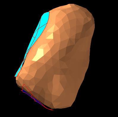
49 Figure 3-5 Dorsal view of the scaphoid with Its Articulation Surfaces, cyan surface articulates with radius, white surface articulates with lunate, yellow surface on the right articulates with capitate, the other yellow surface at the top articulates with trapezium, and dark green surface articulates with trapezoid. 38

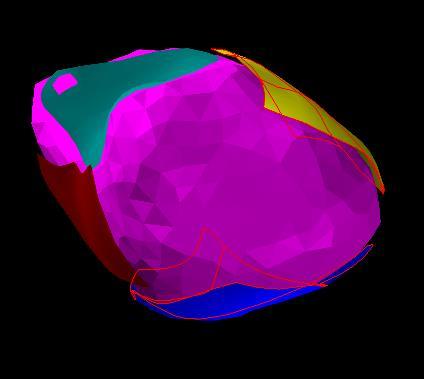
50 Figure 3-6 Radio-scaphoid articulation with the smooth gliding surfaces where the radius has two surfaces - purple surface to articulate with scaphoid and green surface to articulate with lunate these surfaces are fixed at certain distances from its bone. 39


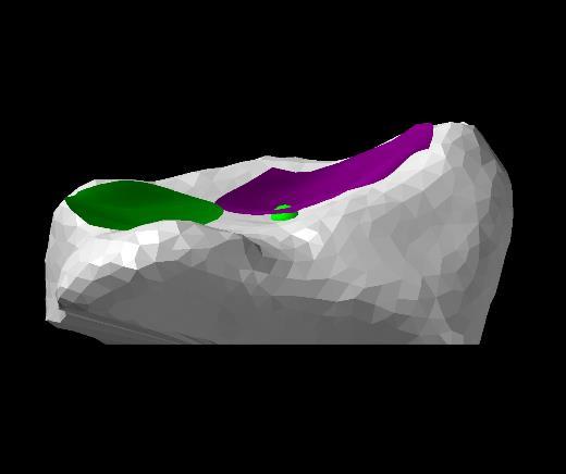
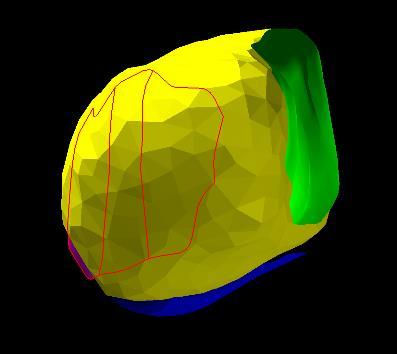
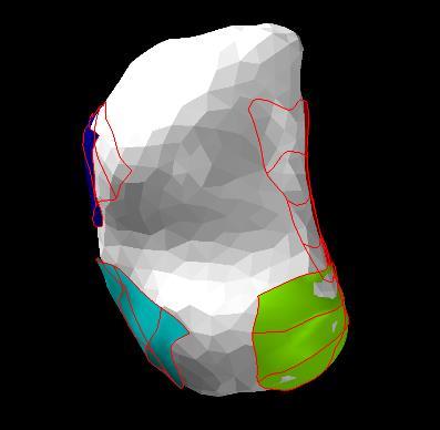
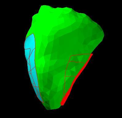
51 A B C D E F G H Figure 3-7 Wrist bones with their articulating surfaces; A, distal radius; B, scaphoid; C, lunate; D, triquetrum; E, trapezium; F, trapezoid; G, capitate; H, hamate. 40
52 The interaction between the two articulating surfaces is established by defining contact force elements in RecurDyn. In RecurDyn, several types of contacts are available. In our study, surface-to-surface contact is used. The contact force elements characterize the behavior of the articulations between the bones. These contact force elements generate forces between interacting structures, in this case, the articulating surfaces of the bones. The contact force element approximates the interaction of contact surfaces as multiple triangular patches. The patch-net and its triangle sizes are set automatically by RecurDyn to achieve a reasonable approximation of the contact behavior and a good computational efficiency. In RecurDyn, the contact normal force is defined as an equation of penetration as follows: F n = kδ m + cδδ n 3.1 Where δ and δ are the penetration and its velocity, respectively, k is the spring coefficient, c is the damping coefficient, order m is the stiffness exponent, and order n is the indentation exponent. The stiffness exponent (m) is used to ensure a non-linear behavior of the contact force. The indentation exponent (n) is used to prevent excessive generation of damping force when relative indentation is very small. In RecurDyn, this equation is used as the default contact equation. This contact force model was found previously [9] to have a comparable behavior to that of the normal carpal bone cartilage contact. The contact stiffness coefficient is set to 200 N/mm based on previous studies that modeled the wrist and its articulations [27-29]. Other parameters are set based on trial and error to provide timely response in the numerical simulation without numerical instability. These parameters are set as follows: the damping coefficient is 2 Ns/mm, the stiffness exponent is 1.3, and the indentation exponent is 2 as in Figure 3-8. In this study, the simple wrist movements are stimulated, therefore, small loads are expected on the articulating surfaces. The suggested parameters values led to reasonable performance in this case. In a normal human joint, friction coefficient is low [85], therefore, no friction forces were simulated. 41

53 Figure 3-8 The Contact Definition dialog in RecurDyn. The carpometacarpal joints were fused since their contribution to wrist motion is very minimal and to improve computational time [2-4, 6-8, 10, 12, 14, 16, 18, 20, 21]. This was done to the piso-triquetral joint as well, for the same reason. The distal radio-ulnar joint is not included in this study because this joint has a negligible effect in radiocarpal and inter-carpal articulations which are the main articulations considered in this study. 3.3 Ligament Modeling The intrinsic and extrinsic wrist ligaments are considered the major stabilizers of most articulations in the wrist. The attachment sites and the surface area of each ligament are well 42
![described in the literature[86]. Figure 3-9 shows the attachment site of the Dorsal Radio-Carpal ligament (DRC) [86]. This ligament connects the distal radius dorsally to the triquetrum.](/docs-images/77/74936297/images/54-0.jpg "This ligament has an intermediate attachment to the dorsum of the lunate. This intermediate connection was not modeled in this study for the sake of simplicity and computational efficiency.")
54 described in the literature[86]. Figure 3-9 shows the attachment site of the Dorsal Radio-Carpal ligament (DRC) [86]. This ligament connects the distal radius dorsally to the triquetrum. This ligament has an intermediate attachment to the dorsum of the lunate. This intermediate connection was not modeled in this study for the sake of simplicity and computational efficiency. In the wrist model, nonlinear tension-only spring elements are used to represent extrinsic and intrinsic ligaments. The major intrinsic and extrinsic ligaments were modeled in this study. Ligaments of fused joints were not included. Table 3-2 and Table 3-3 list the summary of the wrist ligaments. Figure 3-14 and Figure 3-15 show the modeled dorsal and volar wrist ligaments, respectively. Figure 3-9 Dorsal Radio-Carpal Ligament Attachment Sites [86]. Notice the intermediate connection to lunate which was not included in our study. Two wrist models were created with two different ligament modeling approaches. The Single Spring Ligament (SSL) model has each ligament or a major ligament component represented 43
 model has two spring elements representing each ligament or major ligament component.")
55 by a single spring element. The single spring is placed along the central axis of the ligament as much as possible. The Double Spring Ligament (DSL) model has two spring elements representing each ligament or major ligament component. Each of the two springs in the DSL model are placed at the two ends of the plane with the largest distance in its cross-section. To demonstrate, Figure 3-10 shows the selected attachment sites of the dorsal radio-carpal (DRC) ligament in SSL model. Notice that the spring element is placed along the central axis of the ligament. Figure 3-11 shows the two springs that represent the DRC ligament in DSL model. The two springs are placed at the two ends of the longest axis in cross-section. Figure 3-10 Dorsal Radio-Carpal (DRC) ligament is represented in the SSL model by a single spring element (gray line) placed along the ligament central axis. Notice that the intermediate attachment to lunate was not modeled to reduce the complexity of the model. At the middle of the line representing the spring element is a label which is used in RecurDyn to mark each element that is not shown clearly here because of the low resolution. 44

56 Figure 3-11 Dorsal Radio-Carpal (DRC) ligament is represented by two spring elements (gray lines) in the DSL model. The two springs are placed at the two ends of the longest axis of ligament cross-section. Some ligaments with multiple components are represented by one or more spring elements for each component depending on the site of the component and its biomechanical importance. For example, the scapholunate ligament which has three components dorsal, volar, and interosseous components, is either represented by 2 spring element in SSL model or by 4 spring elements in DSL model. Figure 3-12 shows the scapholunate ligaments with a single spring for each of the dorsal and volar components in SSL model. Figure 3-13 illustrates the scapholunate ligament in DSL model with two springs for each of the dorsal and volar components. Figure 3-14 and Figure 3-15 show the wrist ligaments depicted by gray lines modeled in the SSL model from dorsal and volar views respectively. Figure 3-16 and Figure 3-17 demonstrate all the ligaments in the DSL model shown as gray lines connecting the wrist bones from dorsal and volar views respectively. 45
.")
57 Figure 3-12 Scapholunate ligament dorsal and volar components are each represented by a single spring elements (red lines). Notice the labels for each red line indicating the RecurDyn element type. 46

58 Figure 3-13 Scapholunate ligament dorsal and volar components are each represented by two spring elements. Two red lines dorsally for dorsal scapholunate ligament and two red lines volarly for the volar scapholunate ligament. Table 3-2 Wrist extrinsic ligaments. Numbers in round brackets represent ligament stiffness in N/mm. Wrist Extrinsic Ligaments bone 1 carpal 2 Ligament Components DSL springs (stiffness for each) SSL springs (stiffness for each) Stiffness (reference) 47
59 Radius Scaphoid Radial dorsal 1 central 50 collateral [29, 87] and volar (50) [29, 87] ligament (25) Radius Lunate Dorsal 3 radio- 2 1 central 75 triquetrum radiocarpal lunate, luno- proximal Radio- [28] ligament triquetral, radio- triquetral radio- triquetral (75) triquetral and distal [86] radiotriquetral (37.5) Radius Lunate radiolunate 2-4 long 2 long 75 Short and (proximal and short [28] long and distal) (central) [86] and short (37.5) (proximal and distal) (18.75) Ulna Lunate Ulno-lunate 1 2 medial 1 central 40 [28] and lateral (40) [28] (20) Ulna Triquetrum Ulno- 1 2 medial 1 central 40 triquetral [28] and lateral (40) [28] (20) 48
60 Ulna Capitate Ulno- 1 2 medial 1 central 50 capitate [28] and lateral (50) [28] (25) Table 3-3 Wrist intrinsic ligaments. Numbers in round brackets are ligament stiffness in N/mm. Wrist Intrinsic Ligaments Carpal 1 Carpal 2 Ligament Components DSL Springs (stiffnes s for each) SSL Springs (stiffnes s for each) Stiffness (referenc e) Scaphoid Lunate- Dorsal inter triquetru Carpal ligament scapholunate (25) (50) [28] m and lunotriquetral [57] scaphoid lunate Scapholunate(4) 3 dorsal, volar, (57.5) (115) [28] interosseous (not modeled) [57] scaphoid capitate Volar One ligament scaphocapitate [86] (20) (40) [28] 49
61 scaphoid trapezium Palmar Inter Two radial Carpal Ligament and ulnar (50) (150) [28] - scaphotrapezial branches [86] scaphoid trapezoid Scaphotrapezoid One Not modeled al component (volar) [86] Lunate Triquetru Luno-triquetral 3 volar, m dorsal, and (87.5) (175) [28] interosseous (not modeled) [57] Triquetru Capitate Volar One m Triquetrocapitat [28] proximal Central [28] e and (40) distal (20) Triquetru hamate Dorsal One m triquetrohamate [86, 87] proximal Central [28] and (50) distal (25) Trapeziu Trapezoid Palmar trapezio- 3 components m trapezoid and dorsal, volar, dorsal dorsal [28, 88] 50
62 dorsal trapezio- and and 2 and 1 trapezoid interosseous(n volar volar ot modeled) (37.5) (75) [86] Capitate hamate Capitohamte 3 dorsal, volar, dorsal dorsal [28] interosseous and 2 and 1 (not modeled) volar volar [86] (65) (162.5) Capitate Trapeziu volar One m capitotrapezim [29, 86, 87] Proxima Central [29, 87] l and (300) distal (150) Capitate Trapezoid Volar and dorsal 3 volar, capitotrapezoid dorsal, 2 dorsal Dorsal [28] interosseous (proxim and (not modeled) al and central, [86] distal) volar and 2 and volar central (proxim (150) al and distal) (75) 51
63 Pisiform Hamate Pisohamate One [86] medial central [28] and 1 (50) lateral (25) Figure 3-14 Single Spring Ligament (SSL) model showing the wrist ligaments depicted by gray lines connecting carpal bones from dorsal view. 52

64 Figure 3-15 Single Spring Ligament (SSL) model showing wrist ligaments depicted by gray lines connecting carpal bones form a volar view. 53

65 Figure 3-16 Double Spring Ligament (DSL) model showing wrist ligaments depicted by gray lines connecting carpal bones form a dorsal view. 54
66 Figure 3-17 Double Spring Ligament (DSL) model showing wrist ligaments depicted by gray lines connecting carpal bones from a volar view. Ligaments are viscoelastic structures [89-91] and they have a nonlinear response [22, 28, 30, 92, 93]. The load-deflection behavior is modeled using the superposition of a linear and an exponential function[30]: f(ε) = k l 0 (ε 0.03) k l 0 exp ( ε 0.03 ) 3.2 ε = l l 0 l Where k is the ligament stiffness, l is the length of the spring element during the simulation and l 0 is the spring element initial length at the beginning of the simulation. The initial ligament length l 0 55
67 is measured in RecurDyn before the simulation for each spring. Each ligament will exert tensile force once the spring element length during simulation exceeds that of the initial length. This equation provides a nonlinear tensile force after crossing the initial length. The factor 0.03 describes the parabolic toe region to be mostly in the first three percent of the stress-strain curve[29, 30]. The ligament stiffness values are obtained from previous biomechanical studies [4, 28, 29, 52, 88, 93]. The stiffness values used for each ligament in the SSL and DSL models are listed in Table 3-2 and Table 3-3. The study and understanding of wrist ligaments anatomical and biomechanical structures evolved over the last several years. When the stiffness value of a particular ligament is not available in the literature, then it is set to the stiffness of a similar ligament that is closest in anatomy. When a single spring is used to represent a ligament, the spring stiffness is set to take the whole ligament stiffness value. When more than one spring is used to model a ligament, then each spring stiffness is given by the total ligament stiffness divided by the number of springs used. 3.4 Tendon Modeling Five muscle tendons were modeled in this study. These included the extensor carpi radialis longus (ECRL), extensor carpi radialis brevis (ECRB), extensor carpi ulnaris (ECU), flexor carpi radialis (FCR), and flexor carpi ulnaris (FCU)[94]. The wrist tendons are modeled using RecurDyn axial force elements. Axial force element applies force to two markers on bodies it is attached to. The force magnitude can be defined using elements of displacements, velocities, forces, variables, and time. Each tendon was modeled using one axial force element. This element has two attachment sites. Distally (insertion site), it is attached to the center of surface area of tendon insertion site. The insertion sites are all based on previous cadaveric studies on the anatomy of the human wrist. Proximally (origin site), these elements cannot be attached to the corresponding muscles origins. The wrist muscles originates proximally from different sites, mainly of the distal humerus. The extensor tendons (dorsally) pass 56
68 through fibrous tunnels at the dorsum of distal forearm called wrist extensor compartments. These help control the direction of these tendons. The force elements representing these muscles were attached proximally to the area of the tendon where the first constraining tunnels are. This helps maintain the direction of pull of these tendons. The flexor carpi radialis (FCR) passes volarly through a tunnel at the most proximal part of the wrist formed by the flexor retinaculum. Flexor carpi ulnaris (FCU) tendon has no distinct tunnel to pass through but its insertion is far more proximal than other wrist muscles. Table 3-4 lists the wrist tendons modeled and their insertion sites in the wrist model. Table 3-4 Wrist muscles and their corresponding force elements in wrist model; ECRL, extensor carpi radialis longus; ECRB, extensor carpi radialis brevis; ECU, extensor carpi ulnaris; FCR, flexor carpi radialis; FCU, flexor carpi ulnaris; EC, extensor compartment. Tendon Origin model Origin actual Insertion Insertion - actual model ECRL 2 ND EC, dorsal Humerus Base of 2 nd base of 2 nd and 1cm radial lateral metacarpal metacarpal - to Lister supracondylar radial and dorsal radial and dorsal Tubercle ridge side side Lateral intermuscular septum Humerus lateral epicondyle a few fibers 57
69 ECRB 2 nd EC, dorsal humerus Base of 3 rd mainly base and just radial to lateral epicondyle metacarpal of 3 rd metacarpal Lister Tubercle radial collateral dorso-latera dorso-lateral ligament aspect aspect intermuscular small part septa base of 2 nd metacarpal - dorso-medial aspect ECU 6 th EC, dorsal humerus Base of 5 th base of 5 th and ulnar to lateral epicondyle metacarpal metacarpal head of ulna posterior ulnar side border of ulna FCR FCR tunnel at humerus Base of 2 nd mainly base mid wrist, volar medial metacarpal of 2 nd metacarpal and over medial epicondyle volar and ulnar anterior aspect aspect of aspect small slip scaphoid volar base of 3 rd tubercle metacarpal volar aspect small slip trapezial tuberosity 58
70 FCU Volar to middle humeral head Center of the pisiform of ulnar head medial volar aspect of by ligaments volar surface epicondyle pisiform into hamate and ulnar head 5 th metacarpal medial margin of olecranon and proximal 2/3 of dorsal ulna To simulate different wrist movements, different axial forces are applied to the wrist model. Six major wrist movements are simulated. These are: 30 flexion, 65 flexion, 34 extension, 57 extension, ulnar deviation, and radial deviation. The three-dimensional coordinates and orientation of each bone of the wrist in each wrist position are available from CT scan data of a previous study[29]. In our study, the axial force varies according to the motion simulated. The three-dimensional target position of the wrist of each movement is used to compute the force needed to perform the wrist movement. The capitate bone is selected to perform this analysis since it is at the center of the wrist in the lateral axis and is most distal in axial axis [12, 20, 21]. The center of rotation (COR) for radial-ulnar deviation is within the head of capitate. The center of rotation of flexion-extension motion in the proximal row is about the concave surface of the lunate, whereas in the distal row, it is in the capitate neck. A common COR occurs between these two just inside the capitate head as shown in Figure This makes the movement of the capitate the most predictable when applying forces to the wrist tendons. A tracking marker is created in the capitate model at a specific distal and dorsal position relative to the capitate reference frame. Similar tracking markers are created for each capitate in the different wrist positions at the exact same linear and angular position. The x and y displacement components are used in the tendon force equation 59
71 since motion along the lateral axis (x-axis) and along the dorso-volar axis (y axis) are the main components in every wrist motion. Carpal bone motion along the z axis is small and is neglected. Figure 3-18 The common center of rotation (COR) of the wrist in neutral-radial deviation, neutral-ulnar deviation and neutral-flexion/extension are shown respectively [20]. Tracking markers are attached to the capitate in all desired motion position (flexed, extended, ulnarly deviated and radially deviated). The capitate in desired positions are fixed to the simulation environment. For each movement to be simulated, the X and Y distances between the tracking marker of the model capitate in simulation and the capitate in desired position are observed and used to govern the force acting in each tendon. The global COR of the wrist is in the capitate head. Tendons radial to that COR are considered wrist radial deviators (ECRB, ECRL and FCR). Tendons ulnar to the global COR are considered wrist ulnar deviators (ECU and FCU). Tendons that are volar to wrist COR are the wrist flexors (FCR and FCU) whereas tendons dorsal to this COR are wrist extensors (ECRL, ECRB and ECU). Figure 3-19 and Figure 3-20 show the capitate in neutral, radial deviation and ulnar deviation positions with their corresponding tracking markers from anteroposterior and lateral views respectively. To simulate radial deviation, for example, Figure 3-19 shows the X displacement between simulating capitate and capitate in radial deviation is negative, and the radial 60

72 deviators need to contract to minimize this distance whereas ulnar deviators should have minimal contraction to allow radial deviation. The minimal force applied in the opposite direction is to simulate a relaxed muscle tone. Notice that capitate in radial deviation is slightly extended as in Figure 3-20, so, the Y displacement is negative and the wrist extensors need to contract whereas wrist flexors have minimal force to allow extension while maintaining tone. Figure 3-19 The capitate from dorso-volar view in neutral position (white), radial deviation (light brown) and ulnar deviation (gray). Notice the tracking markers attached to capitate in different position. R, radial side; U, ulnar side. 61

73 Figure 3-20 The capitate from radio-ulnar view (lateral view) in neutral position (white), radial deviation (light brown) and ulnar deviation (gray). Notice the tracking markers attached to each one. Radially deviated capitate is slightly extended. V, volar; D, dorsal. In RecurDyn, a feedback based control is not available. To fit within the constraints of the software, the forces are modeled using two different functions utilizing the displacement components. First, the force is modeled using an exponential function (EF). As the distance increases between the target and original capitate, the force increases exponentially and vice versa. f tendon 1 dx 1 dy 3.4 Where dx is the x displacement component and dy is the y displacement component, negative sign to indicate tension force. The force should be greater than or equal to 0 to prevent force in opposite 62
Introduction. The wrist contains eight small carpal bones, which as a group act as a flexible spacer between the forearm and hand.
 Wrist Introduction The wrist contains eight small carpal bones, which as a group act as a flexible spacer between the forearm and hand. Distal forearm Distal forearm 4 Distal end of the radius A. anterior
Forearm and Wrist Regions Neumann Chapter 7
 Forearm and Wrist Regions Neumann Chapter 7 REVIEW AND HIGHLIGHTS OF OSTEOLOGY & ARTHROLOGY Radius dorsal radial tubercle radial styloid process Ulna ulnar styloid process ulnar head Carpals Proximal Row
10/15/2014. Wrist. Clarification of Terms. Clarification of Terms cont
 Wrist Clarification of Terms Palmar is synonymous with anterior aspect of the wrist and hand Ventral is also synonymous with anterior aspect of the wrist and hand Dorsal refers to the posterior aspect
Wrist and Hand Anatomy
 Wrist and Hand Anatomy Bone Anatomy Scapoid Lunate Triquetrium Pisiform Trapeziod Trapezium Capitate Hamate Wrist Articulations Radiocarpal Joint Proximal portion Distal portion Most surface contact found
Wrist and Hand Anatomy/Biomechanics
 Wrist and Hand Anatomy/Biomechanics Kristin Kelley, DPT, OCS, FAAOMPT Orthopaedic Manual Physical Therapy Series Charlottesville 2017-2018 Orthopaedic Manual Physical Therapy Series 2017-2018 Anatomy -
Anatomy - Hand. Wrist and Hand Anatomy/Biomechanics. Osteology. Carpal Arch. Property of VOMPTI, LLC
 Wrist and Hand Anatomy/Biomechanics Kristin Kelley, DPT, OCS, FAAOMPT The wrist The metacarpals The Phalanges Digit 1 thumb Digit 5 digiti minimi Anatomy - Hand Orthopaedic Manual Physical Therapy Series
Sean Walsh Orthopaedic Surgeon Dorset County Hospital
 Sean Walsh Orthopaedic Surgeon Dorset County Hospital Shapes and orientation of articular surfaces Ligaments Oblique positioning of scaphoid Tendons surrounding the joints Other soft tissues Peripheral
8/25/2014. Radiocarpal Joint. Midcarpal Joint. Osteology of the Wrist
 Structure and Function of the Wrist 2 joints and 10 different bones Combine to create wrist motion Anatomical Terms: Wrist/Hand Palmar = anterior aspect of the wrist and hand Dorsal = posterior aspect
Wrist & Hand Ultrasonography 대구가톨릭대학교병원재활의학과 권동락
 Wrist & Hand Ultrasonography 대구가톨릭대학교병원재활의학과 권동락 Dorsal Wrist Evaluation (1 st Compartment) EPB APL Transverse View APL, abductor pollicis longus; EPB, extensor pollicis brevis Dorsal Wrist Evaluation
SURGICAL/APPLIED ANATOMY
 Página 1 de 11 Copyright 2001 Lippincott Williams & Wilkins Bucholz, Robert W., Heckman, James D. Rockwood & Green's Fractures in Adults, 5th Edition SURGICAL/APPLIED ANATOMY Part of "19 - FRACTURES AND
Trapezium is by the thumb, Trapezoid is inside
 Trapezium is by the thumb, Trapezoid is inside Intercarpal Jt Radiocarpal Jt Distal Middle Proximal DIP PIP Interphalangeal Jts Metacarpalphalangeal (MCP) Jt Metacarpal Carpometacarpal (CMC) Jt Trapezium
Ultrasonography of the wrist - a step-by-step approach to study protocols and normal findings
 Ultrasonography of the wrist - a step-by-step approach to study protocols and normal findings Poster No.: C-1779 Congress: ECR 2016 Type: Educational Exhibit Authors: R. R. Domingues Madaleno, A. P. Pissarra,
Netter's Anatomy Flash Cards Section 6 List 4 th Edition
 Netter's Anatomy Flash Cards Section 6 List 4 th Edition https://www.memrise.com/course/1577581/ Section 6 Upper Limb (66 cards) Plate 6-1 Humerus and Scapula: Anterior View 1.1 Acromion 1.2 Greater tubercle
Ligaments of Elbow hinge: sagittal plane so need lateral and medial ligaments
 Ligaments of Elbow hinge: sagittal plane so need lateral and medial ligaments Ulnar Collateral ligament on medial side; arising from medial epicondyle and stops excess valgus movement (lateral movement)
SIMULATION OF WRIST KINEMATICS ON THE BASIS OF A RIGID BODY SPRING MODEL
 SIMULATION OF WRIST KINEMATICS ON THE BASIS OF A RIGID BODY SPRING MODEL by Simon Fischli A thesis submitted to the Department of Mechanical and Materials Engineering in conformity with the requirements
THE DESIGN AND VALIDATION OF A COMPUTATIONAL MODEL OF THE HUMAN WRIST JOINT
 Virginia Commonwealth University VCU Scholars Compass Theses and Dissertations Graduate School 2013 THE DESIGN AND VALIDATION OF A COMPUTATIONAL MODEL OF THE HUMAN WRIST JOINT Afsarul Mir Virginia Commonwealth
This presentation is the intellectual property of the author. Contact them for permission to reprint and/or distribute.
 The Stiff Hand: Manual Therapy Sylvia Dávila, PT, CHT San Antonio, Texas Orthopedic Manual Therapy Common Applications Passive stretch Tensile force to tissue to increase extensibility of length & ROM
MCQWeek2. All arise from the common flexor origin. The posterior aspect of the medial epicondyle is the common flexor origin.
 MCQWeek2. 1. Regarding superficial muscles of anterior compartment of the forearm: All arise from the common flexor origin. The posterior aspect of the medial epicondyle is the common flexor origin. Flexor
LECTURE 8 HANDS: BONES AND MUSCLES
 LECTURE 8 HANDS: BONES AND MUSCLES WRIST AND HAND - Human hand can do power grip and precision grip - Thumb is 90 to the rest of the hand can do fine actions - Often able to do power actions o Take tools
Chapter 7. Anatomy of the Triangular Fibrocartilage Complex: Current Concepts. Introduction. Anatomy. Histology
 Chapter 7 Anatomy of the Triangular Fibrocartilage Complex: Current Concepts Introduction The triangular fibrocartilage complex (TFCC) is one of the intrinsic ligaments of the wrist. It is often injured
3. Ulno lunate, Ulno triquetral ligament. Poirier: Between RSC &LRL. 5. Dorsal intercarpal ligament
 CARPAL INSTABILITY Ligaments Intrinsic Scapho lunate ligament: Dorsal component stronger than volar ligament Luno triquetral ligament: Volar component stronger than dorsal ligament Extrinsic Palmar 1 Radio
Main Menu. Wrist and Hand Joints click here. The Power is in Your Hands
 1 The Wrist and Hand Joints click here Main Menu K.5 http://www.handsonlineeducation.com/classes/k5/k5entry.htm[3/23/18, 1:40:40 PM] Bones 29 bones, including radius and ulna 8 carpal bones in 2 rows of
ARM Brachium Musculature
 ARM Brachium Musculature Coracobrachialis coracoid process of the scapula medial shaft of the humerus at about its middle 1. flexes the humerus 2. assists to adduct the humerus Blood: muscular branches
Hand and wrist emergencies
 Chapter1 Hand and wrist emergencies Carl A. Germann Distal radius and ulnar injuries PEARL: Fractures of the distal radius and ulna are the most common type of fractures in patients younger than 75 years.
PREVIEW ONLY 27/10/2014. Instabilities in the Wrist
 Be sure to convert to your own time zone at Andrew Ellis BSc (Ex. Sci), M. Phty Instabilities in the Wrist Presented by: Ben Cunningham Be sure to convert to your own time zone at Ben Cunningham Member
A Biomechanical Investigation of Load Sharing at the Distal Forearm
 Western University Scholarship@Western Electronic Thesis and Dissertation Repository October 2017 A Biomechanical Investigation of Load Sharing at the Distal Forearm Martine E. McGregor The University
(i) Examination of the wrist surface anatomy of the carpal bones
 Examination of the wrist surface anatomy of the carpal bones") Current Orthopaedics (2005) 19, 171 179 www.elsevier.com/locate/cuor MINI-SYMPOSIUM: THE WRIST (i) Examination of the wrist surface anatomy of the carpal bones R. Srinivas Reddy, J. Compson Upper Limb
Current Orthopaedics (2005) 19, 171 179 www.elsevier.com/locate/cuor MINI-SYMPOSIUM: THE WRIST (i) Examination of the wrist surface anatomy of the carpal bones R. Srinivas Reddy, J. Compson Upper Limb
EXAMINATION OF THE WRIST BEYOND THE BASICS OMA SPORT MED Janice Harvey MD CCFP CFFP Dip. Sp Med.
 EXAMINATION OF THE WRIST BEYOND THE BASICS OMA SPORT MED 2019 Janice Harvey MD CCFP CFFP Dip. Sp Med. CFPC CoI Templates: Slide 1 used in Faculty presentation only. FACULTY/PRESENTER DISCLOSURE Faculty:
The Elbow and the cubital fossa. Prof Oluwadiya Kehinde
 The Elbow and the cubital fossa Prof Oluwadiya Kehinde www.oluwadiya.com Elbow and Forearm Anatomy The elbow joint is formed by the humerus, radius, and the ulna Bony anatomy of the elbow Distal Humerus
Lecture 9: Forearm bones and muscles
 Lecture 9: Forearm bones and muscles Remember, the region between the shoulder and the elbow = brachium/arm, between elbow and wrist = antebrachium/forearm. Forearm bones : Humerus (distal ends) Radius
MR IMAGING OF THE WRIST
 MR IMAGING OF THE WRIST Wrist Instability Dissociative Pattern apparent on routine radiographs Non-dissociative Stress / positional radiographs Dynamic fluoroscopy during stress Arthrography MRI / MR arthrography
Acute Wrist Injuries OUCH!
 Acute Wrist Injuries OUCH! Case the athlete FOOSH from sporting event 2 days ago C/O wrist swelling, pain, worse with movement Hmmm Wrist pain Exam of the wrist - basics Appearance Swelling, bruising,
Physical therapy of the wrist and hand
 Physical therapy of the wrist and hand Functional anatomy wrist and hand The wrist includes distal radius, scaphoid, lunate, triquetrum, pisiform, trapezium, trapezoid, capitate, and hamate. The hand includes
Functional Anatomy of the Elbow
 Functional Anatomy of the Elbow Orthopedic Institute Daryl C. Osbahr, M.D. Chief of Sports Medicine, Orlando Health Chief Medical Officer, Orlando City Soccer Club Orthopedic Consultant, Washington Nationals
Kinesiology of The Wrist and Hand. Cuneyt Mirzanli Istanbul Gelisim University
 Kinesiology of The Wrist and Hand Cuneyt Mirzanli Istanbul Gelisim University Bones The wrist and hand contain 29 bones including the radius and ulna. There are eight carpal bones in two rows of four to
Wrist & Hand Assessment and General View
 Wrist & Hand Assessment and General View Done by; Mshari S. Alghadier BSc Physical Therapy RHPT 366 m.alghadier@sau.edu.sa http://faculty.sau.edu.sa/m.alghadier/ Functional anatomy The hand can be divided
1/13/2013. Anatomy Guy Dissection Sheet Extensor Forearm and Hand. Eastern Virginia Medical School
 Dr. Craig Goodmurphy Anatomy Guy Superficial Extensor Muscles Complete skin removal if necessary then remove the antebrachial fascia starting at the extensor retinaculum and working proximally. Define
Carpal Instability: Clarification of the Most Common Etiologies and Imaging Findings
 Carpal Instability: Clarification of the Most Common Etiologies and Imaging Findings Corey Matthews DO, Nicholas Strle DO, Donald von Borstel DO Oklahoma State University Medical Center, Department of
Chapter 8. The Pectoral Girdle & Upper Limb
 Chapter 8 The Pectoral Girdle & Upper Limb Pectoral Girdle pectoral girdle (shoulder girdle) supports the arm consists of two on each side of the body // clavicle (collarbone) and scapula (shoulder blade)
Index. Springer International Publishing Switzerland 2016 J.N. Lawton (ed.), Distal Radius Fractures, DOI /
, Distal Radius Fractures, DOI /") Index A AAOS. See American Academy of Orthopaedic Surgeons (AAOS) Abductor pollicis longus (APL) tendon, 34, 73 Acute carpal tunnel syndrome, 93 American Academy of Orthopaedic Surgeons (AAOS), 66, 238
Index A AAOS. See American Academy of Orthopaedic Surgeons (AAOS) Abductor pollicis longus (APL) tendon, 34, 73 Acute carpal tunnel syndrome, 93 American Academy of Orthopaedic Surgeons (AAOS), 66, 238
CHAPTER 6: THE UPPER EXTREMITY: THE ELBOW, FOREARM, WRIST, AND HAND
 CHAPTER 6: THE UPPER EXTREMITY: THE ELBOW, FOREARM, WRIST, AND HAND KINESIOLOGY Scientific Basis of Human Motion, 12 th edition Hamilton, Weimar & Luttgens Presentation Created by TK Koesterer, Ph.D.,
Development of an in-vitro passive and active motion Simulator for the investigation of wrist function and Kinematics
 Western University Scholarship@Western Electronic Thesis and Dissertation Repository January 2016 Development of an in-vitro passive and active motion Simulator for the investigation of wrist function
31yo M with chronic basilar thumb and wrist pain that started after cross-country bicycle ride 5 yrs ago.
 31yo M with chronic basilar thumb and wrist pain that started after cross-country bicycle ride 5 yrs ago. EPL EPB APL Full-thickness tear involving the dorsal deltoid ligament of the first carpometacarpal
Muscular Nomenclature and Kinesiology - One
 Chapter 16 Muscular Nomenclature and Kinesiology - One Lessons 1-3 (with lesson 4) 1 Introduction 122 major muscles covered in this chapter Chapter divided into nine lessons Kinesiology study of human
Lab Activity 11: Group II
 Lab Activity 11: Group II Muscles Martini Chapter 11 Portland Community College BI 231 Origin and Insertion Origin: The place where the fixed end attaches to a bone, cartilage, or connective tissue. Insertion:
Joints of the upper limb II
 Joints of the upper limb II Prof. Abdulameer Al-Nuaimi E-mail: a.al-nuaimi@sheffield.ac.uk E. mail: abdulameerh@yahoo.com Elbow joint The elbow joint is connecting the upper arm to the forearm. It is classed
divided by the bones ( redius and ulna ) and interosseous membrane into :
 and interosseous membrane into :") fossa Cubital Has: * floor. * roof : - Skin - superficial fasica - deep fascia ( include bicipital aponeurosis ) Structures within the roof : -cephalic and basilic veins -and between them median cubital
fossa Cubital Has: * floor. * roof : - Skin - superficial fasica - deep fascia ( include bicipital aponeurosis ) Structures within the roof : -cephalic and basilic veins -and between them median cubital
JOINT MOBILITY Joint Mobility of Upper Extremity
 Kinesiology 2017#5: JOINT MOBILITY Joint Mobility of Upper Extremity Huei-Ming Chai, Ph.D., PT School of Physical Therapy National Taiwan University Functions of Synovial Joints Joint Mobility Osteokinematic
The Forearm 2. Extensor & lateral Compartments of the Forearm
 The Forearm 2 Extensor & lateral Compartments of the Forearm 1-Lateral Fascial Compartment (at the lateral side of the forearm ) *Some books mention the lateral compartment contain just the Brachioradialis
Anatomy Workshop Upper Extremity David Ebaugh, PT, PhD Workshop Leader. Lab Leaders: STATION I BRACHIAL PLEXUS
 Anatomy Workshop Upper Extremity David Ebaugh, PT, PhD Workshop Leader Lab Leaders: STATION I BRACHIAL PLEXUS A. Posterior cervical triangle and axilla B. Formation of plexus 1. Ventral rami C5-T1 2. Trunks
Supplied in part by the musculocutaneous nerve. Forms the axis of rotation in movements of pronation and supination
 Anatomy: Upper limb (15 questions) 1. Latissimus Dorsi: Is innervated by the dorsal scapular nerve Lies above feres major muscle Medially rotates the humerus All of the above 2. Supinator muscle is: Deep
Difference Between Angle You Can Bend Your Left Wrist Back vs Your Right Wrist Jenna Priest Science Department Altoona High School January 25, 2017
 Difference Between Angle You Can Bend Your Left Wrist Back vs Your Right Wrist Jenna Priest Science Department Altoona High School January 25, 2017 Background 1- The wrist joint (also known as the radiocarpal
Elbow, Wrist & Hand Evaluation.
 Elbow, Wrist & Hand Evaluation www.fisiokinesiterapia.biz Common Injuries to the Elbow, Wrist, Hand & Fingers Lateral epicondylitis tennis elbow Medial epicondylitis golfer s s elbow, little league elbow
The skeleton consists of: Bones: special connective tissue, hard. Cartilage: special connective tissue, less hard than bones. Joints: joint is the
 The skeleton consists of: Bones: special connective tissue, hard. Cartilage: special connective tissue, less hard than bones. Joints: joint is the location at witch two bones make contact, whereas ligaments
STRUCTURAL BASIS OF MEDICAL PRACTICE EXAMINATION 5 October 6, 2006
 STRUCTURAL BASIS OF MEDICAL PRACTICE EXAMINATION 5 October 6, 2006 PART l. Answer in the space provided. (8 pts) 1. Identify the structures. (2 pts) B C A. _pisiform B. _ulnar artery A C. _flexor carpi
[[Sally Leaning Towards Peter To Take Cold Hand]]
![[[Sally Leaning Towards Peter To Take Cold Hand]]](/thumbs/84/91174469.jpg "[[Sally Leaning Towards Peter To Take Cold Hand]]") In this lecture we will talk about the bones of the hand, and the muscles and contents of the forearm. *The hand bones are: - Carpal bones. -Metacarpals. -Phalanges. *The carpal bones (wrist bones): They
In this lecture we will talk about the bones of the hand, and the muscles and contents of the forearm. *The hand bones are: - Carpal bones. -Metacarpals. -Phalanges. *The carpal bones (wrist bones): They
Figure 1: Bones of the upper limb
 BONES OF THE APPENDICULAR SKELETON The appendicular skeleton is composed of the 126 bones of the appendages and the pectoral and pelvic girdles, which attach the limbs to the axial skeleton. Although the
10/10/2014. Structure and Function of the Hand. The Hand. Osteology of the Hand
 Structure and Function of the Hand 19 bones and 19 joints are necessary to produce all the motions of the hand The Hand Dorsal aspect Palmar aspect The digits are numbered 1-5 Thumb = #1 Little finger
Lecture 04 Osteology Of Hand
 Lecture 04 Osteology Of Hand By: A. Prof. Dr Farooq A. Khan PMC Date: 09 th Jan. 2018 HAND Distal to wrist joint Divided into three parts: Carpus Metacarpus Digits Bones Carpal bones o Prox row S L Tq
Integra. Spider and Mini Spider Limited Wrist Fusion System SURGICAL TECHNIQUE
 Integra Spider and Mini Spider Limited Wrist Fusion System SURGICAL TECHNIQUE Table of contents Description... 02 Indications... 02 Contraindications... 02 Surgical Technique... 03 Spider Introduction-Four
The Muscular System. Chapter 10 Part C. PowerPoint Lecture Slides prepared by Karen Dunbar Kareiva Ivy Tech Community College
 Chapter 10 Part C The Muscular System Annie Leibovitz/Contact Press Images PowerPoint Lecture Slides prepared by Karen Dunbar Kareiva Ivy Tech Community College Table 10.9: Muscles Crossing the Shoulder
Hand & Wrist Injuries. DR MA Manjra
 Hand & Wrist Injuries DR MA Manjra 1 Background Up to 25% of all athletic injuries General population Sport people Sport specific Position specific Multifaceted Time of season Level of athlete Parents
Clinical examination of the wrist, thumb and hand
 Clinical examination of the wrist, thumb and hand 20 CHAPTER CONTENTS Referred pain 319 History 319 Inspection 320 Functional examination 320 The distal radioulnar joint.............. 320 The wrist.......................
Chapter 8 The Skeletal System: The Appendicular Skeleton. Copyright 2009 John Wiley & Sons, Inc.
 Chapter 8 The Skeletal System: The Appendicular Skeleton Appendicular Skeleton It includes bones of the upper and lower limbs Girdles attach the limbs to the axial skeleton The pectoral girdle consists
Acknowledgement. Here are some flash cards all set up in a "pdf" format for you! Thanks to Laura H. (spring 08)
") Acknowledgement Here are some flash cards all set up in a "pdf" format for you! Thanks to Laura H. (spring 08) for her donation to all my anatomy students! t Here is her suggestion for making flashcards
Acknowledgement Here are some flash cards all set up in a "pdf" format for you! Thanks to Laura H. (spring 08) for her donation to all my anatomy students! t Here is her suggestion for making flashcards
TRIQUETRUM FRACTURE. The triquetrum bone is one of the small bones that make up the carpus.
 TRIQUETRUM FRACTURE Introduction The triquetrum bone is one of the small bones that make up the carpus. It is also known as the triquetral bone, (and in the past the pyramidal or triangular bone) Triquetrum
Introduction to Ultrasound Examination of the Hand and upper
 Introduction to Ultrasound Examination of the Hand and upper Emil Dionysian, M.D. Ultrasound of upper ext. Upside Convenient Opens another exam dimension Can be like a stethoscope Helps 3-D D visualization
Anatomy and Physiology II. Review Shoulder Girdle New Material Upper Extremities - Bones
 Anatomy and Physiology II Review Shoulder Girdle New Material Upper Extremities - Bones Anatomy and Physiology II Shoulder Girdle Review Questions From Last Lecture Can you identify the following muscles?
I-A-1) Non-specific thickening of synovial membrane
 Non-specific thickening of synovial membrane") I-A-1) Non-specific thickening of synovial membrane Grayscale Metatarsal Power Doppler Dorsal aspect of metatarsophalangeal joint in right 1 st toe, longitudinal view Asterisks indicate non-specific thickening
I-A-1) Non-specific thickening of synovial membrane Grayscale Metatarsal Power Doppler Dorsal aspect of metatarsophalangeal joint in right 1 st toe, longitudinal view Asterisks indicate non-specific thickening
Connects arm to thorax 3 joints. Glenohumeral joint Acromioclavicular joint Sternoclavicular joint
 Connects arm to thorax 3 joints Glenohumeral joint Acromioclavicular joint Sternoclavicular joint Scapula Elevation Depression Protraction (abduction) Retraction (adduction) Downward Rotation Upward Rotation
Joint Mobilization: Elbow, Wrist, and Hand
 Joint Mobilization: Elbow, Wrist, and Hand Small Joints of the Hand: MCP, PIP, and DIP: distraction and glides (A/P or P/A) Philadelphia Hand Meeting Monday, March 26, 2018 Jane Fedorczyk, PT, PhD, CHT
SPORTS INJURIES IN HAND
 Grundkurs SGSM-SSMS Sion 2015 SPORTS INJURIES IN HAND Dr S. KŠmpfen EPIDEMIOLOGY Incidence of hand, finger and wrist injuries in sports : 3% Ð 9 % RADIAL-SIDED WRIST PAIN 1)! Distal Radius Fractures 2)!
Scaphoid Fractures. Mohammed Alasmari. Orthopaedic Surgery Demonstrator Majmaah University
 Scaphoid Fractures Mohammed Alasmari Orthopaedic Surgery Demonstrator Majmaah University 1 2 Scaphoid Fractures Introduction Anatomy History Clinical examination Radiographic evaluation Classification
Joints. Vi Michelle Austin
 Joints Vi Michelle Austin Joints Overview A joint, otherwise known as an articulation, is a point at which points connect. They are constructed to allow movement (except for skull bones) and provide mechanical
Practical 2 Worksheet
 Practical 2 Worksheet Upper Extremity BONES 1. Which end of the clavicle is on the lateral side (acromial or sternal)? 2. Describe the difference in the appearance of the acromial and sternal ends of the
Carpal ligaments evaluation with ultrasound and MRI: A pictorial review
 Carpal ligaments evaluation with ultrasound and MRI: A pictorial review Poster No.: C-2246 Congress: ECR 2010 Type: Educational Exhibit Topic: Musculoskeletal Authors: A. Kraus, C. Barwick, S. Wenham,
SCAPHOID FRACTURE. Relevant antomy
 SCAPHOID FRACTURE Relevant antomy The proximal row consists of the scaphoid, the lunate, and the triquetrum. The proximal carpal row is regarded as an intercalated segment The keystone in the coordination
Imaging Key Wrist Ligaments: What the Surgeon Needs the Radiologist to Know
 Musculoskeletal Imaging Review ateni et al. Imaging of Wrist Ligaments Musculoskeletal Imaging Review Downloaded from www.ajronline.org by 46.3.201.114 on 12/07/17 from IP address 46.3.201.114. Copyright
Learning Objectives. 07 Aug 12. Article E-1. At the end of this section the learner will be able to:
 Module 1: Comparative Functional Anatomy and Biomechanics Article E-1 Learning Objectives At the end of this section the learner will be able to: Describe the bones of the equine thoracic Describe the
Chapter 51 Wrist and Forearm Episode Overview
 Chapter 51 Wrist and Forearm Episode Overview 1) Describe normal radiographic relationships: a. Radial length measurement b. Radial inclination c. Volar Tilt d. Scapholunate angle e. Capitolunate angle
Scapholunate Ligament Lesions Imaging Which and when?
 Scapholunate Ligament Lesions Imaging Which and when? Kolo Frank Lesions to scapholunate ligament(sl) Most frequent cause of carpal instability Traumatic tears of SL ligament = most common ligament injury
Triangular Fibrocartilage Complex Repair. The triangular fibrocartilage complex (TFCC) is one of the main stabilizers of the
 is one of the main stabilizers of the") Michelle Brandt and Megan Passarelle Surgical Assignment Due: 11/9/14 Triangular Fibrocartilage Complex Repair Abstract The triangular fibrocartilage complex (TFCC) is one of the main stabilizers of the
Michelle Brandt and Megan Passarelle Surgical Assignment Due: 11/9/14 Triangular Fibrocartilage Complex Repair Abstract The triangular fibrocartilage complex (TFCC) is one of the main stabilizers of the
Levels of the anatomical cuts of the upper extremity RADIUS AND ULNA right
 11 CHAPTER 2 Levels of the anatomical cuts of the upper extremity AND right CUT 1 CUT 4 1 2 3 4 5 6 Isolated fixation of the radius is difficult at this level because of the anterolateral vessels and the
medial half of clavicle; Sternum; upper six costal cartilages External surfaces of ribs 3-5
 MUSCLE ORIGIN INSERTION ACTION NERVE Pectoralis Major medial half of clavicle; Sternum; upper six costal cartilages Lateral lip of intertubercular groove of horizontal adduction Medial and lateral pectoral
Introduction to Human Osteology Chapter 3: Hands and Feet
 Introduction to Human Osteology Chapter 3: Hands and Feet Roberta Hall Kenneth Beals Holm Neumann Georg Neumann Gwyn Madden Revised in 1978, 1984, and 2008 Bones of the Hand Eight carpal bones, in two
REFERENCE DIAGRAMS OF UPPER LIMB MUSCLES: NAMES, LOCATIONS, ATTACHMENTS, FUNCTIONS MUSCLES CONNECTING THE UPPER LIMB TO THE AXIAL SKELETON
 REFERENCE DIAGRAMS OF UPPER LIMB MUSCLES: NAMES, LOCATIONS, ATTACHMENTS, FUNCTIONS MUSCLES CONNECTING THE UPPER LIMB TO THE AXIAL SKELETON A25LAB EXERCISES: UPPER LIMB MUSCLES Page 1 MUSCLES CONNECTING
Chapter 09 Articulations Pearson Education, Inc.
 Chapter 09 Articulations An Introduction to Articulations Articulations Body movement occurs at joints (articulations) where two bones connect Joint Structure Determines direction and distance of movement
Ultrasonography of Peripheral Nerve -upper extremity
 Ultrasonography of Peripheral Nerve -upper extremity Department of Physical Medicine and Rehabilitation Korea University Guro Hospital Korea University College of Medicine Yoon Joon Shik Normal median
Elbow Elbow Anatomy. Flexion extension. Pronation Supination. Anatomy. Anatomy. Romina Astifidis, MS., PT., CHT
 Elbow Elbow Anatomy Romina Astifidis, MS., PT., CHT Curtis National Hand Center Baltimore, MD October 6-8, 2017 Link between the arm and forearm to position the hand in space Not just a hinge Elbow = 70%
Nerves of Upper limb. Dr. Brijendra Singh Professor & Head Department of Anatomy AIIMS Rishikesh
 Nerves of Upper limb Dr. Brijendra Singh Professor & Head Department of Anatomy AIIMS Rishikesh 1 Objectives Origin, course & relation of median & ulnar nerves. Motor & sensory distribution Carpal tunnel
POLSKI 2013, 85, 8,
 POLSKI PRZEGLĄD CHIRURGICZNY 2013, 85, 8, 452 459 10.2478/pjs-2013-0069 Radial wrist extensors as a dynamic stabilizers of scapholunate complex Ahmed Elsaftawy Department of General Surgery, Centre of
Viorel Nacu. The clinical anatomy of the Hand
 Viorel Nacu The clinical anatomy of the Hand The distal part of the upper limb is divided in to three regions: 1. The wrist (carpus) 2. The hand (metacarpus) 3. The digits (fingers) The landmarks of this
Interesting Case Series. Perilunate Dislocation
 Interesting Case Series Perilunate Dislocation Tom Reisler, BSc (Hons), MB ChB, MRCS (Ed), Paul J. Therattil, MD, and Edward S. Lee, MD Division of Plastic and Reconstructive Surgery, Department of Surgery,
Index. Note: Page numbers of article titles are in boldface type. Hand Clin 21 (2005)
") Hand Clin 21 (2005) 501 505 Index Note: Page numbers of article titles are in boldface type. A Antibiotics, following distal radius fracture treatment, 295, 296 Arthritis, following malunion of distal
Hand Clin 21 (2005) 501 505 Index Note: Page numbers of article titles are in boldface type. A Antibiotics, following distal radius fracture treatment, 295, 296 Arthritis, following malunion of distal
THE SHORT DESCRIPTION OF THE JOINTS 1. THE UPPER LIMB (Dr. Dóra Reglődi*, version )
") THE SHORT DESCRIPTION OF THE JOINTS 1. THE UPPER LIMB (Dr. Dóra Reglődi*, version 02-2007) Shoulder girdle The shoulder girdle consists of the clavicle and scapula on both sides. The two sides are connected
THE SHORT DESCRIPTION OF THE JOINTS 1. THE UPPER LIMB (Dr. Dóra Reglődi*, version 02-2007) Shoulder girdle The shoulder girdle consists of the clavicle and scapula on both sides. The two sides are connected
compartments of the forearm
 " forearm posterior compartment " compartments of the forearm Posterior Fascial compartment Muscles: ** The superficial group 1. Extensor carpi radialis brevis 2. Ex. digitorum 3. Ex. digiti minimi 4.
Thesis/Dissertation Collections
 Rochester Institute of Technology RIT Scholar Works Theses Thesis/Dissertation Collections 6-5-1992 The Human wrist James A. Perkins Follow this and additional works at: http://scholarworks.rit.edu/theses
Chapter 12 Distal Ulnar Resection
 Chapter 12 Distal Ulnar Resection Introduction Ulnar impaction syndrome is a common but often unrecognized cause of pain on the ulnar side of the wrist. Although it can be congenital (due to a long ulna),
Anatomy of the Forearm
 Anatomy of the Forearm Musculoskeletal block- Anatomy-lecture 8 Editing file Objectives List the names of the Flexors Group of Forearm (superficial & deep muscles). Identify the common flexor origin of
RADIOGRAPHY OF THE WRIST
 RADIOGRAPHY OF THE WRIST Patient Position: WRIST PA Projection, elbow in same plane Part Position: Hand ; fingers centered to IR Central Ray: Structures Shown: NOTE: Optional AP projection best demonstrates