Anatomy of the Hand and Nomenclature. R K Kankate Specialist Registrar St.George s Hospital
|
|
|
- Lynn Green
- 5 years ago
- Views:
Transcription
1 Anatomy of the Hand and Nomenclature R K Kankate Specialist Registrar St.George s Hospital
2 Bony skeleton muscles and ligaments nervous, arterial and venous system

3 Ossification of bones: carpus Most carpal bones are cartilagenous at birth. Each carpal bone ossifies from 1 centre. Capitate ossifies first and the pisiform last.

4 Ossification of bones: carpus Capitate Hamate 2 months 3 months Triquetral 3 years Lunate Scaphoid 4 years Trapezium 4-5years Trapezoid

5 Ossification of bones: metacarpus Primary and secondary centres primary at 9 months and in order of 2nd to 5th metacarpal upto 2 years 1st metacarpal base at 2-3 years
: distal phalanx represents fused middle + distal")
6 Ossification of bones: metacarpus Thumb metacarpal ossifies like a phalanx some believe that the thumb consists of 3 phallanges Broom(1930): distal phalanx represents fused middle + distal phallanges

7 Ossification of bones: phalanges Primary centre for shaft proximal epiphyseal centre shafts: distal epiphyseal: proximal 1m proximal 10 middle 11 8 wks middle and distal 2m
8
 felt that there were 2 apposing")
9 CMC Joint of the Thumb Classical saddle joint Eaton and Littler(1973) felt that there were 2 apposing saddles
10 CMC Joint of the Thumb 1742: Weitbrecht gave an accurate description Pieron in 1973: Anterior Oblique (volar ligament of Kaplan) Dorsoradial Posterior Oblique Intermetacarpal
: stable central post of the hand for the joint between the capitate and the 3rd")
11 2nd to 5th CMC Joints 2nd to 4th are planar gliding joints 5th is a saddle joint Flatt(1959): stable central post of the hand for the joint between the capitate and the 3rd metacarpal
12 2nd to 5th CMC Joints Limited AP gliding movement in the 2nd and the 3rd cmc jt. Maximum movement between the 4th and the 5th metacarpal bases and the hamate. Average of degrees. Gunther s cadaveric study in 1984 showed 8 degrees in the 4th cmc jt. And 15 degrees in the 5th cmc joint.

13 2nd to 5th CMC Joints 2 important soft tissue relationships to the cmc joints. Motor branch of the ulnar nerve: 5th cmc joint. Deep palmar arterial arch : 3rd cmc joint.
14 Intermetacarpal Joints Dorsal ligament Palmar ligament Interrosseous ligament
15 MCP Joint of the Thumb Condyloid joint very wide ROM: degrees Palmer and Louis felt that the ROM depends on the shape of the head Harris: flat head associated with less movt. And more prone to injury
16 MCP Joint of the Thumb Static stabilisers: collateral ligaments, capsule, dorsal aponeurosis,volar plate Dynamic stabilisers: adductor aponeurosis

17 2nd to 5th MCP Joints Condyloid joint Globular head articulates with the reciprocal concave base of the proximal phalanx
18 2nd to 5 MCP Joints Eaton(1971): Key to the stability is the snug like configuration of the 3 ligaments Primary stabilisers Secondary stabilisers

19 2nd to 5 MCP Joints

20 2nd to 5 MCP Joints Extension: collateral ligament is lax and the joint comtact is minimum Maximum joint capacity: short of full extension
21 Interphalangeal Joint Uniaxial hinge joint bicondylar tongue in groove appearance Kuczynski: sloppy hinge designed mainly for flexion and extension ROM: 0 to 105(120) degrees 7-10 degrees of lateral deviation

22 Interphalangeal Joint Same as the MCP joint Cord like collateral ligaments Volar plate Metacarpoglenoidal ligament Capsule and the long extensor tendon

23 Volar Plate Volar plate is less mobile in the PIP joint than in the MCP joint: Kuczynski

24 Interphalangeal joint: check rein ligaments Swallowtail extensions of the of the volar plate which prevent hyperextension of the pip joint: Bowers
25 Interphalangeal Joint The CAM effect of the PIP joint is less significant than that of the MCP joint.
26 Interphalangeal Joint The tension in the collateral ligaments is the same in flexion and extension in the PIP joint.

27 Dorsal Plate of the PIP Joint P G Slattery: J of Hand Surg. 15 B, 1990 histologically similar to the fibrocart. volar plate constant structure in 70 cadaveric fingers

28 Dorsal Plate of the PIP Joint Morphologically similar to the patella

29 Dorsal Plate of the PIP Joint First described by Jacob and Testut in 1906 as fibrocartilagenous glenoidien

30 Dorsal Plate of the PIP Joint: functions Stability of extensor tendon adds to the stability of the PIP joint increases the moment arm of the tendon prevents attrition of the extensor tendon in extreme flexion circulation of the synovial fluid

31 Palmar Aponeurosis 2 layers of deep fascia in the hand deep: covers the interrosseous muscles superficial: palmar aponeurosis

32 Palmar Aponeurosis Originates from the palmaris longus muscle function: improves grip strength and prevents the flexor tendons from bowstringing in the palm

33 Palmar Aponeurosis thins out laterally consists of longitudinal and transverse fibres continues with flexor tendon sheaths of the fingers
34 Palmar Aponeurosis

35 Midpalmar spaces of the hand 3 compartments in the palm: thenar hypothenar intermediate lateral medial

36 Synovial sheaths of the palm Flexor tendons are surrounded by these sheaths Visceral layer Parietal layer Each finger sheath is a double walled hollow tube sealed at both ends
37 Flexor digital sheath Cul-de-sacs -

38 Synovial sheaths of the palm 3 sheaths FPL FCR FDP + FDS (common flexor sheath) In 70% the sheath of the little finger is continuos with the common flexor sheath

39 Synovial sheaths of the palm In 50% there is a communication between the common flexor sheath and the sheath for the FPL at the wrist.
40 Long Flexor tendons: Nutrition Tendon nutrition synovial fluid vincular circulation Vinculum longus superficialis Vinculum longus profundus Vinculum breve superficialis Vinculum breve profundus Potenza(1963): diffusion is most important Manske: both are important
41 Long Flexor tendons: Nutrition Vincula are folds of periosteum carying blood supply to the tendons they exit on the dorsum of the tendon and hence sutures on the volar aspect are advocated
42

43 Long Flexor tendons: nerve supply Mechanoreceptors and free nerve endings present in the fleoxor tendons: Zimny 1986 branch of the digital nerve, accompanying the digital artery present within the vinculum brevis
44 Zones of flexor tendons: Verdan s Zone System
45 Zones of flexor tendons: Verdan s Zone System

46 Pulley system of the fingers 5 annular and 3 cruciate pulleys annular: thicker and prevents tendon bowstringing during flexion cruciate: thinner and allows the sheath to conform in flexion by approximating the annular pulleys.
47 Pulley system of the fingers
48 Pulley system of the fingers A2 and A4 are the most important for optimum tendon function
49 Significance of A1 pulley in RA L T Jager et al J HAND Surg minimal bowstringing MCP volar subluxation with loss of terminal flexion (11 degrees)

50 Pulley system of the thumb 3 constant pulleys: A1 pulley Oblique pulley A2 pulley

51 Thenar eminence Opponens pollicis Abductor pollicis brevis Flexor pollicis brevis

52 Hypothenar eminence Abductor digiti minimi Opponens digiti minimi Flexor digiti minimi
 4 dorsal")
53 Intrinsic muscles Lumbricals: origin from FDP flexion at MCP joint part of extensor apparatus Interrossei 4 palmar (PAD) 4 dorsal (DAB)
54 Extensor apparatus of the hand 2 separate and neurologically independent systems: radial nerve innervated extrinsic extensors ulnar and median nerve innervated intrinsic muscles.
55 Extensor apparatus of the hand Extrinsic extensors arise from multiple muscle bellies in the forearm Ext. digitorum Ext. pollicis longus Ext. pollicis brevis Ext. indicis Ext. digiti minimi
56 Extensor apparatus of the hand Kaplan: Ext. digitorum has 4 distinct tendons Doyle: Ext. digitorum to the little finger is present in less than 50% of fingers and when absent it is replaced by juncturae tendinum of ring finger
57
 Extensor retinaculum is a wide fibrous band measuring 2.9-8.4 cm (avg.")
58 Extensor apparatus of the hand: wrist 6 extensor tunnels: 5 fibrosseous and 1 fibrous( 5th) Extensor retinaculum is a wide fibrous band measuring cm (avg.4.9 cm)
 ECU")
59 Extensor apparatus of the hand: wrist EPB/APL ECRB/ECRL EPL ED/EI EDM (fibrous tunnel) ECU iii
60 Extensor apparatus of the hand: MCP joint Central extensor tendon conjoined tendon of interrossei and the lumbricals transverse lamina (dorsal digital expansion) sagittal band of the volar plate
61 Extensor apparatus of the hand: MCP joint

62 Extensor apparatus of the hand: MCP joint Conjoined tendon is volar to the axis of the MCP axis of rotation Interosseous: medial to central tendon Lumbricals: lateral
63 Extensor apparatus of the hand: MCP joint Extensor hood is movable
64
65 Extensor apparatus of the hand: pip joint Trifurcation of the extensor tendon central slip 2 lateral bands lateral bands dorsal to axis of PIP joint

66 Extensor apparatus of the hand: pip joint
67 Extensor apparatus of the hand: pip joint transverse retinacular ligaments of Landsmeer( link ligaments, 1949)
68 Extensor apparatus of the hand: pip joint
69 Extensor apparatus of the hand: pip joint The fibres are differentialy loaded in flexion, the central fibres are tense in extension, the lateral fibres are tense

70 Extensor apparatus of the hand: dip joint Lateral bands merge to form a conjoint tendon which inserts at the base of the distal phalanx
71 Extensor apparatus of the dip joint-vascular anatomy Warren et al, JBJS 1988 Critical area of avascularity at the osseotendinous junction


72 Extensor tendon: zones of injury- Kleinert and Verdan
73
74
75
76
Wrist and Hand Anatomy
 Wrist and Hand Anatomy Bone Anatomy Scapoid Lunate Triquetrium Pisiform Trapeziod Trapezium Capitate Hamate Wrist Articulations Radiocarpal Joint Proximal portion Distal portion Most surface contact found
Main Menu. Wrist and Hand Joints click here. The Power is in Your Hands
 1 The Wrist and Hand Joints click here Main Menu K.5 http://www.handsonlineeducation.com/classes/k5/k5entry.htm[3/23/18, 1:40:40 PM] Bones 29 bones, including radius and ulna 8 carpal bones in 2 rows of
Wrist and Hand Anatomy/Biomechanics
 Wrist and Hand Anatomy/Biomechanics Kristin Kelley, DPT, OCS, FAAOMPT Orthopaedic Manual Physical Therapy Series Charlottesville 2017-2018 Orthopaedic Manual Physical Therapy Series 2017-2018 Anatomy -
Anatomy - Hand. Wrist and Hand Anatomy/Biomechanics. Osteology. Carpal Arch. Property of VOMPTI, LLC
 Wrist and Hand Anatomy/Biomechanics Kristin Kelley, DPT, OCS, FAAOMPT The wrist The metacarpals The Phalanges Digit 1 thumb Digit 5 digiti minimi Anatomy - Hand Orthopaedic Manual Physical Therapy Series
10/10/2014. Structure and Function of the Hand. The Hand. Osteology of the Hand
 Structure and Function of the Hand 19 bones and 19 joints are necessary to produce all the motions of the hand The Hand Dorsal aspect Palmar aspect The digits are numbered 1-5 Thumb = #1 Little finger
Trapezium is by the thumb, Trapezoid is inside
 Trapezium is by the thumb, Trapezoid is inside Intercarpal Jt Radiocarpal Jt Distal Middle Proximal DIP PIP Interphalangeal Jts Metacarpalphalangeal (MCP) Jt Metacarpal Carpometacarpal (CMC) Jt Trapezium
ARM Brachium Musculature
 ARM Brachium Musculature Coracobrachialis coracoid process of the scapula medial shaft of the humerus at about its middle 1. flexes the humerus 2. assists to adduct the humerus Blood: muscular branches
MCQWeek2. All arise from the common flexor origin. The posterior aspect of the medial epicondyle is the common flexor origin.
 MCQWeek2. 1. Regarding superficial muscles of anterior compartment of the forearm: All arise from the common flexor origin. The posterior aspect of the medial epicondyle is the common flexor origin. Flexor
Kinesiology of The Wrist and Hand. Cuneyt Mirzanli Istanbul Gelisim University
 Kinesiology of The Wrist and Hand Cuneyt Mirzanli Istanbul Gelisim University Bones The wrist and hand contain 29 bones including the radius and ulna. There are eight carpal bones in two rows of four to
Muscles of the hand Prof. Abdulameer Al-Nuaimi
 Muscles of the hand Prof. Abdulameer Al-Nuaimi a.alnuaimi@sheffield.ac.uk abdulameerh@yahoo.com Thenar Muscles Thenar muscles are three short muscles located at base of the thumb. All are innervated by
LECTURE 8 HANDS: BONES AND MUSCLES
 LECTURE 8 HANDS: BONES AND MUSCLES WRIST AND HAND - Human hand can do power grip and precision grip - Thumb is 90 to the rest of the hand can do fine actions - Often able to do power actions o Take tools
Hand and Wrist Editing file. Color Code Important Doctors Notes Notes/Extra explanation
 Hand and Wrist Editing file Color Code Important Doctors Notes Notes/Extra explanation Objectives Describe the anatomy of the deep fascia of the wrist & hand (flexor & extensor retinacula & palmar aponeurosis).
13 13/3/2012. Adel Muhanna
 13 13/3/2012 Adel Muhanna بسم هللا الرحمن الرحيم The Hand Extensor retinaculum: Deep fascia of anterior compartment of the wrist is thickened to form flexor retinaculum : a bridge that have 6 structures
Wrist & Hand Ultrasonography 대구가톨릭대학교병원재활의학과 권동락
 Wrist & Hand Ultrasonography 대구가톨릭대학교병원재활의학과 권동락 Dorsal Wrist Evaluation (1 st Compartment) EPB APL Transverse View APL, abductor pollicis longus; EPB, extensor pollicis brevis Dorsal Wrist Evaluation
Structure and Function of the Hand
 Structure and Function of the Hand Some say it takes a village to raise a child, but it takes 19 bones and 19 joints in the hand for it to function smoothly. The Hand Dorsal aspect 2 3 4 The digits are
Physical therapy of the wrist and hand
 Physical therapy of the wrist and hand Functional anatomy wrist and hand The wrist includes distal radius, scaphoid, lunate, triquetrum, pisiform, trapezium, trapezoid, capitate, and hamate. The hand includes
Nerves of Upper limb. Dr. Brijendra Singh Professor & Head Department of Anatomy AIIMS Rishikesh
 Nerves of Upper limb Dr. Brijendra Singh Professor & Head Department of Anatomy AIIMS Rishikesh 1 Objectives Origin, course & relation of median & ulnar nerves. Motor & sensory distribution Carpal tunnel
The Muscular System. Chapter 10 Part C. PowerPoint Lecture Slides prepared by Karen Dunbar Kareiva Ivy Tech Community College
 Chapter 10 Part C The Muscular System Annie Leibovitz/Contact Press Images PowerPoint Lecture Slides prepared by Karen Dunbar Kareiva Ivy Tech Community College Table 10.9: Muscles Crossing the Shoulder
The hand is full with sweat glands, activated at times of stress. In Slide #2 there was a mistake where the doctor mentioned lateral septum twice.
 We should only know: Name, action & nerve supply Layers - Skin - Superficial fascia - Deep fascia The hand is full with sweat glands, activated at times of stress. Deep fascia In Slide #2 there was a mistake
The hand. it's the most important subject of the upper limb because it has a clinical importance. the palm of the hand**
 Today at 12:48 AM The hand it's the most important subject of the upper limb because it has a clinical importance. the palm of the hand** -the palmar aponeurosis located in the palm of the hand which is
Forearm and Wrist Regions Neumann Chapter 7
 Forearm and Wrist Regions Neumann Chapter 7 REVIEW AND HIGHLIGHTS OF OSTEOLOGY & ARTHROLOGY Radius dorsal radial tubercle radial styloid process Ulna ulnar styloid process ulnar head Carpals Proximal Row
Introduction to Ultrasound Examination of the Hand and upper
 Introduction to Ultrasound Examination of the Hand and upper Emil Dionysian, M.D. Ultrasound of upper ext. Upside Convenient Opens another exam dimension Can be like a stethoscope Helps 3-D D visualization
In the name of Allah, Most gracious, Most merciful
 In the name of Allah, Most gracious, Most merciful This lecture includes the following: The Palmer Oponeurosis. The Carpel tunnel. The palmaris brevis muscle. The anatomical snuffbox. The Fibrous flexor
Ligaments of Elbow hinge: sagittal plane so need lateral and medial ligaments
 Ligaments of Elbow hinge: sagittal plane so need lateral and medial ligaments Ulnar Collateral ligament on medial side; arising from medial epicondyle and stops excess valgus movement (lateral movement)
Biceps Brachii. Muscles of the Arm and Hand 4/4/2017 MR. S. KELLY
 Muscles of the Arm and Hand PSK 4U MR. S. KELLY NORTH GRENVILLE DHS Biceps Brachii Origin: scapula Insertion: radius, fascia of forearm (bicipital aponeurosis) Action: supination and elbow flexion Innervation:
Small muscles of the hand
 By the name of Allah Small muscles of the hand Revision: The palmar aponeurosis is triangular in shape with apex and base. It is divided into 4 bands that radiate to the medial four fingers. Dupuytren
Objectives. How your brain sees your hand. Surface anatomy Bones Joints Muscles Tendons Nerves Arteries
 Hand Therapy Review Course Washington University St. Louis, MO April 7 9, 2017 Over eighty percent of activities of daily living involve grasping or seizing objects with the hand. (Katz et al., 1970) Anatomy
The Forearm 2. Extensor & lateral Compartments of the Forearm
 The Forearm 2 Extensor & lateral Compartments of the Forearm 1-Lateral Fascial Compartment (at the lateral side of the forearm ) *Some books mention the lateral compartment contain just the Brachioradialis
Viorel Nacu. The clinical anatomy of the Hand
 Viorel Nacu The clinical anatomy of the Hand The distal part of the upper limb is divided in to three regions: 1. The wrist (carpus) 2. The hand (metacarpus) 3. The digits (fingers) The landmarks of this
MR: Finger and Thumb Injuries
 MR: Finger and Thumb Injuries Laura W. Bancroft, M.D. Professor of Radiology University of Central Florida Florida State University Outline Normal anatomy of the fingers and thumb MR imaging protocols
Hand Anatomy A Patient's Guide to Hand Anatomy
 Hand Anatomy A Patient's Guide to Hand Anatomy Introduction Few structures of the human anatomy are as unique as the hand. The hand needs to be mobile in order to position the fingers and thumb. Adequate
Elbow, Wrist & Hand Evaluation.
 Elbow, Wrist & Hand Evaluation www.fisiokinesiterapia.biz Common Injuries to the Elbow, Wrist, Hand & Fingers Lateral epicondylitis tennis elbow Medial epicondylitis golfer s s elbow, little league elbow
divided by the bones ( redius and ulna ) and interosseous membrane into :
 and interosseous membrane into :") fossa Cubital Has: * floor. * roof : - Skin - superficial fasica - deep fascia ( include bicipital aponeurosis ) Structures within the roof : -cephalic and basilic veins -and between them median cubital
fossa Cubital Has: * floor. * roof : - Skin - superficial fasica - deep fascia ( include bicipital aponeurosis ) Structures within the roof : -cephalic and basilic veins -and between them median cubital
medial half of clavicle; Sternum; upper six costal cartilages External surfaces of ribs 3-5
 MUSCLE ORIGIN INSERTION ACTION NERVE Pectoralis Major medial half of clavicle; Sternum; upper six costal cartilages Lateral lip of intertubercular groove of horizontal adduction Medial and lateral pectoral
Lecture 9: Forearm bones and muscles
 Lecture 9: Forearm bones and muscles Remember, the region between the shoulder and the elbow = brachium/arm, between elbow and wrist = antebrachium/forearm. Forearm bones : Humerus (distal ends) Radius
Al-Balqa Applied University
 Al-Balqa Applied University Faculty Of Medicine *You can use this checklist as a guide to you for the lab. the items on this checklist represent the main features of the models that you have to know for
Netter's Anatomy Flash Cards Section 6 List 4 th Edition
 Netter's Anatomy Flash Cards Section 6 List 4 th Edition https://www.memrise.com/course/1577581/ Section 6 Upper Limb (66 cards) Plate 6-1 Humerus and Scapula: Anterior View 1.1 Acromion 1.2 Greater tubercle
Anatomy of the Upper Limb
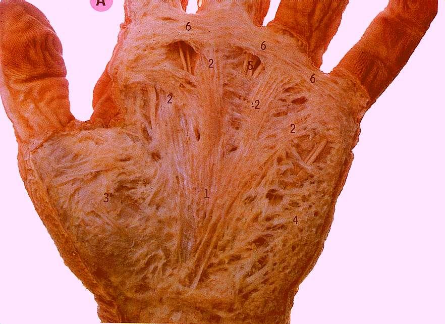
 Anatomy of the Upper Limb Figure 53: The thenar & midpalmar spaces. The synovial (tendon) sheaths of the long flexors [Figure.54] These sheaths surround the tendons of the long flexors; flexor digitorum
Wrist & Hand Assessment and General View
 Wrist & Hand Assessment and General View Done by; Mshari S. Alghadier BSc Physical Therapy RHPT 366 m.alghadier@sau.edu.sa http://faculty.sau.edu.sa/m.alghadier/ Functional anatomy The hand can be divided
Muscular Nomenclature and Kinesiology - One
 Chapter 16 Muscular Nomenclature and Kinesiology - One Lessons 1-3 (with lesson 4) 1 Introduction 122 major muscles covered in this chapter Chapter divided into nine lessons Kinesiology study of human
STRUCTURAL BASIS OF MEDICAL PRACTICE EXAMINATION 5 October 6, 2006
 STRUCTURAL BASIS OF MEDICAL PRACTICE EXAMINATION 5 October 6, 2006 PART l. Answer in the space provided. (8 pts) 1. Identify the structures. (2 pts) B C A. _pisiform B. _ulnar artery A C. _flexor carpi
10/15/2014. Wrist. Clarification of Terms. Clarification of Terms cont
 Wrist Clarification of Terms Palmar is synonymous with anterior aspect of the wrist and hand Ventral is also synonymous with anterior aspect of the wrist and hand Dorsal refers to the posterior aspect
Supplied in part by the musculocutaneous nerve. Forms the axis of rotation in movements of pronation and supination
 Anatomy: Upper limb (15 questions) 1. Latissimus Dorsi: Is innervated by the dorsal scapular nerve Lies above feres major muscle Medially rotates the humerus All of the above 2. Supinator muscle is: Deep
Accreditation of this course does not necessarily imply the FPTA supports the views of the presenter or the sponsors.
 SCS Continuing Education presents: Basic Structure and Function of the Wrist and Hand Copyright 2004 Introduction: Hello and welcome to this program from SCS Continuing Education! Knowledge is the key
Nerves of the upper limb Prof. Abdulameer Al-Nuaimi. E. mail:
 Nerves of the upper limb Prof. Abdulameer Al-Nuaimi E-mail: a.al-nuaimi@sheffield.ac.uk E. mail: abdulameerh@yahoo.com Brachial plexus Median nerve After originating from the brachial plexus in the axilla,
[[Sally Leaning Towards Peter To Take Cold Hand]]
![[[Sally Leaning Towards Peter To Take Cold Hand]]](/thumbs/84/91174469.jpg "[[Sally Leaning Towards Peter To Take Cold Hand]]") In this lecture we will talk about the bones of the hand, and the muscles and contents of the forearm. *The hand bones are: - Carpal bones. -Metacarpals. -Phalanges. *The carpal bones (wrist bones): They
In this lecture we will talk about the bones of the hand, and the muscles and contents of the forearm. *The hand bones are: - Carpal bones. -Metacarpals. -Phalanges. *The carpal bones (wrist bones): They
Key Relationships in the Upper Limb
 Key Relationships in the Upper Limb This list contains some of the key relationships that will help you identify structures in the lab. They are organized by dissection assignment as defined in the syllabus.
Anatomy Workshop Upper Extremity David Ebaugh, PT, PhD Workshop Leader. Lab Leaders: STATION I BRACHIAL PLEXUS
 Anatomy Workshop Upper Extremity David Ebaugh, PT, PhD Workshop Leader Lab Leaders: STATION I BRACHIAL PLEXUS A. Posterior cervical triangle and axilla B. Formation of plexus 1. Ventral rami C5-T1 2. Trunks
compartments of the forearm
 " forearm posterior compartment " compartments of the forearm Posterior Fascial compartment Muscles: ** The superficial group 1. Extensor carpi radialis brevis 2. Ex. digitorum 3. Ex. digiti minimi 4.
Lab Activity 11: Group II
 Lab Activity 11: Group II Muscles Martini Chapter 11 Portland Community College BI 231 Origin and Insertion Origin: The place where the fixed end attaches to a bone, cartilage, or connective tissue. Insertion:
Clinical examination of the wrist, thumb and hand
 Clinical examination of the wrist, thumb and hand 20 CHAPTER CONTENTS Referred pain 319 History 319 Inspection 320 Functional examination 320 The distal radioulnar joint.............. 320 The wrist.......................
musculoskeletal system anatomy muscles of foot sheet done by: dina sawadha & mohammad abukabeer
 musculoskeletal system anatomy muscles of foot sheet done by: dina sawadha & mohammad abukabeer Extensor retinaculum : A- superior extensor retinaculum (SER) : originates from the distal ends of the tibia
First & second layers of muscles of the sole
 The FOOT First & second layers of muscles of the sole introduction The muscles acting on the foot can be divided into two distinct groups; extrinsic and intrinsic muscles. The extrinsic muscles arise from
8/25/2014. Radiocarpal Joint. Midcarpal Joint. Osteology of the Wrist
 Structure and Function of the Wrist 2 joints and 10 different bones Combine to create wrist motion Anatomical Terms: Wrist/Hand Palmar = anterior aspect of the wrist and hand Dorsal = posterior aspect
# Anatomy. Upper Extremities Muscles and anatomy of axilla. Tiba Al-Ani 9/10/2015 Nabil. Page 0 of 16
 #10 25 Anatomy Upper Extremities Muscles and anatomy of axilla Tiba Al-Ani 9/10/2015 Nabil Page 0 of 16 Salam AWN Today s lecture is divided into two parts, the first part is the continuation of the upper
Introduction. The wrist contains eight small carpal bones, which as a group act as a flexible spacer between the forearm and hand.
 Wrist Introduction The wrist contains eight small carpal bones, which as a group act as a flexible spacer between the forearm and hand. Distal forearm Distal forearm 4 Distal end of the radius A. anterior
Intrinsic muscles palsies of the hand Management of Thumb Opposition with BURKHALTER s Procedure
 Intrinsic muscles palsies of the hand Management of Thumb Opposition with BURKHALTER s Procedure TRUONG LE DAO, MD, IFAAD 1 Burkhalter W.E, Cristhensen R.C, Brown P.W, Extensor Indicis Proprius opponensplasty
RHEUMATOID HAND. History Pain Loss of function Neck pain. Diminished ADL assessment:
 RHEUMATOID HAND History Pain Loss of function Neck pain Diminished ADL assessment: Using toothbrush, hairbrush, knife, fork Dressing bra, Pulling up trousers / stockings Operating remote control Hobbies
Upper Limb- Sports Medicine II
 Upper Limb- Sports Medicine II I. Palpation A. With patient sitting, supine, & prone, palpate for pain, specific tenderness, swelling, effusion, local hyperthermia B. Bony Palpation 1. Carpal Bones (8)
ANATOMY. Su~,ect : Lecturer : Maher Hadidi Done by: lecture # : 1 3 Date :
 ANATOMY Su~,ect : Lecturer : Maher Hadidi Done by: lecture # : 1 3 Date : HAND Skin ~ Thick and shovv many flexure creases (folds). ~ Flexure creases are an important landmarks. 111 Proximal palmar crease.
LIST OF STRUCTURES TO BE IDENTIFIED IN LAB: UPPER EXTREMITY REVIEW 2016
 LIST OF STRUCTURES TO BE IDENTIFIED IN LAB: UPPER EXTREMITY REVIEW 2016 BONES Ribs, sternum, clavicle Humerus: Head, greater tubercle, lesser tubercle, intertubercular sulcus, surgical neck, anatomical
The Foot. Dr. Wegdan Moh.Mustafa Medicine Faculty Assistant Professor Mob:
 The Foot Dr. Wegdan Moh.Mustafa Medicine Faculty Assistant Professor Mob: 0127155717 The skeleton of the foot Cutaneous innervations Sole of foot layers of muscles First layer -Abductor hallucis -Flexor
REFERENCE DIAGRAMS OF UPPER LIMB MUSCLES: NAMES, LOCATIONS, ATTACHMENTS, FUNCTIONS MUSCLES CONNECTING THE UPPER LIMB TO THE AXIAL SKELETON
 REFERENCE DIAGRAMS OF UPPER LIMB MUSCLES: NAMES, LOCATIONS, ATTACHMENTS, FUNCTIONS MUSCLES CONNECTING THE UPPER LIMB TO THE AXIAL SKELETON A25LAB EXERCISES: UPPER LIMB MUSCLES Page 1 MUSCLES CONNECTING
Musculoskeletal Imaging of the Digits. Arash David Tehranzadeh, MD UCSD MSK Radiology May 11 th, 2006
 Musculoskeletal Imaging of the Digits Arash David Tehranzadeh, MD UCSD MSK Radiology May 11 th, 2006 Musculoskeletal Imaging of the Digit Anatomy & Internal Derangement The Extensor System The Flexor System
Anatomy of the Forearm
 Anatomy of the Forearm Musculoskeletal block- Anatomy-lecture 8 Editing file Objectives List the names of the Flexors Group of Forearm (superficial & deep muscles). Identify the common flexor origin of
Dynamic 22 Mhz ultrasound evaluation (HR-US) of the finger: a detailed didactic approach.
 of the finger: a detailed didactic approach.") Dynamic 22 Mhz ultrasound evaluation (HR-US) of the finger: a detailed didactic approach. Poster No.: C-2228 Congress: ECR 2014 Type: Educational Exhibit Authors: A. Muda, D. Orlandi, V. Prono, S. Migone,
Dynamic 22 Mhz ultrasound evaluation (HR-US) of the finger: a detailed didactic approach. Poster No.: C-2228 Congress: ECR 2014 Type: Educational Exhibit Authors: A. Muda, D. Orlandi, V. Prono, S. Migone,
Dr Nabil khouri MD. MSc. Ph.D
 Dr Nabil khouri MD. MSc. Ph.D Foot Anatomy The foot consists of 26 bones: 14 phalangeal, 5 metatarsal, and 7 tarsal. Toes are used to balance the body. Metatarsal Bones gives elasticity to the foot in
forearm posterior compartment
 Quick revision: The anterior compartment of the forearm contains of 8 muscles... -4 superficial -1 intermediate -3 deep *All supplied by median nerve except 1 and 1/2 muscle (by ulnar N.) forearm posterior
Dr. Mahir Alhadidi Anatomy Lecture #9 Feb,28 th 2012
 Quick Revision: Upper arm is divided into two compartments: 1. Anterior Compartment: Contains three muscles (Biceps brachii, Coracobrachialis, Brachialis). Innervated by Musculocutaneous nerve. 2. Posterior
1/13/2013. Anatomy Guy Dissection Sheet Extensor Forearm and Hand. Eastern Virginia Medical School
 Dr. Craig Goodmurphy Anatomy Guy Superficial Extensor Muscles Complete skin removal if necessary then remove the antebrachial fascia starting at the extensor retinaculum and working proximally. Define
The Clavicle Right clavicle Deltoid tubercle: Conoid tubercle, conoid ligamen Impression for the
 The Clavicle Muscle Attachment Sites in the Upper Limb Pectoralis major Right clavicle Smooth superior surface of the shaft, under the platysma muscle tubercle: attachment of the deltoid Acromial facet
Hand and Upper Extremity
 Taylor_PS_C1-10.qxd 25/10/2004 04:20 PM Page 101 Chapter 8 Hand and Upper Extremity Subhro K. Sen, MD and Jesse A.Taylor, MD Hands, and in particular opposable thumbs, have played a large role in human
Assessing hand ligaments and tendons lesions using MRI
 Assessing hand ligaments and tendons lesions using MRI Award: Certificate of Merit Poster No.: C-0691 Congress: ECR 2017 Type: Educational Exhibit Authors: A. M. Benitez Vazquez, M. I. Rossi Prieto, C.
Clarification of Terms
 Clarification of Terms The plantar aspect of the foot refers to the role or its bottom The dorsal aspect refers to the top or its superior portion The ankle and foot perform three main functions: 1. shock
Ultrasonography of the wrist - a step-by-step approach to study protocols and normal findings
 Ultrasonography of the wrist - a step-by-step approach to study protocols and normal findings Poster No.: C-1779 Congress: ECR 2016 Type: Educational Exhibit Authors: R. R. Domingues Madaleno, A. P. Pissarra,
SPECIAL ARTICLE. Missed tendon injuries INTRODUCTION
 Archives of Emergency Medicine, 1991, 8, 87-91 SPECIAL ARTICLE Missed tendon injuries H. R. GULY Consultant in A & E, Derriford Hospital, Plymouth INTRODUCTION The timing of the repair of divided tendons
The Elbow and the cubital fossa. Prof Oluwadiya Kehinde
 The Elbow and the cubital fossa Prof Oluwadiya Kehinde www.oluwadiya.com Elbow and Forearm Anatomy The elbow joint is formed by the humerus, radius, and the ulna Bony anatomy of the elbow Distal Humerus
Muscle/Tendon Functions: Thumb. Extensor Tendon Healing. Digital Extension. Digital Extension. Supporting Ligaments: ORL. Supporting Ligaments
 Extensor Tendon Anatomy, Common Injury and Treatment Christina Schmidt, OTR/L, CHT University of California, Irvine Irvine, CA February 9-11, 2018 2 How Extensor Tendons Differ from Flexor Tendons Dorsum
Learning Objectives. 07 Aug 12. Article E-1. At the end of this section the learner will be able to:
 Module 1: Comparative Functional Anatomy and Biomechanics Article E-1 Learning Objectives At the end of this section the learner will be able to: Describe the bones of the equine thoracic Describe the
Tendon Transfers. Variability muscle 2x stronger than another Greatest force at resting length. Drag:
 Tendon Transfers Joanne Mimm, MPT, CHT Restore balance Indications Nerve injury paralyzed muscle damaged tendon/muscle CNS lesion Consider action Functional gain Strength-ability to generate tension number
Wrist and Hand Complaints
 Wrist and Hand Complaints Charles S. Day, M.D., M.B.A. Chief, Hand & Upper Extremity Surgery St. Elizabeth s Medical Center Tufts University School of Medicine Primary Care Internal Medicine 2018 Outline
Abduction of arm until your hand rich your head. Flexion of forearm at elbow joint. Extension of arm at elbow joint. Flexion of fingers 10.
 Num. answer 1. Medialy With the manubrium ( sternum ), and laterally with the acromion of the scapula 2. 1. Trapezius 2. Levator scapulae 3. Rhomboids 3. 1. Pectoralis major 2. Pectoralis minor 3. Latissiumus
Dorsal Digital Expansion Of Thumb
 Dorsal Digital Expansion Of Thumb Joshi, S.S., Joshi, S.D., Aavale S.A., Kishve, P. S. and Jadhav S.D. Rural Medical College,Pravara Institute of Medical Sciences, Loni Abstract: Human hands perform e
Classification of Established Volkmann s Ischemic Contracture and the Program for Its Treatment
 10 Classification of Established Volkmann s Ischemic Contracture and the Program for Its Treatment In spite of the advances made in preventive treatment of muscular ischemia at the forearm and hand, there
Functional Anatomy of the Elbow
 Functional Anatomy of the Elbow Orthopedic Institute Daryl C. Osbahr, M.D. Chief of Sports Medicine, Orlando Health Chief Medical Officer, Orlando City Soccer Club Orthopedic Consultant, Washington Nationals
Nerve Injury. 1) Upper Lesions of the Brachial Plexus called Erb- Duchene Palsy or syndrome.
 Upper Lesions of the Brachial Plexus called Erb- Duchene Palsy or syndrome.") Nerve Injury - Every nerve goes to muscle or skin so if the nerve is injured this will cause paralysis in the muscle supplied from that nerve (paralysis means loss of function) then other muscles and other
Nerve Injury - Every nerve goes to muscle or skin so if the nerve is injured this will cause paralysis in the muscle supplied from that nerve (paralysis means loss of function) then other muscles and other
Muscles of the Upper Limb
 Muscles of the Upper Limb anterior surface of ribs 3 5 coracoid process Pectoralis minor pectoral nerves protracts / depresses scapula Serratus anterior Subclavius ribs 1-8 long thoracic nerve rib 1 ----------------
Morphological Variations in Lumbricals of Hand A Cadaveric Study
 IBIMA Publishing Plastic Surgery: An International Journal http://www.ibimapublishing.com/journals/psij/psij.html Vol. 2013 (2013), Article ID 821692, 7 pages DOI: 10.5171/2013.821692 Morphological Variations
MLT Muscle(s) Patient Position Therapist position Stabilization Limb Position Picture Put biceps on slack by bending elbow.
 Patient Position Therapist position Stabilization Limb Position Picture Put biceps on slack by bending elbow.") MLT Muscle(s) Patient Position Therapist position Stabilization Limb Position Picture Put biceps on slack by bending elbow. Pectoralis Minor Supine, arm at side, elbows extended, supinated Head of Table
MLT Muscle(s) Patient Position Therapist position Stabilization Limb Position Picture Put biceps on slack by bending elbow. Pectoralis Minor Supine, arm at side, elbows extended, supinated Head of Table
Ulnar Neuropathy in the Distal Ulnar Tunnel
 Ulnar Neuropathy in the Distal Ulnar Tunnel DAVID W. SHUPE, PT, ATC' Journal of Orthopaedic & Sports Physical Therapy A brief anatomical review of the ulnar nerve and areas of ulnar nerve entrapment is
CONTINUING MEDICAL EDUCATION
 CONTINUING MEDICAL EDUCATION Management of the injured hand - Principles of assessment K. Karunadasa Plastic and Reconstructive Surgical Unit, North Colombo Teaching Hospital, Ragama, Sri Lanka Keywords:
Peripheral Nervous Sytem: Upper Body
 Peripheral Nervous Sytem: Upper Body MSTN121 - Neurophysiology Session 10 Department of Myotherapy Cervical Plexus Accessory nerve (CN11 + C1-5) Motor: trapezius and sternocleidomastoid Greater auricular
Anatomy Upper Limb Muscles
 Anatomy Upper Limb Muscles Rotator cuff/scapulohumeral muscles 4 muscles (SITS) form musculotendinous rotator cuff around glenohumeral joint, provide stability of joint Supraspinatus Course: med 2/3 supraspinatous
I-A-1) Non-specific thickening of synovial membrane
 Non-specific thickening of synovial membrane") I-A-1) Non-specific thickening of synovial membrane Grayscale Metatarsal Power Doppler Dorsal aspect of metatarsophalangeal joint in right 1 st toe, longitudinal view Asterisks indicate non-specific thickening
I-A-1) Non-specific thickening of synovial membrane Grayscale Metatarsal Power Doppler Dorsal aspect of metatarsophalangeal joint in right 1 st toe, longitudinal view Asterisks indicate non-specific thickening
Interesting Case Series. Swan-Neck Deformity in Cerebral Palsy
 Interesting Case Series Swan-Neck Deformity in Cerebral Palsy Leyu Chiu, BA, a Nicholas S. Adams, MD, a,b and Paul A. Luce, MD, a,b,c a Michigan State University College of Human Medicine, Grand Rapids,
Joints of the upper limb II
 Joints of the upper limb II Prof. Abdulameer Al-Nuaimi E-mail: a.al-nuaimi@sheffield.ac.uk E. mail: abdulameerh@yahoo.com Elbow joint The elbow joint is connecting the upper arm to the forearm. It is classed
Cubital fossa and forearm
 Cubital fossa and forearm Cubital fossa is the triangular space in front of elbow joint. - The Cubital fossa has boundaries: apex, base, roof and floor and it has contents. The base: an imaginary horizontal
Skeletal System. Supplementary Information
 Skeletal System Supplementary Information COMMON ANATOMICAL TERMS Planes run through the body side to side and front to back eg. median plane Surfaces of the body are also named eg. anterior surface This
Anatomy of the lower limb
 Anatomy of the lower limb Arches & sole of the foot Dr. Hayder ARCHES OF THE FOOT The foot as a mechanical unit performs two major functions: - It acts as a pliable platform to support the body weigh during