International Journal of Current Medical Sciences- Vol. 6, Issue,, pp , June, 2016 A B S T R A C T
|
|
|
- Basil Green
- 5 years ago
- Views:
Transcription

1 ISSN: International Journal of Current Medical Sciences- Vol. 6, Issue,, pp , June, 2016 COMPUTED TOMOGRAPHY IN HEPATIC METASTASES Ananthakumar P and Adaikkappan M., Available online at INTERNATIONAL JOURNAL OF CURRENT MEDICAL SCIENCES RESEARCH ARTICLE Department of Radiodiagnosis, Rajah Muthiah Medical College and Hospital, Annamalai University, Annamalai Nagar A R T I C L E I N F O Article History: Received 11th, March, 2016, Received in revised form 6th, April, 2016, Accepted 8th, May, 2016, Published online 28th, June, 2016 Key words: Metastases, Computed tomography, Liver A B S T R A C T Background: The liver is one of the most common organs to be involved with metastatic disease, which arises most frequently from primary sites in the colon, breast, lung, pancreas, and stomach. The accurate detection of metastatic disease at the time of diagnosis or during the course of treatment remains crucial to patient management. Objective: To evaluate the effectiveness of computed tomography in detecting hepatic metastases in patients with focal liver lesions and to provide information regarding probable source of primary lesions. Materials and methods: The study was conducted in the department of Radio diagnosis, Rajah Muthiah Medical College, Chidambaram. Forty patients with hepato-biliary related clinical symptoms or incidentally detected liver masses with USG abdomen were evaluated. Of them, 111 patients with metastases were included in this study. Rest of the patients with other focal lesions was excluded from the study. Results: Out of 40 patients, 11 patients diagnosed with hepatic metastases underwent histopathological examinations. Six patients had stomach carcinoma, two had thyroid malignancy, one patient each had lung, renal and pancreatic carcinoma. Conclusions: CT scanning, which is widely available and familiar, remains the dominant modality in the evaluation of suspected hepatic metastases, for preoperative planning and treatment monitoring. Copyright Ananthakumar P and Adaikkappan M. 2016, This is an open-access article distributed under the terms of the Creative Commons Attribution License, which permits unrestricted use, distribution and reproduction in any medium, provided the original work is properly cited. INTRODUCTION The liver is one of the most common organs to be involved with metastatic disease. About 30 percent of patients, who die from malignancies, have liver metastases (4). Hepatic metastases are times more common than primary liver tumors. Liver metastatic disorders usually occur in patients with stomach, pancreas, breast, colon, lung and other tumors. Most liver metastases are multiple, involving both lobes in 77% of patients and only in 10% of cases there is a solitary metastasis (2). Hypervascular metastases are less common and are seen in renal cell carcinoma, insulinomas, carcinoid, sarcomas, melanoma and breast cancer. Calcified liver metastases are uncommon, can be seen in metastases of colon, stomach, breast, endocrine pancreatic Ca, leiomyosarcoma, osteosarcoma and melanoma. Cystic liver metastases are seen in mucinous ovarian, colon, lung carcinomas, sarcoma, melanoma, and carcinoid tumor. Lesion conspicuity will depend on differential enhancement between lesions and the adjacent liver parenchyma. Vascular (hypervascular) metastases may show significant enhancement during the arterial phase. Most liver metastases are hypovascular and are best imaged during the portal venous phase. During the equilibrium phase, lesions may become less conspicuous or completely obscured. If there is concomitant hepatic steatosis, then the lesions may be iso or even slightly hyperattenuating. Enhancement is typically peripheral, and although there may be central filling in on portal venous phase, delayed phase will show washout; helpful in distinguishing metastases from liver haemangiomas (1). *Corresponding author: Ananthakumar P Department of Radiodiagnosis, Rajah Muthiah Medical College and Hospital, Annamalai University, Annamalai Nagar


2 MATERIALS AND METHODS This study was conducted on patients presenting to the Department of Radiodiagnosis, Rajah Muthiah Medical College & Hospital, Annamalai university, Chidambaram, for evaluation of known or suspected liver lesions. All basic examinations were performed and written informed consent was taken prior to any intervention. The aim of the study was to evaluate the effectiveness of computed tomography in detecting hepatic metastases in patients with focal liver lesions and to provide information regarding probable source of primary lesions. All patients below 20 years, patients with history of trauma, patients allergic to contrast agents and patients with elevated renal parameters were excluded from the study. The exact plan of the study was individualized for each case. The study was performed using TOSHIBA 4 SLICE CT scan machine with non contrast study followed by triphasic scans including arterial phase, venous phase and delayed phase scans in all the patients with breath hold using oral and intravenous contrast agents. Oral contrast was given for suspicious gastrointestinal tract malignancy patients. All patients would be administered 1ml/kg of Intravenous contrast material at an injection rate of 4-6 ml/sec. The phases were obtained with empirically timed scans using a bolus injector with arterial phase obtained between seconds, portal venous phase at seconds and delayed phase were taken at seconds. The patients were followed up and final diagnosis was confirmed by histopathological analysis with FNAC / BIOPSY. Observation and results Out of 40 patients who had focal liver lesions enrolled in our study, 11 patients were diagnosed with liver metastasis. There were 4 men (age range, years) and 7 women (age range, years). The remaining 29 patients were excluded from the study because triple- other than phase helical CT images showed lesions metastases. Out of 11 cases, ten cases showed multiple hypodense and isodense lesions in the plain study. Among them diffuse enhancement in the arterial phase was observed in 2 cases, and peripheral enhancement in portal venous phase seen in the rest of 8 cases. Among the two cases which showed enhancement in the arterial phase, one case showed mass lesion in the kidney and one case showed a hyperdense lesion in the body of stomach which was showing strong enhancement in the arterial phase. The cases were diagnosed as liver metastasis with primary mass in the kidney and as hypervascular tumour in the stomach respectively. Among the 8 cases which showed peripheral enhancement in the portal venous phase, 5 cases had irregular wall thickening in the pyloric antral region of the stomach. These patients were also evaluated with oral contrast and diagnosed with carcinoma stomach and hepatic metastasis. One case showed abnormality in chest radiograph with spiculated mass in left lung and two cases each had mass lesions in thyroid, hence thesee cases were also diagnosed thyroid carcinoma and hepatic metastasis. One case showed hypodense lesion in the right lobe of liver and showed only peripheral enhancement in the arterial and the portal venous phase. This patient also had a mass lesion in the pancreas; hence the lesion was diagnosed as liver metastasis. All the cases reported as metastasis were confirmed as liver metastasis with histopathological findings. By correlation of imaging diagnosis with final histopathological diagnosis, the accuracy of MDCT in evaluation of liver metastasess in our study is found to be 100 %. Table 1 Distribution of primary malignancies in our study Primary Stomach Thyroid Pancreas Kidney Lung Kidney, 1 Lung, 1 Pancreas, 1 Thyroid, 2 CT diagnosis HPE diagnosis Graph 1 Distribution of Primary Malignancies Table 2 Distribution of Hepatic Metastases In Various Ages And Sex Female 4 1 Male 0 2 Total 4 3 No. of patients Age level 1 2 Age in year Stomach, Total Fema le Graph 2 Distribution of hepatic metastases in various ages and sex 123 P a g e


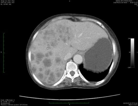
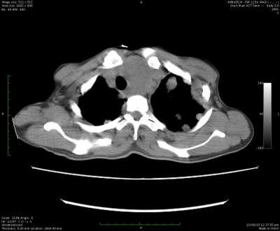
3 1.CT with oral and IV contrast axial images showing multiple hypodense peripherally enhancing liver parenchymal lesions, with primary mass in the body of stomach. 2.CT with oral IV contrast axial images showing multiple hypodense peripherally enhancing liver lesions, with primary mass in the pyloric antrum of stomach. 3.CT with IV contrast axial images showing multiple hypodense peripherally enhancing liver parenchymal lesions, with primary mass in the body and tail of pancreas 4.CT with IV contrast axial images showing multiple hypodense peripherally enhancing liver parenchymal lesions, with primary mass in left thyroid gland 124 P a g e
4 DISCUSSION The early detection of liver metastases is of paramount importance in patients suffering from liver malignancies. In most malignancies, the presence of liver metastases indicates non-resectability of the primary tumor for oncologic reasons. Liver metastatic disorders usually occur in patients with stomach, pancreas, breast, colon, lung and other tumors. A reproducible radiologic examination should be performed in patients before treatment, particularly in preoperative phase. Purposive radiologic evaluations such as US and CT scan are necessary to choose the best therapeutic method and (3) determine the prognosis. Advances in imaging technology lead to improved image quality and accuracy. Since US is an operator- dependent modality, its usage is limited for detecting liver lesions. High spatial resolution enables high quality multiplanar and three dimensional (3D) reformations to be constructed from the raw data. High temporal resolution allows multiple precisely defined imaging phases to be performed (7, 9). which are relevant especially in hepatic imaging In our study metastases were seen in 11 patients, out of 40 focal liver lesions. Majority of these cases were in the age group of years (n=4), 3 cases in years and 2 cases each in years and in years. Lewis et al have described that the liver is the most frequent site of metastasis, being far more common than primary liver tumours. The lesions in our study were multiple in all 11 cases and they were involving both lobes of liver in 90 %. Pain in the abdomen was the commonest symptom followed by weight loss and feeling of lump in the abdomen (8). Most of the lesions were hypodense lesions on plain study and few were isodense which was seen when they were smaller in size. 8 cases showed peripheral enhancement in the arterial phase and portal venous phase with no central enhancement while 3 cases showed diffuse enhancement in arterial and portal venous phases. Foley et al (5) has described that MDCT appearance of hypervascular lesions are hyperenhancing on arterial and portal venous phase either homogeneous or inhomogeneous due to areas of necrosis or haemorrhage. The imaging appearance of these hypervascular tumors is distinct from rim enhancing hypovascular metastatic lesions from common primary sites, such as lung, breast, pancreas, colon, and the genital tract (8). Hypovascular metastasis are the most common type and these lesions on the arterial and portal venous phase may demonstrate a hyperenhancing rim that is different from that of hypervascular metastases which shows diffuse enhancement. Two cases had carcinoma of thyroid with metastasis in liver. The lesions were multiple and involving both the lobes, the lesions were hypodense on plain study and showed peripheral enhancement in portal venous phase and arterial phase with no central or diffuse enhancement. These findings are in correlation with the study by Chen et al who have described similar findings of hypovascular metastases for thyroid malignancy (9). One case of adenocarcinoma of lung had multiple metastatic lesions in both lobes of liver. Lesions showed no central enhancement but only showed peripheral enhancement in portal venous phase. The findings of hypovascular metastasis from the case of lung carcinoma was consistent with the findings of hypovascular metastasis by Philippe soyer et al (10). There was one case of Renal cell carcinoma with metastases to liver in our study. Lesions were multiple, seen in both lobes and were predominantly hypodense and few lesions were hyperdense on plain scans. All the lesions showed strong arterial phase enhancement which reduced in the portal venous phase. On delayed phase the lesions showed no enhancement. According to study by (11) Raptopoulos et al RCC are known to give hypervascular metastasis of which 65% cases were more conspicuous in arterial and 90% cases were more conspicuous in portal venous phase. However in our study of renal cell carcinoma with liver metastasis, lesions strongly enhanced in the arterial phase and became less conspicuous in the portal venous phase. One of the cases with multiple hypodense lesions on plain scan showed no arterial phase enhancement but showed peripheral enhancement in portal venous phase, the lesions in the delayed phase showed no enhancement. The patient also showed a peripherally enhancing mass lesion in the pancreas. The patient was diagnosed as liver metastasis from hypovascular pancreatic malignant mass and was referred to higher centre where the HPE was done and the patient was diagnosed with adenocarcinoma of pancreatic head. 6 cases of carcinoma stomach had multiple lesions in the liver. The lesions were hypodense on plain study with early peripheral enhancement on arterial phase and persistence of peripheral enhancement in the portal venous phase. However no central enhancement was seen. Foley et al have described similar findings in cases of hypovascular liver metastasis (8). In this study we observed that hypervascular metastatic lesions were more conspicuous in the arterial phase and hypovascular metastatic lesions predominantly shows only peripheral thin enhancement in the arterial phase and portal venous phase and no central enhancement. Triple phase CT was helpful to differentiate and characterize hypo vascular and hyper vascular type of metastases based on the features as described by Foley et al (8). Based on the enhancement characteristics of metastases and combining them with clinical and other features, attempt could be made to identify the primary. Triple phase with its arterial phase was helpful in detecting the primary in cases of hyper vascular type of metastasis. CONCLUSION Despite recent advances in radiologic examination, liver metastases are still remaining as a challenge in human oncology. Advances in imaging technology have improved our ability to detect, characterize, and stage metastatic liver disease. Although every modality has 125 P a g e
5 benefited from advances in technology, CT scanning with its speed and three-dimensional volume rendering that can provide detailed vascular anatomy remains a dominant imaging modality not only for lesion detection and preoperative planning, but also for treatment monitoring and post treatment surveillance. References 1. Kalaap C, Nessar G, Ulas M, et al. Preoperative staging of colon cancer in patients: ultrasound can be a valuable alternative to computer tomography. Bratisl Lek Listy. 2011; 112: Sutton D. Textbook of radiology and imaging. D. Textbook of radiology and imaging. 8th ed. Philadelphia: Mosby; D souza MM, Sharma R, Mondal A, et al. Prospective evaluation of CECT and 18F-FDG- PET/CT in detection of hepatic metastases. Nucl Med Commun. 2009; 30: Foley WD. Special focus session: multidetector CT: abdominal visceral imaging. Radiographics 2002; 22: Ros PR, Ji H. Special focus session. Multisection (multidetector) CT: applications in the abdomen. Radiographics 2002; 22: ******* 6. Semelka Rc, Sofka CM. Hepatic Hemangioma Magnetic Resonance Imaging Clin N AM 1997; May ;5(2): Beaty Sd, Silva Ac, Depetrsi G: Incidental hepatic mass Am J Roentgenol 2008, 190:S62-S64. doi: /AJR Foley WD, Mallisee TA, Hohenwalter MD, Wilson CR, Quiroz FA, Taylor AJ. Multiphase hepatic CT with a multirow detector CT scanner. Am J Roentgenol 2000; 175: Lewis Kh, Chezmar Jl. Hepatic Metastases. MRI Clin N Am 1997; May 5(2): Gregory T. Sica, Chen, Hoon Ji, Pablo R Ros. CT and MR imaging of Hepatic Metastases. Ajr 2000 mar; 174(3): Atasoy, Raptopulous, Akyar Ji. Multidetector CT: Contributions in Liver Imaging. European Journal of Radiology. 2004; 52 (1): Ji, 12. Philippe Soyer. Detection of Hypovascular Hepatic Metastases at Triple-Phase Helical CT: sensitivity of phases and comparison with surgical and histopathologic findings. Radiology 2004 MAY; 231(2): Epub 2004 mar P a g e
Imaging in gastric cancer
 Imaging in gastric cancer Gastric cancer remains a deadly disease because of late diagnosis. Adenocarcinoma represents 90% of malignant tumors. Diagnosis is based on endoscopic examination with biopsies.
Newcastle HPB MDM updated radiology imaging protocol recommendations. Author Dr John Scott. Consultant Radiologist Freeman Hospital
 Newcastle HPB MDM updated radiology imaging protocol recommendations Author Dr John Scott. Consultant Radiologist Freeman Hospital This document is intended as a guide to aid radiologists and clinicians
HEPATO-BILIARY IMAGING
 HEPATO-BILIARY IMAGING BY MAMDOUH MAHFOUZ MD PROF.OF RADIOLOGY CAIRO UNIVERSITY mamdouh.m5@gmail.com www.ssregypt.com CT ABDOMEN Indications Patient preparation Patient position Scanogram Fasting 4-6 hours
Common and unusual CT and MRI manifestations of pancreatic adenocarcinoma: a pictorial review
 Review Article Common and unusual CT and MRI manifestations of pancreatic adenocarcinoma: a pictorial review Min-Jie Yang, Su Li, Yong-Guang Liu, Na Jiao, Jing-Shan Gong Department of Radiology, Shenzhen
Imaging of Neuroendocrine Metastases
 Imaging of Neuroendocrine Metastases Aoife Kilcoyne, Shaunagh McDermott, Colin McCarthy,Manuel Patino, Dushyant Sahani, Michael Blake Abdominal Imaging Division Massachusetts General Hospital Disclosure
The Focal Hepatic Lesion: Radiologic Assessment
 The Focal Hepatic Lesion: Radiologic Assessment Kevin Kuo, Harvard Medical School Year III Our Patient: PS 67 y/o female w/ long history of alcohol use Drinking since age 18, up to one bottle of wine/day
The Frequency and Significance of Small (15 mm) Hepatic Lesions Detected by CT
 Hepatic Lesions Detected by CT") 535 Elizabeth C. Jones1 Judith L. Chezmar Rendon C. Nelson Michael E. Bernardino Received July 22, 1991 ; accepted after revision October 16, 1991. Presented atthe annual meeting ofthe American Aoentgen
535 Elizabeth C. Jones1 Judith L. Chezmar Rendon C. Nelson Michael E. Bernardino Received July 22, 1991 ; accepted after revision October 16, 1991. Presented atthe annual meeting ofthe American Aoentgen
Imaging of liver and pancreas
 Imaging of liver and pancreas.. Disease of the liver Focal liver disease Diffusion liver disease Focal liver disease Benign Cyst Abscess Hemangioma FNH Hepatic adenoma HCC Malignant Fibrolamellar carcinoma
Extraosseous myeloma: imaging features
 Extraosseous myeloma: imaging features C. Santos Montón, R. Corrales, J. M. Bastida Bermejo, M. Villanueva Delgado, R. E. Correa Soto, J. M. Alonso Sánchez; Salamanca/ES Learning objectives -To review
Upper GI Malignancies Imaging Guidelines for the Management of Gastric, Oesophageal & Pancreatic Cancers 2012
 Upper GI Malignancies Imaging Guidelines for the Management of Gastric, Oesophageal & Pancreatic Cancers 2012 Version Control This is a controlled document please destroy all previous versions on receipt
Imaging characterization of renal clear cell carcinoma
 Imaging characterization of renal clear cell carcinoma Poster No.: C-0327 Congress: ECR 2011 Type: Educational Exhibit Authors: S. Ballester 1, A. Gaser 2, M. Dotta 1, M. F. CAPPA 1, F. Hammar 1 ; 1 2
Case Report Solitary Osteolytic Skull Metastasis in a Case of Unknown Primary Being latter Diagnosed as Carcinoma of Gall Bladder
 Cronicon OPEN ACCESS CANCER Case Report Solitary Osteolytic Skull Metastasis in a Case of Unknown Primary Being latter Diagnosed as Carcinoma of Gall Kartik Mittal 1, Rajaram Sharma 1, Amit Dey 1, Meet
CT Contrast Protocols for Different Organ Imaging
 CT Contrast Protocols for Different Organ Imaging g Paul Shreve, M.D. Advanced Radiology Services, P.C. & Spectrum Health Grand Rapids, MI, USA Correlative Imaging Council Society of Nuclear Medicine 56
Contrast Enhanced Ultrasound of Parenchymal Masses in Children
 Contrast Enhanced Ultrasound of Parenchymal Masses in Children Sue C Kaste, DO On behalf of Beth McCarville, MD St. Jude Children s Research Hospital Memphis, TN Overview Share St. Jude experience with
Anatomical and Functional MRI of the Pancreas
 Anatomical and Functional MRI of the Pancreas MA Bali, MD, T Metens, PhD Erasme Hospital Free University of Brussels Belgium mbali@ulb.ac.be Introduction The use of MRI to investigate the pancreas has
Dr Claire Smith, Consultant Radiologist St James University Hospital Leeds
 Dr Claire Smith, Consultant Radiologist St James University Hospital Leeds Imaging in jaundice and 2ww pathway Image protocol Staging Limitations Pancreatic cancer 1.2.4 Refer people using a suspected
Intraoperative staging of GIT cancer using Intraoperative Ultrasound
 Intraoperative staging of GIT cancer using Intraoperative Ultrasound Thesis For Fulfillment of MSc Degree In Surgical Oncology By Abdelhalim Salah Abdelhalim Moursi M.B.B.Ch (Cairo University ) Supervisors
Liver Tumors. Prof. Dr. Ahmed El - Samongy
 Liver Tumors Prof. Dr. Ahmed El - Samongy Objective 1. Identify the most important features of common benign liver tumors 2. Know the risk factors, diagnosis, and management of hepatocellular carcinoma
X-ray Corner. Imaging of The Pancreas. Pantongrag-Brown L
 X-ray Corner 125 Imaging of The Pancreas Modern imaging modalities commonly used in pancreas include ultrasound (US), CT, and MRI. Pancreas is a retroperitoneal organ which makes it difficult to visualize
Alison Douglass Gillian Lieberman, MD. November. Colon Cancer. Alison Douglass, Harvard Medical School Year III Gillian Lieberman, MD
 November Colon Cancer Alison Douglass, Harvard Medical School Year III Our Patient Mr. K. is a 67 year old man with no prior medical problems other than hemorrhoids which have caused occasional rectal
performed to help sway the clinician in what the appropriate diagnosis is, which can substantially alter the treatment of management.
 Hello, I am Maura Polansky at the University of Texas MD Anderson Cancer Center. I am a Physician Assistant in the Department of Gastrointestinal Medical Oncology and the Program Director for Physician
Role of triple phase computed tomography findings for evaluation of hepatic lesions
 International Journal of Research in Medical Sciences Ahirwar CP et al. Int J Res Med Sci. 26 Aug;4(8):3576-3583 www.msjonline.org pissn 232-67 eissn 232-62 Research Article DOI: http://dx.doi.org/.823/232-62.ijrms262332
CT & MRI of Benign Liver Neoplasms Srinivasa R Prasad
 CT & MRI of Benign Liver Neoplasms Srinivasa R Prasad No financial disclosures Acknowledgements Many thanks to Drs. Heiken, Narra & Menias (MIR) Dr. Sahani (MGH) for sharing images Benign Liver Tumors:
Gemstone Spectral Imaging quantifies lesion characteristics for a confident diagnosis
 GE Healthcare Gemstone Spectral Imaging quantifies lesion characteristics for a confident diagnosis CT clinical case study lesion characterization Desiree Morgan, MD Vice Chair of Clinical Research Professor
Nonfunctioning Islet Cell Tumors of the Pancreas: Computed Tomography Findings
 Chin J Radiol 2002; 27: 239-243 239 Nonfunctioning Islet Cell Tumors of the Pancreas: Computed Tomography Findings CHAO-HSUAN YEN 1 JEN-HWEY CHIANG 1 JEN-I HUANG 3 CHENG-SHI SU 2 YI-YOU CHIOU 1 CHENG-YEN
Abstract. Introduction. Patients and Methods
 The Evaluation of Hepatocellular Carcinoma with Biphasic Contrast enhanced Helical CT Scan J. Yaqoob, V. Bari, M. U. Usman, K. Munir, F. Mosharaf, W. Akhtar Department of Radiology, Aga Khan University
CASE 1 11/1/2016 HEPATOBILIARY IMAGING CASE PRESENTATIONS DECLARATION. Dr. Chirag Patel ORGAN IMAGING yr old lady
 HEPATOBILIARY IMAGING CASE PRESENTATIONS DECLARATION No financial disclosures or affiliations with commercial organisations No discussion of investigational or off-label use of medical devices, products
HEPATIC METASTASES. We can state 3 types of metastases depending on their treatment options:
 HEPATIC METASTASES 1. Definition Metastasis means the spread of cancer. Cancerous cells can separate from the primary tumor and enter the bloodstream or the lymphatic system (the one that produces, stores,
10 most frequently made mistakes with RECIST 1.1: how Radiologist can fail - and how to avoid them
 10 most frequently made mistakes with RECIST 1.1: how Radiologist can fail - and how to avoid them Poster No.: C-1689 Congress: ECR 2014 Type: Educational Exhibit Authors: M. Kekelidze, P. Lodise, M. Tozakidou,
Ask EuroSafe Imaging. Tips & Tricks. CT Working Group
 Ask EuroSafe Imaging Tips & Tricks CT Working Group The use of bi-phase injection protocols to reduce the number of acquisition phases and radiation dose Alban Gervaise (Medical Imaging Department, HIA
CT evaluation of small bowel carcinoid tumors
 CT evaluation of small bowel carcinoid tumors Poster No.: C-0060 Congress: ECR 2015 Type: Educational Exhibit Authors: N. V. V. P. Costa, L. Nascimento, T. Bilhim ; Estoril/PT, PT, 1 2 3 1 2 3 Lisbon/PT
objectives Pitfalls and Pearls in PET/CT imaging Kevin Robinson, DO Assistant Professor Department of Radiology Michigan State University
 objectives Pitfalls and Pearls in PET/CT imaging Kevin Robinson, DO Assistant Professor Department of Radiology Michigan State University To determine the regions of physiologic activity To understand
Innovations in HCC Imaging: MDCT/MRI
 Innovations in HCC Imaging: MDCT/MRI Anthony E. Cheng, M.D. Cardinal MRI Center Cardinal Santos Medical Center, Wilson Street, San Juan Innovations in HCC Imaging: Goals/Objectives MDCT/MRI Learn the diagnostic
Biliary cancers: imaging diagnosis. Study of 30 cases
 Biliary cancers: imaging diagnosis. Study of 30 cases N Hammoune, S Semlali, M Eddarai, T. Amil, M Zentar, S. El Kandri,, M Benameur,, S Chaouir. Radiology Department. Mohamed V Military Hospital. Rabat-
Molecular Imaging and Cancer
 Molecular Imaging and Cancer Cancer causes one in every four deaths in the United States, second only to heart disease. According to the U.S. Department of Health and Human Services, more than 512,000
Chief Complain. Liver lesion found in routine health check 41 days ago
 Chief Complain Liver lesion found in routine health check 41 days ago Present Illness On 2005-7-26 at 台北署立醫院 he underwent a health check for the first time. Abdominal US showed suspicious of a 6*5 cm hepatoma,
ABDOMINAL DIFFUSION WEIGHTED MR
 ABDOMINAL DIFFUSION WEIGHTED MR Frank Miller, M.D. FACR Professor of Radiology Chief, Body Imaging Section Medical Director, MR Imaging Northwestern University Feinberg School of Medicine fmiller@northwestern.edu
Fusion Ultrasound: Characterization of Abdominal Masses with MR, CT, PET, and Contrast Ultrasound
 Fusion Ultrasound: Characterization of Abdominal Masses with MR, CT, PET, and Contrast Ultrasound Mollie Rashid, MD Corinne Deurdulian, MD Hisham Tchelepi, MD Keck School of Medicine, University of Southern
Case Report Contrast Enhanced Ultrasound of a Gallbladder Lesion in a Patient with a History of Renal Cell and Rectal Cancer
 Case Reports in Gastrointestinal Medicine Volume 2013, Article ID 538534, 4 pages http://dx.doi.org/10.1155/2013/538534 Case Report Contrast Enhanced Ultrasound of a Gallbladder Lesion in a Patient with
Staging Colorectal Cancer
 Staging Colorectal Cancer CT is recommended as the initial staging scan for colorectal cancer to assess local extent of the disease and to look for metastases to the liver and/or lung Further imaging for
State of the Art Imaging for Hepatic Malignancy: My Assignment
 State of the Art Imaging for Hepatic Malignancy: My Assignment CT vs MR vs MRCP Which one to choose for HCC vs Cholangiocarcinoma What special protocols to use for liver tumors Role of PET and Duplex US
JMSCR Vol 05 Issue 06 Page June 2017
 www.jmscr.igmpublication.org Impact Factor 5.84 Index Copernicus Value: 83.27 ISSN (e)-2347-176x ISSN (p) 2455-0450 DOI: https://dx.doi.org/10.18535/jmscr/v5i6.29 MRI in Clinically Suspected Uterine and
Clinical Case. António Pedro Pissarra. March 23, 2018
 Clinical Case António Pedro Pissarra March 23, 2018 Medical Imaging Department, University Hospitals of Coimbra Dir.: Prof. Doutor Filipe Caseiro Alves Case Report 62-year-old woman; Medical history: Obesity;
Liver MRI in 30 minutes
 X Liver MRI in 30 minutes SCBT/MR Annual Meeting Salt Lake City September 18, 2016 Scott B. Reeder, MD, PhD Department of Radiology University of Wisconsin Madison, WI Disclosures University of Wisconsin-Madison
PANCREAS DUCTAL ADENOCARCINOMA PDAC
 CONTENTS PANCREAS DUCTAL ADENOCARCINOMA PDAC I. What is the pancreas? II. III. IV. What is pancreas cancer? What is the epidemiology of Pancreatic Ductal Adenocarcinoma (PDAC)? What are the risk factors
Case Discussion Splenic Abscess
 Case Discussion Splenic Abscess Personal Data Gender: male Birth Date: 1928/Mar/06th Allergy: Mefenamic Smoking: 0.5 PPD for 55 years Alcohol: negative (?) 4 Months Ago Abdominal pain: epigastric area
Prof. Dr. NAGUI M. ABDELWAHAB,M.D.; MARYSE Y. AWADALLAH, M.D. AYA M. BASSAM, Ms.C.
 Role of Whole-body Diffusion MR in Detection of Metastatic lesions Prof. Dr. NAGUI M. ABDELWAHAB,M.D.; MARYSE Y. AWADALLAH, M.D. AYA M. BASSAM, Ms.C. Cancer is a potentially life-threatening disease,
RICCARDO LENCIONI,CLOTILDE DELLA PINA, LAURA CROCETTI,DANIA CIONI. Chapter 1
 RICCARDO LENCIONI,CLOTILDE DELLA PINA, LAURA CROCETTI,DANIA CIONI Chapter 1 Impact of European Federation of Societies for Ultrasound in Medicine and Biology (EFSUMB) Guidelines on the Use of Contrast
Recommendations for cross-sectional imaging in cancer management, Second edition
 www.rcr.ac.uk Recommendations for cross-sectional imaging in cancer management, Second edition Renal and adrenal tumours Faculty of Clinical Radiology www.rcr.ac.uk Contents Renal cell carcinoma 3 Clinical
Role of imaging in RCC. Ultrasonography. Solid lesion. Cystic RCC. Solid RCC 31/08/60. From Diagnosis to Treatment: the Radiologist Perspective
 Role of imaging in RCC From Diagnosis to Treatment: the Radiologist Perspective Diagnosis Staging Follow up Imaging modalities Limitations and pitfalls Duangkamon Prapruttam, MD Department of Therapeutic
Purpose. Methods and Materials. Results
 Prevalence and significance of hypoattenuating hepatic lesions deemed too small to characterise: How are we following up these lesions and what are the outcomes? Poster No.: C-014 Congress: ECR 2009 Type:
Comparison of Radiological Criteria (RECIST - MASS - SACT -Choi) in Antiangiogenic Therapy of Renal Cell Carcinoma
 in Antiangiogenic Therapy of Renal Cell Carcinoma") Universal Journal of Public Health 4(5): 239-243, 2016 DOI: 10.13189/ujph.2016.040503 http://www.hrpub.org Comparison of Radiological Criteria (RECIST - MASS - SACT -Choi) in Antiangiogenic Therapy of
Universal Journal of Public Health 4(5): 239-243, 2016 DOI: 10.13189/ujph.2016.040503 http://www.hrpub.org Comparison of Radiological Criteria (RECIST - MASS - SACT -Choi) in Antiangiogenic Therapy of
Imaging Pancreatic Neuroendocrine Tumors (PNETs): CT, MRI, EUS, Nuclear
: CT, MRI, EUS, Nuclear") Imaging Pancreatic Neuroendocrine Tumors (PNETs): CT, MRI, EUS, Nuclear Eric Tamm, M.D. Department of Diagnostic Radiology Division of Diagnostic Imaging MD Anderson Cancer Center Houston, TX Disclosure
Imaging Pancreatic Neuroendocrine Tumors (PNETs): CT, MRI, EUS, Nuclear Eric Tamm, M.D. Department of Diagnostic Radiology Division of Diagnostic Imaging MD Anderson Cancer Center Houston, TX Disclosure
CTA/MRA of Pediatric Hepatic Masses Radiology-Pathology Correlation
 Acta Radiológica Portuguesa, Vol.XVIII, nº70, pág. 41-50, Abr.-Jun., 2006 CTA/MRA of Pediatric Hepatic Masses Radiology-Pathology Correlation Marilyn J. Siegel Mallinckrodt Institute of Radiology, Washington
MD Spiral CT appearances of pancreatic tail insulinoma
 Article ID: WMC004688 ISSN 2046-1690 MD Spiral CT appearances of pancreatic tail insulinoma Peer review status: No Corresponding Author: Dr. Atanas D Hilendarov, MD, Medical University, Volga 49, ap.14,
Multidetector CT of the Liver and Hepatic Neoplasms: Effect of Multiphasic Imaging on Tumor Conspicuity and Vascular Enhancement
 Isaac R. Francis 1 Richard H. Cohan 1 Nancy J. McNulty 1 Joel F. Platt 1 Melvyn Korobkin 1 Achamyeleh Gebremariam 2 Kartik Ragupathi 2 Received June 3, 2002; accepted after revision October 21, 2002. Presented
Liver Cancer (Hepatocellular Carcinoma or HCC) Overview
 Overview") Liver Cancer (Hepatocellular Carcinoma or HCC) Overview Recent advances in liver cancer care seek to address the rising incidence of liver cancer, which has steadily increased over the past three decades.
Liver Cancer (Hepatocellular Carcinoma or HCC) Overview Recent advances in liver cancer care seek to address the rising incidence of liver cancer, which has steadily increased over the past three decades.
HCC e CEUS. Prof. A. Giorgio. Direttore IX UOC di Malattie Infettive ad Indirizzo Ecointerventistico
 HCC e CEUS Prof. A. Giorgio Direttore IX UOC di Malattie Infettive ad Indirizzo Ecointerventistico The natural history of compensated cirrhosis due to hepatitis C virus: a 17 year cohort study of 214 patients
Case report Osteosarcoma of long bone metastatic to the pancreas-an unusual site of
 Osteosarcoma of long bone metastatic to the pancreas-an unusual site of Dr. Santosh Kumar Singh 1, Col (Dr.) Narayanan Kannan 2, Brig (Dr) Rajnish Talwar 3, ABSTRACT Col (Dr) Arvind Kumar Tyagi 4, Dr Adarsh
Liver Cancer And Tumours
 Liver Cancer And Tumours What causes liver cancer? Many factors may play a role in the development of cancer. Because the liver filters blood from all parts of the body, cancer cells from elsewhere can
Appendix 5. EFSUMB Newsletter. Gastroenterological Ultrasound
 EFSUMB Newsletter 87 Examinations should encompass the full range of pathological conditions listed below A log book listing the types of examinations undertaken should be kept Training should usually
ACG Clinical Guideline: Diagnosis and Management of Focal Liver Lesions
 ACG Clinical Guideline: Diagnosis and Management of Focal Liver Lesions Jorge A. Marrero, MD, 1 Joseph Ahn, MD, FACG, 2 K. Rajender Reddy, MD, FACG 3 1 University of Texas at Southwestern, Dallas, Texas,
US-Guided Radiofrequency Ablation of Hepatic Focal Lesions
 US-Guided Radiofrequency Ablation of Hepatic Focal Lesions Poster No.: C-2219 Congress: ECR 2011 Type: Scientific Exhibit Authors: D. Armario Bel, A. PLA, F. TERREL, X. Serres; BARCELONA/ES Keywords: Neoplasia,
Afternoon Session Cases
 Afternoon Session Cases Case 1 19 year old woman Presented with abdominal pain to community hospital Mild incr WBC a14, 000, Hg normal, lipase 100 (normal to 75) US 5.2 x 3.7 x 4 cm mass in porta hepatis
MDCT signs differentiating retroperitoneal and intraperitoneal lesions- diagnostic pearls
 MDCT signs differentiating retroperitoneal and intraperitoneal lesions- diagnostic pearls Poster No.: C-0987 Congress: ECR 2015 Type: Educational Exhibit Authors: D. V. Bhargavi, R. Avantsa, P. Kala; Bangalore/IN
INTRALUMINAL GAS IN NON-PERFORATED ACUTE APPENDICITIS: A predictor of gangrenous appendicitis
 INTRALUMINAL GAS IN NON-PERFORATED ACUTE APPENDICITIS: A predictor of gangrenous appendicitis DM Plata Ariza, MD; E Martínez Chamorro, MD; D Castaño Pardo, MD; M Arroyo López, MD; E Peghini Gavilanes,
Modern liver imaging techniques - A new era in liver ultrasound
 Modern liver imaging techniques - A new era in liver ultrasound Yuko Kono, M.D., Ph.D. Clinical Professor Departments of Medicine and Radiology University of California, San Diego San Diego, USA How to
CT 101 :Pancreas and Spleen
 CT 101 :Pancreas and Spleen Shikha Khullar,, MD, MPH Division of Radiology University of South Alabama The Pancreas Normal Pancreas 3 Phase Pancreatic CT Non contrast Arterial phase : 30-35 35 second
4,3,2,1...How Many Phases are Needed? Balancing Diagnostic Efficacy and Radiation Modulation for MDCT Imaging of Renal Cell Carcinoma
 4,3,2,1...How Many Phases are Needed? Balancing Diagnostic Efficacy and Radiation Modulation for MDCT Imaging of Renal Cell Carcinoma Jeremy Hackworth, MD, MS Steven P Rowe, MD, PhD Satomi Kawamoto, MD
Disclosure. Acknowledgement. What is the Best Workup for Rectal Cancer Staging: US/MRI/PET? Rectal cancer imaging. None
 What is the Best Workup for Rectal Cancer Staging: US/MRI/PET? Zhen Jane Wang, MD Assistant Professor in Residence UC SF Department of Radiology Disclosure None Acknowledgement Hueylan Chern, MD, Department
Index. Surg Oncol Clin N Am 16 (2007) Note: Page numbers of article titles are in boldface type.
 Note: Page numbers of article titles are in boldface type.") Surg Oncol Clin N Am 16 (2007) 465 469 Index Note: Page numbers of article titles are in boldface type. A Adjuvant therapy, preoperative for gastric cancer, staging and, 339 B Breast cancer, metabolic
Surg Oncol Clin N Am 16 (2007) 465 469 Index Note: Page numbers of article titles are in boldface type. A Adjuvant therapy, preoperative for gastric cancer, staging and, 339 B Breast cancer, metabolic
Solitary Contralateral Adrenal Metastases after Nephrectomy for Renal Cell Carcinoma
 Original Report ISSN 1537-744X; DOI 10.1100/tsw.2004.39 Solitary Contralateral Adrenal after Nephrectomy for Renal Cell Carcinoma Nikolaos Antoniou, M.D. and Demetrios Karanastasis, M.D. General Hospital
Radiology Pathology Conference
 Radiology Pathology Conference Nadia F. Yusaf, M.D. PGY-3 1/29/2010 Presentation material is for education purposes only. All rights reserved. 2010 URMC Radiology Page 1 of 90 Case 1 60 year- old man presents
Imaging Decisions Start Here SM
 Owing to its high resolution and wide anatomic coverage, dynamic first-pass perfusion 320-detector-row CT outperforms PET/CT for distinguishing benign from malignant lung nodules, researchers from Japan
Primary Hepatic Undifferentiated Pleomorphic Sarcoma: CT and angiographic findings in two cases
 J Radiol Sci 2013; 38: 15-19 Primary Hepatic Undifferentiated Pleomorphic Sarcoma: CT and angiographic findings in two cases Jan-Wen Ku Ying-Chi Tseng Kuo-Luon Kung Hsien-Chang Shen Yen-Lin Huang Chi-Jen
Abdomen and Pelvis CT (1) By the end of the lecture students should be able to:
 By the end of the lecture students should be able to:") RAD 451 Abdomen and Pelvis CT (1) By the end of the lecture students should be able to: State the common indications for Abdomen and pelvis CT exams Identify possible contra indications for Abdomen and
RAD 451 Abdomen and Pelvis CT (1) By the end of the lecture students should be able to: State the common indications for Abdomen and pelvis CT exams Identify possible contra indications for Abdomen and
Acknowledgements. Update of Focal Liver Lesions Goals. Focal Liver Lesions. Imaging Choices For Liver Lesions. Focal Liver Lesions
 Acknowledgements Update of Focal Liver Lesions 2012 Giles Boland Massachusetts General Hospital Harvard Medical School No disclosures Dushyant Sahani Mukesh Harisinghani Goals Focal liver lesions Imaging
Radiological Reasoning: Incidentally Discovered Liver Mass
 AJR Integrative Imaging LIFELONG LEARNING FOR RADIOLOGY This Radiological Reasoning article is available for SAM credit and CME credits when completed with the additional educational material provided
What Radiologists do?
 Multimodality Imaging in Oncology 2018 March 5 th 9th Diagnostic Imaging in Oncology What Radiologists do? Chikako Suzuki, MD, PhD Department of Diagnostic Radiology, KS Solna Department of Molecular Medicine
American College of Radiology ACR Appropriateness Criteria
 American College of Radiology ACR Appropriateness Criteria Date of origin: 1998 Last review date: 2005 Clinical Condition: Variant 1: Initial imaging test following detection of primary tumor. CT abdomen
Abdomen Sonography Examination Content Outline
 Abdomen Sonography Examination Content Outline (Outline Summary) # Domain Subdomain Percentage 1 2 3 Anatomy, Perfusion, and Function Pathology, Vascular Abnormalities, Trauma, and Postoperative Anatomy
Cancers of unknown primary : Knowing the unknown. Prof. Ahmed Hossain Professor of Medicine SSMC
 Cancers of unknown primary : Knowing the unknown Prof. Ahmed Hossain Professor of Medicine SSMC Definition Cancers of unknown primary site (CUPs) Represent a heterogeneous group of metastatic tumours,
CT EVALUATION OF GASTRIC LESIONS:
 CT EVALUATION OF GASTRIC LESIONS: Pictural essay Hasni Bouraoui I, Kahloun A, Jemni H, Elouni F, Moulahi H, Daadoucha A, Ben Ali A, Sriha B, Tlili Graies K Departments of Radiology, Gastro enterology,
INTERDISCIPLINARY DISCUSSIONS IN LOCALISED RCC DIAGNOSIS AND SURGICAL STRATEGIES FOR ATYPICAL RENAL CYSTIC LESIONS. Maria Cova
 INTERDISCIPLINARY DISCUSSIONS IN LOCALISED RCC DIAGNOSIS AND SURGICAL STRATEGIES FOR ATYPICAL RENAL CYSTIC LESIONS Maria Cova Radiology Department University of Trieste (IT) Eleventh European International
Liver nodules mimicking metastatic disease
 Liver nodules mimicking metastatic disease Poster No.: C-1703 Congress: ECR 2011 Type: Educational Exhibit Authors: F. Vandenbroucke, B. Ilsen, B. Op de Beeck, J. de Mey ; 1 1 2 2 3 2 3 Brussels/BE, Brussel/BE,
Interesting Cases from Liver Tumor Board. Jeffrey C. Weinreb, M.D.,FACR Yale University School of Medicine
 Interesting Cases from Liver Tumor Board Jeffrey C. Weinreb, M.D.,FACR Yale University School of Medicine jeffrey.weinreb@yale.edu Common Liver Diseases Hemangioma Cyst FNH Focal Fat/Sparing THID Non-Cirrhotic
Hepatobiliary and Pancreatic Malignancies
 Hepatobiliary and Pancreatic Malignancies Gareth Eeson MD MSc FRCSC Surgical Oncologist and General Surgeon Kelowna General Hospital Interior Health Consultant, Surgical Oncology BC Cancer Agency Centre
Multidetector CT evaluation of acute pancreatitis and its complications and its correlation with clinical outcome
 INTERNATIONAL JOURNAL OF CURRENT RESEARCH IN BIOLOGY AND MEDICINE ISSN: 2455-944X www.darshanpublishers.com DOI:10.22192/ijcrbm Volume 3, Issue 1-2018 Original Research Article Multidetector CT evaluation
Common Occurrence of Benign Liver Lesions in Patients With Newly Diagnosed Breast Cancer Investigated by MRI for Suspected Liver Metastases
 JOURNAL OF MAGNETIC RESONANCE IMAGING 10:165 169 (1999) Original Research Common Occurrence of Benign Liver Lesions in Patients With Newly Diagnosed Breast Cancer Investigated by MRI for Suspected Liver
Complete Summary GUIDELINE TITLE. Liver lesion characterization. BIBLIOGRAPHIC SOURCE(S)
") Complete Summary GUIDELINE TITLE Liver lesion characterization. BIBLIOGRAPHIC SOURCE(S) Foley WD, Bree RL, Gay SB, Glick SN, Heiken JP, Huprich JE, Levine MS, Ros PR, Rosen MP, Shuman WP, Greene FL, Rockey
Complete Summary GUIDELINE TITLE Liver lesion characterization. BIBLIOGRAPHIC SOURCE(S) Foley WD, Bree RL, Gay SB, Glick SN, Heiken JP, Huprich JE, Levine MS, Ros PR, Rosen MP, Shuman WP, Greene FL, Rockey
Dr Sneha Shah Tata Memorial Hospital, Mumbai.
 Dr Sneha Shah Tata Memorial Hospital, Mumbai. Topics covered Lymphomas including Burkitts Pediatric solid tumors (non CNS) Musculoskeletal Ewings & osteosarcoma. Neuroblastomas Nasopharyngeal carcinomas
Radiology Pathology Conference
 Radiology Pathology Conference Sharlin Johnykutty,, MD, Cytopathology Fellow Sara Majewski, MD, Radiology Resident Friday, August 28, 2009 Presentation material is for education purposes only. All rights
BJUI. Solitary, isolated metastatic disease to the kidney: Memorial Sloan-Kettering Cancer Center experience
 ; 2010 Urological Oncology SOLITARY, ISOLATED METASTATIC DISEASE TO THE KIDNEY ADAMY ET AL. BJUI Solitary, isolated metastatic disease to the kidney: Memorial Sloan-Kettering Cancer Center experience Ari
Management of Rare Liver Tumours
 Gian Luca Grazi Hepato-Biliary-Pancreatic Surgery National Cancer Institute Regina Elena Rome Fibrolamellar Carcinoma Mixed Hepato Cholangiocellular Carcinoma Hepatoblastoma Carcinosarcoma Primary Hepatic
Alice Fung, MD Oregon Health and Science University
 Alice Fung, MD Oregon Health and Science University Disclosure Comments The speaker Alice Fung, MD Has relevant financial relationships to disclose. Received honorarium from (Guerbet). This individual
Solitary Skull Metastasis as Initial Manifestation of Hepatocellular Carcinoma A Case Report
 Solitary Skull Metastasis as Initial Manifestation of Hepatocellular Carcinoma A Case Report Ellyda MN a and Mohd Shafie A b a Department of Radiology, International Islamic University Malaysia, Kuantan,
SELF-ASSESSMENT MODULE REFERENCE SPR 2018 Oncologic Imaging Course Adrenal Tumors November 10, :00 12:10 p.m.
 SELF-ASSESSMENT MODULE REFERENCE SPR 2018 Oncologic Imaging Course Adrenal Tumors November 10, 2018 10:00 12:10 p.m. Staging Susan E. Sharp, MD 1. In the International Neuroblastoma Risk Group Staging
Essentials of Clinical MR, 2 nd edition. 65. Benign Hepatic Masses
 65. Benign Hepatic Masses Pulse sequences acquired for abdominal MRI typically consist of fast acquisition schemes such as single-shot turbo spin echo (i.e. HASTE) and gradient echo schemes such as FLASH
Pancreatic Adenocarcinoma: Everything You Need to Know From Cross-Sectional Imaging to Treatment
 Pancreatic Adenocarcinoma: Everything You Need to Know From Cross-Sectional Imaging to Treatment Andrew W. Bowman, MD PhD Assistant Professor of Radiology Mayo Clinic Florida SCBT-MR Annual Meeting Nashville,
Liver imaging takes a step forward with Ingenia
 Publication for the Philips MRI Community ISSUE 49 2013 / 2 Liver imaging takes a step forward with Ingenia Lyon South Hospital strives to move from several studies first CT, then MR or PET to using just