What Radiologists do?
|
|
|
- Darlene Carroll
- 5 years ago
- Views:
Transcription

1 Multimodality Imaging in Oncology 2018 March 5 th 9th Diagnostic Imaging in Oncology What Radiologists do? Chikako Suzuki, MD, PhD Department of Diagnostic Radiology, KS Solna Department of Molecular Medicine and Surgery
2 Contents Imaging of Oncology? How tumors look like? Why? Novel imaging tools and parameters, Radiomics & artificial intelligence (AI), deep learning Mind the Gap!
3 Diagnostic Imaging of Oncology to see is to believe How cancers look like? Key characters of cancers, such as How to catch those characters? Novel imaging techniques, such as..


4 Oncological Imaging
5 Diagnostic Imaging of Oncology
6 Diagnostic Imaging of Oncology Anatomical imaging Functional imaging

7 How cancers (should) look like? A patient with uterine cancer 2014-May Which one is cancer/metastasis?
8 How cancers (should) look like? A patient with uterine cancer 2014-May 2015-July

9 How cancers (should) look like? A patient with uterine cancer 2014-May 2015-July Metastasis (Ca) Hamartoma (Benign)

10 How cancers (should) look like? 2015-March Is this cancer?
11 How cancers (should) look like? 2015-March 2015-August
 look")


")
12 How cancers (should) look like? Cancer (malignant, bad ones) Not Cancer (benign, good ones) Proliferation
13 How cancers (should) look like? Pat with abdominal pain Is this cancer?
14 How cancers (should) look like?
 look")


15 How cancers (should) look like? Metastasis = Cancer Cecal cancer with lung and lymph nodes metastases
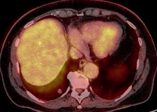
16 How cancers (should) look like? Without contrast agent With contrast agent
17 How cancers (should) look like? Without contrast agent With contrast agent Renal cyst (Benign) Heterogenous enhancement Renal cancer (Malignant)
 look like?")

")

18 How cancers (should) look like? Without contrast agent With contrast agent Renal cyst (Benign) Angiogenesis Heterogenous enhancement Renal cancer (Malignant)

19 How cancers (should) look like? A patient with uterine cancer
20 How cancers (should) look like? 18FDG-PET FDG-hot FDG-cold


21 How cancers (should) look like? 18FDG-PET Metabolism FDG-hot FDG-cold
22 Diagnostic Imaging of Oncology to see is to believe How cancers look like? Key characters of cancers, such as Proliferation, Angiogenesis, Metabolism etc. How to catch those characters? Novel imaging techniques, such as..


23 Novel Imaging Techniques Dual Energy Multidetector CT
24 Novel Imaging Techniques Conventional CT Water material density image Iodine material density image Dual-Energy (Spectral) CT: applications in Abdominal Imaging, Silva et al, Radiographics 2011;31:


25 Novel Imaging Techniques Conventional CT Water material density image Iodine material density image Cysts Cancer Dual-Energy (Spectral) CT: applications in Abdominal Imaging, Silva et al, Radiographics 2011;31:
26 Novel Imaging Techniques CT Spectral attenuation curves Imaging of tumor vascularity A patient with multiple liver metastases after treatment Green: reference, Pink: a meta with high vascularity (viable tumor cells) Yellow: a meta with moderate vascl. Blue: with less vascl (close to necrosis) Dual-Energy (Spectral) CT: applications in Abdominal Imaging, Silva et al, Radiographics 2011;31:

27 Novel Imaging Techniques Magnetic resonance imaging (MRI) Magnetic field and radio frequency waves Superior contrast resolution compared to other imaging modalities Functional imaging capabilities 27

28 MRI

29 Diffusion-weighted MRI Water diffusion correlates with cell density. Tumor= high cell density=diffusion restriction Diffuson Weighted MRI in the Body, Koh et al, AJR 2007; 188:
30 Diffusion-weighted MRI Ovarian cancer with peritoneal dissemination CT

31 Diffusion-weighted MRI Ovarian cancer with peritoneal dissemination CT DWI


32 Diffusion-weighted MRI A patient with rectal cancer

33 Diffusion-weighted MRI A patient with prostate cancer T2WI DWI ADC map
 Prostate")
 Dynamic")
 MR")
34 Novel Imaging Techniques Magnetic resonance imaging (MRI) Prostate cancer T1, T2WI (morphology) DWI, ADC-map (cell density) Dynamic contrast enhanced MR (permeability, vascularity) MR spectroscopy (choline)


35 Cervical cancer, MR: Diffusion, ADC, Choline spectroscopy Garcia-Figueiras et al, Magn Reson Imaging Clin N Am 24 (2016) 261


36 Novel Imaging Techniques PET-CT, PET-MR Make positron emitting tracers, such as 18-FDG, via cyclotron Administrate tracers via injection/inhalation etc Image by PET camera t ½ 11 C 20 min 13 N 10 min 15 O 2 min 18 F 110 min 68 Ga 68 min


37 Novel Imaging Techniques PET-CT, PET-MR Basic principle of FDG-PET


38 PET/MR, Pancreatic cancer Pre treatment Post treatment
39 Diagnostic Imaging of Oncology to see is to believe How cancers look like? Key characters of cancers, such as Proliferation, Angiogenesis, Metabolism etc. How to catch those characters? Novel imaging techniques, such as.. CT (computed tomography), MRI (magnet resonance imaging), PET-CT (positron emission tomography-ct) etc


40 Can macroscopic imaging features correlate/indicate with genetic features?
41 Diagnostic Imaging of Oncology How cancers look like? Morphology: Reflection of DNA, RNA, genes, proteins etc. Function: increased/decreased metabolic level. Can macroscopic imaging features correlate/indicate with genetic features? Radiomics
42 Diagnostic Imaging of Oncology Radiomics Imaging feature correlates with long noncoding RNA (lncrna) expression And may indicate poor metastasis-free survival (Radiogenomic Biomarker Reveals Associations among Dynamic Contrastenhanced MR Imaging, Long Noncoding RNA, and Metastasis: Yamamot et al, Radiology vol 275; ) Measure ERF (enhancing rim fraction)
43 Diagnostic Imaging of Oncology ERF: enhancing rim fraction Triple negative br ca Metastasis free survival Radiogenomic Biomarker Reveals Associations among Dynamic Contrast-enhanced MR Imaging, Long Noncoding RNA, and Metastasis: Yamamot et al, Radiology vol 275;


44 AI, deep learning for diagnosis Ben-Cohen, Acad Radiolo 2017;24:

:883,")

45 Radiomics, AI, deep learning for Health Care NEJM Gerlinger, 2012;366(10):883, Nature Clinical Oncology Lambin,2017;14:749
46 NEJM Gerlinger, 2012;366(10):883 What We Want to Do To catch the cancer s dynamic Clonal Evolution Intratumor Heterogenity Spatial and Temporal Heterogenity In One Patient
47 What We Want to Do A patient with multiple liver metastases after treatment Green: reference, Pink: a meta with high vascularity (viable tumor cells) Yellow: a meta with moderate vascl. Blue: with less vascl (close to necrosis) NEJM Gerlinger, 2012;366(10):883, Radiographics 2011;31:
 Yellow: a meta with moderate vascl.")
48 What We Don t Want to Do A patient with multiple liver metastases after treatment Green: reference, Pink: a meta with high vascularity (viable tumor cells) Yellow: a meta with moderate vascl. Blue: with less vascl (close to necrosis) NEJM Gerlinger, 2012;366(10):883, Radiographics 2011;31:
49 Radiomics, AI, deep learning for Health Care Nature Clinical Oncology Lambin,2017;14:749
50 Contents Imaging of Oncology? How tumors look like? Why? Mind the Gap!


51 MIND THE GAP! Flow cytometry, tumor markers

52 MIND THE GAP!
53 Thank you!
Case Reports: Tumor Detection by Diffusion-Weighted MRI and ADC-Mapping with Correlation to PET/CT Results
 Case Reports: Tumor Detection by Diffusion-Weighted MRI and ADC-Mapping with Correlation to PET/CT Results Matthias Philipp Lichy, M.D.; Philip Aschoff, M.D.; Christina Pfannenberg, M.D.; Schlemmer Heinz-Peter,
Staging Colorectal Cancer
 Staging Colorectal Cancer CT is recommended as the initial staging scan for colorectal cancer to assess local extent of the disease and to look for metastases to the liver and/or lung Further imaging for
Molecular Imaging and Cancer
 Molecular Imaging and Cancer Cancer causes one in every four deaths in the United States, second only to heart disease. According to the U.S. Department of Health and Human Services, more than 512,000
Multiparametric imaging in oncology
 Multiparametric imaging in oncology p1 T p2 p2 T T p3 p1 p3 T Marco Ravanelli Roberto Maroldi The goal of traditional imaging is high spatial and contrast resolution diagnosis, tumor extent treatment planning,
Radiological assessment of neoadjuvent chemotherapy for breast cancer
 XV th Balkan Congress of Radiology Budapest, Hungary, October 12 15, 2017 Radiological assessment of neoadjuvent chemotherapy for breast cancer V. Bešlagić C l i n i c o f R a d i o l o g y, U n i v e
PET-MRI in malignant bone tumours. Lars Stegger Department of Nuclear Medicine University Hospital Münster, Germany
 PET-MRI in malignant bone tumours Lars Stegger Department of Nuclear Medicine University Hospital Münster, Germany Content From PET to PET/MRI General considerations Bone metastases Primary bone tumours
Imaging in gastric cancer
 Imaging in gastric cancer Gastric cancer remains a deadly disease because of late diagnosis. Adenocarcinoma represents 90% of malignant tumors. Diagnosis is based on endoscopic examination with biopsies.
بسم هللا الرحمن الرحيم. Prof soha Talaat
 بسم هللا الرحمن الرحيم Ovarian tumors The leading indication for gynecologic surgery. Preoperative characterization of complex solid and cystic adnexal masses is crucial for informing patients about possible
Bone PET/MRI : Diagnostic yield in bone metastases and malignant primitive bone tumors
 Bone PET/MRI : Diagnostic yield in bone metastases and malignant primitive bone tumors Lars Stegger, Benjamin Noto Department of Nuclear Medicine University Hospital Münster, Germany Content From PET to
PET IMAGING (POSITRON EMISSION TOMOGRAPY) FACT SHEET
 FACT SHEET") Positron Emission Tomography (PET) When calling Anthem (1-800-533-1120) or using the Point of Care authorization system for a Health Service Review, the following clinical information may be needed to
Positron Emission Tomography (PET) When calling Anthem (1-800-533-1120) or using the Point of Care authorization system for a Health Service Review, the following clinical information may be needed to
Functional aspects of anatomical imaging techniques
 Functional aspects of anatomical imaging techniques Nilendu Purandare Associate Professor & Consultant Radiologist Tata Memorial Centre Functional/metabolic/molecular imaging (radioisotope scanning) PET
Medical Policy An independent licensee of the Blue Cross Blue Shield Association
 PET Scanning: Oncologic Applications Page 1 of 88 Medical Policy An independent licensee of the Blue Cross Blue Shield Association Title: Positron Emission Tomography (PET) Scanning: Oncologic Applications
Prof. Dr. NAGUI M. ABDELWAHAB,M.D.; MARYSE Y. AWADALLAH, M.D. AYA M. BASSAM, Ms.C.
 Role of Whole-body Diffusion MR in Detection of Metastatic lesions Prof. Dr. NAGUI M. ABDELWAHAB,M.D.; MARYSE Y. AWADALLAH, M.D. AYA M. BASSAM, Ms.C. Cancer is a potentially life-threatening disease,
Indications of PET/CT in oncology
 Monday, August 27, 2012 Session 1, 10:00-10:40 Indications of PET/CT in oncology Helle Westergren Hendel MD, PhD, assistant professor Bacelor in Leadership & Health Ecomomics Head of Clinical PET, Herlev
Index. Surg Oncol Clin N Am 16 (2007) Note: Page numbers of article titles are in boldface type.
 Note: Page numbers of article titles are in boldface type.") Surg Oncol Clin N Am 16 (2007) 465 469 Index Note: Page numbers of article titles are in boldface type. A Adjuvant therapy, preoperative for gastric cancer, staging and, 339 B Breast cancer, metabolic
Surg Oncol Clin N Am 16 (2007) 465 469 Index Note: Page numbers of article titles are in boldface type. A Adjuvant therapy, preoperative for gastric cancer, staging and, 339 B Breast cancer, metabolic
Emerging Referral Patterns for Whole-Body Diffusion Weighted Imaging (WB-DWI) in an Oncology Center
 in an Oncology Center") Emerging Referral Patterns for Whole-Body Diffusion Weighted Imaging (WB-DWI) in an Oncology Center Poster No.: C-1296 Congress: ECR 2014 Type: Scientific Exhibit Authors: G. Petralia 1, G. Conte 1, S.
Emerging Referral Patterns for Whole-Body Diffusion Weighted Imaging (WB-DWI) in an Oncology Center Poster No.: C-1296 Congress: ECR 2014 Type: Scientific Exhibit Authors: G. Petralia 1, G. Conte 1, S.
Dr Claire Smith, Consultant Radiologist St James University Hospital Leeds
 Dr Claire Smith, Consultant Radiologist St James University Hospital Leeds Imaging in jaundice and 2ww pathway Image protocol Staging Limitations Pancreatic cancer 1.2.4 Refer people using a suspected
ABDOMINAL DIFFUSION WEIGHTED MR
 ABDOMINAL DIFFUSION WEIGHTED MR Frank Miller, M.D. FACR Professor of Radiology Chief, Body Imaging Section Medical Director, MR Imaging Northwestern University Feinberg School of Medicine fmiller@northwestern.edu
Appendix 1: Regional Lymph Node Stations for Staging Esophageal Cancer
 Appendix 1: Regional Lymph Node Stations for Staging Esophageal Cancer Locoregional (N stage) disease was redefined in the seventh edition of the AJCC Cancer Staging Manual as any periesophageal lymph
Essentials of Clinical MR, 2 nd edition. 73. Urinary Bladder and Male Pelvis
 73. Urinary Bladder and Male Pelvis Urinary bladder carcinoma is best locally staged with MRI. It is important however to note that a thickened wall (> 5 mm) is a non-specific finding seen in an underfilled
LYMPHATIC DRAINAGE IN THE HEAD & NECK
 LYMPHATIC DRAINAGE IN THE HEAD & NECK Like other parts of the body, the head and neck contains lymph nodes (commonly called glands). Which form part of the overall Lymphatic Drainage system of the body.
POSITRON EMISSION TOMOGRAPHY (PET)
") Status Active Medical and Behavioral Health Policy Section: Radiology Policy Number: V-27 Effective Date: 08/27/2014 Blue Cross and Blue Shield of Minnesota medical policies do not imply that members should
Status Active Medical and Behavioral Health Policy Section: Radiology Policy Number: V-27 Effective Date: 08/27/2014 Blue Cross and Blue Shield of Minnesota medical policies do not imply that members should
Oncology - Evolution of imaging From helpful to essential
 Oncology - Evolution of imaging From helpful to essential 1990s 2000 1980s 18 FDG PET/CT MRI/MRSI 2010 1970s MRI 1960s CT 2015 HP13C-MRSI Ultrasound Nuc Med X-Ray - IVU MRI/PET Imaging 2016: Essential
F NaF PET/CT in the Evaluation of Skeletal Malignancy
 F NaF PET/CT in the Evaluation of Skeletal Malignancy Andrei Iagaru, MD September 26, 2013 School of of Medicine Ø Introduction Ø F NaF PET/CT in Primary Bone Cancers Ø F NaF PET/CT in Bone Metastases
Imaging. Personalized Cancer Medicine Program Dec. Chikako Suzuki. Per Grybäck. Lennart Blomqvist. PCM report Imaging
 Personalized Cancer Medicine Program Imaging 2016 Dec. Chikako Suzuki Per Grybäck Lennart Blomqvist Chikako.Suzuki@ki.se Personalised Cancer Medicine Program Karolinska Institutet Science for Life Laboratory
Role of MRI Diffusion in Assessment of Mediastinal Lymphadenopathy
 Med. J. Cairo Univ., Vol. 85, No. 3, June: 925-931, 2017 www.medicaljournalofcairouniversity.net Role of MRI Diffusion in Assessment of Mediastinal Lymphadenopathy YOUSSRIAH Y. SABRI, M.D.*; MARIAN FAYEK,
FDG-PET/CT in Gynaecologic Cancers
 Friday, August 31, 2012 Session 6, 9:00-9:30 FDG-PET/CT in Gynaecologic Cancers (Uterine) cervical cancer Endometrial cancer & Uterine sarcomas Ovarian cancer Little mermaid (Edvard Eriksen 1913) honoring
MRI-PET: Oncologic Applications
 MRI-PET: Oncologic Applications Pablo R. Ros, MD University Hospitals Case Medical Center Case Western Reserve University SCBT-MR Boston, MA October, 2012 Pablo.Ros@UHhospitals.org Acknowledgement Osman
MR imaging of FIGO stage I uterine cervical cancer: The diagnostic impact of 3T-MRI
 MR imaging of FIGO stage I uterine cervical cancer: The diagnostic impact of 3T-MRI Poster No.: C-1191 Congress: ECR 2010 Type: Educational Exhibit Topic: Genitourinary Authors: M. Takeuchi, K. Matsuzaki,
EUROPEAN BOARD OF RADIOLOGY (EUROPEAN COLLEGE OF RADIOLOGY), VIENNAAUSTRIA. EDiR. Radiology March 2012
, VIENNAAUSTRIA. EDiR. Radiology March 2012") DR.SIKANDAR SHAIKH Department of PET-CT and Nuclear Medicine Yashoda Hospitals Somajiguda. Hyderabad.Andhra Pradesh 500082 Email: drsikandar_s@yahoo.co.in, Office: 91 40 67779999 CONSULTANT PET-CT & RADIOLOGY
DR.SIKANDAR SHAIKH Department of PET-CT and Nuclear Medicine Yashoda Hospitals Somajiguda. Hyderabad.Andhra Pradesh 500082 Email: drsikandar_s@yahoo.co.in, Office: 91 40 67779999 CONSULTANT PET-CT & RADIOLOGY
Principles of nuclear metabolic imaging. Prof. Dr. Alex Maes AZ Groeninge Kortrijk and KULeuven Belgium
 Principles of nuclear metabolic imaging Prof. Dr. Alex Maes AZ Groeninge Kortrijk and KULeuven Belgium I. Molecular imaging probes A. Introduction - Chemical disturbances will precede anatomical abnormalities
performed to help sway the clinician in what the appropriate diagnosis is, which can substantially alter the treatment of management.
 Hello, I am Maura Polansky at the University of Texas MD Anderson Cancer Center. I am a Physician Assistant in the Department of Gastrointestinal Medical Oncology and the Program Director for Physician
Acknowledgements. Update of Focal Liver Lesions Goals. Focal Liver Lesions. Imaging Choices For Liver Lesions. Focal Liver Lesions
 Acknowledgements Update of Focal Liver Lesions 2012 Giles Boland Massachusetts General Hospital Harvard Medical School No disclosures Dushyant Sahani Mukesh Harisinghani Goals Focal liver lesions Imaging
Laura Tormoehlen, M.D. Neurology and EM-Toxicology Indiana University
 Laura Tormoehlen, M.D. Neurology and EM-Toxicology Indiana University Disclosures! No conflicts of interest to disclose Neuroimaging 101! Plain films! Computed tomography " Angiography " Perfusion! Magnetic
Radionuclides in Medical Imaging. Danielle Wilson
 Radionuclides in Medical Imaging Danielle Wilson Outline Definitions History and development Radionuclide applications & techniques in imaging Conclusion Definition #1 : Radionuclide An unstable nucleus
Option D: Medicinal Chemistry
 Option D: Medicinal Chemistry Basics - unstable radioactive nuclei emit radiation in the form of smaller particles alpha, beta, positron, proton, neutron, & gamma are all used in nuclear medicine unstable
FieldStrength. Leuven research is finetuning. whole body staging
 FieldStrength Publication for the Philips MRI Community Issue 40 May 2010 Leuven research is finetuning 3.0T DWIBS for whole body staging The University Hospital of Leuven is researching 3.0T whole body
MEASUREMENT OF EFFECT SOLID TUMOR EXAMPLES
 MEASUREMENT OF EFFECT SOLID TUMOR EXAMPLES Although response is not the primary endpoint of this trial, subjects with measurable disease will be assessed by standard criteria. For the purposes of this
Diffusion Weighted Imaging in Prostate Cancer
 Diffusion Weighted Imaging in Prostate Cancer Disclosure Information Vikas Kundra, M.D, Ph.D. No financial relationships to disclose. Education Goals and Objectives To describe the utility of diffusion-weighted
Computed Diffusion-Weighted Image in the Abdomen
 Computed Diffusion-Weighted Image in the Abdomen Poster No.: C-0234 Congress: ECR 2014 Type: Scientific Exhibit Authors: T. Yoshikawa 1, N. Aoyama 1, Y. Ohno 1, K. Kyotani 1, Y. Kassai 2, K. Sofue 1, M.
Dosimetry, see MAGIC; Polymer gel dosimetry. Fiducial tracking, see CyberKnife radiosurgery
 Subject Index Acoustic neuroma, neurofibromatosis type 2 complications 103, 105 hearing outcomes 103, 105 outcome measures 101 patient selection 105 study design 101 tumor control 101 105 treatment options
Optimized. clinical pathway. propels high utilization of PET/MR at Pitié-Salpêtrière Hospital
 Optimized propels high utilization of PET/MR at Pitié-Salpêtrière Hospital clinical pathway As one of Europe s largest teaching hospitals, Pitié-Salpêtrière Hospital is renowned for its innovative research
Localized Prostate Cancer Have we finally got it right? Shingai Mutambirwa Professor & Chair-Division Urology DGMAH & SMU Pretoria SOUTH AFRICA
 Localized Prostate Cancer Have we finally got it right? Shingai Mutambirwa Professor & Chair-Division Urology DGMAH & SMU Pretoria SOUTH AFRICA ESMO Cape Town 14 Feb 2018 Disclosures Advisory boards/lecturer/consultant-
Disclosure. Acknowledgement. What is the Best Workup for Rectal Cancer Staging: US/MRI/PET? Rectal cancer imaging. None
 What is the Best Workup for Rectal Cancer Staging: US/MRI/PET? Zhen Jane Wang, MD Assistant Professor in Residence UC SF Department of Radiology Disclosure None Acknowledgement Hueylan Chern, MD, Department
Assessment of renal cell carcinoma by two PET tracer : dual-time-point C-11 methionine and F-18 fluorodeoxyglucose
 Assessment of renal cell carcinoma by two PET tracer : dual-time-point C-11 methionine and F-18 fluorodeoxyglucose Poster No.: C-0805 Congress: ECR 2015 Type: Scientific Exhibit Authors: S. Ito, K. Kato,
T2, T2*, ute. Yeo Ju Kim. Radiology, Inha University Hospital, Incheon, Korea
 SY28-1 T2, T2*, ute Yeo Ju Kim Radiology, Inha University Hospital, Incheon, Korea T2 relaxation times relate to the rate of transverse magnetization decay, caused by the loss of phase coherence induced
Nuclear Medicine in Thyroid Cancer. Phillip J. Koo, MD Division Chief of Diagnostic Imaging
 Nuclear Medicine in Thyroid Cancer Phillip J. Koo, MD Division Chief of Diagnostic Imaging Financial Disclosures Bayer Janssen Learning Objectives To learn the advantages and disadvantages of SPECT/CT
Imaging features of malignant transformation and benign malignant-mimicking lesions in the genitourinary tracts
 Imaging features of malignant transformation and benign malignant-mimicking lesions in the genitourinary tracts Poster No.: C-2639 Congress: ECR 2015 Type: Scientific Exhibit Authors: S. B. Park, J. B.
HEPATIC METASTASES. We can state 3 types of metastases depending on their treatment options:
 HEPATIC METASTASES 1. Definition Metastasis means the spread of cancer. Cancerous cells can separate from the primary tumor and enter the bloodstream or the lymphatic system (the one that produces, stores,
PET imaging of cancer metabolism is commonly performed with F18
 PCRI Insights, August 2012, Vol. 15: No. 3 Carbon-11-Acetate PET/CT Imaging in Prostate Cancer Fabio Almeida, M.D. Medical Director, Arizona Molecular Imaging Center - Phoenix PET imaging of cancer metabolism
Proc. Intl. Soc. Mag. Reson. Med. 22 (2014)
") Tumor Physiology Natalie J. Serkova, PhD Department of Anesthesiology and Radiology, University of Colorado at Denver, Anschutz Medical Center, Aurora, CO This course will describe the distinct characteristics
Tumor Physiology Natalie J. Serkova, PhD Department of Anesthesiology and Radiology, University of Colorado at Denver, Anschutz Medical Center, Aurora, CO This course will describe the distinct characteristics
PET/MR:Techniques, Indications and Applications
 PET/MR:Techniques, Indications and Applications Franz Wolfgang Hirsch Professor and Head of the Department of Pediatric Radiology University Hospital Leipzig / Germany Children s Hospital University Leipzig
Clinical indications for positron emission tomography
 Clinical indications for positron emission tomography Oncology applications Brain and spinal cord Parotid Suspected tumour recurrence when anatomical imaging is difficult or equivocal and management will
Pancreatic Cancer. What is pancreatic cancer?
 Scan for mobile link. Pancreatic Cancer Pancreatic cancer is a tumor of the pancreas, an organ that is located behind the stomach in the abdomen. Pancreatic cancer does not always cause symptoms until
Role of DE-CT in Oncology
 Role of DE-CT in Oncology Dushyant Sahani, M.D Director of CT Associate Professor of Radiology Massachusetts General Hospital Harvard Medical School Email-dsahani@partners.org Disclosure Research Grant
Molecular Imaging and the Brain
 Molecular imaging technologies are playing an important role in neuroimaging, a branch of medical imaging, by providing a window into the living brain. Where CT and conventional MR imaging provide important
Innovations in HCC Imaging: MDCT/MRI
 Innovations in HCC Imaging: MDCT/MRI Anthony E. Cheng, M.D. Cardinal MRI Center Cardinal Santos Medical Center, Wilson Street, San Juan Innovations in HCC Imaging: Goals/Objectives MDCT/MRI Learn the diagnostic
International Journal of Current Medical Sciences- Vol. 6, Issue,, pp , June, 2016 A B S T R A C T
 ISSN: 2320-8147 International Journal of Current Medical Sciences- Vol. 6, Issue,, pp. 122-126, June, 2016 COMPUTED TOMOGRAPHY IN HEPATIC METASTASES Ananthakumar P and Adaikkappan M., Available online
Utility of 18 F-FDG PET/CT in metabolic response assessment after CyberKnife radiosurgery for early stage non-small cell lung cancer
 Utility of F-FDG PET/CT in metabolic response assessment after CyberKnife radiosurgery for early stage non-small cell lung cancer Ngoc Ha Le 1*, Hong Son Mai 1, Van Nguyen Le 2, Quang Bieu Bui 2 1 Department
Advanced Imaging Techniques in Evaluation of Colorectal Cancer 1
 Gastrointestinal Imaging 740 Advanced Imaging Techniques in Evaluation of Colorectal Cancer 1 Roberto García-Figueiras, MD, PhD Sandra Baleato-González, MD, PhD Anwar R. Padhani, MBBS, MRCP, FRCR Antonio
Imaging Decisions Start Here SM
 Owing to its high resolution and wide anatomic coverage, dynamic first-pass perfusion 320-detector-row CT outperforms PET/CT for distinguishing benign from malignant lung nodules, researchers from Japan
Medical imaging X-ray, CT, MRI, scintigraphy, SPECT, PET Györgyi Műzes
 Medical imaging X-ray, CT, MRI, scintigraphy, SPECT, PET Györgyi Műzes Semmelweis University, 2nd Dept. of Medicine Medical imaging: definition technical process of creating visual representations about
Anatomic Imaging of Prostate Cancer
 Masoom Haider, MD, FRCP(C) Professor of Radiology, University of Toronto Clinician Scientist, Ontario Institute of Cancer Research Senior Scientist, Sunnybrook Research Institute Chief, Dept of Medical
Medical Policy An independent licensee of the Blue Cross Blue Shield Association
 PET Scanning: Oncologic Applications Page 1 of 42 Medical Policy An independent licensee of the Blue Cross Blue Shield Association Title: See also: Positron Emission Tomography (PET) Scanning: Oncologic
Improving Patients' Understanding of Radiology Reports: Comparing Coverage of a Lay-Language Radiology Glossary to MedlinePlus
 Improving Patients' Understanding of Radiology Reports: Comparing Coverage of a Lay-Language Radiology Glossary to MedlinePlus American College of Radiology National Meeting May 2017 Teresa Martin-Carreras,
Molecular Imaging and Breast Cancer
 Molecular Imaging and Breast Cancer Breast cancer forms in tissues of the breast usually in the ducts, tubes that carry milk to the nipple, and lobules, the glands that make milk. It occurs in both men
Whole Body MRI. Dr. Nina Tunariu. Prostate Cancer recurrence, progression and restaging
 Whole Body MRI Prostate Cancer recurrence, progression and restaging Dr. Nina Tunariu Consultant Radiology Drug Development Unit and Prostate Targeted Therapies Group 12-13 Janeiro 2018 Evolving Treatment
COLORECTAL CARCINOMA
 QUICK REFERENCE FOR HEALTHCARE PROVIDERS MANAGEMENT OF COLORECTAL CARCINOMA Ministry of Health Malaysia Malaysian Society of Colorectal Surgeons Malaysian Society of Gastroenterology & Hepatology Malaysian
ADRENAL LESIONS 10/09/2012. Adrenal + lesion. Introduction. Common causes. Anatomy. Financial disclosure. Dr. Boraiah Sreeharsha. Nothing to declare
 ADRENAL LESIONS Financial disclosure Nothing to declare Dr. Boraiah Sreeharsha MBBS;FRCR;FRCPSC Introduction Adrenal + lesion Adrenal lesions are common 9% of the population Increase in the detection rate
Imaging Tissue Response to Therapeutic Radiation
 1 Imaging Tissue Response to Therapeutic Radiation Sean P. Frigo, Ph.D. Assistant Professor Department of Human Oncology School of Medicine and Public Health University of Wisconsin Madison Slide 1 1 Based
Index. mri.theclinics.com. Note: Page numbers of article titles are in boldface type.
 Index Note: Page numbers of article titles are in boldface type. A Angiogenesis, and cancer of prostate, 689 690 Angiography, MR. See MR angiography. Apoptosis, MR imaging of, 637 Apparent diffusion coefficient,
Comparison of whole-body diffusion-weighted MRI and PET/ CT for detection of metastases
 Comparison of whole-body diffusion-weighted MRI and PET/ CT for detection of metastases Poster No.: R-0021 Congress: RANZCR-AOCR 2012 Type: Scientific Exhibit Authors: T. D. Nguyen Keywords: Oncology,
MRI/MRS Biomarkers. Robert E. Lenkinski, Ph.D.
 MRI/MRS Biomarkers Robert E. Lenkinski, Ph.D. Disclosure GE Healthcare-Research Grant Aspect MR-Scientific Advisor Aposense-Scientific Advisor Brainwatch-Scientific Advisor I will be discussing off-label
Nuclear Medicine and PET. D. J. McMahon rev cewood
 Nuclear Medicine and PET D. J. McMahon 150504 rev cewood 2018-02-15 Key Points Nuclear Medicine and PET: Imaging: Understand how Nuc Med & PET differ from Radiography & CT by the source of radiation. Be
Current Clinical Practice. MR Imaging Evaluations. MRI Anatomic Review. Imaging to Address Clinical Challenges. Prostate MR
 BETH ISRAEL DEACONESS MEDICAL CENTER Prostate MR Neil M. Rofsky, MD Harvard Medical School Current Clinical Practice DIGITAL RECTAL EXAMINATION PSA ( ~ 20% False negative) BIOPSY (18-25% False negative)
Imaging of Neuroendocrine Metastases
 Imaging of Neuroendocrine Metastases Aoife Kilcoyne, Shaunagh McDermott, Colin McCarthy,Manuel Patino, Dushyant Sahani, Michael Blake Abdominal Imaging Division Massachusetts General Hospital Disclosure
Role of imaging in RCC. Ultrasonography. Solid lesion. Cystic RCC. Solid RCC 31/08/60. From Diagnosis to Treatment: the Radiologist Perspective
 Role of imaging in RCC From Diagnosis to Treatment: the Radiologist Perspective Diagnosis Staging Follow up Imaging modalities Limitations and pitfalls Duangkamon Prapruttam, MD Department of Therapeutic
Oncologic Applications of PET Scanning
 6.01.26 Oncologic Applications of PET Scanning Section 6.0 Radiology Subsection Effective Date February 15, 2015 Original Policy Date January 26, 2009 Next Review Date December 2015 Description Positron
8/10/2016. PET/CT for Tumor Response. Staging and restaging Early treatment response evaluation Guiding biopsy
 PET/CT for Tumor Response Evaluation August 4, 2016 Wei Lu, PhD Department of Medical Physics www.mskcc.org Department of Radiation Oncology www.umaryland.edu FDG PET/CT for Cancer Imaging Staging and
Staging recurrent ovarian cancer with 18 FDG PET/CT
 ONCOLOGY LETTERS 5: 593-597, 2013 Staging recurrent ovarian cancer with FDG PET/CT SANJA DRAGOSAVAC 1, SOPHIE DERCHAIN 2, NELSON M.G. CASERTA 3 and GUSTAVO DE SOUZA 2 1 DIMEN Medicina Nuclear and PET/CT
Introduction to the Course and the Techniques. Jeffry R. Alger, PhD Ahmanson-Lovelace Brain Mapping Center Department of Neurology
 Introduction to the Course and the Techniques Jeffry R. Alger, PhD Ahmanson-Lovelace Brain Mapping Center Department of Neurology (jralger@ucla.edu) CTSI Neuroimaging April 2014 Rationale for the Course
Anthem Blue Cross and Blue Shield Virginia Advanced Imaging Procedures Requiring Precertification Revised 02/13/2013
 Anthem Blue Cross and Blue Shield Virginia Advanced Imaging Procedures Requiring Precertification Revised 02/13/2013 Modality and CT Head CTA Head: Cerebrovascular MRI Head MRA Head: Cerebrovascular Functional
Page: 1 of 29. For this policy, PET scanning is discussed for the following 4 applications in oncology:
 Emission Tomography Scanning Page: 1 of 29 Last Review Status/Date: June 2015 Description Positron emission tomography (PET) scans are based on the use of positron-emitting radionuclide tracers coupled
PELVIC MRI COURSE. November 12-14, 2016
 Memorial Sloan Kettering Cancer Center is pleased to announce: PELVIC MRI COURSE November 12-14, 2016 Conference Location: Memorial Sloan Kettering Cancer Center Rockefeller Research Laboratories 430 East
FDG-PET/CT for cancer management
 195 REVIEW FDG-PET/CT for cancer management Hideki Otsuka, Naomi Morita, Kyo Yamashita, and Hiromu Nishitani Department of Radiology, Institute of Health Biosciences, The University of Tokushima, Graduate
Cardiac Imaging Tests
 Cardiac Imaging Tests http://www.medpagetoday.com/upload/2010/11/15/23347.jpg Standard imaging tests include echocardiography, chest x-ray, CT, MRI, and various radionuclide techniques. Standard CT and
Hybrid systems in Medical Imaging
 Hybrid systems in Medical Imaging from PET/CT to PET/MR Osman Ratib, MD, PhD, FAHA Professor and chair Department of Medical Imaging and Information Sciences Head of division of Nuclear Medicine University
Zurich, January 19, 2018
 Brain metastases as first presentation of malignancy: Immediate management, differential diagnosis; prevalence of primaries and suggested work-up Symposium on Brain Metastasis Cancer Center Zurich Zurich,
Positron emission tomography/computer tomography in the evaluation of head and neck cancer treatment
 Positron emission tomography/computer tomography in the evaluation of head and neck cancer treatment Severina Šedienė 1, Ilona Kulakienė 1, Viktoras Rudžianskas 2 1 Lithuanian University of Health Sciences,
FOR CMS (MEDICARE) MEMBERS ONLY NATIONAL COVERAGE DETERMINATION (NCD) FOR MAGNETIC RESONANCE IMAGING:
 MEMBERS ONLY NATIONAL COVERAGE DETERMINATION (NCD) FOR MAGNETIC RESONANCE IMAGING:") National Imaging Associates, Inc. Clinical guidelines BONE MARROW MRI Original Date: July 2008 Page 1 of 5 CPT Codes: 77084 Last Review Date: September 2014 NCD 220.2 MRI Last Effective Date: July 2011
National Imaging Associates, Inc. Clinical guidelines BONE MARROW MRI Original Date: July 2008 Page 1 of 5 CPT Codes: 77084 Last Review Date: September 2014 NCD 220.2 MRI Last Effective Date: July 2011
Case Report Multiple Hypovascular Tumors in Kidney: A Rare Case Report and Differential Diagnosis
 Volume 2013, Article ID 595193, 4 pages http://dx.doi.org/10.1155/2013/595193 Case Report Multiple Hypovascular Tumors in Kidney: A Rare Case Report and Differential Diagnosis Pei-Yu Wu, 1 Sheng-Fung Lin,
How to integrate surgery in the treatment of patients with liver-only metastatic disease
 How to integrate surgery in the treatment of patients with liver-only metastatic disease Luis Sabater Ortí MD, PhD Associate Professor University of Valencia European Board Surgical Qualification HBP (EBSQ-HPB)
Wednesday 12 September Advanced imaging science to practice 14:30 14:55. Recent advances in computed tomography (CT) technology
 technology") 14:30 14:55 Recent advances in computed tomography (CT) technology Dr Gareth Iball, Leeds Teaching Hospitals NHS Trust All modern CT systems incorporate iterative reconstruction (IR) techniques. Their
14:30 14:55 Recent advances in computed tomography (CT) technology Dr Gareth Iball, Leeds Teaching Hospitals NHS Trust All modern CT systems incorporate iterative reconstruction (IR) techniques. Their
MR Functional Imaging to Guide Radiotherapy: Challenges and Opportunities
 Abstract No. 1234 MR Functional Imaging to Guide Radiotherapy: Challenges and Opportunities Michael Milosevic, MD Department of Radiation Oncology, University of Toronto Radiation Medicine Program, Princess
Volumetric Functional MRI Criteria for Assessing Tumor Response
 Volumetric Functional MRI Criteria for Assessing Tumor Response Ihab R. Kamel, M.D., Ph.D. ikamel@jhmi.edu Associate Professor of Radiology and Oncology Clinical Director, MRI Department of Radiology Johns
8/3/2016. Consultant for / research support from: Astellas Bayer Bracco GE Healthcare Guerbet Medrad Siemens Healthcare. Single Energy.
 U. Joseph Schoepf, MD Prof. (h.c.), FAHA, FSCBT-MR, FNASCI, FSCCT Professor of Radiology, Medicine, and Pediatrics Director, Division of Cardiovascular Imaging Consultant for / research support from: Astellas
Journal of Radiology Case Reports
 Critical Pitfall: Varices in Cancer Patients mimicking Lymphadenopathy; Differentiation of varicose veins and enlarged lymph nodes in routine staging Tilman Schubert 1*, Michele Pansini 1, Georg Bongartz
Liver Cancer (Hepatocellular Carcinoma or HCC) Overview
 Overview") Liver Cancer (Hepatocellular Carcinoma or HCC) Overview Recent advances in liver cancer care seek to address the rising incidence of liver cancer, which has steadily increased over the past three decades.
Liver Cancer (Hepatocellular Carcinoma or HCC) Overview Recent advances in liver cancer care seek to address the rising incidence of liver cancer, which has steadily increased over the past three decades.
PET/CT in lung cancer
 PET/CT in lung cancer Andrei Šamarin North Estonia Medical Centre 3 rd Baltic Congress of Radiology 08.10.2010 Imaging in lung cancer Why do we need PET/CT? CT is routine imaging modality for staging of
Imaging Findings of Primary Angiomyolipoma of the Pancreas: A Case Report 췌장의원발성혈관근육지방종의영상소견 1 예 : 증례보고
 Case Report pissn 1738-2637 / eissn 2288-2928 https://doi.org/10.3348/jksr.2017.77.1.9 Imaging Findings of Primary Angiomyolipoma of the Pancreas: A Case Report 췌장의원발성혈관근육지방종의영상소견 1 예 : 증례보고 Hye Hee Kim,