Liver MRI in 30 minutes
|
|
|
- Jocelin Stanley
- 6 years ago
- Views:
Transcription

1 X Liver MRI in 30 minutes SCBT/MR Annual Meeting Salt Lake City September 18, 2016 Scott B. Reeder, MD, PhD Department of Radiology University of Wisconsin Madison, WI
2 Disclosures University of Wisconsin-Madison receives research support from GE Healthcare, and Bracco Diagnostics Founder Calimetrix, LLC Shareholder Elucent Medical Consulting - Parexel International


")

")
")


3 Overview: Standard Abdominal Protocol Coronal T2w SSFSE (1-2 BH) T1w In/Out Phase (1-2 BH) Injection Pre-contrast 3D-T1w (1 BH) Late Arterial 3D-T1w (1 BH) Portal Venous 3D-T1w (1 BH) Axial T2w FSE (3-4 min) Equilibrium 3D-T1w (1 BH) DWI (2-3 BH) Dynamic T1w Imaging (5-7 min) b=0-50 b= ADC
4 Localizers Three plane localizers Perform during same breath-holding maneuver Practice for patient, more accurate prescription Help determine how long patient can hold breath Check coil position to ensure well positioned SSFSE (HASTE) SSFP (FIESTA, true-fisp, BFFE) Contain a great deal of diagnostic information Don t forget to review Corner shots!

5 Signal T2 Weighted Imaging Fluid Tumor Liver Echo Time (TE)
6 Coronal SSFSE: 1-2 breath-holds High resolution Rapid, robust overview of anatomy and pathology Surgeons like the coronal view Coronal useful to prescribe axial volumes Should it replace conventional T2w-FSE?? Altered contrast Extended echo train = mix of spin-echoes & stimulated echoes Metastatic disease difficult to visualize Great for cysts and hemangiomas


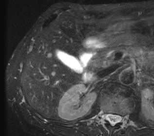
7 Examples Hemangioma and Renal Cyst Patient 1 Patient 2 Patient 3 Biliary cystadenoma Caroli s disease


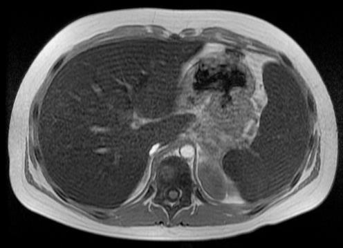
8 Case: Metastatic Adenocarcinoma T2w FSE with FS T2w SSFSE DWI (b=500) Metastases Inconspicuous On T2w SSFSE Is this a problem??


9 Hepatic metastasis, rectal CA Precontrast Arterial Delayed T2 FS DWI Case, courtesy Bobby Kalb, MD, University of Arizona


10 21 yo F, intrahepatic cholangioca Precontrast Arterial Delayed T2 Ax MRCP Case, courtesy Bobby Kalb, MD, University of Arizona
11 Fat-Iron Sensitive Sequences In-phase and opposed-phase Liver Detection (qualitative) of hepatic steatosis Mass-mimicking steatosis Focal fat Focal sparing Mass characterization HCC Hepatic adenoma Angiomyolipoma Adrenal masses Renal masses (AML, RCC)

")
12 Detection of Fat with MRI: In and Opposed Phase (2-point Dixon) Opposed-Phase (W-F) In-Phase (W+F)


13 Case : Renal Mass Histology: Clear Cell Renal Cell Carcinoma


14 Case: Hemosiderosis Axial out-of-phase Axial in-phase Axial T2w FSE Coronal T2w SSFSE

15 Enhancement Phases of Enhancement: Extracellular Contrast Agents HA PV Liver Tumor Time inject


16 Enhancement Phases of Enhancement: Extracellular Contrast Agents Arterial Late Phase Arterial Phase Portal Venous HA PV Liver Tumor Time Pre inject Late HAP PVP Equilibrium


17 Patient 3 Patient 2 Patient 1 Cases: Contrast Enhanced T1w Normal Focal Nodular Hyperplasia Renal Cell Carcinoma
18 Diffusion Weighted Imaging Unrestricted (fluid) Larger ADC (more signal loss) Restricted (cells) Smaller ADC (less signal loss)
19 DWI: Optional Sequence Suppresses tissues with freely diffusing water Increased conspicuity: lesions with high cellularity Applications Detection and characterization of malignancy helpful for metastatic disease in the liver Not helpful for HCC Pearls: b = 0 is just T2-weighted S=S o e -TE/T2 b > 0 is both T2- & diffusion-weighted S=S o e -TE/T2 e -badc
20 DWI: Optional Sequence Image quality insufficiently reliable Used with echo planar imaging Distortion, signal dropout (left lobe of liver) Failed fat suppression Problem on all vendors Cannot rely on this sequence alone for detection Pearl: important, but least reliable. Perform last!


 ADC Map")
21 Cases: 58yo M with Colorectal Cancer 2x10⁻³ mm 2 /s Restricted diffusion: high cellularity Axial DWI (b=500) ADC Map (mm 2 /s) 0 mm 2 /s


22 Abdominal MRI: Current Protocol Dynamic T1w CE-MRI - Detect lesions - Characterize - Vascular T2w MRI - Detect lesions - Characterize - Biliary, renal T1w Fat/Iron Sensitive - liver - pancreas - adrenal glands DWI - Detect lesions - Characterize - Diffuse liver dz Neuroendocrine Mets MRCP - Biliary dz - Pancreas - Surgical Cx s Pancreatic AdenoCA

23 Enhancement Phases of Enhancement: Extracellular Contrast Agents Late Arterial Phase Portal Venous HA PV Liver Tumor Equilibrium Time inject 3-4 minutes
24 Time Resolved MRI: Interleaved Variable Density Injection 24s Pre-Contrast T1 # 1 # 2 # 3 # 4 # 5 Portal Venous Phase Equilibrium Phase Dynamic IVD k y k z


25 Time Resolved MRI: Interleaved Variable Density 4s temporal resolution Focal Nodular Hyperplasia x 4
 x 32 (A/P) x 20 (S/I) cm 3 4.")

26 Data Acquisition: 3D Cartesian High Temporal & Spatial Resolution 2.0 mm isotropic 40 (L/R) x 32 (A/P) x 20 (S/I) cm sec/frame 3x 20s breath-holds 3T, GE Discovery MR channel Torso Phase 5, end of 1 st BH Phase 6, start of 2 nd BH Wang et al ISMRM 2012, 3867
27 Basic Liver MRI: Current Protocol Contrast Localizers (1 min) T1w IOP (1 min) T2W (1 min) DWI (3-5min) 3D T1w (3-4 min) ~20-25 minutes
 T2 weighted")
 Dual Echo")
 (3 minutes) T2W (1 min)")
 (3-5min) 3D")


28 Basic Liver MRI: Suggested Approach DWI (b=0) T2 weighted Contrast image! Localizers (1 min) Dual Echo (Dixon) T1w IOP 3D T1w DCE-MRI (1 min) (3 minutes) T2W (1 min) Advanced DWI (b=500) T2w + DWI DWI (1 breath-hold) (3-5min) 3D T1w (3-4 min) Water Fat < 5 minutes Combine In-phase Dixon Opposed fat-water Phase imaging with 3D CE-MRI Combine DWI with T2 weighted imaging** Eliminate localizers**

29 Thank you! Alex Frydrychowicz, MD Scott Nagle, MD, PhD Jeff Weinreb, MD Claude Sirlin, MD Grant Support WARF Accelerator NIH: R01 DK R01 DK R01 DK RC1 EB010384
Hepatobiliary Contrast Agents
 Hepatobiliary Contrast Agents SCBT/MR Annual Meeting Salt Lake City September 21, 2016 Scott B. Reeder, MD, PhD Department of Radiology University of Wisconsin Madison, WI Disclosures University of Wisconsin-Madison
40 th Annual Meeting of the SCBT/MR
 Liver Fat and Iron Quantification 40 th Annual Meeting of the SCBT/MR Nashville, TN September 11, 2017 Scott B. Reeder, MD, PhD 100% 0% Department of Radiology University of Wisconsin Madison, WI Disclosures
Hepatobiliary Contrast Agents for Liver MRI
 Hepatobiliary Contrast Agents for Liver MRI Scott B. Reeder, MD, PhD International Society for Magnetic Resonance in Medicine Sociedad Mexicana de Radiologia e Imagen (SMRI) Mexico City June 4, 2014 Department
Abdominal MRI Techniques in Pediatric Oncology
 Abdominal MRI Techniques in Pediatric Oncology Jonathan R. Dillman, M.D. Assistant Professor Departments of Radiology & Urology Section of Pediatric Radiology C.S. Mott Children s Hospital Disclosures
Essentials of Clinical MR, 2 nd edition. 65. Benign Hepatic Masses
 65. Benign Hepatic Masses Pulse sequences acquired for abdominal MRI typically consist of fast acquisition schemes such as single-shot turbo spin echo (i.e. HASTE) and gradient echo schemes such as FLASH
ABDOMINAL DIFFUSION WEIGHTED MR
 ABDOMINAL DIFFUSION WEIGHTED MR Frank Miller, M.D. FACR Professor of Radiology Chief, Body Imaging Section Medical Director, MR Imaging Northwestern University Feinberg School of Medicine fmiller@northwestern.edu
MRI Abdomen Protocol Pancreas/MRCP with Contrast
 MRI Abdomen Protocol Pancreas/MRCP with Contrast Reviewed By: Brett Mollard, MD; Anna Ellermeier, MD Last Reviewed: July 2018 Contact: (866) 761-4200 Standard uses: 1. Characterization of cystic and solid
ADRENAL MR: PEARLS AND PITFALLS
 ADRENAL MR: PEARLS AND PITFALLS Frank Miller, M.D. Lee F. Rogers MD Professor of Medical Education Chief, Body Imaging Section and Fellowship Medical Director, MR Imaging Professor of Radiology Northwestern
CT & MRI of Benign Liver Neoplasms Srinivasa R Prasad
 CT & MRI of Benign Liver Neoplasms Srinivasa R Prasad No financial disclosures Acknowledgements Many thanks to Drs. Heiken, Narra & Menias (MIR) Dr. Sahani (MGH) for sharing images Benign Liver Tumors:
MRI of the Hepatobiliary System
 BODY APPLICATIONS OF MRI 51 Chapter Four C H A P T E R F O U R MRI of the Hepatobiliary System After completing this chapter, the reader will be able to: Develop a protocol for liver MRI Identify benign
BODY MRI PROTOCOLS 1.5T
 BODY MRI PROTOCOLS 1.5T Table of Contents Non Specific...3-5 Renals.....6-8 Adrenals.....9-11 Pancreas..12-14 Liver 15-17 Liver No Gad.18-21 Liver Eovist..22-24 Liver Iron Quant 25-29 MRCP.30-34 ENTEROGRAPHY.35-38
Newcastle HPB MDM updated radiology imaging protocol recommendations. Author Dr John Scott. Consultant Radiologist Freeman Hospital
 Newcastle HPB MDM updated radiology imaging protocol recommendations Author Dr John Scott. Consultant Radiologist Freeman Hospital This document is intended as a guide to aid radiologists and clinicians
MRI OF FOCAL LESIONS IN
 Introduction MRI OF FOCAL LESIONS IN THE NON-CIRRHOTIC LIVER Ivan Pedrosa M.D. Ph.D. Associate Professor of Radiology and Advanced Imaging Research Center University of Texas Southwestern. Dallas, TX Incidental
Acknowledgements. Update of Focal Liver Lesions Goals. Focal Liver Lesions. Imaging Choices For Liver Lesions. Focal Liver Lesions
 Acknowledgements Update of Focal Liver Lesions 2012 Giles Boland Massachusetts General Hospital Harvard Medical School No disclosures Dushyant Sahani Mukesh Harisinghani Goals Focal liver lesions Imaging
Pancreatic Adenocarcinoma: Everything You Need to Know From Cross-Sectional Imaging to Treatment
 Pancreatic Adenocarcinoma: Everything You Need to Know From Cross-Sectional Imaging to Treatment Andrew W. Bowman, MD PhD Assistant Professor of Radiology Mayo Clinic Florida SCBT-MR Annual Meeting Nashville,
Liver imaging takes a step forward with Ingenia
 Publication for the Philips MRI Community ISSUE 49 2013 / 2 Liver imaging takes a step forward with Ingenia Lyon South Hospital strives to move from several studies first CT, then MR or PET to using just
CASE 1 11/1/2016 HEPATOBILIARY IMAGING CASE PRESENTATIONS DECLARATION. Dr. Chirag Patel ORGAN IMAGING yr old lady
 HEPATOBILIARY IMAGING CASE PRESENTATIONS DECLARATION No financial disclosures or affiliations with commercial organisations No discussion of investigational or off-label use of medical devices, products
Anatomical and Functional MRI of the Pancreas
 Anatomical and Functional MRI of the Pancreas MA Bali, MD, T Metens, PhD Erasme Hospital Free University of Brussels Belgium mbali@ulb.ac.be Introduction The use of MRI to investigate the pancreas has
Alice Fung, MD Oregon Health and Science University
 Alice Fung, MD Oregon Health and Science University Disclosure Comments The speaker Alice Fung, MD Has relevant financial relationships to disclose. Received honorarium from (Guerbet). This individual
Innovations in HCC Imaging: MDCT/MRI
 Innovations in HCC Imaging: MDCT/MRI Anthony E. Cheng, M.D. Cardinal MRI Center Cardinal Santos Medical Center, Wilson Street, San Juan Innovations in HCC Imaging: Goals/Objectives MDCT/MRI Learn the diagnostic
Emergency Abdominal MRI Protocols
 2018 SPR Annual Meeting & Postgraduate Course May 15-19, 2018 Nashville, Tennessee Emergency Abdominal MRI Protocols Unni Udayasankar MD Associate Professor Department of Medical Imaging University of
Disclosures. Diffusion and Perfusion Imaging in the Head and Neck. Learning objectives ???
 Disclosures No relevant financial disclosures Diffusion and Perfusion Imaging in the Head and Neck Ashok Srinivasan, MD Associate Professor Director of Neuroradiology University of Michigan Health System
Revised June Body MR Protocols
 Body MR Protocols Abdomen focus protocols A 1: Pre- and post-contrast abdomen MRI A 1L: Abdomen MRI without contrast A 1P: Pre- and post-contrast abdomen MRI (pancreas protocol) A 1R: Pre- and post-contrast
High Field MR of the Spine
 Department of Radiology University of California San Diego 3T for MR Applications Advantages High Field MR of the Spine Increased signal-to-noise Better fat suppression Increased enhancement with gadolinium
Imaging of liver and pancreas
 Imaging of liver and pancreas.. Disease of the liver Focal liver disease Diffusion liver disease Focal liver disease Benign Cyst Abscess Hemangioma FNH Hepatic adenoma HCC Malignant Fibrolamellar carcinoma
Magnetization Preparation Sequences
 Magnetization Preparation Sequences Acquisition method may not give desired contrast Prep block adds contrast (and/or encoding) MP-RAGE = Magnetization prepared rapid acquisition with gradient echo (Mugler,
Interesting Cases from Liver Tumor Board. Jeffrey C. Weinreb, M.D.,FACR Yale University School of Medicine
 Interesting Cases from Liver Tumor Board Jeffrey C. Weinreb, M.D.,FACR Yale University School of Medicine jeffrey.weinreb@yale.edu Common Liver Diseases Hemangioma Cyst FNH Focal Fat/Sparing THID Non-Cirrhotic
Indications: Abdomen and pelvis for malignancy. If there is history of small bowel or mesentery tumor, see "contrast" section for oral contrast**
 MRB AB01AMR 01aAbdomen (with dynamics) + Pelvis (pre/post) A74183 A72197 Indications: Abdomen and pelvis for malignancy. If there is history of small bowel or mesentery tumor, see "contrast" section for
Diffusion Weighted Imaging in Pediatric Body: Update
 Diffusion Weighted Imaging in Pediatric Body: Update Govind Chavhan, MD DABR Diagnostic Imaging Department The Hospital for Sick Children and University of Toronto, Canada 2 Outline Principles of diffusion
Hepatobiliary MRI: Current Concepts and Controversies
 JOURNAL OF MAGNETIC RESONANCE IMAGING 25:681 695 (2007) Review Article Hepatobiliary MRI: Current Concepts and Controversies James F. Glockner, MD, PhD* Evaluation of the liver and biliary system is a
Customizing Contrast Injection for Body MDCT: Algorithmic Approach
 Customizing Contrast Injection for Body MDCT: Algorithmic Approach Lincoln L. Berland, M.D., F.A.C.R. University of Alabama at Birmingham Before Contrast Prep and Hydration Hydration single most important
Enhancements in Hepatobiliary Imaging:
 Enhancements in Hepatobiliary Imaging: S. Channual 1, MD; A. Pahwa 2, MD; S. Raman 1, MD. 1 UCLA Medical Center, Department of Radiologic Sciences 2 Olive-View UCLA Medical Center, Department of Radiology
CTA/MRA of Pediatric Hepatic Masses Radiology-Pathology Correlation
 Acta Radiológica Portuguesa, Vol.XVIII, nº70, pág. 41-50, Abr.-Jun., 2006 CTA/MRA of Pediatric Hepatic Masses Radiology-Pathology Correlation Marilyn J. Siegel Mallinckrodt Institute of Radiology, Washington
Imaging in gastric cancer
 Imaging in gastric cancer Gastric cancer remains a deadly disease because of late diagnosis. Adenocarcinoma represents 90% of malignant tumors. Diagnosis is based on endoscopic examination with biopsies.
LIVER IMAGING TIPS IN VARIOUS MODALITIES. M.Vlychou, MD, PhD Assoc. Professor of Radiology University of Thessaly
 LIVER IMAGING TIPS IN VARIOUS MODALITIES M.Vlychou, MD, PhD Assoc. Professor of Radiology University of Thessaly Hepatocellular carcinoma is a common malignancy for which prevention, screening, diagnosis,
Hepatobiliary and Pancreatic Malignancies
 Hepatobiliary and Pancreatic Malignancies Gareth Eeson MD MSc FRCSC Surgical Oncologist and General Surgeon Kelowna General Hospital Interior Health Consultant, Surgical Oncology BC Cancer Agency Centre
Essentials of Clinical MR, 2 nd edition. 73. Urinary Bladder and Male Pelvis
 73. Urinary Bladder and Male Pelvis Urinary bladder carcinoma is best locally staged with MRI. It is important however to note that a thickened wall (> 5 mm) is a non-specific finding seen in an underfilled
IMAGING OF ACUTE AND CHRONIC PANCREATITIS, INCLUDING EXOCRINE FUNCTION
 IMAGING OF ACUTE AND CHRONIC PANCREATITIS, INCLUDING EXOCRINE FUNCTION Andrew T. Trout, MD @AndrewTroutMD Disclosures Grant support National Pancreas Foundation In-kind support - ChiRhoClin modified from:
Objectives. HCC Incidence and Mortality. Disclosure Statement HCC. Imaging of Hepatocellular Carcinoma. Treatment of Hepatocellular Carcinoma
 Imaging of Hepatocellular Carcinoma and the use of LI RADS Treatment of Hepatocellular Carcinoma Aaron D. Anderson, D.O. AOCR April 2015 Objectives Show how the use of LI RADS can simplify the diagnosis
International Journal of Current Medical Sciences- Vol. 6, Issue,, pp , June, 2016 A B S T R A C T
 ISSN: 2320-8147 International Journal of Current Medical Sciences- Vol. 6, Issue,, pp. 122-126, June, 2016 COMPUTED TOMOGRAPHY IN HEPATIC METASTASES Ananthakumar P and Adaikkappan M., Available online
Abdominal applications of DWI
 Postgraduate course, SPR San Antonio (Texas), May 14-15, 2013 Abdominal applications of DWI Rutger A.J. Nievelstein Wilhelmina Children s s Hospital, Utrecht (NL) Outline What is DWI? How to perform? Challenges
Evaluation of Liver Mass Lesions. American College of Gastroenterology 2013 Regional Postgraduate Course
 Evaluation of Liver Mass Lesions American College of Gastroenterology 2013 Regional Postgraduate Course Lewis R. Roberts, MB ChB, PhD Division of Gastroenterology and Hepatology Mayo Clinic College of
IT 의료융합 1 차임상세미나 복부질환초음파 이재영
 IT 의료융합 1 차임상세미나 2013-4-3 복부질환초음파 이재영 나는오늘누구를위하여 종을울리나? 전통적의료 의사 공학설계자 의사 최첨단진단장비들 USG, CT, MRI 환자 환자 현대의료 사용자중심의사고 US in the Abdomen Detection DDx Look Behavior Response by external stimuli Guiding Tool
ADRENAL LESIONS 10/09/2012. Adrenal + lesion. Introduction. Common causes. Anatomy. Financial disclosure. Dr. Boraiah Sreeharsha. Nothing to declare
 ADRENAL LESIONS Financial disclosure Nothing to declare Dr. Boraiah Sreeharsha MBBS;FRCR;FRCPSC Introduction Adrenal + lesion Adrenal lesions are common 9% of the population Increase in the detection rate
Prostate Cancer DFP Case of the Week
 Prostate Cancer DFP Case of the Week Antonio C. Westphalen, MD PhD Clinical Prostate MR Imaging Program, Director Associate Professor of Radiology and Urology University of California, San Francisco Case
HEPATO-BILIARY IMAGING
 HEPATO-BILIARY IMAGING BY MAMDOUH MAHFOUZ MD PROF.OF RADIOLOGY CAIRO UNIVERSITY mamdouh.m5@gmail.com www.ssregypt.com CT ABDOMEN Indications Patient preparation Patient position Scanogram Fasting 4-6 hours
Hepatocellular carcinoma Cholangiocarcinoma. Jewels of hepatobiliary cancer imaging : what to look for? Imaging characteristics of HCC.
 Outline : Imaging Jewels Jewels of hepatobiliary cancer imaging : what to look for? Hepatocellular carcinoma Cholangiocarcinoma Surachate Siripongsakun, M.D. Chulabhorn Cancer Center Imaging characteristics
IMAGING OF LIVER, BILIARY TREE, PANCREAS
 IMAGING OF LIVER, BILIARY TREE, PANCREAS Department of Radiology West China Hospital, Sichuan University Yao Jin Learning Points The methodology for imaging the LBP (liver, biliary tree, and pancreas )
Contrast Enhanced Ultrasound of Parenchymal Masses in Children
 Contrast Enhanced Ultrasound of Parenchymal Masses in Children Sue C Kaste, DO On behalf of Beth McCarville, MD St. Jude Children s Research Hospital Memphis, TN Overview Share St. Jude experience with
DETECTING EARLY LIVER METASTASIS: THE POWER OF MRI WITH LIVER SPECIFIC CONTRAST
 DETECTING EARLY LIVER METASTASIS: THE POWER OF MRI WITH LIVER SPECIFIC CONTRAST LINDA PANTONGRARG-BROWN, MD ADVANCED DIAGNOSTIC IMAGING, RAMATHIBODI HOSPITAL, BANGKOK, THAILAND OUTLINE Choice of imaging
OASIS 1.2T: MULTIPARAMETRIC MRI OF PROSTATE CANCER
 OASIS 1.2T: MULTIPARAMETRIC MRI OF PROSTATE CANCER By Dr. John Feller, MD, Radiologist Desert Medical Imaging, Palm Springs, CA MRI is clinically accepted as the best imaging modality for displaying anatomical
MR Tumor Staging for Treatment Decision in Case of Wilms Tumor
 MR Tumor Staging for Treatment Decision in Case of Wilms Tumor G. Schneider, M.D., Ph.D.; P. Fries, M.D. Dept. of Diagnostic and Interventional Radiology, Saarland University Hospital, Homburg/Saar, Germany
Common and unusual CT and MRI manifestations of pancreatic adenocarcinoma: a pictorial review
 Review Article Common and unusual CT and MRI manifestations of pancreatic adenocarcinoma: a pictorial review Min-Jie Yang, Su Li, Yong-Guang Liu, Na Jiao, Jing-Shan Gong Department of Radiology, Shenzhen
Imaging of Neuroendocrine Metastases
 Imaging of Neuroendocrine Metastases Aoife Kilcoyne, Shaunagh McDermott, Colin McCarthy,Manuel Patino, Dushyant Sahani, Michael Blake Abdominal Imaging Division Massachusetts General Hospital Disclosure
Jeffrey C. Weinreb, MD, FACR Yale School of Medicine Yale-New Haven Hospital
 Jeffrey C. Weinreb, MD, FACR Yale School of Medicine Yale-New Haven Hospital jeffrey.weinreb@yale.edu 1991 1997 Whole body MRI: multistation approach x z Isocenter: Table Move: Multiple Steps Whole body
Disclosure. Acknowledgement. What is the Best Workup for Rectal Cancer Staging: US/MRI/PET? Rectal cancer imaging. None
 What is the Best Workup for Rectal Cancer Staging: US/MRI/PET? Zhen Jane Wang, MD Assistant Professor in Residence UC SF Department of Radiology Disclosure None Acknowledgement Hueylan Chern, MD, Department
ESUR 2018, Sept. 13 th.-16 th., 2018 Barcelona, Spain
 ESUR 2018, Sept. 13 th.-16 th., 2018 Barcelona, Spain OUR APPROACH Incidental adrenal nodule/mass Isaac R Francis, M.B;B.S University of Michigan, Ann Arbor, Michigan Disclosures None (in memory) M Korobkin,
CARDIAC MRI. Cardiovascular Disease. Cardiovascular Disease. Cardiovascular Disease. Overview
 CARDIAC MRI Dr Yang Faridah A. Aziz Department of Biomedical Imaging University of Malaya Medical Centre Cardiovascular Disease Diseases of the circulatory system, also called cardiovascular disease (CVD),
Outline. Neuroradiology. Diffusion Imaging in. Clinical Applications of. Basics of Diffusion Imaging. Basics of Diffusion Imaging
 Clinical Applications of Diffusion Imaging in Neuroradiology No disclosures Stephen F. Kralik Assistant Professor of Radiology Indiana University School of Medicine Department of Radiology and Imaging
Objectives 8/17/2011. Challenges in Cardiac Imaging. Challenges in Cardiac Imaging. Basic Cardiac MRI Sequences
 8/17/2011 Traditional Protocol Model for Tomographic Imaging Cardiac MRI Sequences and Protocols Frandics Chan, M.D., Ph.D. Stanford University Medical Center Interpretation Lucile Packard Children s Hospital
Introduction. Body MRI overview
 Section 1 Chapter 1 Body MRI overview Introduction Introduction Body MRI is a dynamic, exciting modality. If read carefully, you will find in this small book the essentials to protocol, understand, and
Evaluation of adrenal masses using FIESTA-MRI sequence
 Evaluation of adrenal masses using FIESTA-MRI sequence 14.September.2018/Friday / Room 3 / 11.00-1230 GÖKHAN PEKİNDİL, FATMA CAN, CELAL BAYAR UNIVERSITY MEDICAL FACULTY DEP. OF RADIOLOGY MANİSA-TURKEY
Feasibility and initial dosimetric findings for a randomized trial using dose painted multi-parametric-mri defined targets in prostate cancer
 Feasibility and initial dosimetric findings for a randomized trial using dose painted multi-parametric-mri defined targets in prostate cancer Thoughts on the use of MRI in the treatment of prostate cancer
T2, T2*, ute. Yeo Ju Kim. Radiology, Inha University Hospital, Incheon, Korea
 SY28-1 T2, T2*, ute Yeo Ju Kim Radiology, Inha University Hospital, Incheon, Korea T2 relaxation times relate to the rate of transverse magnetization decay, caused by the loss of phase coherence induced
11/10/2015. Prostate cancer in the U.S. Multi-parametric MRI of Prostate Diagnosis and Treatment Planning. NIH estimates for 2015.
 Multi-parametric MRI of Prostate Diagnosis and Treatment Planning Temel Tirkes, M.D. Associate Professor of Radiology Director, Genitourinary Radiology Indiana University School of Medicine Department
Jesse Civan, M.D. Medical Director, Jefferson Liver Tumor Center
 Liver Tumors Jesse Civan, M.D. Medical Director, Jefferson Liver Tumor Center Differential Diagnosis Malignant Metastatic from non-hepatic primary Hepatocellular carcinoma Cholangiocarcinoma Biliary cystcarcinoma
Diagnostic Challenges and Pitfalls in MR Imaging with Hepatocyte-specific
 Note: This copy is for your personal non-commercial use only. To order presentation-ready copies for distribution to your colleagues or clients, contact us at www.rsna.org/rsnarights. ABDOMINAL AND GASTROINTESTINAL
Gemstone Spectral Imaging quantifies lesion characteristics for a confident diagnosis
 GE Healthcare Gemstone Spectral Imaging quantifies lesion characteristics for a confident diagnosis CT clinical case study lesion characterization Desiree Morgan, MD Vice Chair of Clinical Research Professor
Computed Diffusion-Weighted Image in the Abdomen
 Computed Diffusion-Weighted Image in the Abdomen Poster No.: C-0234 Congress: ECR 2014 Type: Scientific Exhibit Authors: T. Yoshikawa 1, N. Aoyama 1, Y. Ohno 1, K. Kyotani 1, Y. Kassai 2, K. Sofue 1, M.
MRI of Acute Abdominal Pain in Pregnancy
 MRI of Acute Abdominal Pain in Pregnancy Ivan Pedrosa, M.D. Chief of MRI Jack Reynolds MD Chair in Radiology Associate Professor of Radiology UT Southwestern Medical Center Advanced Imag ing Research Center
8/3/2016. Consultant for / research support from: Astellas Bayer Bracco GE Healthcare Guerbet Medrad Siemens Healthcare. Single Energy.
 U. Joseph Schoepf, MD Prof. (h.c.), FAHA, FSCBT-MR, FNASCI, FSCCT Professor of Radiology, Medicine, and Pediatrics Director, Division of Cardiovascular Imaging Consultant for / research support from: Astellas
6/23/2009. Inversion Recovery (IR) Techniques and Applications. Variations of IR Technique. STIR, FLAIR, TI and TI Null. Applications of IR
 Techniques and Applications. Variations of IR Technique. STIR, FLAIR, TI and TI Null. Applications of IR") The Anatomy of Basic R Pulse Sequences Inversion Recovery () Techniques and Applications Chen Lin, PhD Indiana University School of edicine & Clarian Health Partners agnetization Preparation Section Chemical
The Anatomy of Basic R Pulse Sequences Inversion Recovery () Techniques and Applications Chen Lin, PhD Indiana University School of edicine & Clarian Health Partners agnetization Preparation Section Chemical
Liver surgery, acute GI tract bleeding
 Semmelweis University, Faculty of Medicine, 1 st Department of Surgery Liver surgery, acute GI tract bleeding Oszkár HAHN M.D. LIVER CYST US, CT, MRI Parasite (ELISA, eosinophil, anaphylaxy) Echinococcus
Diffusion-weighted MRI of metastatic liver lesions: is there a difference between hypervascular and hypovascular metastases?
 Original Article Diffusion-weighted MRI of metastatic liver lesions: is there a difference between hypervascular and hypovascular metastases? Acta Radiologica 2014, Vol. 55(5) 515 523! The Foundation Acta
Update on liver MRI at 3T
 REVIEW Update on liver MRI at 3T 3T MRI of the liver has become widely available and is routinely utilized in the clinical setting, but published scientific data comparing 1.5T with 3T is limited. Theoretically,
Dr Claire Smith, Consultant Radiologist St James University Hospital Leeds
 Dr Claire Smith, Consultant Radiologist St James University Hospital Leeds Imaging in jaundice and 2ww pathway Image protocol Staging Limitations Pancreatic cancer 1.2.4 Refer people using a suspected
Focal fatty sparing and focal fatty infiltration can be observed in the
 Diagn Interv Radiol 2011; 17:323 327 Turkish Society of Radiology 2011 ABDOMINAL IMAGING ORIGINAL ARTICLE Focal sparing of iron and fat in liver tissue in patients with hemosiderosis: diagnosis with combination
State of the Art Imaging for Hepatic Malignancy: My Assignment
 State of the Art Imaging for Hepatic Malignancy: My Assignment CT vs MR vs MRCP Which one to choose for HCC vs Cholangiocarcinoma What special protocols to use for liver tumors Role of PET and Duplex US
GASTROINTESTINAL IMAGING STUDY GUIDE
 GASTROINTESTINAL IMAGING STUDY GUIDE Pharynx Diverticula Foreign bodies Trauma o Motility Disorders Esophagus Diverticula Trauma Esophagitis Barrett esophagus Rings, webs, and strictures Varices Benign
Timothy L. Miao 1, Ania Z. Kielar 2,3, Rebecca M. Hibbert 2, Nicola Schieda 2,3
 DOES LESION T1 SIGNAL INTENSITY RELATIVE TO LIVER PARENCHYMA PREDICT VISIBILITY ON ULTRASOUND? A clinical tool to determine feasibility of ultrasound-guided percutaneous interventions Timothy L. Miao 1,
Liver nodules mimicking metastatic disease
 Liver nodules mimicking metastatic disease Poster No.: C-1703 Congress: ECR 2011 Type: Educational Exhibit Authors: F. Vandenbroucke, B. Ilsen, B. Op de Beeck, J. de Mey ; 1 1 2 2 3 2 3 Brussels/BE, Brussel/BE,
Non Contrast MRA. Mayil Krishnam. Director, Cardiovascular and Thoracic Imaging University of California, Irvine
 Non Contrast MRA Mayil Krishnam Director, Cardiovascular and Thoracic Imaging University of California, Irvine No disclosures Non contrast MRA-Why? Limitations of CTA Radiation exposure Iodinated contrast
ASL BASICS II. Learning Objectives. Outline. Acquisition. M. A. Fernández-Seara, Ph. D. Arterial spin labeled perfusion MRI: basic theory
 Acquisition ASL BASICS II M. A. Fernández-Seara, Ph. D. Neuroimaging Laboratory Center for Applied Medical Research University of Navarra Pamplona, Spain Outline Arterial spin labeled perfusion MRI: basic
Recommendations for cross-sectional imaging in cancer management, Second edition
 www.rcr.ac.uk Recommendations for cross-sectional imaging in cancer management, Second edition Renal and adrenal tumours Faculty of Clinical Radiology www.rcr.ac.uk Contents Renal cell carcinoma 3 Clinical
Altought liver (75%) and lung (15%) are the most commonly involved
 and lung (15%) are the most commonly involved") Diagn Interv Radiol 2010; 16:168 174 Turkish Society of Radiology 2010 MAGNETIC RESONANCE IMAGING ORIGINAL ARTICLE Conventional and diffusion-weighted MRI of extrahepatic hydatid cysts Nagihan İnan, Nilay
Diagn Interv Radiol 2010; 16:168 174 Turkish Society of Radiology 2010 MAGNETIC RESONANCE IMAGING ORIGINAL ARTICLE Conventional and diffusion-weighted MRI of extrahepatic hydatid cysts Nagihan İnan, Nilay
بسم هللا الرحمن الرحيم. Prof soha Talaat
 بسم هللا الرحمن الرحيم Ovarian tumors The leading indication for gynecologic surgery. Preoperative characterization of complex solid and cystic adnexal masses is crucial for informing patients about possible
Erratum to: Fertility-sparing for young patients with gynecologic cancer: How MRI can guide patient selection prior to conservative management
 Abdominal Radiology ª Springer Science+Business Media, LLC 2017 Published online: 11 November 2017 Abdom Radiol (2017) 42:2966 2973 DOI: 10.1007/s00261-017-1205-5 Erratum to: Fertility-sparing for young
Functional Chest MRI in Children Hyun Woo Goo
 Functional Chest MRI in Children Hyun Woo Goo Department of Radiology and Research Institute of Radiology Asan Medical Center, University of Ulsan College of Medicine, Seoul, Korea No ionizing radiation
Monitoring bony metastases response with diffusion MRI
 Monitoring bony metastases response with diffusion MRI Anwar Padhani MD Mount Vernon Hospital Cancer Centre London, UK Objectives To illustrate the potential of whole body DWI in the therapy response assessment
DTI fiber tracking at 3T MR using b-1000 value in the depiction of periprostatic nerve before and after nervesparing prostatectomy
 DTI fiber tracking at 3T MR using b-1000 value in the depiction of periprostatic nerve before and after nervesparing prostatectomy Poster No.: C-2328 Congress: ECR 2012 Type: Scientific Paper Authors:
Tips and Tricks of State of the art MRA
 Tips and Tricks of State of the art MRA Mayil Krishnam, MD,MBA, MRCP,FRCR(UK) Professor of Radiology Director, Cardiovascular and Thoracic Imaging University of California, Irvine Objectives Technical
Liver Specific MRI using Gd-EOB-DTPA Disodium (Primovist) Effects Change in Management of Indeterminate Liver Lesions.
 Effects Change in Management of Indeterminate Liver Lesions.") Liver Specific MRI using Gd-EOB-DTPA Disodium (Primovist) Effects Change in Management of Indeterminate Liver Lesions. Poster No.: C-1751 Congress: ECR 2012 Type: Authors: Keywords: DOI: Educational Exhibit
Liver Specific MRI using Gd-EOB-DTPA Disodium (Primovist) Effects Change in Management of Indeterminate Liver Lesions. Poster No.: C-1751 Congress: ECR 2012 Type: Authors: Keywords: DOI: Educational Exhibit
Financial Disclosure
 Benign Liver Masses Adil Abdalla, MBBS Creighton University-CHI Health August 25, 2018 Financial Disclosure Nothing to disclose Financial Disclosure 1 Objectives To assess patients with benign liver tumors
Fulfilling the Promise
 Fulfilling the Promise of Cardiac MR Non-contrast, free-breathing technique generates comprehensive evaluation of the coronary arteries By Maggie Fung, MR Cardiovascular Clinical Development Manager; Wei
Malignant Focal Liver Lesions
 Malignant Focal Liver Lesions Other Than HCC Pablo R. Ros, MD, MPH, PhD Departments of Radiology and Pathology University Hospitals Cleveland Medical Center Case Western Reserve University Pablo.Ros@UHhospitals.org
Prof. Dr. NAGUI M. ABDELWAHAB,M.D.; MARYSE Y. AWADALLAH, M.D. AYA M. BASSAM, Ms.C.
 Role of Whole-body Diffusion MR in Detection of Metastatic lesions Prof. Dr. NAGUI M. ABDELWAHAB,M.D.; MARYSE Y. AWADALLAH, M.D. AYA M. BASSAM, Ms.C. Cancer is a potentially life-threatening disease,
Hepatic Imaging: What Every Practitioner Should Know
 Hepatic Imaging: What Every Practitioner Should Know Shuchi K. Rodgers, MD Section Chief, Abdominal Imaging Director of Ultrasound Department of Radiology Einstein Medical Center rodgerss@einstein.edu
Liver imaging the revolution
 Liver imaging the revolution Valérie Vilgrain Hôpital Beaujon, Paris, France PHC 2018 - www.aphc.info At the Beginning of the story Radiology in the 1970s US Garrett Radiology 1976 abscess Taylor Radiology
MRI/MRS Biomarkers. Robert E. Lenkinski, Ph.D.
 MRI/MRS Biomarkers Robert E. Lenkinski, Ph.D. Disclosure GE Healthcare-Research Grant Aspect MR-Scientific Advisor Aposense-Scientific Advisor Brainwatch-Scientific Advisor I will be discussing off-label
RECENT ADVANCES IN CLINICAL MR OF ARTICULAR CARTILAGE
 In Practice RECENT ADVANCES IN CLINICAL MR OF ARTICULAR CARTILAGE By Atsuya Watanabe, MD, PhD, Director, Advanced Diagnostic Imaging Center and Associate Professor, Department of Orthopedic Surgery, Teikyo