Anatomy of Pituitary Gland
|
|
|
- Constance Stanley
- 6 years ago
- Views:
Transcription


1 Anatomy of Pituitary Gland Please view our Editing File before studying this lecture to check for any changes. Color Code Important Doctors Notes Notes/Extra explanation
2 Objectives At the end of the lecture, students should be able to: Describe the position of the pituitary gland. List the structures related to the pituitary gland. Differentiate between the lobes of the gland. Describe the blood supply of pituitary gland & the hypophyseal portal system.


 lactation But only the pituitary gland will only increase in")
3 الغدة النخامية Pituitary Gland (also called Hypophysis Cerebri) o It is referred to as the master of endocrine glands. o It is a small oval structure 1 cm in diameter. o It doubles its size during pregnancy. A women experiences changes in her hormone levels during menstruation ( ضلحي,(ا pregnancy,(الحمل) lactation But only the pituitary gland will only increase in size during pregnancy.(سن اليأس ( menopause and,(الرضاعة) X-RAY SKULL: LATERAL VIEW SAGITTAL SECTION OF HEAD & NECK Extra

 of body of sphenoid o It")
.")


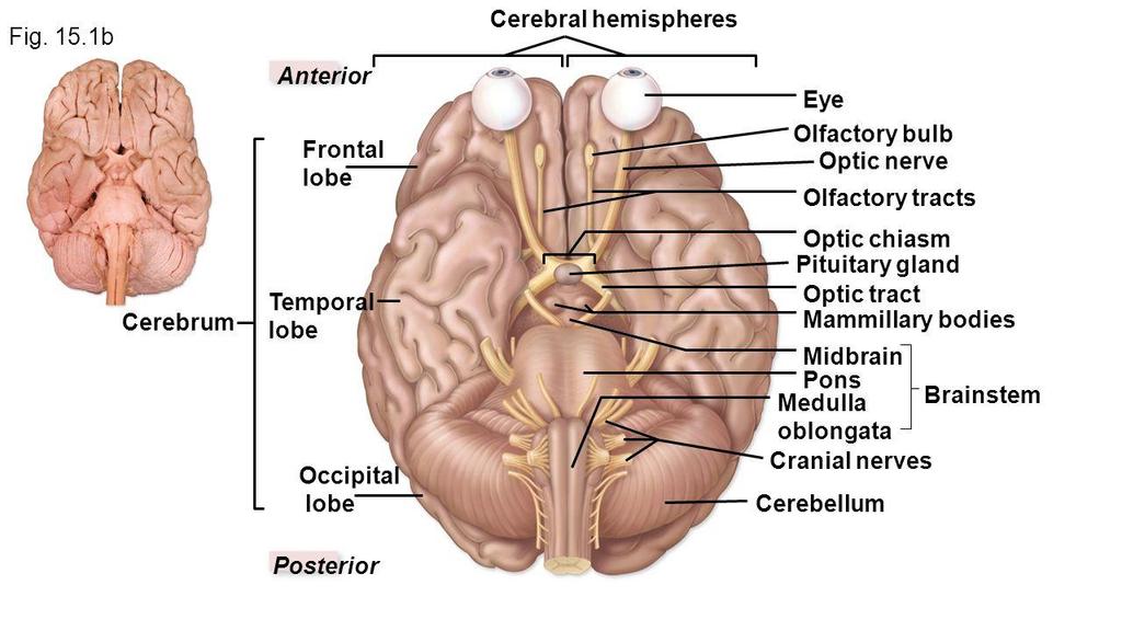
4 Pituitary Gland Position o It lies in the middle cranial fossa. o It is well protected in sella turcica* (hypophyseal fossa) of body of sphenoid o It lies between optic chiasma (anteriorly) & mamillary bodies** (posteriorly). Clinical point: Anterior to the pituitary gland is the optic chiasm, so if there was a tumor in the pituitary gland or it was enlarged this could press on the chiasm and disrupt the patients vision (loss of temporal field). *سرج الحصان ** Part of hypothalamus


5 The purple part is the sphenoid bone Hypophyseal fossa Extra Pictures
 connecting the")
 LATERAL: Cavernous sinuses Clinical point: 2")

6 Pituitary Gland Important Relations The relations are important SUPERIOR: Diaphragma sellae: A fold of dura mater covers the pituitary gland & has an opening for passage of infundibulum (pituitary stalk) connecting the gland to hypothalamus. INFERIOR: Sphenoidal air sinuses (recall from respiratory block) LATERAL: Cavernous sinuses Clinical point: 2 structures are present in the cavernous sinus: abducens nerve and the internal carotid artery. So when a surgeon is working on the pituitary he must be careful not to injure the internal carotid artery (which passes through the cavernous sinus) because it supplies the brain and may lead to a stroke or coma. Extra Extra Extra

: connected to hypothalamus")
, stores hormones secreted by")
7 Pituitary Gland Subdivisions 02:00 Hypothalamo -hypophyseal tract The gland is subdivided into: o Anterior Lobe (Adenohypophysis): it is the True gland, secretes hormones. o Posterior Lobe (Neurohypophysis): connected to hypothalamus through hypothalamo-hypophyseal tract (which passes through the stalk or infundibulum), stores hormones secreted by hypothalamic nuclei. Extra

 to the pituitary gland.")
8 Pituitary Gland Blood supply o Arteries: superior & inferior hypophyseal arteries (branches from internal carotid artery). Remember when we studied the circle of willis we said it surrounded the optic chiasm and pituitary gland. Remember also that it was formed by the internal carotid and basilar arteries. So the circle of willis will give a branch (from the internal carotid) to the pituitary gland. o Veins: hypophyseal veins drain into cavernous sinuses. To remember the supply recall that the pituitary gland is also called hypophysis cerebri hence hypophyseal. Extra


9 Pituitary Gland Distribution of Arteries o Superior Hypophyseal: supplies infundibulum and anterior lobe forms a capillary network from which vessels pass downward & form sinusoids into the anterior lobe of pituitary gland hypophyseal portal system. (AKA: hypothalamo-hypophyseal portal vessel/ system) A portal system is a system of blood vessels between 2 capillary beds, just like the one in the liver. The difference between the portal system in the liver and the one in the pituitary gland is that here it started with an artery and contains hormone releasing factors while in the liver it is a vein and carries nutrients. o Inferior Hypophyseal: supplies posterior lobe of pituitary gland. a hypothalamohypophyseal portal vessel
 o The Neurohypophysis receives a nerve supply from some of the hypothalamic nuclei (supraoptic &")

10 مهمة Pituitary Gland Lobes Explained further in physiology Anterior Lobe (adenohypophysis) o Hormone releasing & inhibiting factors produced by hypothalamus use Hypophyseal Portal System of vessels to reach the Anterior lobe of pituitary gland Posterior Lobe (neurohypophysis) o The Neurohypophysis receives a nerve supply from some of the hypothalamic nuclei (supraoptic & paraventricular) o The axons of these nuclei convey their neuro-secretion to the Posterior lobe of pituitary gland through Hypothalamo- Hypophyseal tract from where it passes into the blood stream. Hypophyseal Portal System: vascular connection between hypothalamus & anterior pituitary Hypothalamo-Hypophyseal tract: Neural connection between hypothalamus & posterior pituitary
11 PITUITARY GLAND (HYPOPHYSIS CEREBRI) master of endocrine glands. a small oval structure 1 cm in diameter. doubles its size during pregnancy. It lies in the middle cranial fossa. It is well protected in sella turcica (hypophyseal fossa) of body of sphenoid. IMPORTANT RELATIONS ANTERIOR : Optic chiasma POSTERIOR : Mamillary bodies SUPERIOR: Diaphragma sellae INFERIOR: Sphenoidal air sinuses LATERAL: Cavernous sinuses SUMMARY BLOOD SUPPLY ARTERIES: Superior & Inferior hypophyseal arteries - Internal Carotid artery branches Superior hypophyseal: supplies infundibulum and the anterior lobe of pituitary gland (hypophyseal portal system). Inferior hypophyseal: supplies posterior lobe of pituitary gland VEINS: Hypophyseal veins drain into Cavernous Sinuses. SUBDIVISION S OF PITUITARY GLAND Anterior Lobe (Adenohypophysis): it is the True gland, Secretes hormones Hormone-releasing & inhibiting factors produced by hypothalamus use Hypophyseal Portal System of vessels to reach the Anterior lobe of pituitary gland. Posterior Lobe (Neurohypophysis): connected to hypothalamus through hypothalamo-hypophyseal tract, Stores hormones secreted. It receives a nerve supply from some of the hypothalamic nuclei (supraoptic & paraventricular) -The axons of these nuclei convey their neurosecretion to the Posterior lobe of pituitary gland through Hypothalamo- Hypophyseal tract from where it passes into the blood stream.
12 MCQs 1. Which part of the pituitary gland secret hormones? A- The posterior part B- Neurohypophysis part C- Adenohypophysis part 2. Inferior hypophyseal artery branch from which of the following? A- Internal carotid artery B- External carotid artery C- Posterior cerebral artery 3. Which of artery forms the hypophyseal portal system? A- Inferior hypophyseal B- Superior hypophyseal C- Internal carotid 4. Which of the following nuclei supply the neurohypophysis? A- Paraventricular B- Mammillary body C- Dentate 5. Which one of the following structures is superior to the pituitary gland? A- Optic chiasma B- Diaphragma sellae C- Mammillary bodies D- Sphenoidal air sinuses 6. Which one of the following venous sinuses drains hypophyseal veins? A- Superior sagittal B- Cavernous C- Transverse D- Sigmoid 7. Which of the following is posterior to the pituitary gland? A- Optic chiasma B- Diaphragma sellae C- Mammillary bodies D- Sphenoidal air sinuses Answers: 1. C, 2. A, 3. B, 4. A, 5. B, 6. B, 7. C
13 SAQ 1. Enumerate the relations of pituitary gland? Anteriorly: Optic Chiasm Posteriorly: Mammillary Bodies Superiorly: Diaphragma sellae Inferiorly: Sphenoidal air sinuses Laterally: Cavernous sinuses 2. In case of pituitary gland enlargement which structure lie anteriorly will be compressed? The optic chiasm 3. When performing surgery on the pituitary gland which structure should the surgeon be most careful not to injure? And what may happen if he does injure it? He should be careful not to injure the internal carotid artery. If it is severed it will decrease blood supply to the brain and result in a stroke or coma.


14 Leaders: Nawaf AlKhudairy Jawaher Abanumy Members: Alanoud Abuhaimed Anwar Alajmi Ghaida Alsaeed Lama Alfawzan Lama AlTamimi Rawan AlWadee Safa Al-Osaimi Shatha Alghaihb Wejdan alzaid References: 1- Girls & Boys Slides 2- Greys Anatomy for Students 3- TeachMeAnatomy.com
Lecture 03. Hyophyseal Cerebri or Pituitary Gland. By: Dr Farooq Khan PMC Date: 16 th March. 2018
 Lecture 03 Hyophyseal Cerebri or Pituitary Gland By: Dr Farooq Khan PMC Date: 16 th March. 2018 The pituitary gland Also called as Hypophyseal Cerebri. Hypo.Under. Physis..Growth Cerebri Cerebrum. Small
The Endocrine System
 Collin College BIOL 2402 Anatomy/Physiology 2 Chapter 18 The Endocrine System 1 Pituitary Gland or Hypophysis The Pituitary Gland Also called hypophysis Lies within sella turcica Hangs inferior to hypothalamus
Endocrine System. Dr. Rajaa Ali
 Endocrine System Dr. Rajaa Ali Structure and Function of the Pituitary Gland Anterior Lobe of the Pituitary Gland (Adenohypophysis) The anterior lobe of the pituitary gland regulates other endocrine glands.
Clinical Anatomy of the Endocrine System HYPOPTHALAMUS; HYPOPHYSIS; PINEAL GLAND
 STUDY COMPONENT Clinical Anatomy of the Endocrine System UNIT THEME 1: UNIT THEME 2: UNIT THEME 3: UNIT THEME 4: HYPOPTHALAMUS; HYPOPHYSIS; PINEAL GLAND THYROID AND PARATHYROID PANCREAS; ADRENAL GLANDS
RADIOANATOMY OF SELLA TURCICA
 RADIOANATOMY OF SELLA TURCICA O.BAKKACHA, H.MALAJATI, M.RHISSASSI, H. BENCHAABOUNE, N.CHAKIR, My R. EL HASSANI,M.JIDDANE Department of Neuroradiology specialties Hospital. Rabat Objective: New imaging
Gross Morphology of the Endocrine Glands
 Gross Morphology of the Endocrine Glands A Pituitary Gland (Hypophysis Cerebri) Hypo means below, and physis means growth, so it is the gland that grows from below because it is located below the brain.
ENDOCRINE SYSTEM. Endocrine
 ENDOCRINE SYSTEM Endocrine Function Help regulate internal functions Use chemical messengers Recall: Endocrine vs. Exocrine glands Nervous System vs Endocrine System Target Specificity Lock n Key action
Superior View of the Skull (Norma Verticalis) Anteriorly the frontal bone articulates with the two parietal bones AT THE CORONAL SUTURE
 Anteriorly the frontal bone articulates with the two parietal bones AT THE CORONAL SUTURE") Superior View of the Skull (Norma Verticalis) Anteriorly the frontal bone articulates with the two parietal bones AT THE CORONAL SUTURE 1 The two parietal bones articulate in the midline AT THE SAGITTAL
Superior View of the Skull (Norma Verticalis) Anteriorly the frontal bone articulates with the two parietal bones AT THE CORONAL SUTURE 1 The two parietal bones articulate in the midline AT THE SAGITTAL
Unit 18: Cranial Cavity and Contents
 Unit 18: Cranial Cavity and Contents Dissection Instructions: The calvaria is to be removed without damage to the dura mater which is attached to the inner surface of the calvaria. Cut through the outer
Thyroid and Parathyroid Glands
 Thyroid and Parathyroid Glands Please view our Editing File before studying this lecture to check for any changes. Color Code Important Doctors Notes Notes/ explanation Objectives: By the end of the lecture,
NEUROENDOCRINOLOGY. Danil Hammoudi.MD
 NEUROENDOCRINOLOGY Danil Hammoudi.MD The hypothalamus and pituitary gland are key regulators of the hormone system. Sensory and endocrine information is processed and integrated t in the brain and hormone
Cranial Cavity REFERENCES: OBJECTIVES OSTEOLOGY. Stephen A. Gudas, PT, PhD
 Stephen A. Gudas, PT, PhD Cranial Cavity REFERENCES: Moore and Agur, Essential Clinical Anatomy (ECA), 3rd ed., pp. 496 498; 500 507; 512 514 Grant s Atlas 12 th ed., Figs 7.6; 7.19 7.30. Grant s Dissector
PTA 106 Unit 1 Lecture 3
 PTA 106 Unit 1 Lecture 3 The Basics Arteries: Carry blood away from the heart toward tissues. They typically have thicker vessels walls to handle increased pressure. Contain internal and external elastic
T HE visual field changes that accompany
 J. Neurosurg. / Volume 31 / September, 1969 The Arterial Supply of the Human Optic Chiasm RICHARD BERGLAND, M.D.,* AND BRONSON S. RAY, M.D. Department of Surgery (Neurosurgery), New York Hospital-Cornell
OBJECTIVES. At the end of the lecture, students should be able to: List the cerebral arteries.
 DR JAMILA EL MEDANY OBJECTIVES At the end of the lecture, students should be able to: List the cerebral arteries. Describe the cerebral arterial supply regarding the origin, distribution and branches.
Principles Arteries & Veins of the CNS LO14
 Principles Arteries & Veins of the CNS LO14 14. Identify (on cadaver specimens, models and diagrams) and name the principal arteries and veins of the CNS: Why is it important to understand blood supply
Endocrine Glands: Hormone-secreting organs are called endocrine glands
 University of Jordan Department of Physiology and Biochemistry Nursing students, Academic year 2017/2018. ******************************************************************* Ref: Principles of Anatomy
panhypopituitarism Pattawan Wongwijitsook Maharat Nakhon Ratchasima hospital 17 Nov 2013
 panhypopituitarism Pattawan Wongwijitsook Maharat Nakhon Ratchasima hospital 17 Nov 2013 PITUITARY GLAND (HYPOPHYSIS CEREBRI) The master of endocrine glands master of endocrine glands It is a small oval
NEUROENDOCRINOLOGY. Danil Hammoudi.MD
 NEUROENDOCRINOLOGY Danil Hammoudi.MD The hypothalamus and pituitary gland are key regulators of the hormone system. Sensory and endocrine information is processed and integrated t in the brain and hormone
Brain ميهاربا لض اف دمح ا د The Meninges 1- Dura Mater of the Brain endosteal layer does not extend meningeal layer falx cerebri tentorium cerebelli
 .احمد د فاضل ابراهيم Lecture 15 Brain The Meninges Three protective membranes or meninges surround the brain in the skull: the dura mater, the arachnoid mater, and the pia mater 1- Dura Mater of the Brain
Pituitary Gland (Hypophysis)
") Endocrine Organs Pituitary Gland (Hypophysis) Function o Production of hormones Location o Connected to the hypothalamus via an infundibulum situated within the sella turcica of the sphenoid bone Structure
Endocrine Organs Pituitary Gland (Hypophysis) Function o Production of hormones Location o Connected to the hypothalamus via an infundibulum situated within the sella turcica of the sphenoid bone Structure
ENDOCRINE SYSTEM ENDOCRINE SYSTEM
 Endocrine system consists of organs that produce and secrete hormones "endocrine" = internal secretion into capillaries Hormones carried by the blood to another organ; exert effects Hormones manipulate
Cranial cavity. Dr. Heba Kalbouneh Associate Professor of Anatomy and Histology
 Cranial cavity Dr. Heba Kalbouneh Associate Professor of Anatomy and Histology The Meninges The brain in the skull is surrounded by three membranes or meninges: 1-DURA MATER 2-ARACHNOID MATER 3-PIA MATER
Morphology of the endocrine glands. Done by : Areej Al-Hadidi
 Morphology of the endocrine glands Done by : Areej Al-Hadidi *nervous and endocrine systems work together &the nervous system control the endocrine *the nervous system is fast because of the action potential
TRANSVERSE SECTION PLANE Scalp 2. Cranium. 13. Superior sagittal sinus
 TRANSVERSE SECTION PLANE 1 1. Scalp 2. Cranium 3. Superior sagittal sinus 4. Dura mater 5. Falx cerebri 6. Frontal lobes of the cerebrum 7. Middle meningeal artery 8. Cortex, grey matter 9. Cerebral vessels
Pancreas and Biliary System
 Pancreas and Biliary System Please view our Editing File before studying this lecture to check for any changes. Color Code Important Doctors Notes Notes/Extra explanation Objectives At the end of the lecture,
Neuro-Physiology Kamal Mohammed Lecturer Of Physiology LECTURE NO (-) Hypothalamus. Faculty Of Medicine Dept.Of Physiology
 Hypothalamus. Faculty Of Medicine Dept.Of Physiology") LECTURE NO (-) Neuro-Physiology Kamal Mohammed Lecturer Of Physiology Hypothalamus Faculty Of Medicine Dept.Of Physiology Hypothalamus Less than 1% of the brain mass Many connect the hypothalamus to the
LECTURE NO (-) Neuro-Physiology Kamal Mohammed Lecturer Of Physiology Hypothalamus Faculty Of Medicine Dept.Of Physiology Hypothalamus Less than 1% of the brain mass Many connect the hypothalamus to the
Skull-2. Norma Basalis Interna. Dr. Heba Kalbouneh Assistant Professor of Anatomy and Histology
 Skull-2 Norma Basalis Interna Dr. Heba Kalbouneh Assistant Professor of Anatomy and Histology Norma basalis interna Base of the skull- superior view The interior of the base of the skull is divided into
The Endocrine System Pearson Education, Inc.
 19 The Endocrine System Introduction The nervous system and the endocrine system work together to monitor the body s activities The nervous system: produces short-term, very specific responses The endocrine
NANOS Patient Brochure
 NANOS Patient Brochure Pituitary Tumor Copyright 2015. North American Neuro-Ophthalmology Society. All rights reserved. These brochures are produced and made available as is without warranty and for informational
NROSCI/BIOSC 1070 and MSNBIO 2070 September 11, 2017 Control Mechanisms 2: Endocrine Control
 NROSCI/BIOSC 1070 and MSNBIO 2070 September 11, 2017 Control Mechanisms 2: Endocrine Control Hormones are chemical messengers that are secreted into the blood by endocrine cells or specialized neurons.
Brainstem and Cerebellum
 Brainstem and Cerebellum Lecture two Objectives: 1. Identify radiological anatomy of brain stem and cerebellum. 2. Compares CT and MRI imaging of brain stem and cerebellum. 3. Recognize the imaging findings
SKULL AS A WHOLE + ANTERIOR CRANIAL FOSSA
 SKULL AS A WHOLE + ANTERIOR CRANIAL FOSSA LEARNING OBJECTIVES At the end of this lecture, the student should be able to know: Parts of skeleton (axial and appendicular) Parts of skull Sutures of skull
BASIC CONCEPTS OF NEURAL AND ENDOCRINE REGULATION. [ Academic Script ]
![BASIC CONCEPTS OF NEURAL AND ENDOCRINE REGULATION. [ Academic Script ]](/thumbs/87/96904971.jpg "BASIC CONCEPTS OF NEURAL AND ENDOCRINE REGULATION. [ Academic Script ]") BASIC CONCEPTS OF NEURAL AND ENDOCRINE REGULATION [ Academic Script ] Course : Zoology Name : B.Sc. 2nd Year Paper No. : Z-203B & Title : Vertebrate Endocrinology And Reproductive Biology Topic No. : 1
BASIC CONCEPTS OF NEURAL AND ENDOCRINE REGULATION [ Academic Script ] Course : Zoology Name : B.Sc. 2nd Year Paper No. : Z-203B & Title : Vertebrate Endocrinology And Reproductive Biology Topic No. : 1
YOU MUST BRING GLOVES FOR THIS ACTIVITY
 ACTIVITY 10: VESSELS AND CIRCULATION OBJECTIVES: 1) How to get ready: Read Chapter 23, McKinley et al., Human Anatomy, 5e. All text references are for this textbook. 2) Observe and sketch histology slide
The Endocrine System. PowerPoint Lecture Presentations prepared by Jason LaPres. Lone Star College North Harris
 18 The Endocrine System PowerPoint Lecture Presentations prepared by Jason LaPres Lone Star College North Harris NOTE: Presentations extensively modified for use in MCB 244 & 246 at the University of Illinois
The Endocrine System: An Overview
 C H A P T E R 17 The Endocrine System The Endocrine System: An Overview A system of ductless glands Secrete messenger molecules called hormones Hormones travel to distant body cells and signal characteristic
*in general the blood supply of the nose comes from branches of the internal and external carotid arteries.
 In the previous lecture we talked about the anatomy of the nasal cavity, today we will talk about its blood supply, venous drainage, innervations, and finally about the paranasal sinuses. When we describe
Behavioral and Motivational mechanisms of Brain. Limbic system and the Hypothalamus
 Behavioral and Motivational mechanisms of Brain Limbic system and the Hypothalamus 1 General functions 1. Control of behavior 2. Control level of activities in different parts of brain 3. Motivational
HEAD/NECK VESSELS. Objectives
 Objectives Arterial Supply to Head and Neck Arteries to Head Surrounding Brain Common carotid arteries Arteries to Head Surrounding Brain External carotid arteries Arteries to Head Surrounding Brain External
The orbit-1. Dr. Heba Kalbouneh Assistant Professor of Anatomy and Histology
 The orbit-1 Dr. Heba Kalbouneh Assistant Professor of Anatomy and Histology Orbital plate of frontal bone Orbital plate of ethmoid bone Lesser wing of sphenoid Greater wing of sphenoid Lacrimal bone Orbital
Cranial cavity. Dr. Heba Kalbouneh Assistant Professor of Anatomy and Histology
 Cranial cavity Dr. Heba Kalbouneh Assistant Professor of Anatomy and Histology Cerebrum Cerebral hemispheres The Meninges The brain in the skull is surrounded by three membranes or meninges: 1-THE DURA
Health Sciences 1110 Module 10 Endocrine System LAB 10
 Health Sciences 1110 Module 10 Endocrine System LAB 10 View the Film on Pituitary Tumor Surgery and answer the questions on your laboratory worksheet. Anatlab o On campus students: Double-click on the
Human Anatomy, First Edition. Endocrine System. Chapter 20 Lecture Outline: Endocrine System. McKinley & O'Loughlin
 Human Anatomy, First Edition McKinley & O'Loughlin Chapter 20 Lecture Outline: Endocrine System 1 Endocrine System Endocrine system and the nervous system often work together to bring about homeostasis.
VESSELS: GROSS ANATOMY
 ACTIVITY 10: VESSELS AND CIRCULATION OBJECTIVES: 1) How to get ready: Read Chapter 23, McKinley et al., Human Anatomy, 4e. All text references are for this textbook. 2) Observe and sketch histology slide
Skull-2. Norma Basalis Interna Norma Basalis Externa. Dr. Heba Kalbouneh Associate Professor of Anatomy and Histology
 Skull-2 Norma Basalis Interna Norma Basalis Externa Dr. Heba Kalbouneh Associate Professor of Anatomy and Histology Norma basalis interna Base of the skull- superior view The interior of the base of the
Medical Neuroscience Tutorial Notes
 Medical Neuroscience Tutorial Notes Blood Supply to the Brain MAP TO NEUROSCIENCE CORE CONCEPTS 1 NCC1. The brain is the body's most complex organ. LEARNING OBJECTIVES After study of the assigned learning
Brain, Cranial Nerves, and Spinal Cord
 Bio101 Laboratory 13 Neuron/Spinal Cord Histology Brain Anatomy Ear & Eye Anatomy 1 Brain, Cranial Nerves, and Spinal Cord Objectives for today s lab Become familiar with the gross anatomy of the brain
WADE H. RENN, M.D., AND ALBERT L. RHOTON, JR., M.D.
 Microsurgical anatomy of the sellar region WADE H. RENN, M.D., AND ALBERT L. RHOTON, JR., M.D. Division of Neurological Surgery, University of Florida Health Center, Gainesville, Florida v' Fifty adult
Dr. Sami Zaqout, IUG Medical School
 The skull The skull is composed of several separate bones united at immobile joints called sutures. Exceptions? Frontal bone Occipital bone Vault Cranium Sphenoid bone Zygomatic bones Base Ethmoid bone
M555 Medical Neuroscience Lab 1: Gross Anatomy of Brain, Crainal Nerves and Cerebral Blood Vessels
 M555 Medical Neuroscience Lab 1: Gross Anatomy of Brain, Crainal Nerves and Cerebral Blood Vessels Anatomical Directions Terms like dorsal, ventral, and posterior provide a means of locating structures
HEAD AND NECK IMAGING. James Chen (MS IV)
") HEAD AND NECK IMAGING James Chen (MS IV) Anatomy Course Johns Hopkins School of Medicine Sept. 27, 2011 OBJECTIVES Introduce cross sectional imaging of head and neck Computed tomography (CT) Review head
HEAD AND NECK IMAGING James Chen (MS IV) Anatomy Course Johns Hopkins School of Medicine Sept. 27, 2011 OBJECTIVES Introduce cross sectional imaging of head and neck Computed tomography (CT) Review head
Blood Supply of the CNS
 Blood Supply of the CNS Lecture Objectives Describe the four arteries supplying the CNS. Follow up each artery to its destination. Describe the circle of Willis and its branches. Discuss the principle
Embryology and Histology of Pituitary and Adrenal gland
 Embryology and Histology of Pituitary and Adrenal gland Prof. Abdulameer Al-Nuaimi E-mail: a.al-nuaimi@sheffield.ac.uk E. mail: abdulameerh@yahoo.com ituitary gland, is a pea-sized gland that sits in a
Chapter 16: Endocrine System 1
 Ch 16 Endocrine System Bi 233 Endocrine system Endocrine System: Overview Body s second great controlling system Influences metabolic activities of cells by means of hormones Slow signaling Endocrine glands
ACTIVITY 7: NERVOUS SYSTEM HISTOLOGY, BRAIN, CRANIAL NERVES
 ACTIVITY 7: NERVOUS SYSTEM HISTOLOGY, BRAIN, CRANIAL NERVES LABORATORY OBJECTIVES: 1. Histology: Identify structures indicated on three different slides or images of nervous system tissue. These images
Anatomic Relations Summary. Done by: Sohayyla Yasin Dababseh
 Anatomic Relations Summary Done by: Sohayyla Yasin Dababseh Anatomic Relations Lecture 1 Part-1 - The medial wall of the nose is the septum. - The vestibule lies directly inside the nostrils (Nares). -
Endocrine System. Always willing to lend a helping gland
 Endocrine System Always willing to lend a helping gland Functions of the Endocrine System Regulates metabolic activities through hormones Controls reproduction, growth and development, cellular metabolism,
Brain Meninges, Ventricles and CSF
 Brain Meninges, Ventricles and CSF Lecture Objectives Describe the arrangement of the meninges and their relationship to brain and spinal cord. Explain the occurrence of epidural, subdural and subarachnoid
Superior View of the Skull (Norma Verticalis) Anteriorly the frontal bone articulates with the two parietal bones AT THE CORONAL SUTURE
 Anteriorly the frontal bone articulates with the two parietal bones AT THE CORONAL SUTURE") Superior View of the Skull (Norma Verticalis) Anteriorly the frontal bone articulates with the two parietal bones AT THE CORONAL SUTURE 1 The two parietal bones articulate in the midline AT THE SAGITTAL
Superior View of the Skull (Norma Verticalis) Anteriorly the frontal bone articulates with the two parietal bones AT THE CORONAL SUTURE 1 The two parietal bones articulate in the midline AT THE SAGITTAL
University of Palestine. Midterm Exam 2013/2014 Total Grade:
 Course No: DNTS2208 Course Title: Head and Neck Anatomy Date: 09/11/2013 No. of Questions: (50) Time: 1hour Using Calculator (No) University of Palestine Midterm Exam 2013/2014 Total Grade: Instructor
Omran Saeed. Luma Taweel. Mohammad Almohtaseb. 1 P a g e
 2 Omran Saeed Luma Taweel Mohammad Almohtaseb 1 P a g e I didn t include all the photos in this sheet in order to keep it as small as possible so if you need more clarification please refer to slides In
THE BLOOD VESSELS OF THE HUMAN OPTIC CHIASMA AND THEIR RELATION TO THOSE OF THE HYPOPHYSIS AND HYPOTHALAMUS
 207 THE BLOOD VESSELS OF THE HUMAN OPTIC CHIASMA AND THEIR RELATION TO THOSE OF THE HYPOPHYSIS AND HYPOTHALAMUS BY B. H. DAWSON 1 (From the Department of Anatomy, the University of Manchester) THE ophthalmological
No Financial Interest
 Pituitary Apoplexy Michael Vaphiades, D.O. Professor Department of Ophthalmology, Neurology, Neurosurgery University of Alabama at Birmingham, Birmingham, AL No Financial Interest N E U R O L O G I C
4/23/2018. Endocrine System: Overview. Endocrine System: Overview
 Endocrine System: Overview With nervous system, coordinates and integrates activity of body cells Influences metabolic activities via hormones transported in blood Response slower but longer lasting than
Laurie A. Loevner, MD
 Laurie A. Loevner, MD Chief, Division of Neuroradiology UPHS Professor of Radiology, Otorhinolaryngology: Head & Neck Surgery, Neurosurgery, and Ophthalmology University of Pennsylvania Health System Disclosures
Hypothalamus. To learn how the brain regulates neuroendocrine secretions NTA Ch 14, pgs Key Figs: 14-3; 14-4,
 Hypothalamus Objectives To learn the general organization of the hypothalamus and the functions of the major nuclei NTA Ch 14, pgs. 419-422 Key Figs: 14-2, 14-3 To learn how the brain regulates neuroendocrine
Hypothalamus & pituitary gland
 Hypothalamus & pituitary gland Huiping Wang ( 王会平 ), PhD Department of Physiology Rm C541, Block C, Research Building, School of Medicine Tel: 88208292 Outline Hypothalamus Relationship between the hypothalamus
ORIGINAL ARTICLE. Age and sex related morphology and morphometry of sellar region of sphenoid in prenatal and postnatal human cadavers ANATOMY
 ORIGINAL ARTICLE www.ijrdh.com ISSN: 2321-1431 i Age and sex related morphology and morphometry of sellar region of sphenoid in prenatal and postnatal human cadavers ANATOMY Subhadra Devi V*, Baburao S
b. The groove between the two crests is called 2. The neural folds move toward each other & the fuse to create a
 Chapter 13: Brain and Cranial Nerves I. Development of the CNS A. The CNS begins as a flat plate called the B. The process proceeds as: 1. The lateral sides of the become elevated as waves called a. The
Lecture 4 The BRAINSTEM Medulla Oblongata
 Lecture 4 The BRAINSTEM Medulla Oblongata Introduction to brainstem 1- Medulla oblongata 2- Pons 3- Midbrain - - - occupies the posterior cranial fossa of the skull. connects the narrow spinal cord
OPTIC CHIASM. project ipsilaterally, and thus Zic2 may endow these cells with response properties that influence their trajectory at the
 EMBRYOLOGY, ANATOMY, AND PHYSIOLOGY OF THE AFFERENT VISUAL PATHWAY A 35 B Figure 1.37. Wilbrand s knee in human tissue. A, Woelcke myelin stain of a horizontal section through the optic chiasm from a patient
Hand and Wrist Editing file. Color Code Important Doctors Notes Notes/Extra explanation
 Hand and Wrist Editing file Color Code Important Doctors Notes Notes/Extra explanation Objectives Describe the anatomy of the deep fascia of the wrist & hand (flexor & extensor retinacula & palmar aponeurosis).
HISTOGENESIS OF HUMAN FOETAL PITUITARY AT VARIOUS GESTATIONAL AGES S. Babu Rao * 1, G. Geetha vani 2, V. Subhadra Devi 3.
 Original Research Article HISTOGENESIS OF HUMAN FOETAL PITUITARY AT VARIOUS GESTATIONAL AGES S. Babu Rao * 1, G. Geetha vani 2, V. Subhadra Devi 3. ABSTRACT International Journal of Anatomy and Research,
Cranial Nerves IX-X (Glossopharyngeal & Vagus Nerves)
") Cranial Nerves IX-X (Glossopharyngeal & Vagus Nerves) Please view our Editing File before studying this lecture to check for any changes. Color Code Important Doctors Notes Notes/Extra explanation Objectives
Cranial Nerves IX-X (Glossopharyngeal & Vagus Nerves) Please view our Editing File before studying this lecture to check for any changes. Color Code Important Doctors Notes Notes/Extra explanation Objectives
213: HUMAN FUNCTIONAL ANATOMY: PRACTICAL CLASS 12 Cranial cavity, eye and orbit
 213: HUMAN FUNCTIONAL ANATOMY: PRACTICAL CLASS 12 Cranial cavity, eye and orbit OSTEOLOGY Identify the bones which comprise the walls of the orbit: maxilla, zygomatic, ethmoid, lachrymal, frontal, and
ACTIVITY 7: NERVOUS SYSTEM HISTOLOGY, BRAIN, CRANIAL NERVES NERVOUS SYSTEM TISSUES: HISTOLOGY SLIDES
 ACTIVITY 7: NERVOUS SYSTEM HISTOLOGY, BRAIN, CRANIAL NERVES OBJECTIVES: 1) How to get ready: Read Chapter 14 & 15 McKinley et al., Human Anatomy, 4e. All text references are for this textbook. Read dissection
Cranial Nerve VIII (The Vestibulo-Cochlear Nerve)
") Cranial Nerve VIII (The Vestibulo-Cochlear Nerve) Please view our Editing File before studying this lecture to check for any changes. Color Code Important Doctors Notes Notes/Extra explanation Objectives
Cranial Nerve VIII (The Vestibulo-Cochlear Nerve) Please view our Editing File before studying this lecture to check for any changes. Color Code Important Doctors Notes Notes/Extra explanation Objectives
Hypophysis or Pituitary Gland
 Hypophysis or Pituitary Gland It is also called master gland because it not only secretes hormones for physiological effects, it also controls the development and functions of other endocrine glands whereas
Biology 218 Human Anatomy. Adapted from Martini Human Anatomy 7th ed. Chapter 6 The Skeletal System: Axial Division
 Adapted from Martini Human Anatomy 7th ed. Chapter 6 The Skeletal System: Axial Division Introduction The axial skeleton: Composed of bones along the central axis of the body Divided into three regions:
Lab Photo Review Sheet
 9 8 0. Posterior Median Sulcus. Central Canal. Dorsal (Posterior) Horn. Ventral (Anterior) Horn. Grey Matter. White Matter. Anterior Median Fissure 8. Ventral (Anterior) Root (ramus) 9. Dorsal (Posterior)
Mohammad Hisham Al-Mohtaseb. Lina Mansour. Reyad Jabiri. 0 P a g e
 2 Mohammad Hisham Al-Mohtaseb Lina Mansour Reyad Jabiri 0 P a g e This is only correction for the last year sheet according to our record. If you already studied this sheet just read the yellow notes which
Where Has My Vision Gone? Evaluation of Sellar Lesions. Caleb Stowell,, HMS III Gillian Lieberman, MD November 2008
 Where Has My Vision Gone? Evaluation of Sellar Lesions Caleb Stowell,, HMS III Gillian Lieberman, MD November 2008 Objectives Present a case highlighting the clinical presentation and evaluation of a sellar
The sebaceous glands (glands of Zeis) open directly into the eyelash follicles, ciliary glands (glands of Moll) are modified sweat glands that open
 open directly into the eyelash follicles, ciliary glands (glands of Moll) are modified sweat glands that open") The Orbital Region The orbits are a pair of bony cavities that contain the eyeballs; their associated muscles, nerves, vessels, and fat; and most of the lacrimal apparatus upper eyelid is larger and more
The Orbital Region The orbits are a pair of bony cavities that contain the eyeballs; their associated muscles, nerves, vessels, and fat; and most of the lacrimal apparatus upper eyelid is larger and more
CARDIOVASCULAR DANIL HAMMOUDI.MD
 CARDIOVASCULAR DANIL HAMMOUDI.MD 18 Systemic Circulation Figure 19.19 Pulmonary Circulation Figure 19.18b 1. Thyroid gland 2. Trachea 3. Brachiocephalic 4. Common carotid 5. Internal jugular 6. Superior
Mediastinum. Respiratory block-anatomy-lecture 6. Editing file
 Mediastinum Respiratory block-anatomy-lecture 6 Editing file Objectives At the end of the lecture, students should be able to: Define the Mediastinum. Differentiate between the divisions of the mediastinum.
Sheep Brain Dissection
 Sheep Brain Dissection Mammalian brains have many features in common. Human brains may not be available, so sheep brains often are dissected as an aid to understanding the mammalian brain since he general
Anatomy & Physiology Central Nervous System Worksheet
 1. What are the two parts of the CNS? 2. What are the four functions of the CNS Anatomy & Physiology Central Nervous System Worksheet 3. What are the four functions of the meninges? (p430) 4. Starting
Temporal fossa Infratemporal fossa Pterygopalatine fossa Terminal branches of external carotid artery Pterygoid venous plexus
 Outline of content Temporal fossa Infratemporal fossa Pterygopalatine fossa Terminal branches of external carotid artery Pterygoid venous plexus Boundary Content Communication Mandibular division of trigeminal
The Endocrine System. The Endocrine System
 The Endocrine System Like nervous system, endocrine system provides communication and control. Messages are relayed from one cell to another via chemical messengers (hormones). Unlike nervous system which
The Endocrine System
 Collin College BIOL 2402 Anatomy/Physiology 2 Chapter 18 The Endocrine System 1 Pituitary Gland or Hypophysis The Pituitary Gland Also called hypophysis Lies within sella turcica Hangs inferior to hypothalamus
Endocrine System. Organs and Tissues: Pituitary Adrenals Pancreas Thyroid Parathyroids
 Endocrine System Organs and Tissues: Pituitary Adrenals Pancreas Thyroid Parathyroids Bruce A. Fenderson, Ph.D. Pathology, Anatomy & Cell Biology Sidney Kimmel Medical College Bruce.Fenderson@Jefferson.edu
Lab Activity 25. Blood Vessels & Circulation. Portland Community College BI 232
 Lab Activity 25 Blood Vessels & Circulation Portland Community College BI 232 Artery and Vein Histology Walls have 3 layers: Tunica intima Tunica media Tunica externa 2 Tunica Intima Is the innermost layer
The Endocrine System WSO School of Biomedical Sciences, HKU
 The Endocrine System WSO School of Biomedical Sciences, HKU Objectives: 1. Be able to identify the endocrine glands and tissues. 2. Be able to describe their locations in the body and the functions of
Anatomy of the hypothalamus and pituitary
 J. clin. Path., 30, Suppl. (Ass. Clin. Path.), 7, 1-7 Anatomy of the hypothalamus and pituitary gland P. M. DANIEL From the Department of Applied Physiology and Surgical Science, Royal College of Surgeons
Hypothalamus. Small, central, & essential.
 Hypothalamus Small, central, & essential. Summary: You can t live without a hypothalamus. Located at the junction between the brain stem and the forebrain Medial hypothalamus: interface between the brain
BIOLOGY 2402 Anatomy and Physiology Lecture. Chapter 18 ENDOCRINE GLANDS
 BIOLOGY 2402 Anatomy and Physiology Lecture Chapter 18 ENDOCRINE GLANDS 1 ENDOCRINE GLANDS Homeostasis depends on the precise regulation of the organs and organ systems of the body. Together the nervous
Developmental sequence of brain
 Cerebellum Developmental sequence of brain Fourth week Fifth week Location of cerebellum Lies above and behind the medullar and pons and occupies posterior cranial fossa Location of cerebellum External
2. capillaries - allow exchange of materials between blood and tissue fluid
 Chapter 19 - Vascular System A. categories and general functions: 1. arteries - carry blood away from heart 2. capillaries - allow exchange of materials between blood and tissue fluid 3. veins - return