HYDATID LIVER DISEASE SPECTRUM OF APPEARANCES
|
|
|
- Mae Casey
- 5 years ago
- Views:
Transcription

1 HYDATID LIVER DISEASE SPECTRUM OF APPEARANCES DR JAIKISHOR JOTHIRAJ 1 ST YEAR POSTGRADUATE MDRD SRMMCH CHENNAI GUIDED BY PROF & HOD DR SRINIVASA MUDALI PROF DR VINAYAGAM.S PROF DR SARAVANAN K.C
2 CASE 1 5/F 5yr old girl came with the c/o pain in the rt hypochondrium for the past 3 months

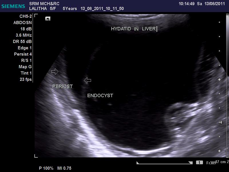
3

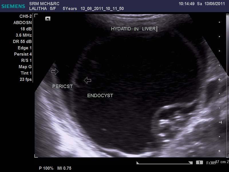
4

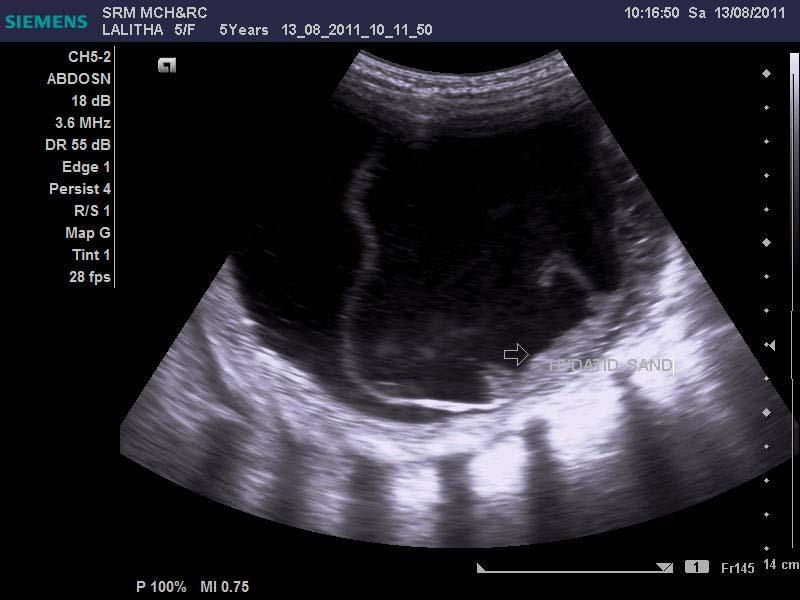
5 SNOWSTORM SIGN=HYDATID SAND

6
7 DAUGHTER CYSTS
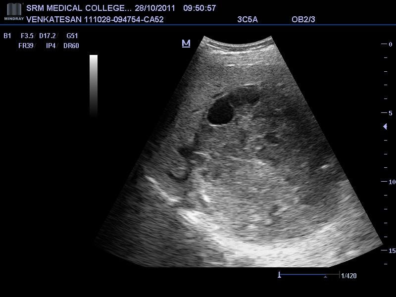
8 CASE 2 60 yr old female with C/O pain in the LEFT hypochondrium

9 RACEMOSE OR HONEYCOMB APPEARANCE


10 Speck of marginal calcification with multiloculated cyst in LLL

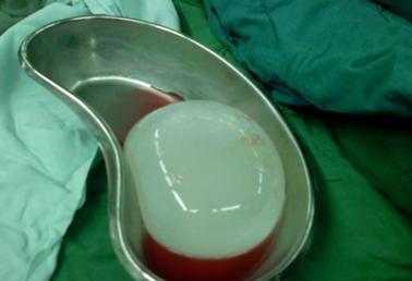
11 OPERATIVE SPECIMEN

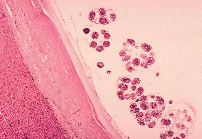
12 HISTOPATHOLOGICAL FINDING
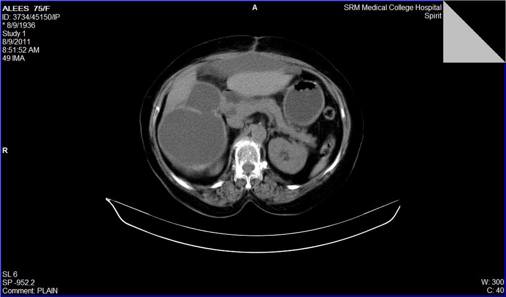
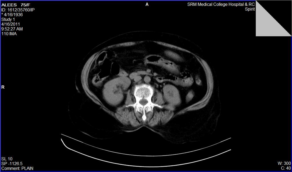
13 CASE 3 75/f 75/f came with the following c/o pain in the pain in the rt hypochondrium for the past 4 months after 3 months she developed jaundice,intermittent fever,urticaria


14 Multiple peripheral loculated cysts within a large cyst honeycomb pattern
15
16
17
18
19 CASE 4 48/M 48/m came with the following c/o pain in the rt & lt hypochondrium for the past 4 months
20
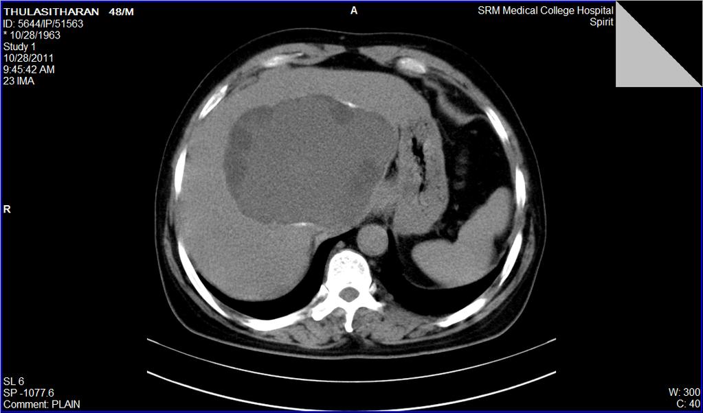
21
22 MRI T1 SEQUENCE

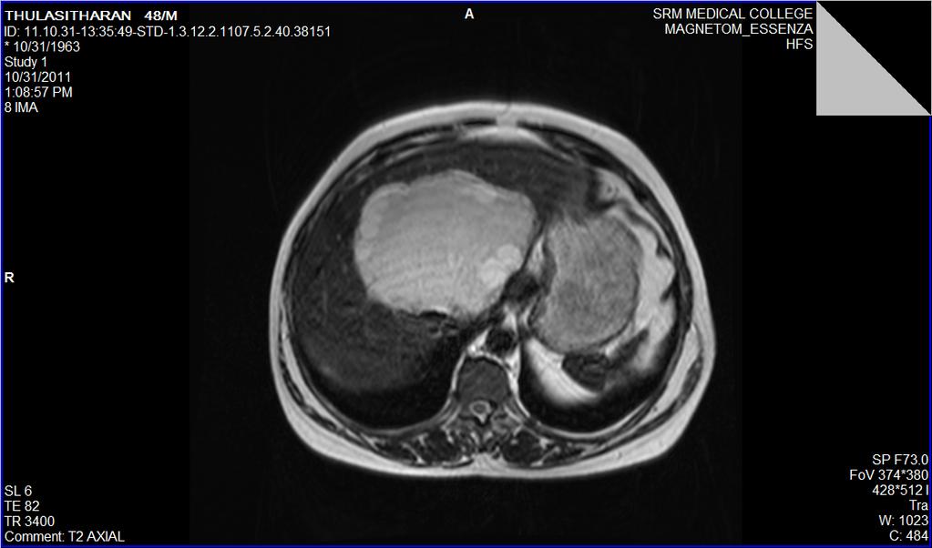
23 MRI T2 SEQUENCE
24 HIPPOCRATES recognized human hydatid over 2000 yrs ago. The arab physician AL RHAZES made reference to hydatid disease of the liver in AD 900
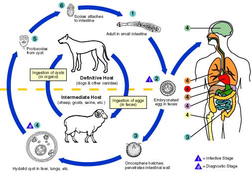
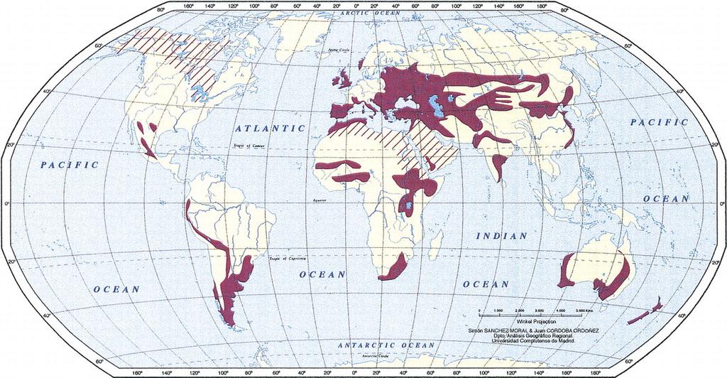
25 Etiology infestation of the parasite echinococcus granulosus echinococcus multilocularis echinococcus Vogeli paca brazil echinococcus oligarthrus
26
27
28 Cyst wall consits of 3 layers Ectocyst (laminated membrane) external membrane which may calcify Pseudocyst/Pericyst(adventitia) dense connective tissue capsule formed around the cyst as a part of host response Endocyst(germinal epithelium) the inner germinal that gives rise to brood capsules This brood capsules may separate from the wall & form a fine sediment called HYDATID SAND
29 CLINICAL FEATURES Simple/uncomplicated/multivesicular/univesicular cysts are asymptomatic Pressure on the adjacent organs symptoms Abdominal pain,tenderness,palpable mass. Secondary infection hepatomegaly,chills,fever Urticaria,erythema generalized anaphylactic reactions Biliary rupture jaundice,biliary colic,urticaria
30 CLASSIFICATION TYPE 1 Simple Cyst 2A TYPE 2 ACTIVE PHASE 2B 2C 3A TYPE 3 CALCIFIED CYSTS 3B 3C TYPE 4 COMPLICATED
31 Type I: Simple Cyst Appear as a well defined anechoic mass with or without hydatid sand and septa. usg Unilocular cysts are considered to be an initial stage in the development of the parasite SNOWSTORM SIGN==HYDATID SAND Rolling the patient during evaluation disperses the sand, creating small echogenic foci, or falling snowflakes.
32 WATER LILLY SIGN =partial /complete detachment of endocyst from pericyst (due to intracystic pressure as a sign of degeneration /trauma /host response /response to therapy is pathognomic. Floating membranes doesn t indicate the death of the parasite At CT, a type I HC appears as a well defined, water attenuation mass. The septa and cyst wall enhance in contrast CT and magnetic resonance
33 MR imaging features T1W1 and T2WI MR imaging, a finding that helps to differentiate type I HC from a simple liver cyst. A low signal intensity rim ( rim sign ), which is more evident on T2W1, is characteristic of hydatidosis
34 Type II hydatid cysts It is the active phase of hydatid disease type II HCs can spread to near by tissue by outpouching a new cyst from the main cyst An hourglass appearance can be seen. This type is classified into three stages
35 MRI shows daughter cysts as hypointense or isointense relative to the maternal matrix on T1 and T2 weighted images. Type IIB HCs a rosette appearance RACEMOSE/HONEYCOMB APPEARANCE multiple septa between the daughter cysts inside the mother cysts occupying the entire volume SPOKE WHEEL PATTERN daughter cysts separated by echogenic material of hydatid matrix likely to be hydatid
36 type IIC cysts that contain scattered calcifications within the cyst wall and daughter cysts within the cavity. Scattered calcification at the cyst wall does not imply a dead cyst but degeneration at the cyst wall CT is the preferred imaging method to evaluate these types of HCs due to calcification
37 Type III: Calcified HCs IT is the inactive or dead phase of HD. 3 types: (1) Total and thick continuous calcification (ringlike) of the cyst wall (2) total calcification within the cyst matrix and a decrease in cyst size (3) curvilinear calcification within the ruptured internal membranes EGG SHELL CALCIFICATION in cyst wall least common
38 Type IV: Complicated HCs Transdiaphragmatic growth through bare area of liver 1. Chronic biliary fistula (5 15%) 2.Peritoneum 13% 3.rupture into pleural cavity 4. Seeding in pulmonary parenchyma 5.Hollow viscus infection following rupture (5 8%)
39 Internal rupture leads to the water lily sign With time, cystic fluid decreases, and the HC mimics a solid mass Collapsed membranes within the cavity are detected as serpentine structures Compression of vital structures (bileduct,portal vein ) Coexisting cholelithiasis recurrence
40 KEY POINTS CT helps in differentiating parasitic from nonparasitic cysts. Parasitic cysts pathognomically have a thick wall A solid heterogeneous mass is difficult to differentiate from granulomas or tumors, although calcification suggests echinococcal cyst.
41 MRI may have some advantages over CT in the evaluation of postsurgical residual lesions, recurrences, and selected extrahepatic infections. It is also superior in identifying changes of the intrahepatic and extrahepatic venous system and in identifying cysto biliary fistulas
42 MR PROTON SPECTROSCOPY pyruvate levels DIRECT CHOLANGIOGRAPHY is indicated in intrabiliary rupture & bile duct obstruction RADIO NUCLIDE SCAN bronchobiliary fistula ELISA SEROLOGICAL TESTS IgM & IgA antibodies active phase CASONI S INTRADERMAL TEST
43 DIFFERENTIAL DIAGNOSIS SIMPLE CYST AMOEBIC LIVER ABSCESS HAEMATOMA METASTASIS MESENCHYMAL HAMARTOMA
44 THANK YOU
45 REFERENCES GRAINGER & ALLISON DAVID SUTTON RADIOGRAPHICS Pinar Polat, MD Mecit Kantarci, MD Fatih Alper, MD,Selami Suma, MD Melike Bedel Koruyucu, MD Adnan Okur, MDmarch april 2003 Polat P, Kantarci M, Alper F, Suma S, Koruyucu MB, Okur A. Hydatid Disease from Head totoe. RadioGraphics 2003; 23: Mortele KJ, Segatto E, Ros PR. The infected liver: radiologic pathologic correlation. Radiographics.2004; 24:
Case Report Unusual Presentation of Hydatid Cyst in Breast with Magnetic Resonance Imaging Findings
 Hindawi Case Reports in Medicine Volume 2017, Article ID 6237435, 4 pages https://doi.org/10.1155/2017/6237435 Case Report Unusual Presentation of Hydatid Cyst in Breast with Magnetic Resonance Imaging
Atypical manifestations, complications and pathological correlation of hydatid disease.
 Atypical manifestations, complications and pathological correlation of hydatid disease. Poster No.: C-2501 Congress: ECR 2012 Type: Educational Exhibit Authors: R. Lerma Ortega, D. J. López Ruiz, L. A.
Imaging of ruptured endocyst in an isolated intramuscular hydatid cyst
 Imaging of ruptured endocyst in an isolated intramuscular hydatid cyst Nitin P Ghonge 1*, Sriram Rajan 1, Bharat Aggarwal 1, Amit K Sahu 1 1. Department Of Radiology, Diwan Chand Satyapal Aggarwal Imaging
Cardiac Mass in a 15-Year-Old Boy
 Cardiac Mass in a 15-Year-Old Boy Echocardiographic Case Report Hortensia Vuçini Department of Cardiology and Cardiac Surgery UHC Mother Theresa Tirana, Albania October 20, 2007 Case Presentation 15 year-old
International Journal of Health Sciences and Research ISSN:
 International Journal of Health Sciences and Research www.ijhsr.org ISSN: 2249-9571 Case Report Parietal Abdominal Wall Swelling Turning Out To Be a Parietal Complication of Hydatid Cyst of Liver - A Case
X-Ray Corner. Imaging Approach to Cystic Liver Lesions. Pantongrag-Brown L. Solitary cystic liver lesions. Hepatic simple cyst (Figure 1)
") THAI J 136 Imaging Approach to Cystic Liver Lesions GASTROENTEROL 2013 X-Ray Corner Imaging Approach to Cystic Liver Lesions Pantongrag-Brown L Cystic liver lesions are common findings in daily practice
THAI J 136 Imaging Approach to Cystic Liver Lesions GASTROENTEROL 2013 X-Ray Corner Imaging Approach to Cystic Liver Lesions Pantongrag-Brown L Cystic liver lesions are common findings in daily practice
Science & Technologies
 A GIANT LIVER HYDATIDE CYST SIMULTANEOUSLY PERFORATED TO PERITONEAL AND PLEURAL CAVITIES A RARE CASE REPORT. Ivan P. Novakov Department of Special Surgery; Medical University - Plovdiv Abstract. Background.
PRIMARY MUSCULOSKELETAL HYDATID CYST DISEASE. Radiology Department, Military Hospital Mohamed V. Rabat Morocco
 PRIMARY MUSCULOSKELETAL HYDATID CYST DISEASE. A. El Kharras S. Semlali A. Darbi S. Akjouj J. El Fenni A. Hanine M. Benameur D. Bassou Radiology Department, Military Hospital Mohamed V. Rabat Morocco Introduction
Primary Intramuscular Hydatid Cyst of the thigh Muscle a Rare Case Report
 IOSR Journal of Dental and Medical Sciences (IOSR-JDMS) e-issn: 2279-0853, p-issn: 2279-0861.Volume 15, Issue 3 Ver. VIII (Mar. 2016), PP 07-11 www.iosrjournals.org Primary Intramuscular Hydatid Cyst of
HEPATO-BILIARY IMAGING
 HEPATO-BILIARY IMAGING BY MAMDOUH MAHFOUZ MD PROF.OF RADIOLOGY CAIRO UNIVERSITY mamdouh.m5@gmail.com www.ssregypt.com CT ABDOMEN Indications Patient preparation Patient position Scanogram Fasting 4-6 hours
Cystic lesions of the liver
 Cystic lesions of the liver Poster No.: C-0408 Congress: ECR 2014 Type: Educational Exhibit Authors: E. Rosado, J. Pereira, S. El Bouchaibi, M. A. A. Bali ; 1 1 2 2 3 3 4 4 Amadora/PT, Lisboa/PT, Bruxelles/BE,
Complicated echinococcal cyst to Biopsy or not to biopsy. V. Rusanov MR Kramer Pulmonary Institute, Rabin medical center
 Complicated echinococcal cyst to Biopsy or not to biopsy V. Rusanov MR Kramer Pulmonary Institute, Rabin medical center Case 1 84 y.o. Male, Iraq descend, past smoker 40 PY Medical History- HTN, Rheumatoid
Greater Manchester and Cheshire HPB Unit Guidelines for the Assessment & Management of Hepatobiliary and Pancreatic Disease Chapter 7
 Greater Manchester and Cheshire HPB Unit Guidelines for the Assessment & Management of Hepatobiliary and Pancreatic Disease Chapter 7 Contents 7. Benign liver conditions 79 7.1. Pyogenic liver abscess
Liver Cancer (Hepatocellular Carcinoma or HCC) Overview
 Overview") Liver Cancer (Hepatocellular Carcinoma or HCC) Overview Recent advances in liver cancer care seek to address the rising incidence of liver cancer, which has steadily increased over the past three decades.
Liver Cancer (Hepatocellular Carcinoma or HCC) Overview Recent advances in liver cancer care seek to address the rising incidence of liver cancer, which has steadily increased over the past three decades.
Splenic Cystic Lesions - Differential Diagnosis
 Splenic Cystic Lesions - Differential Diagnosis Poster No.: C-1343 Congress: ECR 2013 Type: Educational Exhibit Authors: N. Neto, P. G. M. G. Ferreira, A. Vasconcelos ; Lisboa/PT, 1 2 2 3 1 3 Amadora/PT,
ACG Clinical Guideline: Diagnosis and Management of Focal Liver Lesions
 ACG Clinical Guideline: Diagnosis and Management of Focal Liver Lesions Jorge A. Marrero, MD, 1 Joseph Ahn, MD, FACG, 2 K. Rajender Reddy, MD, FACG 3 1 University of Texas at Southwestern, Dallas, Texas,
Multidisciplinary imaging of liver hydatidosis
 Online Submissions: http://www.wjgnet.com/1007-9327office wjg@wjgnet.com doi:10.3748/wjg.v18.i13.1438 World J Gastroenterol 2012 pril 7; 18(13): 1438-1447 ISSN 1007-9327 (print) ISSN 2219-2840 (online)
IT 의료융합 1 차임상세미나 복부질환초음파 이재영
 IT 의료융합 1 차임상세미나 2013-4-3 복부질환초음파 이재영 나는오늘누구를위하여 종을울리나? 전통적의료 의사 공학설계자 의사 최첨단진단장비들 USG, CT, MRI 환자 환자 현대의료 사용자중심의사고 US in the Abdomen Detection DDx Look Behavior Response by external stimuli Guiding Tool
Financial Disclosure
 Benign Liver Masses Adil Abdalla, MBBS Creighton University-CHI Health August 25, 2018 Financial Disclosure Nothing to disclose Financial Disclosure 1 Objectives To assess patients with benign liver tumors
A Rare Cause of Recurrent Vaginal Hydrocele: Herniating Mesenteric Hydatid Cyst
 Iran J Parasitol Tehran University of Medical Sciences Publication http:// tums.ac.ir Open access Journal at http:// ijpa.tums.ac.ir Iranian Society of Parasitology http:// isp.tums.ac.ir Case Report A
Cystic Lesions of the Liver
 Residents Section Pattern of the Month Vachha et al. Cystic Lesions of the Liver Residents Section Pattern of the Month Residents inradiology ehroze Vachha 1 Maryellen R. M. Sun ettina Siewert Ronald L.
Diagnosis and treatment of the hydatid disease of the liver
 J. Nippon Med. Sch., Vol. 51, No. 1 (1984) Note for Clinical Doctors (107) 107 Diagnosis treatment disease liver Xu Ming-qian Department Surgery, The People's General Hospital Xinjiang Uighur Autonomous
Role of imaging in RCC. Ultrasonography. Solid lesion. Cystic RCC. Solid RCC 31/08/60. From Diagnosis to Treatment: the Radiologist Perspective
 Role of imaging in RCC From Diagnosis to Treatment: the Radiologist Perspective Diagnosis Staging Follow up Imaging modalities Limitations and pitfalls Duangkamon Prapruttam, MD Department of Therapeutic
Common and unusual CT and MRI manifestations of pancreatic adenocarcinoma: a pictorial review
 Review Article Common and unusual CT and MRI manifestations of pancreatic adenocarcinoma: a pictorial review Min-Jie Yang, Su Li, Yong-Guang Liu, Na Jiao, Jing-Shan Gong Department of Radiology, Shenzhen
State of the Art Imaging for Hepatic Malignancy: My Assignment
 State of the Art Imaging for Hepatic Malignancy: My Assignment CT vs MR vs MRCP Which one to choose for HCC vs Cholangiocarcinoma What special protocols to use for liver tumors Role of PET and Duplex US
Case Discussion Splenic Abscess
 Case Discussion Splenic Abscess Personal Data Gender: male Birth Date: 1928/Mar/06th Allergy: Mefenamic Smoking: 0.5 PPD for 55 years Alcohol: negative (?) 4 Months Ago Abdominal pain: epigastric area
Cholangiocarcinoma (Bile Duct Cancer)
") Cholangiocarcinoma (Bile Duct Cancer) The Bile Duct System (Biliary Tract) A network of bile ducts (tubes) connects the liver and the gallbladder to the small intestine. This network begins in the liver
Cholangiocarcinoma (Bile Duct Cancer) The Bile Duct System (Biliary Tract) A network of bile ducts (tubes) connects the liver and the gallbladder to the small intestine. This network begins in the liver
Renal masses - the role of diagnostic imaging
 Renal masses - the role of diagnostic imaging Poster No.: C-2471 Congress: ECR 2015 Type: Educational Exhibit Authors: V. Rai#; Bjelovar/HR Keywords: Cysts, Cancer, Structured reporting, Ultrasound, MR,
Interesting Cases from Liver Tumor Board. Jeffrey C. Weinreb, M.D.,FACR Yale University School of Medicine
 Interesting Cases from Liver Tumor Board Jeffrey C. Weinreb, M.D.,FACR Yale University School of Medicine jeffrey.weinreb@yale.edu Common Liver Diseases Hemangioma Cyst FNH Focal Fat/Sparing THID Non-Cirrhotic
Essentials of Clinical MR, 2 nd edition. 65. Benign Hepatic Masses
 65. Benign Hepatic Masses Pulse sequences acquired for abdominal MRI typically consist of fast acquisition schemes such as single-shot turbo spin echo (i.e. HASTE) and gradient echo schemes such as FLASH
Normal Sonographic Anatomy
 hapter 2:The Liver DUNSTAN ABRAHAM Normal Sonographic Anatomy Homogeneous, echogenic texture (Figure 2-1) Measures approximately 15 cm in length and 10 12.5 cm anterior to posterior; measurement taken
CELL AND TISSUE INJURY COURSE-II PATHOLOGY LABORATORY
 CELL AND TISSUE INJURY COURSE-II PATHOLOGY LABORATORY PATHOLOGY of INFECTIOUS DISEASES MICROSCOPY Rengin Ahıskalı Macroscopy samples are shown in the macroscopy presentations of the first two courses.
Radiological Investigations of Abdominal Trauma
 76 77 Investigations of Abdominal Trauma Introduction: Trauma to abdominal organs is a common cause of patient morbidity and mortality among trauma patients. Causes of abdominal trauma include blunt injuries,
INTRODUCTION. Hydatidosis is a tapeworm infection caused by the Echinococcus species (1). Involvement of
. Involvement of") A New Approach for Treatment of Bone Hydatid Cyst Disease: A Case Report of Primer Femur Hydatid Cyst H Çabuk 1, AÇ Tekin 1, S Ayanoğlu 1, CD Büyükkurt 1, H Gürbüz 1, FK Çabuk 2 INTRODUCTION Hydatidosis
A New Approach for Treatment of Bone Hydatid Cyst Disease: A Case Report of Primer Femur Hydatid Cyst H Çabuk 1, AÇ Tekin 1, S Ayanoğlu 1, CD Büyükkurt 1, H Gürbüz 1, FK Çabuk 2 INTRODUCTION Hydatidosis
Chief Complaint. Retroperitoneal cystic mass incidentally found at health examination center.
 Personal Information Age: 34 y/o Sex: female Past history: major systemic medical history(-) surgical history(-), family history(-) Denied food or drug allergy Chief Complaint Retroperitoneal cystic mass
UPDATES AND SPOTLIGHTS ON SOME HEPATOBILIARY PARASITES
 UPDATES AND SPOTLIGHTS ON SOME HEPATOBILIARY PARASITES Dr. Manar Sobh Azab Associate Professor of Parasitology Faculty of Medicine Mansoura University Parasites are common worldwide and are classified
Role of Therapeutic Endoscopy in Hepatic Hydatid Disease after Surgical Intervention: Case Report
 ISPUB.COM The Internet Journal of Gastroenterology Volume 10 Number 2 Role of Therapeutic Endoscopy in Hepatic Hydatid Disease after Surgical Intervention: Case Report H Ono, M Okabe, T Kimura, M Kawakami,
Imaging of liver and pancreas
 Imaging of liver and pancreas.. Disease of the liver Focal liver disease Diffusion liver disease Focal liver disease Benign Cyst Abscess Hemangioma FNH Hepatic adenoma HCC Malignant Fibrolamellar carcinoma
RING ENCHANCING LESION BY M.S. HEMHNATH
 RING ENCHANCING LESION BY M.S. HEMHNATH A 21 YRS FEMALE CAME WITH H/O HEADACHE AND SEIZURE FOR THE PAST ONE MONTH. NO OTHER FOCAL NEUROLOGICAL DEFICIT. DIFFERENTIAL DIAGNOSIS For this case are Neurocysticerosis
Case Report Pulmonary Embolism Originating from a Hepatic Hydatid Cyst Ruptured into the Inferior Vena Cava: CT and MRI Findings
 Case Reports in Radiology Volume 2016, Article ID 3589812, 4 pages http://dx.doi.org/10.1155/2016/3589812 Case Report Pulmonary Embolism Originating from a Hepatic Hydatid Cyst Ruptured into the Inferior
Clinical Management of Hydatid Disease of the Urinary Tract
 The Journal of International Medical Research 2002; 30: 346 352 Clinical Management of Hydatid Disease of the Urinary Tract İ ÖZBEY, Y AKSOY, Ö POLAT, AF ATMACA AND A DEMIREL Department of Urology, Atatürk
Pediatric Retroperitoneal Masses Radiologic-Pathologic Correlation
 Acta Radiológica Portuguesa, Vol.XVIII, nº 70, pág. 61-70, Abr.-Jun., 2006 Pediatric Retroperitoneal Masses Radiologic-Pathologic Correlation Marilyn J. Siegel Mallinckrodt Institute of Radiology, Washington
Common surgical disease of the liver and portal hypertension
 Common surgical disease of the liver and portal hypertension Liver functhion: Responsible for storing glucose as glycogen, or converting it to lactate Utilization of: Amino acids for hepatic and plasma
THE POSTOPERATIVE APPEARANCE OF THE HYDATID CYST SURGERY LIVER ON ULTRASONOGRAPHY FOLLOWING
 HPB Surgery, 1990, Vol. 2, pp. 253-260 Reprints available directly from the publisher Photocopying permitted by license only 1990 Harwood Academic Publishers GmbH Printed in the United Kingdom THE POSTOPERATIVE
Surgical Management of Calcified
 HPB Surgery, 1999, Vol. 11, pp. 253-259 Reprints available directly from the publisher Photocopying permitted by license only (C) 1999 OPA (Overseas Publishers Association) N.V. Published by license under
Radiology of hepatobiliary diseases
 GI cycle - Lecture 14 436 Teams Radiology of hepatobiliary diseases Objectives 1. To Interpret plan x-ray radiograph of abdomen with common pathologies. 2. To know the common pathologies presentation.
A CASE OF HEPATIC CYST AND HEPATIC LOBE TORSION IN A CHOW-CHOW MALE
 Scientific Works. Series C. Veterinary Medicine. Vol. LXIII (1) ISSN 2065-1295; ISSN 2343-9394 (CD-ROM); ISSN 2067-3663 (Online); ISSN-L 2065-1295 A CASE OF HEPATIC CYST AND HEPATIC LOBE TORSION IN A CHOW-CHOW
IMAGING OF LIVER, BILIARY TREE, PANCREAS
 IMAGING OF LIVER, BILIARY TREE, PANCREAS Department of Radiology West China Hospital, Sichuan University Yao Jin Learning Points The methodology for imaging the LBP (liver, biliary tree, and pancreas )
Case Fibrothecoma of the ovary
 Case 10646 Fibrothecoma of the ovary Elisa Melo Abreu, Teresa Margarida Cunha Section: Genital (Female) Imaging Published: 2015, Jan. 2 Patient: 70 year(s), female Authors' Institution Department of Radiology,
A CASE OF A Huge Submandibular Pleomorphic Adenoma
 ISPUB.COM The Internet Journal of Head and Neck Surgery Volume 4 Number 2 S VERMA Citation S VERMA.. The Internet Journal of Head and Neck Surgery. 2009 Volume 4 Number 2. Abstract Pleomorphic adenoma
Cystic Disease of the Liver Work Up and Management. Louis Ferrari MD, PGY 3 6/9/16 SUNY Downstate Medical Center
 Cystic Disease of the Liver Work Up and Management Louis Ferrari MD, PGY 3 6/9/16 SUNY Downstate Medical Center The Case 73F presents to clinic after diagnostic laparoscopy at OSH. Known liver mass for
Recently role of non-invasive diagnostics methods
 CERTAIN ASPECTS OF NLS-DIAGNOSTICS OF LIVER FOCAL PATHOLOGY A.Y. Shvack, V.I. Nesterov, N.L. Ogluzdina This article contains information about NLS-graphy application in diagnostics of liver focal affections:
Basic Data. Sex:Male 31 years old Occupation: 搬家工人
 Basic Data Sex:Male 31 years old Occupation: 搬家工人 Chief Complaint Intermittent chest pain with shortness of breath for 2-3 months. Present Illness 4 months ago, he started having occasional chest pain
Focal Hepatic Masses in Pediatric Patients
 Residents Section Pattern of the Month deyiga et al. Focal Hepatic Masses in Pediatric Patients Residents Section Pattern of the Month Downloaded from www.ajronline.org by 148.251.232.83 on 04/27/18 from
Abdominal ultrasound:
 Abdominal ultrasound: Non-traumatic acute abdomen Wittanee Na-ChiangMai, MD Department of Radiology ChiangMai University 26/04/2017 Contents Technique of examination Normal anatomy Emergency conditions
The Unusual Mass of Retrovesical Space: A Secondary Hydatid Cyst Dısease
 Case Study TheScientificWorldJOURNAL (2006) 6, 2481 2485 TSW Urology ISSN 1537-744X; DOI 10.1100/tsw.2006.386 The Unusual Mass of Retrovesical Space: A Secondary Hydatid Cyst Dısease Can Tuygun*, Hasan
Hydatid Lung Disease: An Analysis of Five years Cumulative Data from Kolkata
 12 JAPI july 2012 VOL. 60 Original Article Hydatid Lung Disease: An Analysis of Five years Cumulative Data from Kolkata AG Ghoshal *, Supriya Sarkar **, Kaushik Saha ***, Uday Sarkar ****, Susmita Kundu
Category Term Definition Comments 1 Major Categories 1a
 Working Lexicon Categories, Terms & Definitions Category Term Definition Comments 1 Major Categories 1a Physiologic Category (consistent with normal ovarian physiology) Follicle Simple 3 cm in premenopausal
BILIARY CYSTADENOMA OF LIVER.
 BILIARY CYSTADENOMA OF LIVER. K. Kuberan [1], G. Chandrasekar [1] Abstract Biliary cystadenoma is very rare non parasitic benign neoplasm of liver with less than 200 cases reported all over the world in
Case 7729 Child with choledochal cyst presenting with episodes of vomitting and jaundice
 Case 7729 Child with choledochal cyst presenting with episodes of vomitting and jaundice dos Santos R 1, Almeida J 1, Mendes PP 2, Pereira S 3, Borges C 3, Soares E 4. 1) Radiology resident, 2) Radiology
MRI XR, CT, NM. Principal Modality (2): Case Report # 2. Date accepted: 15 March 2013
: Case Report # 2. Date accepted: 15 March 2013") Radiological Category: Musculoskeletal Principal Modality (1): Principal Modality (2): MRI XR, CT, NM Case Report # 2 Submitted by: Hannah Safia Elamir, D.O. Faculty reviewer: Naga R. Chinapuvvula, M.D.
Radiological Category: Musculoskeletal Principal Modality (1): Principal Modality (2): MRI XR, CT, NM Case Report # 2 Submitted by: Hannah Safia Elamir, D.O. Faculty reviewer: Naga R. Chinapuvvula, M.D.
Cerebro-vascular stroke
 Cerebro-vascular stroke CT Terminology Hypodense lesion = lesion of lower density than the normal brain tissue Hyperdense lesion = lesion of higher density than normal brain tissue Isodense lesion = lesion
Case Report Isolated Retroperitoneal Hydatid Cyst Invading Splenic Hilum
 Case Reports in Surgery, Article ID 303401, 4 pages http://dx.doi.org/10.1155/2014/303401 Case Report Isolated Retroperitoneal Hydatid Cyst Invading Splenic Hilum Safak Ozturk, 1 Mutlu Unver, 1 Burcin
INTERNATIONAL JOURNAL OF PHARMACEUTICAL RESEARCH AND BIO-SCIENCE
 INTERNATIONAL JOURNAL OF PHARMACEUTICAL RESEARCH AND BIO-SCIENCE HEPATIC MESENCHYMAL HAMARTOMA: A CASE REPORT N JAIN 1, S SIDHU 2, K SAGGAR 3 1. Junior Resident, Department of Radiodiagnosis, Dayanand
Malignant Focal Liver Lesions
 Malignant Focal Liver Lesions Other Than HCC Pablo R. Ros, MD, MPH, PhD Departments of Radiology and Pathology University Hospitals Cleveland Medical Center Case Western Reserve University Pablo.Ros@UHhospitals.org
Name : 黃 XX Age : 52 Sex : 女 Occupation : 廚房阿姨 Marital status : 已婚
 Name : 黃 XX Age : 52 Sex : 女 Occupation : 廚房阿姨 Marital status : 已婚 Chief Complaint Mild postprandial fullness for 2 months Present Illness This 52 year-old female suffered from intermittent post-prandial
Altought liver (75%) and lung (15%) are the most commonly involved
 and lung (15%) are the most commonly involved") Diagn Interv Radiol 2010; 16:168 174 Turkish Society of Radiology 2010 MAGNETIC RESONANCE IMAGING ORIGINAL ARTICLE Conventional and diffusion-weighted MRI of extrahepatic hydatid cysts Nagihan İnan, Nilay
Diagn Interv Radiol 2010; 16:168 174 Turkish Society of Radiology 2010 MAGNETIC RESONANCE IMAGING ORIGINAL ARTICLE Conventional and diffusion-weighted MRI of extrahepatic hydatid cysts Nagihan İnan, Nilay
Cross sectional imaging of Intracranial cystic lesions Abdel Razek A
 Cross sectional imaging of Intracranial cystic lesions Abdel Razek A Department of Radiology. Mansoura Faculty of Medicine, Mansoura. Egypt. arazek@mans.edu.eg Introduction Intracranial cystic lesions
IMAGING IN FAMILY MEDICINE
 IMAGING IN FAMILY MEDICINE Imaging Methods Ultrasonography (USG) X-ray Computed tomography (CT) Magnetic resonance imaging (MRI) Scintigraphy Angiography SNC MEDIASTINUM LUNGS ABDOMEN PELVIS USG SNC -
Imaging features of benign hepatic lesions at US and CT imaging: a pictorial review.
 Imaging features of benign hepatic lesions at US and CT imaging: a pictorial review. Poster No.: C-1557 Congress: ECR 2016 Type: Educational Exhibit Authors: M. Barros, L. A. Ferreira, P. B. Oliveira,
Case Report Ruptured Hydatid Cyst with an Unusual Presentation
 Case Reports in Surgery Volume 2011, Article ID 730604, 4 pages doi:10.1155/2011/730604 Case Report Ruptured Hydatid Cyst with an Unusual Presentation Deepak Puri, Amit Kumar Mandal, Harinder Pal Kaur,
Diagnostic Studies Then. It s important to be able to distinguish. Diagnostic Studies Now
 Jonathan S. Fisher, MD, FACS It s important to be able to distinguish Diagnostic Studies Then Diagnostic Studies Then History Biopsy Diagnostic Studies Now History Biopsy Serum markers (AFP, CA19 9, CEA)
Evaluation of rare male breast masses using multimodality approach with their histopathological correlation: a case series
 Evaluation of rare male breast masses using multimodality approach with their histopathological correlation: a case series Poster No.: C-0662 Congress: ECR 2013 Type: Educational Exhibit Authors: P. S.
Imaging in breast cancer. Mammography and Ultrasound Donya Farrokh.MD Radiologist Mashhad University of Medical Since
 Imaging in breast cancer Mammography and Ultrasound Donya Farrokh.MD Radiologist Mashhad University of Medical Since A mammogram report is a key component of the breast cancer diagnostic process. A mammogram
Biliary cancers: imaging diagnosis. Study of 30 cases
 Biliary cancers: imaging diagnosis. Study of 30 cases N Hammoune, S Semlali, M Eddarai, T. Amil, M Zentar, S. El Kandri,, M Benameur,, S Chaouir. Radiology Department. Mohamed V Military Hospital. Rabat-
DOI: /SE Immunopathology of hydatid infection in human liver. PhD Thesis. Ali Vatankhah. Doctoral School of Pathological Sciences
 Immunopathology of hydatid infection in human liver PhD Thesis Ali Vatankhah Doctoral School of Pathological Sciences Semmelweis University Supervisor: József Tímár MD, D.Sc. Official Reviewers: Károly
1 yr old girl presented with Fever on and off 3 months H/o frequent semisolid bulky stools 3 months Progressive abdominal distension 3 months Failure
 Dr Rajasree S Dr Srinivas S, Dr Bagdi RK, Dr Satheesh C Apollo Childrens Hospital, Chennai 1 yr old girl presented with Fever on and off 3 months H/o frequent semisolid bulky stools 3 months Progressive
Sonographic Assessment of Cystic Hepatic Lesions in Sudanese
 568724JDMXXX10.1177/8756479314568724Journal of Diagnostic Medical SonographyBabiker and Eisa research-article2015 Original Research Sonographic Assessment of Cystic Hepatic Lesions in Sudanese Journal
Cystic Pancreatic Lesions: Approach to Diagnosis
 Cystic Pancreatic Lesions: Approach to Diagnosis Poster No.: R-0130 Congress: RANZCR-AOCR 2012 Type: Educational Exhibit Authors: A. AGARWAL, R. M. Mendelson; Perth/AU Keywords: Cysts, Biopsy, Endoscopy,
Biliary Cystadenoma Causing Esophageal Varices
 https://doi.org/10.7180/kmj.2016.31.2.191 KMJ Case Report Biliary Cystadenoma Causing Esophageal Varices Sung Ju Kang, Tae Hee Lee, Min Gyu Seok, Hyo Jin Yun, Ye Seul Jang, Jun Hyun Byun Department of
World Journal of Radiology. Presumptive case of sparganosis manifesting as a hepatic mass: A case report and literature review
 W J R World Journal of Radiology Submit a Manuscript: http://www.wjgnet.com/esps/ Help Desk: http://www.wjgnet.com/esps/helpdesk.aspx DOI: 10.4329/wjr.v8.i10.846 World J Radiol 2016 October 28; 8(10):
Title: An intrahepatic cavoportal collateral pathway due to a liver hydatid cyst obstructing the inferior vena cava
 Title: An intrahepatic cavoportal collateral pathway due to a liver hydatid cyst obstructing the inferior vena cava Authors: Alba Manuel Vázquez, José Manuel Ramia Ángel, Luis Gijón, Roberto de la Plaza
Neckmasses in infancy and childhood: Clinical and radiological classification and imaging approaches M. Mearadji
 Neckmasses in infancy and childhood: Clinical and radiological classification and imaging approaches M. Mearadji International Foundation for Pediatric Imaging Aid Introduction Neck masses are a frequent
TB Intensive Houston, Texas
 TB Intensive Houston, Texas October 15-17, 17 2013 Diagnosis of TB: Radiology Rosa M Estrada-Y-Martin, MD MSc FCCP October 16, 2013 Rosa M Estrada-Y-Martin, MD MSc FCCP, has the following disclosures to
Liver Tumors. Prof. Dr. Ahmed El - Samongy
 Liver Tumors Prof. Dr. Ahmed El - Samongy Objective 1. Identify the most important features of common benign liver tumors 2. Know the risk factors, diagnosis, and management of hepatocellular carcinoma
CT & MRI of Benign Liver Neoplasms Srinivasa R Prasad
 CT & MRI of Benign Liver Neoplasms Srinivasa R Prasad No financial disclosures Acknowledgements Many thanks to Drs. Heiken, Narra & Menias (MIR) Dr. Sahani (MGH) for sharing images Benign Liver Tumors:
CT & MRI Evaluation of Brain Tumour & Tumour like Conditions
 CT & MRI Evaluation of Brain Tumour & Tumour like Conditions Dr. Anjana Trivedi 1, Dr. Jay Thakkar 2, Dr. Maulik Jethva 3, Dr. Ishita Virda 4 1 M.D. Radiology, Professor and Head, P.D.U. Medical College
Radiologic Pathologic Correlation of Intraosseous Lipomas. Tim Propeck 1, Mary Anne Bullard 1, John Lin 1, Kei Doi 2, William Martel 1
 Downloaded from www.ajronline.org by 148.251.232.83 on 04/10/18 from IP address 148.251.232.83. opyright RRS. For personal use only; all rights reserved Radiologic Pathologic orrelation of Intraosseous
Cystic liver lesion and eosinophilia
 November, 2005 Cystic liver lesion and eosinophilia Jakob Begun, Harvard medical School Year III Patient Presentation 55 year old Cape Verde female presented to her PCP with 6 month history of variable
Kurdistan Center for Gastroenterology & Hepatology, Asulaimaneyah, Iraqi Kurdistan, Iraq
 Case Reports in Clinical Medicine, 2014, 3, 533-543 Published Online September 2014 in SciRes. http://www.scirp.org/journal/crcm http://dx.doi.org/10.4236/crcm.2014.39117 The Role of Endoscopic Retrograde
What s Your Diagnosis??? Renée Fahrenholz, Class of 2012
 Renée Fahrenholz, Class of 2012 What s Your Diagnosis??? Signalment Emma, a 9 year old, Female, Spayed, Domestic Short Haired Feline Presenting Complaint Weight loss, vomited the morning of her visit,
INTERDISCIPLINARY DISCUSSIONS IN LOCALISED RCC DIAGNOSIS AND SURGICAL STRATEGIES FOR ATYPICAL RENAL CYSTIC LESIONS. Maria Cova
 INTERDISCIPLINARY DISCUSSIONS IN LOCALISED RCC DIAGNOSIS AND SURGICAL STRATEGIES FOR ATYPICAL RENAL CYSTIC LESIONS Maria Cova Radiology Department University of Trieste (IT) Eleventh European International
Hilar cholangiocarcinoma. Frank Wessels, Maarten van Leeuwen, UMCU utrecht
 Hilar cholangiocarcinoma Frank Wessels, Maarten van Leeuwen, UMCU utrecht Content Anatomy Biliary strictures (Hilar) Cholangiocarcinoom Staging Biliary tract 1 st order Ductus hepatica dextra Ductus hepaticus
A CASE REPORT OF SPONTANEOUS BILOMA - AN ENIGMATIC SURGICAL PROBLEM
 A CASE REPORT OF SPONTANEOUS BILOMA - AN ENIGMATIC SURGICAL PROBLEM *Sumanta Kumar Ghosh and Biswajit Mukherjee ESIC Medical College, Joka, Kolkata, India *Author for Correspondence ABSTRACT Occurrence
Guidelines, Policies and Statements D5 Statement on Abdominal Scanning
 Guidelines, Policies and Statements D5 Statement on Abdominal Scanning Disclaimer and Copyright The ASUM Standards of Practice Board have made every effort to ensure that this Guideline/Policy/Statement
Diagnostic evaluation and surgical management of recurrent hydatid cysts in an endemic region
 doi:10.2478/v10019-009-0032-x research article Diagnostic evaluation and surgical management of recurrent hydatid cysts in an endemic region Mehmet Yildirim 1, Omer Engin 1, Ozgür Oztekin 2, Fatih Akdamar
Intrabiliary Rupture of Hepatic Hydatid Cysts: Diagnostic Accuracy of MR Cholangiopancreatography
 Erden et al. MR Cholangiopancreatography of Hydatid Cyst Hepatobiliary Imaging Clinical Observations Ayşe Erden 1 Necati Örmeci 2 Suat Fitoz 1 İlhan Erden 1 Sumru Tanju 1 Yasemin Genç 3 Erden A, Örmeci
Various kinds of cystic lesions in the Liver: Case-based analysis
 Various kinds of cystic lesions in the Liver: Case-based analysis Poster No.: R-0018 Congress: RANZCR-AOCR 2012 Type: Educational Exhibit Authors: J.-H. Yoon, S.-H. Kim, Y.-J. Lim, J.-H. Ryu, H.-D. Kim