IMAGING OF LIVER, BILIARY TREE, PANCREAS
|
|
|
- Rafe Daniel
- 5 years ago
- Views:
Transcription
1 IMAGING OF LIVER, BILIARY TREE, PANCREAS Department of Radiology West China Hospital, Sichuan University Yao Jin
2 Learning Points The methodology for imaging the LBP (liver, biliary tree, and pancreas ) The normal appearances and the spectrum of abnormal imaging findings of the LBP on various imaging modalities The imaging diagnosis of some common diseases of the LBP
3 I.Imaging Methodology
4 I Methods of Imaging Examination 1. Plain film 2. Contrast enhancedx-ray Exam 3. Digital Subtraction Angiography (DSA) 4. Computed Tomography (CT) 5. Magnetic Resonance Image (MRI), Magnetic Resonance Cholangiopancreatography (MRCP)
5 I Methods of Imaging Examination 6. Ultrasonography (USG) 7. Endoscopic Retrograde Cholangiopancreatography (ERCP) 8. Percutaneous Transhepatic Cholangiography (PTC)

6 1 X-ray Exam Fluoroscopy Radiography (plain film) The X-ray examination has very limited value in the imaging of the LBPS for all of the abdominal organs manifest as soft tissue
7 2 Contrast EnhancedX-ray Examination Oral or intravenous cholecystography Oral or intravenous cholangiography T-tube retrograde cholangiography, etc Limitations ---- Inconvenient; indirect; low sensitivity & specificity These two methods were replaced by US or/and MRCP for display the biliary tree and gallbladder
8 3 Digital Subtraction Agiogrophy (DSA) Selective celiac arteriography Superselective hepatic arteriography Direct / indirect portography, etc. Invasive procedure Identify blood supple, vascularity & perfusion characteristics the nature of the lesion Interventional therapy

9 Selective Celiac Angiography


10 4 CT 1 CT Scan Techniques Unenhanced CT Scan Contrast-enhanced CT Scan


11 On the unenhanced image, the lesion can not be displayed
12 Contrast-enhanced CT Scan 2 Methods Duo- or multiphase helical CT scan CT angiography:combination of CT technique and angiography CTHA CTAP
13 Duo-phase Helical CECT Based on the double blood supply of the Liver Hepatic arterial supply(25%) Portal venous supply(75%)
14 Hemodynamics of the liver The pathway of the contrast medium flow after injection by venous The contrast material arrive to the liver by the hepatic artery earlier about 20 to 25 seconds than via the portal vein. Contrast material Peripheral v. Superior vena cava Pulmonary circulation Aorta Celiac A SMA/IMA Hepatic A Splenic A Portal Vein Liver
15 CECT Scan Time Schedule HA phase: second PV phase: second Equilibrium phase: second Delay phase: 3 5 min The time is from the beginning of contrast medium injection to the start of CT scan
16 Contrast Agent for CECT Iodine material: ironic vs. non-ironic Allergic test: unnecessary! Injection route: transvenous Injection rate: 3 5 ml/s
17 HAP Unenhanced Scan PVP
18 Merits and Diagnostic Values of CT 1 Perfect spatial resolution fine anatomic images Improve the Identification of small lesions Correct and meticulous assess the extent of lesions Evaluate the invasion of surrounding structures Detect if metastasis exists in peritoneal cavity, retroperitoneal space and distant organs
19 Merits and Diagnostic Values of CT 2 Duo/multiphase scan For characterization of blood supply and status of microcirculation of lesions Determine the nature of lesions Assessment of the biological behavior of lesions and monitoring the response to treatment
20 Limitations of CT with the state-of-the-art, multi-detector row spiral CT, many of the technical limitations have been overcome, such as coronal/sagittal image reconstruction, slice misregistration phenomenon, motion artifact, etc. High energy X-ray may be potentially harmful, espetially for children and pregnant women.
21 5 MR IMAGING Special features of MR imaging Super tissue contrast, good for both lesion detection and characterization MR hydrography (MRCP) for non-invasive depiction of the biliary tree and gallbladder Functional MR imaging can provide functional and metabolic information of organs and lesions
22 MR Imaging Techniques Spin-echo (SE) Pulse sequences Gradient-echo (GE) MR angiography Fat saturation MR hydrography (MRCP) Functional MR (DWI, SWI, ASL, et al ) MR spectroscopy
23 MR Sequences for LBPS Imaging T1WI unenhanced +contrast enhanced scan Organ anatomy and tissue characteristics Blood supply and extent of lesions T2WI unenhanced scan Very sensitive to changes of water content, so very good for lesion detection, especially for the small lesions But not good at displaying the characteristics of specific tissue texture.
24 MR Imaging Sequences Reasonable combination of several MR imaging sequences is crucial to the early detection and characterization of LBP abnormalities!

25 MRI,the sequences of unenhancement Fat saturation T1WI Fat saturationt2wi T2WI DWI


26 VIBE contrast enhanced dynamic scanning

27 MRCP 1 The Principle MR hydrography Hollow organs with abundant water are depicted nicely the bile duct and the pancreatic duct 2D or 3D reconstruction images to clearly illustrate the spatial relationship
28 MRCP 2 Diagnostic Values To determinate the location and extent and causes of obstructive jaundice non-invasively To help identify necrosis, liquefaction and pancreatic duct disruption in acute pancreatitis
29 Merits and Diagnostic Values of MRI Multiple scanning sequences can generate more diagnostic information MRI is superior to CT in the following aspects: Early depiction of certain lesions, especially depiction the small HCC in the cirrhosis liver Ability to characterize lesions
30 Merits and Diagnostic Values of MRI Multi-directional imaging are suitable for evaluating the anatomic location of lesions and relationship with surrounding structures, such as vessels, organs, peritoneal spaces, ligments, et al. Though the spatial resolution of MRI is lower, but MRI is equal to CT in aspects of : Displaying vascular invasion of tumors Preoperative tumor staging To help select the therapy methods such as surgery or conservative treatment and to evaluate the resectability of lesions.

31 Merits and Diagnostic Values of MRI Harmless, no radiation hazard to human body
32 MRI Limitations Relatively longer data acquisition time compared to MSCT, and motion artifact affects image quality Not sensitive enough to calcification
33 Contraindications for MRI Patients with cardiac pacemaker and certain magnetic metallic implants cannot or cautiously be examined with MRI Critically ill patients better not to be examined with MRI, for: 1 Time-consuming 2 Metallic instrument for resuscitation can not be placed in MR room
34 6 Ultra-sonography 1 Diagnostic Values Able to show lesions in all directions Useful for determinate the origination and localization of lesions Good at visualizing cystic conditions and bile stones (cholelithiasis) Cheap and easy to perform
35 6 Ultra-sonography 2 Limitations Not good for obesity Bowel gas influence for imaging the pancreas and imaging the lower half of CBD Highly operator-dependent
36 7 ERCP
37 8 PTC
38 Selection of Imaging Techniques 1 Liver: USG : for screening and general exam purposes CT vs. MR: Each has respective applications. MR is superior to CT in detection and characterization of lesions (especially in patients with cirrhotic background ) DSA: mainly used for interventional procedures, its role as an imaging diagnostic tool is downplaying
39 Selection of Imaging Techniques 2 Billary System: USG: The method of choice to exam the gallbladder; Valuable in biliary diseases (especially cholelithiasis) CT & MR: of great value in determining the extent of biliary carcinoma and accurate preoperative staging MRCP: the method of choice in displaying biliary tree and pancreatic duct ERCP & PTC: valuable, but invasive

40 Comparison of ERCP and MRCP
41 Selection of Imaging Techniques 3 Pancreas: CT and MR are of great value in displaying pancreatic abnormalities. The value of USG is limited MRCP can clearly show the pancreatic duct, thus can replace ERCP and PTC for most circumstances DSA is powerful and the final resort in qualitative diagnosis of pancreatic island endocrinal tumors
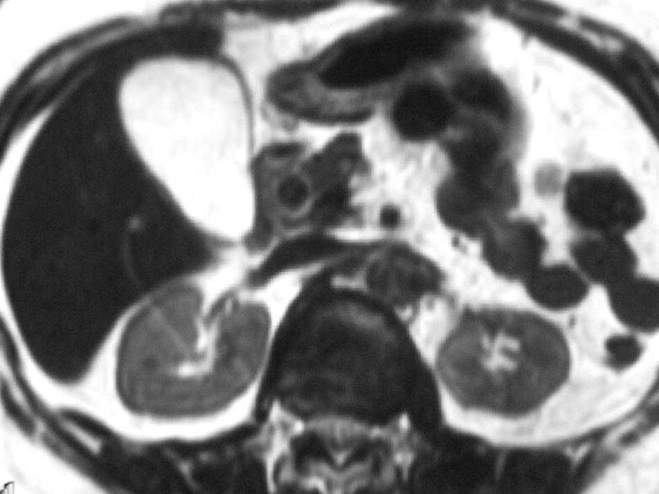
42 II Normal Imaging Appearance of live, biliary tract, pancreas and spleen
43
44
45
46


47 On T2WI the gallbladder appears very high signal as bright lamp bulb, and on T1WI it looks as low signal or miscellaneous signal
48

49
50
51
52 III IMAGING APPEARANCES OF COMMON DISEASES
53 Hepatic Diseases 1. Liver Cirrhosis 2. Hepatic Neoplasm 3. Hepatic Cyst 4. Hepatic Abscess
54 1 Liver cirrhosis early stage liver size advanced contour:the contour was smooth on the early stage and became nodolar gradually as the disease developing. lobar or segmental deformity widening of hepatic fissures changes secondary to portal hypertension ascites regenerative nodules splenomegaly collateral varices


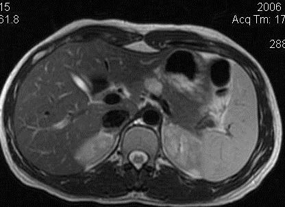
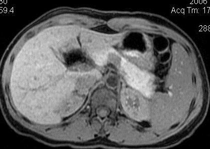
55 Liver cirrhosis, the contour of the liver is nodolar and the hepatic fissures was widening with portal hypertension and ascites The paraumbilical vein and the superficial epigastric veins and the left branch of portal vein were dilatation
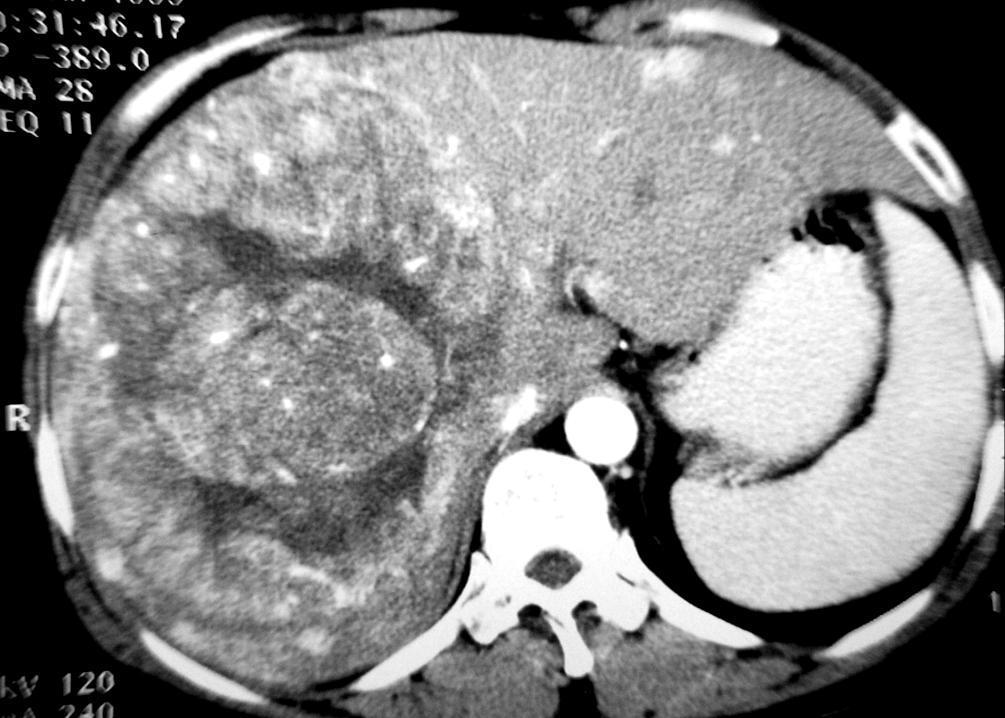
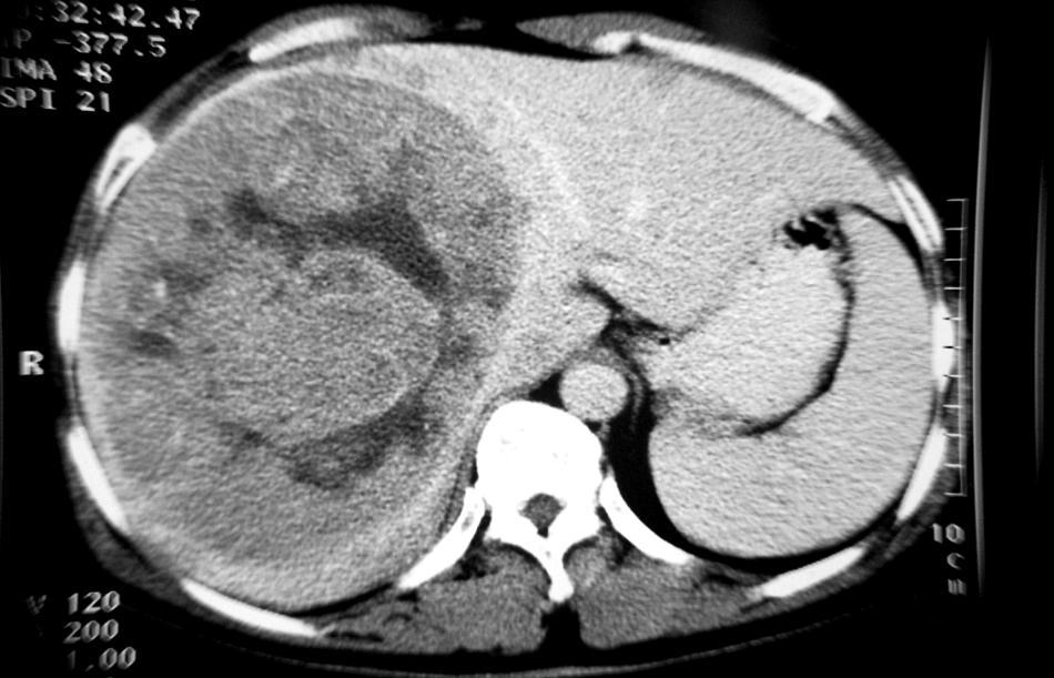
56 2 Tumors of Liver Malignant tumors primary malignant tumor Metastatic tumor Benign tumors cavernous hemangioma adenoma
57 MALIGNANT TUMORS Primary liver cancer hepatocellular carcinoma cholangiocarcinoma Secondary or metastatic liver malignant tumor
58 Hepatocellular Carcinoma (HCC) CT appearance: Liver cirrhotic background On unenhanced CT scanning, the lesion appears as well-defined or obscure, round or irregular lower density area than the relatively normal liver parenchyma
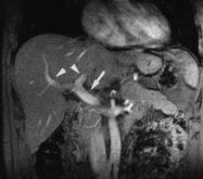
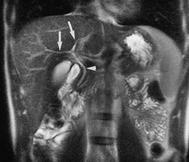
59 CT appearance: HCC Enhanced Scan: HAP --- heterogeneous enhancement; many enhancing patchy or dots within large tumor mass; dilated and tortuous arterial branches to the mass; necrotic low density areas within the lesion without enhancement. Rich & dominant arterial supply
60 CT appearance: HCC Enhanced Scan: PVP --- quickly return to hypodensity relatively to the neighboring liver parenchyma Wash-in and wash-out enhancement pattern
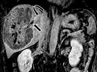
61 CT appearance: Secondary changes HCC (1) Abnormal blood perfusion around the lesion (displays as markedly enhancement of the parenchyma nearing the lesion) (2)Tumor thrombosis of PV and hepatic veins and IVC (3)Metastasis (hematogenic/seeding/lymphatic): The mets include the daughter foci in the liver and the metastatic lesions in the peritoneal cavity and other organs.
62 Dual-phase Scanning of HCC


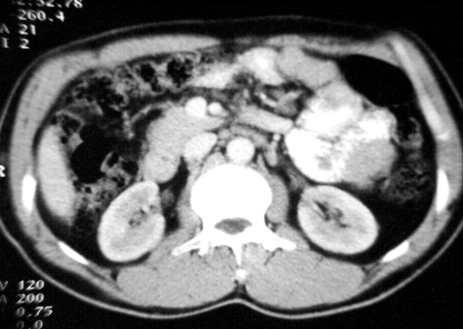
63 HAP Plain Scanning PVP HCC in right lobe with tumor emboli in the right branch of portal vein


64 Small HCC CT-Plain scan CT-HAP CT-PVP Specimen On unenhanced CT scan the lesion can not be differentiated definitely for it appears as isodense as the adjacent liver parenchyma and the lesion can be seen easily on hepatic artery phase for it was enhanced markedly and the density of the lesion became lower than the adjacent liver parenchyma on portal venous phase
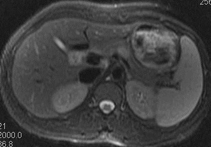
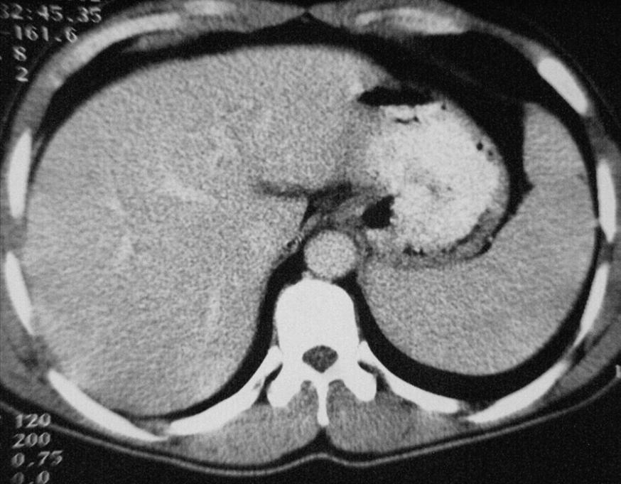
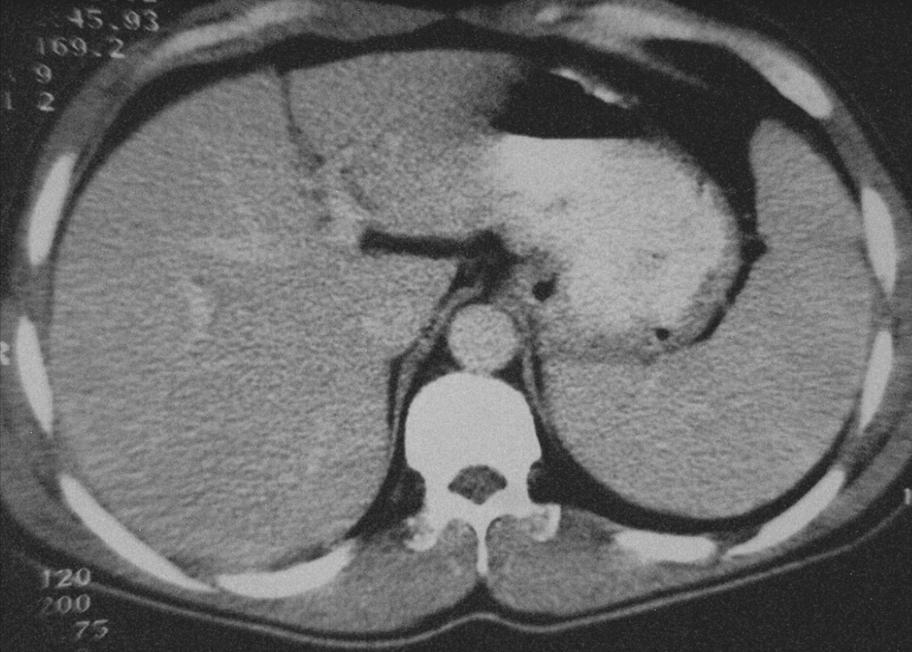
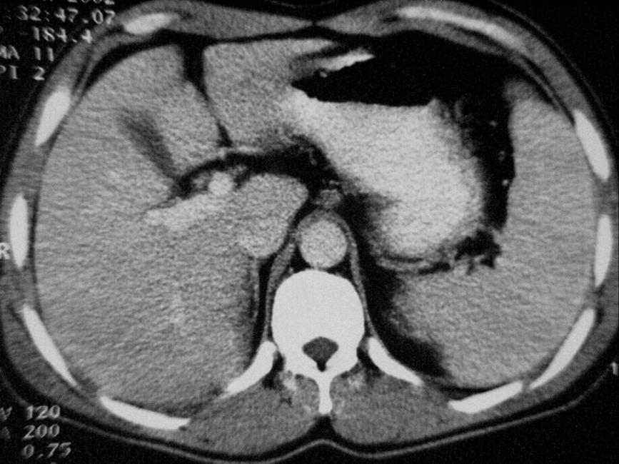
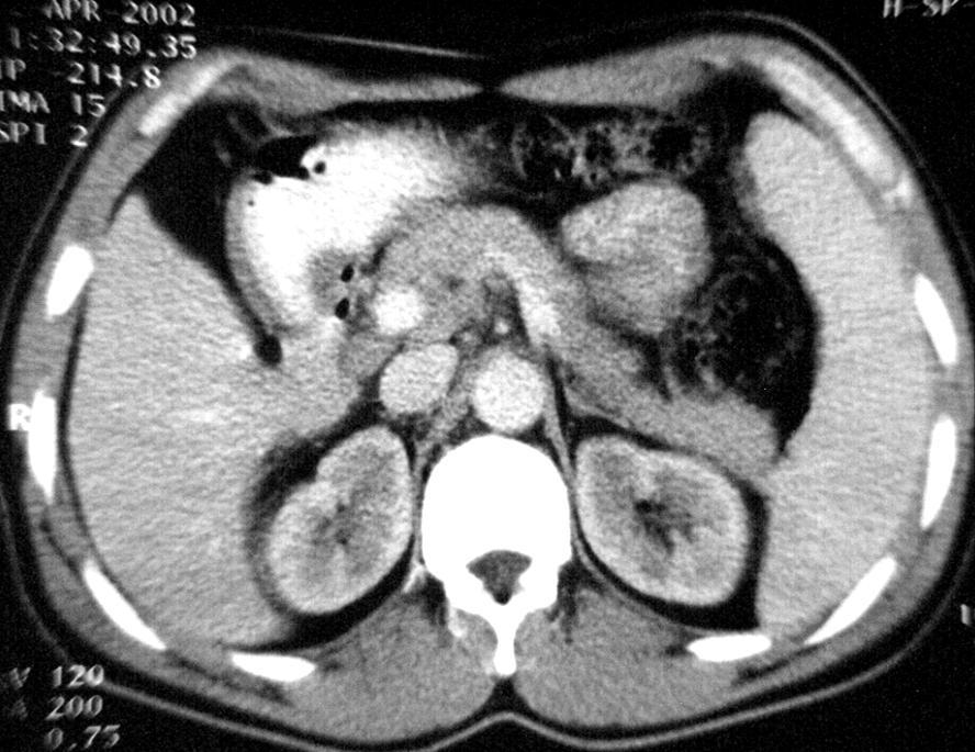
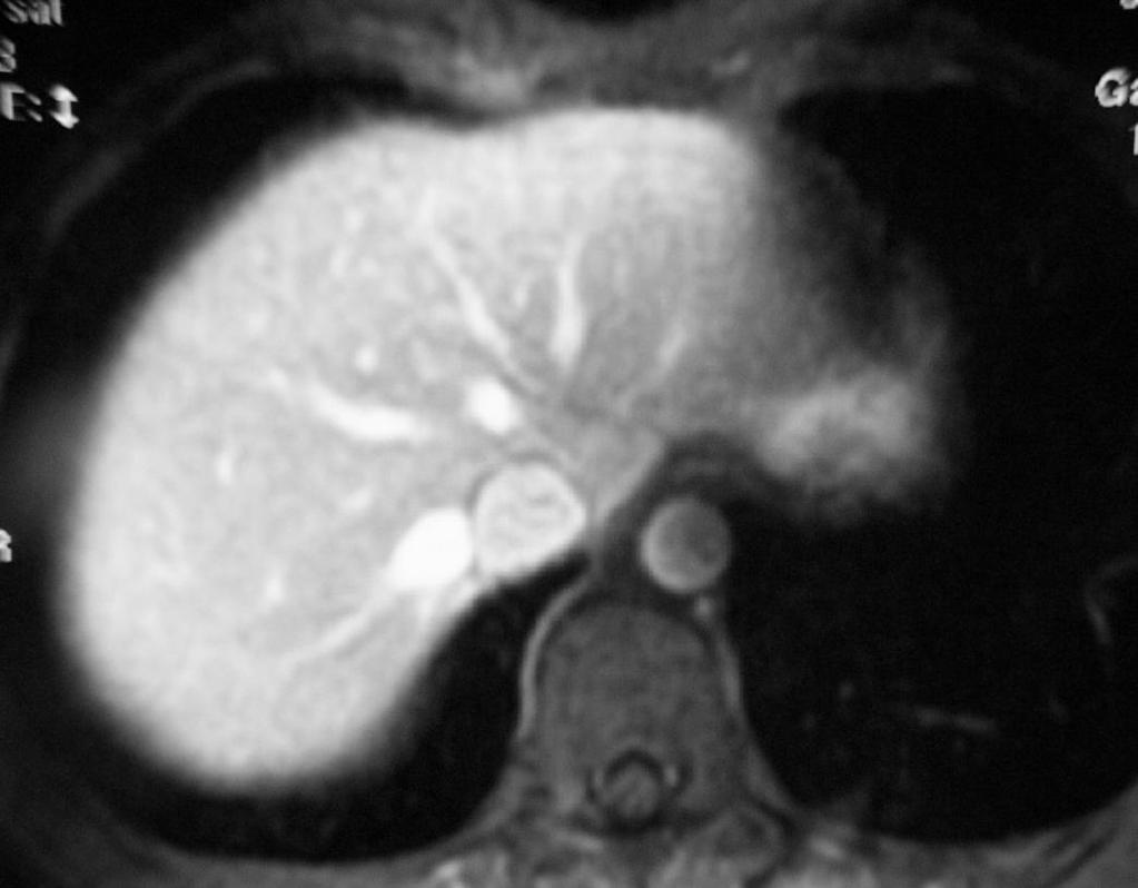
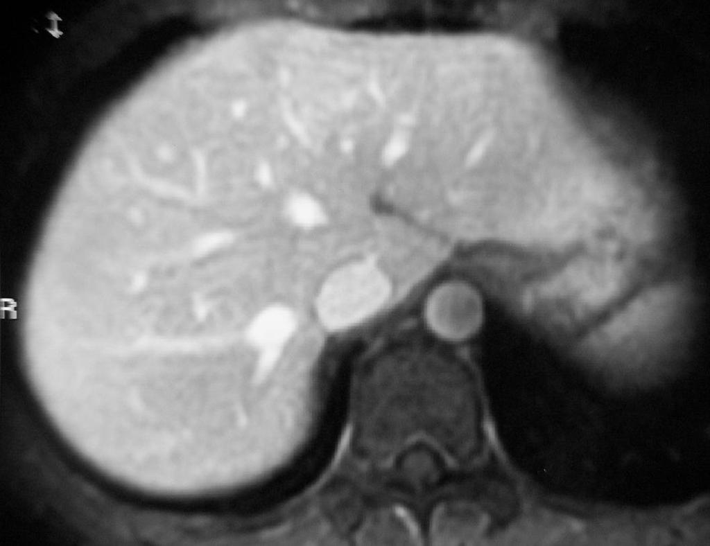
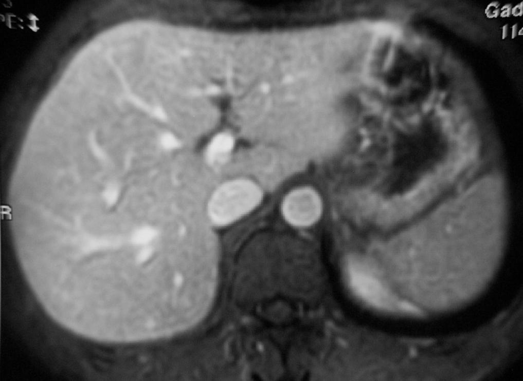
65 HCC MRI appearance: Mosaic pattern on T2WI MRI is comparable to CT in the diagnosis of large HCC lesions (>3 cm); but is superior to CT in the detection and characterization of small HCC nodules, especially in the cirrhotic liver very good for HCC staging


66 HCC Tumor thrombus GE T1WI-PVP GE T1WI

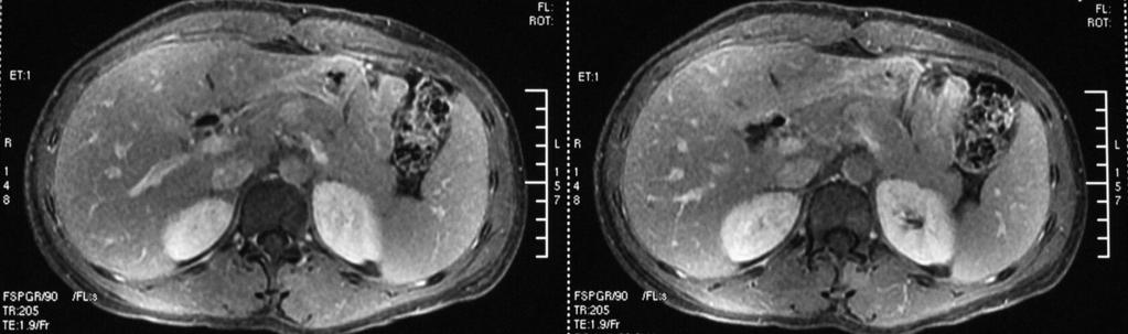
67 Mosaic Pattern SE T2W


68 Cholangiocarcinoma CT appearance: The lesion was enhanced heterogeneously as lace-like with intrahepatic bile duct dilatation HAP PVP


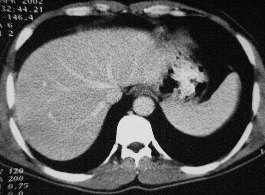
69 Cholangiocarcinoma MRI Findings: The mass was irregular as lobular with heterogeneous low signal at T1WI and heterogeneous high signal at T2WI. And the intrahepatic bile ducts were dilatation T1WI T2WI MRCP


70 Metastatic Liver Carcinoma CT appearance: always multiple lesions with periphery enhancement and obscure margin Mets from Breast Cancer


71 Mets from colorectal carcinoma Target Sign
72 Cavernous Hemangioma CT appearance: Low density area with smooth and clearly margin Initial peripheral nodular enhancement Gradual centripetal push-on enhancement Isodense fill-in at delayed scan The lesion was enhanced remarkable as periphery nodular at the hepatic artery phase and centripetal push-on enhancement gradually with the time delay of contrast injection and the lesion may become isodense at delay time scanning imaging

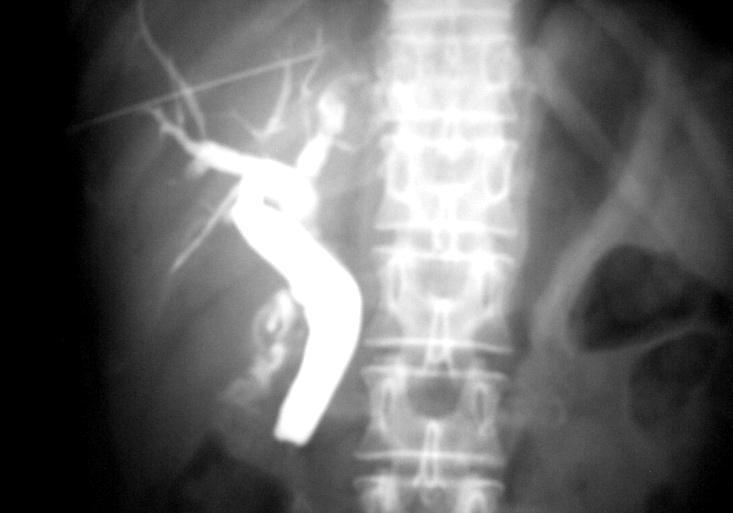
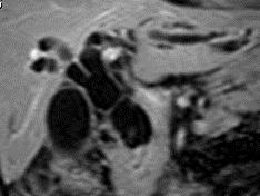
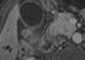
73 Enhancement evolution over time


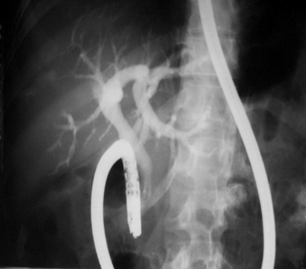
74 Cavernous Hemangioma MR appearance: The enhancement characteristics of the lesion in MR imaging is similar to that of CT scan. The lesion was enhanced centripetal gradually from the HAP to PVP and EP


75 3 Liver Cyst CT appearance: The liver cyst appears as round or oval shape of low density without enhancement and the margin is clear and regular


76 4 Liver Abscess CT appearance: The lesion was enhanced lace-like with periphery edema and the rest area of the liver parenchyma was normal
77 Biliary Tree Diseases 1. Cholelithiasis 2. Obstructive jaundice Cholangiocarcinoma Bile Duct Carcinoma Gallbladder Carcinoma
78 1 Cholelithiasis 1 Gall Stone CT density and MR signal vary based on the compositions of stones (calcium, bilirubin, steroid): CT high density stones low density stones isodense stones mixed density M R T1W: low or high signal some isointense T2W: always low signal


79 Cholecystitis with gallstone The gallbladder wall was edema with obscure margin

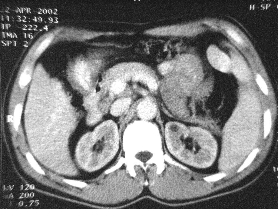
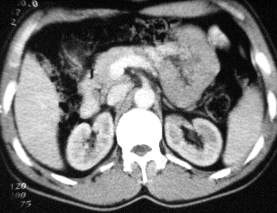
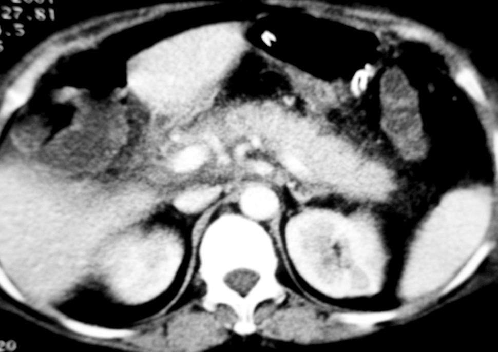
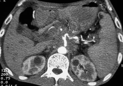
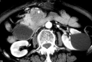
80 Cholelithiasis 2 Choledocholithiasis T2W MR


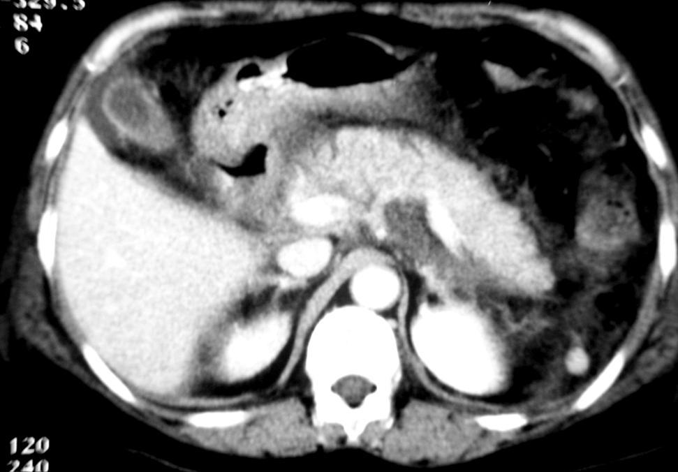
81 On MRCP, the stone appears as filling defect as low signal with bile duct and gallbladder dilatation moderately MRCP
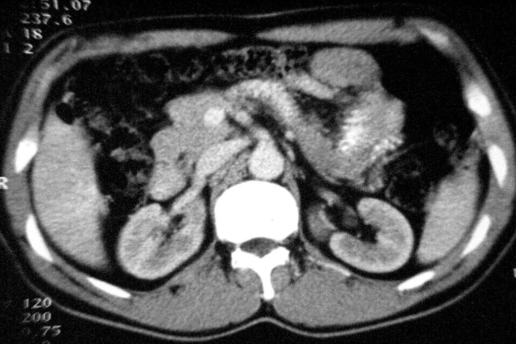
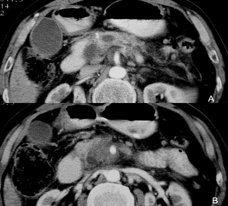
82 2 Obstructive jaundice Cholangiocarcinoma CT appearances: Dilatation of bile duct proximal to the obstruction lesion, abrupt narrowing or stenosis at the site of the tumor Nodule protruding to bile duct lumen Mild to moderate enhancement of the thicken bile duct wall and intraluminal nodule on PVP


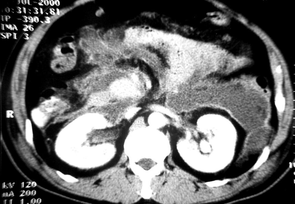
83 Hilar Cholangiocarcinoma Hilar cholangiocarcinoma appears as bile duct wall thicken with moderate enhancement at PVP and the intrahepatic bile duct dilatation markedly
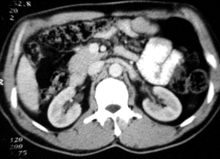
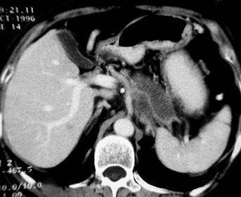
84 Obstructive Jaundice Bile Duct Carcinoma MR Display size, extent and surroundings of mass, superior to CT MRCP: able to display subtle morphological changes, comparable to ERCP or PTC


85 CBD carcinoma The low segment of the CBD wall thicken and enhanced moderately with proximal CBD dilatation
86 Pancreatic Diseases 1 Pancreatitis Acute pancreatitis Chronic pancreatitis 2 Pancreatic carcinoma


87 1 Acute Pancreatitis CT appearance: AP appears as pancreatic edema with inflammation effusion in the retroperitoneal space and the effusion extend along the mesentery. The irregular low density area in pancreas neck and body indicate pancreas necrosis AP with gland necrosis

88 AP with retroperitoneal spreading
89 2 Chronic Pancreatitis CT appearance: Atrophy of parenchyma Dilatation or stricture of pancreatic duct Stones or calculi within pancreatic duct


90 Chronic pancreatitis with a lot of calculi in pancreatic duct and parenchyma and there is a pseudocyst on the neck of pancreas with active bleeding


91 3 Pancreatic Carcinoma CT appearances: An irregular low density mass on the head of pancreas which encase the celiac truck and it s branches, and there is a mets on right lobe of the liver
92

93 Thank you for listening!
Imaging of liver and pancreas
 Imaging of liver and pancreas.. Disease of the liver Focal liver disease Diffusion liver disease Focal liver disease Benign Cyst Abscess Hemangioma FNH Hepatic adenoma HCC Malignant Fibrolamellar carcinoma
Anatomical and Functional MRI of the Pancreas
 Anatomical and Functional MRI of the Pancreas MA Bali, MD, T Metens, PhD Erasme Hospital Free University of Brussels Belgium mbali@ulb.ac.be Introduction The use of MRI to investigate the pancreas has
Radiology of hepatobiliary diseases
 GI cycle - Lecture 14 436 Teams Radiology of hepatobiliary diseases Objectives 1. To Interpret plan x-ray radiograph of abdomen with common pathologies. 2. To know the common pathologies presentation.
Alice Fung, MD Oregon Health and Science University
 Alice Fung, MD Oregon Health and Science University Disclosure Comments The speaker Alice Fung, MD Has relevant financial relationships to disclose. Received honorarium from (Guerbet). This individual
Imaging of common diseases of hepatobiliary and GI system
 Imaging of common diseases of hepatobiliary and GI system Natthaporn Tanpowpong, M.D. Diagnostic radiology Faculty of Medicine, Chulalongkorn University Normal plain radiograph A = Common bile duct
Liver imaging takes a step forward with Ingenia
 Publication for the Philips MRI Community ISSUE 49 2013 / 2 Liver imaging takes a step forward with Ingenia Lyon South Hospital strives to move from several studies first CT, then MR or PET to using just
Dr Claire Smith, Consultant Radiologist St James University Hospital Leeds
 Dr Claire Smith, Consultant Radiologist St James University Hospital Leeds Imaging in jaundice and 2ww pathway Image protocol Staging Limitations Pancreatic cancer 1.2.4 Refer people using a suspected
Appendix 5. EFSUMB Newsletter. Gastroenterological Ultrasound
 EFSUMB Newsletter 87 Examinations should encompass the full range of pathological conditions listed below A log book listing the types of examinations undertaken should be kept Training should usually
Biliary cancers: imaging diagnosis. Study of 30 cases
 Biliary cancers: imaging diagnosis. Study of 30 cases N Hammoune, S Semlali, M Eddarai, T. Amil, M Zentar, S. El Kandri,, M Benameur,, S Chaouir. Radiology Department. Mohamed V Military Hospital. Rabat-
MRI Abdomen Protocol Pancreas/MRCP with Contrast
 MRI Abdomen Protocol Pancreas/MRCP with Contrast Reviewed By: Brett Mollard, MD; Anna Ellermeier, MD Last Reviewed: July 2018 Contact: (866) 761-4200 Standard uses: 1. Characterization of cystic and solid
State of the Art Imaging for Hepatic Malignancy: My Assignment
 State of the Art Imaging for Hepatic Malignancy: My Assignment CT vs MR vs MRCP Which one to choose for HCC vs Cholangiocarcinoma What special protocols to use for liver tumors Role of PET and Duplex US
Guidelines, Policies and Statements D5 Statement on Abdominal Scanning
 Guidelines, Policies and Statements D5 Statement on Abdominal Scanning Disclaimer and Copyright The ASUM Standards of Practice Board have made every effort to ensure that this Guideline/Policy/Statement
Essentials of Clinical MR, 2 nd edition. 65. Benign Hepatic Masses
 65. Benign Hepatic Masses Pulse sequences acquired for abdominal MRI typically consist of fast acquisition schemes such as single-shot turbo spin echo (i.e. HASTE) and gradient echo schemes such as FLASH
Hilar cholangiocarcinoma. Frank Wessels, Maarten van Leeuwen, UMCU utrecht
 Hilar cholangiocarcinoma Frank Wessels, Maarten van Leeuwen, UMCU utrecht Content Anatomy Biliary strictures (Hilar) Cholangiocarcinoom Staging Biliary tract 1 st order Ductus hepatica dextra Ductus hepaticus
Original Policy Date 12:2013
 MP 6.01.30 Magnetic Resonance Cholangiopancreatography Medical Policy Section Radiology Is12:2013sue 3:2005 Original Policy Date 12:2013 Last Review Status/Date 12:2013 Return to Medical Policy Index Disclaimer
Post-operative complications following hepatobiliary surgery: imaging findings and current radiological treatment options
 Post-operative complications following hepatobiliary surgery: imaging findings and current radiological treatment options Poster No.: C-1501 Congress: ECR 2015 Type: Educational Exhibit Authors: A. Hadjivassiliou,
CT & MRI of Benign Liver Neoplasms Srinivasa R Prasad
 CT & MRI of Benign Liver Neoplasms Srinivasa R Prasad No financial disclosures Acknowledgements Many thanks to Drs. Heiken, Narra & Menias (MIR) Dr. Sahani (MGH) for sharing images Benign Liver Tumors:
Biliary tree dilation - and now what?
 Biliary tree dilation - and now what? Poster No.: C-1767 Congress: ECR 2012 Type: Educational Exhibit Authors: I. Ferreira, A. B. Ramos, S. Magalhães, M. Certo; Porto/PT Keywords: Pathology, Diagnostic
Newcastle HPB MDM updated radiology imaging protocol recommendations. Author Dr John Scott. Consultant Radiologist Freeman Hospital
 Newcastle HPB MDM updated radiology imaging protocol recommendations Author Dr John Scott. Consultant Radiologist Freeman Hospital This document is intended as a guide to aid radiologists and clinicians
Hepatic Imaging: What Every Practitioner Should Know
 Hepatic Imaging: What Every Practitioner Should Know Shuchi K. Rodgers, MD Section Chief, Abdominal Imaging Director of Ultrasound Department of Radiology Einstein Medical Center rodgerss@einstein.edu
4/9/2018 OBJECTIVES PANCREAOTO BILIARY ULTRASOUND: BEYOND CHOLECYSTITIS
 PANCREAOTO BILIARY ULTRASOUND: BEYOND CHOLECYSTITIS Jean Yves Sewah Kaiser Permanente West Los Angeles 1 OBJECTIVES Discuss the role of ultrasound in the evaluation of the gallbladder, biliary tree and
Normal Sonographic Anatomy
 hapter 2:The Liver DUNSTAN ABRAHAM Normal Sonographic Anatomy Homogeneous, echogenic texture (Figure 2-1) Measures approximately 15 cm in length and 10 12.5 cm anterior to posterior; measurement taken
Common and unusual CT and MRI manifestations of pancreatic adenocarcinoma: a pictorial review
 Review Article Common and unusual CT and MRI manifestations of pancreatic adenocarcinoma: a pictorial review Min-Jie Yang, Su Li, Yong-Guang Liu, Na Jiao, Jing-Shan Gong Department of Radiology, Shenzhen
MRI of the Hepatobiliary System
 BODY APPLICATIONS OF MRI 51 Chapter Four C H A P T E R F O U R MRI of the Hepatobiliary System After completing this chapter, the reader will be able to: Develop a protocol for liver MRI Identify benign
Nasogastric tube. Stomach. Pylorus. Duodenum 1. Duodenum 2. Duodenum 3. Duodenum 4
 Esophagus Barium Swallow Stomach and Duodenum 4 year old Upper GI Nasogastric tube Stomach and Duodenum 4 year old Upper GI Nasogastric tube Stomach Pylorus Duodenum 1 Duodenum 2 Duodenum 3 Duodenum 4
Portal Venous Thrombosis: Tumor VS Bland Thrombus
 June 2015 Portal Venous Thrombosis: Tumor VS Bland Thrombus SERGIO ALFARO, HARVARD MEDICAL SCHOOL YEAR III GILLIAN LIEBERMAN, MD Overview 2 Index Patient History Portal Venous Thrombosis (PVT) Imaging
HEPATO-BILIARY IMAGING
 HEPATO-BILIARY IMAGING BY MAMDOUH MAHFOUZ MD PROF.OF RADIOLOGY CAIRO UNIVERSITY mamdouh.m5@gmail.com www.ssregypt.com CT ABDOMEN Indications Patient preparation Patient position Scanogram Fasting 4-6 hours
Abdomen Sonography Examination Content Outline
 Abdomen Sonography Examination Content Outline (Outline Summary) # Domain Subdomain Percentage 1 2 3 Anatomy, Perfusion, and Function Pathology, Vascular Abnormalities, Trauma, and Postoperative Anatomy
Anatomy Jessica Ferguson Ashley Dobos May 31, 2006 LIVER
 Anatomy Jessica Ferguson Ashley Dobos May 31, 2006 LIVER 1) Other Names: Reidel s Lobe normal anatomic variant; projection of the right lobe that can extend as far as the iliac crest (Tempkin, p.54, Anatomy).
Radiological Investigations of Abdominal Trauma
 76 77 Investigations of Abdominal Trauma Introduction: Trauma to abdominal organs is a common cause of patient morbidity and mortality among trauma patients. Causes of abdominal trauma include blunt injuries,
Personal Profile. Name: 劉 XX Gender: Female Age: 53-y/o Past history. Hepatitis B carrier
 Personal Profile Name: 劉 XX Gender: Female Age: 53-y/o Past history Hepatitis B carrier Chief complaint Fever on and off for 2 days Present illness 94.10.14 Sudden onset of epigastric pain 94.10.15 Fever
IT 의료융합 1 차임상세미나 복부질환초음파 이재영
 IT 의료융합 1 차임상세미나 2013-4-3 복부질환초음파 이재영 나는오늘누구를위하여 종을울리나? 전통적의료 의사 공학설계자 의사 최첨단진단장비들 USG, CT, MRI 환자 환자 현대의료 사용자중심의사고 US in the Abdomen Detection DDx Look Behavior Response by external stimuli Guiding Tool
Cholangiocarcinoma (Bile Duct Cancer)
") Cholangiocarcinoma (Bile Duct Cancer) The Bile Duct System (Biliary Tract) A network of bile ducts (tubes) connects the liver and the gallbladder to the small intestine. This network begins in the liver
Cholangiocarcinoma (Bile Duct Cancer) The Bile Duct System (Biliary Tract) A network of bile ducts (tubes) connects the liver and the gallbladder to the small intestine. This network begins in the liver
Case Study: #3: Gallbladder Carcinoma?
 Case Study: #3: Gallbladder Carcinoma? By: Megan Wyatt K. SON Wyatt 225 2B1 RDMS, RVT Patient: Male 85 YOA Caucasian Indication: Elevated Alkaline Phosphatase History Annual physical showed elevated alkaline
Abdominal ultrasound:
 Abdominal ultrasound: Non-traumatic acute abdomen Wittanee Na-ChiangMai, MD Department of Radiology ChiangMai University 26/04/2017 Contents Technique of examination Normal anatomy Emergency conditions
Liver Cancer And Tumours
 Liver Cancer And Tumours What causes liver cancer? Many factors may play a role in the development of cancer. Because the liver filters blood from all parts of the body, cancer cells from elsewhere can
Recently role of non-invasive diagnostics methods
 CERTAIN ASPECTS OF NLS-DIAGNOSTICS OF LIVER FOCAL PATHOLOGY A.Y. Shvack, V.I. Nesterov, N.L. Ogluzdina This article contains information about NLS-graphy application in diagnostics of liver focal affections:
Liver, Pancreas and Biliary System. Wirana Angthong, M.D.
 Liver, Pancreas and Biliary System Wirana Angthong, M.D. Objectives Outline Anatomy Imaging Techniques Common Diseases Outline Anatomy Imaging Techniques Common Diseases Liver anatomy Morphological anatomy:
Lab Monitor Images Dissection of the Abdominal Vasculature + Lower Digestive System
 Lab Monitor Images Dissection of the Abdominal Vasculature + Lower Digestive System Stomach & Duodenum Frontal (AP) View Nasogastric tube 2 1 3 4 Stomach Pylorus Duodenum 1 Duodenum 2 Duodenum 3 Duodenum
Pancreas Case Scenario #1
 Pancreas Case Scenario #1 An 85 year old white female presented to her primary care physician with increasing abdominal pain. On 8/19 she had a CT scan of the abdomen and pelvis. This showed a 4.6 cm mass
Job Task Analysis for ARDMS Abdomen Data Collected: June 30, 2011
 Job Task Analysis for ARDMS Abdomen Data Collected: June 30, 2011 Reported: Analysis Summary for: Abdomen Examination Survey Dates 06/13/2011-06/26/2011 Invited Respondents 6,000 Surveys with Demographics
Interesting Cases from Liver Tumor Board. Jeffrey C. Weinreb, M.D.,FACR Yale University School of Medicine
 Interesting Cases from Liver Tumor Board Jeffrey C. Weinreb, M.D.,FACR Yale University School of Medicine jeffrey.weinreb@yale.edu Common Liver Diseases Hemangioma Cyst FNH Focal Fat/Sparing THID Non-Cirrhotic
6 th August 2018 Day 1 - Gallbladder & Bile duct Topic
 Venue: Sterling Hospital Auditorium, Sterling Hospitals, Gurukul Road Ahmedabad, Gujarat 6 th August 2018 Day 1 - Gallbladder & Bile duct Registration(8:00am-8:15am) Inauguration(8:15am-8:30am) Welcome
Imaging in gastric cancer
 Imaging in gastric cancer Gastric cancer remains a deadly disease because of late diagnosis. Adenocarcinoma represents 90% of malignant tumors. Diagnosis is based on endoscopic examination with biopsies.
Diagnosis of tumor extension in biliary carcinoma has. Differential Diagnosis and Treatment of Biliary Strictures
 CLINICAL GASTROENTEROLOGY AND HEPATOLOGY 2009;7:S79 S83 Differential Diagnosis and Treatment of Biliary Strictures KAZUO INUI, JUNJI YOSHINO, and HIRONAO MIYOSHI Department of Internal Medicine, Second
Malignant Focal Liver Lesions
 Malignant Focal Liver Lesions Other Than HCC Pablo R. Ros, MD, MPH, PhD Departments of Radiology and Pathology University Hospitals Cleveland Medical Center Case Western Reserve University Pablo.Ros@UHhospitals.org
pitfall Table 1 4 disorientation pitfall pitfall Table 1 Tel:
 11 687 692 2002 pitfall 1078 29 17 9 1 2 3 dislocation outflow block 11 1 2 3 9 1 2 3 4 disorientation pitfall 11 687 692 2002 Tel: 075-751-3606 606-8507 54 2001 8 27 2002 10 31 29 4 pitfall 16 1078 Table
Jaundice. Agnieszka Dobrowolska- Zachwieja, MD, PhD
 Jaundice Agnieszka Dobrowolska- Zachwieja, MD, PhD Jaundice definition Jaundice, as in the French jaune, refers to the yellow discoloration of the skin. It arises from the abnormal accumulation of bilirubin
Hepatocellular carcinoma Cholangiocarcinoma. Jewels of hepatobiliary cancer imaging : what to look for? Imaging characteristics of HCC.
 Outline : Imaging Jewels Jewels of hepatobiliary cancer imaging : what to look for? Hepatocellular carcinoma Cholangiocarcinoma Surachate Siripongsakun, M.D. Chulabhorn Cancer Center Imaging characteristics
ACUTE PANCREATITIS: NEW CLASSIFICATION OF AN OLD FOE. T Barrow, A Nasrullah, S Liong, V Rudralingam, S A Sukumar
 ACUTE PANCREATITIS: NEW CLASSIFICATION OF AN OLD FOE T Barrow, A Nasrullah, S Liong, V Rudralingam, S A Sukumar LEARNING OBJECTIVES q Through a series of cases illustrate the updated Atlanta symposium
Evidence based imaging of the pancreas
 Evidence based imaging of the pancreas D.Vanbeckevoort, D.Bielen, K.Op de beeck, R.Vanslembrouck Department of Radiology Chairman Prof. Dr. R.Oyen Non-invasive imaging tests available for the diagnosis
CT 101 :Pancreas and Spleen
 CT 101 :Pancreas and Spleen Shikha Khullar,, MD, MPH Division of Radiology University of South Alabama The Pancreas Normal Pancreas 3 Phase Pancreatic CT Non contrast Arterial phase : 30-35 35 second
Multiple Primary Quiz
 Multiple Primary Quiz Case 1 A 72 year old man was found to have a 12 mm solid lesion in the pancreatic tail by computed tomography carried out during a routine follow up study of this patient with adult
Navigating the Biliary Tract with CT & MR: An Imaging Approach to Bile Duct Obstruction
 Navigating the Biliary Tract with CT & MR: An Imaging Approach to Bile Duct Obstruction Ann S. Fulcher, MD Medical College of Virginia Virginia Commonwealth University Richmond, Virginia Objectives To
Gemstone Spectral Imaging quantifies lesion characteristics for a confident diagnosis
 GE Healthcare Gemstone Spectral Imaging quantifies lesion characteristics for a confident diagnosis CT clinical case study lesion characterization Desiree Morgan, MD Vice Chair of Clinical Research Professor
Innovations in HCC Imaging: MDCT/MRI
 Innovations in HCC Imaging: MDCT/MRI Anthony E. Cheng, M.D. Cardinal MRI Center Cardinal Santos Medical Center, Wilson Street, San Juan Innovations in HCC Imaging: Goals/Objectives MDCT/MRI Learn the diagnostic
Body MRI from the Liver to the Bladder
 Body MRI from the Liver to the Bladder I Want You! Audience Participation Methodist Hospital Continuing Education Seminar Jordan Swensson, MD November 7, 2015 Objectives Observe the uses of MRI for organs
Question 1 History. Likely Diagnosis Differential. Further Investigation or Management. Requires Paediatric Surgical referral for laparotomy
 Question 1 Male newborn spilling green tinged vomit day 1 of life Imaging Abdominal X-Rays performed on 03/05/2012 Upper and lower gastrointestinal contrast studies performed on 03/05/2012 Abdominal X-Rays
Intraoperative staging of GIT cancer using Intraoperative Ultrasound
 Intraoperative staging of GIT cancer using Intraoperative Ultrasound Thesis For Fulfillment of MSc Degree In Surgical Oncology By Abdelhalim Salah Abdelhalim Moursi M.B.B.Ch (Cairo University ) Supervisors
MR imaging of primary sclerosing cholangitis (PSC) using the hepatobiliary specific contrast agent Gd-EOB-DTPA
 using the hepatobiliary specific contrast agent Gd-EOB-DTPA") MR imaging of primary sclerosing cholangitis (PSC) using the hepatobiliary specific contrast agent Gd-EOB-DTPA Poster No.: C-0019 Congress: ECR 2010 Type: Educational Exhibit Topic: Abdominal Viscera (Solid
MR imaging of primary sclerosing cholangitis (PSC) using the hepatobiliary specific contrast agent Gd-EOB-DTPA Poster No.: C-0019 Congress: ECR 2010 Type: Educational Exhibit Topic: Abdominal Viscera (Solid
Appendix 9: Endoscopic Ultrasound in Gastroenterology
 Appendix 9: Endoscopic Ultrasound in Gastroenterology This curriculum is intended for clinicians who perform endoscopic ultrasonography (EUS) in gastroenterology. It includes standards for theoretical
Vascular Imaging in the Pediatric Abdomen. Jonathan Swanson, MD
 Vascular Imaging in the Pediatric Abdomen Jonathan Swanson, MD Goals and Objectives To understand the imaging approach, appearance, and clinical manifestations of the common pediatric abdominal vascular
Primary Sclerosing Cholangitis and Cholestatic liver diseases. Ahsan M Bhatti MD, FACP Bhatti Gastroenterology Consultants
 Primary Sclerosing Cholangitis and Cholestatic liver diseases Ahsan M Bhatti MD, FACP Bhatti Gastroenterology Consultants I have nothing to disclose Educational Objectives What is PSC? Understand the cholestatic
Cirrhosis and Portal Hypertension Gastroenterology Teaching Project American Gastroenterological Association
 CIRRHOSIS AND PORTAL HYPERTENSION Cirrhosis and Portal Hypertension Gastroenterology Teaching Project American Gastroenterological Association WHAT IS CIRRHOSIS? What is Cirrhosis? DEFINITION OF CIRRHOSIS
Diagnostic Imaging of the Liver
 S. Sakuma T. Ishigaki T. Takeuchi Diagnostic Imaging of the Liver Biliary Tract and Pancreas Data Analysis and Diagnostic Procedures With 312 Figures in 932 Separate Illustrations Springer-Verlag Berlin
Imaging abdominal vascular emergencies. V.Stoynova
 Imaging abdominal vascular emergencies V.Stoynova Abdominal vessels V. Stoynova 2 Acute liver bleeding trauma anticoagulant therapy liver disease : HCC, adenoma, meta, FNH, Hemangioma Diagnosis :CT angiography
Review. Computed tomography in the diagnosis and staging of cholangiocarcinoma and pancreatic carcinoma
 Annals of Oncology 10 Suppl. 4: S12-S17, 1999. 1999 Kluwer Academic Publishers. Printed in the Netherlands. Review Computed tomography in the diagnosis and staging of cholangiocarcinoma and pancreatic
Policies, Standards, and Guidelines. Guidelines for Abdominal Ultrasound Examination
 Policies, Standards, and Guidelines Guidelines for Abdominal Ultrasound Examination Approved by Council Feb 2018 Disclaimer and Copyright The ASUM Standards of Practice Board have made every effort to
Gall bladder cancer. Information for patients Hepatobiliary
 Gall bladder cancer Information for patients Hepatobiliary page 2 of 12 Who will provide my care? You will be cared for by a number of professionals who work together. These professionals will be specialist
A patient with an unusual congenital anomaly of the pancreaticobiliary tree
 A patient with an unusual congenital anomaly of the pancreaticobiliary tree Thomas Hocker, HMS IV BIDMC Core Radiology Case Presentation September 17, 2007 Review of Normal Pancreaticobiliary Tract Anatomy
Case Scenarios. 12/28/12 MRI Liver: multiple focal arterially enhancing liver lesions, indeterminate. Repeat MRI in 4 months.
 Case Scenarios Case Scenario 1 A 63 year old black female presented with a history of liver lesions present for the past three years being monitored by serial scans. The patient has a history of esophageal
ROLE OF RADIOLOGY IN INVESTIGATION OF JAUNDICE
 ROLE OF RADIOLOGY IN INVESTIGATION OF JAUNDICE Dr. Sohan kumar sah *, Dr. Liu Sibin, Dr. sumendra raj pandey, Dr. Prakashmaan shah, Dr. Gaurishankar pandit, Dr. Suraj kurmi and Dr. Sanjay kumar jaiswal
Liver MRI in 30 minutes
 X Liver MRI in 30 minutes SCBT/MR Annual Meeting Salt Lake City September 18, 2016 Scott B. Reeder, MD, PhD Department of Radiology University of Wisconsin Madison, WI Disclosures University of Wisconsin-Madison
Autoimmune Pancreatitis: A Great Imitator
 Massachusetts General Hospital Harvard Medical School Autoimmune Pancreatitis: A Great Imitator Dushyant V Sahani MD dsahani@partners.org Autoimmune Pancreatitis: Learning Objectives Clinical manifestations
Gastrointestinal System: Accessory Organ Disorders
 Gastrointestinal System: Accessory Organ Disorders Mary DeLetter, PhD, RN Associate Professor Dept. of Baccalaureate and Graduate Nursing Eastern Kentucky University Disorders of Accessory Organs Portal
TOPICS FOR PRACTICAL LESSONS, DISCIPLINE RADIOLOGY For the IIIrd year students Faculty of Medicine, university year
 TOPICS FOR PRACTICAL LESSONS, DISCIPLINE RADIOLOGY For the IIIrd year students Faculty of Medicine, university year 2018-2019 I. Evolution of radiology. Notion of Radiophysics. 1. Medical imaging definition.
INVESTIGATIONS OF GASTROINTESTINAL DISEAS
 INVESTIGATIONS OF GASTROINTESTINAL DISEAS Lecture 1 and 2 دز اسماعيل داود فرع الطب كلية طب الموصل Radiological tests of structure (imaging) Plain X-ray: May shows soft tissue outlines like liver, spleen,
Computed tomography. Department of Radiology, University Medical School, Szeged
 Computed tomography Department of Radiology, University Medical School, Szeged voxel +1-4 +2 +5 +3 +1 0-2 pixel -2 0 +1-4 -6 +5 +2 +1 Department of Radiology, University Medical School, Szeged
Liver Specific MRI using Gd-EOB-DTPA Disodium (Primovist) Effects Change in Management of Indeterminate Liver Lesions.
 Effects Change in Management of Indeterminate Liver Lesions.") Liver Specific MRI using Gd-EOB-DTPA Disodium (Primovist) Effects Change in Management of Indeterminate Liver Lesions. Poster No.: C-1751 Congress: ECR 2012 Type: Authors: Keywords: DOI: Educational Exhibit
Liver Specific MRI using Gd-EOB-DTPA Disodium (Primovist) Effects Change in Management of Indeterminate Liver Lesions. Poster No.: C-1751 Congress: ECR 2012 Type: Authors: Keywords: DOI: Educational Exhibit
Approach to the Patient with Liver Disease
 Approach to the Patient with Liver Disease Diagnosis of liver disease Careful history taking Physical examination Laboratory tests Radiologic examination and imaging studies Liver biopsy Liver diseases
The role for contrast-enhanced ultrasonography outside of focal liver lesions
 The role for contrast-enhanced ultrasonography outside of focal liver lesions Paul S. Sidhu King s College Hospital, London, UK Introduction Contrast-enhanced ultrasonography (US) of focal liver lesions
Hepatobiliary Ultrasound Rimon Bengiamin, MD, RDMS Assistant Clinical Professor Director of Emergency Ultrasound UCSF Fresno. Objectives. Why?
 Hepatobiliary Ultrasound Rimon Bengiamin, MD, RDMS Assistant Clinical Professor Director of Emergency Ultrasound UCSF Fresno Objectives Discuss the goals of point-of-care biliary ultrasound Review the
Intrahepatic Sarcomatoid Cholangiocarcinoma with Portal Vein Thrombosis: A Case Report 1
 Intrahepatic Sarcomatoid Cholangiocarcinoma with Portal Vein Thrombosis: A Case Report 1 Jae-Hoon Lim, M.D., Jin Woong Kim, M.D., Suk Hee Heo, M.D., Yong Yeon Jeong, M.D., Heoung Keun Kang, M.D. A 53-year-old
CTA/MRA of Pediatric Hepatic Masses Radiology-Pathology Correlation
 Acta Radiológica Portuguesa, Vol.XVIII, nº70, pág. 41-50, Abr.-Jun., 2006 CTA/MRA of Pediatric Hepatic Masses Radiology-Pathology Correlation Marilyn J. Siegel Mallinckrodt Institute of Radiology, Washington
TABLES. Imaging Modalities Evidence Tables Table 1 Computed Tomography (CT) Imaging. Conclusions. Author (Year) Classification Process/Evid ence Class
 Imaging. Conclusions. Author (Year) Classification Process/Evid ence Class") TABLES Imaging Modalities Evidence Tables Table 1 Computed Tomography (CT) Imaging Author Clark (1986) 9 Reformatted sagittal images in the differential diagnosis meningiomas and adenomas with suprasellar
TABLES Imaging Modalities Evidence Tables Table 1 Computed Tomography (CT) Imaging Author Clark (1986) 9 Reformatted sagittal images in the differential diagnosis meningiomas and adenomas with suprasellar
Portal biliopathy from the image to the diagnostic: a systematic illustrated overview
 Portal biliopathy from the image to the diagnostic: a systematic illustrated overview Poster No.: C-1690 Congress: ECR 2018 Type: Educational Exhibit Authors: F. Stefan, M. D. M. Boros, E. M. Preda, I.
Financial Disclosure
 Benign Liver Masses Adil Abdalla, MBBS Creighton University-CHI Health August 25, 2018 Financial Disclosure Nothing to disclose Financial Disclosure 1 Objectives To assess patients with benign liver tumors
MR Advance Techniques. Vascular Imaging. Class II
 MR Advance Techniques Vascular Imaging Class II 1 Vascular Imaging There are several methods that can be used to evaluate the cardiovascular systems with the use of MRI. MRI will aloud to evaluate morphology
Imaging iconography of gallbladder cancer. Assessment by CT.
 1 REVISTA DE IMAGENOLOGIA- EII / Vol. XVI / Num. 2 Imaging iconography of gallbladder cancer. Assessment by CT. Doctors Crisci, Alejandro (1); Landó, Fernando.(2). CASMU CT Department Hospital of Tacuarembó
International Journal of Current Medical Sciences- Vol. 6, Issue,, pp , June, 2016 A B S T R A C T
 ISSN: 2320-8147 International Journal of Current Medical Sciences- Vol. 6, Issue,, pp. 122-126, June, 2016 COMPUTED TOMOGRAPHY IN HEPATIC METASTASES Ananthakumar P and Adaikkappan M., Available online
Spleen indications of splenectomy complications OPSI
 Intestinal obstruction Differences between adynamic ileus and mechanical obstruction Aetiology Pathophysiology (Cluster contractions- bowel proximal to the obstruction dilate- wall of obstructed gut is
Clinics in Diagnostic Imaging (79)
") Singapore Med J 2002 Vol 43(11) : 591-596 M e d i c a l E d u c a t i o n Clinics in Diagnostic Imaging (79) P Lerttumnongtum, M Muttarak, K Wasanavijit 1a Fig. 1a Subcostal US scan taken at the porta
Singapore Med J 2002 Vol 43(11) : 591-596 M e d i c a l E d u c a t i o n Clinics in Diagnostic Imaging (79) P Lerttumnongtum, M Muttarak, K Wasanavijit 1a Fig. 1a Subcostal US scan taken at the porta
Liver Cancer (Hepatocellular Carcinoma or HCC) Overview
 Overview") Liver Cancer (Hepatocellular Carcinoma or HCC) Overview Recent advances in liver cancer care seek to address the rising incidence of liver cancer, which has steadily increased over the past three decades.
Liver Cancer (Hepatocellular Carcinoma or HCC) Overview Recent advances in liver cancer care seek to address the rising incidence of liver cancer, which has steadily increased over the past three decades.
Looking Outside the Box: Incidental Extracardiac Finding in Echo
 Looking Outside the Box: Incidental Extracardiac Finding in Echo Dr. Aijaz Shah Head of Division, Adult Echocardiography Laboratory Prince Sultan Cardiac Centre Riyadh Case 1 17 year old boy presented
Pancreatic Cancer. What is pancreatic cancer?
 Scan for mobile link. Pancreatic Cancer Pancreatic cancer is a tumor of the pancreas, an organ that is located behind the stomach in the abdomen. Pancreatic cancer does not always cause symptoms until
Acknowledgements. Update of Focal Liver Lesions Goals. Focal Liver Lesions. Imaging Choices For Liver Lesions. Focal Liver Lesions
 Acknowledgements Update of Focal Liver Lesions 2012 Giles Boland Massachusetts General Hospital Harvard Medical School No disclosures Dushyant Sahani Mukesh Harisinghani Goals Focal liver lesions Imaging
Hepatobiliary investigations
 Hepatobiliary investigations Hepatobiliary Services Information for patients Liver i Stomach Pancreas Gall bladder Introduction You have been referred to the Hepatobiliary Unit. We specialise in procedures
PET IMAGING (POSITRON EMISSION TOMOGRAPY) FACT SHEET
 FACT SHEET") Positron Emission Tomography (PET) When calling Anthem (1-800-533-1120) or using the Point of Care authorization system for a Health Service Review, the following clinical information may be needed to
Positron Emission Tomography (PET) When calling Anthem (1-800-533-1120) or using the Point of Care authorization system for a Health Service Review, the following clinical information may be needed to
Interactive Exhibit On Imaging Updates For Staging And Response Assessment In Pancreatic Cancer
 Interactive Exhibit On Imaging Updates For Staging And Response Assessment In Pancreatic Cancer 1 Vinit Baliyan, MD; 1 Hamed Kordbacheh, MD; 2 Eric P Tamm, MD; 3 Theodore S Hong, MD; 4 Carlos Fernandez-Del
GASTROINTESTINAL IMAGING STUDY GUIDE
 GASTROINTESTINAL IMAGING STUDY GUIDE Pharynx Diverticula Foreign bodies Trauma o Motility Disorders Esophagus Diverticula Trauma Esophagitis Barrett esophagus Rings, webs, and strictures Varices Benign