Neckmasses in infancy and childhood: Clinical and radiological classification and imaging approaches M. Mearadji
|
|
|
- Felicity Haynes
- 5 years ago
- Views:
Transcription

1
2 Neckmasses in infancy and childhood: Clinical and radiological classification and imaging approaches M. Mearadji International Foundation for Pediatric Imaging Aid
3 Introduction Neck masses are a frequent finding in pediatric age. From clinical point of view the neck masses should be classified in different age groups. Congenital malformations and anomalies will reveal in the first 3 months of life. Acquired neck masses should be suspected later in life including complications of congenital anomalies.
4 Introduction Lymphadenitis as a neck mass is a frequent finding appearing in late infancy and early childhood and is mostly unspecific. In late childhood neoplastic masses including malignant lymphoma and soft tissue tumours are more frequently observed.
 C.")
5 A. Submandibular triangle (suprahyoid division) B. Submental triangle (suprahyoid division) C. Infrahyoid division of anterior triangle D. Posterior triangle
6 Imaging procedures Sonography MRI CT Nuclear scanning Fistulography or esophagography Angiography
7 Sonographic imaging Sonography is the first modality of choice in diagnostics of neck masses. It should be used as a modality in screening, follow-up and sonographic guided biopsy of neck masses. Cystic lesions are easily differentiated from solid masses. The anatomic location and its origin can often be recognized. Additional colour Doppler contributes largely in characterizing tumour type by its vascularity.
8 MR imaging Evaluation of benign or malignant neurogenic tumours and its relation to central nervous system. To assess the nature and extension of other soft tissue malignant masses. In addition to sonography in large and extended lymphangiomas and hemangiomas.
9 CT and other imaging modalities CT angiography in vascular malformation. CT in diagnostics of cervicothoracic masses such as malignant lymphomas or others. CT of neck masses originated from the skeleton or secundary involvement. Nuclear scanning of neurogenic tumour (MIBG) masses originated from thyroid gland. Fistulography in thyroglossus cyst or branchogenic cysts with cutaneous fistula. Angiography in large vascular malformation.
10 Congenital benign neck masses Cystic Thyroglossal duct cysts Branchial cleft cyst Solid Cervical thymus Fibromatosis colli Cystic hygroma (lymphangioma) Hemangioma Dermoid cyst, teratoma Hamartoma Cervical thymus cyst


11 Thyroglossal duct cysts Location: Echogenicity: midline, mostly below the hyoid bone with close relation to the bone an- or hypoechoic, ovoid shaped


12 Branchial cleft cysts Location: Echogenicity: Fistulography: anywhere along the anterior border of musculus sternocleidomastoideus mostly anechoic, increased echogenicity in cases with infection communication of cyst with laryngeal cavity
13 Cystic hygroma Type: Location: a. simple composed or capillary sized lymphatics b. cavernous c. cystic hygroma mostly in posterior triangle with extension to other compartments Echogenicity: diverse echogenicity depending on the type, varying from echogenic masses to large anechoic hygroma


14 Left-sided giant cystic hygroma
")


15 Dermoidcyst(1) and teratomas(2) Location: Echogenicity: mostly midline echogenic or complex with or without calcification 1 1 2


16 Location: Cervicothoracal cystic thymus infrahyoid or anterior triangle with continuation in thoracic cage. Echogenicity: hypoechoic fluctuating giant cystic mass MRI: hyperintens on T2 with air-fluid level.


17 Cervical thymus Location: Echogenicity: infrahyoid or anterior triangle with continuation in thoracic cage hypoechoic (similar to normal located thymus)


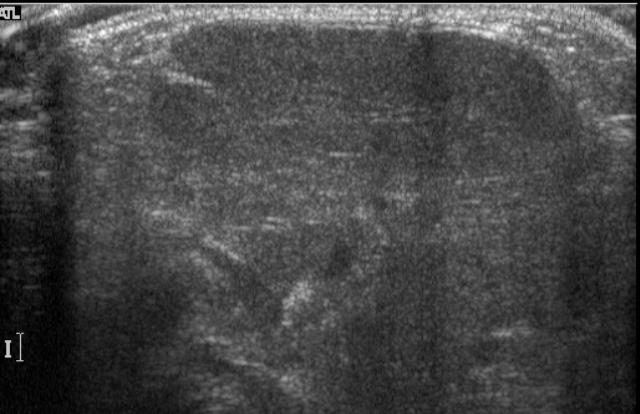
18 Fibromatosis colli Location: Echogenicity: sternocleidomastoid muscle right- or left-sided increased echogenicity with respect to normal muscle
19 Hemangioma Capillary or cavernous type Location in all layers and regions of the neck Echogenicity: echogenic or predominantly hypoechoic with echogenic septation (cavernous type) Color Doppler: highly and low vascularized mass MRI: hyperintens highly vascularized mass (MRA) with extension to other parts of neck

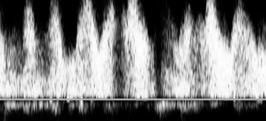


20 Hemangioma


21 Hamartomatous tumor of neck Location: variable Echogenicity: inhomogenous with calcification CT: calcificated tumor
22 Acquiered inflammatory masses 1. Unsupperative and supperative lymphadenitis 2. Specified lymphadenitis


23 Non-supperative lymphadenitis Location: Echogenicity: Color Doppler: jugodigastric, along internal carotid sheath hypoechoic, larger than 5 mm only hilar vascularization


24 Supperative lymphadenitis Location: Echogenicity: Color Doppler: jugodigastric, deep internal jugular well-defined mass with fluid-filled areas, irregular hypo- or anechoic often with hypervascular rim


25 Specified lymphadenitis Location: Echogenicity: deep lateral cervical nodes inhomogeneous hypoechoic, frequently with calcification TBC lymphadenitis


26 Specified lymphadenitis Location: Echogenicity: deep lateral cervical nodes inhomogeneous hypoechoic Sarcoidosis Chronic granulomatous disease


27 Atypical mycobacterial lymphadenitis Location: Echogenicity: deep lateral cervical nodes inhomogeneous hypoechoic, frequently with calcification
28 Non-inflammatory benign and malignant masses 1. Benign neck masses 2. Lymphoma, Hodgkin disease, etc 3. Neuroblastoma 4. Rhabdomyosarcoma


29 Lipoma Location: Echogenicity: MRI: anywhere in the body, especially subcutis inhomogenous hypo- or echogenic hyperintens on T1WI


30 Agressive fibromatosis Location: Echogenicity: MRI: around mandible inhomogenous, hypo- and hyper echogenic mass hypointens on T1, moderate hyperintens on T2


31 Neurofibromas or schwannomas Location: paraspinal Echogenicity: hypoechoic MRI: hyperintens on T2W1


32 Burkitt lymphoma Location: Echogenicity: lateral cervical chain mostly large in size, hypoechoic or heterogenous echopattern


33 Location: Echogenicity: CT: Hodgkin disease lateral cervical chain mostly large in size, hypoechoic or heterogenous echopattern hypodens masses in neck extending into mediastinum


34 Location: Echogenicity: MRI: Neuroblastoma paraspinal echogenic mostly with calcified notches hypointens on T1 and T2 MIBG


35 Location: Echogenicity: MRI: Rhabomyosarcoma mostly originating from parapharyngeal spaces hypoechoic hypointens on T1 and mildly hyperintens on T2
36 Conclusion Neck masses recognized in the first 3 months of age are mostly congenital. Acquired inflammatory masses such as lymphadenitis are observed in late infancy and early childhood. Lymphadenitis is rarely specific. Malignant lymphoma appears mostly in late childhood.
37 Conclusion Sonography is the first modality of choice in screening and follow up of neck masses. MRI should be performed in all benign and malignant neurogenic masses as well as other malignant soft tissue tumours and giant cystic masses. CT is a useful modality in assessment of cervicothoracic masses
38 Conclusion The use of other modalities depends on clinical and sonographic findings. Attention should be paid to the choice of modality in relation to radiation dose and its invasivity.
PEDIATRICS WK 3 HEAD AND NECK ALISON WALLACE MD, PHD
 PEDIATRICS WK 3 HEAD AND NECK ALISON WALLACE MD, PHD Topics 1. Cervical lymphadenopathy 2. Lymphatic malformation 3. Thyroglossal duct cysts 4. Branchial cleft cysts 5. Thyroid masses CASE 1 Case 1 A 2
"Mummy what's this on my neck? - A pictorial review of paediatric neck masses"
 "Mummy what's this on my neck? - A pictorial review of paediatric neck masses" Poster No.: C-0405 Congress: ECR 2014 Type: Educational Exhibit Authors: A. Farrugia, A. S. Gatt; Msida/MT Keywords: Education,
Evaluation of Neck Mass. Disclosure. Learning Objectives 3/24/2014. Karen T. Pitman MD, FACS Banner MDACC, Gilbert AZ. Nothing to disclose
 Evaluation of Neck Mass Karen T. Pitman MD, FACS Banner MDACC, Gilbert AZ Nothing to disclose Disclosure Learning Objectives 1. Describe a systematic method to evaluate a patient with a neck mass 2. Select
Contents. Basic Ultrasound Principles and Terminology. Ultrasound Nodule Characteristics
 Contents Basic Ultrasound Principles and Terminology Basic Ultrasound Principles... 1 Ultrasound System... 2 Linear Transducer for Superficial Images and Ultrasound-Guided FNA... 3 Scanning Planes... 4
Cervical Lymph Nodes
 Cervical Lymph Nodes Diana Gaitini, MD Unit of Ultrasound, Department of Medical Imaging Rambam Medical Center and Faculty of Medicine Technion, Israel Institute of Technology Haifa, Israel Learning Targets
Shadow because the air
 Thyroid Ultrasound Thyroid US examination needs: 1. high frequency transducer 2. extended patient's neck 3. check all the neck area because the swelling could be in areas other than the thyroid such as
Sonography of soft-tissue vascular lesions
 Sonography of soft-tissue vascular lesions Oscar M. Navarro Associate Professor, University of Toronto Dept. of Diagnostic Imaging, The Hospital for Sick Children Toronto, Canada Declaration of Disclosure
Pelvic tumor in childhood Classification, imaging approach and radiological findings
 Pelvic tumor in childhood Classification, imaging approach and radiological findings M. Mearadji International Foundation for Pediatric Imaging Aid Rotterdam, The Netherlands Solid pelvic masses in childhood
Dr Nick McIvor. Dr John Chaplin. Head & Neck Surgeon Auckland City Hospital Auckland. Auckland Head & Neck Surgeon Gillies Hospital Auckland
 Dr Nick McIvor Head & Neck Surgeon Auckland City Hospital Auckland Dr John Chaplin Auckland Head & Neck Surgeon Gillies Hospital Auckland 14:00-14:55 WS #148: Case Studies of Lumps in the Neck 15:05-16:00
Neck lumps in children
 Neck lumps in children Midline Lateral Midline neck lumps Thyroglossal cyst - 80% Dermoid cyst Submental lymph node Ectopic thyroid Some rare lesions Thyroglossal cyst Diagnosis: midline, usually overlying
The Reliability of Ultrasonography in Neck Masses Evaluation.
 IOSR Journal of Dental and Medical Sciences (IOSR-JDMS) e-issn: 2279-0853, p-issn: 2279-0861.Volume 16, Issue 10 Ver. XII (Oct. 2017), PP 33-40 www.iosrjournals.org The Reliability of Ultrasonography in
Evaluation of congenital masses of the neck in children: embryological, clinical and radiological review
 Evaluation of congenital masses of the neck in children: embryological, clinical and radiological review Poster No.: C-1731 Congress: ECR 2014 Type: Educational Exhibit Authors: N. C. S. D. Ferreira, A.
Chapter 13: Mass in the Neck. Raymond P. Wood II:
 Chapter 13: Mass in the Neck Raymond P. Wood II: In approaching the problem of a mass in the neck, one immediately encounters the fact that there are normally palpable masses in the neck (eg, almost all
Imaging Work-Up of a Neck Mass - Adults & Children
 Disclosures Imaging Work-Up of a Neck Mass - Adults & Children I have nothing to disclose Christine M Glastonbury MBBS Professor of Radiology & Biomedical Imaging Otolaryngology-Head & Neck Surgery and
Sonographic imaging of pediatric thyroid disorders in childhood. Experiences and report in 150 cases
 Sonographic imaging of pediatric thyroid disorders in childhood. Experiences and report in 150 cases M. Mearadji International Foundation for Pediatric Imaging Aid Sonographic technique. Use of high frequency
Congenital Neck Masses C. Stefan Kénel-Pierre, MD
 Congenital Neck Masses C. Stefan Kénel-Pierre, MD SUNY-LICH Medical Center Department of Surgery Case Presentation xx year old male presents with sudden onset left lower neck swelling x 1 week Denies pain,
Ultrasound of soft-tissue vascular anomalies
 Ultrasound of soft-tissue vascular anomalies Oscar M. Navarro Associate Professor, University of Toronto Dept. of Diagnostic Imaging, The Hospital for Sick Children Toronto, Canada Declaration of Disclosure
"The Space Between Us:" A Radiographic Review of Common and Uncommon Pathologic Findings within the Deep Spaces of the Neck
 "The Space Between Us:" A Radiographic Review of Common and Uncommon Pathologic Findings within the Deep Spaces of the Neck Poster No.: C-2457 Congress: ECR 2015 Type: Educational Exhibit Authors: A. K.
Head and Neck Image 頭頸部放射影像學
 Head and Neck Image 頭頸部放射影像學 陳家媛 台北醫學大學 - 市立萬芳醫院 cychen@wanfang.gov.tw Normal Suprahyoid neck: the old way Nasopharynx Oropharynx Oral cavity Staging of SCC Spaces of Suprahyoid Neck: a New Way Deep
MDCT and Ultrasound Evaluation of Primary Masses of Neck with Pathological Correlation
 IOSR Journal of Dental and Medical Sciences (IOSR-JDMS) e-issn: 2279-0853, p-issn: 2279-0861.Volume 15, Issue 9 Ver. IX (September). 2016), PP 63-71 www.iosrjournals.org MDCT and Ultrasound Evaluation
Thyroid and Parathyroid Ultrasound Protocol
 Thyroid and Parathyroid Ultrasound Protocol Reviewed By: Anna Ellermeier, MD Last Reviewed: December 2017 Contact: (866) 761-4200, Option 1 **NOTE for all examinations: 1. If documenting possible flow
Pediatric Ocular Sonography
 Pediatric Ocular Sonography Cicero J Torres A Silva, MD Associate Professor of Radiology 2016 SPR Pediatric Ultrasound Course Yale University School of Medicine None Disclosures Objectives of Presentation
Uncommon ultrasound findings in the cervical region
 Uncommon ultrasound findings in the cervical region Poster No.: C-1132 Congress: ECR 2014 Type: Educational Exhibit Authors: V. Donciu, S. M. Petrescu, E. NEGUT, C. ZAHARIA ; 1 1 1 2 1 2 Bucharest/RO,
Cystic Head and Neck Lesions
 Cystic Head and Neck Lesions Disclosures None Brad Wright, MD 19 March 2018 Key points Huge variety of cystic lesions in H&N May be cystic, necrotic, or solid but cystic-appearing Patient age, clinical
Ultrasound Interpretation of Non-Thyroid Neck Pathology
 Ultrasound Interpretation of Non-Thyroid Neck Pathology Kevin T. Brumund, M.D., F.A.C.S. Associate Professor of Surgery Head and Neck Surgery University of California, San Diego Health Sciences VA Medical
Deepak M. Sampathu MD, PhD Assistant Professor of Clinical Radiology University of Pennsylvania
 Deepak M. Sampathu MD, PhD Assistant Professor of Clinical Radiology University of Pennsylvania Objectives Recognize benign masses and masslike lesions of the neck and skull base Understand the imaging
Mediastinal Tumors: Imaging
 Mediastinal Tumors: Imaging References Imaging in Oncology, Husband and Reznek Computed Tomography and Magnetic Resonance of the thorax, Naidich, Zerhouni, Siegelman, Mediastinal compartments Anterior:
AACE/ACE Principles of Endocrine Neck Sonography Course
 AACE/ACE Principles of Endocrine Neck Sonography Course Primary objective of thyroid ultrasound: assess for malignant disease Nodular Disease Benign Malignant Goiter Iodine deficient Thyroiditis Organification
PAPILLARY THYROID CARCINOMA PRESENTING AS A LATERAL NECK MASS MASS. Dr. Pamela Hanson DO PGY3
 PAPILLARY THYROID CARCINOMA PRESENTING AS A LATERAL NECK MASS MASS Dr. Pamela Hanson DO PGY3 MK CASE PRESENTATION 28 yo Female presented to the ENT Clinic in October 2016, with the complaint of chronic
Preoperative Evaluation
 Preoperative Evaluation Lateral compartment lymph nodes are easier to detect and are amenable to FNA Central compartment lymph nodes are much more difficult to detect and FNA (Tg washout testing is compromised)
Salivary ultrasound. Dr T J Beale Royal National Throat Nose & Ear and UCLH Hospitals London UK
 Salivary ultrasound Dr T J Beale Royal National Throat Nose & Ear and UCLH Hospitals London UK Two main groups of patients with presenting symptoms of: Obstructive or chronic inflammatory symptoms (salivary
THYROID & PARATHYROID. By Prof. Saeed Abuel Makarem & Dr. Sanaa Al-Sharawy
 THYROID & PARATHYROID By Prof. Saeed Abuel Makarem & Dr. Sanaa Al-Sharawy 1 OBJECTIVES By the end of the lecture, the student should be able to: Describe the shape, position, relations and structure of
Adrenal masses in infancy and childhood: A clinical and radiological overview M. Mearadji
 Adrenal masses in infancy and childhood: A clinical and radiological overview M. Mearadji International Foundation for Pediatric Imaging Aid Introduction Neoplastic adrenal masses usually originate from
Major Salivary Glands: Sonographic Anatomy and Pathologic Conditions
 Major Salivary Glands: Sonographic Anatomy and Pathologic Conditions Poster No.: C-0452 Congress: ECR 2014 Type: Educational Exhibit Authors: E. Papadaki, I. Tritou, E. Sfakianaki ; Rethymno, CR/GR, 1
Branchial cleft cyst at an unusual location: a rare case with a brief review
 (2012) 41, 696 702 2012 The British Institute of Radiology http://dmfr.birjournals.org CASE REPORT Branchial cleft cyst at an unusual location: a rare case with a brief review AS Panchbhai*,1 and MS Choudhary
C. Douglas Phillips, MD FACR Director of Head and Neck Imaging Weill Cornell Medical Center NewYork Presbyterian Hospital
 C. Douglas Phillips, MD FACR Director of Head and Neck Imaging Weill Cornell Medical Center NewYork Presbyterian Hospital Objectives Review basics of head and neck imaging Discuss our spatial approach
Oropharynx and Oral Cavity
 455 17 Oropharynx and Oral Cavity Th. Vogl 17.1 Anatomy, Imaging Techniques and Imaging Findings 456 17.1.1 Normal Anatomy 456 17.1.2 Imaging Techniques 456 17.1.3 Imaging Findings 456 17.2 Congenital
Neck mass Evaluation & Management OTOLARYNGOLOGY, HEAD & NECK SURGICAL ONCOLOGY
 Neck mass Evaluation & Management MOHAMMED ALESSA MBBS,FRCSC ASSISTANT PROFESSOR CONSULTANT OTOLARYNGOLOGY, HEAD & NECK SURGICAL ONCOLOGY KSU, MEDICAL CITY & KKUH Objectives Obtain map overview in neck
Does MDCT Really Have a Role in the Evaluation of Neck Masses?
 Does MDCT Really Have a Role in the Evaluation of Neck Masses? 1 Dr Ravi N, 2 Dr Lakshmeesha M T, 3 Dr Manjappa B H, 3 Dr Naveen K G, 4 Dr.Ramesh V, 4 Dr.Nagaraj B R 1 Associate Professor, 2 Resident,
LUMPS AND BUMPS: EVALUATION AND MANAGEMENT OF SOFT TISSUE MASSES IN PEDIATRICS. By Elizabeth A. Paton, MSN, RN-BC, PPCNP-BC, FAEN
 LUMPS AND BUMPS: EVALUATION AND MANAGEMENT OF SOFT TISSUE MASSES IN PEDIATRICS By Elizabeth A. Paton, MSN, RN-BC, PPCNP-BC, FAEN I. Objectives II. By the end of this presentation, the learner will be able
Thyroglossal cyst our experience
 Volume 3 Issue 1 2013 ISSN: 2250-0359 Thyroglossal cyst our experience Balasubramanian Thiagarajan 1 Ulaganathan Venkatesan 2 Geetha Ramamoorthy 1 1 Stanley Medical College 2 Meenakshi Medical College
The Neck the lower margin of the mandible above the suprasternal notch and the upper border of the clavicle
 The Neck is the region of the body that lies between the lower margin of the mandible above and the suprasternal notch and the upper border of the clavicle below Nerves of the neck Cervical Plexus Is formed
Diagnostic imaging of lymphatic malformations
 Diagnostic imaging of lymphatic malformations Poster No.: C-1440 Congress: ECR 2016 Type: Educational Exhibit Authors: M. M. Coman 1, M. T. A. Buzan 2, S. Manole 3, S. M. Dudea 3 ; 1 2 3 Campia Turzii/RO,
Paraspinal Venous Malformation Joseph Junewick, MD FACR
 Paraspinal Venous Malformation Joseph Junewick, MD FACR 06/04/2010 History 2 year old with history of fall. Rule out spinal injury. Diagnosis Paraspinal Venous Malformation Additional Clinical CT of the
THYROID NODULES: THE ROLE OF ULTRASOUND
 THYROID NODULES: THE ROLE OF ULTRASOUND NOVEMBER 2017 DR. DEAN DURANT DEFINITION Thyroid nodule: Focal area within the thyroid gland with echogenicity different from surrounding parenchyma. THYROID NODULES
Thyroid in a Nutshell Dublin Catherine Kirkpatrick Consultant Sonographer ULHT
 Thyroid in a Nutshell Dublin 2017 Catherine Kirkpatrick Consultant Sonographer ULHT Acknowledgements Dr. Steve Colley Dr. Rhodri Evans Dr. Rhian Rhys Dr. Andrew McQueen Aims Anatomy & Physiology Incidence
Anterior triangle of neck
 Anterior triangle of neck Dept. of Anatomy Zhou Hong Ying Outline boundary and subdivisions of ant. triangle contents of the triangle Muscles: suprahyoid muscles, infrahyoid muscles Nerves: CNⅩ, CNⅪ, CNⅫ,
A CASE OF A Huge Submandibular Pleomorphic Adenoma
 ISPUB.COM The Internet Journal of Head and Neck Surgery Volume 4 Number 2 S VERMA Citation S VERMA.. The Internet Journal of Head and Neck Surgery. 2009 Volume 4 Number 2. Abstract Pleomorphic adenoma
Branchial Cleft and Pouch Anomalies
 Branchial Cleft and Pouch Anomalies Prof.Mohamed Hesham Alexandria Faculty of Medicine Alexandria, Egypt Emberyological Basis Branchial Clefts 1st 2nd Pinna EAC 3rd 4th 4th 6th Cervical sinus Branchial
Causes of pleural effusion and its imaging approach in pediatrics. M. Mearadji International Foundation for Pediatric Imaging Aid
 Causes of pleural effusion and its imaging approach in pediatrics M. Mearadji International Foundation for Pediatric Imaging Aid Pleural fluid A tiny amount of fluid in the pleural cavity is physiological.
Hemangioma of Tongue with Phlebolith: A Rare presentation
 Journal of Government Dental College and Hospital, October 2017, Vol.-04, Issue- 01, P. 20-25 Original article: Hemangioma of Tongue with Phlebolith: A Rare presentation 1 Dr. Jigna S Shah (MDS) 1, 2 Dr.
Sonographic Features of Thyroid Nodules & Guidelines for Management
 Sonographic Features of Thyroid Nodules & Guidelines for Management Mark A. Lupo, MD, FACE, ECNU Thyroid & Endocrine Center of Florida Assistant Clinical Professor of Medicine Florida State University,
Thyroid gland. importance. relations and connections. external laryngeal nerves. malformations.
 Thyroid gland 1. Recognize and understand the coverings of the thyroid gland and their clinical importance. 2. Recognize and understand the main parts of the thyroid gland and their locations, relations
CERVICAL LYMPH NODES
 CERVICAL LYMPH NODES (ANATOMY & EXAMINATION) Hemant (DTCD 1 st YEAR) 1. Lymphatic Tissues: A Type of connective tissue that contains large numbers of lymphocytes. 2. Lymphatic Vessels: Are Tubes that assist
AACE 2018 Advanced Endocrine Neck Ultrasound and UGFNA Course
 AACE 2018 Advanced Endocrine Neck Ultrasound and UGFNA Course Describe the sonographic appearance of diffuse thyroid diseases: autoimmune thyroid disease Review non thyroidal findings that can be encountered
Evaluation of Head and Neck Masses in Adults
 Evaluation of Head and Neck Masses in Adults Kristi Chang, MD Associate Professor Department of Otolaryngology-Head and Neck Surgery University of Iowa Hospitals and Clinics Annual Refresher Course for
Case Presentation. Jayer Chung, MD University of Colorado August 6, 2007
 Case Presentation Jayer Chung, MD University of Colorado August 6, 2007 J.E.S Cc: Neck mass HPI: The pt is a 68 y.o.. male with neck mass on the left neck. The patient began noticing the mass several months
Anatomy: head and Neck (6 questions) 1. Prevertebral Flexor Musculature (lying in front of the vertebrae) include all, EXCEPT: Longus Colli.
 1. Prevertebral Flexor Musculature (lying in front of the vertebrae) include all, EXCEPT: Longus Colli.") Anatomy: head and Neck (6 questions) 1. Prevertebral Flexor Musculature (lying in front of the vertebrae) include all, EXCEPT: Longus Colli. Rectus Capitis Anterior. Rectus Capitis Lateralis. Rectus Capitis
Anatomy: head and Neck (6 questions) 1. Prevertebral Flexor Musculature (lying in front of the vertebrae) include all, EXCEPT: Longus Colli. Rectus Capitis Anterior. Rectus Capitis Lateralis. Rectus Capitis
CTA/MRA of Pediatric Hepatic Masses Radiology-Pathology Correlation
 Acta Radiológica Portuguesa, Vol.XVIII, nº70, pág. 41-50, Abr.-Jun., 2006 CTA/MRA of Pediatric Hepatic Masses Radiology-Pathology Correlation Marilyn J. Siegel Mallinckrodt Institute of Radiology, Washington
Management of unknown primary with neck node metastasis: Current evidence
 Management of unknown primary with neck node metastasis: Current evidence Dr. Pooja Nandwani Patel Associate Professor Dept. of Radiation Oncology GCRI, Ahmedabad Introduction- Approach to Topic What is
Index. oralmaxsurgery.theclinics.com. Note: Page numbers of article titles are in boldface type.
 Index Note: Page numbers of article titles are in boldface type. A Adenomatoid odontogenic tumor, pediatric, 50 51 Ameloblastic carcinoma, pediatric, 17, 49 Ameloblastic fibro-odontoma, pediatric, 54 Ameloblastic
The importance of knowing the lymphatic spread patterns of head and neck cancer for accurate nodal staging on CT: A practical schematic guide
 The importance of knowing the lymphatic spread patterns of head and neck cancer for accurate nodal staging on CT: A practical schematic guide Poster No.: C-0483 Congress: ECR 2014 Type: Educational Exhibit
LYMPHATIC DRAINAGE IN THE HEAD & NECK
 LYMPHATIC DRAINAGE IN THE HEAD & NECK Like other parts of the body, the head and neck contains lymph nodes (commonly called glands). Which form part of the overall Lymphatic Drainage system of the body.
INFECTION. HIV Infection DWI
 HIV Infection INFECTION DWI Fig Axial CT and MRI images show multiple enlarged lymph nodes in the neck as well as in the parotid gland bilaterally. These nodes were suppurative with positive diffusion.
Imaging Technique. Ultrasound Imaging of the Salivary Glands. Parotid Gland. The Major Salivary Glands. Parotid Gland: Stenson s Duct.
 Ultrasound Imaging of the Salivary Glands Edward G. Grant MD Professor & Chairman, Dept of Radiology USC Keck School of Medicine edgrant@usc.edu edgrant@usc.edu Imaging Technique Linear array transducer
Parathyroid Imaging: Current Concepts. Maria Gule-Monroe, M.D. Nancy Perrier, M.D.
 Parathyroid Imaging: Current Concepts Maria Gule-Monroe, M.D. Nancy Perrier, M.D. Disclosures None Objectives Ultrasound characteristics of parathyroid adenomas vs. lymph nodes 4D-CT evaluation of hyperparathyroidism
HEAD & NECK SWELLINGS
 HEAD & NECK SWELLINGS EXCLUDING GOITRE FAISAL GHANI SIDDIQUI MBBS; FCPS; MCPS-HPE; PGDIP-BIOETHICS PROFESSOR OF SURGERY J I N N A H S I N D H M E D I C A L U N I V E R S I T Y MIDLINE SWELLINGS NECK SWELLINGS
Retroperitoneal Teratoma Heather Borders, MD
 Retroperitoneal Teratoma Heather Borders, MD 03/04/2012 History Newborn with congenitally diagnosed mass. No other clinical symptoms. Diagnosis Retroperitoneal Teratoma; Immature teratoma, grade 1, with
Neck-2. Dr. Heba Kalbouneh Associate Professor of Anatomy and Histology
 Neck-2 ` Dr. Heba Kalbouneh Associate Professor of Anatomy and Histology Triangles of the neck Side of the neck Midline Lower border of mandible Line between angle of mandible and mastoid Superior nuchal
Neoplasms that present as a swelling in the neck may be either
 Problems in otolaryngology Inflammatory swellings Viral and bacterial infection are frequent causes of swellings in the neck. Enlargement of the cervical lymph nodes is most likely but a dormant branchial
Role of imaging in RCC. Ultrasonography. Solid lesion. Cystic RCC. Solid RCC 31/08/60. From Diagnosis to Treatment: the Radiologist Perspective
 Role of imaging in RCC From Diagnosis to Treatment: the Radiologist Perspective Diagnosis Staging Follow up Imaging modalities Limitations and pitfalls Duangkamon Prapruttam, MD Department of Therapeutic
Thyroid and Parathyroid Glands
 Thyroid and Parathyroid Glands Please view our Editing File before studying this lecture to check for any changes. Color Code Important Doctors Notes Notes/ explanation Objectives: By the end of the lecture,
J of Evolution of Med and Dent Sci/ eissn , pissn / Vol. 4/ Issue 39/ May 14, 2015 Page 6787
 ROLE OF HIGH RESOLUTION SONOGRAPHY IN CHARACTERIZATION OF SOLID SALIVARY GLAND TUMORS Sheetal Singh 1, Amlendu Nagar 2, Pramod Sakhi 3, Sachin Kataria 4, Kumud Julka 5, Anup Gupta 6 HOW TO CITE THIS ARTICLE:
AACE/ACE Advanced Endocrine Neck Ultrasound Training Course 2016
 AACE/ACE Advanced Endocrine Neck Ultrasound Training Course 2016 This 9mm left inferior nodule should remind us all why we re here! There is no absolute number of images required for documentation
Advanced Anatomy of the Neck
 AACE 2018 Advanced Anatomy of the Neck Alex Tessnow, MD, MBA, FACE, ECNU University of Texas Southwestern Dallas, TX Content contributed by: H. Jack Baskin, Daniel Duick, Diana Dean, Robert A. Levine,
Case Presentation. x year old African American male seen in Pediatric Surgery Clinic. History: NKDA
 Case Presentation ALIREZA SADEGHI MD Kings County Hospital Center University Hospital of Brooklyn Downstate Medical Center Division of Pediatric Surgery July 7 th 2006 Case Presentation x year old African
ANTERIOR CERVICAL TRIANGLE (Fig. 2.1 )
") 2 Neck Anatomy ANTERIOR CERVICAL TRIANGLE (Fig. 2.1 ) The boundaries are: Lateral: sternocleidomastoid muscle Superior: inferior border of the mandible Medial: anterior midline of the neck This large triangle
2 Neck Anatomy ANTERIOR CERVICAL TRIANGLE (Fig. 2.1 ) The boundaries are: Lateral: sternocleidomastoid muscle Superior: inferior border of the mandible Medial: anterior midline of the neck This large triangle
Financial Disclosures. Do we really need fetal MRI? Fetal airway compromise. Facial lesions
 Financial Disclosures In-utero imaging, Decision making and surgical planning Fetal Imaging No relevant financial relationships with any commercial interests. Mariana L. Meyers, MD Director of Fetal Imaging
Imaging Of Cystic Paravertebral Masses:
 Imaging Of Cystic Paravertebral Masses: Differential Diagnosis and Key Discriminators John P. Lichtenberger III, MD, Maj, USAF, MC Brent McCarragher, MD, CPT, USA John R. Dryden, MD, LT, USN P. Gabriel
Plunging Ranula. 1. Department of Diagnostic Radiology, Yale University, New Haven, CT, USA
 Plunging Ranula Vivek Kalra 1, Khurram Mirza 2, Ajay Malhotra 1 1. Department of Diagnostic Radiology, Yale University, New Haven, CT, USA 2. Ross University School of Medicine, North Brunswick, New Jersey,
Ectopic cervical thymic cyst in a seven year old: A diagnostic challenge.
 ecommons@aku Section of Otolaryngology, Head & Neck Surgery Department of Surgery May 2018 Ectopic cervical thymic cyst in a seven year old: A diagnostic challenge. Haissan Iftikhar Aga Khan University,
Imaging of Pediatric Neck Masses
 ijhns IJHNS Review Article 10.5005/jp-journals-10001-1271 1 Payam Sajedi, 2 Narendra Shet Abstract Pediatric neck masses comprise a wide spectrum of entities, benign and malignant. The role of imaging
Pre-operative Ultrasound of Lymph Nodes in Thyroid Cancer
 Pre-operative Ultrasound of Lymph Nodes in Thyroid Cancer AACE - Advances in Medical and Surgical Management of Thyroid Cancer - 2018 Robert A. Levine, MD, FACE, ECNU Thyroid Center of New Hampshire Geisel
SKIN SONOGRAPHY IN CHILDREN. CRISTIAN J. GARCIA MD Santiago, Chile
 SKIN SONOGRAPHY IN CHILDREN CRISTIAN J. GARCIA MD Santiago, Chile I HAVE NO DISCLOSURES OBJECTIVES RELEVANCE OF SKIN LESIONS IN CHILDREN ROLEN OF THE RADIOLOGIST CLINICAL CORRELATION US TECHNIQUE NORMAL
Renal masses - the role of diagnostic imaging
 Renal masses - the role of diagnostic imaging Poster No.: C-2471 Congress: ECR 2015 Type: Educational Exhibit Authors: V. Rai#; Bjelovar/HR Keywords: Cysts, Cancer, Structured reporting, Ultrasound, MR,
Imaging features of orbital neoplasm developed in pediatrics
 Imaging features of orbital neoplasm developed in pediatrics Poster No.: C-1119 Congress: ECR 2015 Type: Educational Exhibit Authors: J. H. Yoo; Seoul/KR Keywords: Eyes, Head and neck, Paediatric, CT,
Ultrasound imaging of vascular anomalies: pearls and pitfalls
 Ultrasound imaging of vascular anomalies: pearls and pitfalls Oscar Navarro, MD Dept. of Medical Imaging, University of Toronto Dept. of Diagnostic Imaging, The Hospital for Sick Children Declaration of
Imaging in Pediatric Thyroid disorders: US and Radionuclide imaging. Deepa R Biyyam, MD Attending Pediatric Radiologist
 Imaging in Pediatric Thyroid disorders: US and Radionuclide imaging Deepa R Biyyam, MD Attending Pediatric Radiologist Imaging in Pediatric Thyroid disorders: Imaging modalities Outline ACR-SNM-SPR guidelines
Principal Site Investigator ENHANCE (Evaluation of Thyroid FNA Genomic Signature) study: An IRB approved study with funding to Rochester Regional
 study: An IRB approved study with funding to Rochester Regional") October 20 th 2018 Principal Site Investigator ENHANCE (Evaluation of Thyroid FNA Genomic Signature) study: An IRB approved study with funding to Rochester Regional Health from Veracyte Review ultrasound
October 20 th 2018 Principal Site Investigator ENHANCE (Evaluation of Thyroid FNA Genomic Signature) study: An IRB approved study with funding to Rochester Regional Health from Veracyte Review ultrasound
Tania Gallant MD, FRCPC Internal Medicine Update April
 Tania Gallant MD, FRCPC Internal Medicine Update April 28 2017 Disclosures Honoraria/Ad board: Sanofi-Aventis, Janssen, Merck Frosst, Eli-Lilly, Astra Zeneca, Boehringer-Ingelheim Objectives By the end
Case Report Cervical Thymic Cyst in an Adult
 Case Reports in Surgery, Article ID 801745, 4 pages http://dx.doi.org/10.1155/2014/801745 Case Report Cervical Thymic Cyst in an Adult Hassan A. Alzahrani, Javeria M. Iqbal, Amani K. Abu Shaheen, and Bandar
Evaluation of Thyroid Nodules
 Evaluation of Thyroid Nodules Stephan Kowalyk, MD January 25 28, 2018 1 Primary goal Exclude malignancy Incidental thyroid nodules If found on CT, MRI, PET scan, carotid Doppler ULTRASOUND!! January 25
Case 9087 Retropharyngeal nodular fasciitis
 Case 9087 Retropharyngeal nodular fasciitis Santiago I 1; Cavalheiro F 2; Noruégas MJ 3; Sanches MC3 1 Hospital Infante D. Pedro, Aveiro, Portugal 2 Hospitais da Universidade de Coimbra, Portugal 3 Hospital
Thyroid Nodules: US Risk Stratification. Alex Tessnow, MD, FACE, ECNU University of Texas Southwestern Associate Professor of Medicine Dallas, Texas
 Thyroid Nodules: US Risk Stratification Alex Tessnow, MD, FACE, ECNU University of Texas Southwestern Associate Professor of Medicine Dallas, Texas Which of the following is true? A. All echogenic foci
Ultrasound 5/1/2017. Ultrasound in the FNA Clinic. Uses of Ultrasound Outside of. Cardiology
 Ultrasound in the FNA Clinic Martha Bishop Pitman, M.D. Director, Cytopathology Massachusetts General Hospital Professor of Pathology Harvard Medical School Boston, MA Uses of Ultrasound Outside of Radiology
Soft Tissue Hemangiomas: High-resolution Grayscale and Color Doppler Ultrasonographic Features in 43 Patients
 O R I G I N A L A R T I C L E Soft Tissue Hemangiomas: High-resolution Grayscale and Color Doppler Ultrasonographic Features in 43 Patients Chia-Yu Keng 1,2, Howard Haw-Chang Lan 1,2,3 *, Clayton Chi-Chang
Radiology of hepatobiliary diseases
 GI cycle - Lecture 14 436 Teams Radiology of hepatobiliary diseases Objectives 1. To Interpret plan x-ray radiograph of abdomen with common pathologies. 2. To know the common pathologies presentation.