Moffitt Weekends in Pathology Head & Neck, and Endocrine Pathology Course Outline
|
|
|
- Ambrose Marshall
- 5 years ago
- Views:
Transcription
1 Moffitt Weekends in Pathology Head & Neck, and Endocrine Pathology Course Outline Squamous Cell Lesions Lecture BMW: 8:30-9:15 Break: 9:15-9:30 Case Review LK: 9:30-10:15 Case Review JHP: 10:30-11:15 Break: 11:15-11:30
2 Moffitt Weekends in Pathology Head & Neck, and Endocrine Pathology Course Outline Salivary Gland Lesions Lecture JHP: 11:30-12:15 Lunch: 12:15-1:15 Case Review LK: 1:15-2:00 Case Review BMW: 2:00-2:45 Break: 2:45-3:00
3 Moffitt Weekends in Pathology Head & Neck, and Endocrine Pathology Course Outline Thyroid Lesions Lecture LK: 3:00-3:30 Case Review JHP: 3:30-4:15 Case Review BMW: 4:15-5:00 Audience Response Q & A 5:00-5:15


4 Premalignant Squamous Cell Lesions and Variants of Squamous Cell Carcinoma Weekend in Pathology Moffitt Cancer Center November 4, 2017
5 Head & Neck Squamous Cell Lesions Outline Keratinizing Dysplasia Squamous cell carcinoma: Microinvasive and Invasive Select Variants

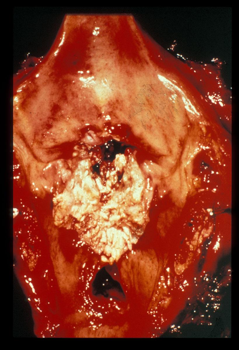
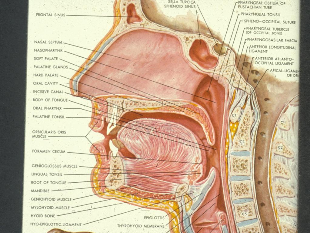
6 Vocal cord

7 Floor of Mouth

8 Buccal Mucosa
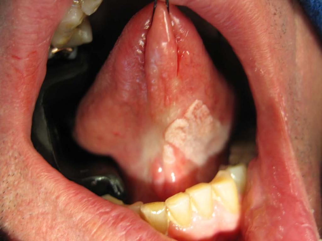
9 Oral Leukoplakia
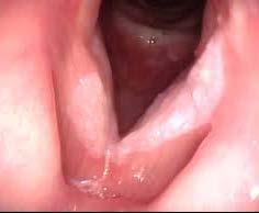
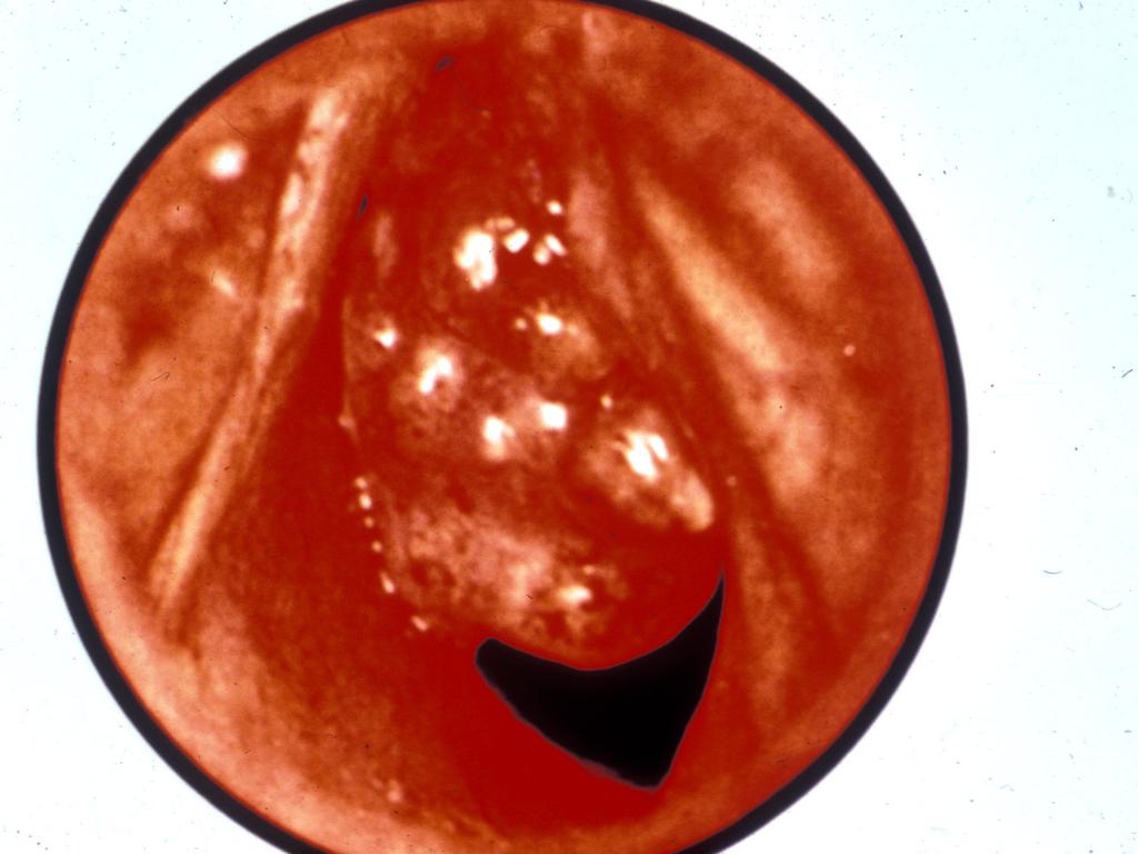
10 Vocal Cord Leukoplakia

11 Laryngeal Speckled Leukoplakia
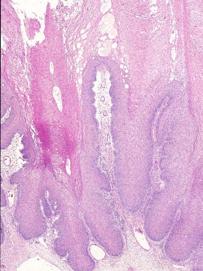
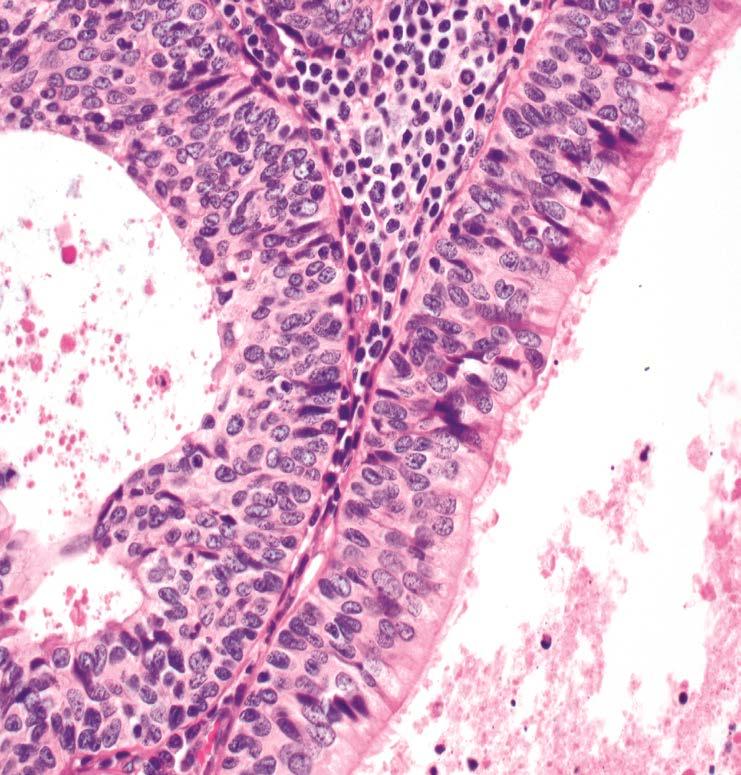
12 Epithelial Alterations Histopathology (Hyper)keratosis Hyperplasia Dysplasia: Spectrum of architectural and cytological epithelial changes caused by a gradual accumulation of genetic changes with an increased likelihood of progression to squamous cell carcinoma

13 Criteria for Dysplasia 2017 WHO Blue Book
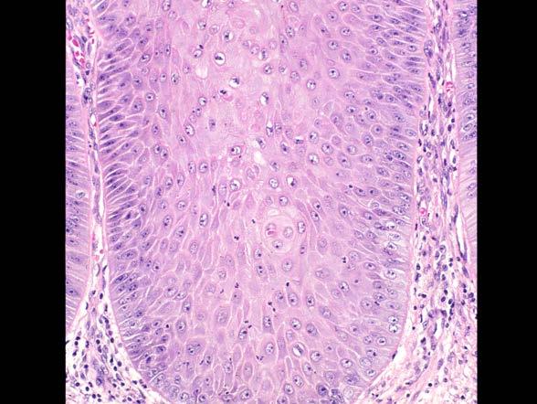
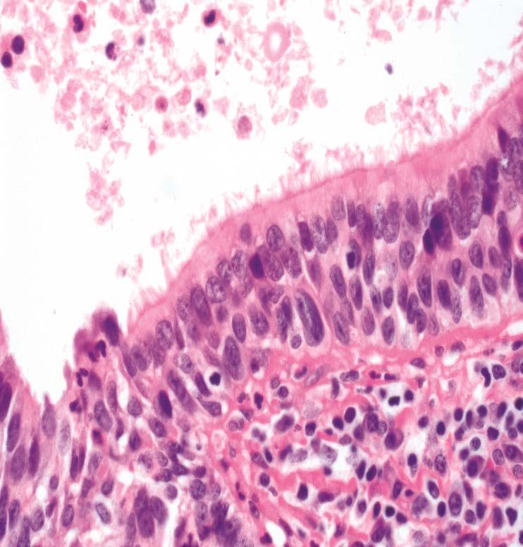
14 Dyskeratosis Keratin not on the surface Individual cell keratinization Keratin pearl(s) in the middle or lower half of the epithelium Pink or glassy cytoplasm Paradoxical maturation
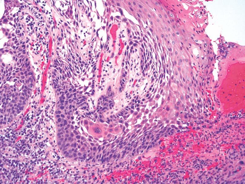
15 Dyskeratosis
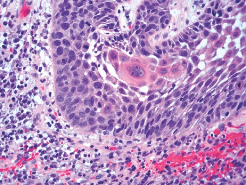
16 Dyskeratosis
17 Paradoxical Maturation

18 Keratosis without Dysplasia

19 Keratosis without Dysplasia

20 (Papillary or verrucoid) Keratosis without Dysplasia
21 Upper Aerodigestive Tract Epithelial Dysplasia Classic or Non-Keratinizing: Mild dysplasia Moderate dysplasia Severe dysplasia = Carcinoma in situ

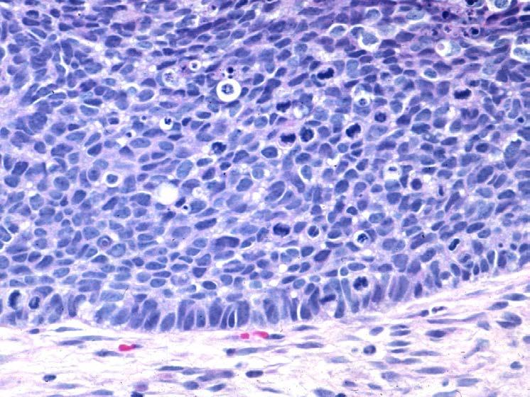
22 Carcinoma In Situ
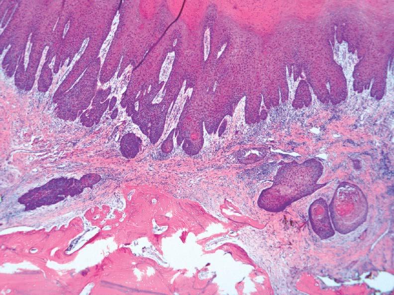
23 Upper Aerodigestive Tract Epithelial Dysplasia Keratinizing >>>> Nonkeratinizing: Mild dysplasia Moderate dysplasia Severe dysplasia

24 Keratinizing Mild Dysplasia

25 Keratinizing Mild Dysplasia

26 Keratinizing Moderate Dysplasia

27 Moderate? Severe? CIS?

28 Moderate? Severe? CIS?

29 Moderate? Severe? CIS?

30 Drop Off Carcinoma

31 Drop Off Carcinoma
32 Carcinoma In Situ (CIS) In the absence of full thickness intraepithelial dysplasia is the use of CIS justified? Does keratinizing severe dysplasia = CIS? Is it important to separate moderate and severe dysplasia/cis?
33 Upper Aerodigestive Tract Keratinizing Dysplasia Goal of any grading system is: Reproducible and Applicable Convey to the clinician the potential risk for progression of disease
34 Upper Aerodigestive Tract Grading Keratinizing Dysplasia Imprecise and subjective Preferred grading based on degree and extent of cellular and maturation alterations mild dysplasia moderate dysplasia severe dysplasia
35
36
37
38 Grading Keratinizing Dysplasia No statistical difference in progression to invasive SCC between keratinizing moderate dysplasia and keratinizing severe dysplasia/cis Justification to 2-Tier grading scheme: Low-grade Dysplasia = Mild dysplasia High-grade Dysplasia = Moderate Dysplasia, Severe Dysplasia, CIS Better reproducibility

39 Binary Grading Laryngeal Dysplasia 2017 WHO Blue Book

40 Binary Grading Oral Dysplasia 2017 WHO Blue Book
41 Keratinizing Dysplasia Etiology Tobacco (smoking, chewing) Alcohol Areca nut, with or without tobacco, causes oral submucous fibrosis with a relatively high frequency of oral dysplasia High risk human papillomavirus? Generally not considered a risk factor
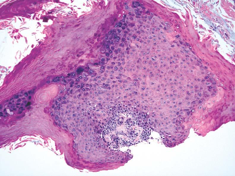
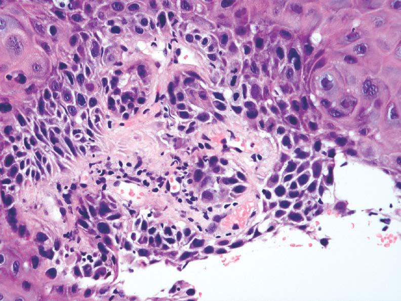
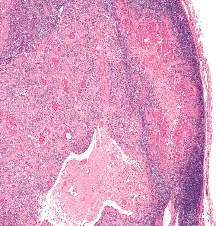
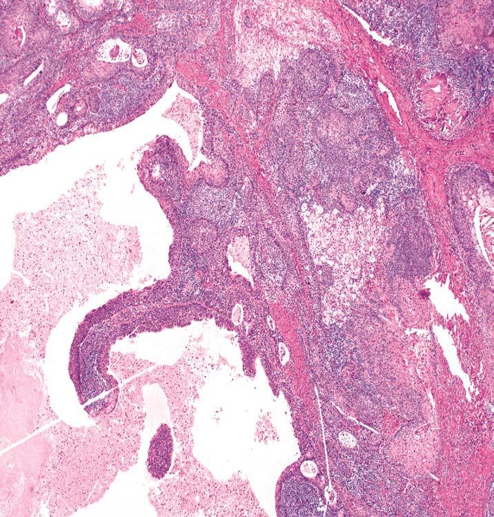
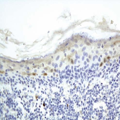
42 Oral Dysplasia and High Risk Human Papillomavirus (HR-HPV) Presence of HR-HPV infection has been convincingly demonstrated in some oral keratinizing dysplasias*: Majority clinically oral leukoplakias Most adult men; Tongue > FOM >> other sites Karyorrhexis and apoptosis with brightly eosinophilic apoptotic cells throughout the thickness of the epithelium, surrounded by keratinocytes exhibiting conventional dysplastic changes Positive for p16 and high-risk HPV subtypes *Woo SB, et al. Modern Pathol 2013;26:
43 Oral Dysplasia and HR-HPV HR-HPV associated with a subset of severe epithelial dysplasia or carcinoma in situ characterized by: Most adult men; ventral tongue or FOM Diffuse loss of squamous differentiation & high proliferation index throughout basal and suprabasal epithelial layers Identified by p16 IHC staining followed by ISH with probes for HPV DNA McCord C, et al. Oral Surg Oral Med Oral Pathol Oral Radiol 2013;115:541-9

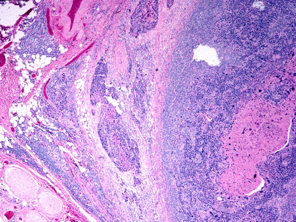
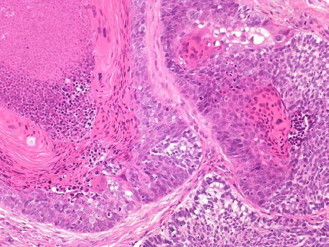
44 HR-HPV FOM Dysplasia

45 HR-HPV FOM Dysplasia

46 HR-HPV FOM Dysplasia p16
47 Keratinizing Dysplasia IHC Staining p16, p53 and Ki67 (MIB1): p16 of limited diagnostic utility in keratinizing dysplasias of the UADT p53: increase expression Ki67: increase intraepithelial proliferation rate through all epithelial layers Overall of limited utility

48 High-Grade Keratinizing Dysplasia Ki67 Ki67
49 Squamous Cell Carcinoma Early or Microinvasive Carcinoma Neoplastic cells penetrated basement membrane with invasion into submucosa Develops as a continuum from keratinizing high-grade dysplasia Classically defined CIS is not a prerequisite to the development of invasive carcinoma
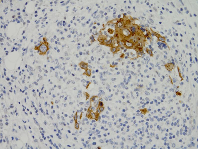
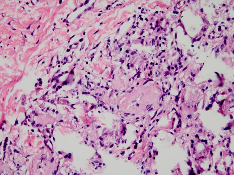
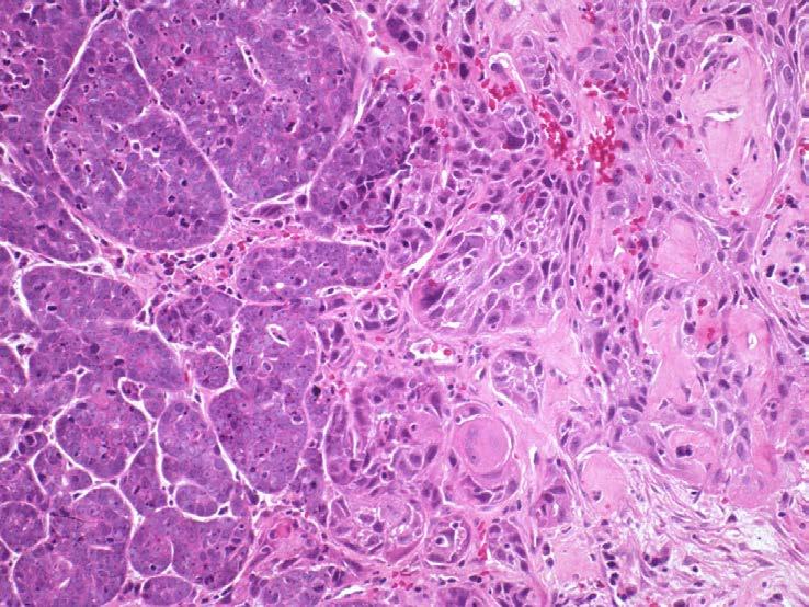
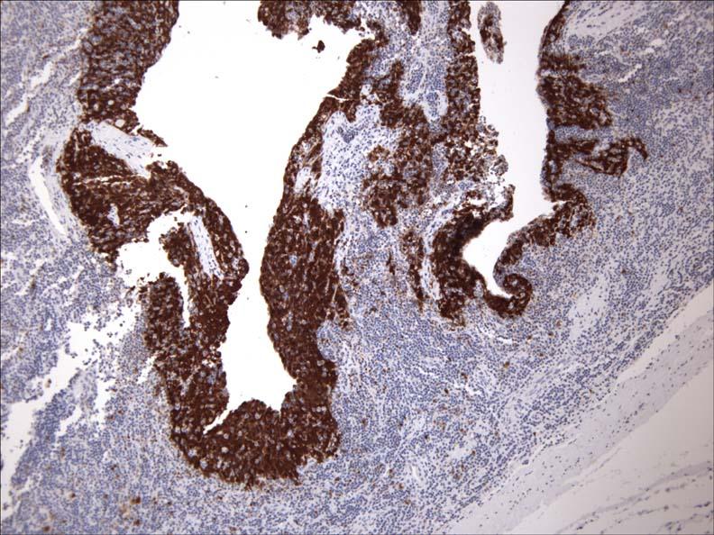
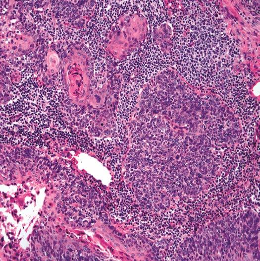
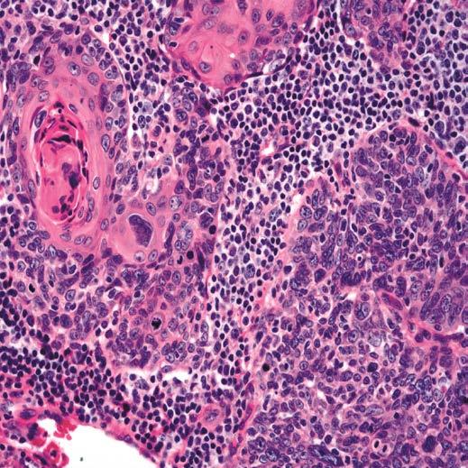
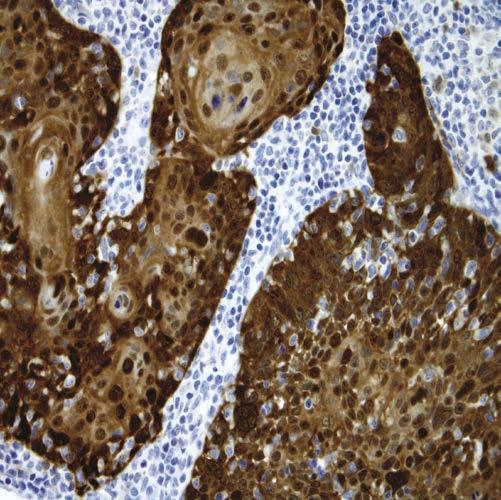
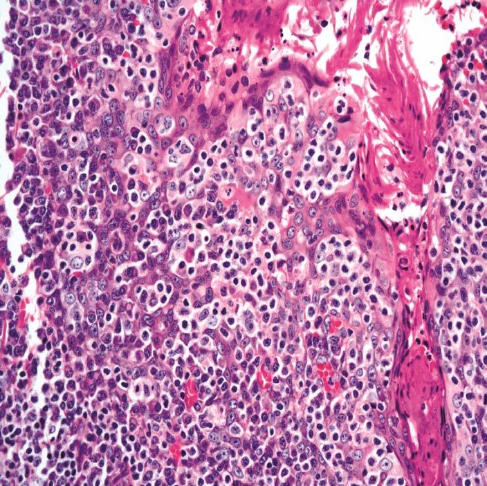
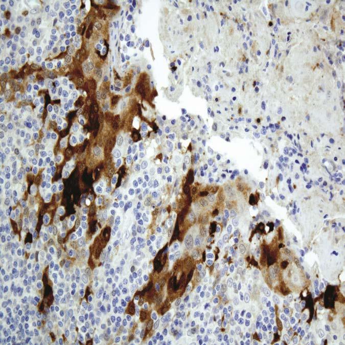
50 Squamous Cell Carcinoma Microinvasive Carcinoma (MIC) No uniformity in defining MIC: small number of cells below BM invasion through the BM invasion through the BM limited to 1-2mm of BM without angioinvasion invasion no more than 0.5mm from epithelial BM with no angioinvasion
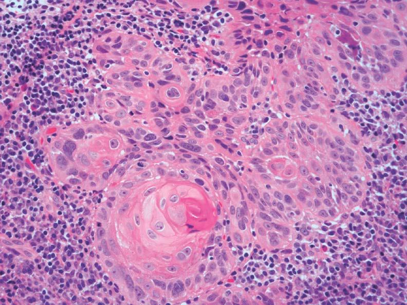
51 Invasive SCC Diagnostic Features Irregular shaped nests within the submucosa with associated dysplastic changes: Hyperchromasia; N:C; dyskeratosis; mitotic activity including atypical mitoses Desmoplasia Invasion: Lymph-vascular invasion; perineural invasion; invasion of soft tissues and/or bone Keratin granuloma formation

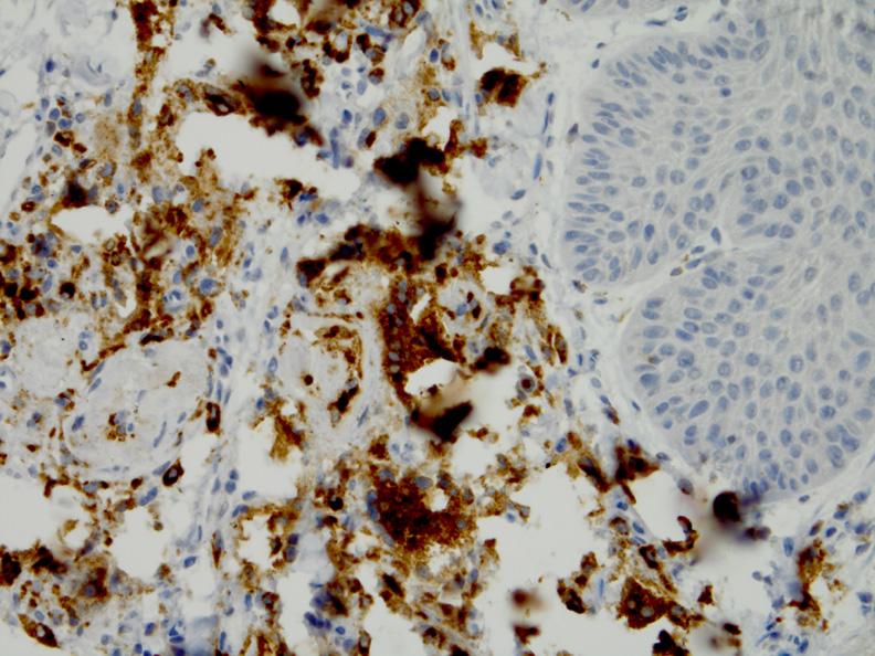
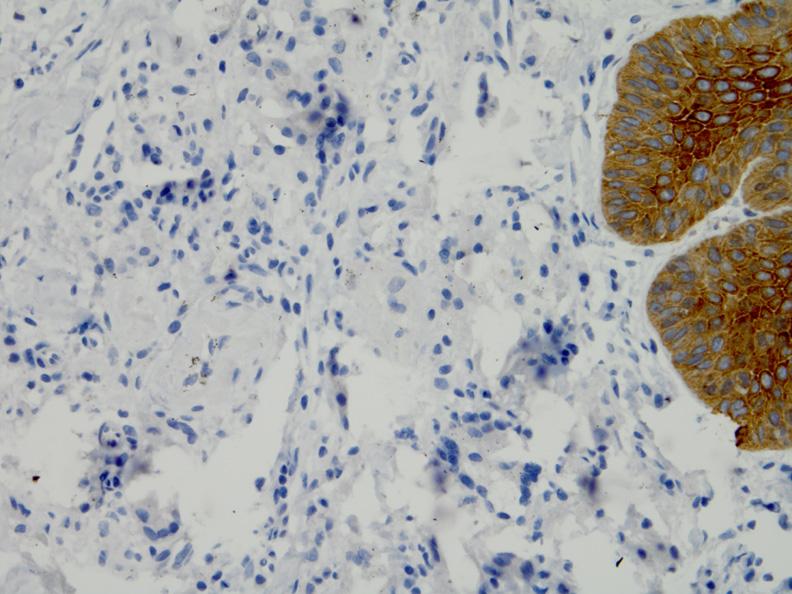
52 Invasive SCC

53 Invasive SCC

54 (Micro)Invasive SCC?
55 (Micro)Invasive SCC?
56 (Micro)Invasive SCC?

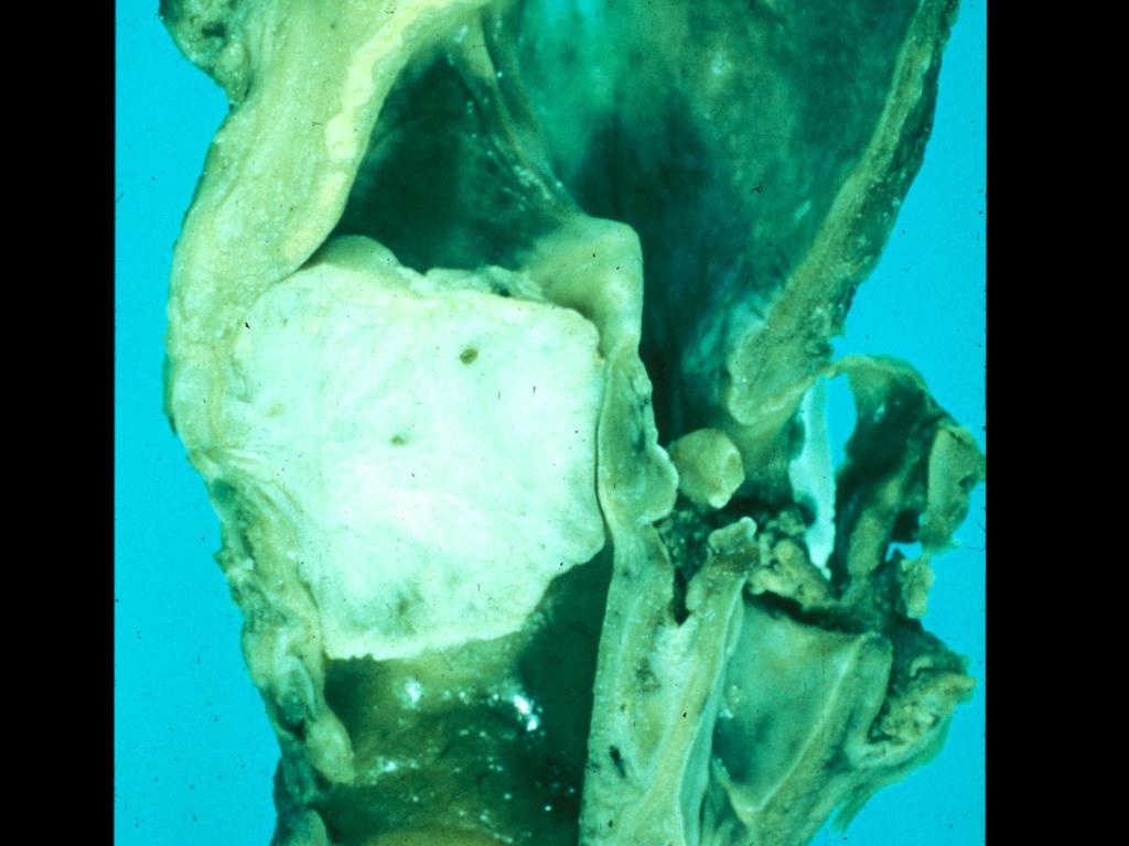
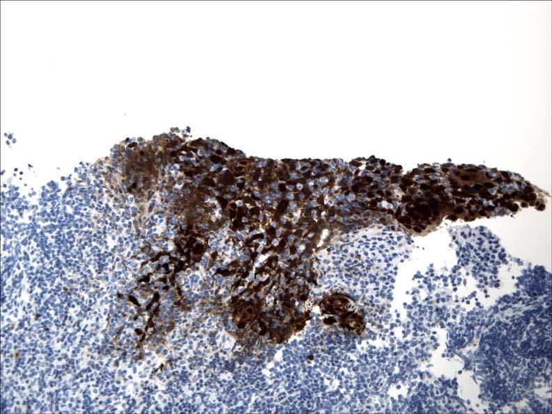
57 Keratin Granuloma AE1/AE3
58 Keratin Granuloma

59 CD68 AE1/AE3
60 HNSCC Factors Associated with Prognosis Adequacy of resection (surgical margins) Pattern of invasion: cohesive v dyscohesive Tumor size, thickness, location LVI, neurotropism and soft tissue invasion Regional metastasis - Extranodal Extension Distant metastasis Angiogenesis; Host immune response Second malignancy
61 Extranodal Extension
62 Variants of Squamous Cell Carcinoma of the Upper Aerodigestive Tract
63 Squamous Cell Carcinoma Variants Verrucous Carcinoma Spindle Cell Squamous Carcinoma Basaloid Squamous Cell Carcinoma Viral-Associated Carcinomas (HPV; EBV) Papillary (Exophytic) SCC Adenoid SCC (angiosarcoma-like or acantholytic) Adenosquamous Carcinoma Lymphoepithelial-like Carcinoma Other variants
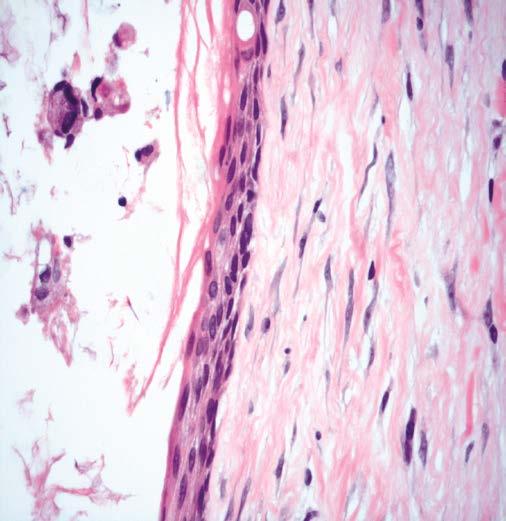
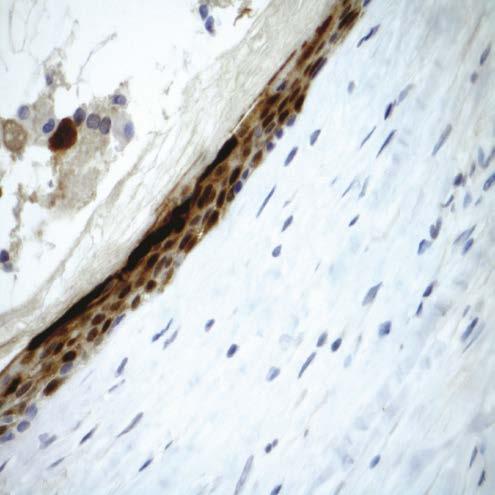
64 Verrucous Carcinoma (VC) Highly differentiated variant of squamous cell carcinoma with locally destructive but not metastatic capabilities
65 Verrucous Carcinoma Clinical Features M > F; generally occurs in older age groups (6 th 7 th decades of life) Sites: oral cavity (4%) > larynx (1-3%) > other (sinonasal tract; nasopharynx) Symptoms vary according to site
66 Verrucous Carcinoma Etiology Tobacco (smoking, chewing) use HPV may play an active role in the multistep progression to cancer by binding (via protein products) to the RB gene product removing regulatory block in the cell cycle (Science 1989;243:934-7 Recent studies using highly sensitive and specific molecular methods suggest that VC is not associated with human papillomavirus infection
67
68
69
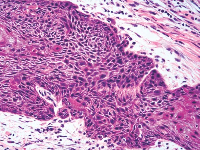
70 Hybrid Carcinoma Tumor showing mixed histology including verrucous carcinoma and conventional SCC Oral cavity > larynx >>> other sites Biologic risk that of conventional SCC potential for metastasis Treatment that of conventional SCC


71 Hybrid Carcinoma
72 Verrucous Carcinoma Biopsy Diagnosis Biopsy diagnosis of VC extremely difficult Adequate material is critical to interpretation and should include ample epithelial-stromal interface: Pathologists should not over interpret a verrucoid lesion as a carcinoma without adequate tissue Diagnosis of VC at initial presentation and biopsy is challenging given overall bland cytomorphology and shared features with reactive verrucoid lesions
73 Verrucous Carcinoma Biopsy Diagnosis Well-differentiated verrucoid squamous epithelial proliferation, NOS complete excision & follow-up Recurrence of tumor at a future time may be the most important clue/evidence to diagnosis of VC
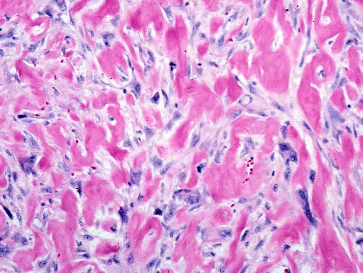
74 Verrucous Carcinoma Differential Diagnosis Conventional squamous cell carcinoma Reactive verrucoid hyperplasia Proliferative verrucous leukoplakia (PVL) Papilloma

75 Proliferative Verrucous Leukoplaia
76 Verrucous Carcinoma Treatment and Prognosis Surgery is the treatment of choice Radiotherapy used in select settings Excellent prognosis: for laryngeal VC: 5-yr survival rates of 86-95% Local recurrence but no metastases may cause extensive destruction if left untreated Does not metastasize Hybrid carcinoma has potential for metastasis and should be treated as conventional SCC
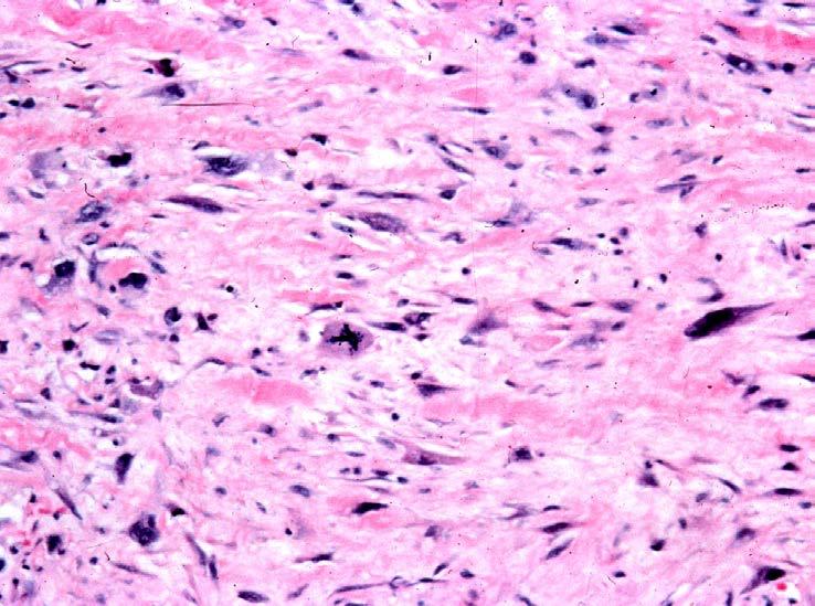
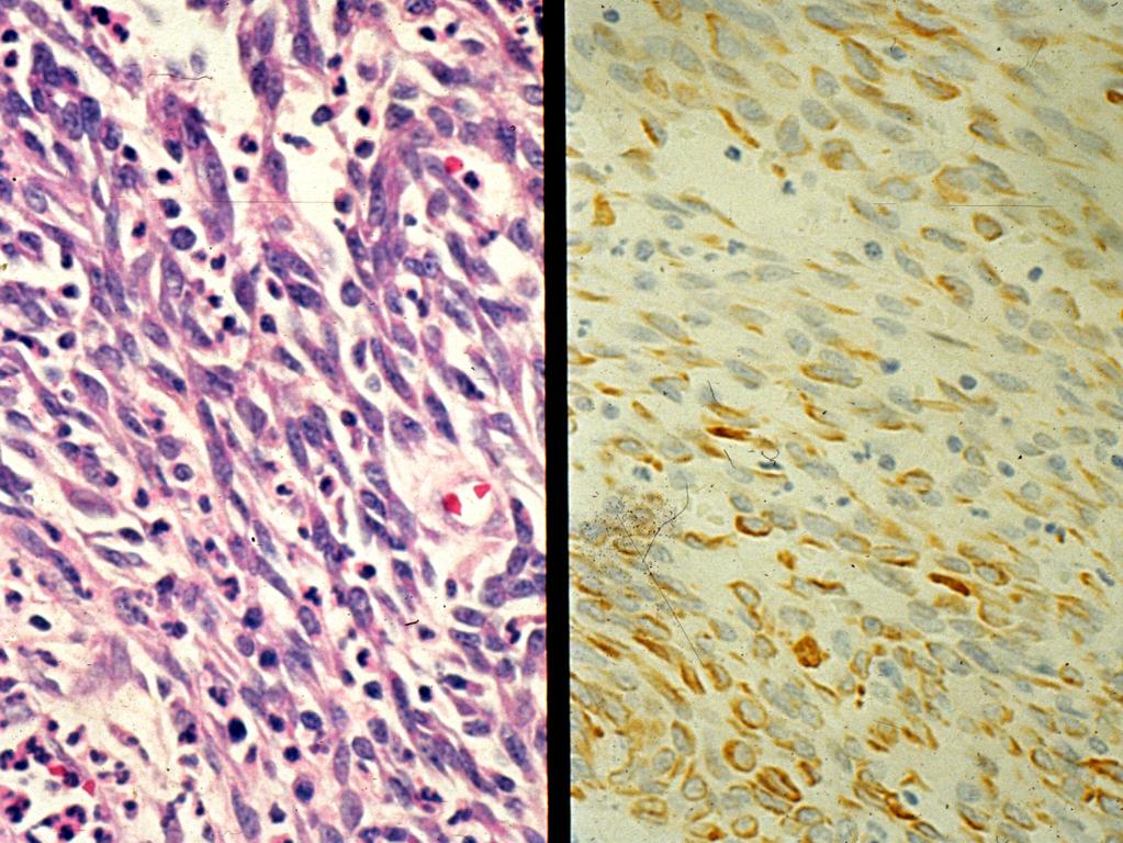
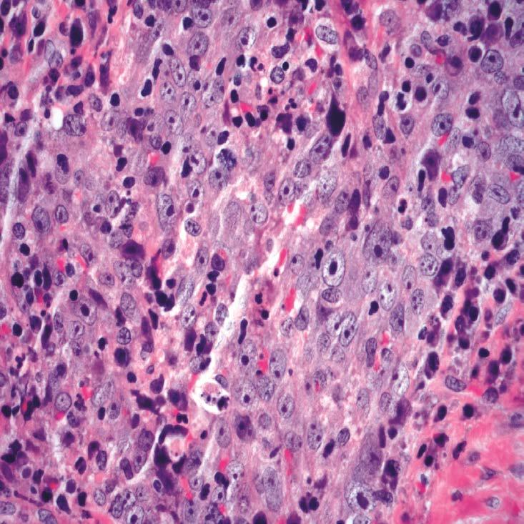
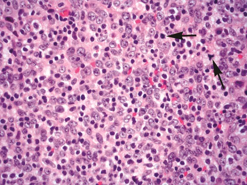
77 Spindle Cell Squamous Carcinoma (SCSC) Variant of SCC characterized by prominent or even exclusive malignant spindle-shaped cell and/or pleomorphic cells with or without identifiable conventional squamous cell carcinoma component (intraepithelial dysplasia and/or invasive differentiated SCC) Synonym: Sarcomatoid carcinoma
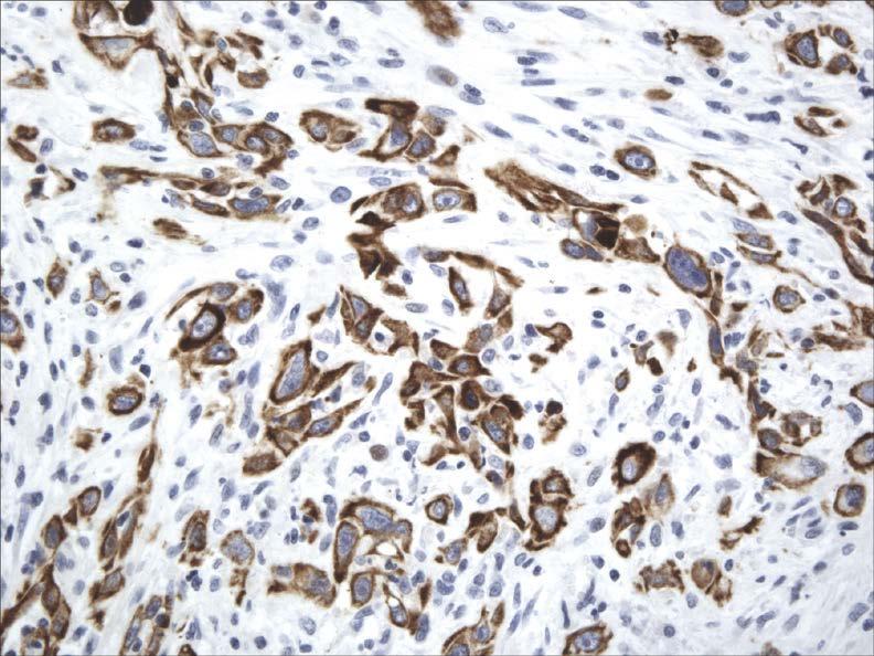
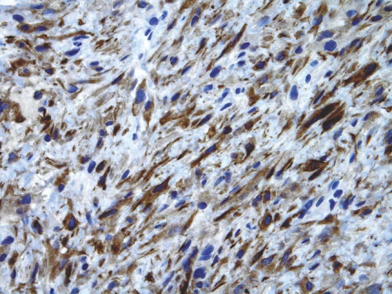
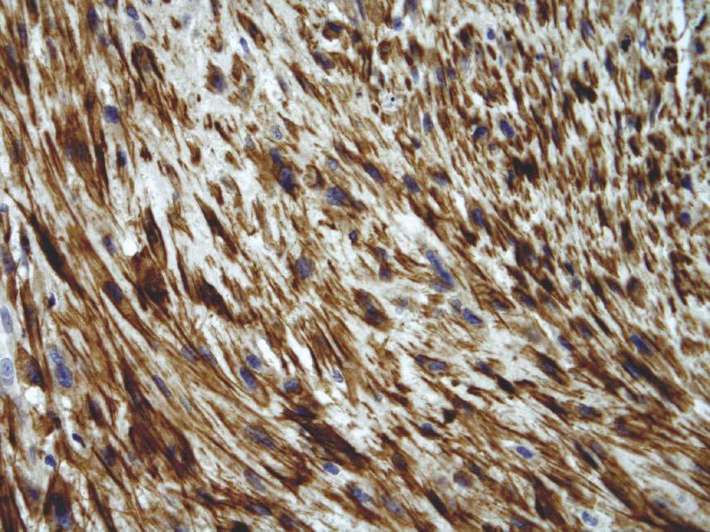
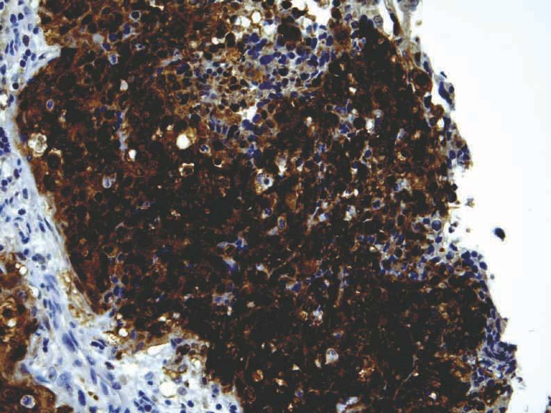
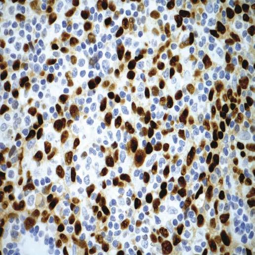
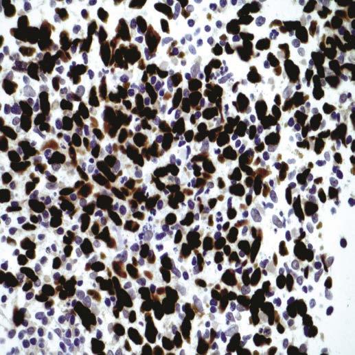
78 SCSC Clinical Features Uncommon tumor type M >> F; primarily occurs in older age groups (6 th 8 th decades) Sites of occurrence: Larynx (TVC) > oral cavity > cutaneous > tonsil > pharynx, other Symptoms vary according to site Linked to tobacco and alcohol use/abuse No specific correlation with HPV
79
80


81 Invasive differentiated SCC and malignant spindle-shaped cells = SCSC

82 Spindle cells Epithelioid cells

83 Granulation tissuelike appearance
84 Collagenized (hypocellular) SCSC
85 Spindle Cell Squamous Carcinoma IHC Staining Cytokeratins (AE1/AE3, CAM5.2, CK5/6, OSCAR) p63, p40 Vimentin Mesenchymal markers (actins, desmin)


86 AE1/AE3 CAM5.2 CK5/6 OSCAR
87 p63
88 Spindle Cell Squamous Carcinoma Keratin Expression 71 of 122 cases (58%) expressed keratin Lewis J, et al. Hum Pathol 1997;28:664-73
89
90 VIM
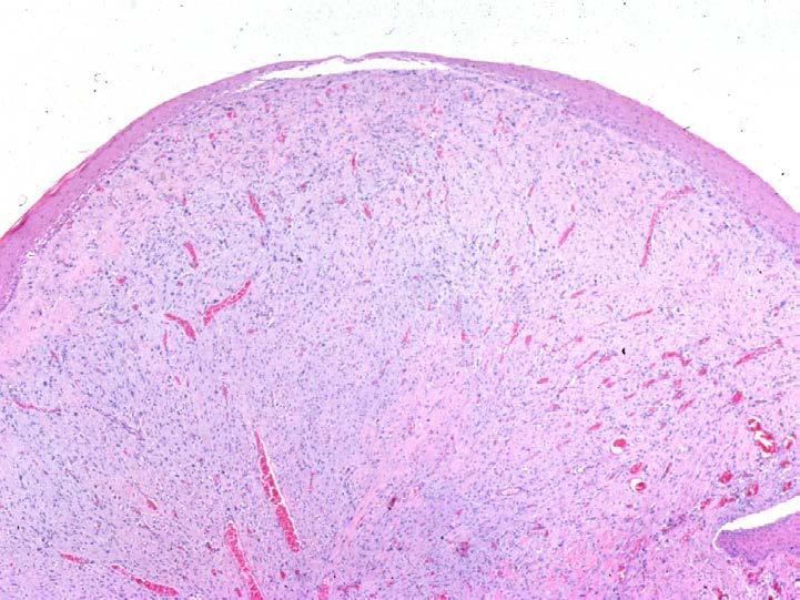
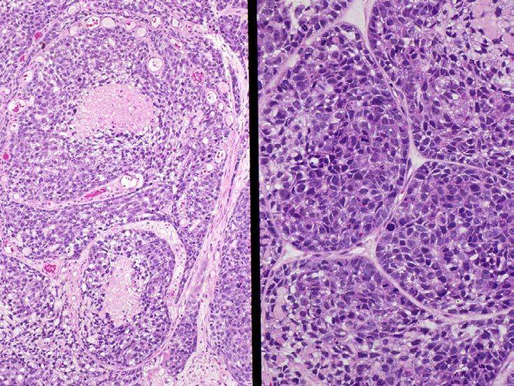
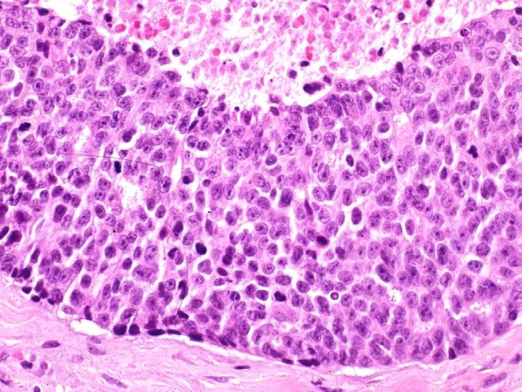
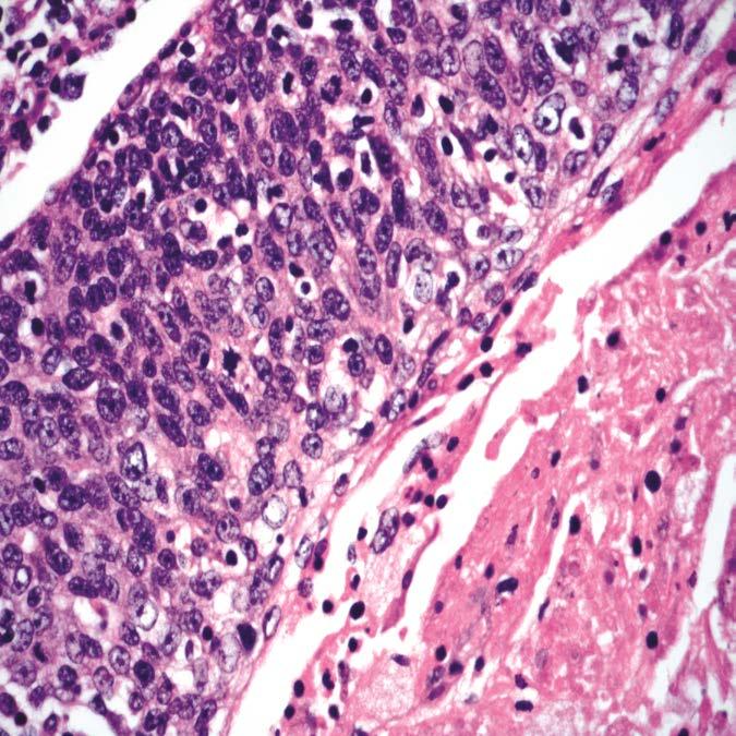
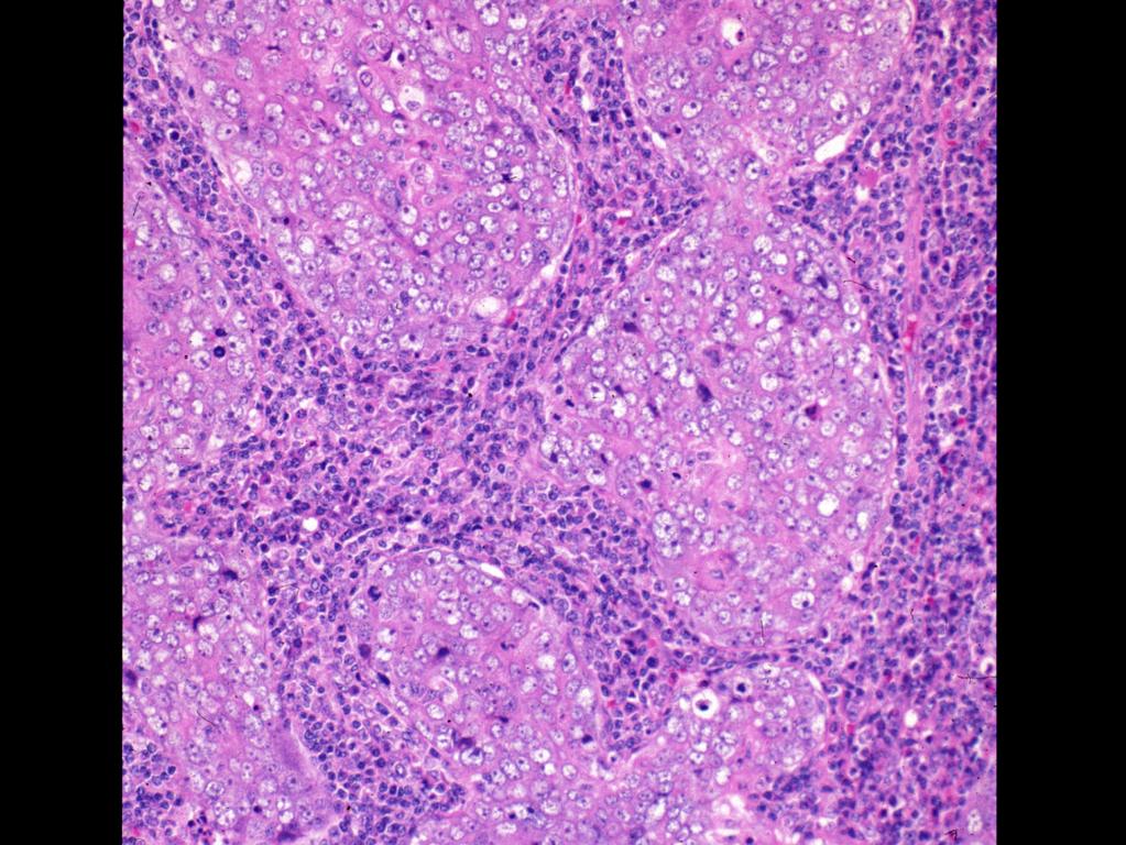
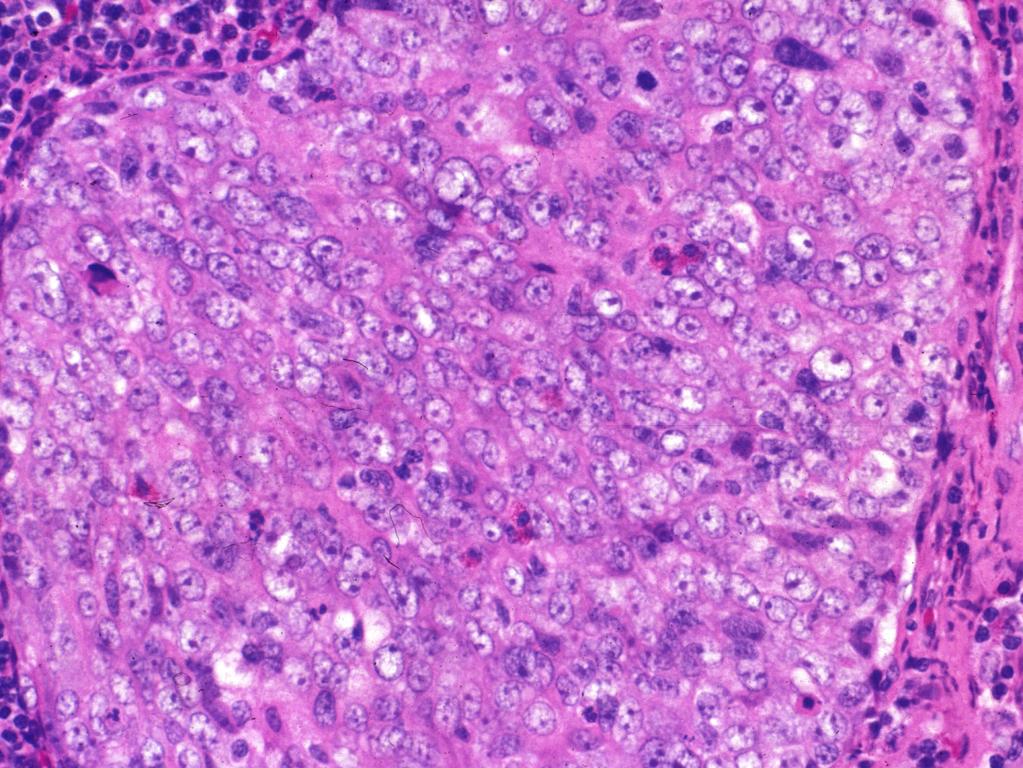
91 SMA
92 MSA
93 DES
94 Spindle Cell Squamous Carcinoma Epithelial Differentiation Identical p53 expression patterns in epithelial and spindle cell components support concept that phenotypically divergent cell populations share similar (epithelial) developmental pathway Ansari-Lari MA, et al. Am J Surg Pathol 2002;26:
95 Spindle Cell Squamous Carcinoma Sarcomas: Differential Diagnosis heterologous elements may be present in SCSC (e.g., rhabdomyosarcoma, osteosarcoma, chondrosarcoma) Inflammatory myofibroblastic tumor Reactive processes: myofibroblastic-based Inflammatory (e.g., contact ulcers) post-radiation changes
96 Spindle Cell Squamous Carcinoma Association with HPV Majority of SCSC not related to HPV Rare p16-positive oropharyngeal SCSC harboring HPV identified: Watson RF, Chernock RD, Wang X, Liu W, Ma XJ, Luo Y, Wang H, El-Mofty SK, Lewis JS Jr. Head Neck Pathol 2013;7:
97 Spindle Cell Squamous Carcinoma Treatment and Prognosis Surgical excision is the treatment of choice Adjunctive therapeutic modalities of questionable utility Prognosis dependent on clinical stage but overall prognosis is considered to be poor Metastasis to regional lymph nodes and to the lungs No known ameliorating effect associated with HPV
98 Basaloid Squamous Cell Carcinoma (BSCC) Biologically aggressive histologically high-grade variant of conventional squamous cell carcinoma characterized by invasive growth and predominantly composed of basaloid (pleomorphic) cell population and often limited evidence of squamous cell component
99 Basaloid Squamous Cell Carcinoma Clinical Features M > F; primarily occurs in older age groups (6 th 7 th decades) Sites of predilection: supraglottic larynx hypopharynx (piriform sinus) oropharynx: base of tongue and tonsil Symptoms vary according to site: at presentation tendency to be multifocal, deeply invasive and/or metastatic
100 Basaloid Squamous Cell Carcinoma Etiology Strongly related to alcohol and tobacco Non-oropharyngeal BSCC: Transcriptionally-active high risk human papillomavirus (HPV) is consistently absent in BSCC arising outside the oropharynx

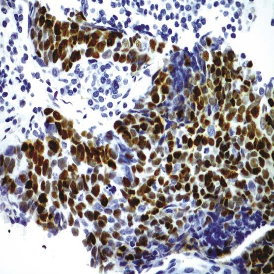
101
102

103 Lobular growth, comedonecrosis Reduplicated basement membrane-like material

104 BSCC - In Situ Component

105 BSCC with squamous differentiation

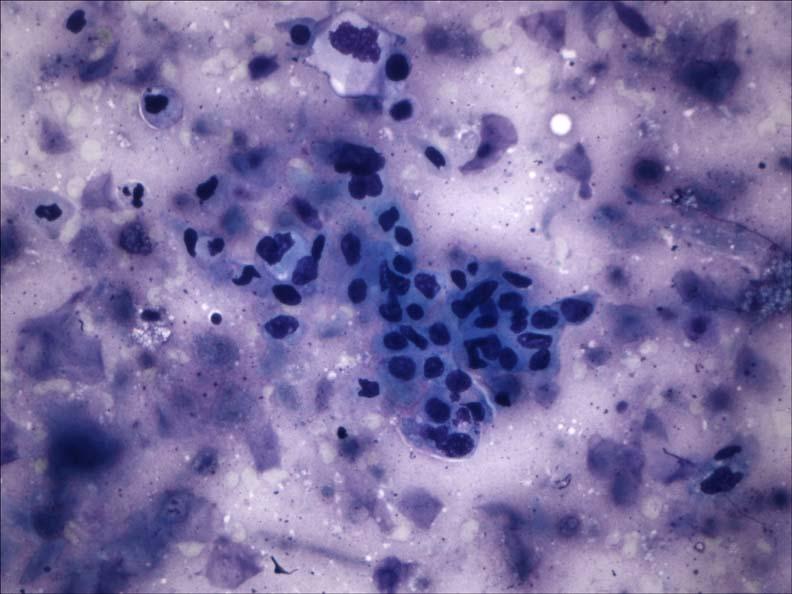
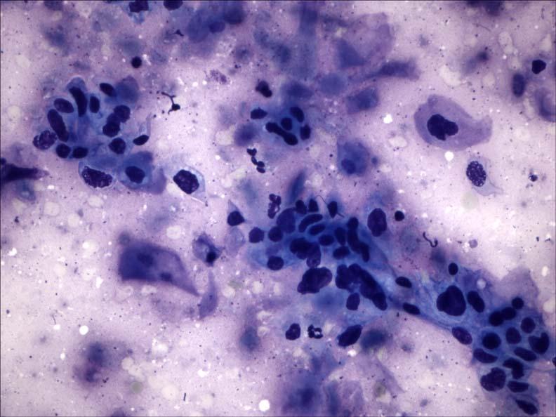
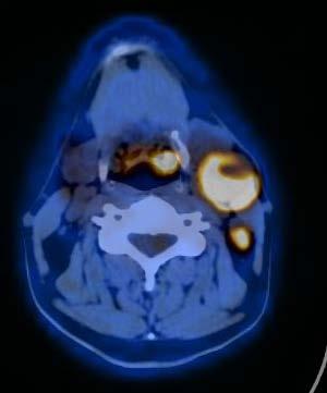
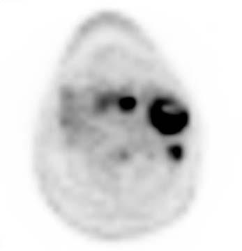
106 Rosettes and Nuclear Palisading
107 Perineural invasion
108 Basaloid Squamous Cell Carcinoma IHC Findings IHC: Cytokeratins p63/p40 (diffusely positive) Variable reactivity for S100 protein, NSE Mesenchymal: Vimentin, SMA Negative for neuroendocrine, melanocytic and lymphoid markers p16: Most non-oropharyngeal HPV-negative Most oropharyngeal HPV-positive
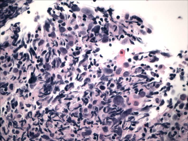
109 Basaloid Squamous Cell Carcinoma Differential Diagnosis Adenoid cystic carcinoma Small cell (neuroendocrine) carcinoma Conventional squamous cell carcinoma Adenosquamous carcinoma Spindle cell squamous carcinoma Others
110 Basaloid Squamous Cell Carcinoma Treatment and Prognosis Aggressive management: Complete surgical resection Radiotherapy and chemotherapy HPV-negative: dismal prognosis Active smokers and those with nodal metastases at presentation have worse prognosis Lymphatic and hematogenous spread: Regional lymph nodes (50-70%) Lung, bone, skin and brain
111 Basaloid Squamous Cell Carcinoma HPV-positive: Better overall prognosis than histologically similar non-hpv associated head and neck BSCC (Am J Surg Pathol 2008;32: ) Any tumor appearing to arise in the larynx/hypopharynx but that involves the oropharynx should be tested for HPV (p16)
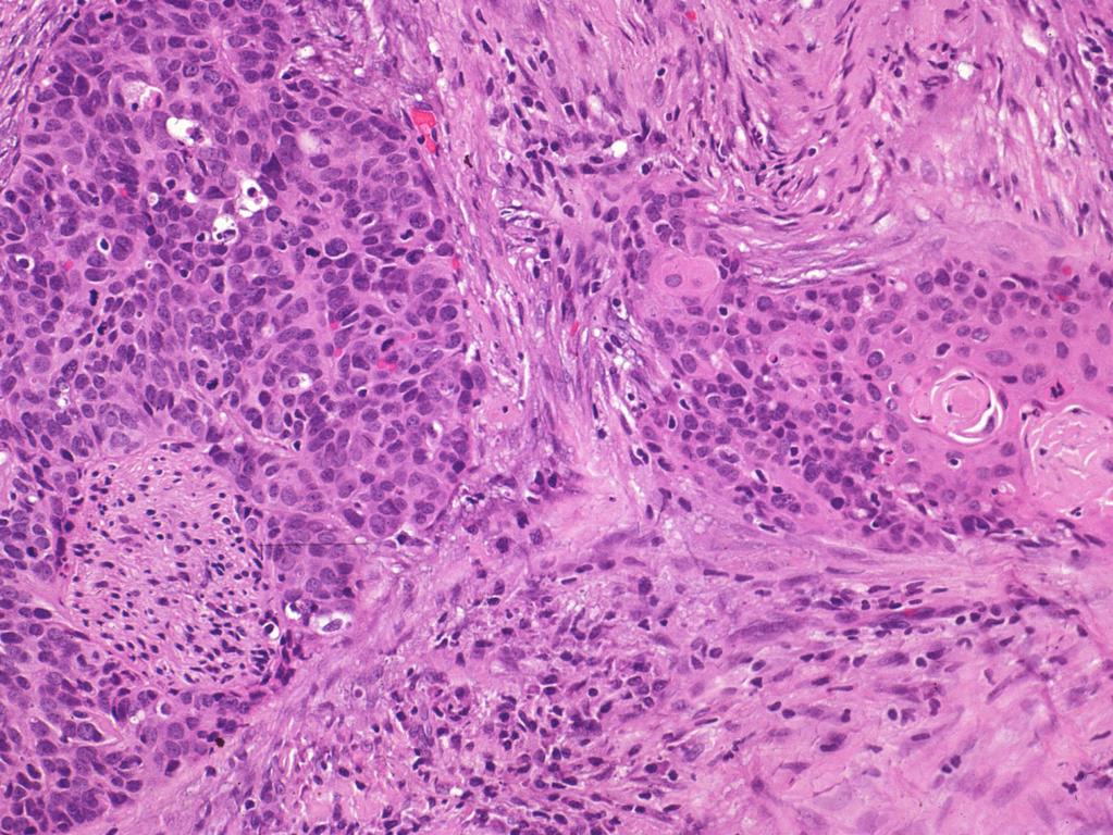
112 Viral-Associated Head and Neck Squamous Cell Carcinoma (SCC) Oropharyngeal HPV-associated squamous cell carcinoma (WHO 2017 SCC, HPV-positive) Nasopharyngeal EBV-associated squamous cell carcinoma
113 MRI T1 T2
114 FNAB Metastatic poorly-differentiated carcinoma favor squamous cell carcinoma
115 PET/CT
 Bx")
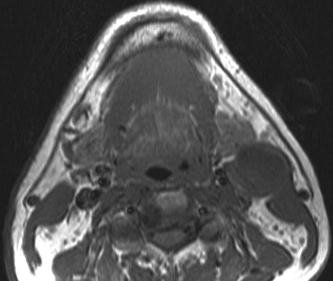
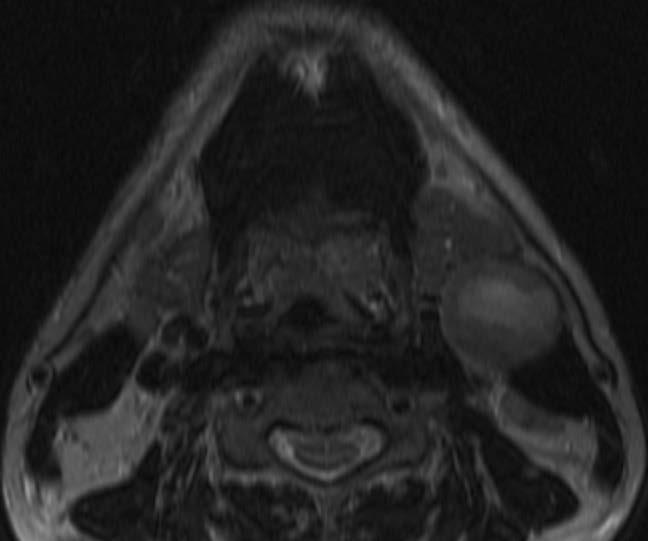
116 Base of Tongue (BOT) Bx Biopsy

117 BOT Biopsy

118 CAM5.2 p16
119 Diagnosis Oropharyngeal (Tonsillar) Carcinoma: Poorly-differentiated squamous cell carcinoma Squamous cell carcinoma with basaloid features Nonkeratinizing carcinoma
120 Nonkeratinizing Carcinoma Human Papillomavirus (HPV) Oropharyngeal Carcinoma (HPV-associated SCC)
121 HPV-positive SCC vs HPV-negative SCC HPV-positive SCC HPV-negative SCC Incidence Increasing Stable to decreasing Age Younger Older Gender M = F M > F Race Caucasian >>>> African American African American > Caucasian Risk factors HPV Smoking, alcohol Primary location Oropharynx (base of tongue; tonsil) All mucosal sites of the UADT Histology Nonkeratinizing SCC Keratinizing SCC p16 Positive Negative AJCC staging (TNM) ChemoXRT response Prognosis Lower T, higher N Good with low rate of recurrence Better disease-free and overall survival (worse if smokers) Higher T, lower N Good with high rate recurrence Worse disease-free and overall survival

122 Cystic Tonsillar Crypt NK Carcinoma


123 Cystic Tonsillar Crypt NK Carcinoma p16

124 Cystic Neck Mass

125 Cystic Neck Mass p16
126 Hybrid Oropharyngeal SCC (OPSCC) p16
127
128

129 p16 p16
130 Ciliated HPV-Associated Carcinoma (aka Ciliated Adenosquamous Carcinoma) Bishop JA, Westra WH. Am J Surg Pathol 2015;39: Radkay-Gonzalez L, et al. Head Neck Pathol 2016;10:
131 Oropharyngeal Reticulated Epithelium

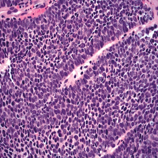
132 Oropharyngeal Reticulated Epithelium Immunohistochemistry AE1/AE3 p16


133 Tonsillar Crypt with Carcinoma CIS p16
134 HPV 16 p16 immunostaining and/or HPV DNA (ISH, PCR) correlates to the presence of HPV16 Representing a reliable predictor of origin from the oropharynx
135 Guidelines for the Detection of High Risk (HR)-HPV in H&N SCC College of American Pathologists (2017) The tumors of all adults presenting with oropharyngeal SCC should be tested for HR- HPV Neck nodal tissue from all patients with metastatic SCC of unknown primary should be tested for HR-HPV Staining with IHC p16 should be used as an initial screening method (70% cut off)
136 Guidelines for the Detection of High Risk (HR)-HPV in H&N SCC When to Test All newly diagnosed oropharyngeal squamous cell carcinomas (from primary or metastasis) All carcinomas of unknown primary presenting as neck metastases Don ts: Other sites: not currently recommended Low-risk HPV
137
138 Nasopharyngeal Carcinoma (NPC) WHO Classification (2017) Keratinizing SCC: well-, moderately, poorly-differentiated (WHO 1) Nonkeratinizing SCC: Differentiated type (Transitional Cell or Cylindrical Cell Carcinoma; WHO 2) Undifferentiated type (Lymphoepithelioma; WHO 3) Basaloid SCC
139 Percent Sex/Age EBV XRT Respons e 5-Yr survival Nasopharyngeal Carcinoma (NPC) Keratinizing Approximately 25% M > F; 4 th - 6th decades Weak association Radioresponsiveness is not good Nonkeratinizing Differentiated Least common < 15% M > F; 4 th - 6th decades Strong association Radioresponsive Nonkeratinizing Undifferentiated Most common > 60% M > F; 4 th - 6th decades; may occur in children Strong association Radioresponsive 20-40% 75% 75%
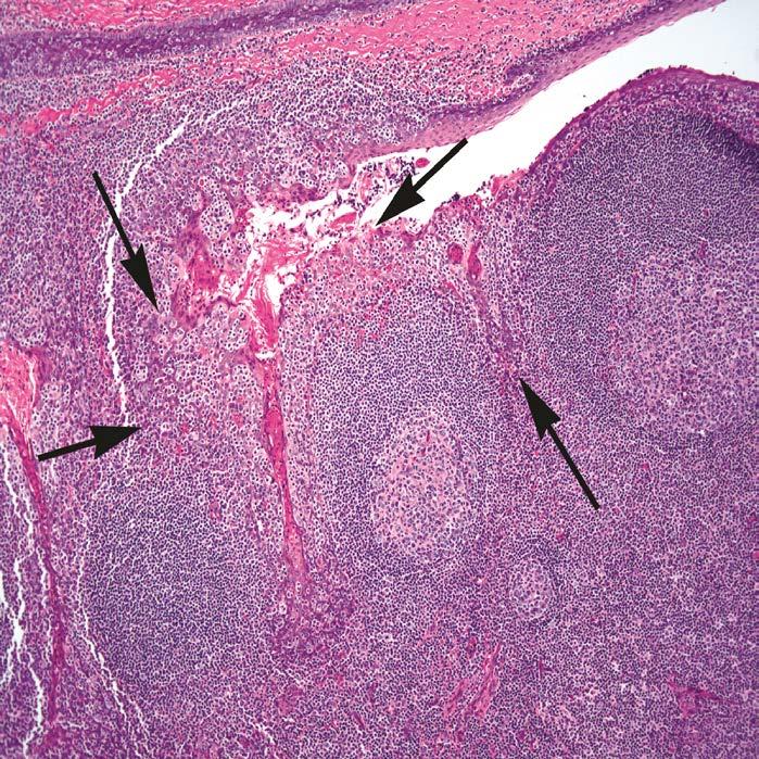
140 NPC, nonkeratinizing differentiated EBER
141 NPC, nonkeratinizing undifferentiated

142 Where is the carcinoma?
143 NPC, nonkeratinizing undifferentiated
144 NPC, nonkeratinizing undifferentiated AE1/AE3 p63 EBER
145 Nasopharyngeal Carcinoma Differential Diagnosis Non-Hodgkin lymphoma (DLBCL) Mucosal malignant melanoma Mesenchymal (e.g., rhabdomyosarcoma)

146 Cervical Neck Lymph Node Topography

147 Site Specific Lymph Node Drainage
148 Metastatic Cervical Carcinoma with an Unknown Primary Tumor (MCCUP) Definition: - Overt neck mass harboring a cytologically or histologically proven metastatic carcinoma in the absence of signs and symptoms of a primary neoplasm or of a clinically detectable mass: no history of previous malignancy or cancer ablation of any indeterminate lesion no history of definite symptoms related to a specific organ system no clinical or laboratory evidence of a primary neoplasm
149 Luna MA. Chapter 17. In: Barnes L, ed. Surgical Pathology of the H&N. 2009
150 Luna MA. Chapter 17. In: Barnes L, ed. Surgical Pathology of the H&N. 2009
151 Waldeyer Tonsillar Tissues
152 Luna MA. Chapter 17. In: Barnes L, ed. Surgical Pathology of the H&N. 2009
153 Branchiogenic Carcinoma Criteria* Cervical tumor occurs along line extending from anterior to the tragus along the anterior border of the SCM to the clavicle Histology c/w origin from tissue known to be present in branchial vestige No primary source for carcinoma on at least 5-year f/u Histologic evidence of carcinoma arising in wall of epithelial-lined cyst *Martin et al. Ann Surg 1950;132:
154 Branchial Cleft Cyst Benign lateral neck cyst most often of 2 nd branchial cleft apparatus Bimodal age: (75%); <5 yrs (20%): - rare ( 5%) in ages >40 years Painless cervical swelling typically near angle of mandible along border of SCM


155 Branchial Cleft Cyst p16 p16 p16
156 AJCC Staging 8 th Edition HPV-mediated (p16+) Oropharyngeal Cancer (Chapter 10): - Descriptor poorly-differentiated at odds with known improved prognosis so use should be avoided - Use of the designation oropharyngeal SCC, nonkeratinizing type is recommended - Histologic grading is not relevant - Presence of keratinization in a p16+/hr HPV+ carcinoma does not exclude using this staging system - Cervical lymph node metastases (to level II/III) from unknown primary tumor (pt0) that is p16+ and histology is consistent with HPV-mediated carcinogenesis are staged according to the guidelines in this chapter - Diagnosis of malignant transformation of branchial cleft cyst should be rejected
157 Thank you
Keratinizing Dysplasia and Select Variants of Head & Neck Squamous Cell Carcinoma
 Keratinizing Dysplasia and Select Variants of Head & Neck Squamous Cell Carcinoma Napa Valley Pathology Conference Silverado Resort & Spa May 18, 2018 Bruce M. Wenig, MD Moffitt Cancer Center Tampa, FL
5/22/2018. Keratinizing Dysplasia and Select Variants of Head & Neck Squamous Cell Carcinoma
 Keratinizing Dysplasia and Select Variants of Head & Neck Squamous Cell Carcinoma Napa Valley Pathology Conference Silverado Resort & Spa May 18, 2018 Bruce M. Wenig, MD Moffitt Cancer Center Tampa, FL
Squamous Cell Carcinoma of the Head and Neck (SCCHN)
") Squamous Cell Carcinoma of the Head and Neck (SCCHN) Part 1 Bruce M. Wenig, M.D. Dept. of Pathology & Laboratory Medicine Continuum Health Partners New York, NY College of American Pathologists 2004. Materials
Squamous Cell Carcinoma of the Head and Neck (SCCHN) Part 1 Bruce M. Wenig, M.D. Dept. of Pathology & Laboratory Medicine Continuum Health Partners New York, NY College of American Pathologists 2004. Materials
Head and Neck Squamous Subtypes
 1 Head and Neck Squamous Subtypes Adel K. El-Naggar, M.D., Ph.D. The University of Texas MD Anderson Cancer Center, Houston, Texas HNSCC 5 th -6 th most common cancer 400,000/year 50% mortality Considerable
Dysplasia, Mimics and Other Controversies
 Dysplasia, Mimics and Other Controversies Mary S. Richardson, MD Dept. of Pathology Medical University of South Carolina Charleston, SC Notice of Faculty Disclosure In accordance with ACGME guidelines,
04/09/2018. Squamous Cell Neoplasia and Precursor Lesions. Agenda. Squamous Dysplasia. Squamo-proliferative lesions. Architectural features
 Squamous Cell Neoplasia and Precursor Lesions Jennifer L. Hunt, MD, MEd Aubrey J. Hough Jr, MD, Endowed Professor of Pathology Chair of Pathology and Laboratory Medicine University of Arkansas for Medical
Human Papillomavirus and Head and Neck Cancer. Ed Stelow, MD
 Human Papillomavirus and Head and Neck Cancer Ed Stelow, MD No conflict of interest Declaration Cancer 1974 Lancet Oncol 2016; 17: e477-8 JAMA 1984; 252: 1857 JAMA 1988;259(13):1943-1944 Clin Cancer Res
Squamous cell carcinoma of the upper aerodigestive tract: dysplasia and select variants
 S112 2017 USCAP, Inc All rights reserved 0893-3952/17 $32.00 Squamous cell carcinoma of the upper aerodigestive tract: dysplasia and select variants Bruce M Wenig Department of Pathology, Moffitt Cancer
Notice of Faculty Disclosures
 William C. Faquin, MD, PhD Professor of Pathology Harvard Medical School Director of Head and Neck Pathology Massachusetts Eye and Ear Massachusetts General Hospital FNA OF SQUAMOUS CYSTS OF THE HEAD AND
Reporting HPV related carcinomas of the head and neck. dr. Nina Zidar Institute of Pathology Faculty of Medicine University of Ljubljana Slovenia
 Reporting HPV related carcinomas of the head and neck dr. Nina Zidar Institute of Pathology Faculty of Medicine University of Ljubljana Slovenia Conflict of interest/funding X None Company: Product royalties
Basaloid neoplasms of the head and neck. Basaloid SCC. Clinico-pathologic features 5/5/11. Basaloid Tumors Head and Neck
 Basaloid neoplasms of the head and neck Richard Jordan DDS PhD FRCPath Professor & Director UCSF Oral Pathology Laboratory University of California San Francisco Basaloid Tumors Head and Neck Basaloid
Evaluation and Management of Head and Neck Cancer in Patients with Fanconi anemia David I. Kutler, M.D., F.A.C.S.
 Evaluation and Management of Head and Neck Cancer in Patients with Fanconi anemia David I. Kutler, M.D., F.A.C.S. Residency Site Director Weill Cornell Medical Center Associate Professor Division of Head
Human Papillomavirus Testing in Head and Neck Carcinomas
 Human Papillomavirus Testing in Head and Neck Carcinomas Guideline from the College of American Pathologists Early Online Release Publication: Archives of Pathology & Laboratory Medicine 12/18/2017 Overview
Molecular Diagnostics of Head and Neck Tumors Justin A. Bishop, M.D. Associate Professor of Pathology The Johns Hopkins University Baltimore, Maryland
 Molecular Diagnostics of Head and Neck Tumors Justin A. Bishop, M.D. Associate Professor of Pathology The Johns Hopkins University Baltimore, Maryland Two Main Topics Molecular insights in salivary gland
Head and Neck Cancer in FA: Risks, Prevention, Screening, & Treatment Options David I. Kutler, M.D., F.A.C.S.
 Head and Neck Cancer in FA: Risks, Prevention, Screening, & Treatment Options David I. Kutler, M.D., F.A.C.S. Associate Professor Division of Head and Neck Surgery Department of Otolaryngology-Head and
Desmoplastic Melanoma R/O BCC. Clinical Information. 74 y.o. man with lesion on left side of neck r/o BCC
 R/O BCC Sabine Kohler, M.D. Professor of Pathology and Dermatology Dermatopathology Service Stanford University School of Medicine Clinical Information 74 y.o. man with lesion on left side of neck r/o
57th Annual HSCP Spring Symposium 4/16/2016
 An Unusual Malignant Spindle Cell Lesion to Involve the Breast Erinn Downs-Kelly, D.O. Associate Professor of Pathology University of Utah & ARUP Laboratories No disclosures Case 39 y/o female with no
HPV and Head and Neck Cancer: What it means for you and your patients
 HPV and Head and Neck Cancer: What it means for you and your patients Financial Disclosure: None November 8, 2013 Steven J. Wang, MD Associate Professor Department of Otolaryngology-Head and Neck Surgery
Nasal Cavity and Paranasal Sinuses
 Chapter 2 Nasal Cavity and Paranasal Sinuses Introduction Included in this chapter are nasal cavities, frontal sinus, ethmoid complex, sphenoid sinus, and maxillary sinuses. These cavities and sinuses
Mody. AIS vs. Invasive Adenocarcinoma of the Cervix
 Common Problems in Gynecologic Pathology Michael T. Deavers, M.D. Houston Methodist Hospital, Houston, Texas Common Problems in Gynecologic Pathology Adenocarcinoma in-situ (AIS) of the Cervix vs. Invasive
Head & Neck Squamous Carcinoma: Artifacts, Challenges, and Controversies. Agenda
 Head & Neck Squamous Carcinoma: Artifacts, Challenges, and Controversies Jennifer L. Hunt, MD, MEd Aubrey J. Hough Jr, MD, Endowed Professor of Pathology Chair of Pathology and Laboratory Medicine University
Diagnostic difficulties with lesions of the oral mucosa
 BDIAP London, November 2010 School of Clinical Dentistry University of Sheffield Diagnostic difficulties with lesions of the oral mucosa Paul M Speight Dept Oral & Maxillofacial Pathology University of
Squamous Cell Neoplasia and Precursor Lesions
 Squamous Cell Neoplasia and Precursor Lesions Jennifer L. Hunt, MD, MEd Aubrey J. Hough Jr, MD, Endowed Professor of Pathology Chair of Pathology and Laboratory Medicine University of Arkansas for Medical
NEOPLASMS OF THE SURFACE EPITHELIUM (KERATINOCYTES)
") NEOPLASMS OF THE SURFACE EPITHELIUM (KERATINOCYTES) Papillary Lesions Precancerous Lesions Keratinocyte Proliferations Carcinomas Melanotic Lesions Melanomas Normal Mucosa Keratin layer Spinous layer Basal
NEOPLASMS OF THE SURFACE EPITHELIUM (KERATINOCYTES) Papillary Lesions Precancerous Lesions Keratinocyte Proliferations Carcinomas Melanotic Lesions Melanomas Normal Mucosa Keratin layer Spinous layer Basal
HEAD AND NECK PATHOLOGY
 Bosnian-British School of Pathology November 2012 HEAD AND NECK PATHOLOGY Slide seminar: Oral Pathology Preferred Diagnoses Dr A Sandison, Slide seminar: Pathology of the Oral Cavity Page 1 of 5 1. Female
Neuroendocrine Carcinoma. Lebanon Neuroendocrine Neoplasms of H&N Nov /7/2011. Broad Classification:
 H&N Neuroendocrine Neoplasms: Classification and Diagnostic Considerations Adel K. El-Naggar, M.D., Ph.D. The University of Texas MD Anderson Cancer Center, Houston, Texas Broad Classification: A. Epithelial:
Diseases of oral cavity
 Diseases of oral cavity Diseases of Teeth and Supporting Structures Inflammatory/Reactive Lesions Infections Oral Manifestations of Systemic Disease Precancerous and Cancerous Lesions Odontogenic Cysts
Head and Neck SCC. HPV in Tumors of the Head and Neck. Overview. Role of HPV in Pathogenesis of Head & Neck Tumors
 HPV in Tumors of the Head and Neck Christina Kong, M.D. Associate Professor, Stanford Dept of Pathology Director, Cytopathology Laboratory & Cytopathology Fellowship ckong@stanford.edu Head and Neck SCC
Lesions Mimicking Adenoid Cystic Carcinoma. Diagnostic Problems in Salivary Gland Pathology An Update 5/29/2009
 Diagnostic Problems in Salivary Gland Pathology An Update Lesions Mimicking Adenoid Cystic Carcinoma Stacey E. Mills, M.D. W.S. Royster Professor of Pathology Director of Surgical and Cytopathology University
HPV Analysis of Head and Neck Squamous Cell Carcinomas based on Fine-Needle. Aspiration Specimens. William H. Westra M.D.
 HPV Analysis of Head and Neck Squamous Cell Carcinomas based on Fine-Needle Aspiration Specimens William H. Westra M.D. The Department of Pathology, The Johns Hopkins Medical Institutions, Baltimore Maryland
Differential Diagnosis of Oral Masses. Palatal Lesions
 Differential Diagnosis of Oral Masses Palatal Lesions Palatal Masses Periapical Abscess Torus Palatinus Mucocele Lymphoid Hyperplasia Adenomatous Hyperplasia Benign Salivary Neoplasms Malignant Salivary
Head and Neck Pathology. Macroscopy and Dissection Dr Tim Bracey Consultant Pathologist (Derriford Hospital)
") Head and Neck Pathology Macroscopy and Dissection Dr Tim Bracey Consultant Pathologist (Derriford Hospital) Outline Introduction - importance of macro Lip, tongue and oropharynx Larynx Bony resections
Head and Neck Pathology Macroscopy and Dissection Dr Tim Bracey Consultant Pathologist (Derriford Hospital) Outline Introduction - importance of macro Lip, tongue and oropharynx Larynx Bony resections
Problem diagnoses. Current issues in Anatomic pathology. Problem Diagnoses in Tumors of the Oral Cavity 5/29/2009
 Current issues in Anatomic pathology Problem Diagnoses in Tumors of the Oral Cavity Richard Jordan DDS PhD FRCPath Professor of Oral Pathology & Pathology Director, UCSF Oral Pathology Diagnostic Laboratory
Sarcomatoid (spindle cell) carcinoma of the cricopharynx presenting as dysphagia
 carcinoma of the cricopharynx presenting as dysphagia") Case Report Sarcomatoid (spindle cell) carcinoma of the cricopharynx presenting as dysphagia Jagtap Sunil V. 1, Shukla Dhirajkumar B. 2, Jagtap Swati S. 3, Havle Abhay D. 4 1 Associate Professor, Department
Case Report Sarcomatoid (spindle cell) carcinoma of the cricopharynx presenting as dysphagia Jagtap Sunil V. 1, Shukla Dhirajkumar B. 2, Jagtap Swati S. 3, Havle Abhay D. 4 1 Associate Professor, Department
Catholic University of Louvain, St - Luc University Hospital Head and Neck Oncology Programme. Anatomopathology. Pathology 1 Sept.
 Anatomopathology Pathology 1 Anatomopathology Biopsies Frozen section Surgical specimen Peculiarities for various tumor site References Pathology 2 Biopsies Minimum data, which should be given by the pathologist
Management of Neck Metastasis from Unknown Primary
 Management of Neck Metastasis from Unknown Primary.. Definition Histologic evidence of malignancy in the cervical lymph node (s) with no apparent primary site of original tumour Diagnosis after a thorough
Carcinoma of Unknown Primary site (CUP) in HEAD & NECK SURGERY
 in HEAD & NECK SURGERY") Carcinoma of Unknown Primary site (CUP) in HEAD & NECK SURGERY SEARCHING FOR THE PRIMARY? P r o f J P P r e t o r i u s H e a d : C l i n i c a l U n i t C r i t i c a l C a r e U n i v e r s i t y O f
Carcinoma of Unknown Primary site (CUP) in HEAD & NECK SURGERY SEARCHING FOR THE PRIMARY? P r o f J P P r e t o r i u s H e a d : C l i n i c a l U n i t C r i t i c a l C a r e U n i v e r s i t y O f
Squamous Cell Carcinoma of Thyroid: possible thymic origin, so-called ITET/CASTLE 2012/03/22
 Squamous Cell Carcinoma of Thyroid: possible thymic origin, so-called ITET/CASTLE 2012/03/22 History of ITET/CASTLE First Report Gross Appearance and Prognosis 1) Miyauchi A et al: Intrathyroidal epithelial
Neoplasia 2018 Lecture 2. Dr Heyam Awad MD, FRCPath
 Neoplasia 2018 Lecture 2 Dr Heyam Awad MD, FRCPath ILOS 1. List the differences between benign and malignant tumors. 2. Recognize the histological features of malignancy. 3. Define dysplasia and understand
3/28/2017. Head and Neck/Endocrine Pathology Specialty Conference Case 4 Raja R. Seethala, M.D. University of Pittsburgh Medical Center
 Head and Neck/Endocrine Pathology Specialty Conference Case 4 Raja R. Seethala, M.D. University of Pittsburgh Medical Center Disclosure of Relevant Financial Relationships Disclosure of Relevant Financial
Basal cell carcinoma 5/28/2011
 Goal of this Presentation A practical approach to the diagnosis of cutaneous carcinomas and their mimics Thaddeus Mully, MD University of California San Francisco To review common non-melanoma skin cancers
VULVAR CARCINOMA. Page 1 of 5
 VULVAR CARCINOMA EXAMPLE OF A VULVAR CARCINOMA USING PROPOSED TEMPLATE Case: Invasive squamous cell carcinoma arising in D-VIN Tumor in left labia major Left partial vaginectomy and sentinel lymph node
Non Small Cell Lung Cancer Histopathology ד"ר יהודית זנדבנק
 Non Small Cell Lung Cancer Histopathology ד"ר יהודית זנדבנק 26.06.09 Lecture outlines WHO histological classification Macro/Micro assessment Early diagnosis Minimal pathology Main subtypes SCC, AdCa, LCLC
Pathology of Selected Head and Neck Lesions. Adel Assaad MD Department of Pathology
 Pathology of Selected Head and Neck Lesions Adel Assaad MD Department of Pathology 1 NOSE Infections 2 Zygomycosis (Mucormycosis) Opportunistic infection caused by "bread mold fungi," including Rhizopus,
Premalignant lesions may expose to a promoting. factor & may be induced to undergo malignant. Carcinoma in situ displays the cytologic features of
 بسم رلاهللا Def. Premalignant lesions may expose to a promoting factor & may be induced to undergo malignant transformation. Carcinoma in situ displays the cytologic features of malignancy without invasion
LARYNGEAL DYSPLASIA. Tomas Fernandez M; 3 rd year ENT resident, Son Espases University Hospital
 LARYNGEAL DYSPLASIA Tomas Fernandez M; 3 rd year ENT resident, Son Espases University Hospital INTRODUCTION Laryngeal cancer constitutes 1-2% of all malignancies diagnosed worldwide Survival is related
Human Papillomavirus and Epstein Barr Virus in Head and Neck Carcinomas: Suggestions for the New WHO Classification
 Head and Neck Pathol (2014) 8:50 58 DOI 10.1007/s12105-014-0528-6 PROCEEDINGS OF THE NORTH AMERICAN SOCIETY OF HEAD AND NECK PATHOLOGY COMPANION MEETING, MARCH 2, 2014, SAN DIEGO, CALIFORNIA Human Papillomavirus
Physician to Physician AJCC 8 th Edition. Head and Neck. Summary of Changes. AJCC Cancer Staging Manual, 7 th Ed. Head and Neck Chapters
 Physician to Physician Head and Neck William M. Lydiatt, MD Chair of Surgery Nebraska Methodist Hospital Clinical Professor of Surgery, Creighton University Validating science. Improving patient care.
Head & Neck Staging. Donna M. Gress, RHIT, CTR Technical Editor, AJCC Cancer Staging Manual First Author, Chapter 1: Principles of Cancer Staging
 AJCC 8 th Edition Staging Head & Neck Staging Donna M. Gress, RHIT, CTR Technical Editor, AJCC Cancer Staging Manual First Author, Chapter 1: Principles of Cancer Staging Validating science. Improving
AJCC 8 th Edition Staging. Head & Neck Staging. Learning Objectives. This webinar is sponsored by. the Centers for Disease Control and Prevention.
 AJCC 8 th Edition Staging Head & Neck Staging Donna M. Gress, RHIT, CTR Technical Editor, AJCC Cancer Staging Manual First Author, Chapter 1: Principles of Cancer Staging Validating science. Improving
Penile cancer teams in UK. Common variants. Penile cancer teams. Basaloid squamous carcinoma. The Pathology of Penile Tumours
 The Pathology of Penile Tumours Dr Jonathan H Shanks The Christie NHS Foundation Trust, Manchester, UK Penile cancer teams in UK 12 centres for penile cancer work (10 in England and Wales, 2 in Scotland)
My Journey into the World of Salivary Gland Sebaceous Neoplasms
 My Journey into the World of Salivary Gland Sebaceous Neoplasms Douglas R. Gnepp Warren Alpert Medical School at Brown University Rhode Island Hospital Pathology Department Providence RI Asked to present
When Immunostains Can Get You in Trouble: Gynecologic Pathology p16: Panacea or Pandora s Box?
 When Immunostains Can Get You in Trouble: Gynecologic Pathology p16: Panacea or Pandora s Box? Teri A. Longacre, MD Stanford Medicine Stanford California pi6 in Gynecologic Pathology: Panacea or Pandora
Benign and malignant epithelial lesions: Seborrheic keratosis: A common benign pigmented epidermal tumor occur in middle-aged or older persons more
 Benign and malignant epithelial lesions: Seborrheic keratosis: A common benign pigmented epidermal tumor occur in middle-aged or older persons more common on the trunk; but extremities, head and neck are
3/27/2017. Pulmonary Pathology Specialty Conference. Disclosure of Relevant Financial Relationships. Clinical History:
 Pulmonary Pathology Specialty Conference Saul Suster, M.D. Medical College of Wisconsin Disclosure of Relevant Financial Relationships USCAP requires that all planners (Education Committee) in a position
THYMIC CARCINOMAS AN UPDATE
 THYMIC CARCINOMAS AN UPDATE Mark R. Wick, M.D. University of Virginia Medical Center Charlottesville, VA CARCINOMA OF THE THYMUS General Clinical Features No apparent gender predilection Age range of 35-75
Oral Cancer Risk and Detection
 Oral Cancer Risk and Detection Evan M. Graboyes, MD Assistant Professor Department of Otolaryngology-Head & Neck Surgery Cancer Control Program, Hollings Cancer Center Medical University of South Carolina
Pitfalls in Sinonasal Pathology
 Pitfalls in Sinonasal Pathology Jennifer L. Hunt, MD, MEd Aubrey J. Hough Jr, MD, Endowed Professor of Pathology Chair of Pathology and Laboratory Medicine University of Arkansas for Medical Sciences jhunt2@uams.edu
Objectives. Salivary Gland FNA: The Milan System. Role of Salivary Gland FNA 04/26/2018
 Salivary Gland FNA: The Milan System Dr. Jennifer Brainard Section Head Cytopathology Cleveland Clinic Objectives Introduce the Milan System for reporting salivary gland cytopathology Define cytologic
Case 2. Dr. Sathima Natarajan M.D. Kaiser Permanente Medical Center Sunset
 Case 2 Dr. Sathima Natarajan M.D. Kaiser Permanente Medical Center Sunset History 24 year old male presented with a 3 day history of right flank pain, sharp in nature Denies fever, chills, hematuria or
Objectives. Atypical Glandular Cells. Atypical Endocervical Cells. Reactive Endocervical Cells
 2013 California Society of Pathologists 66 th Annual Meeting San Francisco, CA Atypical Glandular Cells to Early Invasive Adenocarcinoma: Cervical Cytology and Histology Christina S. Kong, MD Associate
FINE NEEDLE ASPIRATION OF ENLARGED LYMPH NODE: Metastatic squamous cell carcinoma
 Case Scenario 1 HNP: A 70 year old white male presents with dysphagia. The patient is a current smoker, current user of alcohol and is HPV positive. A CT of the Neck showed mass in the left pyriform sinus.
Central Poorly Differentiated Adenocarcinoma of the Maxilla: Report of a Case
 Kobe J. Med. Sci., Vol. 49, No. 2, pp. 45-49, 2003 Central Poorly Differentiated Adenocarcinoma of the Maxilla: Report of a Case MASAHIRO UMEDA 1), SATOSHI YOKOO 1), YASUYUKI SHIBUYA 1), TAKAHIDE KOMORI
Papillary Lesions of the breast
 Papillary Lesions of the breast Emad Rakha Professor of Breast Pathology The University of Nottingham Papillary lesions of the breast are a heterogeneous group of disease, which are characterised by neoplastic
Polymorphous Low-Grade. December 5 th, 2008
 Polymorphous Low-Grade Adenocarcinoma December 5 th, 2008 Epidemiology Represents 2 nd or 3 rd most common minor salivary gland malignancy (17-26%) 1 st mucoepidermoid carcinoma Rare in reported Asian
Kidney Case 1 SURGICAL PATHOLOGY REPORT
 Kidney Case 1 Surgical Pathology Report February 9, 2007 Clinical History: This 45 year old woman was found to have a left renal mass. CT urography with reconstruction revealed a 2 cm medial mass which
Head and Neck Pathology Macroscopy and Dissection Dr Tim Bracey
 Head and Neck Pathology Macroscopy and Dissection Dr Tim Bracey Outline Introduction - importance of macro Lip, tongue and oropharynx Larynx Bony resections Salivary glands* will not be covered specifically
Diseases of the vulva
 Diseases of the vulva 1. Bartholin Cyst - Infection of the Bartholin gland produces an acute inflammation within the gland (adenitis) and may result in an abscess. Bartholin duct cysts - Are relatively
AGGRESSIVE VARIANTS OF PAPILLARY THYROID CARCINOMA DIAGNOSIS AND PROGNOSIS
 AGGRESSIVE VARIANTS OF PAPILLARY THYROID CARCINOMA DIAGNOSIS AND PROGNOSIS PAPILLARY THYROID CARCINOMA Clinical Any age Microscopic to large Female: Male= 2-4:1 Radiation history Lymph nodes Prognosis
1.Acute and Chronic Cervicitis - At the onset of menarche, the production of estrogens by the ovary stimulates maturation of the cervical and vaginal
 Diseases of cervix I. Inflammations 1.Acute and Chronic Cervicitis - At the onset of menarche, the production of estrogens by the ovary stimulates maturation of the cervical and vaginal squamous mucosa
LEUKOPLAKIA Definition Epidemiology Clinical presentation
 LEUKOPLAKIA Definition Leukoplakia is the most common premalignant or "potentially malignant" lesion of the oral mucosa. Leukoplakia is a predominantly white lesion of the oral mucosa than cannot be clinicopathologically
أملس عضلي غرن = Leiomyosarcoma. Leiomyosarcoma 1 / 5
 Leiomyosarcoma 1 / 5 EPIDEMIOLOGY Exact incidence is unknown, but older studies suggest that leiomyosarcomas comprise approximately 3 percent of soft-tissue sarcomas. Superficial leiomyosarcoma occurs
Case year old female presented with asymmetric enlargement of the left lobe of the thyroid
 Case 4 22 year old female presented with asymmetric enlargement of the left lobe of the thyroid gland. No information available relative to a prior fine needle aspiration biopsy. A left lobectomy was performed.
Case 4 Diagnosis 2/21/2011 TGB
 Case 4 22 year old female presented with asymmetric enlargement of the left lobe of the thyroid gland. No information available relative to a prior fine needle aspiration biopsy. A left lobectomy was performed.
Case Presentation. Maha Akkawi, MD, Fatima Obeidat, MD, Tariq Aladily, MD. Department of Pathology Jordan University Hospital Amman, Jordan
 Case Presentation Maha Akkawi, MD, Fatima Obeidat, MD, Tariq Aladily, MD Department of Pathology Jordan University Hospital Amman, Jordan The 25th Annual Congress of the ADIAP The 8/11/2013 1 5th International
2018 SEER Solid Tumor Manual 2018 KCR SPRING TRAINING
 2018 SEER Solid Tumor Manual 2018 KCR SPRING TRAINING Eight Groups are Revised for 2018 Head & Neck Colon (includes rectosigmoid and rectum for cases diagnosed 1/1/2018 forward) Lung (2018 Draft not yet
Enterprise Interest Nothing to declare
 Enterprise Interest Nothing to declare Diagnoses one would not like to miss in soft tissue pathology early in your career Marta Sbaraglia, MD Department of Pathology Hospital of Treviso University of Padua
4/17/2015. Case 1. A 37 year old man with a 2.2 cm solitary left thyroid mass.
 Case 1 A 37 year old man with a 2.2 cm solitary left thyroid mass. Case 1 Case 1 1 Case 1: Diagnosis? A. Benign B. Atypia of undetermined significance/follicular lesion of undetermined significance C.
Salivary Glands 3/7/2017
 Salivary Glands 3/7/2017 Goals and objectives Focus on the entities unique to H&N Common board type facts Information for your future practice Salivary Glands Salivary Glands Major gland. Paratid. Submandibular.
3/24/2017 DENDRITIC CELL NEOPLASMS: HISTOLOGY, IMMUNOHISTOCHEMISTRY, AND MOLECULAR GENETICS. Disclosure of Relevant Financial Relationships
 DENDRITIC CELL NEOPLASMS: HISTOLOGY, IMMUNOHISTOCHEMISTRY, AND MOLECULAR GENETICS Jason L. Hornick, M.D., Ph.D. Director of Surgical Pathology and Immunohistochemistry Brigham and Women s Hospital Professor
ONCOLOGY. Csaba Bödör. Department of Pathology and Experimental Cancer Research november 19., ÁOK, III.
 ONCOLOGY Csaba Bödör Department of Pathology and Experimental Cancer Research 2018. november 19., ÁOK, III. bodor.csaba1@med.semmelweis-univ.hu ONCOLOGY Characteristics of Benign and Malignant Neoplasms
A neoplasm is defined as "an abnormal tissue proliferation, which exceeds that of adjacent normal tissue. This proliferation continues even after
 NEOPLASIA Neoplasia is a very important topic in pathology because neoplasms are both common and serious diseases. A neoplasm literally means a new growth, and this term is used interchangeably with a
CODING TUMOUR MORPHOLOGY. Otto Visser
 CODING TUMOUR MORPHOLOGY Otto Visser INTRODUCTION The morphology describes the tissue of the tumour closest to normal tissue Well differentiated tumours are closest to normal Undifferentiated tumours show
Salivary Gland Cytology
 Salivary Gland Cytology Diagnostic challenges and potential pitfalls Tarik M. Elsheikh, MD Professor and Medical Director Anatomic Pathology Cleveland Clinic FNA Salivary Gland Lesions Indications Distinguish
Histological Typing Of Cancer And Precancer Of The Oral Mucosa
 Histological Typing Of Cancer And Precancer Of The Oral Mucosa 1 / 7 2 / 7 3 / 7 Histological Typing Of Cancer And Within the last decade, histologic grading has become widely accepted as a powerful indicator
Basement membrane in lobule.
 Bahram Memar, MD Basement membrane in lobule. Normal lobule-luteal phase Normal lobule-follicular phase Lactating breast Greater than 95% are adenocarcinomas in situ carcinomas and invasive carcinomas.
(CYLINDROMA) ATLAS OF HEAD AND NECK PATHOLOGY ADENOID CYSTIC CARCINOMA
 ATLAS OF HEAD AND NECK PATHOLOGY ADENOID CYSTIC CARCINOMA") (CYLINDROMA) This malignant tumor is poorly encapsulated and while seemingly well defined within the affected gland, there is usually infiltration of surrounding tissue on closer examination. The cut surface
(CYLINDROMA) This malignant tumor is poorly encapsulated and while seemingly well defined within the affected gland, there is usually infiltration of surrounding tissue on closer examination. The cut surface
Cystic carcinoma of the neck
 Case Report Brunei Int Med J. 2010; 6 (1): 56-60 Cystic carcinoma of the neck Prathibha Parampalli SUBRHAMANYA, Ghazala KAFEEL, Hla OO, Pemasiri Upali TELISINGHE, Department of Pathology, RIPAS Hospital,
Neoplasia part I. Dr. Mohsen Dashti. Clinical Medicine & Pathology nd Lecture
 Neoplasia part I By Dr. Mohsen Dashti Clinical Medicine & Pathology 316 2 nd Lecture Lecture outline Review of structure & function. Basic definitions. Classification of neoplasms. Morphologic features.
Ben Witt, MD University of Utah/ARUP Laboratories Assistant Professor of Anatomic Pathology
 Ben Witt, MD University of Utah/ARUP Laboratories Assistant Professor of Anatomic Pathology Review some of the more common cytodiagnoses of the Head and Neck Establish an approach to some of the diagnostic
Small (and large) Blue Cell Tumors of the Skull Base
 Blue Cell Tumors of the Skull Base") Small (and large) Blue Cell Tumors of the Skull Base Jennifer L. Hunt, MD, MEd Aubrey J. Hough Jr, MD, Endowed Professor of Pathology Chair of Pathology and Laboratory Medicine University of Arkansas for
Small (and large) Blue Cell Tumors of the Skull Base Jennifer L. Hunt, MD, MEd Aubrey J. Hough Jr, MD, Endowed Professor of Pathology Chair of Pathology and Laboratory Medicine University of Arkansas for
Basaloid Squamous Cell Carcinoma of the Oral Cavity: An Analysis of 92 Cases
 The Laryngoscope VC 2013 The American Laryngological, Rhinological and Otological Society, Inc. Basaloid Squamous Cell Carcinoma of the Oral Cavity: An Analysis of 92 Cases Valerie A. Fritsch, MD; Daniel
04/09/2018. Salivary Gland Pathology in the Molecular Era Old Friends, Old Foes, & New Acquaintances
 Salivary Gland Pathology in the Molecular Era Old Friends, Old Foes, & New Acquaintances Jennifer L. Hunt, MD, MEd Aubrey J. Hough Jr, MD, Endowed Professor of Pathology Chair of Pathology and Laboratory
New Diagnoses Need New Approaches: A Glimpse into the Near Future of Gynecologic Pathology
 New Diagnoses Need New Approaches: A Glimpse into the Near Future of Gynecologic Pathology United States and Canadian Academy of Pathology 102 nd Annual Meeting Baltimore, Maryland Christina S. Kong, M.D.
The many faces of extranodal lymphoma
 The many faces of extranodal lymphoma Frank Pameijer Departments of Radiology and Radiation Oncology University Medical Center Utrecht Special thanks to Ilona M Schmalfuss, MD University of Florida Gainesville,
Cutaneous Adnexal Tumors
 Cutaneous Adnexal Tumors Lesions with Predominant Follicular Differentiation Special Emphasis on Basal Cell Carcinoma 2014-04-01 Prof. Dr. med. Katharina Glatz Pathologie Cutaneous Adnexal Tumors Hair
NEOPLASIA-I CANCER. Nam Deuk Kim, Ph.D.
 NEOPLASIA-I CANCER Nam Deuk Kim, Ph.D. 1 2 Tumor in the hieroglyphics of the Edwin Smith papyrus (1,600 B.C., Breasted s translation 1930) 3 War on Cancer (National Cancer Act, 1971) 4 Cancer Acts in Korea
CASE year old male with a PET avid nodule in the left adrenal gland
 CASE 1 55 year old male with a PET avid nodule in the left adrenal gland Case 1 Adrenal gland parenchyma partly replaced by a spindle cell tumour with mild nuclear pleomorphism Atypical mitoses present
Update on Cutaneous Mesenchymal Tumors. Thomas Brenn
 Update on Cutaneous Mesenchymal Tumors Thomas Brenn Cutaneous Mesenchymal Tumours Wide morphological and biological spectrum Myofibroblastic, smooth muscle, neural, vascular, apidocytic, undifferentiated;
4/12/2018. MUSC Pathology Symposium Kiawah Island April 18, Jesse K. McKenney, MD
 MUSC Pathology Symposium Kiawah Island April 18, 2018 Jesse K. McKenney, MD 1 Urothelial Carcinoma with Alternative Differentiation 2 Urothelial Carcinoma with Alternative Differentiation Recognition as