Tumor Imaging in Nuclear Medicine: Current Status and
|
|
|
- Nora Vanessa Nelson
- 6 years ago
- Views:
Transcription
1 Tumor Imaging in Nuclear Medicine: Current Status and Future Prospects Bennett S. Greenspan, MD SNM MWM Albuquerque, NM January 30, 2010
2 Tumor Imaging Part I Current Status Current status agents that are FDA approved for routine clinical use.
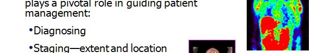
3 Tumor Imaging Indications for tumor imaging: Identification, diagnosis Staging/re staging Identification of recurrence, residual disease Monitoring response to therapy Evaluating prognosis
4 Tumor Imaging Agents Ga 67 citrate Organ imaging, e.g. thyroid, bone Thallium 201 Tc 99m Sestamibi Breast imaging Lbld Labeled monoclonal lantibodies Peptide receptor imaging In 111 pentetreotide Adrenal tumor imaging I 123 MIBG F 18 FDG
5 Ga 67 citrate Lymphoma
6 Ga 67 citrate Melanoma
7 Ga 67 citrate Mechanism of uptake bound to transferrin, uptake in tumor cells by lysosomes and endoplasmic reticulum Now nearly obsolete as a tumor imaging agent outperformed by FDG PET
8 Ga 67 citrate Probable only remaining indication for Ga 67 citrate in tumor imaging: Differentiating hepatocellular carcinoma from regenerating nodules in patients with cirrhosis
9 Thyroid Carcinoma Indications for imaging with I 131: 1. Detect active residual disease (papillary or follicular thyroid CA) 2. Detect functioning metastases 3. Assess results of treatment
10 Papillary Thyroid Cancer


11 Papillary Thyroid Cancer
12 Papillary Thyroid Carcinoma


13 Metastatic Thyroid Carcinoma
14 I 131 Thyroid Carcinoma I 131: Oldest radionuclide (RN) in clinical use Images are not very pretty, due to the high gamma energy, but theinformation obtained is extremely useful. Having a gamma emission i and a beta emission i makes this RN uniquely suited to therapy, esp. for thyroid iddisease. There is no replacement on the horizon.


15 Bone Scan Prostate Carcinoma widespread dbone metastases
16 Bone scan Agent: Tc 99m MDP (orhdp) Uptake related to blood flow and osteoblastic activity Very sensitive for metastases that generate an osteoblastic response (most tumors) Useful for staging/re staging, assessing response to therapy, detecting recurrence or residual disease


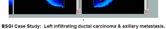
17 Tc 99m Sestamibi Breast CA BSGI


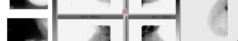
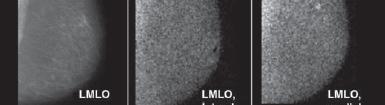
18 Breast Specific Gamma Imaging (BSGI)

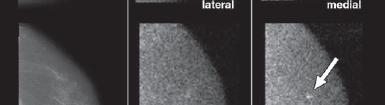
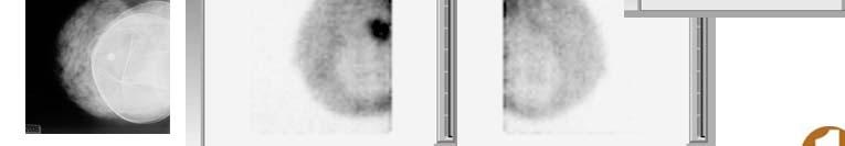
19 BSGI 4 mm tumor
20 BSGI Fairly new technique utilizes small gamma camera optimized to image the breast Will not replace mammography, but may be a useful adjunct in certain circumstances, particularly if MRI is indicated but cannot be done. Greater sensitivity and specificity than conventional scintimammography. SNM Procedure Guideline under development
21 Prostascint Monoclonal Antibodies Prostascint Capromab Pendetide, labeled with In 111 Used in patients with prostate cancer, to detect recurrent or residual disease.

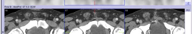
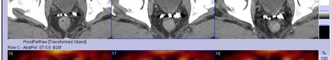
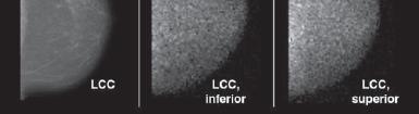
22 SPECT/CT Normal

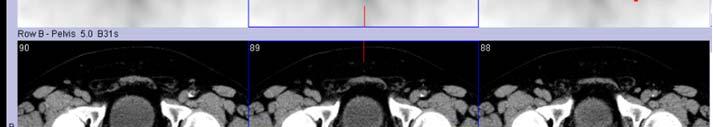
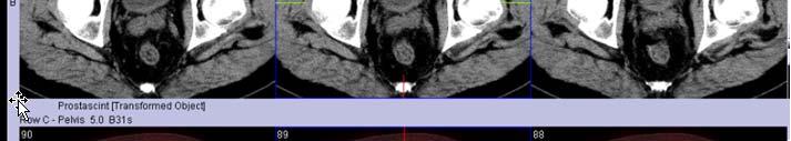
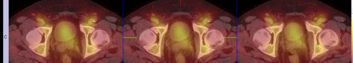
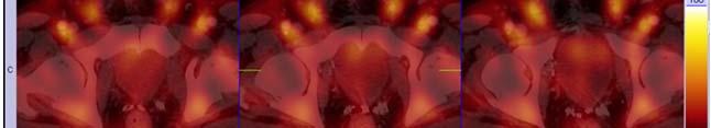
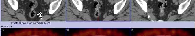
23 Tumor in Prostate Bed

24 Nodal metastases pelvis


25 Para aortic nodes Coronal images SPECT/CT
26 Prostascint Only monoclonal antibody in routine clinical use. Not a very good agent less sensitive than is desirable, and images are often difficult to read. Also, test is performed over 4 7 days.
27 Peptide Receptor Imaging Somatostatin receptor imaging In 111 pentetreotide (Octreotide, Octreoscan). Neuroendocrine tumors derived fromapud (Amine Precursor Uptake and Decarboxylation) system cells Examples: carcinoid, pituitary adenoma, pancreatic islet cell tumor, small cell lung cancer, pheochromocytoma, neuroblastoma


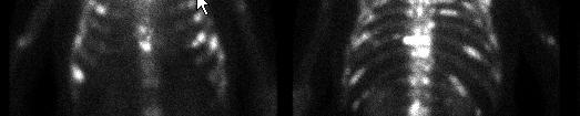
28 In 111 Pentetreotide (Octreoscan) Merkel klcell tumor
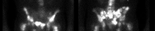
29 Metastatic carcinoid, with meningioma
30 Adrenal Tumor Imaging Adrenal tumor imaging I 123 MIBG: Pheochromocytoma, Neuroblastoma, Paraganglioma MIBG (metaiodobenzylguanidine) is an analog of norepinephrine i Taken up by chromaffin cells, and therefore useful in imaging sympathetic adrenergic tissue.
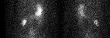
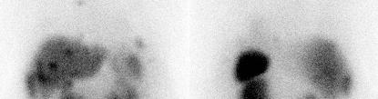
31 Recurrent Malignant Pheochromocytoma h
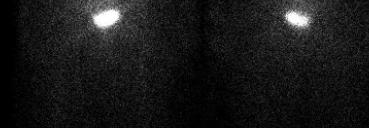
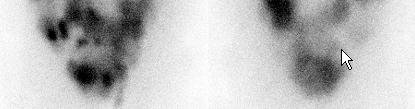
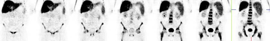
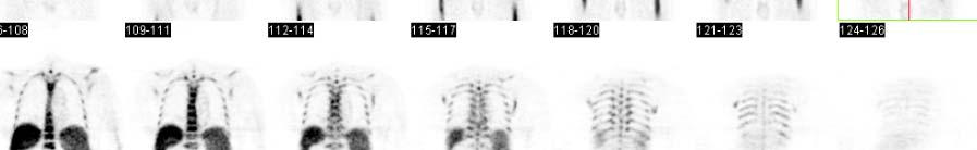
32 Stage IV Recurrent Neuroblastoma bone marrow and liver metastases
33 Tumor FDG PET F 18 FDG Used for many tumors for staging/re staging, monitoring response to therapy, detecting recurrent or residual disease For example: Head and neck, lung, lymphoma, melanoma, esophageal, colorectal, breast, cervical lca
34 Positron Decay Mettler and Guiberteau, Essentials of Nuclear Medicine Imaging, 2006, p. 361
35 Metabolism of F 18 FDG Mettler and Guiberteau, Essentials of Nuclear Medicine Imaging, 2006, page 372
36 F 18 FDG H&N Base of Tongue CA
37 F 18 FDG Lung CA


38 DLBCL Extensive Bone Marrow Involvement
39 F 18 FDG Metastatic Melanoma
40 F 18 FDG Esophageal CA with Liver Metastases
41 F 18 FDG Disseminated cervical cancer metastases
42 F 18 FDG While F 18 FDG is a non specific agent, it is useful for many different malignancies. It measures glycolysis, which is increased in many tumors. Photon flux is 100 times greater than for conventional single photon agents, allowing for better spatial resolution.
43 Current Molecular Imaging in routine clinical luse in Oncology (Part I) I 131 thyroid In 111 octreotide (pentetreotide) I 123/131 3 MIBG In 111 monoclonal antibody Capromab Pendetide (Prostascint) Ga 67 citrate (essentially obsolete as a tumor imaging agent) F 18 FDG


44
45 Tumor Imaging Part II Futureprospects Note Except for F 18 FDG, the following agents are not FDA approved. Many of these are in clinical trials.
46 Detection Treatment especially as a precursor for targeted therapy Early Intervention Drug Discovery and Development
47 Molecular Imaging Molecular imaging (MI) MI will have an expanding clinical relevance as it will becomeincreasingly important in patient care and management in the near future; and PET is the most sensitive and the most specific technique to image molecular l pathways in patients
48 Why the Interest in Molecular Imaging? The ultimate goal is targeted therapy to provide personalized medicine. Targeted imaging: finding the right molecular probe for the right target to monitor the right disease in theright patient. Streamlining drug discovery: finding the right drug against the right ihtarget to treat the right ih disease in the right patient

49 Molecular Imaging
50 Molecular Imaging
51 Molecular Imaging Therapeutic response criteria Will be based on metabolic characteristics rather than size alone Translational research bringing experimental imaging i and therapeutic techniques to the clinic after extensive testing in experimental models dl(bench to bdid) bedside)
52 Molecular Imaging
53 Radiotracer Imaging of cancer Categories: Proliferation/DNA synthesis Hypoxia Receptors Angiogenesis Metabolism F 18 FDG/Amino acid transport
54 Cancer Imaging Agents Radiopharmaceuticals for imaging cellular proliferation 3 deoxy 3 [ 18 F]fluorothymidine ([ 18 F]FLT) Imaging with 18 F labeled sigma 2 receptor ligands Imaging tumor hypoxia 60/64 Cu ATSM 18 F MISO Imaging upregulation of receptors in tumors 68 Ga labeled somatostatin analogs
55 Cancer Imaging Agents Radiopharmaceuticals for imaging cellular proliferation 3 deoxy 3 [ 18 F]fluorothymidine ([ 18 F]FLT) Imaging with 18 F labeled sigma 2 receptor ligands Imaging tumor hypoxia 60/64 Cu ATSM 18 F MISO Imaging upregulation of receptors in tumors 68 Ga labeled somatostatin analogs
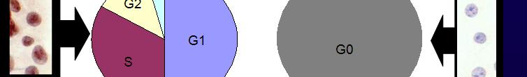
56 Why Image Cellular Proliferation Rationale Proliferative status of tumors may indicate which patients are at high risk of recurrence, as that has a profound effect on outcome from therapy. A change in the proliferative status of a tumor during or after therapy may also be an indicator of response and allow further tailoring of therapy.
57 18 F Labeled Thymidine Analogs O O O O HN CH 3 HN CH 3 HN I HN Br O N O N O N O N HO H H 18 F O H H H HO H H OH O 18 F H H HO H H OH O 18 F H H HO H H OH O 18 F H H [ 18 F]FLT [ 18 F]FMAU [ 18 F]FIAU [ 18 F]FBAU
58 3 deoxy 3 [ 18 F]fluorothymidine: [ 18 F] FLT F-18 is the radiolabeled l d form of the pyrimidine idi nucleoside, thymidine FLT is retained within the cell after phosphorylation providing a measure of cellular l thymidine kinase (TK1) activity, it an enzyme which is closely related to cellular proliferation. TK1 is up-regulated in the S phase of the cell cycle O O HO O N NH O TK-1 HO O P O O O N NH O DNA synthesis OH OH THYMIDINE O O HO O N NH O TK-1 HO O P O O O N NH O DNA synthesis 18 F 18 F FLUOROTHYMIDINE [ 18 F]FLT dtmp
59 F 18 FLT F 18 FLT F 18 replaces OH at 3 position it cannot be incorporated into DNA, and is trapped in tumor cells following phosphorylation of the 5 hydroxy group by thymidine kinase (TK 1) 1). Analogous to trapping of F 18 FDG in cells following phosphorylation by hexokinase. F 18 FLT is a marker of cell proliferation, but does not directly measure DNA synthesis.
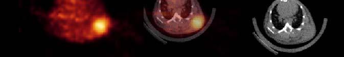
60 F 18 FLT Proliferation F 18 Fluorothymidine (F 18 FLT) Mach, et al, PET Clinics, Jan, 2009
61 F 18 FLT Responder vs. Non Responder Bading and Shields JNM 2008; 49 (6, Suppl), 65S
62 F 18 FLT Responder vs. Non Responder Bading and Shields JNM 2008; 49 (6, Suppl), 65S
63 F 18 FLT Responder vs. Non Responder Bading and Shields JNM 2008; 49 (6, Suppl), 65S
64 F 18 FLT Responder vs. Non Responder Bading and Shields JNM 2008; 49 (6, Suppl), 65S
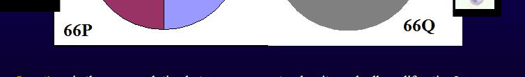
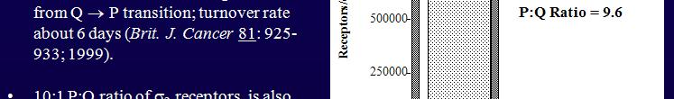
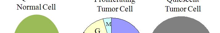
65 Proliferation
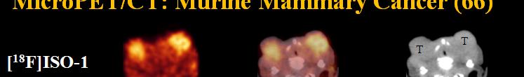
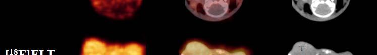
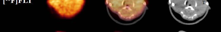
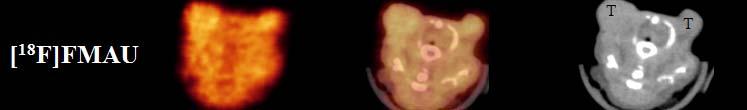
66 Proliferation
67


68 Proliferation Sigma 2 Receptors


69 Sigma 2 Receptors

70 Sigma 2 Receptors


71 Proliferation

72 Proliferation: F 18 ISO
73 F 18 FLT, F 18 FMAU, F 18 ISO 1
74 Proliferation Agents
75 F 18 FLT vs. F 18 ISO FLT shows some promise, and is in current clinical trials sponsored by SNM However, FLT only shows proliferating cells in S phase, about 2% of proliferating cells. F ISO shows all of the proliferating i cells, and may turn out to be a better agent for detecting proliferating i cells.
76 Cancer Imaging Agents Radiopharmaceuticals for imaging cellular proliferation 3 deoxy 3 [ 18 F]fluorothymidine ([ 18 F]FLT) Imaging with 18 F labeled sigma 2 receptor ligands Imaging tumor hypoxia 60/64 Cu ATSM 18 F MISO Imaging upregulation of receptors in tumors 68 Ga labeled somatostatin analogs
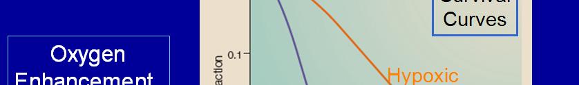
77 Hypoxia Tumor cells that outgrow their blood supply become hypoxic, and slow their growth rate. Chemotherapy and radiotherapy becomeless effective chemotherapy depends on proliferation rate to be effective, and cytotoxicity of radiotherapy depends on level of intracellular oxygen.
78 Why Image Hypoxia? Hypoxia influences response to treatment: (1)Radiotherapy - hypoxic cells are protected from lethal effects of conventional ionizing radiation therapy (2)Chemotherapy - effect of hypoxia on special genes and drug delivery Imaging of hypoxia is required in order to predict response to traditional therapies Imaging of hypoxia in the brain, heart and cancer have been explored


79 Hypoxia
80 Hypoxia PET imaging agents that can be used to assess regional tumor hypoxia: F 18 Misonidazole (FMISO) Cu 64 ATSM
81 Hypoxia F 18 FMISO
82 Hypoxia F 18 FMISO Most widely used PET agent for regional hypoxia. It is retained in hypoxic cells; it enters by passive diffusion and undergoes reduction, eventuallyforming covalent bonds with macromolecules, and is trapped in the cell. Images are of low contrast, but it can identify if clinically significant regional hypoxia.
83 Hypoxia F 18 FMISO It requires a venous blood sample taken during imaging, to determine a normalized map of a tissue to blood ratio, T/B. The hypoxic portion of a tumor can be characterized by a maximum T/B, or T/B greater than a defined threshold.
84 Hypoxia F 18 FMISO Identification of hypoxic tumor may help facilitate image directed radiotherapy. It appears to have the potential to predict the response to treatment (better than F 18 FDG) and provide prognostic information. However, there are some drawbacks to this agent.
85 Hypoxia F 18 FMISO
86 Hypoxia Cu ATSM Theory: H 3 C CH 3 TRAPPED N N N N Cu HN S S NH CH 3 CH 3 NOT TRAPPED Cu H 3 C CH 3 N N N N HN SH HS NH CH 3 CH 3 H 3 C CH 3 N N N N Cu HN S S NH CH 3 CH 3 H 3 C CH 3 N N N N Cu HN S S NH CH 3 CH 3 Hypoxic cell (-O 2 ) Normal cell (+O 2 )
87 Cu ATSM In the hypoxic cell, Cu (II) ATSM is reduced to Cu (I) ATSM. Cu (I) is then released from ATSM and is trapped in the hypoxic cell. Cu (II) is not trapped in normoxic cells.
88 Copper Radionuclides Isotope Half- life Decay modes /% Maximum + Reaction Natural energy abundance (MeV) of target isotope 60 Cu 23.7 m / Ni(p,n) 26.1% EC/ Cu 3.32 h + / EC/ Cu 12.7 h + / Ni(p,n) 1.25% 64 Ni(p,n) 1.16% EC/ /38.4 * *Qaim et al. Radiochimica Acta 2007; 95:67-73
89 Cu 60 /Cu 64 Most of the early clinical trials were done with Cu 60. However, Cu 60 has too short a half life life for multicenter clinical trials. The FDA wanted to confirm that Cu 64 gave similar results, and could be used in place of Cu 60; with iha 12.7 hr hlflif half life, it can be used for multi center clinical trials.
90 Uptake of 64 Cu(ATSM), 64 Cu(PTSM) and 18 F MISO in EMT6 cells after 1 h at varying levels of oxygen Cu(PTSM) % up ptake Cu(ATSM) Oxygen concentration (ppm) 18 F-MISO
91 Cu ATSM
92 Cu 60 ATSM NSCLC
93 Cu ATSM NSCLC
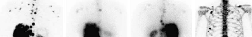
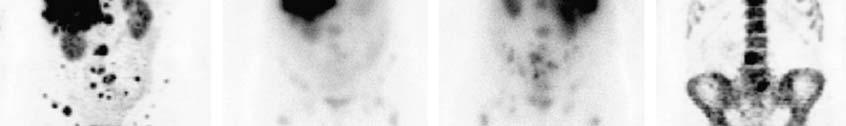
94 Cu 60 ATSM vs. F 18 FDG
95 Cu 60 ATSM
96 Cu 60 ATSM
97 Cu 60 ATSM Predictor of Survival
98 Cu 60 ATSM vs. F 18 FDG
99 Cu 64 ATSM vs. Cu 60 ATSM
100 Cu 64 ATSM vs. Cu 60 ATSM
101
102 Hypoxia In cervical CA, tumor hypoxia is predictive of decreased disease freesurvival survival and poorer overall survival. Cu 64 ATSM provides prognostic information in cervical cancer that F 18 FDG is unable to provide. Cu 64 ATSM is strongly correlated with response to therapy and overall survival. Currently an ACRIN clinical trial is ongoing with Cu 64 ATSM in patients with cervical cancer.
103 Cancer Imaging Agents Radiopharmaceuticals for imaging cellular proliferation 3 deoxy 3 [ 18 F]fluorothymidine ([ 18 F]FLT) Imaging with 18 F labeled sigma 2 receptor ligands Imaging tumor hypoxia 60/64 Cu ATSM 18 F MISO Imaging upregulation of receptors in tumors 68 Ga labeled somatostatin analogs


104 Receptor Imaging
105 Receptor Imaging Tumor receptors have an important role in carcinogenesis and tumor growth. Evaluation of tumor receptor expression is critical in cancer therapy directed at tumor receptors. The ability to measure expression of tumor receptors is essential ilfor selecting patients for receptor targeted therapy.
106 Receptor Imaging Tumor receptor imaging can: 1. characterize tumor biology, 2. identify if therapeutic targets, and 3. delineate the pharmacodynamics of targeted cancer therapy. Advantages: Noninvasive, measurement of receptor expression of entire disease burden, and potential for serial studies.

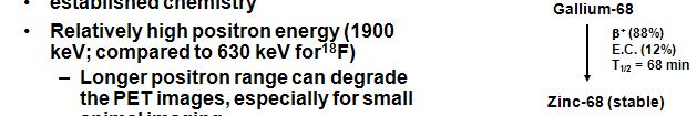
107 Gallium 68


108 Gallium 68


109 Ga 68

110 Receptor Imaging
111 Summary Proliferation, Hypoxia Proliferation: FLT has applications in determining proliferative status of tumors having implications in predicting aggressiveness and monitoring therapy Radiolabeled DNA precursors underestimate the P:Q ratio 2 receptor imaging agents show promise in animal studies but need to be validated in human imaging studies Hypoxia 60 Cu ATSM has shown promise in several clinical studies for imaging hypoxia in NSCLC, head and neck, rectal and cervical cancers 64 Cu ATSM provides higher quality images and is currently under IND with a multi center trial to begin soon Caution should be advised that this agent does not image hypoxia in all tumor types, i.e. prostate cancer
112 Summary Receptors Receptor Targeted Agents: Somatostatin is one tumor receptor that has been heavily studied both pre clinically as well as clinically The implementation of PET agents ( 68 Ga) compared to SPECT ( 111 In) has greatly improved the tumor targeting and non target tissue clearance
113 Translational Molecular Imaging in Oncology Current Near Future I 131 Thyroid Proliferation In 111 octreotide F 18 FLT/ISO /SO I 123/131 MIBG Hypoxia In 111 capromab F 18 FDG Cu 64 ATSM/FMISO Receptors Angiogenesis
114
115 Tumor Imaging Future Prospects I would like to thank the following colleagues: Carolyn Anderson Jon McConathy Robert Mach Mike Graham Barry Siegel Mike Welch Steve Larson
Objective. Assessment Question. I. Theranostics II. Classic Theranostic Agent
 Up and Coming Research Radiopharmaceuticals Dao Le, Pharm.D, BCNP Director, Nuclear Medicine, Radiopharmacy University of Texas MD Anderson Cancer Center Objective I. Theranostics II. Classic Theranostic
Nuclear Medicine in Oncology
 Radiopharmaceuticals Nuclear Medicine in Oncology Practice Pharmaceutical Radionuc lide Function Tumor type Diphosphonates Tc-99m Osteoblast Bone tumor & metast. Ga-citrate Ga-67 Fe-analogue Bronchogenous
Nuclear medicine in oncology. 1. Diagnosis 2. Therapy
 Nuclear medicine in oncology 1. Diagnosis 2. Therapy Diagnosis - Conventional methods - Nonspecific radiopharmaceuticals cumulating in tumours - Specific radiopharmaceuticals (receptor- and immunoscintigraphy)
Molecular Imaging and Cancer
 Molecular Imaging and Cancer Cancer causes one in every four deaths in the United States, second only to heart disease. According to the U.S. Department of Health and Human Services, more than 512,000
Molecular Imaging Guided Therapy: The Perfect Storm. David M Schuster, MD Emory University Department of Radiology Atlanta, GA
 Molecular Imaging Guided Therapy: The Perfect Storm David M Schuster, MD Emory University Department of Radiology Atlanta, GA Talk can be found at radiology.emory.edu Let s start with a case 74 year
Indications of PET/CT in oncology
 Monday, August 27, 2012 Session 1, 10:00-10:40 Indications of PET/CT in oncology Helle Westergren Hendel MD, PhD, assistant professor Bacelor in Leadership & Health Ecomomics Head of Clinical PET, Herlev
Physical Bases : Which Isotopes?
 Physical Bases : Which Isotopes? S. Gnesin Institute of Radiation Physics, Lausanne University Hospital, Lausanne, Switzerland 1/53 Theranostic Bruxelles, 2 Octobrer 2017 Theranostic : use of diagnostic
PET/CT in oncology. Positron emission tomography
 Clinical Medicine 2012, Vol 12, No 4: 368 72 PET/CT in oncology Fahim-Ul-Hassan, SpR Nuclear Medicine, Guy s Hospital, London; Gary J Cook, professor of Clinical PET, KCL Division of Imaging Sciences &
Radionuclide Therapy: History, Present and Future Promise. History of Radionuclide (RN) Therapy. History of RN Therapy 8/2/2017
 Therapy. History of RN Therapy 8/2/2017") Radionuclide Therapy: History, Present and Future Promise Bennett S. Greenspan, MD, MS Professor, Dept. of Radiology, MCG / AU President, SNMMI AAPM Annual Meeting, Denver, CO History Symposium 8/02/2017
Radionuclide Therapy: History, Present and Future Promise Bennett S. Greenspan, MD, MS Professor, Dept. of Radiology, MCG / AU President, SNMMI AAPM Annual Meeting, Denver, CO History Symposium 8/02/2017
Subject: PET Scan With or Without CT Attenuation. Original Effective Date: 11/7/2017. Policy Number: MCR: 610. Revision Date(s): Review Date:
: Review Date:") Subject: PET Scan With or Without CT Attenuation Policy Number: MCR: 610 Revision Date(s): MHW Original Effective Date: 11/7/2017 Review Date: DISCLAIMER This Molina Clinical Review (MCR) is intended to
Subject: PET Scan With or Without CT Attenuation Policy Number: MCR: 610 Revision Date(s): MHW Original Effective Date: 11/7/2017 Review Date: DISCLAIMER This Molina Clinical Review (MCR) is intended to
PET Assessment of Tumor Hypoxia
 PET Assessment of Tumor Hypoxia Farrokh Dehdashti, M.D. Mallinckrodt Institute of Radiology Washington University St. Louis, Missouri 9/30/10 This work was supported by National Institute of Health R21
Molecular Imaging as a Cancer Biomarker:
 Molecular Imaging as a Cancer Biomarker: Imaging to Guide Targeted Cancer Therapy 51 st Annual AAPM Meeting, Anaheim CA David A. Mankoff, MD, PhD Seattle Cancer Care Alliance University of Washington Seattle,
Index. Surg Oncol Clin N Am 16 (2007) Note: Page numbers of article titles are in boldface type.
 Note: Page numbers of article titles are in boldface type.") Surg Oncol Clin N Am 16 (2007) 465 469 Index Note: Page numbers of article titles are in boldface type. A Adjuvant therapy, preoperative for gastric cancer, staging and, 339 B Breast cancer, metabolic
Surg Oncol Clin N Am 16 (2007) 465 469 Index Note: Page numbers of article titles are in boldface type. A Adjuvant therapy, preoperative for gastric cancer, staging and, 339 B Breast cancer, metabolic
The Drug Development Paradigm in Oncology: Role of Imaging Hedvig Hricak MSKCC
 The Drug Development Paradigm in Oncology: Role of Imaging Hedvig Hricak MSKCC Precision Medicine: Imaging in Clinical Trials and Drug Development* Critical elements: Patient Selection, Dose Finding &
NUCLEAR MEDICINE Molecular Imaging + Endo-Radiotherapy
 NUCLEAR MEDICINE Molecular Imaging + Endo-Radiotherapy Istvan Szilvási Dept. of Nuclear Medicine Semmelweis University and HDF Medical Centre 2016 DEFINITION OF NUCLEAR MEDICINE Medical applications of
Principles of nuclear metabolic imaging. Prof. Dr. Alex Maes AZ Groeninge Kortrijk and KULeuven Belgium
 Principles of nuclear metabolic imaging Prof. Dr. Alex Maes AZ Groeninge Kortrijk and KULeuven Belgium I. Molecular imaging probes A. Introduction - Chemical disturbances will precede anatomical abnormalities
Dr Sneha Shah Tata Memorial Hospital, Mumbai.
 Dr Sneha Shah Tata Memorial Hospital, Mumbai. Topics covered Lymphomas including Burkitts Pediatric solid tumors (non CNS) Musculoskeletal Ewings & osteosarcoma. Neuroblastomas Nasopharyngeal carcinomas
Chapter 10. Summary, conclusions and future perspectives
 Chapter 10 Summary, conclusions and future perspectives 10.1 SUMMARY In this thesis, a new tumor imaging tracer in nuclear medicine is studied. This 123 tracer, L-3-[ I]Iodo-alpha-methyl-tyrosine (IMT),
Preclinical imaging and therapy. Marion de Jong
 Preclinical imaging and therapy Marion de Jong Content Introduction Preclinical Imaging Preclinical Therapy to raise awareness about problems related to translation of animal studies Radiopharmaceuticals
Molecular Imaging and Breast Cancer
 Molecular Imaging and Breast Cancer Breast cancer forms in tissues of the breast usually in the ducts, tubes that carry milk to the nipple, and lobules, the glands that make milk. It occurs in both men
The Use of PET Scanning in Urologic Oncology
 The Use of PET Scanning in Urologic Oncology Dr Nicholas C. Buchan Uro-oncology Fellow 1 2 Aims To understand the basic concepts underlying PET scanning. Understand the emerging role of PET Scanning for
New imaging techniques: let there be light. Felix M. Mottaghy Department of Nuclear Medicine University Hospital KU Leuven
 New imaging techniques: let there be light Felix M. Mottaghy Department of Nuclear Medicine University Hospital KU Leuven Medical imaging and the pathology cascade Molecular/Cellular disturbance Alterations
Hybrid Imaging SPECT/CT PET/CT PET/MRI. SNMMI Southwest Chapter Aaron C. Jessop, MD
 Hybrid Imaging SPECT/CT PET/CT PET/MRI SNMMI Southwest Chapter 2014 Aaron C. Jessop, MD Assistant Professor, Department of Nuclear Medicine UT MD Anderson Cancer Center, Houston, Texas Complimentary role
Clinical indications for positron emission tomography
 Clinical indications for positron emission tomography Oncology applications Brain and spinal cord Parotid Suspected tumour recurrence when anatomical imaging is difficult or equivocal and management will
Medical Policy An independent licensee of the Blue Cross Blue Shield Association
 PET Scanning: Oncologic Applications Page 1 of 88 Medical Policy An independent licensee of the Blue Cross Blue Shield Association Title: Positron Emission Tomography (PET) Scanning: Oncologic Applications
Cabozantinib for medullary thyroid cancer. February 2012
 Cabozantinib for medullary thyroid cancer February 2012 This technology summary is based on information available at the time of research and a limited literature search. It is not intended to be a definitive
An Introduction to PET Imaging in Oncology
 January 2002 An Introduction to PET Imaging in Oncology Janet McLaren, Harvard Medical School Year III Basics of PET Principle of Physiologic Imaging: Allows in vivo visualization of structures by their
THE PARATHYROID GLAND THEORY AND NUCLEAR MEDICINE PRACTICE
 THE PARATHYROID GLAND THEORY AND NUCLEAR MEDICINE PRACTICE George N. Sfakianakis MD Professor of Radiology and Pediatrics Director, Division of Nuclear Medicine UM/JMMC Miami FL October 2009 ENDONCRINE
Molecular Imaging: - SPECT agents under development - Imaging challenges
 Molecular Imaging: - SPECT agents under development - Imaging challenges Jody Garrard, CNMT Gamma Camera Product Manager Philips Nuclear Medicine jody.garrard@philips.com When you reach turning points
Positron Emission Tomography in Lung Cancer
 May 19, 2003 Positron Emission Tomography in Lung Cancer Andrew Wang, HMS III Patient DD 53 y/o gentleman presented with worsening dyspnea on exertion for the past two months 30 pack-year smoking Hx and
Nuclear Medicine and PET. D. J. McMahon rev cewood
 Nuclear Medicine and PET D. J. McMahon 150504 rev cewood 2018-02-15 Key Points Nuclear Medicine and PET: Imaging: Understand how Nuc Med & PET differ from Radiography & CT by the source of radiation. Be
PET IMAGING (POSITRON EMISSION TOMOGRAPY) FACT SHEET
 FACT SHEET") Positron Emission Tomography (PET) When calling Anthem (1-800-533-1120) or using the Point of Care authorization system for a Health Service Review, the following clinical information may be needed to
Positron Emission Tomography (PET) When calling Anthem (1-800-533-1120) or using the Point of Care authorization system for a Health Service Review, the following clinical information may be needed to
Studying tissue pharmacokinetics by Positron Emission Tomography (PET)
") BIOMATH M263 Clinical Pharmacology Lecture Studying tissue pharmacokinetics by Positron Emission Tomography (PET) Imaging tumor responses to therapy Caius G. Radu, M.D. Ahmanson Translational Imaging Division
BIOMATH M263 Clinical Pharmacology Lecture Studying tissue pharmacokinetics by Positron Emission Tomography (PET) Imaging tumor responses to therapy Caius G. Radu, M.D. Ahmanson Translational Imaging Division
Index. radiologic.theclinics.com. Note: Page numbers of article titles are in boldface type.
 Index Note: Page numbers of article titles are in boldface type. A ACC. See Adrenal cortical carcinoma. Acromegaly and the pituitary gland, 551 Acute suppurative thyroiditis, 405, 406 Addison, Thomas and
Recent initiatives of the FANC. Michel Biernaux Health Protection Service Health and Environment Department
 Recent initiatives of the FANC Michel Biernaux Michel.biernaux@fanc.fgov.be Health Protection Service Health and Environment Department Reminder objectives of the national survey : 1.Review the average
PET in Prostate Cancer
 PET in Prostate Cancer Tom R. Miller, M.D., Ph.D. Mallinckrodt Institute of Radiology Washington University School of Medicine St. Louis, Missouri, USA Prostate Imaging Bone Scintigraphy primarily for
Physics of MBI (~10 slides)
") Physics of MBI (~10 slides) Molecular Breast Imaging (MBI) physics and performance testing JW Hugg, BR Simrak, PD Smith, BE Patt Gamma Medica, Inc., Northridge, CA Molecular Breast Imaging (MBI) is an
Physics of MBI (~10 slides) Molecular Breast Imaging (MBI) physics and performance testing JW Hugg, BR Simrak, PD Smith, BE Patt Gamma Medica, Inc., Northridge, CA Molecular Breast Imaging (MBI) is an
F NaF PET/CT in the Evaluation of Skeletal Malignancy
 F NaF PET/CT in the Evaluation of Skeletal Malignancy Andrei Iagaru, MD September 26, 2013 School of of Medicine Ø Introduction Ø F NaF PET/CT in Primary Bone Cancers Ø F NaF PET/CT in Bone Metastases
PET/CT Imaging in Cancer: Current Applications and Future Directions
 PET/CT Imaging in Cancer: Current Applications and Future Directions Michael D. Farwell, MD; Daniel A. Pryma, MD; and David A. Mankoff, MD, PhD Positron emission tomography (PET) is a radiotracer imaging
Individualised Treatment Planning for Radionuclide therapy (Molecular Radiotherapy)
") Individualised Treatment Planning for Radionuclide therapy (Molecular Radiotherapy) FMU/ICRP workshop 2017 Glenn Flux Royal Marsden Hospital & Institute of Cancer Research Sutton UK ICRP Task Group 101
Individualised Treatment Planning for Radionuclide therapy (Molecular Radiotherapy) FMU/ICRP workshop 2017 Glenn Flux Royal Marsden Hospital & Institute of Cancer Research Sutton UK ICRP Task Group 101
Theranostics in Nuclear Medicine
 Theranostics in Nuclear Medicine Patrick FLAMEN, MD, PhD Head Nuclear Medicine Institut Jules Bordet Université Libre de Bruxelles (U.L.B.) n Theranostics in Nuclear Medicine n A form of (nuclear) diagnostic
Page: 1 of 29. For this policy, PET scanning is discussed for the following 4 applications in oncology:
 Emission Tomography Scanning Page: 1 of 29 Last Review Status/Date: June 2015 Description Positron emission tomography (PET) scans are based on the use of positron-emitting radionuclide tracers coupled
Nuclear medicine in general practice. Dr Reza Garzan MD, FRACP, FAANMS
 Nuclear medicine in general practice Dr Reza Garzan MD, FRACP, FAANMS Myocardial perfusion study Bone scans in general practice Thyroid scans in general practice Gamma camera Detection of gamma rays Myocardial
New Visions in PET: Surgical Decision Making and PET/CT
 New Visions in PET: Surgical Decision Making and PET/CT Stanley J. Goldsmith, MD Director, Nuclear Medicine Professor, Radiology & Medicine New York Presbyterian Hospital- Weill Cornell Medical Center
METROLOGY TO SUPPORT INNOVATION IN MOLECULAR RADIOTHERAPY. Glenn Flux
 METROLOGY TO SUPPORT INNOVATION IN MOLECULAR RADIOTHERAPY Glenn Flux Head of Radioisotope Physics Royal Marsden Hospital & Institute of Cancer Research Sutton UK CGPM 2018 glenn.flux@icr.ac.uk Overview
Policy. Number: (Replaces CPBs 239, 309, 320) *Please see amendment for Pennsylvania Medicaid at the end
 *Please see amendment for Pennsylvania Medicaid at the end") 1 of 99 Number: 0168 (Replaces CPBs 239, 309, 320) Policy *Please see amendment for Pennsylvania Medicaid at the end of this CPB. ProstaScint Aetna considers ProstaScint scans medically necessary for either
1 of 99 Number: 0168 (Replaces CPBs 239, 309, 320) Policy *Please see amendment for Pennsylvania Medicaid at the end of this CPB. ProstaScint Aetna considers ProstaScint scans medically necessary for either
THE ROLE OF CONTEMPORARY IMAGING AND HYBRID METHODS IN THE DIAGNOSIS OF CUTANEOUS MALIGNANT MELANOMA(CMM) AND MERKEL CELL CARCINOMA (MCC)
 AND MERKEL CELL CARCINOMA (MCC)") THE ROLE OF CONTEMPORARY IMAGING AND HYBRID METHODS IN THE DIAGNOSIS OF CUTANEOUS MALIGNANT MELANOMA(CMM) AND MERKEL CELL CARCINOMA (MCC) I.Kostadinova, Sofia, Bulgaria CMM some clinical facts The incidence
THE ROLE OF CONTEMPORARY IMAGING AND HYBRID METHODS IN THE DIAGNOSIS OF CUTANEOUS MALIGNANT MELANOMA(CMM) AND MERKEL CELL CARCINOMA (MCC) I.Kostadinova, Sofia, Bulgaria CMM some clinical facts The incidence
Oncologic Applications of PET Scanning
 6.01.26 Oncologic Applications of PET Scanning Section 6.0 Radiology Subsection Effective Date February 15, 2015 Original Policy Date January 26, 2009 Next Review Date December 2015 Description Positron
Medical Policy An independent licensee of the Blue Cross Blue Shield Association
 PET Scanning: Oncologic Applications Page 1 of 42 Medical Policy An independent licensee of the Blue Cross Blue Shield Association Title: See also: Positron Emission Tomography (PET) Scanning: Oncologic
Lung Cancer Imaging. Terence Z. Wong, MD,PhD. Department of Radiology Duke University Medical Center Durham, NC 9/9/09
 Lung Cancer Imaging Terence Z. Wong, MD,PhD Department of Radiology Duke University Medical Center Durham, NC 9/9/09 Acknowledgements Edward F. Patz, Jr., MD Jenny Hoang, MD Ellen L. Jones, MD, PhD Lung
PET/CT Value: Rocky Mountain Cancer Centers
 PET/CT Value: Rocky Mountain Cancer Centers Glenn Balasky Executive Director Rocky Mountain Cancer Centers glenn.balasky@usoncology.com CANM/CAMRT Joint Conference March 22, 2018 Vancouver, British Columbia
OTHER NON-CARDIAC USES OF Tc-99m CARDIAC AGENTS Tc-99m Sestamibi for parathyroid imaging, breast tumor imaging, and imaging of other malignant tumors.
 DEFINITION OF CARDIAC RADIOPHARMACEUTICAL: A radioactive drug which, when administered for purpose of diagnosis of heart disease, typically elicits no physiological response from the patient. Even though
General Nuclear Medicine
 General Nuclear Medicine What is General Nuclear Medicine? What are some common uses of the procedure? How should I prepare? What does the equipment look like? How does the procedure work? How is the procedure
FMU-ICRP Workshop on Radiological Protection in Medicine Current Status in Radionuclide Therapy Tuesday, October 3, 2017 Makoto Hosono, MD PhD Kindai
 FMU-ICRP Workshop on Radiological Protection in Medicine Current Status in Radionuclide Therapy Tuesday, October 3, 2017 Makoto Hosono, MD PhD Kindai University Faculty of Medicine, Osaka, Japan Current
Nuclear Medicine Head and Neck Region. Bán Zsuzsanna, MD University of Pécs, Department of Nuclear Medicine
 Nuclear Medicine Head and Neck Region Bán Zsuzsanna, MD University of Pécs, Department of Nuclear Medicine Thyroid scintigraphy Parathyroid scintigraphy F18-FDG PET examinations in head and neck cancer
Option D: Medicinal Chemistry
 Option D: Medicinal Chemistry Basics - unstable radioactive nuclei emit radiation in the form of smaller particles alpha, beta, positron, proton, neutron, & gamma are all used in nuclear medicine unstable
Systemic Therapy for Pheos/Paras: Somatostatin analogues, small molecules, immunotherapy and other novel approaches in the works.
 Systemic Therapy for Pheos/Paras: Somatostatin analogues, small molecules, immunotherapy and other novel approaches in the works. Arturo Loaiza-Bonilla, MD, FACP Assistant Professor of Clinical Medicine
FDG-PET/CT for cancer management
 195 REVIEW FDG-PET/CT for cancer management Hideki Otsuka, Naomi Morita, Kyo Yamashita, and Hiromu Nishitani Department of Radiology, Institute of Health Biosciences, The University of Tokushima, Graduate
performed to help sway the clinician in what the appropriate diagnosis is, which can substantially alter the treatment of management.
 Hello, I am Maura Polansky at the University of Texas MD Anderson Cancer Center. I am a Physician Assistant in the Department of Gastrointestinal Medical Oncology and the Program Director for Physician
FACULTY MEMBERSHIP APPLICATION Tulane Cancer Center
 FACULTY MEMBERSHIP APPLICATION Tulane Cancer Center 1430 Tulane Ave., Box SL-68, New Orleans, LA 70112-2699 J. Bennett Johnston Building, Mezzanine (Floor 1A), Suite A102 (504) 988-6060, fax (504) 988-6077,
Evaluation of Lung Cancer Response: Current Practice and Advances
 Evaluation of Lung Cancer Response: Current Practice and Advances Jeremy J. Erasmus I have no financial relationships, arrangements or affiliations and this presentation will not include discussion of
Radionuclide Therapy. Prof. Dr. Çetin Önsel. Cerrahpaşa Medical School Department of Nuclear Medicine. Radionuclide Therapy
 Prof. Dr. Çetin Önsel Cerrahpaşa Medical School Department of Nuclear Medicine Principles of radionuclide therapy (1) The radionuclide has to be concentrated at the site of the abnormality (tumor) with
SUMMARY OF PRODUCT CHARACTERISTICS
 SUMMARY OF PRODUCT CHARACTERISTICS 1. NAME OF THE MEDICINAL PRODUCT Octreoscan Kit for preparation of 111 In-Pentetreotide 111 MBq/ ml 2. QUALITATIVE AND QUANTITATIVE COMPOSITION Octreoscan is supplied
Nuclear medicine studies of the digestiv system. Zámbó Katalin Department of Nuclear Medicine
 Nuclear medicine studies of the digestiv system Zámbó Katalin Department of Nuclear Medicine Anatomy of the liver Liver scintigraphy The labelled colloid (200 MBq 99mTc-Fyton) is phagocyted by the Kuppfer-cells
Radiology Pathology Conference
 Radiology Pathology Conference Nadia F. Yusaf, M.D. PGY-3 1/29/2010 Presentation material is for education purposes only. All rights reserved. 2010 URMC Radiology Page 1 of 90 Case 1 60 year- old man presents
Understanding Biological Activity to Inform Drug Development
 National Cancer Policy Forum Understanding Biological Activity to Inform Drug Development December 12, 2016 Wolfgang Weber Molecular Imaging and Therapy Service Department of Radiology RECIST Response
Ryan Niederkohr, M.D. Slides are not to be reproduced without permission of author
 Ryan Niederkohr, M.D. CMS: PET/CT CPT CODES 78814 Limited Area (e.g., head/neck only; chest only) 78815 78816 Regional (skull base to mid-thighs) True Whole Body (skull vertex to feet) SELECTING FIELD
Nuclear medicine studies of the digestiv system. Zámbó Katalin Department of Nuclear Medicine
 Nuclear medicine studies of the digestiv system Zámbó Katalin Department of Nuclear Medicine Imaging tehniques Anatomy Physiology Metabolism Molecular Rtg. / CT PET / SPECT MRI MR spectroscopy fmri Ultrasound
Quantitative Theranostics in Nuclear Medicine
 Quantitative Theranostics in Nuclear Medicine M. Lassmann Klinik und Poliklinik für Nuklearmedizin Direktor: Prof. Dr. A. Buck Contents What is Theranostics? Potential Targets Basic Principles of Quantitative
Prostate Cancer and PSMA:
 Prostate Cancer and PSMA: The Clinical Perspective from Liverpool. Dr. Christopher Mayes. Professor Sobhan Vinjamuri. Department of Nuclear Medicine Royal Liverpool University Hospital (BSUR 2016 Plymouth)
Nuclear Medicine: Basics to therapy
 Nuclear Medicine: Basics to therapy RCP Medical careers day Dr Sabina Dizdarevic MD MSc PhD FRCP Dr Deena Neriman MBBS FRCR Ms Charlotte Weston CEO BNMS On behalf of the British Nuclear Medicine Society
PSMA PET SCANNING AND THERANOSTICS IN PROSTATE CANCER KEVIN TRACEY, MD, FRCPC PRECISION DIAGNSOTIC IMAGING REGIONAL PET/CT CENTRE
 PSMA PET SCANNING AND THERANOSTICS IN PROSTATE CANCER KEVIN TRACEY, MD, FRCPC PRECISION DIAGNSOTIC IMAGING REGIONAL PET/CT CENTRE DISCLOSURES/CONFLICTS NONE OBJECTIVES Understand current diagnostic role
Ga68 Imaging. Roland HUSTINX Division of Nuclear Medicine and Oncologic Imaging Centre Hospitalier Universitaire de Liège Belgium
 Ga68 Imaging Roland HUSTINX Division of Nuclear Medicine and Oncologic Imaging Centre Hospitalier Universitaire de Liège Belgium 68 Ga Produced by a 68 Ge/ 68 Ga generator Decays by positron emission
PET tracer development a tale of mice and men
 Cancer Imaging (2006) 6, S102 S106 DOI: 10.1102/1470-7330.2006.9098 CI MINI SYMPOSIUM: PET THE FUTURE Tuesday 17 October 2006, 14:00 15:30 PET tracer a tale of mice and men Rodney J Hicks, Donna Dorow
Nuclear Medicine in Thyroid Cancer. Phillip J. Koo, MD Division Chief of Diagnostic Imaging
 Nuclear Medicine in Thyroid Cancer Phillip J. Koo, MD Division Chief of Diagnostic Imaging Financial Disclosures Bayer Janssen Learning Objectives To learn the advantages and disadvantages of SPECT/CT
Imaging of Neuroendocrine Metastases
 Imaging of Neuroendocrine Metastases Aoife Kilcoyne, Shaunagh McDermott, Colin McCarthy,Manuel Patino, Dushyant Sahani, Michael Blake Abdominal Imaging Division Massachusetts General Hospital Disclosure
PET/CT for Therapy Assessment in Oncology
 PET/CT for Therapy Assessment in Oncology Rodolfo Núñez Miller, M.D. Nuclear Medicine Section Division of Human Health International Atomic Energy Agency Vienna, Austria Clinical Applications of PET/CT
Radioisotopes for staging and follow-up of prostate cancer. F. Scopinaro
 Radioisotopes for staging and follow-up of prostate cancer F. Scopinaro Specific Radiotracers Gamma ray emitters Positron emitters 111 In capromab 111 In octreotide 99m Tc Tyr-octr. (more than one tracer)
RADIONUCLIDE THERAPY FOR PALLIATION OF PAIN FROM BONY METASTASES
 RADIONUCLIDE THERAPY FOR PALLIATION OF PAIN FROM BONY METASTASES Overview and Topics to Be Covered Reviews the use of radionuclide therapy in nuclear medicine for palliation of pain due to bony metastases.
PET/CT Frequently Asked Questions
 PET/CT Frequently Asked Questions General Q: Is FDG PET specific for cancer? A: No, it is a marker of metabolism. In general, any disease that causes increased metabolism can result in increased FDG uptake
Nuclear Medicine: Manuals. Nuclear Medicine. Nuclear imaging. Emission imaging: study types. Bone scintigraphy - technique
 Nuclear Medicine - Unsealed radioactive preparations the tracer mixes with the patients body fluids on a molecular level (e.g. after intravenous injection) - 3 main fields: - In vitro : measuring concentrations
στη σταδιοποίηση του καρκίνου του προστάτη Γ. Αρσος, Γ Εργ. Πυρηνικής Ιατρικής ΑΠΘ, ΓΝΘ Παπαγεωργίου
 Η θέση του PET/CT στη σταδιοποίηση του καρκίνου του προστάτη Γ. Αρσος, Γ Εργ. Πυρηνικής Ιατρικής ΑΠΘ, ΓΝΘ Παπαγεωργίου 2014 : the Guidelines year. PRINCIPLES OF IMAGING Imaging is performed for the detection
The Role of PET / CT in Lung Cancer Staging
 July 2004 The Role of PET / CT in Lung Cancer Staging Vlad Vinarsky, Harvard Medical School Year IV Patient AM HPI: 81 yo F p/w hemoptysis x 1 month LLL lesion on CXR, not responsive to Abx 35 pack-year
Nuclear medicine in oncology. 1. Diagnosis 2. Therapy
 Nuclear medicine in oncology 1. Diagnosis 2. Therapy Diagnosis - Conventional methods - Nonspecific radiopharmaceuticals cumulating in tumours - Specific radiopharmaceuticals, receptor- and immunoscintigraphy
Itroduction to the Nuclear Medicine: biophysics and basic principles. Zámbó Katalin Department of Nuclear Medicine
 Itroduction to the Nuclear Medicine: biophysics and basic principles Zámbó Katalin Department of Nuclear Medicine NUCLEAR MEDICINE Application of the radioactive isotopes in the diagnostics and in the
New Challenge of Nuclear Medicine in Oncology -The dawn of functional imaging-
 New Challenge of Nuclear Medicine in Oncology -The dawn of functional imaging- Nihon Medi-Physics Co., Ltd. Corporate Planning and Coordination Office Strategic Marketing Hideshi HATTORI Research and Development
PET-CT for radiotherapy planning in lung cancer: current recommendations and future directions
 PET-CT for radiotherapy planning in lung cancer: current recommendations and future directions Gerry Hanna Centre for Cancer Research and Cell Biology Queen s University of Belfast @gerryhanna Talk Outline
Chapter 19: Radionuclide Therapy
 Chapter 19: Radionuclide Therapy Slide set of 40 slides based on the chapter authored by G. Flux and Y. Du of the publication (ISBN 978 92 0 143810 2): Nuclear Medicine Physics: A Handbook for Teachers
Radiopharmacy. Prof. Dr. Çetin ÖNSEL. CTF Nükleer Tıp Anabilim Dalı
 Prof. Dr. Çetin ÖNSEL CTF Nükleer Tıp Anabilim Dalı What is Nuclear Medicine? Nuclear Medicine is the branch of medicine concerned with the use of radionuclides in the study and the diagnosis of diseases.
Overview Therapeutic Radiopharmaceuticals in Nuclear Medicine Definition: Characteristics of the Ideal Therapeutic Radiopharmaceutical
 Overview After reviewing this tutorial, the users should be able to list the most important characteristics of the ideal therapeutic radiopharmaceutical and briefly discuss each of these as it relates
Thyroid Cancer: Imaging Techniques (Nuclear Medicine)
") Thyroid Cancer: Imaging Techniques (Nuclear Medicine) Andrei Iagaru, MD MIPS Molecular Imaging Program at Stanford Stanford University School of Medicine Department of Radiology Introduction Ø There are
Thyroid Cancer: Imaging Techniques (Nuclear Medicine) Andrei Iagaru, MD MIPS Molecular Imaging Program at Stanford Stanford University School of Medicine Department of Radiology Introduction Ø There are
Austin Radiological Association Nuclear Medicine Procedure PROSTATE CANCER STUDY (In-111-Capromab Pendetide [ProstaScint ])
![Austin Radiological Association Nuclear Medicine Procedure PROSTATE CANCER STUDY (In-111-Capromab Pendetide [ProstaScint ])](/thumbs/81/82771892.jpg "Austin Radiological Association Nuclear Medicine Procedure PROSTATE CANCER STUDY (In-111-Capromab Pendetide [ProstaScint ])") Austin Radiological Association Nuclear Medicine Procedure PROSTATE CANCER STUDY (In-111-Capromab Pendetide [ProstaScint ]) Overview Indications The Prostate Cancer Study with an indium-111 labeled murine
Austin Radiological Association Nuclear Medicine Procedure PROSTATE CANCER STUDY (In-111-Capromab Pendetide [ProstaScint ]) Overview Indications The Prostate Cancer Study with an indium-111 labeled murine
Use of imaging systems for patient modeling - PET and SPECT
 Use of imaging systems for patient modeling - PET and SPECT Sasa Mutic Department of Radiation Oncology Siteman Cancer Center Mallinckrodt Institute of Radiology Washington University School of Medicine
Typical PET Image. Elevated uptake of FDG (related to metabolism) Lung cancer example: But where exactly is it located?
 Lung cancer example: But where exactly is it located?") Typical PET Image Elevated uptake of FDG (related to metabolism) Lung cancer example: But where exactly is it located? PET/CT Oncology Imaging Anatometabolic fusion images are useful in the management
Typical PET Image Elevated uptake of FDG (related to metabolism) Lung cancer example: But where exactly is it located? PET/CT Oncology Imaging Anatometabolic fusion images are useful in the management
Index. Surg Oncol Clin N Am 15 (2006) Note: Page numbers of article titles are in boldface type.
 Note: Page numbers of article titles are in boldface type.") Surg Oncol Clin N Am 15 (2006) 681 685 Index Note: Page numbers of article titles are in boldface type. A Ablative therapy, for liver metastases in patients with neuroendocrine tumors, 517 with radioiodine
Surg Oncol Clin N Am 15 (2006) 681 685 Index Note: Page numbers of article titles are in boldface type. A Ablative therapy, for liver metastases in patients with neuroendocrine tumors, 517 with radioiodine
SARCOPHAGINE CHELATORS AND COPPER ISOTOPES FOR IMAGING AND THERAPY
 SEEING IS BELIEVING SARCOPHAGINE CHELATORS AND COPPER ISOTOPES FOR IMAGING AND THERAPY Sydney Vital and STEaM Neuroendocrine Tumour Preceptorship May 2018 Amos Hedt Head of Clinical Development 1 CLARITY
Specialised Services Policy CP66: 68-gallium DOTA- peptide scanning for the Management of Neuroendocrine Tumours (NETs)
") Specialised Services Policy CP66: Management of Neuroendocrine Tumours (NETs) Document Author: Assistant Planner for Cancer and Blood Executive Lead: Director of Quality and Nursing Approved by: Management
Specialised Services Policy CP66: Management of Neuroendocrine Tumours (NETs) Document Author: Assistant Planner for Cancer and Blood Executive Lead: Director of Quality and Nursing Approved by: Management
Use of molecular and functional imaging for treatment planning The Good, The Bad and The Ugly
 Use of molecular and functional imaging for treatment planning The Good, The Bad and The Ugly Robert Jeraj, PhD Associate Professor of Medical Physics, Human Oncology, Radiology and Biomedical Engineering
Radionuclid Therapy. dr. Erzsébet Schmidt Dept. of Nuclear Medicine University of Pécs
 Radionuclid Therapy dr. Erzsébet Schmidt Dept. of Nuclear Medicine University of Pécs Therapy Principle of radionuclid therapy - carrying a cytotoxic agent, such as radionuclid, direct to an aberrant cell
SUGGESTED PRESCRIBED DOSAGE LIST
 approved and issued by a noted Authorized User on your Materials License. CENTRAL NERVOUS SYSTEM Brain Metabolism Brain Perfusion Cisternography Ventricular Shunt 18 F-FDG 10-20 mci (370 740 MBq) 99m Tc