The Role of EZH2 in Breast Cancer Progression and Metastasis
|
|
|
- Shon Robinson
- 6 years ago
- Views:
Transcription
1 The Role of EZH2 in Breast Cancer Progression and Metastasis by Heather Marie Moore A dissertation submitted in partial fulfillment of the requirements for the degree of Doctor of Philosophy (Cellular and Molecular Biology) in the University of Michigan 2013 Doctoral Committee: Professor Celina G. Kleer, Chair Professor Andrzej A. Dlugosz Professor Gregory R. Dressler Professor Sofia D. Merajver Associate Professor Sooryanarayana Varambally
2 Heather M. Moore 2013

3 DEDICATION Kay Lavon Dickson I dedicate my thesis in loving memory of my aunt Kay Dickson. Kay passed away in October of 2012 from pancreatic cancer and her memory reminds me every day why the research I conduct is so very important. Kay overflowed with love and warmth and she generously shared these with those who were lucky enough to have known her. ii
4 ACKNOWLEDGEMENTS I strongly believe that the well-known proverb It takes a village to raise a child can be applied to those who decide to pursue a biomedical graduate degree. A small village was definitely required and aided me every step of the way through completion of this dissertation. First and foremost, I would like to express my sincerest gratitude to my mentor, Dr. Celina Kleer. I really am amazed at how much I have grown as a scientist under her guidance. It is astounding and humbling how much my scientific understanding, not to mention proficiency in lab skills, has progressed and improved with her as my mentor. I appreciate that her door is always open and that she always welcomes me with a smile, even if she has a presentation or grant due the next day. She always exemplifies patience and heartfelt concern for those surrounding her, and I know she has genuinely wished for my success in all aspects of my graduate career. I have been extremely fortunate that Celina took a chance on me years ago when I needed to change labs. I know that my time in the Kleer lab has prepared me for what lies ahead. As for the Kleer lab itself, I must thank all the members past and present. Although some of the faces have changed over the years, the positive attitude of the lab has stayed the same. Particularly, I thank Maria Gonzalez for keeping the lab afloat as an efficient lab manager. No matter how many times I come over to her desk with a question, she always patiently takes the time to give me an answer and insist me. Also, I must thank Kathy Toy for her never failing happy attitude and excellent organizational iii
5 skills. Even in the next room, I can her laughter on a daily basis. Maria and Kathy have been pivotal in my research and I cannot thank them enough for helping me accomplish my experimental goals. To my committee members, I thank you for your invaluable input and patience over the years. Like all graduate students, I was terrified at the thought of committee meetings in the beginning, but you made them anything but. Your insight on my research was incredibly helpful and kept me on track. You opened my eyes to questions and perspectives that I had not even considered and therefore improved my work. In addition, I would like to thank Chris Edwards in the Microscopy and Imaging analysis Laboratory for his assistance and training in the live cell imaging assays. Also, those within the Cellular and Molecular Biology Program cannot be forgotten. Jessica Schwartz, Bob Fuller and most recently Cathy Mitchell for making sure I stayed on track and for helping with all of the little details that graduate students forget about. I must also share my gratitude with those who know me the best, my friends and family. All of the friends I have made over the years in graduate school played a role in my success. The numerous sports teams and get-togethers always provided an escape from stress. But, having scientific friends is a priceless asset, as they also understood my daily frustrations and problems. My parents, Davy and Marlene Krueger, have always supported and encouraged me to succeed and go farther in my endeavors. They have shown unbelievable understanding during my time in graduate school and have never faltered in their belief that I could accomplish my goals (even when longer and longer periods of time occurred between calls). Lastly, I must thank my husband, Paul Moore. He is my rock and has never wavered in his support for me. He is there to iv
6 celebrate the good times and to provide a steady shoulder when times are rough. As a fellow scientist, he has always understood the odd hours and the pressures, as we have labored through graduate school together. I had no idea that when I came to Ann Arbor years ago, I would meet the love of my life and be so incredibly happy. I wish the absolute best to all I have encountered throughout this journey! v
7 TABLE OF CONTENTS Dedication... ii Acknowledgements... iii List of Figures... vii List of Abbreviations... ix Abstract... x Chapters 1. Introduction The Human Mammary Gland and Breast Cancer Development The Breast Cancer Metastatic Cascade Breast Cancer and the Cancer Stem Cell Hypothesis The Tumorigenic Role of EZH Figures References EZH2 inhibition decreases p38 signaling and suppresses breast cancer motility and metastasis Abstract Introduction Materials and Methods Results Discussion Figures References EZH2 expands the stem cell population in benign and tumorigenic breast cells through regulation of Notch1 signaling Abstract Introduction Materials and Methods Results Discussion Figures References Conclusions Discussion and Future Directions Clinical and Therapeutic Implications References vi
8 LIST OF FIGURES Figure 1-1 The continuum of breast cancer progression and the metastatic cascade Figure 1-2 Polycomb Repressive Complexes 1 and 2 (PRC1 & PRC2) coordinately work together to repress gene expression Figure 2-1 EZH2 knockdown in breast cancer cell lines can be achieved with targeted shrna or DZNeP treatment and leads to decreased proliferation Figure 2-2 EZH2 knockdown induces a mesenchymal-to-epithelial transition and decreases invasion in breast cancer cells Figure 2-3 EZH2 downregulation decreases proliferation and invasion in primary patient breast cancer cells Figure 2-4 EZH2 knockdown decreases breast cancer cell motility Figure 2-5 EZH2 regulates the activation of the p38 MAPK signaling pathway Figure 2-6 EZH2 regulates the activation of total and isoform specific protein levels of p Figure 2-7 EZH2, along with the PRC2 complex, binds to phosphorylated p38 and can methylate p38α in vitro Figure 2-8 EZH2 knockdown in MDA-MB-231 cells is sufficient to reduce distant metastasis Figure 2-9 Primary xenografts exhibit MET and lung metastases have significantly reduced expression of Ki67 and p-p38 with EZH2 knockdown in MDA-MB-231 cells Figure 2-10 EZH2 and p-p38 are significantly upregulated in human breast cancer metastases when compared to matched primary tumors from the same patient Figure 2-11 Table of the complete clinical and pathological information, including EZH2 and p-p38 protein expression, for the tumor microarrays containing 16 human primary breast carcinomas with matched metastases vii
9 Figure 3-1 EZH2 levels regulate stem cell numbers in SUM149 breast cancer cells Figure 3-2 EZH2 levels regulate stem cell numbers in MDA-MB-231 breast cancer cells and in patient primary breast cancer cells Figure 3-3 EZH2 knockdown in SUM149 ALDH1 + cells decreased the growth rate and time to tumor initiation of in vivo breast tumors Figure 3-4 EZH2 downregulation reduces Notch1 and downstream signaling in breast cancer cells Figure 3-5 EZH2 expression is associated with Notch1 expression in independent datasets of human invasive breast carcinomas Figure 3-6 Notch1 is required for EZH2-induced expansion of MCF10A breast stem cells, part Figure 3-7 Notch1 is required for EZH2-induced expansion of MCF10A breast stem cells, part Figure 3-8. Full length EZH2 is required for Notch1 promoter activation and expansion of mammosphere sizes and numbers Figure 3-9 EZH2 binds to the Notch1 gene promoter in benign breast cells, breast cancer cell lines and in patient primary breast cancer cells Figure 3-10 Characterization of EZH2 + /neu and EZH2 wt /neu transgenic mice Figure 3-11 Transgenic EZH2 overexpression accelerates tumor initiation and upregulates Notch1 in MMTV-neu mice Figure 3-12 EZH2 expression is associated with NICD and CD44 + /CD24 - expression in human invasive breast cancer tissues viii
10 LIST OF ABBREVIATIONS AH Atypical Hyperplasia ALDH1 Aldehyde Dehydrogenase 1 BM Basement Membrane ChIP Chromatin Immunoprecipitation CSC Cancer Stem Cell CTC Circulating Tumor Cell DCIS Ductal Carcinoma in situ DNMT DNA Methyltransferase DOX Doxycycline DZNeP 3-deazaneplanocin A EED Embryonic Ectoderm Development EMT Epithelial-to-Mesenchymal Transition ER - Estrogen Receptor Negative EZH2 Enhancer of Zeste Homolog 2 GSI γ-secretase Inhibitor HDAC Histone Deacetylase H3K27me3 trimethylation of histone H3 at lysine 27 IP Immunoprecipitation LCIS Lobular Carcinoma in situ MAPK Mitogen-Activated Protein Kinase MET Mesenchymal-to-Epithelial Transition MMP Matrix Metalloproteinase MMTV Mouse Mammary Tumor Virus PcG Polycomb Group PRC1 Polycomb Repressive Complex 1 PRC2 Polycomb Repressive Complex 2 shezh2 EZH2-targeted short hairpin RNA shrna short hairpin RNA SUZ12 Suppressor of Zeste 12 TDLU Terminal-Ductal-Lobular-Unit ix
11 ABSTRACT The Role of EZH2 in Breast Cancer Progression and Metastasis Breast cancer is the second leading cause of cancer-related deaths for women in the United States, with the majority due to the development of distant metastasis. Understanding how breast cancer cells disseminate and metastasize is essential to develop more efficacious treatments and to improve survival. Enhancer of Zeste Homolog 2 (EZH2) is a Polycomb group protein which functions mainly as a transcriptional repressor through histone trimethylation. Our laboratory has found that EZH2 overexpression in clinical samples of invasive breast carcinomas is associated with worse survival. Here, we have focused our work on elucidating the functions and mechanisms by which EZH2 promotes aggressive breast carcinomas with metastatic potential. We have found that EZH2 regulates two important processes for metastasis: the epithelial-to-mesenchymal transition and migration, and the numbers of breast cancer stem cells. We discovered that downregulation of EZH2 in aggressive and metastasizing breast cancer cells promotes a mesenchymal-to-epithelial transition and reduces motility and invasion. In vivo, EZH2 knockdown in breast cancer cells decreased spontaneous metastasis to the lungs. We uncovered an unexpected role of EZH2 in inducing the p38 signaling pathway, a known regulator of breast cancer invasion and metastasis. EZH2 was demonstrated to bind phosphorylated-p38 (p-p38) in association x
12 with other core members of the Polycomb Repressive Complex 2 (PRC2). Moreover, the effect of p-p38 was confirmed in vivo and correlated with decreased spontaneous metastasis. Through analysis of invasive human breast cancers, we found that EZH2 expression was upregulated in all cases, and that EZH2 and p-p38 were co-expressed in 63% of cases, consistent with the functional results. In our studies on the role of EZH2 in breast cancer stem cell biology, we found that EZH2 expression levels regulate stem cell numbers in nontumorigenic and malignant breast cells. Mechanistically, we revealed a novel role of EZH2 in activating Notch1 signaling through binding of the Notch1 promoter. Binding was independent of its catalytic methyltransferase activity and PRC2, and correlated instead with transcriptional activation. Notch1 inhibition was sufficient in preventing the EZH2- induced expansion of the stem cell population. In a transgenic mouse model with targeted EZH2 overexpression, we found that EZH2 promoted earlier breast cancer initiation and correlated with Notch1 expression. Additionally, EZH2, Notch1 and stem cell markers were found to correlate in human breast cancer. Taken together, these findings reveal important and novel functional links between EZH2, stem cells and breast cancer migration and invasion, and their underlying mechanisms involving EZH2- mediated regulation of p38 and Notch1 signaling pathways. Our work establishes EZH2 as a regulator of breast cancer progression and metastasis, and identifies potential targets for treatment of this deadly malignancy. xi
13 CHAPTER 1 Introduction 1-1. The Human Mammary Gland and Breast Cancer Development Mammals are distinguished from all other animal groups by the presence of a unique organ, the mammary gland, which functions in secreting milk to nourish young. The development of the mammary gland is divided into three stages: embryonic, pubertal and reproductive [1]. Proper maintenance and control of these temporal stages are essential for correct development and tissue homeostasis. Unfortunately, when aberrations in these developmental processes transpire during the postnatal life of a female, uncontrolled cell growth and subsequent breast cancer may result. For women in the United States, breast cancer is the most common malignancy and is also the second most common cause of cancer-related deaths behind lung cancer [2]. It is currently estimated that a women living in the United States has a 1 in 8 lifetime risk of being diagnosed with breast cancer. Better screening and treatment strategies have resulted in improved survival and quality of life in breast cancer patients as demonstrated in a yearly 2.2% decrease in breast cancer death rates since 1990 [2]. However, although death rates are decreasing, approximately 40,000 women were expected to die from breast cancer in 2011 alone [2]. Even though breast cancer incidence rates have remained relatively stable since 2003, approximately 288,000 new cases of in situ and invasive breast cancer were expected to be diagnosed among 1
14 women in 2011 [2]. Research, ranging from basic to clinical, into the mechanisms behind breast cancer development and progression are responsible for the improving trend in breast cancer statistics, but more research is needed to see further reductions in incidence and death rates. It is important to note that breast cancer cannot be considered a single disease. It is rather a heterogeneous mix of breast malignancies that exhibit various genetic, epigenetic and genomic alterations, which can have diverse histological presentations and outcomes in patients. This diversity has led to the subdivision of breast cancer into four main molecular classes based on gene expression profiling: luminal A, luminal B, basal-like or triple-negative and HER2/ERBB2-overexpressing [3, 4]. Our current theory on breast cancer is that it develops along a continuum within the epithelium of breast [5]. Breast ducts and acini exhibit a bilayer of two cell types present in roughly equal numbers: an outer layer of elongated, myoepithelial cells surrounding an inner lining of polarized, luminal epithelial cells [1, 6-8]. The bilayered ducts form a branching structure throughout the breast that end in clusters of small secretory acini that compose the terminal-ductal-lobular-units (TDLUs), or lobules. By birth, a female has a rudimentary ductal tree that grows isometrically to the rest of the body up until puberty. During puberty and pregnancy, the epithelium undergoes great proliferation and expansion, but the mammary glands do not reach full maturity until pregnancy. During pregnancy, the luminal cells within the TDLUs become alveolar cells that produce milk proteins. Functionally, the myoepithelial cells contract upon hormone stimulation during lactation allowing for release of milk from the luminal cells into the lumen, which travels through the ducts to the nipple. Following pregnancy, the TDLUs involute decreasing in number, 2
15 but the cycle of proliferation and expansion can be repeated with subsequent pregnancies until a woman is no longer able to conceive. At all stages, the ducts and lobules are enclosed by a continuous, laminin-rich basement membrane (BM), which separates the epithelium from the collagenous stroma. The tree-like structure of the epithelium is supported by the stroma, which is also referred to as the mammary fat pad, extending its branches throughout this tissue. The stroma is composed of adipocytes, blood vessels, nerves, fibroblasts and immune cells, all of which help in the development and maintenance of the mammary gland. The linear continuum of breast cancer development postulates that epithelial cells within the TDLUs progress through stages of alterations that may eventually advance into invasive breast carcinoma and metastasis (Figure 1-1, A) [5, 9]. Breast cancer may initiate with epithelial hyperplasia in the TDLUs. A small proportion of these hyperplastic cells may develop atypia and progress into atypical hyperplasia (AH), either ductal or lobular, if the hyperplastic cells begin to layer and take on an abnormal appearance. AH is considered a non-obligate precursor to cancer as only about 20% of patients with AH will go on to develop to cancer within 15 years of diagnosis [10]. AH may progress into ductal carcinoma in situ (DCIS) or lobular carcinoma in situ (LCIS), which are defined as noninvasive malignant lesions. Of the estimated 58,000 in situ cases diagnosed yearly, the majority, approximately 83%, are classified as DCIS, while LCIS accounts for about 11% of in situ cancer cases [2]. As DCIS and LCIS lesions are in most cases excised, there are limited available data on the percentage of in situ carcinomas that would be expected to progress to invasive breast cancer. However, data indicate that the 10-year mortality rate for patients with DCIS is less than 2% after 3
16 excision or mastectomy [11-13]. A diagnosis of invasive breast cancer is confirmed when the malignant epithelial cells escape the confines of the duct or lobule by breaking through the surrounding BM into the stroma. Once invasive breast cancer develops, the risk for developing metastasis significantly increases. The five-year survival rate for women with regional breast cancer metastasis, meaning it has mainly spread to the axillary lymph nodes, is 84%, but unfortunately, the same rate for women with metastasis to distant organs is a mere 23% [2]. Even with progress in research, however, metastatic breast cancer is considered essentially incurable and most deaths from breast cancer occur as a result of metastasis. More research aiming to understand the mechanisms involved in the progression of breast cancer from in situ to invasive and metastatic stages is needed for the development of better treatment strategies and survival. The linear model of tumor progression holds to the traditional view that a single cell of origin gains genetic variability and a clonal expansion of more aggressive cells evolves with time [5, 9]. This generally implies that most primary cells have low metastatic potential and that metastasis-driving mutations are acquired during later stages of tumorigenesis. With this model, metastatic dissemination is expected to occur as a late process and to correlate with primary tumor size, and this does hold true in most cases [14]. Further support for this model is seen in studies showing that primary tumors and their matched metastases have similar molecular signatures [15-17]. In addition, mutations associated with driving metastasis are more likely to be seen in metastatic cells versus cells from the primary tumor site [18, 19]. 4
17 It is important to recognize that the linear progression for breast cancer development described above is only a model and deviations do occur. For instance, observations have suggested that breast cancer cells may acquire the ability to disseminate early during tumor progression, perhaps even during a premalignant stage [20]. This parallel model for metastasis implies that metastatic breast cancer cells may evolve independently from the primary tumor [20, 21]. Indeed, clinical analysis of a large cohort of breast cancer patients indicated that metastasis may be initiated before diagnosis of the primary tumor and that survival following metastasis was almost unrelated to primary tumor size [22]. Husemann and colleagues demonstrated that malignant cells were detected in the circulation and in the bone marrow of patients with DCIS [23]. In further support, research has shown that disseminated cancer cells from breast cancer patients display fewer genetic alterations when compared to cells isolated from the primary tumor [21, 23-25]. In reference to this model, attention should be paid to delineating the genetic alterations in disseminated cancer cells, as presence of these cells in the bone marrow of breast cancer patients has been shown to be an independent indicator of recurrence [26]. It is possible to characterize breast cancer cells circulating in the peripheral blood or disseminated to the bone marrow, and these characterizations may provide important information on predicting response to therapies in metastatic breast cancer [27]. In addition, genetic studies on disseminated tumor cells may allow for earlier detection of metastasis. Taken together, there is no defined set of standards on how, when or where breast cancer progresses to a metastatic state. The linear and parallel models should remain as no more than broad guidelines being 5
18 applied on an individual basis as metastatic capability may arise early or late during breast cancer progression The Breast Cancer Metastatic Cascade The metastatic cascade collectively describes the complex multistep process that allows tumor cells to leave the primary site and establish a distant colony (Figure 1-1, B). The major stages breast cancer cells undergo in this succession of biological events are characterized as (1) local invasion through the basement membrane into surrounding stromal tissues, (2) intravasation into blood vessels, (3) survival during dissemination through vasculature, (4) arrest and extravasation at distant sites, (5) survival in new microenvironment and (6) re-initiation of proliferative capabilities at the metastatic site [18, 20]. These basic steps will be described in more detail in the following sections. Invasion, Migration and the Role of the Epithelial-to-Mesenchymal Transition As previously mentioned, progression from carcinoma in situ to invasive breast carcinoma occurs when tumor cells gain access to the stroma by breaking through the well-confined barrier of the surrounding BM. One of the first alterations required of tumor cells to invade is the loss of cell-to-cell adhesion and adhesion to the BM. Studies have shown that tumor cells may utilize the process of an epithelial-to-mesenchymal transition (EMT), where epithelial cells undergo transdifferentiation to mesenchymal cells to move and invade [28-32]. Expanding on the previous definition, breast epithelial cells are tightly bound together through cell-to-cell adhesion complexes forming a sheet 6
19 of columnar cells that display an apico-basal cell polarity. In contrast, mesenchymal cells throughout the body lack intercellular junctions, act individually and possess motile abilities. Thus, a switch of tumorigenic epithelial cells to a mesenchymal phenotype might enable migration and invasion. EMT events were first described in nontumorigenic cells [29, 30]. Type 1 EMT occurs during embryogenesis and is first observed at gastrulation with the formation of the primitive streak. Type 2 EMT takes place during instances of inflammation, such as at times of wound healing and tissue regeneration. Focus on metastasis research has led to the development of Type 3 EMT, or oncogenic EMT, where carcinoma cells may gain characteristics of mesenchymal cells allowing for detachment from the primary tumor, invasion and intravasation. Recent studies in mouse models and in human tumors provide evidence for oncogenic EMT [33-35]. In a study where almost 500 invasive breast carcinomas were analyzed for different markers, protein expression patterns associated with a mesenchymal phenotype were found to associate in basallike tumors, the most aggressive breast cancer molecular subtype, signifying a relationship between mesenchymal differentiation and metastatic capabilities [36]. In most cases of oncogenic EMT, the transition is considered only partial, as tumor cells may not fully lose epithelial characteristics [30]. Also, as metastases usually appear histologically similar to their primary tumor counterparts, it has been suggested that tumor cells at a metastatic site may undergo a mesenchymal-to-epithelial transition (MET) emphasizing the plasticity of these transitions [37]. E-cadherin, considered a marker protein of epithelial cells, is well documented in mediating cell-to-cell junctions and its downregulation has been associated with 7
20 metastasis and poor prognosis in breast cancer [28, 38-40]. Several mesenchymalrelated transcription factors, such as Snail1, Snail2, Twist1, ZEB1 and ZEB2, have been shown to regulate EMT, with some even directly repressing levels of E-cadherin, and their upregulation is also associated with poor prognosis [18, 28-30]. Moreover, several signaling pathways, including TGF-β, EGF, Wnt, Notch, and Hedgehog, have all been found to induce EMT in breast cancer, sometimes activating the above transcription factors [41-46]. For example, the induction of EMT in mammary epithelial cells through TGF-β and active Ras leads to the phosphorylation of Twist1 by p38 mitogen activated protein kinase (MAPK) and promotes invasiveness [47]. When a loss in cell adhesion occurs through reprogramming to a mesenchymallike protein expression pattern, a change in polarity from apico-basal to front-rear is observed, which initiates invasion and motility with cytoskeleton remodeling [28, 48]. Specifically, activation of p38 signaling, especially of the p38γ isoform, through overexpression of RhoC GTPase has been found to be important in breast cancer cell motility and invasion [49, 50]. The induction of proteases, especially matrix metalloproteinases (MMPs), which degrade BM components, is also associated with EMT, and it has been demonstrated that levels of these enzymes are highest at the in situ stage prior to invasion [51-53]. It is believed that cancer cell motility is enhanced with these BM degrading enzymes as they may make channels through the BM and stroma allowing for movement. In breast cancer cells, p38α has been shown to be important in orchestrating motility and invasion as it mediates the expression of several MMPs [54]. Once free from the constraints of the BM, research suggests that cells within the stroma may enhance the aggressive behaviors of the cancer cells and 8
21 promote further motility and invasion [25, 55-57]. For example, secretion of IL-6 by adipocytes can stimulate invasion in breast cancer cells [25], and secretion of IL-4 by breast cancer cells can promote protease activity in macrophages [57]. A feedback loop may occur between carcinoma and stromal cells as carcinoma cells may create stroma with attributes associated with inflammation, and reactive stroma may then enhance aggressive traits within the carcinoma cells. Besides the need to lose cell-to-cell contacts and degrade the BM, breast tumor cells may also have to breach the outer layer of myoepithelial cells within the epithelium before even encountering the BM. Normal myoepithelial cells form a physical border between tumorigenic cells within the lumen and the underlying basement membrane. Studies suggest that myoepithelial cells can influence tumorigenic cells by blocking proliferation through promotion of growth arrest and apoptosis, and by blocking invasion through inhibition of angiogenesis and basement membrane degradation [58-60]. Thus, differentiated myoepithelial cells act as natural tumor suppressors, and it is not surprising then that gene expression profiles in myoepithelial cells surrounding a DCIS show significant differences from myoepithelial cells surrounding a healthy gland [61]. DCIS-associated myoepithelial cells exhibit overexpression of several chemokines, which boost proliferation, migration, and invasion of neighboring epithelial cells [61]. Moreover, the same cells show overexpression of enzymes implicated in degradation of the basement membrane and extracellular matrix, such as MMPs [61]. Interestingly, once a tumor is classified as invasive, the adjacent myoepithelial layer can no longer be found [60]. The mechanism behind this disappearance remains unknown but may be due to degradation of the myoepithelial cells by the very proteolytic enzymes they were 9
22 shown to overexpress. Mammary stem cells may also prevent myoepithelial differentiation or selective apoptosis may be responsible [62, 63]. How myoepithelial cells are eliminated needs to be deduced, but once they are gone, tumorigenic cells gain easier access to the basement membrane and stroma. Before their complete disappearance, myoepithelial cells play an important role in invasion influencing the tumor microenvironment through secretion of key proteins. Intravasation and Survival in the Circulation Intravasation occurs when invasive breast cancer cells enter the lumen of lymphatic or blood vessels. Although invasion of tumor cells into regional lymph nodes classifies a tumor as metastatic, it is believed that tumor cell dissemination via blood circulation is primarily responsible for distant metastasis [19]. Many aspects of EMT are believed to be important in intravasation as carcinoma cells are required to break through vessel walls. For instance, TGF-β signaling, a pathway known to induce EMT, has been shown to enhance breast cancer intravasation [64]. Outside of EMT, Wyckoff and colleagues have demonstrated that perivascular macrophages are associated with breast cancer intravasation implying the importance of the associated microenvironment [65]. Tumor cells may also initiate neoangiogenesis and create new blood vessels within their local microenvironment [18]. These vessels, unlike normal blood vessels, are characterized as being in a constant state of reconfiguration and are susceptible to leaks, which allows for easier intravasation [66, 67]. Once successful intravasation occurs, breast carcinoma cells have the ability to widely travel throughout the blood circulation system and are termed circulating tumor 10
23 cells (CTCs). Recent advances have allowed for detection and characterization of CTCs in the bloodstream of patients [27, 68-70], and studies have revealed a correlation between the number of CTCs and patient survival [71-74]. In order for a successful metastasis to occur, CTCs must survive or evade certain stressors, such as anoikis caused by matrix detachment and detection by the immune system. The formation of large platelet-coated emboli has been shown to allow for survival by providing a shield from vascular turbulence and immune cells [75, 76]. Even though invasion and migration might be considered effective processes, metastatic outgrowth is deemed quite inefficient as only 0.01% of CTCs are able to produce a single bone metastasis [77, 78]. Likewise, CTCs have been detected in disease-free breast cancer patients up to 22 years after treatment implicating that CTCs in the bloodstream are required, but not sufficient, for distant metastasis [21]. In addition, how long a CTC can persist in the bloodstream remains an unanswered question. As carcinoma cells at 20-30µm in diameter outsize the diameter of capillaries, it is expected that CTCs would become trapped quickly after intravasation, and this short time in circulation may allow for anoikis evasion by CTCs [18]. Arrest and Extravasation at Distant Sites CTCs, if they survive circulation, do eventually arrest at a distant site and the most common metastatic organs for breast CTCs are the brain, bone, liver and lungs [18]. It is unknown whether this organotropism is due to the structure and size restrictions of capillaries in certain organs or an ability to selectively target to specific organs. It is confounding when carcinoma cells arrest in a distant organ that is 11
24 downstream of capillary beds whose diameters should have not allowed passage of CTCs. But, this does occur, and the plasticity of CTCs may be accountable and allow for entrapment at more distal sites. In contrast, CTCs may exhibit features that allow them to home to specific organs, implying that distinct adaptive programs may be developed for each metastatic site [20]. For instance, the expression of metadherin in breast cancer cells may allow for specific binding to the pulmonary vasculature [79]. Research has also found that chemokines may be involved in tissue tropism as breast cancer cells highly express chemokine receptor 4 (CXCR4), while its respective ligand, CXCL12, is highly expressed in lymph nodes, lung, liver and bone, but weakly expressed in other sites such as the kidney and skeletal muscle [80]. Several other studies in human breast cancer have described gene expression signatures associated with specific metastasis to bone, lung, liver and brain [81-84]. How CTCs become lodged at a distant site requires further research, but the process of CTCs invading through the luminal wall of a vessel and gaining access to the stromal tissue of a distant organ is known as extravasation [18]. Alternately, CTCs may become trapped and form a microcolony, and with additional growth, the microcolony may burst vessel walls and allow access to stromal tissue [85]. Although both mechanisms are possible, the following section will only focus on extravasation. It would seem logical that extravasation would be the reverse of intravasation, but the specific microenvironments at metastatic sites greatly differ from those surrounding the primary tumor site. As previously mentioned, blood vessels created by carcinoma cells within the primary tumor via neoangiogenesis are quite permeable and allow for easier intravasation. In contrast, blood vessels at the distant metastatic site are 12
25 expected to be normal and functional, and much less penetrable. This may partially be why nearly all CTCs arrested at a distant site die shortly after lodging [86, 87]. However, factors secreted by the primary tumor may create a pre-metastatic niche that aids in surmounting the vessel wall barrier by inducing vascular hyper-permeability. Secreted factors, such as Angptl4, MMP1 and MMP4, have been demonstrated to upset cell-tocell junctions in the pulmonary vasculature [67, 88]. Additionally, secretion of VEGF promotes extravasation of breast cancer cells through recruitment of inflammatory monocytes to pulmonary metastatic sites [89]. Taken together, the specific microenvironment present at possible distant sites plays a significant role in extravasation and metastasis formation. Survival and Metastatic Colonization Disseminated cancer cells are most likely poorly adapted to the microenvironment present at the metastatic site as the types of stromal cells and extracellular matrix components may differ from those present at the primary tumor site. Survival in the foreign microenvironment might be accomplished through establishment of the aforementioned pre-metastatic niche [90, 91]. This model proposes that cells from the primary tumor release factors prior to the arrival of CTCs that prime metastatic sites for colonization. These factors may include those previously mentioned that aid in organotropism and extravasation. Further evidence for a pre-metastatic niche has been illustrated through clustering of hematopoietic progenitor cells positive for the receptor VEGFR-1 to metastatic sites [91]. These hematopoietic progenitor cells work to modify the local microenvironment through release of MMP9, which may free the cancer cell 13
26 chemoattractant SDF-1. Survival may also be promoted through cell-autonomous programs and has been demonstrated in the activation of Src tyrosine kinase signaling in breast carcinoma cells disseminated to bone marrow. Although Src signaling was deemed dispensible for tissue homing, Src signaling provided survival and outgrowth signals for the tumor cells [92]. Survival in a foreign microenvironment does not guarantee the formation of large, proliferating metastases (macrometastases) as breast cancer patients may go years or even decades without relapse after mastectomy [93]. In fact, disseminated cancer cells may undergo periods of dormancy, which may be due to a lack of growth signals or an incompatibility with the microenvironment [94, 95]. This quiescent state of mammary carcinoma cells has been shown to be dependent on a lack of integrin β1 signaling at distant sites [96-98]. Additionally, cell-nonautonomous programs may be necessary for metastatic proliferation. Breast cancer cells may stimulate the mobilization and recruitment of bone marrow-derived cells to a metastatic site through secretion of factors, such as osteopontin and SDF-1, and they may trigger outgrowth [99, 100]. Conversely, proliferation of cancer cells may occur freely in a metastatic site, but a net gain in size may not be seen and may be due to a high rate of apoptosis or a failure in neoangiogenesis [95]. Ongoing research has delineated many of the steps and mechanisms involved in breast cancer metastasis as demonstrated in the previous sections, but much remains to be discovered. Hopefully, ongoing research will help to make a diagnosis of metastatic breast cancer not so devastating and better treatment strategies will be developed. 14
27 1-3. Breast Cancer and the Cancer Stem Cell Hypothesis The cancer stem cell (CSC) hypothesis postulates that malignant tumors are initiated and maintained by a subpopulation of neoplastic cells that possess similar properties to normal adult stem cells. In order to qualify as a CSC, three functional characteristics must be exhibited [101, 102]. First, CSCs must have the ability to initiate a phenocopy of the primary tumor in immunocompromised or syngeneic mice, which explains why CSCs are interchangeably referred to as tumor-initiating cells. Second, a capacity for self-renewal must be demonstrated in secondary mice. Cells from newly formed tumors initiated by potential CSCs, when serially transplanted, must form additional tumors that recapitulate the primary tumor. Third, CSCs must have the capacity to form tumors that contain the original heterogeneity of cell types found in the bulk of the primary tumor from which they were derived. This characteristic shows a capacity for pluripotency as CSCs must be able to differentiate into a population of nonself-renewing cells, which may constitute the majority of a tumor. Two prominent theories aim to describe the origin of CSCs [103]. One model builds on the linear progression of cancer described earlier where cancers are believed to develop through an accumulation of mutations over a longer period of time. As adult stem cells are long-lived and have a high proliferative capacity, they have the potential to acquire the numerous mutations that lead to a malignant state. Indirect evidence supporting this model has been demonstrated in vitro as adult stem cells were shown to spontaneously transform into tumorigenic stem cells [ ]. Alternately, the other model proposes that CSCs result from mutations in lineage-committed cells, which lead to dedifferentiation and the acquisition of self-renewal capacity. Takahashi and 15
28 colleagues showed that pluripotent stem cells could be induced in differentiated cells through expression of a small subset of transcription factors [109]. Although both models are possible, they each are consistent with the concept of the CSC hypothesis in that tumorigenic cells exhibit stem-like properties in propagating a malignancy. The profound expansion and proliferation of the mammary gland during puberty and pregnancy implies the existence of adult breast stem cells. Indeed, early studies in mice demonstrated that an entire mammary gland could be generated in vivo in a cleared fat pad from serially transplanted portions of mammary epithelium [ ]. Building on this work, other researchers have found that a functional mammary gland could be produced from transplantation of a single cell in mice [114, 115]. Kuperwasser and colleagues extended these studies to a human relevance through development of a xenograft model in which functional mammary glands from human epithelial cells were generated in mouse fat pads humanized by injections with irradiated human fibroblasts [116]. The use of these in vivo transplant assays has allowed for the identification of breast stem cell markers. For example, human breast stem cells have found to be enriched in CD49f hi EpCAM - fractions and in fractions positive for Aldehyde Dehydrogenase 1 (ALDH1 + ) activity [ ]. Evidence for the existence of mammary stem cells has been accumulating since the 1950s; however, the identification of human breast CSCs has been a more recent event. Al-Hajj and colleagues were the first in 2003 to show that a minority of breast cancer cells had the ability to form new tumors when serially transplanted in NOD/SCID mice, and these breast CSCs were identified and isolated as being CD44 + /CD24 -/low [120]. In another study, an invasiveness gene expression profile was generated for 16
29 CD44 + /CD24 -/low breast CSCs compared to cells of normal breast epithelium, and researchers found that this profile predicted shorter metastasis-free survival in patients [121]. The CD44 + /CD24 -/low profile has also been associated with CSCs in other solid tumors, including gastric and prostate [122, 123]. However, the cell surface profile of breast CSCs has been extended to include ESA, ALDH1, and CD133, amongst others [103, 118]. Additionally, the development of an in vitro cultivation system, known as the mammosphere assay, has shown that the ability to form non-adherent spheres in culture is a property of breast stem cells and CSCs and allows for enrichment of these populations [118, ]. More, established breast cancer cell lines have been shown to contain subpopulations of CSCs identified by ALDH1 or CD44 + /CD24 -/low [ ]. In all, CSCs expressing these markers have been found to display enhanced invasive and metastatic capabilities and to associate with poor clinical outcome in breast cancer [118, , 129]. Recent studies also suggest that CSCs may be more resistant to radiation and chemotherapy implying that these treatments mainly target non-cscs, while CSCs may remain behind to proliferate and repopulate the tumor [ ]. Recently, an interesting link between CSCs and EMT has been put forth. Two independent studies have found that induction of EMT in differentiated human mammary epithelial cells increases the number of cells that express surface stem cell markers and that form mammospheres [133, 134]. It was also observed that stem-like cells isolated from normal human mammary glands and breast carcinomas expressed EMT markers at higher levels when compared to non-stem cells [133]. When EMT was induced in transformed human mammary epithelial cells, a CSC phenotype was induced as these cells formed mammospheres and tumors more efficiently. In addition, 17
30 the acquisition of a stem cell-like gene expression signature has been associated with poor prognosis and high grade estrogen receptor negative (ER - ) breast cancers, often of the aggressive basal-like subtype [102]. As previously mentioned, a mesenchymal phenotype was found to associate in basal-like tumors, signifying that the relationship between mesenchymal differentiation and metastatic capabilities may be extended to include a stem cell-like gene expression [36]. These findings revealed for the first time a direct relationship between EMT and CSC properties, and imply that CSCs may play an important role in breast cancer metastasis. It is not surprising that many signaling pathways, such as Wnt, Hedgehog and Notch, which are important in the regulation of EMT have been implicated in CSCs [101, 103]. Specifically, in the normal mouse mammary gland, Notch was found to regulate the expansion of stem cells and differentiation to a luminal lineage establishing a role for Notch in normal breast development [135, 136]. Many studies have implicated a correlation between the expression of Notch receptors and ligands in breast cancer progression, which associates with poor prognosis and survival [ ]. Research utilizing pre-clinical models of DCIS, Notch signaling was shown to have a role in DCIS acini growth and mammosphere formation through treatment with the Notch inhibitor DAPT [145]. In a study of breast CSCs, Notch4 signaling activity was shown to be increased when compared to differentiated cells and that inhibition of Notch4 reduced stem cell activity and tumor formation [146]. A number of recent studies investigating Notch1 have established its role in breast CSCs as inhibition of Notch1 through several methods reduces CSC populations, tumor incidence and the formation of metastasis [ ]. In further support, knockdown of nicastrin, a component of the γ-secretase 18
31 protein complex that is responsible for the release of the activated intracellular domain of Notch1, in breast cancer cells led to a decrease in CSCs and invasion accompanied by a morphological change to an epithelial-like phenotype in vitro and decreased tumorigenicity in vivo [153]. Mao and colleagues found that Notch1 may play a critical role in the resistance of CSCs to chemotherapy as knockdown of Notch1 in breast cells treated with paclitaxel decreased CSC populations and tumor growth [154]. Further research delineating the signaling pathways involved in CSCs will aid in drug discovery and hopefully help in targeting the cells that may be responsible for a good portion of tumor propagation The Tumorigenic Role of EZH2 Overexpression of the epigenetic regulator Enhancer of Zeste Homolog 2 (EZH2) in a wide range of malignancies has been established in cancer research. EZH2 was first associated with aggressive and metastatic prostate cancer through analyses of gene expression in human tumor microarrays [155]. Through similar microarray profiling and other studies, EZH2 expression was found to strongly correlate with breast cancer aggressiveness acting as an independent predictor of recurrence and survival [ ]. EZH2 was also found increased in histologically normal breast epithelium with a higher risk of developing cancer, indicating that EZH2 may prove as a valuable marker for detecting preneoplastic lesions [160, 161]. Elevated EZH2 expression has since been described in other types of cancers: bladder [ ], liver [165], colon [ ], lung [169], gastric [170], enodometrial [157], skin [157, 171], lymphoma [ ], pancreatic [176], Ewing s sarcoma [177, 178], and myeloma [179]. In all reported cancer studies, the common discovery is that EZH2 expression is increased in cancer 19
32 compared to normal tissues, being the highest in the most advanced stages of cancer, and correlates with poor prognosis in patients. Polycomb-Mediated Epigenetic Silencing EZH2 is classified as a member of the Polycomb-group (PcG) family of proteins. PcG proteins were initially identified as regulators of body patterning by silencing homoeotic (Hox) genes in Drosophila melanogaster [ ]. Upon mutation of PcG genes, flies displayed defects in body segmentation in the anterior-posterior axis that were attributed to the expression of Hox genes outside their normal spatial regions. Repressive functions of PcG proteins have been conserved through vertebrates as several PcG mutants exhibit skeletal malformations [ ]. In addition, the crucial role of PcG proteins in mammalian development is emphasized by studies showing that deletion of some genes encoding for PcG proteins leads to early embryonic lethality in mice [ ]. Our current understanding of the role PcG proteins play in transcription regulation has expanded to include genes outside the Hox family. Through numerous genome wide chromatin immunoprecipitation (ChIP) studies and other approaches, PcG proteins have been found to accumulate at hundreds of target genes in Drosophila [ ] and mammalian cells [ ]. These reports indicate that PcG proteins in flies and vertebrates regulate approximately 1.3% and 3-4% of genes, respectively, and that the most prevalent target genes are transcription factors. Interestingly, several studies have demonstrated differences in PcG protein binding profiles signifying that PcG gene regulation can vary in different cell types and at different stages of development [193, 199, 202]. With the wide spectrum of target genes identified thus far, it is not surprising 20
33 that PcG proteins have emerged as regulators of key processes such as multicellular development, cell fate determination and tumor formation [ ]. At the molecular level, PcG proteins are mainly divided into two distinct groups dependent on their formation of multimeric complexes, termed Polycomb Repressive Complex 1 (PRC1) and PRC2 (Figure 1-2) [203, ]. PRC2, which is conserved from Drosophila to mammals, consists of three core mammalian members that are required for catalytic activity: EZH2, Suppressor of Zeste 12 (Suz12), and Embryonic Ectoderm Development (EED). Other proteins have been shown to transiently interact with PRC2 include RbAp46/48 [ ], AEBP2 [ ], JARID2 [ ], the mammalian orthologs of Drosophila PCL proteins (PHF1, MTF2, & PHF19) [214, ], SIRT1 [224], and EPC1 [121]. Although these factors are not required for PRC2 enzymatic activity in vitro, they generally confer modulating and/or recruiting functions as they have been shown to be necessary for optimum PRC2 transcriptional repression. The core composition of PRC1 is much more variable and contains one subunit of the CBX, RING1, SCML, PHC, and PCGF paralog protein groups, with many of these paralogs exhibiting overlapping and redundant functions [132, , 225]. Altogether, multiple versions of PRC1 and PRC2 exist in mammalian systems, and the differing configurations may confer distinct functions. Functionally, PRC2 is responsible for initiating gene repression and occurs when the catalytically active member EZH2 methylates histone H3 at lysine 27 [126, 209, 211, 226]. Even though EZH2 is capable of adding three methyl groups to the ε-amino group of the lysine side chain, the trimethylated form, H3K27me3, is predominant and considered to convey gene repression as its genome-wide distribution coincides with 21
34 PcG complexes. Furthermore, the C-terminal cysteine-rich and SET domains of EZH2 were shown to be required for this catalytic activity. Studies suggest that once methylated, the H3K27me3 mark is specifically recognized by the chromodomain of a CBX protein within PRC1 [227]. The recruitment of PRC1 leads to monoubiquitination of histone H2A on lysine 119 (H2AK119ub) by the E3 ubiquitin ligases RING1 [ ]. This monoubiquitination is believed to block binding of transcription factors, inhibit transcription initiation by RNA polymerase II and compact chromatin [231, 232]. Although it is generally accepted that PRC1 functions downstream of PRC2 as outlined above, this may not always be the case. Studies have demonstrated that some genes targeted by PRC2 lack PRC1 or H2AK119ub, and conversely, some genes targeted by PRC1 do so in the absence of PRC2 [ ]. Generally, though, both of the posttranslational modifications rendered by PRC1 and PRC2 are often required for gene repression. Recent studies in human cells have demonstrated physical and functional links between PcG repression and other epigenetic modifications. Vire and colleagues found PRC2 regulates DNA methylation as EZH2 co-immunoprecipitated with three DNA methyltransferases (DNMTs), which resulted in recruitment of DNMTs to PRC2 target genes and subsequent methylation [236]. Additionally, BMI1and two CBX paralogs, members of PRC1, have been shown to interact with DNMTs [ ]. It is estimated that approximately 47% of genes regulated by DNMT3B are also bound by PRC1 and PRC2 in colon cancer cells [240]. The link between EZH2 and DNA methylation has been expanded as PRC2 and H3K27me3 have been found at gene promoters containing aberrant CpG island hypermethylation in cancer cells [ ]. These 22
35 hypermethylated gene promoters were seen to correlate with sites displaying H3K27me3 during normal development suggesting that these genes may be tagged to undergo hypermethylation during transformation. In support, another study by Ku and colleagues showed that >97% of genes bound by EZH2 in embryonic stem cells associated with CG-rich DNA sequences or CpG islands [233]. In relation to other histone methylating marks, researchers have found that the H3K4 demethylase RBP2 associates with PRC2 at a number of PcG target genes in mouse embryonic stem cells [210]. As trimethylation of H3K4 is normally associated with active gene transcription, this mechanism suggests a coordinated regulation of H3K4 demethylation and H3K27 trimethylation during development. Lastly, histone deacteylase (HDAC) activity has been shown to be required for mediating gene repression by PcG proteins, and EED directly interacts with HDAC1 [155, 156, 244, 245]. In order to better understand how PcG proteins regulate gene expression, a better comprehension of how PcG complexes are recruited to specific target genes is required. In Drosophila, PcG proteins are recruited to specific DNA sequences upstream of target genes, which are defined as Polycomb Repressive Elements [205, 207]. These elements contain several hundred base pairs, can be distally located away from the transcription start sites of target genes and contain DNA-binding consensus sites for transcription factors [246, 247]. Until recently, truly similar motifs had yet to be identified in vertebrates. A 3 kb murine PRE, termed PRE-kr, was found by Sing and colleagues to regulate expression of the mouse MafB/Kreisler gene [234]. PRE-kr contains a palindromic double PHO-binding site, which is not present in the human PRE-kr sequence. Interestingly, PRC1 bound the PRE-kr with higher affinity than PRC2, 23
36 which indicated that PRC1 gene recruitment may not be completely dependent upon PRC2. Even more recently, a potential 1.8 kb PRE has been identified in human embryonic stem cells [248]. Located between the HOXD11 and HOXD12 loci, the region contains binding sites for YY1, a transcriptional regulator whose knockdown removes EZH2 and H3K27me3 from target genes in mouse myoblasts [248, 249]. YY1, in addition to PRC1 and PRC2, was found recruited to this PRE. Recruitment of PcG complexes may also occur through intermediary molecules. For instance, DNA-binding protein JARID2 has been demonstrated to form a stable complex with PRC2 and was shown to be required for recruitment of PRC2 to target genes [217]. The phosphatase NIPP1 also has been found to complex with PRC2 on chromatin and they silence a common set of genes [250]. NIPP1 knockdown results in the dissociation of EZH2 from some target genes whereas NIPP1 overexpression causes a redistribution of EZH2 between target genes [251]. In another example, Gupta and colleagues found that overexpression of the long noncoding RNA HOTAIR in epithelial cancer cells retargeted PRC2 genome-wide to specific targets, silencing metastasis suppressor genes [252]. Expression of HOTAIR was found to be associated with aggressive breast cancer and the authors observed that loss of HOTAIR inhibited cancer invasiveness, suggesting that lincrnas may have a modulating role in recruiting PRC2 to genes involved in cancer progression. In another study, HOTAIR was found to target PRC2 to the human HOXD cluster as its depletion led to decreased H3K27me3 and the re-expression of genes in this locus [253]. Lastly, leukemic fusion proteins PML- RARα, PLZF-RARα and TMPRSS2-ERG have the capability to recruit PcG proteins to 24
37 specific target loci implying oncogenes may play a role in PcG-related carcinogenesis [ ]. EZH2 and Cancer As previously mentioned, EZH2 is overexpressed and associated with poor prognosis and metastasis in numerous types of cancer. EZH2 has been established as an oncogene as its overexpression in cancer cells in vitro leads to increased cell proliferation, invasion and colony formation [155, 156, 257, 258]. Additionally, overexpression of EZH2 induces tumor growth in xenograft mouse models [179, 258, 259]. Conversely, when EZH2 is downregulated in cancer cells, a concomitant decrease in cell proliferation in vitro and in tumor growth in vivo is observed [155, 179, ]. Multiple studies have indicated that overexpression of EZH2 in cancer can occur at various levels. At the transcriptional level, Bracken and colleagues have revealed that the prb-e2f pathway regulates the expression of EZH2 and EED through direct binding of E2F transcriptions factors to EZH2 and EED promoters, which leads to activation and proliferation [257]. In Ewing s sarcoma, the EWS/FLI1 fusion protein binds to the EZH2 promoter and induces expression in tumor cells and mesenchymal stem cells [178]. Loss of the SNF5 tumor suppressor in prostate cancer leads to increased expression of EZH2 and activation of stem cell-associated gene expression signatures [262]. Additionally, micro-rnas have been shown to regulate EZH2 levels as loss of mir-26a and mir-101 in lymphoma and prostate cancer cells, respectively, leads to EZH2 overexpression [ ]. Recent work in lymphoma has also identified a heterozygous missense mutation in EZH2 at amino acid Y641, within the SET domain 25
38 [266, 267]. Wild type EZH2 has a high catalytic activity for monomethylation of H3K27, whereas Y641 mutants have enhanced catalytic activity for di- and trimethylation of H3K27. Therefore, the combination of the two catalytic activities of mutant and wild type EZH2 resulted in increased gene repression through H3K27me3 in a mechanism equivalent to EZH2 overexpression. EZH2 plays a multi-faceted role in cancer through regulation of a plethora of genes, many of which have been implicated in development of metastasis. For example, Cao and colleagues have found in prostate cancer cells that EZH2 promotes EMT by repression of E-cadherin expression through interaction with Snail1, and this influence on E-cadherin has since been demonstrated in many other types of cancer cells [268]. In addition, EZH2 has been found in breast cancer cells to directly repress ADAMTS1, a gene encoding for a metallopeptidase which degrades proteins in the extracellular matrix disrupting cell adhesion and promoting migration [269]. EZH2 has also been found to be a direct driver of tumor initiation and metastasis in prostate cancer cells through silencing of DAB2IP, a RasGap gene known to regulate Ras and NF-κB [270, 271]. Moreover, repression of FOXC1 by EZH2 in breast cancer cells was seen to inhibit differentiation, whereas stable overexpression of FOXC1 protein reduced migration and invasion in vitro and metastasis in vivo [272]. Ren and colleagues have reported that EZH2 directly represses the metastasis suppressor RKIP in breast and prostate cancer cells leading to increased invasion through an interaction with Snail1 [273]. Likewise, in hepatocarcinoma cells, EZH2 has been found to epigenetically repress several micro-rnas characterized as tumor suppressors for their anti-tumor or anti-metastatic established roles [274]. In addition, EZH2 has been implicated in 26
39 promoting tumor angiogenesis and ovarian cancer growth in vivo as VEGF-stimulated overexpression of EZH2 leads to the repression of VASH1, a negative regulator of angiogenesis [275]. Our laboratory has discovered a novel link between EZH2 and DNA repair mechanisms as overexpression of EZH2 in breast cells impaired the formation of RAD51 repair foci at sites of DNA damage leading to decreased cell survival [276]. In another recent study, EZH2 expression-mediated downregulation of RAD51 led to RAF1 gene amplification and an expansion in breast tumor-initiating cells, linking EZH2 to regulation of CSCs [277]. Taken together, these studies confirm the essential roles EZH2 plays in tumor progression and suggest that blocking EZH2 expression or activity may have therapeutic implications. Although primarily known as a gene silencer, evidence has emerged indicating EZH2 in activating functions. In genome-wide mapping ChIP experiments, 10-20% of PcG target genes were found actively transcribed in embryonic stem cells, and 2% of genes bound by PcG proteins were also bound by RNA Polymerase II [200, 201]. Indeed, EZH2 has been demonstrated to be required in the expression of several genes important in cell cycle regulation providing a proliferative advantage [257]. In another study using breast cancer cells, EZH2 was demonstrated to directly activate the transcription of c-myc and Cyclin D1, genes commonly targeted by Wnt signaling, through interaction with Estrogen Receptor α and β-catenin [278]. This activity was found to be independent of the SET domain and instead required the two N-terminal homology domains of EZH2. Another study utilizing glioma CSCs and ChIP experiments also revealed that c-myc is a positively regulated direct target of EZH2 as c-myc expression was strongly repressed upon EZH2 downregulation [279]. Su and 27
40 colleagues have reported the existence of cytosolic EZH2, which still forms a complex with SUZ12 and EED [280]. They found EZH2 to regulate Actin polymerization, and suggested post-translational lysine methylation by EZH2 as a novel function in signal transduction. In further support, Lee and colleagues demonstrated that EZH2 physically interacts with RelA and RelB proteins to promote the expression of NF-κB target genes in basal-like breast cancer cells, independent of the SET domain [281]. Moreover, EZH2 has been indicated to activate the transcription of AXL, a gene encoding for a receptor tyrosine kinase implicated in mesenchymal development, in a methylation independent manner in glioma cells [282]. Another recent study demonstrated in prostate cancer cells that EZH2 binding to transcriptionally active gene sites occurred independent of PRC2 complex members and H3K27me3 [283]. Although independent of its H3K27me3, the authors found the SET domain to be required for gene activation. Taken together, these studies establish context-specific, non-repressive roles for EZH2, and promote the need for further research into understanding all functions of EZH2. * * * It is well known that EZH2 is vital in a number of key cellular processes, such as embryonic and adult stem cell maintenance and tumor progression. Although an association between EZH2 and breast cancer metastasis has been established, a causal relationship has not. The over-arching hypothesis of this thesis is that EZH2 overexpression in cancer cells is required for breast cancer metastasis. In Chapter 2, the regulation of EZH2 on key steps in metastasis, such as EMT, invasion and motility, and the underlying mechanisms will be more thoroughly examined in vitro. Using a 28
41 xenograft mouse model system, we will directly address whether knockdown of EZH2 in breast cancer cells affects metastatic burden in vivo. In Chapter 3, the regulation of EZH2 on CSCs, which are implicated in metastasis, will be studied in both tumorigenic and malignant breast cells. Do the levels of EZH2 influence the numbers and tumorigenicity of CSCs? And, if so, what signaling pathways are essential for this function? These important questions will be the primary focus in the following chapters. 29
42 1-5. Figures Figure 1-1 The continuum of breast cancer progression and the metastatic cascade. (A) Breast cancer is a believed to develop along a continuum. The normal breast terminal ductal lobular unit (TDLU) contains lobules and ducts that consist of a bilayered epithelium of luminal and myoepithelial cells. Atypical ductal hyperplasia (ADH) is a premalignant lesion characterized by abnormal cell layers within the duct or lobule. ADH is thought to be the precursor of ductal carcinoma in situ (DCIS), which is a noninvasive lesion that contains abnormal cells still confined within the duct. With each stage, the risk of developing malignant or invasive breast cancer (IBC) increases. DCIS may give rise to IBC (indicated by a blue star adjacent to a DCIS lesion), although how to predict which lesions will progress is still unknown. Once cells have invaded through the surrounding basement membrane into the stroma, the risk for developing metastasis significantly increases. The lymph nodes are the primary site for breast cancer metastasis (indicated by a blue star). (B) A schematic of breast cancer progression is shown. The transformation of breast epithelial cells to metastatic breast cancer is an accumulation of epigenetic and genetic changes and aberrant interactions within the microenvironment. During this multistage process, control of proliferation, survival, differentiation and migration become deregulated, and aberrant tumor stromal cell interactions facilitate this process. To form metastases, cells must invade through the basement membrane, enter the vasculature through intravasation, survive in the absence of adhesion in the circulatory system, exit the vasculature through extravasation, and establish a new tumor in a foreign microenvironment. (From Vargo-Gogola and Rosen, Nature Reviews Cancer, 2007) (Figure on following page) 30

43 31
 PcG proteins are mainly divided into two distinct groups dependent on their formation of the multimeric complexes PRC1 and PRC2.")
44 Figure 1-2 Polycomb Repressive Complexes 1 and 2 (PRC1 & PRC2) coordinately work together to repress gene expression. (A) PcG proteins are mainly divided into two distinct groups dependent on their formation of the multimeric complexes PRC1 and PRC2. The methyltransferase EZH2 is the catalytic core member of PRC2, which trimethylates histone H3 at lysine 27 utilizing its SET domain (H3K27me3) in association with other members EED and SUZ12. The core composition of PRC1 is much more variable and contains one subunit of the CBX, RING1, SCML, PHC, and PCGF paralog protein groups. It is believed that PRC1 is recruited to the H3K27me3 mark through the affinity of chromodomains in CBX proteins. PRC1 recruitment leads to the monoubiquitination of histone H2A on lysine 119 by the RING1 proteins, which is thought to block the binding of transcription factors and inhibit transcription initiation by RNA Polymerase II resulting in transcriptional repression. (From Bracken and Helin, Nature Reviews Cancer, November 2009, Volume 9) 32
45 1-6. References 1. Macias, H. and L. Hinck, Mammary Gland Development. Wiley Interdiscip Rev Dev Biol, (4): p American Cancer Society: Breast Cancer Facts & Figures Visvader, J.E., Keeping abreast of the mammary epithelial hierarchy and breast tumorigenesis. Genes Dev, (22): p Sotiriou, C. and L. Pusztai, Gene-expression signatures in breast cancer. N Engl J Med, (8): p Vargo-Gogola, T. and J.M. Rosen, Modelling breast cancer: one size does not fit all. Nat Rev Cancer, (9): p Petersen, O.W. and K. Polyak, Stem cells in the human breast. Cold Spring Harb Perspect Biol, (5): p. a Tiede, B. and Y. Kang, From milk to malignancy: the role of mammary stem cells in development, pregnancy and breast cancer. Cell Res, (2): p Gjorevski, N. and C.M. Nelson, Integrated morphodynamic signalling of the mammary gland. Nat Rev Mol Cell Biol, (9): p Bombonati, A. and D.C. Sgroi, The molecular pathology of breast cancer progression. J Pathol, (2): p Simpson, J.F., Update on atypical epithelial hyperplasia and ductal carcinoma in situ. Pathology, (1): p Ernster, V.L., et al., Mortality among women with ductal carcinoma in situ of the breast in the population-based surveillance, epidemiology and end results program. Arch Intern Med, (7): p Fisher, E.R., et al., Pathologic variables predictive of breast events in patients with ductal carcinoma in situ. Am J Clin Pathol, (1): p Virnig, B.A., et al., Diagnosis and management of ductal carcinoma in situ (DCIS). Evid Rep Technol Assess (Full Rep), 2009(185): p Koscielny, S., et al., Breast cancer: relationship between the size of the primary tumour and the probability of metastatic dissemination. Br J Cancer, (6): p Ramaswamy, S., et al., A molecular signature of metastasis in primary solid tumors. Nat Genet, (1): p Albini, A., V. Mirisola, and U. Pfeffer, Metastasis signatures: genes regulating tumor-microenvironment interactions predict metastatic behavior. Cancer Metastasis Rev, (1): p Navin, N., et al., Tumour evolution inferred by single-cell sequencing. Nature, (7341): p Valastyan, S. and R.A. Weinberg, Tumor metastasis: molecular insights and evolving paradigms. Cell, (2): p Chaffer, C.L. and R.A. Weinberg, A perspective on cancer cell metastasis. Science, (6024): p Lorusso, G. and C. Ruegg, New insights into the mechanisms of organ-specific breast cancer metastasis. Semin Cancer Biol, (3): p Klein, C.A., Parallel progression of primary tumours and metastases. Nat Rev Cancer, (4): p
46 22. Engel, J., et al., The process of metastasisation for breast cancer. Eur J Cancer, (12): p Husemann, Y., et al., Systemic spread is an early step in breast cancer. Cancer Cell, (1): p Schmidt-Kittler, O., et al., From latent disseminated cells to overt metastasis: genetic analysis of systemic breast cancer progression. Proc Natl Acad Sci U S A, (13): p Ding, L., et al., Genome remodelling in a basal-like breast cancer metastasis and xenograft. Nature, (7291): p Slade, M.J. and R.C. Coombes, The clinical significance of disseminated tumor cells in breast cancer. Nat Clin Pract Oncol, (1): p Pantel, K., R.H. Brakenhoff, and B. Brandt, Detection, clinical relevance and specific biological properties of disseminating tumour cells. Nat Rev Cancer, (5): p Scully, O.J., et al., Breast cancer metastasis. Cancer Genomics Proteomics, (5): p Foroni, C., et al., Epithelial-mesenchymal transition and breast cancer: role, molecular mechanisms and clinical impact. Cancer Treat Rev, (6): p Drasin, D.J., T.P. Robin, and H.L. Ford, Breast cancer epithelial-to-mesenchymal transition: examining the functional consequences of plasticity. Breast Cancer Res, (6): p Tiwari, N., et al., EMT as the ultimate survival mechanism of cancer cells. Semin Cancer Biol, (3): p Yang, J. and R.A. Weinberg, Epithelial-mesenchymal transition: at the crossroads of development and tumor metastasis. Dev Cell, (6): p Asiedu, M.K., et al., TGFbeta/TNF(alpha)-mediated epithelial-mesenchymal transition generates breast cancer stem cells with a claudin-low phenotype. Cancer Res, (13): p Dubois-Marshall, S., et al., Two possible mechanisms of epithelial to mesenchymal transition in invasive ductal breast cancer. Clin Exp Metastasis, (8): p Fang, X., et al., Twist2 contributes to breast cancer progression by promoting an epithelial-mesenchymal transition and cancer stem-like cell self-renewal. Oncogene, (47): p Sarrio, D., et al., Epithelial-mesenchymal transition in breast cancer relates to the basal-like phenotype. Cancer Res, (4): p Tsuji, T., S. Ibaragi, and G.F. Hu, Epithelial-mesenchymal transition and cell cooperativity in metastasis. Cancer Res, (18): p Wendt, M.K., et al., Down-regulation of epithelial cadherin is required to initiate metastatic outgrowth of breast cancer. Mol Biol Cell, (14): p Gould Rothberg, B.E. and M.B. Bracken, E-cadherin immunohistochemical expression as a prognostic factor in infiltrating ductal carcinoma of the breast: a systematic review and meta-analysis. Breast Cancer Res Treat, (2): p
47 40. Kowalski, P.J., M.A. Rubin, and C.G. Kleer, E-cadherin expression in primary carcinomas of the breast and its distant metastases. Breast Cancer Res, (6): p. R Taylor, M.A., J.G. Parvani, and W.P. Schiemann, The pathophysiology of epithelial-mesenchymal transition induced by transforming growth factor-beta in normal and malignant mammary epithelial cells. J Mammary Gland Biol Neoplasia, (2): p Hardy, K.M., et al., ErbB/EGF signaling and EMT in mammary development and breast cancer. J Mammary Gland Biol Neoplasia, (2): p Kasper, M., et al., Hedgehog signalling in breast cancer. Carcinogenesis, (6): p Yook, J.I., et al., A Wnt-Axin2-GSK3beta cascade regulates Snail1 activity in breast cancer cells. Nat Cell Biol, (12): p Leong, K.G., et al., Jagged1-mediated Notch activation induces epithelial-tomesenchymal transition through Slug-induced repression of E-cadherin. J Exp Med, (12): p del Barco Barrantes, I. and A.R. Nebreda, Roles of p38 MAPKs in invasion and metastasis. Biochem Soc Trans, (1): p Hong, J., et al., Phosphorylation of serine 68 of Twist1 by MAPKs stabilizes Twist1 protein and promotes breast cancer cell invasiveness. Cancer Res, (11): p Godde, N.J., et al., Cell polarity in motion: redefining mammary tissue organization through EMT and cell polarity transitions. J Mammary Gland Biol Neoplasia, (2): p van Golen, K.L., et al., Mitogen activated protein kinase pathway is involved in RhoC GTPase induced motility, invasion and angiogenesis in inflammatory breast cancer. Clin Exp Metastasis, (4): p Rosenthal, D.T., et al., p38gamma promotes breast cancer cell motility and metastasis through regulation of RhoC GTPase, cytoskeletal architecture, and a novel leading edge behavior. Cancer Res, (20): p Goldfarb, R.H. and L.A. Liotta, Proteolytic enzymes in cancer invasion and metastasis. Semin Thromb Hemost, (4): p Bonnomet, A., et al., Epithelial-to-mesenchymal transitions and circulating tumor cells. J Mammary Gland Biol Neoplasia, (2): p Ota, I., et al., Induction of a MT1-MMP and MT2-MMP-dependent basement membrane transmigration program in cancer cells by Snail1. Proc Natl Acad Sci U S A, (48): p Hsieh, M.J., et al., Carbonic anhydrase XII promotes invasion and migration ability of MDA-MB-231 breast cancer cells through the p38 MAPK signaling pathway. Eur J Cell Biol, (8): p Orimo, A., et al., Stromal fibroblasts present in invasive human breast carcinomas promote tumor growth and angiogenesis through elevated SDF- 1/CXCL12 secretion. Cell, (3): p DeNardo, D.G., et al., CD4(+) T cells regulate pulmonary metastasis of mammary carcinomas by enhancing protumor properties of macrophages. Cancer Cell, (2): p
48 57. Gocheva, V., et al., IL-4 induces cathepsin protease activity in tumor-associated macrophages to promote cancer growth and invasion. Genes Dev, (3): p Adriance, M.C., et al., Myoepithelial cells: good fences make good neighbors. Breast Cancer Res, (5): p Deugnier, M.A., et al., The importance of being a myoepithelial cell. Breast Cancer Res, (6): p Pandey, P.R., J. Saidou, and K. Watabe, Role of myoepithelial cells in breast tumor progression. Front Biosci, : p Allinen, M., et al., Molecular characterization of the tumor microenvironment in breast cancer. Cancer Cell, (1): p Li, F., et al., Expression of stromelysin-1 and TIMP-1 in the involuting mammary gland and in early invasive tumors of the mouse. Int J Cancer, (4): p Tobacman, J.K., Filament disassembly and loss of mammary myoepithelial cells after exposure to lambda-carrageenan. Cancer Res, (14): p Giampieri, S., et al., Localized and reversible TGFbeta signalling switches breast cancer cells from cohesive to single cell motility. Nat Cell Biol, (11): p Wyckoff, J.B., et al., Direct visualization of macrophage-assisted tumor cell intravasation in mammary tumors. Cancer Res, (6): p Carmeliet, P. and R.K. Jain, Principles and mechanisms of vessel normalization for cancer and other angiogenic diseases. Nat Rev Drug Discov, (6): p Gupta, G.P., et al., Mediators of vascular remodelling co-opted for sequential steps in lung metastasis. Nature, (7137): p Nagrath, S., et al., Isolation of rare circulating tumour cells in cancer patients by microchip technology. Nature, (7173): p Stott, S.L., et al., Isolation of circulating tumor cells using a microvortexgenerating herringbone-chip. Proc Natl Acad Sci U S A, (43): p Powell, A.A., et al., Single cell profiling of circulating tumor cells: transcriptional heterogeneity and diversity from breast cancer cell lines. PLoS One, (5): p. e Racila, E., et al., Detection and characterization of carcinoma cells in the blood. Proc Natl Acad Sci U S A, (8): p Gaforio, J.J., et al., Detection of breast cancer cells in the peripheral blood is positively correlated with estrogen-receptor status and predicts for poor prognosis. Int J Cancer, (6): p Cristofanilli, M., et al., Circulating tumor cells, disease progression, and survival in metastatic breast cancer. N Engl J Med, (8): p Franken, B., et al., Circulating tumor cells, disease recurrence and survival in newly diagnosed breast cancer. Breast Cancer Res, (5): p. R Joyce, J.A. and J.W. Pollard, Microenvironmental regulation of metastasis. Nat Rev Cancer, (4): p
49 76. Felding-Habermann, B., et al., Integrin activation controls metastasis in human breast cancer. Proc Natl Acad Sci U S A, (4): p Luzzi, K.J., et al., Multistep nature of metastatic inefficiency: dormancy of solitary cells after successful extravasation and limited survival of early micrometastases. Am J Pathol, (3): p Panteleakou, Z., et al., Detection of circulating tumor cells in prostate cancer patients: methodological pitfalls and clinical relevance. Mol Med, (3-4): p Brown, D.M. and E. Ruoslahti, Metadherin, a cell surface protein in breast tumors that mediates lung metastasis. Cancer Cell, (4): p Muller, A., et al., Involvement of chemokine receptors in breast cancer metastasis. Nature, (6824): p Kang, Y., et al., A multigenic program mediating breast cancer metastasis to bone. Cancer Cell, (6): p Minn, A.J., et al., Genes that mediate breast cancer metastasis to lung. Nature, (7050): p Bos, P.D., et al., Genes that mediate breast cancer metastasis to the brain. Nature, (7249): p Tabaries, S., et al., Claudin-2 is selectively enriched in and promotes the formation of breast cancer liver metastases through engagement of integrin complexes. Oncogene, (11): p Al-Mehdi, A.B., et al., Intravascular origin of metastasis from the proliferation of endothelium-attached tumor cells: a new model for metastasis. Nat Med, (1): p Heyn, C., et al., In vivo magnetic resonance imaging of single cells in mouse brain with optical validation. Magn Reson Med, (1): p MacDonald, I.C., A.C. Groom, and A.F. Chambers, Cancer spread and micrometastasis development: quantitative approaches for in vivo models. Bioessays, (10): p Padua, D., et al., TGFbeta primes breast tumors for lung metastasis seeding through angiopoietin-like 4. Cell, (1): p Qian, B.Z. and J.W. Pollard, Macrophage diversity enhances tumor progression and metastasis. Cell, (1): p Psaila, B., et al., Priming the 'soil' for breast cancer metastasis: the premetastatic niche. Breast Dis, : p Psaila, B. and D. Lyden, The metastatic niche: adapting the foreign soil. Nat Rev Cancer, (4): p Zhang, X.H., et al., Latent bone metastasis in breast cancer tied to Srcdependent survival signals. Cancer Cell, (1): p Karrison, T.G., D.J. Ferguson, and P. Meier, Dormancy of mammary carcinoma after mastectomy. J Natl Cancer Inst, (1): p Aguirre-Ghiso, J.A., Models, mechanisms and clinical evidence for cancer dormancy. Nat Rev Cancer, (11): p Chambers, A.F., A.C. Groom, and I.C. MacDonald, Dissemination and growth of cancer cells in metastatic sites. Nat Rev Cancer, (8): p
50 96. Barkan, D., et al., Metastatic growth from dormant cells induced by a col-ienriched fibrotic environment. Cancer Res, (14): p Barkan, D., et al., Inhibition of metastatic outgrowth from single dormant tumor cells by targeting the cytoskeleton. Cancer Res, (15): p Shibue, T. and R.A. Weinberg, Integrin beta1-focal adhesion kinase signaling directs the proliferation of metastatic cancer cells disseminated in the lungs. Proc Natl Acad Sci U S A, (25): p McAllister, S.S., et al., Systemic endocrine instigation of indolent tumor growth requires osteopontin. Cell, (6): p Hiratsuka, S., et al., C-X-C receptor type 4 promotes metastasis by activating p38 mitogen-activated protein kinase in myeloid differentiation antigen (Gr-1)- positive cells. Proc Natl Acad Sci U S A, (1): p McDermott, S.P. and M.S. Wicha, Targeting breast cancer stem cells. Mol Oncol, (5): p Ben-Porath, I., et al., An embryonic stem cell-like gene expression signature in poorly differentiated aggressive human tumors. Nat Genet, (5): p Lawson, J.C., G.L. Blatch, and A.L. Edkins, Cancer stem cells in breast cancer and metastasis. Breast Cancer Res Treat, (2): p Burns, J.S., et al., Tumorigenic heterogeneity in cancer stem cells evolved from long-term cultures of telomerase-immortalized human mesenchymal stem cells. Cancer Res, (8): p Rosland, G.V., et al., Long-term cultures of bone marrow-derived human mesenchymal stem cells frequently undergo spontaneous malignant transformation. Cancer Res, (13): p Shi, C., et al., Spontaneous transformation of a clonal population of dermisderived multipotent cells in culture. In Vitro Cell Dev Biol Anim, (8-9): p Siebzehnrubl, F.A., et al., Spontaneous in vitro transformation of adult neural precursors into stem-like cancer cells. Brain Pathol, (3): p Rubio, D., et al., Spontaneous human adult stem cell transformation. Cancer Res, (8): p Takahashi, K. and S. Yamanaka, Induction of pluripotent stem cells from mouse embryonic and adult fibroblast cultures by defined factors. Cell, (4): p Daniel, C.W. and K.B. Deome, Growth of Mouse Mammary Glands in Vivo after Monolayer Culture. Science, (3684): p Daniel, C.W., et al., The influence of mammogenic hormones on serially transplanted mouse mammary gland. Exp Gerontol, (1): p Young, L.J., et al., The influence of host and tissue age on life span and growth rate of serially transplanted mouse mammary gland. Exp Gerontol, (1): p Smith, G.H. and D. Medina, A morphologically distinct candidate for an epithelial stem cell in mouse mammary gland. J Cell Sci, ( Pt 1): p Shackleton, M., et al., Generation of a functional mammary gland from a single stem cell. Nature, (7072): p
51 115. Kordon, E.C. and G.H. Smith, An entire functional mammary gland may comprise the progeny from a single cell. Development, (10): p Kuperwasser, C., et al., Reconstruction of functionally normal and malignant human breast tissues in mice. Proc Natl Acad Sci U S A, (14): p Eirew, P., et al., A method for quantifying normal human mammary epithelial stem cells with in vivo regenerative ability. Nat Med, (12): p Ginestier, C., et al., ALDH1 is a marker of normal and malignant human mammary stem cells and a predictor of poor clinical outcome. Cell Stem Cell, (5): p Lim, E., et al., Aberrant luminal progenitors as the candidate target population for basal tumor development in BRCA1 mutation carriers. Nat Med, (8): p Al-Hajj, M., et al., Prospective identification of tumorigenic breast cancer cells. Proc Natl Acad Sci U S A, (7): p Attwooll, C., et al., A novel repressive E2F6 complex containing the polycomb group protein, EPC1, that interacts with EZH2 in a proliferation-specific manner. J Biol Chem, (2): p Takaishi, S., et al., Identification of gastric cancer stem cells using the cell surface marker CD44. Stem Cells, (5): p Palapattu, G.S., et al., Selective expression of CD44, a putative prostate cancer stem cell marker, in neuroendocrine tumor cells of human prostate cancer. Prostate, (7): p Dontu, G., et al., In vitro propagation and transcriptional profiling of human mammary stem/progenitor cells. Genes Dev, (10): p Charafe-Jauffret, E., et al., Breast cancer cell lines contain functional cancer stem cells with metastatic capacity and a distinct molecular signature. Cancer Res, (4): p Muller, J., et al., Histone methyltransferase activity of a Drosophila Polycomb group repressor complex. Cell, (2): p Sheridan, C., et al., CD44+/CD24- breast cancer cells exhibit enhanced invasive properties: an early step necessary for metastasis. Breast Cancer Res, (5): p. R Fillmore, C.M. and C. Kuperwasser, Human breast cancer cell lines contain stem-like cells that self-renew, give rise to phenotypically diverse progeny and survive chemotherapy. Breast Cancer Res, (2): p. R Liu, H., et al., Cancer stem cells from human breast tumors are involved in spontaneous metastases in orthotopic mouse models. Proc Natl Acad Sci U S A, (42): p Magee, J.A., E. Piskounova, and S.J. Morrison, Cancer stem cells: impact, heterogeneity, and uncertainty. Cancer Cell, (3): p Dean, M., T. Fojo, and S. Bates, Tumour stem cells and drug resistance. Nat Rev Cancer, (4): p Levine, S.S., et al., The core of the polycomb repressive complex is compositionally and functionally conserved in flies and humans. Mol Cell Biol, (17): p
52 133. Mani, S.A., et al., The epithelial-mesenchymal transition generates cells with properties of stem cells. Cell, (4): p Morel, A.P., et al., Generation of breast cancer stem cells through epithelialmesenchymal transition. PLoS One, (8): p. e Bouras, T., et al., Notch signaling regulates mammary stem cell function and luminal cell-fate commitment. Cell Stem Cell, (4): p Dontu, G., et al., Role of Notch signaling in cell-fate determination of human mammary stem/progenitor cells. Breast Cancer Res, (6): p. R Dickson, B.C., et al., High-level JAG1 mrna and protein predict poor outcome in breast cancer. Mod Pathol, (6): p Reedijk, M., et al., High-level coexpression of JAG1 and NOTCH1 is observed in human breast cancer and is associated with poor overall survival. Cancer Res, (18): p Reedijk, M., et al., JAG1 expression is associated with a basal phenotype and recurrence in lymph node-negative breast cancer. Breast Cancer Res Treat, (3): p Parr, C., G. Watkins, and W.G. Jiang, The possible correlation of Notch-1 and Notch-2 with clinical outcome and tumour clinicopathological parameters in human breast cancer. Int J Mol Med, (5): p Stylianou, S., R.B. Clarke, and K. Brennan, Aberrant activation of notch signaling in human breast cancer. Cancer Res, (3): p Dievart, A., N. Beaulieu, and P. Jolicoeur, Involvement of Notch1 in the development of mouse mammary tumors. Oncogene, (44): p Imatani, A. and R. Callahan, Identification of a novel NOTCH-4/INT-3 RNA species encoding an activated gene product in certain human tumor cell lines. Oncogene, (2): p Soriano, J.V., et al., Expression of an activated Notch4(int-3) oncoprotein disrupts morphogenesis and induces an invasive phenotype in mammary epithelial cells in vitro. Int J Cancer, (5): p Farnie, G., et al., Combined inhibition of ErbB1/2 and Notch receptors effectively targets breast ductal carcinoma in situ (DCIS) stem/progenitor cell activity regardless of ErbB2 status. PLoS One, (2): p. e Harrison, H., et al., Regulation of breast cancer stem cell activity by signaling through the Notch4 receptor. Cancer Res, (2): p Qiu, M., et al., Specific inhibition of Notch1 signaling enhances the antitumor efficacy of chemotherapy in triple negative breast cancer through reduction of cancer stem cells. Cancer Lett, (2): p McGowan, P.M., et al., Notch1 inhibition alters the CD44hi/CD24lo population and reduces the formation of brain metastases from breast cancer. Mol Cancer Res, (7): p Ling, H., J.R. Sylvestre, and P. Jolicoeur, Notch1-induced mammary tumor development is cyclin D1-dependent and correlates with expansion of premalignant multipotent duct-limited progenitors. Oncogene, (32): p
53 150. Sharma, A., et al., A monoclonal antibody against human Notch1 ligand-binding domain depletes subpopulation of putative breast cancer stem-like cells. Mol Cancer Ther, (1): p Simmons, M.J., et al., NOTCH1 inhibition in vivo results in mammary tumor regression and reduced mammary tumorsphere-forming activity in vitro. Breast Cancer Res, (5): p. R Zhang, C.C., et al., Synergistic effect of the gamma-secretase inhibitor PF and docetaxel in breast cancer models. Stem Cells Transl Med, (3): p Lombardo, Y., et al., Nicastrin regulates breast cancer stem cell properties and tumor growth in vitro and in vivo. Proc Natl Acad Sci U S A, (41): p Mao, J., et al., ShRNA targeting Notch1 sensitizes breast cancer stem cell to paclitaxel. Int J Biochem Cell Biol, (6): p Varambally, S., et al., The polycomb group protein EZH2 is involved in progression of prostate cancer. Nature, (6907): p Kleer, C.G., et al., EZH2 is a marker of aggressive breast cancer and promotes neoplastic transformation of breast epithelial cells. Proc Natl Acad Sci U S A, (20): p Bachmann, I.M., et al., EZH2 expression is associated with high proliferation rate and aggressive tumor subgroups in cutaneous melanoma and cancers of the endometrium, prostate, and breast. J Clin Oncol, (2): p Collett, K., et al., Expression of enhancer of zeste homologue 2 is significantly associated with increased tumor cell proliferation and is a marker of aggressive breast cancer. Clin Cancer Res, (4): p Raaphorst, F.M., et al., Poorly differentiated breast carcinoma is associated with increased expression of the human polycomb group EZH2 gene. Neoplasia, (6): p Ding, L., et al., Identification of EZH2 as a molecular marker for a precancerous state in morphologically normal breast tissues. Cancer Res, (8): p Ding, L. and C.G. Kleer, Enhancer of Zeste 2 as a marker of preneoplastic progression in the breast. Cancer Res, (19): p Weikert, S., et al., Expression levels of the EZH2 polycomb transcriptional repressor correlate with aggressiveness and invasive potential of bladder carcinomas. Int J Mol Med, (2): p Raman, J.D., et al., Increased expression of the polycomb group gene, EZH2, in transitional cell carcinoma of the bladder. Clin Cancer Res, (24 Pt 1): p Arisan, S., et al., Increased expression of EZH2, a polycomb group protein, in bladder carcinoma. Urol Int, (3): p Sudo, T., et al., Clinicopathological significance of EZH2 mrna expression in patients with hepatocellular carcinoma. Br J Cancer, (9): p Mimori, K., et al., Clinical significance of enhancer of zeste homolog 2 expression in colorectal cancer cases. Eur J Surg Oncol, (4): p
54 167. Fluge, O., et al., Expression of EZH2 and Ki-67 in colorectal cancer and associations with treatment response and prognosis. Br J Cancer, (8): p Wang, C.G., et al., EZH2 and STAT6 expression profiles are correlated with colorectal cancer stage and prognosis. World J Gastroenterol, (19): p Breuer, R.H., et al., Increased expression of the EZH2 polycomb group gene in BMI-1-positive neoplastic cells during bronchial carcinogenesis. Neoplasia, (6): p Matsukawa, Y., et al., Expression of the enhancer of zeste homolog 2 is correlated with poor prognosis in human gastric cancer. Cancer Sci, (6): p McHugh, J.B., et al., Expression of polycomb group protein EZH2 in nevi and melanoma. J Cutan Pathol, (8): p Raaphorst, F.M., et al., Coexpression of BMI-1 and EZH2 polycomb group genes in Reed-Sternberg cells of Hodgkin's disease. Am J Pathol, (3): p van Kemenade, F.J., et al., Coexpression of BMI-1 and EZH2 polycomb-group proteins is associated with cycling cells and degree of malignancy in B-cell non- Hodgkin lymphoma. Blood, (12): p Visser, H.P., et al., The Polycomb group protein EZH2 is upregulated in proliferating, cultured human mantle cell lymphoma. Br J Haematol, (4): p Dukers, D.F., et al., Unique polycomb gene expression pattern in Hodgkin's lymphoma and Hodgkin's lymphoma-derived cell lines. Am J Pathol, (3): p Ougolkov, A.V., V.N. Bilim, and D.D. Billadeau, Regulation of pancreatic tumor cell proliferation and chemoresistance by the histone methyltransferase enhancer of zeste homologue 2. Clin Cancer Res, (21): p Riggi, N., et al., EWS-FLI-1 expression triggers a Ewing's sarcoma initiation program in primary human mesenchymal stem cells. Cancer Res, (7): p Richter, G.H., et al., EZH2 is a mediator of EWS/FLI1 driven tumor growth and metastasis blocking endothelial and neuro-ectodermal differentiation. Proc Natl Acad Sci U S A, (13): p Croonquist, P.A. and B. Van Ness, The polycomb group protein enhancer of zeste homolog 2 (EZH 2) is an oncogene that influences myeloma cell growth and the mutant ras phenotype. Oncogene, (41): p Lewis, E.B., A gene complex controlling segmentation in Drosophila. Nature, (5688): p Struhl, G., A gene product required for correct initiation of segmental determination in Drosophila. Nature, (5827): p Breen, T.R. and I.M. Duncan, Maternal expression of genes that regulate the bithorax complex of Drosophila melanogaster. Dev Biol, (2): p
55 183. Akasaka, T., et al., A role for mel-18, a Polycomb group-related vertebrate gene, during theanteroposterior specification of the axial skeleton. Development, (5): p Core, N., et al., Altered cellular proliferation and mesoderm patterning in Polycomb-M33-deficient mice. Development, (3): p del Mar Lorente, M., et al., Loss- and gain-of-function mutations show a polycomb group function for Ring1A in mice. Development, (23): p van der Lugt, N.M., et al., Posterior transformation, neurological abnormalities, and severe hematopoietic defects in mice with a targeted deletion of the bmi-1 proto-oncogene. Genes Dev, (7): p Faust, C., et al., The Polycomb-group gene eed is required for normal morphogenetic movements during gastrulation in the mouse embryo. Development, (22): p Schumacher, A., C. Faust, and T. Magnuson, Positional cloning of a global regulator of anterior-posterior patterning in mice. Nature, (6610): p O'Carroll, D., et al., The polycomb-group gene Ezh2 is required for early mouse development. Mol Cell Biol, (13): p Pasini, D., et al., Suz12 is essential for mouse development and for EZH2 histone methyltransferase activity. EMBO J, (20): p Voncken, J.W., et al., Rnf2 (Ring1b) deficiency causes gastrulation arrest and cell cycle inhibition. Proc Natl Acad Sci U S A, (5): p Schwartz, Y.B., et al., Genome-wide analysis of Polycomb targets in Drosophila melanogaster. Nat Genet, (6): p Negre, N., et al., Chromosomal distribution of PcG proteins during Drosophila development. PLoS Biol, (6): p. e Tolhuis, B., et al., Genome-wide profiling of PRC1 and PRC2 Polycomb chromatin binding in Drosophila melanogaster. Nat Genet, (6): p Oktaba, K., et al., Dynamic regulation by polycomb group protein complexes controls pattern formation and the cell cycle in Drosophila. Dev Cell, (6): p Kwong, C., et al., Stability and dynamics of polycomb target sites in Drosophila development. PLoS Genet, (9): p. e Schuettengruber, B., et al., Functional anatomy of polycomb and trithorax chromatin landscapes in Drosophila embryos. PLoS Biol, (1): p. e Kirmizis, A., et al., Silencing of human polycomb target genes is associated with methylation of histone H3 Lys 27. Genes Dev, (13): p Bracken, A.P., et al., Genome-wide mapping of Polycomb target genes unravels their roles in cell fate transitions. Genes Dev, (9): p Boyer, L.A., et al., Polycomb complexes repress developmental regulators in murine embryonic stem cells. Nature, (7091): p Lee, T.I., et al., Control of developmental regulators by Polycomb in human embryonic stem cells. Cell, (2): p Squazzo, S.L., et al., Suz12 binds to silenced regions of the genome in a celltype-specific manner. Genome Res, (7): p
56 203. Margueron, R. and D. Reinberg, The Polycomb complex PRC2 and its mark in life. Nature, (7330): p Bracken, A.P. and K. Helin, Polycomb group proteins: navigators of lineage pathways led astray in cancer. Nat Rev Cancer, (11): p Simon, J.A. and R.E. Kingston, Mechanisms of polycomb gene silencing: knowns and unknowns. Nat Rev Mol Cell Biol, (10): p O'Meara, M.M. and J.A. Simon, Inner workings and regulatory inputs that control Polycomb repressive complex 2. Chromosoma, (3): p Morey, L. and K. Helin, Polycomb group protein-mediated repression of transcription. Trends Biochem Sci, (6): p Sauvageau, M. and G. Sauvageau, Polycomb group proteins: multi-faceted regulators of somatic stem cells and cancer. Cell Stem Cell, (3): p Kuzmichev, A., et al., Histone methyltransferase activity associated with a human multiprotein complex containing the Enhancer of Zeste protein. Genes Dev, (22): p Pasini, D., et al., Coordinated regulation of transcriptional repression by the RBP2 H3K4 demethylase and Polycomb-Repressive Complex 2. Genes Dev, (10): p Cao, R., et al., Role of histone H3 lysine 27 methylation in Polycomb-group silencing. Science, (5595): p Cao, R. and Y. Zhang, SUZ12 is required for both the histone methyltransferase activity and the silencing function of the EED-EZH2 complex. Mol Cell, (1): p Kim, H., K. Kang, and J. Kim, AEBP2 as a potential targeting protein for Polycomb Repression Complex PRC2. Nucleic Acids Res, (9): p Li, G., et al., Jarid2 and PRC2, partners in regulating gene expression. Genes Dev, (4): p Peng, J.C., et al., Jarid2/Jumonji coordinates control of PRC2 enzymatic activity and target gene occupancy in pluripotent cells. Cell, (7): p Shen, X., et al., Jumonji modulates polycomb activity and self-renewal versus differentiation of stem cells. Cell, (7): p Pasini, D., et al., JARID2 regulates binding of the Polycomb repressive complex 2 to target genes in ES cells. Nature, (7286): p Landeira, D., et al., Jarid2 is a PRC2 component in embryonic stem cells required for multi-lineage differentiation and recruitment of PRC1 and RNA Polymerase II to developmental regulators. Nat Cell Biol, (6): p Nekrasov, M., et al., Pcl-PRC2 is needed to generate high levels of H3-K27 trimethylation at Polycomb target genes. EMBO J, (18): p Walker, E., et al., Polycomb-like 2 associates with PRC2 and regulates transcriptional networks during mouse embryonic stem cell self-renewal and differentiation. Cell Stem Cell, (2): p Sarma, K., et al., Ezh2 requires PHF1 to efficiently catalyze H3 lysine 27 trimethylation in vivo. Mol Cell Biol, (8): p
57 222. Savla, U., et al., Recruitment of Drosophila Polycomb-group proteins by Polycomblike, a component of a novel protein complex in larvae. Development, (5): p Cao, R., et al., Role of hphf1 in H3K27 methylation and Hox gene silencing. Mol Cell Biol, (5): p Kuzmichev, A., et al., Composition and histone substrates of polycomb repressive group complexes change during cellular differentiation. Proc Natl Acad Sci U S A, (6): p Valk-Lingbeek, M.E., S.W. Bruggeman, and M. van Lohuizen, Stem cells and cancer; the polycomb connection. Cell, (4): p Czermin, B., et al., Drosophila enhancer of Zeste/ESC complexes have a histone H3 methyltransferase activity that marks chromosomal Polycomb sites. Cell, (2): p Fischle, W., et al., Molecular basis for the discrimination of repressive methyllysine marks in histone H3 by Polycomb and HP1 chromodomains. Genes Dev, (15): p de Napoles, M., et al., Polycomb group proteins Ring1A/B link ubiquitylation of histone H2A to heritable gene silencing and X inactivation. Dev Cell, (5): p Wang, H., et al., Role of histone H2A ubiquitination in Polycomb silencing. Nature, (7010): p Cao, R., Y. Tsukada, and Y. Zhang, Role of Bmi-1 and Ring1A in H2A ubiquitylation and Hox gene silencing. Mol Cell, (6): p Francis, N.J., R.E. Kingston, and C.L. Woodcock, Chromatin compaction by a polycomb group protein complex. Science, (5701): p Zhou, W., et al., Histone H2A monoubiquitination represses transcription by inhibiting RNA polymerase II transcriptional elongation. Mol Cell, (1): p Ku, M., et al., Genomewide analysis of PRC1 and PRC2 occupancy identifies two classes of bivalent domains. PLoS Genet, (10): p. e Sing, A., et al., A vertebrate Polycomb response element governs segmentation of the posterior hindbrain. Cell, (5): p Schoeftner, S., et al., Recruitment of PRC1 function at the initiation of X inactivation independent of PRC2 and silencing. EMBO J, (13): p Vire, E., et al., The Polycomb group protein EZH2 directly controls DNA methylation. Nature, (7078): p Kim, S.H., et al., DNA methyltransferase 3B acts as a co-repressor of the human polycomb protein hpc2 to repress fibroblast growth factor receptor 3 transcription. Int J Biochem Cell Biol, (11): p Mohammad, H.P., et al., Polycomb CBX7 promotes initiation of heritable repression of genes frequently silenced with cancer-specific DNA hypermethylation. Cancer Res, (15): p Negishi, M., et al., Bmi1 cooperates with Dnmt1-associated protein 1 in gene silencing. Biochem Biophys Res Commun, (4): p
58 240. Jin, B., et al., DNMT1 and DNMT3B modulate distinct polycomb-mediated histone modifications in colon cancer. Cancer Res, (18): p Widschwendter, M., et al., Epigenetic stem cell signature in cancer. Nat Genet, (2): p Schlesinger, Y., et al., Polycomb-mediated methylation on Lys27 of histone H3 pre-marks genes for de novo methylation in cancer. Nat Genet, (2): p Ohm, J.E., et al., A stem cell-like chromatin pattern may predispose tumor suppressor genes to DNA hypermethylation and heritable silencing. Nat Genet, (2): p Tonini, T., et al., Ezh2 reduces the ability of HDAC1-dependent prb2/p130 transcriptional repression of cyclin A. Oncogene, (28): p van der Vlag, J. and A.P. Otte, Transcriptional repression mediated by the human polycomb-group protein EED involves histone deacetylation. Nat Genet, (4): p Muller, J. and J.A. Kassis, Polycomb response elements and targeting of Polycomb group proteins in Drosophila. Curr Opin Genet Dev, (5): p Ringrose, L. and R. Paro, Polycomb/Trithorax response elements and epigenetic memory of cell identity. Development, (2): p Woo, C.J., et al., A region of the human HOXD cluster that confers polycombgroup responsiveness. Cell, (1): p Caretti, G., et al., The Polycomb Ezh2 methyltransferase regulates muscle gene expression and skeletal muscle differentiation. Genes Dev, (21): p Nuytten, M., et al., The transcriptional repressor NIPP1 is an essential player in EZH2-mediated gene silencing. Oncogene, (10): p Van Dessel, N., et al., The phosphatase interactor NIPP1 regulates the occupancy of the histone methyltransferase EZH2 at Polycomb targets. Nucleic Acids Res, (21): p Gupta, R.A., et al., Long non-coding RNA HOTAIR reprograms chromatin state to promote cancer metastasis. Nature, (7291): p Rinn, J.L., et al., Functional demarcation of active and silent chromatin domains in human HOX loci by noncoding RNAs. Cell, (7): p Villa, R., et al., Role of the polycomb repressive complex 2 in acute promyelocytic leukemia. Cancer Cell, (6): p Boukarabila, H., et al., The PRC1 Polycomb group complex interacts with PLZF/RARA to mediate leukemic transformation. Genes Dev, (10): p Yu, J., et al., An integrated network of androgen receptor, polycomb, and TMPRSS2-ERG gene fusions in prostate cancer progression. Cancer Cell, (5): p Bracken, A.P., et al., EZH2 is downstream of the prb-e2f pathway, essential for proliferation and amplified in cancer. EMBO J, (20): p
59 258. Karanikolas, B.D., M.L. Figueiredo, and L. Wu, Polycomb group protein enhancer of zeste 2 is an oncogene that promotes the neoplastic transformation of a benign prostatic epithelial cell line. Mol Cancer Res, (9): p Gonzalez, M.E., et al., Downregulation of EZH2 decreases growth of estrogen receptor-negative invasive breast carcinoma and requires BRCA1. Oncogene, (6): p Crea, F., et al., Pharmacologic disruption of Polycomb Repressive Complex 2 inhibits tumorigenicity and tumor progression in prostate cancer. Mol Cancer, : p Chen, Y., et al., Lentivirus-mediated RNA interference targeting enhancer of zeste homolog 2 inhibits hepatocellular carcinoma growth through downregulation of stathmin. Hepatology, (1): p Wilson, B.G., et al., Epigenetic antagonism between polycomb and SWI/SNF complexes during oncogenic transformation. Cancer Cell, (4): p Sander, S., et al., MYC stimulates EZH2 expression by repression of its negative regulator mir-26a. Blood, (10): p Varambally, S., et al., Genomic loss of microrna-101 leads to overexpression of histone methyltransferase EZH2 in cancer. Science, (5908): p Cao, P., et al., MicroRNA-101 negatively regulates Ezh2 and its expression is modulated by androgen receptor and HIF-1alpha/HIF-1beta. Mol Cancer, : p Morin, R.D., et al., Somatic mutations altering EZH2 (Tyr641) in follicular and diffuse large B-cell lymphomas of germinal-center origin. Nat Genet, (2): p Sneeringer, C.J., et al., Coordinated activities of wild-type plus mutant EZH2 drive tumor-associated hypertrimethylation of lysine 27 on histone H3 (H3K27) in human B-cell lymphomas. Proc Natl Acad Sci U S A, (49): p Cao, Q., et al., Repression of E-cadherin by the polycomb group protein EZH2 in cancer. Oncogene, (58): p Tyan, S.W., et al., Breast cancer cells induce stromal fibroblasts to secrete ADAMTS1 for cancer invasion through an epigenetic change. PLoS One, (4): p. e Min, J., et al., An oncogene-tumor suppressor cascade drives metastatic prostate cancer by coordinately activating Ras and nuclear factor-kappab. Nat Med, (3): p Chen, H., S.W. Tu, and J.T. Hsieh, Down-regulation of human DAB2IP gene expression mediated by polycomb Ezh2 complex and histone deacetylase in prostate cancer. J Biol Chem, (23): p Du, J., et al., FOXC1, a target of polycomb, inhibits metastasis of breast cancer cells. Breast Cancer Res Treat, (1): p Ren, G., et al., Polycomb protein EZH2 regulates tumor invasion via the transcriptional repression of the metastasis suppressor RKIP in breast and prostate cancer. Cancer Res, (12): p Au, S.L., et al., Enhancer of zeste homolog 2 epigenetically silences multiple tumor suppressor micrornas to promote liver cancer metastasis. Hepatology, (2): p
60 275. Lu, C., et al., Regulation of tumor angiogenesis by EZH2. Cancer Cell, (2): p Zeidler, M., et al., The Polycomb group protein EZH2 impairs DNA repair in breast epithelial cells. Neoplasia, (11): p Chang, C.J., et al., EZH2 promotes expansion of breast tumor initiating cells through activation of RAF1-beta-catenin signaling. Cancer Cell, (1): p Shi, B., et al., Integration of estrogen and Wnt signaling circuits by the polycomb group protein EZH2 in breast cancer cells. Mol Cell Biol, (14): p Suva, M.L., et al., EZH2 is essential for glioblastoma cancer stem cell maintenance. Cancer Res, (24): p Su, I.H., et al., Polycomb group protein ezh2 controls actin polymerization and cell signaling. Cell, (3): p Lee, S.T., et al., Context-specific regulation of NF-kappaB target gene expression by EZH2 in breast cancers. Mol Cell, (5): p Ott, M., et al., Promotion of Glioblastoma Cell Motility by Enhancer of Zeste Homolog 2 (EZH2) Is Mediated by AXL Receptor Kinase. PLoS One, (10): p. e Xu, K., et al., EZH2 oncogenic activity in castration-resistant prostate cancer cells is Polycomb-independent. Science, (6113): p
61 CHAPTER 2 EZH2 inhibition decreases p38 signaling and suppresses breast cancer motility and metastasis The contents of following chapter have been published in Breast Cancer Research and Treatment (DOI: /s x) under the above title by Heather M. Moore, Maria E. Gonzalez, Kathy A. Toy, Ashley Cimino-Mathews, Pedram Argani and Celina G. Kleer Abstract EZH2 is a Polycomb group protein that exerts oncogenic functions in breast cancer, where its overexpression is associated with metastatic disease. While it reportedly acts a transcriptional repressor through trimethylation of histone H3 at lysine 27, EZH2 may exhibit context-dependent activating functions. Despite associations with worse outcome and metastasis in breast cancer, a functional role of EZH2 in breast cancer metastasis in vivo has not been demonstrated. Furthermore, whether EZH2 regulates cancer cell phenotype and motility are unknown. In this study, we discovered that knockdown of EZH2 induces a phenotypic reprograming from mesenchymal to epithelial, reduces motility, and blocks invasion in breast cancer cells. In vivo, EZH2 downregulation in MDA-MB-231 cells decreases spontaneous metastasis to the lungs. We uncover an unexpected role of EZH2 in inducing the p38 mitogen-activated protein kinase signaling pathway, an important regulator of breast cancer invasion and 49
62 metastasis. In breast cancer cells EZH2 binds to phosphorylated-p38 (p-p38) in association with other core members of the Polycomb Repressive Complex 2 (PRC2), EED and SUZ12, and EZH2 overexpression leads to increased levels of p-p38 and of activated, downstream pathway proteins. The effect on p-p38 was confirmed in vivo where it correlated with decreased spontaneous metastasis. In clinical specimens of matched primary and invasive breast carcinomas, we found that EZH2 expression was upregulated in 100% of the metastases, and that EZH2 and p-p38 were coexpressed in 63% of cases, consistent with the functional results. Together our findings reveal a new mechanism by which EZH2 functions in breast cancer, and provide direct evidence that EZH2 inhibition reduces breast cancer metastasis in vivo Introduction Breast cancer is the 2 nd most common cause of cancer-related deaths for women in the United States [1]. Despite advances in breast cancer detection and treatment strategies, metastatic breast cancer is essentially incurable and the 5 year survival rate for women diagnosed with distant metastatic disease is only 23% [2]. The degree of breast cancer cell differentiation directly impacts its metastatic ability; the more undifferentiated the primary invasive carcinoma, the greater likelihood to develop metastasis [3]. Thus, it is not surprising that dysregulation of cell type identity and differentiation programs directly impact breast cancer metastasis. Polycomb group proteins are major regulators of cellular memory that function in multimeric complexes to regulate the expression of specific genes, mainly through transcriptional repression. Enhancer of Zeste Homolog 2 (EZH2) is the catalytic core member of the Polycomb Repressive Complex 2 (PRC2), which catalyzes the 50
63 trimethylation of histone H3 lysine27 (H3K27me3) [4-6]. Although primarily functioning in gene repression, EZH2 has been shown to exhibit gene activating functions, at times through mechanisms independent of its histone methyltransferase activity [7-13]. EZH2 is highly expressed in a wide range of human cancers and has been shown to mediate the expression of target genes involved in tumorigenesis, including cell cycle regulation and proliferation, stem cell maintenance, cell differentiation, and neoplastic cell transformation [14-16]. EZH2 protein is overexpressed in 55% of invasive breast carcinomas, and is significantly associated with poorly differentiated, estrogen receptor negative (ER - ) tumors. We have demonstrated that EZH2 is an independent prognostic biomarker in breast cancer as women with tumors expressing high EZH2 have worse disease free and overall survival than women with tumors expressing low EZH2 [17-21]. Despite these associations, direct demonstration that EZH2 downregulation decreases breast cancer metastasis is lacking. The p38 mitogen-activated protein kinase (MAPK) signaling pathway plays a complex and key role in cancer progression by translating extracellular signals into cellular responses through phosphorylation of specific serine and threonine residues of downstream effector proteins, especially transcription factors and protein kinases. Four p38 isoforms have been identified, whose implications in tumorigenesis may depend on cell context and tumor type [22, 23]. Once activated, p38 has been associated with regulation of the epithelial-to-mesenchymal transition (EMT), invasion and motility of cancer cells, all cellular processes that are crucial to metastasis [22, 23]. Recently, elevated p38γ expression was shown to be associated with a lower overall survival of patients with breast cancer [24]. 51
64 In this study, we demonstrate a previously undescribed function of EZH2: its role in cancer cell motility and cell phenotype. EZH2 knockdown in breast cancer cells induces a mesenchymal-to-epithelial transition (MET), decreases cancer cell motility and the speed of movement. We provide first evidence that EZH2 knockdown in breast cancer cells reduces lung metastasis in vivo. Mechanistically, EZH2 binds to phosphorylated p38 (p-p38) and upregulates p38 downstream signaling, while EZH2 inhibition in breast cancer cells decreases p-p38 binding, expression, and downstream signaling. The relevance of our in vivo and in vitro studies to human breast cancer is highlighted by the finding that human breast cancer distant metastases express high levels of EZH2 and p-p38. Taken together, this study identifies a novel function of EZH2 in controlling p-p38 activity, breast cancer cell motility, invasion and metastasis Materials and Methods Cell culture Breast cancer cell lines MDA-MB-231 and MCF7 and mammary epithelial cell line MCF10A were obtained from the American Type Culture Collection (Manassas, VA). All obtained cell lines were grown under recommended conditions. The SUM149 breast cancer cell line was obtained from the S. Ethier laboratory (Karmanos Cancer Institute, Detroit, MI) and cultured as previously reported [25]. The primary breast cancer cells (designated as Primary Tumor in text and figures) were derived from a donated tissue from a consenting 36 years old patient in accordance with a protocol approved by the Institutional Review Board at the University of Michigan (IRB# ). The tumor was 2.4cm in size and classified as a Grade 3, triple negative, invasive ductal carcinoma. The tumor sample was obtained from the 52
65 operating room and immediately processed in the laboratory. A portion of the tumor was formalin fixed and paraffin embedded for staining with hematoxylin and eosin (H&E) and immunohistochemical staining with anti-ezh2 (Cell Signaling, #5246, 1:100); see the Immunohistochemistry section for a detailed staining protocol. Another portion was processed a to single cell suspension as previously described [26]. Briefly, the tumor specimen was manually minced and then dissociated in a collagenase-hyaluronidase solution (StemCell Technologies, #07912). Next, red blood cells were lysed with RBC Lysis Solution (Qiagen, #158902). For further purification, the tissue was treated with Trypsin-EDTA (GIBCO, # ), then DNase1 (StemCell Technologies, #07900) and finally filtered through a 40µm cell strainer. Cells were then cultured in Mammary Epithelial Cell Medium (ScienCell, #7611) completed with Mammary Epithelial Cell Growth Supplement and penicillin-streptomycin (ScienCell, #7652 and #0503). EZH2 knockdown using stable short-hairpin interfering RNA in lentivirus was completed as previously reported [20]. For targeting EZH2 (NM_152998, NCBI), the shrna oligo ID# V2LHS_17507 was used and corresponded to #RHS from Open Biosystems (Hunstville, AL); this oligo was cloned into a plko.1-puro vector. The shrna containing plasmid was packaged into lentiviral particles at the Vector Core at the University of Michigan. A lentivirus containing a plasmid encoding a scrambled shrna oligo was used as a control. Cells were transduced and selected for antibiotic resistance with puromycin (Sigma-Aldrich, #P9620). EZH2 knockdown was also achieved using 3-Deazaneplanocin A (Cayman Chemical, #13828) at 1µM for 5 days treating every other day. As previously reported, transient EZH2 overexpression was achieved through infection with a wild type EZH2-encoding, myc-tagged pcmv for 53
66 48 hours, kind gift of A. Chinnaiyan [27, 28]. The p-p38 inhibitors, SB (Cell Signaling, #5633) or SB (abcam, #120638) were used at 10 or 20µM for 48 hours. Western blotting and immunoprecipitations Cells were lysed in RIPA lysis buffer with added protease and phosphatase inhibitors (Thermo Scientific, #89900, #78410 & #78420) and Western blot analyses were carried out using 50μg of whole cell extract. Samples were boiled in laemmli sample buffer, separated by SDS-PAGE gels and transferred onto PVDF membranes. Membranes were blocked and incubated with primary antibodies in 3% BSA (Sigma- Aldrich, #A3059) in TBS-T (Bio-Rad, # , with 0.05% Tween20) at 4 C overnight. Protein signals were visualized via chemiluminescence as described by manufacturer (Thermo Scientific, #32106). β-actin-hrp (Santa Cruz, #47778) was used to confirm equal loading. The following antibodies from Cell Signaling antibodies: rabbit monoclonals anti-ezh2 (#5246), anti-e-cadherin (#3195), anti-suz12 (#3737), antip38β (#2339), anti-p38δ (#2308), anti-mapkapk-2 (#3042), anti-snail1 (#3879); rabbit polyclonals anti-p38 (#9212), anti-p38α (#9218), anti-p38γ (#2307), anti-phospho- HSP27 (Ser82, #2401), anti-phospho-mapkapk-2 ( #3007); mouse monoclonals anti- Snail1 (#3895) and anti-hsp27 (#2402). From Abcam: rabbit monoclonal anti- Cytokeratin-18 (#32118), rabbit polyclonal anti-eed (#4469) and mouse monoclonal anti-trimethyl-histoneh3 ( #6002). Additionally: rabbit polyclonal anti-active -p38 MAPK (ptgpy, Promega, #V1211) and rabbit monoclonal anti-vimentin (Epitomics, #2707-1). 54
67 Immunoprecipitations (IPs) were conducted following protocol instructions (Sigma-Aldrich, #IP50). Protein was extracted from 70% confluent cells with provided IP buffer and added protease and phosphatase inhibitors (Thermo Scientific, #78410 & #78420). Whole protein extracts were precleared with provided Protein G agarose beads for 3 hours and then incubated with antibody (anti-normal mouse IgG [Santa Cruz, #2025], anti-p38 [Novus Biologicals, #NBP ], anti-eed [abcam, #4469], anti-ezh2, anti-phospho-p38, or anti-suz12 [Cell Signaling, #5246, #9216, #3737, respectively]) overnight at 4 C. Next day, protein antibody complexes were captured with Protein G agarose beads for 2 hours, washed in stringent conditions and eluted in laemmli sample buufer. Inputs and IPs were separated as by above described Western blot protocol. Immunoprecipitated EED was detected using Clean-Blot IP HRP (Thermo Scientific, #21230) to avoid interference from denatured IgG. Proliferation Assays Proliferation assays were conducted using the FluoReporter Blue Fluorometric dsdna Quantitation Kit (Molecular Probes, #F-2962) following protocol instructions. SUM149, MDA-MB-231 and Primary Tumor breast cancer cells were plated at a density of cells per well in a 96 well plate with at least 8 wells per condition. For the next seven days, Hoechst DNA staining was completed using kit reagents and fluorescence above blank background was measured at 460nm. 55
68 Invasion and motility assays In vitro invasion was done using Matrigel Invasion Chambers (BD Biosciences, #354480) according to the manufacturer's instructions, in triplicate. Invasive cells located on the lower sides of chambers were crystal violet stained, air-dried and photographed. They were quantified using ImageJ to count colored pixels, or for colorimetric assays, inserts were treated with 10% acetic acid to remove dye and the absorbance was measured at 560nm using a spectrophotometer. Random motion cell motility assays were completed as previously described [24]. Briefly, cells were plated on collagen-coated chambered coverslips at low density attaching overnight. Next day, cells were imaged every 10 minutes at 37⁰C for 24 hours using the DeltaVision RT Live Cell Imaging System (Applied Precision, GE Healthcare) equipped with a UPlanAo 20X/0.7 NA lens at the University of Michigan Microscopy and Image analysis Laboratory. DIC images were acquired using SoftWoRx software, and individual cell movements were quantified using MTrackJ /ImageJ software. Real-time RT-PCR Quantitative real-time reverse transcription polymerase chain reaction (RT-PCR) amplifications were carried out with 1µg of total RNA isolated from the indicated breast cells and conditions. Reactions were performed in triplicate using an Applied Biosystems StepOnePlus RT-PCR System available in the Michigan MicroArray Core with Qiagen primers and SYBR Green Master Mix (Applied Biosystems, # ). All primers were purchased from Qiagen: GAPDH #PPH00150E-200, MAPK1 (Total p38) 56
69 #PPH00715B-200, MAPK14 (p38α) #PPH00750B-200, MAPK11 (p38β) #PPH01778B- 200, MAPK12 (p38γ) #PPH01779A-200, and MAPK13 (p38δ) #PPH00188B-200. In vitro methylation The in vitro methylation assay was completed as previously described [29]. Briefly, 1µg recombinant p38α-gst (BPS Bioscience, #40070) was incubated with 5μl methylation buffer (20mM Tris-HCl [ph 7.8], 5mM DTT, 0.5mM EDTA, 10% glycerol) in the presence of 10μM H 3 -S-Adenosylmethionine, with or without the presence of 2µg purified PRC2 complex (total protein amount), at 30 C for 1 hr. Samples were then separated via SDS-PAGE, the gel was coomassie-stained to visualize proteins, and methylation was detected by autoradiograph at 21 days. We thank Yali Dou and Bo Zhou for assistance with this assay. Spontaneous metastasis assay Ten-week-old severe combined immunodeficiency mice (Jackson Laboratories) were used for examining tumorigenicity as previously reported [20]. Briefly, MDA-MB- 231 cells expressing shezh2 or scrambled control were orthotopically injected into the mammary fat pad at a concentration of cells in 20 mice (n=10 per group). Tumor size was measured twice a week until tumors reached 2 cm 3 (tumor volume=(length x width 2 )/2), at which time mice were sacrificed and primary tumor and lung tissues were collected. Tissues were formalin-fixed and paraffin-embedded for H&E staining and immunohistochemical staining with anti-ezh2 (Cell Signaling, #5246), anti-cytokeratin- 18 (abcam, #32118, 1:100), anti-snail1 (Cell Signaling, #3895, 1:800), anti-ki67 (Fisher, 57
70 #RM-9106, 1:2000), or anti-active -p38 MAPK (Promega, #V1211, 1:325); see the Immunohistochemistry section for a detailed staining protocol. Image analysis and quantification of only metastatic cells to determine the percentage of relative stained area was completed using FRIDA (FRamework for Image Dataset Analysis), a custom open source image analysis software package for the analysis of RGB color image datasets [30]. Additional information can be found at All procedures involving mice were approved by the University Committee on Use and Care of Animals at the University of Michigan and conform to their relevant regulatory standards. Human breast tissue microarray A high-density tissue microarray (TMA) containing 16 human primary invasive breast carcinomas with matched metastases was employed as previously reported [31, 32]. Immunohistochemistry on formalin-fixed, paraffin-embedded tissue blocks was performed using anti-ezh2 (Cell Signaling, #5246, 1:150) and anti-phospho-p38 MAPK (Cell Signaling, #9216, 1:3000); see the Immunohistochemistry section for a detailed staining protocol. EZH2 and p-p38 expression was evaluated as low or high based on intensity of staining and percentage of staining cells, following published literature [33]. The complete clinical and pathological information on these tumors is shown in Figure We thank Dr. Ashley Cimino-Mathews and Dr. Pedram Argani at The Johns Hopkins Hospital for making the tissue microarray available to our laboratory for staining and analysis. 58
71 Immunohistochemistry Formalin fixed, paraffin embedded tissue blocks were sectioned at 5u and placed on charged slides. Slides were deparaffinized in xylene and rehydrated through graded alcohols. Heat Induced Epitope Retrieval (HIER) was performed in the Decloaking Chamber (Biocare Medical) with Target Retrieval, ph 6.0 (DakoCytomation). Slides were incubated in 3% Hydrogen Peroxide for 5 minutes to quench endogenous peroxidases. Primary tumor tissue, mouse tissue and human TMA tissue slides were incubated for 1.5 hours at room temperature with previously indicated antibodies. Antibodies were detected with either anti-rabbit or anti-mouse Envision + HRP Labelled Polymer (DakoCytomation) for 30 minutes at room temperature. HRP staining was visualized with the DAB + Kit (DakoCytomation). Negative control slides were run. Slides were counterstained in hematoxylin, blued in running tap water, dehydrated through graded alcohols, cleared in xylene and then mounted with Permount Results EZH2 knockdown induces a mesenchymal-to-epithelial transition and decreases the ability of breast cancer cells to move. EZH2 overexpressing breast carcinomas have aggressive clinical behavior, high frequency of estrogen receptor negative status (ER - ) and are associated with a high propensity to metastasize [17]. However, direct evidence that EZH2 regulates cancer cell phenotype and motility are lacking. To test the hypothesis that EZH2 knockdown may reduce the ability of breast cancer cells to move and invade into the surrounding tissues, we employed breast cancer cell lines MDA-MB-231 and SUM149, both of which are ER -, invasive, tumorigenic in vivo and express high levels of EZH2 protein in 59
72 comparison to nontumorigenic breast cell lines [20]. We utilized two independent and complementary methods to downregulate EZH2 protein levels in breast cancer cells: stable expression of a short hairpin RNA interference (shrna) in a lentiviral vector and pharmacologic inhibition using 3-Deazaneplanocin A (DZNeP), a histone methyltransferase inhibitor which disrupts PRC2 (Fig. 2-1, A). As we previously reported that shrna knockdown of EZH2 significantly reduced proliferation [20], we performed proliferation assays to confirm that DZNeP treatment also induced a decrease in proliferation (Fig. 2-1, B). EZH2 knockdown through either shrna or DZNeP treatment was sufficient to induce a morphologic and a molecular mesenchymal-to-epithelial transition (MET) of SUM149 and MDA-MB-231 cells when compared to scrambled shrna or untreated controls, respectively (Fig. 2-2, A-D). The observed morphologic change was associated with a protein expression profile characteristic of MET: increased expression of the epithelial markers Cytokeratin-18 and E-cadherin and decreased expression of the mesenchymal markers Vimentin and Snail1 (Fig. 2-2, A-B). Knockdown of EZH2 with shrna or DZNeP significantly reduced invasion in MDA-MB-231 and SUM149 cells when compared to corresponding controls (Fig. 2-2, E-F). Extending the relevance to human disease, EZH2 knockdown with DZNeP significantly decreased proliferation and invasion in primary breast cancer cells derived from a patient diagnosed with a triple negative, grade 3, invasive ductal carcinoma (Fig 2-3, A-D). We next investigated the role of EZH2 on cell motility, a critical step in metastasis. Random cell motion was quantified using live cell imaging with time-lapse microscopy as previously described [24]. EZH2 downregulation by shrna or DZNeP in 60
73 MDA-MB-231 cells significantly decreased the average cell velocity when compared to controls (Fig. 2-4, A-B). Furthermore, rescue of EZH2 expression using a myc-tagged EZH2 adenovirus partially reversed the decreased motility induced by EZH2 knockdown in MDA-MB-231 cells (Fig. 2-4, C). Collectively, these experiments show that EZH2 downregulation promotes a MET and reduces the motility and invasiveness of breast cancer cells. EZH2 regulates the levels of phosphorylated p38 protein and signaling pathway. p38 has emerged as an important regulator of cell migration and metastasis in breast cancer models [23, 24, 33]. Whether EZH2 influences the levels and function of p38 in human breast cancer is unknown. We found that EZH2 downregulation with shrna or DZNeP reduced p-p38 protein and the phosphorylation of downstream targets MAPKAPK-2 (MK2) and Heat Shock Protein 27 (HSP27) in breast cancer cell lines and primary breast cancer cells when compared to controls (Fig. 2-5, A-B). No significant effect on the protein levels of total p38 was observed by EZH2 knockdown. Conversely, adenoviral overexpression of myc-tagged EZH2 in nontumorigenic MCF10A breast cells and in MCF7 breast cancer cells consistently led to upregulation of p-p38 protein levels when compared to controls (Fig. 2-5, C). To further define the mechanistic link between EZH2 and p38, we tested whether p-p38 regulates the levels of EZH2 and other core components of PRC2. Treatment with SB or SB202190, which inhibit the ability of activated p-p38 to phosphorylate downstream targets, such as HSP27, had no effect on EZH2, SUZ12, EED, or H3K27me3 protein levels in MDA-MB-231 cells (Fig. 2-6, A). These data 61
74 indicate that EZH2 levels in breast cancer cells are not affected by the p38 signaling pathway. We next investigated whether EZH2 regulates a specific p38 isoform by testing the effect of EZH2 knockdown on the expression of phosphorylated p38α, p38β, p38γ and p38δ. As phospho-specific isoform antibodies are not available, total p-p38 was immunoprecipitated from whole cell extracts of MDA-MB-231 and SUM149 cells expressing scrambled or EZH2-targeted shrna followed by Western blot analysis for the four isoforms. EZH2 knockdown decreased the phosphorylated levels of all isoforms when compared to controls, while total p38 isoform protein levels remained unaffected (Fig. 2-6, B). Further supporting a non-transcriptional role for EZH2 in the regulation of p-p38, quantitative real-time RT-PCR showed that neither knockdown nor overexpression of EZH2 affected the mrna levels of total p38 or the individual p38 isoforms when compared to controls (Fig. 2-6, C). Collectively, these results show that EZH2 regulates the phosphorylated levels of all p38 isoforms in breast cancer cells and suggest either an indirect transcriptional or a post-transcriptional regulatory mechanism. EZH2 protein binds with phosphorylated p38 protein. To further understand the mechanism by which EZH2 regulates p-p38, we tested the hypothesis that EZH2 may bind to p-p38 in breast cancer cells. Immunoprecipitation and Western blot analyses revealed that endogenous EZH2 protein interacts with p-p38 in SUM149 and MDA-MB-231 breast cancer cells; and that the binding is reduced by EZH2 knockdown, thereby supporting the specificity of the interaction (Fig. 2-7, A-B). Furthermore, reciprocal immunoprecipitation experiments demonstrated that EZH2 62
75 protein binds to p38/p-p38 protein in association with EED and SUZ12 (Fig. 2-7, B). As PRC2 functions in protein methylation, we hypothesized that PRC2 may methylate p38. Even though all p-p38 isoforms were affected by EZH2 protein expression, we chose to analyze p38α because it is the most abundant and ubiquitously expressed [34]. An in vitro methylation assay shows that addition of PRC2 leads to p38α protein methylation (Fig. 2-7, C). Collectively, these data demonstrate that EZH2 binds to p-p38 in association with PRC2, and show that PRC2 can methylate p38α in vitro, which paves the way for future mechanistic investigations. EZH2 knockdown is sufficient to reduce distant metastasis. We previously demonstrated in a xenograft mouse model that stable EZH2 knockdown in MDA-MB-231 cells decreased the rate of breast tumor growth and the volume of tumors and improved survival, but the effect of EZH2 downregulation on the development of distant metastases in these mice was not determined [20]. Compared to the scrambled shrna control, EZH2 knockdown reduced the ability of MDA-MB-231 cells to form spontaneous lung metastasis when injected into the mammary fat pads of NOD/SCID mice (Fig. 2-8, A-B). Histological analysis of lung tissues collected when primary tumors reached 2 cm 3, revealed that 8 of 10 (80%) MDA-MB-231/control mice developed metastases compared with 6 of 10 (60%) of MDA-MB-231/shEZH2 mice. Although no significant difference was observed in the number of mice developing lung metastases between conditions, the metastatic burden as determined by the number of lung metastases per mouse was significantly reduced in shezh2 mice in comparison to control mice (Fig. 2-8, B). In addition, the metastases formed by shezh2 cells were 63
76 smaller than controls; the average sizes of the largest lung metastasis per mouse were 304µm and 737µm from shezh2 and control mice, respectively. Consistent with our functional findings, pathological analyses revealed a change in the invasive pattern of the breast cancer cells at the metastatic site. The metastases formed by control cells exhibited irregular and infiltrative borders, and encased preexisting normal structures such as bronchioles and blood vessels (Fig. 2-8, A). In contrast, metastases formed by shezh2 cells were smaller and circumscribed, with round borders and minimal parenchymal infiltration (Fig. 2-8, A). Consistent with the in vitro data and the histopathological findings, metastases formed by shezh2 cells exhibited increased Cytokeratin-18 and decreased Snail1 proteins compared to metastases formed by the controls, as demonstrated by double immunohistochemical analyses (Fig. 2-8, A); this effect was also observed in the primary xenografts (Fig. 2-9, A). EZH2 knockdown metastases had significantly reduced cell proliferation as measured by Ki67 immunohistochemical staining compared to controls, complementing the previous data which showed decreased mitotic activity in the primary breast cancers (Fig. 2-9, B). Supporting our in vitro observations and mechanistic studies, shezh2 lung metastases had decreased p-p38 levels when compared to controls (Fig. 2-9, C). Human breast cancer metastases exhibit high EZH2 and p-p38 protein expression. The relevance of these novel findings to human breast cancer was validated by examining the expression of EZH2 and p-p38 proteins in a unique cohort of primary invasive carcinomas and their matched metastases from 16 patients, arrayed in tissue 64
77 microarrays [31, 32]. The patients in the cohort varied in age from 33 to 58 years old at diagnosis, and all had been initially diagnosed with invasive carcinoma. The interval to metastasis in the patients ranged from at time of initial diagnosis to seven years. Distant metastases were found in lung, brain, ovarian, pancreatic and bowel tissues. When present, both proteins predominantly localized to the nuclei of breast cancer cells (Fig. 2-10, A-B). EZH2 and p-p38 were scored as exhibiting low or high expression according to a previously validated schema [17, 33]. We found that EZH2 was significantly upregulated in all metastases when compared to primary carcinomas, and that EZH2 and p-p38 were co-expressed in 63% of the metastases (Fig. 2-10, C). The complete clinical and pathological information, including individual EZH2 and p-p38 scoring, on these tumors is shown in Figure Discussion The data presented here reveal the previously undescribed findings that downregulation of EZH2 leads to MET and decreases motility in breast cancer cells. We show for the first time that EZH2 knockdown is sufficient to reduce distant metastasis in vivo. We uncovered a novel mechanism of EZH2 function by which EZH2 protein binds to p-p38 and leads to upregulated expression of p-p38 protein and its signaling pathway in breast cancer cells. It has become increasingly evident that cancer cell plasticity influences the biologic behavior of breast cancer by allowing the conversion between epithelial and mesenchymal states [35]. EMT describes the reversible and dynamic process in which epithelial cells, characterized as organized and polarized cells closely attached by intercellular adhesion complexes, undergo a change into mesenchymal-like cells, 65
78 characterized by a lack of polarization and intercellular junctions. The process of epithelial cancer cells acquiring attributes of mesenchymal-like cells and being driven towards a motile state and metastasis is referred to as oncogenic EMT [35]. One of the hallmarks of EMT is the reduction of normal expression of the cell-cell junction protein E-cadherin [36]. We and other investigators have reported that EZH2 overexpression induces invasion in nontumorigenic breast and prostate cells, and decreases the expression of E-cadherin [27]. However, whether EZH2 can influence EMT and motility of breast cancer cells has not been previously considered. In this study, we show that EZH2 downregulation in breast cancer cell lines is sufficient to reprogram the phenotype of the cells from a spindle to an epithelial morphology with upregulation of E-cadherin and Cytokeratin-18, and downregulation of the mesenchymal protein Vimentin and the EMT transcription factor Snail1. The molecular and morphologic features indicative of EMT are tightly associated with the ability of cancer cells to migrate and invade, enabling metastasis. We found that EZH2 downregulation decreased invasion and motility in breast cancer cell lines. Time-lapse microscopy demonstrated that EZH2 downregulation decreased average cell velocity compared to controls. p38 has been established as a regulator of transitions between epithelial and mesenchymal states as well as cancer cell migration [23, 24, 33]. Activated p-p38 regulates transcription factors responsible for E-cadherin repression including Snail1, Slug and Twist inducing a mesenchymal-like phenotype [37-40]. During TGF-β-induced EMT, p38 activation increases breast cancer lung metastasis [41]. p38α activity is required for the invasive capability of breast, pancreatic, hepatocellular, and head and neck squamous carcinoma cell lines, in part through regulation of matrix 66
79 metalloproteinases implicated in extracellular remodeling and degradation [33, 42-50]. Also, p38δ has been proposed to regulate the invasion of squamous cell carcinoma, while p38γ has been associated with Ras-induced invasion [43, 51]. Recently, downregulation of p38γ markedly decreased the cell motility of breast cancer cells in vitro [24]. In human cancers, increased expression of p38α has been associated with cancer progression and poor prognosis in a number of malignancies, including breast carcinoma [43, 44, 52-55]. Despite the important role of p38 in motility, invasion and metastasis of human cancer, the mechanisms regulating its activation are still being defined. Through a combination of knockdown and overexpression strategies, EZH2 emerges as a novel regulator of p-p38 protein levels and the levels of its downstream signaling proteins in nontumorigenic breast cells and breast cancer cell lines. The mechanisms implicated in the oncogenic role of EZH2 need further investigation. EZH2 has been considered largely a transcriptional repressor of tumor suppressor genes as part of PRC2, but recent evidence supports contextual, activating functions of EZH2 [7-13]. Here, we demonstrate that EZH2 regulates p-p38 via a nontranscriptional mechanism. We found that EZH2 had no effect on the mrna levels of p38 isoforms using quantitative RT-PCR. Unexpectedly, in breast cancer cells, endogenous EZH2, EED and SUZ12 proteins bind to p-p38 protein, and that downregulation of EZH2 abrogates the binding of EZH2 and p-p38. Our in vitro methylation assay results suggest that PRC2 may methylate p-p38, and paves the way for future studies. Our data lead to the novel hypothesis that EZH2 in association with other PRC2 members may influence p-p38 activity, which is under investigation in our laboratory. 67
80 Our group has previously reported that high EZH2 protein expression is associated with the development of metastasis in breast cancer and worse clinical outcome [17]. Data presented here show for the first time that EZH2 knockdown reduces the number of distant breast cancer metastases in vivo. EZH2 knockdown in highly aggressive MDA-MB-231 cells decreased the metastatic burden and reduced the invasiveness of breast cancer cells at the metastatic site, as well as the expression of p- p38. In paired human samples of primary and metastatic carcinomas, EZH2 was significantly overexpressed at the metastatic site. Furthermore, co-expression of EZH2 and p-p38 were detected in 63% of the metastatic carcinomas. In conclusion, our results demonstrate a previously unknown mechanism of EZH2 function in breast cancer metastasis. We have shown that EZH2 inhibition in breast cancer cell lines leads to a phenotypic change from mesenchymal to epithelial, with reduced motility, invasion and metastasis. We uncover a previously unknown molecular mechanism by which EZH2 binds to p-p38 and regulates the activation of the p38 signaling pathway. From a clinical perspective, the role of EZH2 in p38 signaling is of particular interest as activation of this pathway can be detectable and targetable in tumors to halt breast cancer metastasis. 68
 Immunoblots of SUM149 and MDA-MB-231 breast cancer cells show that downregulation of EZH2 protein can be accomplished with either [top] EZH2-targeted shrna [shezh2] or [bottom] 1µM DZNeP")
81 2-6. Figures Figure 2-1 EZH2 knockdown in breast cancer cell lines can be achieved with targeted shrna or DZNeP treatment and leads to decreased proliferation. (A) Immunoblots of SUM149 and MDA-MB-231 breast cancer cells show that downregulation of EZH2 protein can be accomplished with either [top] EZH2-targeted shrna [shezh2] or [bottom] 1µM DZNeP treatment when compared to scrambled control shrna [Scr] or untreated controls, respectively. (B) EZH2 downregulation with DZNeP reduces proliferation of SUM149 [top] and MDA-MB-231 [bottom] breast cancer cells as measured by Hoechst DNA staining and fluorescence emission at 460nm ± SD [Student s t-test, *p<0.001]. 69
82 Figure 2-2 EZH2 knockdown induces a mesenchymal-to-epithelial transition and decreases invasion in breast cancer cells. (A&B) Immunoblots of SUM149 and MDA-MB-231 breast cancer cells show that downregulation of EZH2 protein with either (A) EZH2-targeted shrna [shezh2] or (B) 1µM DZNeP treatment leads to a protein expression profile indicative of epithelial differentiation compared to scrambled control shrna [Scr] or untreated controls, respectively. E-cadherin and Cytokeratin-18 [CK-18] represent epithelial marker proteins, and Vimentin and Snail1 represent mesenchymal marker proteins. (C&D) Representative phase contrast images show that EZH2 knockdown with either (C) shezh2 or (D) DZNeP in SUM149 and MDA-MB-231 cells leads to a morphological change from mesenchymal-like to epithelial when compared to corresponding controls [200X magnification]. (E&F) EZH2 knockdown with either (E) shezh2 or (F) DZNeP reduces invasion of SUM149 and MDA-MB-231 cells compared to corresponding controls using a reconstituted Boyden basement membrane invasion chamber assay. Left, representative images of either (E) entire chambers or (F) fields of chambers [200X magnification] which have been stained are shown; right, mean invaded area ± SD was calculated by either (E) quantifying stained image pixels using ImageJ [Student s t-test, *p<0.0002, **p=0.03] or (F) using colorimetry with absorbance at 560nm ±SD [Student s t-test, *p=0.002, **p=0.01]. (Figure on following page) 70


83 71
 Representative photomicrographs of hematoxylin and eosin [H&E] staining and EZH2 immunostaining of a patient tumor sample obtained from a primary invasive ductal breast carcinoma [Primary Tumor,")
![400X magnification].](/docs-images/74/70224539/images/84-1.jpg "(B) Immunoblots for EZH2 in primary cell cultures derived from the tumor sample described in (A) demonstrate EZH2 protein downregulation with 1µM DZNeP treatment compared to untreated controls.")
84 Figure 2-3 EZH2 downregulation decreases proliferation and invasion in primary patient breast cancer cells. (A) Representative photomicrographs of hematoxylin and eosin [H&E] staining and EZH2 immunostaining of a patient tumor sample obtained from a primary invasive ductal breast carcinoma [Primary Tumor, 400X magnification]. (B) Immunoblots for EZH2 in primary cell cultures derived from the tumor sample described in (A) demonstrate EZH2 protein downregulation with 1µM DZNeP treatment compared to untreated controls. (C) EZH2 downregulation reduces proliferation of primary tumor cells as measured by Hoechst DNA staining and fluorescence emission at 460nm ± SD [Student s t-test, *p<0.001]. (D) EZH2 downregulation reduces invasion of primary tumor cells. Left, representative fields of invaded and stained Boyden chambers are shown [200X magnification]; right, invasion was quantified using colorimetry with absorbance at 560nm ± SD [Student s t-test, *p=0.008]. 72
 Left, representative images displaying MTrackJ individual MDA-MB-231 cell tracks, colored dots and connecting lines, from 24 hour time-lapse videos of (A) scrambled shrna control and shezh2")
![or (B) untreated and DZNeP treated cells [200X magnification]. Each dot represents a 10 minute time span and closely spaced dots indicate less movement over the elapsed time versus widely spaced dots.](/docs-images/74/70224539/images/85-1.jpg "Right, bar graphs show that EZH2 KD cells are significantly slower than controls as demonstrated by the average cell velocity ± SEM [Student s t-test, *p<1 10 5, n 25 cells per condition].")
85 Figure 2-4 EZH2 knockdown decreases breast cancer cell motility. (A & B) Left, representative images displaying MTrackJ individual MDA-MB-231 cell tracks, colored dots and connecting lines, from 24 hour time-lapse videos of (A) scrambled shrna control and shezh2 or (B) untreated and DZNeP treated cells [200X magnification]. Each dot represents a 10 minute time span and closely spaced dots indicate less movement over the elapsed time versus widely spaced dots. Right, bar graphs show that EZH2 KD cells are significantly slower than controls as demonstrated by the average cell velocity ± SEM [Student s t-test, *p<1 10 5, n 25 cells per condition]. (C) Transient rescue of EZH2 expression in MDA-MB-231 EZH2 KD cells using a myc-tagged EZH2-encoding adenovirus reverses the decreased motility of EZH2 KD cells. Representative images displaying cell tracks of shezh2 cells infected with either control or EZH2 adenovirus [200X magnification]. The bar graph shows that shezh2 cells with EZH2 adenoviral rescue are significantly faster than control adenoviral infected cells as demonstrated by the average cell velocity ± SEM [Student s t-test, *p< , n 90 cells per condition]. 73
 Immunoblots of SUM149 and MDA-MB-231 breast cancer cells show downregulation of EZH2 protein with EZH2-targeted shrna [shezh2] have decreased levels of phosphorylated p38 [p-p38] and its activity")
![as demonstrated by the phosphorylation of downstream signaling targets, MK2 and HSP27 when compared to scrambled shrna control cells [Scr].](/docs-images/74/70224539/images/86-1.jpg "(B) Immunoblots of SUM149, MDA-MB-231 and Primary Tumor breast cancer cells show that EZH2 knockdown with DZNeP treatment decreases the levels of p-p38 and its downstream phosphorylating activity.")
86 Figure 2-5 EZH2 regulates the activation of the p38 MAPK signaling pathway. (A) Immunoblots of SUM149 and MDA-MB-231 breast cancer cells show downregulation of EZH2 protein with EZH2-targeted shrna [shezh2] have decreased levels of phosphorylated p38 [p-p38] and its activity as demonstrated by the phosphorylation of downstream signaling targets, MK2 and HSP27 when compared to scrambled shrna control cells [Scr]. (B) Immunoblots of SUM149, MDA-MB-231 and Primary Tumor breast cancer cells show that EZH2 knockdown with DZNeP treatment decreases the levels of p-p38 and its downstream phosphorylating activity. (C) Immunoblots of MCF7 breast cancer cells and nontumorigenic MCF10A breast epithelial cells show that p-p38 levels are increased with adenoviral myc-tagged EZH2 over-expression when compared to adenoviral controls. 74


87 75
88 Figure 2-6 EZH2 regulates the activation of total and isoform specific protein levels of p38. (A) Immunoblots show that inhibition of p-p38 phosphorylating activity in MDA-MB-231 cells with SB or SB at two different concentrations for 48 hours does not affect the levels of EZH2, SUZ12, EED or H3K27me3. (B) Activated, phosphorylated levels of all four p38 isoforms, but not total isoform protein levels, are decreased in SUM149 [left] and MDA-MB-231 [right] shezh2 cells when compared to scrambled shrna control cells [Scr]. Total p-p38 was immunoprecipitated from whole cell extracts followed by Western blot analysis for the four individual isoforms. (C) Quantitative realtime RT-PCR reveals that EZH2 knockdown in SUM149 and MDA-MB-231 breast cancer cells or transient adenoviral overexpression in nontumorigenic MCF10A breast cells has no significant effect on the mrna levels of total p38 or of any of the four p38 isoforms when compared to controls. mrna expression is shown relative to GAPDH mrna levels ± SD. (Figure on previous page). Figure 2-7 EZH2, along with the PRC2 complex, binds to phosphorylated p38 and can methylate p38α in vitro. (A) Co-immunoprecipitations from whole cell extracts of SUM149 shezh2 and scrambled shrna control cells show that endogenous EZH2 immunoprecipitates with endogenous p-p38. Extracts were immunoprecipitated with EZH2, p-p38 or control IgG and bound proteins were revealed by Western blot via antibodies against EZH2 and p- p38. (B) Co-immunoprecipitations from whole cell extracts of SUM149 cells show that EZH2 binds p38/p-p38 in association with PRC2 members SUZ12 and EED. Extracts were immunoprecipitated with EZH2, p38, p-p38, EED, SUZ12 or control IgG and bound proteins were revealed by Western blot via antibodies against EZH2, p38, p-p38, EED and SUZ12. (C) In vitro methylation assay reveals that the addition of the PRC2 complex leads to the methylation of GST-p38α. (Figure on following page) 76


89 77

90 78
91 Figure 2-8 EZH2 knockdown in MDA-MB-231 cells is sufficient to reduce distant metastasis. (A) Representative photomicrographs of mouse lung metastases of MDA-MB-231 scrambled shrna control or EZH2-targeted shrna [shezh2] cells. EZH2 knockdown changed the tumor morphology from poorly circumscribed and highly invasive areas towards small, circumscribed foci. The asterisk shows a vessel encased by metastatic carcinoma. The arrows indicate metastases formed by MDA-MB-231 shezh2 cells. Double immunostain with anti-cytokeratin-18 [CK-18, red] and anti-snail1 [brown] antibodies show that shezh2 metastases exhibit upregulation of epithelial marker CK- 18 and decreased expression of mesenchymal marker Snail1 in the nuclei of cancer cells compared to controls [H&E: 200X magnification; H&E-Inset: 400X magnification; CK-18/Snail1, EZH2: 600X magnification]. (B) EZH2 knockdown significantly reduced the number of lung metastases per mouse. Whiskers indicate the minimum and maximum number of lung metastases per mouse for each condition [Student s t-test, *p<0.05]. (Figure on previous page) Figure 2-9 Primary xenografts exhibit MET and lung metastases have significantly reduced expression of Ki67 and p-p38 with EZH2 knockdown in MDA-MB-231 cells. (A) Representative photomicrographs of MDA-MB-231 scrambled control and shezh2 primary xenografts with immunostaining for the epithelial marker CK-18 [red] and the mesenchymal marker Snail1 [brown, 400X magnification]. Arrow indicates the invasive edge of the tumor, on right, into the surrounding stroma, on left. (B) Left, representative photomicrographs of MDA-MB-231 control and shezh2 lung metastases with immunostaining for the proliferative marker Ki67 [400X magnification]. Dotted line indicates boundaries of the shezh2 lung metastasis. Right, bar graph shows Ki67 protein expression ± SEM in shezh2 and control lung metastases quantified using FRIDA software [Student s t-test, *p=0.03]. (C) Left, photomicrographs of lung metastases of MDA-MB-231 control and shezh2 cells exhibit decreased p-p38 protein [600X magnification]. Right, bar graph shows p-p38 protein expression ± SEM in shezh2 and control lung metastases quantified using FRIDA software [Student s t-test, *p=0.01]. (Figure on following page) 79


92 80
 Representative images of matched primary human breast carcinomas and metastases (n=16 patients) immunostained for EZH2 [100X magnification, Inset: 400X magnification].")
 Representative images of two metastases showing concordant high EZH2 and p- p38 expression [600X magnification] (C) The table shows the distribution of EZH2 and p-p38 protein")
93 Figure 2-10 EZH2 and p-p38 are significantly upregulated in human breast cancer metastases when compared to matched primary tumors from the same patient. (A) Representative images of matched primary human breast carcinomas and metastases (n=16 patients) immunostained for EZH2 [100X magnification, Inset: 400X magnification]. EZH2 is upregulated in the metastasis compared to the primary tumor. (B) Representative images of two metastases showing concordant high EZH2 and p- p38 expression [600X magnification] (C) The table shows the distribution of EZH2 and p-p38 protein expression in the 16 primary breast carcinomas and matched metastases; 62.5% of metastases exhibited high expression of both EZH2 and p-p38. 81


94 Figure 2-11 Table of the complete clinical and pathological information, including EZH2 and p-p38 protein expression, for the tumor microarrays containing 16 human primary breast carcinomas with matched metastases. 82
95 2-7. References 1. Jemal, A., et al., Cancer statistics, CA Cancer J Clin, (1): p American Cancer Society: Breast Cancer Facts & Figures Hayes, D.F., C. Isaacs, and V. Stearns, Prognostic factors in breast cancer: current and new predictors of metastasis. J Mammary Gland Biol Neoplasia, (4): p Laible, G., et al., Mammalian homologues of the Polycomb-group gene Enhancer of zeste mediate gene silencing in Drosophila heterochromatin and at S. cerevisiae telomeres. EMBO J, (11): p Ringrose, L. and R. Paro, Epigenetic regulation of cellular memory by the Polycomb and Trithorax group proteins. Annu Rev Genet, : p Satijn, D.P. and A.P. Otte, Polycomb group protein complexes: do different complexes regulate distinct target genes? Biochim Biophys Acta, (1): p Shi, B., et al., Integration of estrogen and Wnt signaling circuits by the polycomb group protein EZH2 in breast cancer cells. Mol Cell Biol, (14): p Su, I.H., et al., Polycomb group protein ezh2 controls actin polymerization and cell signaling. Cell, (3): p Lee, S.T., et al., Context-specific regulation of NF-kappaB target gene expression by EZH2 in breast cancers. Mol Cell, (5): p Bracken, A.P., et al., EZH2 is downstream of the prb-e2f pathway, essential for proliferation and amplified in cancer. EMBO J, (20): p Suva, M.L., et al., EZH2 is essential for glioblastoma cancer stem cell maintenance. Cancer Res, (24): p Xu, K., et al., EZH2 oncogenic activity in castration-resistant prostate cancer cells is Polycomb-independent. Science, (6113): p Ott, M., et al., Promotion of Glioblastoma Cell Motility by Enhancer of Zeste Homolog 2 (EZH2) Is Mediated by AXL Receptor Kinase. PLoS One, (10): p. e Chang, C.J. and M.C. Hung, The role of EZH2 in tumour progression. Br J Cancer, (2): p Sauvageau, M. and G. Sauvageau, Polycomb group proteins: multi-faceted regulators of somatic stem cells and cancer. Cell Stem Cell, (3): p Chase, A. and N.C. Cross, Aberrations of EZH2 in cancer. Clin Cancer Res, (9): p Kleer, C.G., et al., EZH2 is a marker of aggressive breast cancer and promotes neoplastic transformation of breast epithelial cells. Proc Natl Acad Sci U S A, (20): p Bachmann, I.M., et al., EZH2 expression is associated with high proliferation rate and aggressive tumor subgroups in cutaneous melanoma and cancers of the endometrium, prostate, and breast. J Clin Oncol, (2): p
96 19. Collett, K., et al., Expression of enhancer of zeste homologue 2 is significantly associated with increased tumor cell proliferation and is a marker of aggressive breast cancer. Clin Cancer Res, (4): p Gonzalez, M.E., et al., Downregulation of EZH2 decreases growth of estrogen receptor-negative invasive breast carcinoma and requires BRCA1. Oncogene, (6): p Ding, L., et al., Identification of EZH2 as a molecular marker for a precancerous state in morphologically normal breast tissues. Cancer Res, (8): p Wagner, E.F. and A.R. Nebreda, Signal integration by JNK and p38 MAPK pathways in cancer development. Nat Rev Cancer, (8): p del Barco Barrantes, I. and A.R. Nebreda, Roles of p38 MAPKs in invasion and metastasis. Biochem Soc Trans, (1): p Rosenthal, D.T., et al., p38gamma promotes breast cancer cell motility and metastasis through regulation of RhoC GTPase, cytoskeletal architecture, and a novel leading edge behavior. Cancer Res, (20): p Kleer, C.G., et al., WISP3 (CCN6) is a secreted tumor-suppressor protein that modulates IGF signaling in inflammatory breast cancer. Neoplasia, (2): p McLean, K., et al., Human ovarian carcinoma-associated mesenchymal stem cells regulate cancer stem cells and tumorigenesis via altered BMP production. J Clin Invest, (8): p Cao, Q., et al., Repression of E-cadherin by the polycomb group protein EZH2 in cancer. Oncogene, (58): p Varambally, S., et al., The polycomb group protein EZH2 is involved in progression of prostate cancer. Nature, (6907): p Wu, L., et al., The RING finger protein MSL2 in the MOF complex is an E3 ubiquitin ligase for H2B K34 and is involved in crosstalk with H3 K4 and K79 methylation. Mol Cell, (1): p Gurel, B., et al., Nuclear MYC protein overexpression is an early alteration in human prostate carcinogenesis. Mod Pathol, (9): p Cimino-Mathews, A., et al., Androgen receptor expression is usually maintained in initial surgically resected breast cancer metastases but is often lost in endstage metastases found at autopsy. Hum Pathol, (7): p Singhi, A.D., et al., MYC gene amplification is often acquired in lethal distant breast cancer metastases of unamplified primary tumors. Mod Pathol, (3): p Pal, A., et al., CCN6 modulates BMP signaling via the Smad-independent TAK1/p38 pathway, acting to suppress metastasis of breast cancer. Cancer Res, (18): p Cuadrado, A. and A.R. Nebreda, Mechanisms and functions of p38 MAPK signalling. Biochem J, (3): p Drasin, D.J., T.P. Robin, and H.L. Ford, Breast cancer epithelial-to-mesenchymal transition: examining the functional consequences of plasticity. Breast Cancer Res, (6): p
97 36. Tiwari, N., et al., EMT as the ultimate survival mechanism of cancer cells. Semin Cancer Biol, (3): p Thiery, J.P., et al., Epithelial-mesenchymal transitions in development and disease. Cell, (5): p Hong, J., et al., Phosphorylation of serine 68 of Twist1 by MAPKs stabilizes Twist1 protein and promotes breast cancer cell invasiveness. Cancer Res, (11): p Hipp, S., et al., Interaction of Snail and p38 mitogen-activated protein kinase results in shorter overall survival of ovarian cancer patients. Virchows Arch, (6): p Zohn, I.E., et al., p38 and a p38-interacting protein are critical for downregulation of E-cadherin during mouse gastrulation. Cell, (5): p Parvani, J.G., M.A. Taylor, and W.P. Schiemann, Noncanonical TGF-beta signaling during mammary tumorigenesis. J Mammary Gland Biol Neoplasia, (2): p Hsieh, Y.H., et al., p38 mitogen-activated protein kinase pathway is involved in protein kinase Calpha-regulated invasion in human hepatocellular carcinoma cells. Cancer Res, (9): p Junttila, M.R., et al., p38alpha and p38delta mitogen-activated protein kinase isoforms regulate invasion and growth of head and neck squamous carcinoma cells. Oncogene, (36): p Demuth, T., et al., MAP-ing glioma invasion: mitogen-activated protein kinase kinase 3 and p38 drive glioma invasion and progression and predict patient survival. Mol Cancer Ther, (4): p Hsieh, M.J., et al., Carbonic anhydrase XII promotes invasion and migration ability of MDA-MB-231 breast cancer cells through the p38 MAPK signaling pathway. Eur J Cell Biol, (8): p Johansson, N., et al., Expression of collagenase-3 (MMP-13) and collagenase-1 (MMP-1) by transformed keratinocytes is dependent on the activity of p38 mitogen-activated protein kinase. J Cell Sci, Pt 2: p Kumar, P., et al., Tetrathiomolybdate inhibits head and neck cancer metastasis by decreasing tumor cell motility, invasiveness and by promoting tumor cell anoikis. Mol Cancer, : p Xu, L., S. Chen, and R.C. Bergan, MAPKAPK2 and HSP27 are downstream effectors of p38 MAP kinase-mediated matrix metalloproteinase type 2 activation and cell invasion in human prostate cancer. Oncogene, (21): p Park, S.Y., et al., Lysophosphatidic acid augments human hepatocellular carcinoma cell invasion through LPA1 receptor and MMP-9 expression. Oncogene, (11): p Dreissigacker, U., et al., Oncogenic K-Ras down-regulates Rac1 and RhoA activity and enhances migration and invasion of pancreatic carcinoma cells through activation of p38. Cell Signal, (8): p Loesch, M. and G. Chen, The p38 MAPK stress pathway as a tumor suppressor or more? Front Biosci, : p
98 52. Elenitoba-Johnson, K.S., et al., Involvement of multiple signaling pathways in follicular lymphoma transformation: p38-mitogen-activated protein kinase as a target for therapy. Proc Natl Acad Sci U S A, (12): p Greenberg, A.K., et al., Selective p38 activation in human non-small cell lung cancer. Am J Respir Cell Mol Biol, (5): p Esteva, F.J., et al., Prognostic significance of phosphorylated P38 mitogenactivated protein kinase and HER-2 expression in lymph node-positive breast carcinoma. Cancer, (3): p Pomerance, M., et al., High-level expression, activation, and subcellular localization of p38-map kinase in thyroid neoplasms. J Pathol, (3): p
99 CHAPTER 3 EZH2 expands the stem cell population in benign and tumorigenic breast cells through regulation of Notch1 signaling The following chapter represents a manuscript in progress and will be submitted for publication under the above title by Maria E. Gonzalez, Heather M. Moore, Xin Li, Kathy A. Toy, Kelley Kidwell, and Celina G. Kleer Abstract Breast cancer is the second leading cause of cancer-related deaths in women, but the details of how it begins remain elusive. Increasing evidence supports the association of aggressive breast carcinomas with heightened expression of the Polycomb group transcriptional repressor EZH2 and increased numbers of tumorinitiating cells, often termed cancer stem cells (CSCs). However, mechanistic links between EZH2 and CSCs have remained unclear, and direct demonstration that EZH2 induces breast cancer development is lacking. We demonstrate in vitro that levels of EZH2 expression strongly correlate with stem cell numbers in nontumorigenic and tumorigenic breast cells. EZH2 overexpression in nontumorigenic breast cells leads to an increase in stem cells, whereas EZH2 downregulation in breast cancer cells leads to a decrease in CSCs. Mechanistically, we uncover a novel role of EZH2 in activating, rather than repressing, Notch1 signaling through binding to the Notch1 promoter. EZH2 87
100 binding was found to be independent of its catalytic methyltransferase activity and the PRC2 complex, but corresponded instead to transcriptional activation marks. Inhibition of Notch1 activity prevented the EZH2-induced expansion of stem cell populations. We demonstrate in vivo that EZH2 overexpression promotes earlier breast cancer initiation and correlates with Notch1 expression in a transgenic breast cancer mouse model. In human clinical breast cancer tissues, we found that EZH2 and Notch1 were coexpressed in 44% of invasive breast carcinomas, while the expression of both proteins were absent in 35% of cases. In addition, EZH2, Notch1 and CD44 + /CD24 -/low were coexpressed in 26% of tumor tissues, consistent with the in vitro functional results. These findings reveal a novel functional and mechanistic link between EZH2 expression, Notch1 signaling, and stem cell levels, and provide evidence that EZH2 enhances breast cancer initiation Introduction It is thought that breast cancer arises through a continuum from epithelial cells in the terminal-ductal-lobular-unit, the functional unit of the breast. Normal cells may undergo hyperplasia and develop epithelial atypia, which may progress to ductal carcinoma in situ, and eventually to invasive carcinoma. Invasive carcinoma is characterized by abnormal proliferation and differentiation, and by the ability to invade normal tissues. The presence of cells lacking structural or functional differentiation, known as anaplasia, is considered a hallmark of cancer. Thus, it is not surprising that dysregulation of genes which govern cell type identify may lead to malignant transformation [1-4]. Members of the Polycomb protein family group play a major role in 88
101 maintaining cellular memory by transcriptionally repressing target genes involved in a wide array of cellular processes including differentiation, proliferation and stem cell maintenance [5-9]. Enhancer of zeste homolog 2 (EZH2) is the catalytic core member of the Polycomb Repressive Complex 2 (PRC2) where its most described function is in trimethylation of histone 3 lysine 27 (H3K27me3) leading to transcriptional repression [10]. EZH2, as a regulator of a number of critical cellular pathways, has been found to be upregulated in multiple malignancies of hematopoietic and solid organ origin, where its oncogenic activity is thought to be primarily mediated by silencing tumor suppressor genes [7-9, 11-13]. However, it has been recently demonstrated in several studies that EZH2 may exert transcriptional activating functions in context-dependent manners providing a contrasting role for EZH2 in cancer development [14-20]. EZH2 is overexpressed in clinically aggressive breast carcinomas where it independently predicts survival [3]. In benign breast tissues, elevated levels of EZH2 protein signal future development of breast cancer up to 12 years prior to diagnosis indicating that EZH2 upregulation precedes morphological changes of atypia or carcinoma [21, 22]. Recently, EZH2 has been shown to play a role in the self-renewal of cancer stem cells (CSCs) [23]. However, direct demonstration that EZH2 promotes breast cancer initiation is lacking and the responsible mechanisms remain to be elucidated. The details of how human breast cancer initiates remain unknown. Studies have implicated CSCs in having enhanced tumor initiating capacity when compared to nonstem cells [24-26]. Breast CSCs are defined as a subpopulation of cells having an indefinite potential for self-renewal and the ability to recapitulate the cellular 89
102 heterogeneity of the primary tumor when transplanted into mice [27]. CSCs can be successfully isolated from primary tumor tissue and cultured cell lines. In human breast cancer, CSCs are enriched within cell subpopulations with a CD44 + /CD24 -/low surface marker profile and by positive activity of the detoxifying enzyme aldehyde dehydrogenase-1 (ALDH1 + ) [24, 25, 28-32]. Substantive data exist on the signaling dynamics and regulatory pathways that control breast CSCs, which include the Notch, Hedgehog, Wnt, PI3K, NF-κB, and JAK/STAT pathways [33-35]. In breast cancer, these pathways are dynamically regulated rather than activated via mutation [36]. However, the molecular mechanisms involved in the maintenance of the CSC pool and in breast cancer initiation remain poorly understood. In this study, we demonstrate that EZH2 protein levels correlate strongly with the numbers of stem cells in nontumorigenic and malignant breast cells. The overexpression of EZH2 observed in breast cancer cell lines translates to an increase in stem cells, which can be reduced with EZH2 knockdown. We discover Notch1 expression, at both the mrna and protein levels, to be regulated by the levels of EZH2 expression. Specifically, EZH2 is found to activate transcription of the Notch1 promoter and control downstream signaling by interacting with the Notch1 promoter through a mechanism independent of H3K27 trimethylation. Notch1 inhibition decreases the expansion of stem cells facilitated by EZH2 overexpression. We further show that EZH2 overexpression confers a distinct tumor initiation advantage to breast CSCs in vivo. In analysis of human breast cancers, we found a significant association between EZH2, Notch1 and CD44 + /CD24 -/low protein expression. Together, these findings identify a novel role of EZH2 in regulating stem cell populations and Notch1 expression and 90
103 activity, and establish the first evidence that transgenic EZH2 overexpression accelerates breast cancer initiation in vivo Materials and Methods Cell culture The breast cancer cell line MDA-MB-231 and the immortalized human mammary epithelial cell line MCF10A were obtained from the American Type Culture Collection and grown under recommended conditions. The SUM149 breast cancer cell line was obtained from the S. Ethier laboratory (Karmanos Cancer Institute, Detroit, MI) and cultured as previously reported [37]. Human mammary epithelial cells were purchased from ScienCell Research Laboratories (#7610) and maintained following the provider s instructions. These cells were delivered frozen after being isolated from normal human breast tissue and being cryopreserved at first passage. A fresh breast tumor tissue sample was obtained through the Tissue Procurement Service at the University of Michigan (IRB# ). The donated sample was immediately processed in the laboratory. A portion was formalin fixed and paraffin embedded for staining with hematoxylin and eosin (H&E) and immunohistochemical staining with anti-ezh2 (Cell Signaling, #5246, 1:150); see the Immunohistochemistry section for a detailed staining protocol. Another portion was processed to a single cell suspension as previously described [38]. Briefly, the tumor specimen was manually minced and then dissociated in a collagenase-hyaluronidase solution (StemCell Technologies, #07912). Next, red blood cells were lysed with RBC Lysis Solution (Qiagen, #158902). For further purification, the tissue was treated with 91
104 Trypsin-EDTA (GIBCO, # ), then DNase1 (StemCell Technologies, #07900) and finally filtered through a 40µm cell strainer. Cells were then cultured in Mammary Epithelial Cell Medium (ScienCell, #7611) completed with Mammary Epithelial Cell Growth Supplement and penicillin-streptomycin (ScienCell, #7652 and #0503). Vectors/viral infections and inhibitors EZH2 knockdown using stable short-hairpin interfering RNA in lentivirus was completed as previously reported [39]. For targeting EZH2 (NM_ NCBI), the shrna oligo ID# V2LHS_17507 was used and corresponded to #RHS from Open Biosystems; this oligo was cloned into a plko.1-puro vector and packaged into lentiviral particles at the University of Michigan Vector Core. A lentivirus containing a plasmid encoding a scrambled shrna oligo was used for control. Cells were transduced and selected for antibiotic resistance with puromycin (Sigma-Aldrich, #P9620). Conditional overexpression of EZH2 in MCF10A cells (plvx-ezh2) was achieved through a doxycycline-inducible system previously developed and reported in our laboratory [40]. Doxycycline treatments were done for 24 hours at a final concentration of 1µg/mL (Clontech, #631311). Transient EZH2 overexpression was also achieved through infection with myc-tagged pcmv encoding either wild type or specific deletion mutants of EZH2 for 48 hours, as previously reported [3, 41]. A pgreenfire lentiviral vector containing a GFP reporter gene driven by four transcriptional response elements of the Notch1 gene promoter (GTGGGAACGGCATTGTAGCG) was purchased from System Bioscience (#TR020PA- 1) and used for Notch1 transcription reporter assays. Notch1 signaling inhibition was 92
105 completed by treating cells with γ-secretase inhibitor (GSI) (Calbiochem, #565750) for three days at final 17nM concentration in regular cell culture, and for seven days at a final 1.7nM concentration in mammosphere assays. Western blot analyses Western blot analyses were carried out with 100µg of whole cell extract derived as previously reported [40]. Membranes were blocked and incubated with primary antibodies in 4% milk (Sigma-Aldrich, #A3059) in TBS-T (Bio-Rad, # , with 0.05% Tween20) at 4 C overnight. Mouse monoclonals β-actin-hrp (Santa Cruz, #47778), anti-gapdh (abcam, #ab9484), anti-α-tubulin (Sigma-Aldrich, #T9026) and rabbit polyclonal anti-lamin B1 (abcam, #ab16048) were used to confirm equal loading. Primary antibodies from Cell Signaling included rabbit monoclonals anti-ezh2 (#5246), anti-ccnd1 (#2978), anti-phospho-stat3 (#9145), rabbit polyclonal anti-stat3 (#9132), and mouse monoclonal anti-myc-tag (#2276). The mouse monoclonals anti- Notch1 from Santa Cruz (#SC-32745) and anti-active-β-catenin from Fisher (#05-665) and the rabbit polyclonal anti-hes1 from abcam (#AB71559) were used. Mammosphere assays Single cell dissociation for mammosphere formation assays was performed following established protocols with SUM149 and MCF10A cells plated at density of 1x10 4 cells/ml [25]. Mammospheres were cultured in MammoCult Human Basal Medium with added Proliferation Supplement (StemCell Technologies, #05621 & #05622) on Costar Ultra Low Attachment tissue culture plates (Corning, #3471). At the 93
106 end of seven days, for both primary and secondary generations, mammosphere sizes and numbers were determined using a Leica inverted microscope. Size was measured as the widest diameter with the scale bar. Signaling microarrays Pathway focused PCR arrays from SABiosciences were used to identify the gene expression profiles in the ALDH1 + sorted, SUM149 scrambled and EZH2-targeted shrna cells. The Notch Signaling Pathway PCR array (#PAHS-059Z) and the Stem Cell PCR array (#PAHS-0405Z) were performed in triplicate for each condition. Real-time RT-PCR and ChIP assays Quantitative real-time reverse transcription polymerase chain reaction (RT-PCR) amplifications were carried out with 1µg of total RNA isolated from the indicated breast cells and conditions. Reactions were performed in triplicate using an Applied Biosystems StepOnePlus RT-PCR System available in the Michigan MicroArray Core with Qiagen primers and SYBR Green Master Mix (Applied Biosystems, # ). All primers were purchased from Applied Biosystems: Actin (#Hs _m1), GAPDH (#Hs _m1), EZH2 (#Hs _m1), and Notch1 (#Hs _m1). The ChIP-IT Express Enzymatic kit (Active Motif, #53009) was used following the manufacturer s instructions to perform ChIP assays. Antibodies were used at the manufacturer s recommended dilutions and included anti-ezh2, anti-histone H3K27me2me3, anti-suz12, anti-h3k4me2 (Active Motif, #39875, #39535, #39877 & #39679) and anti-rna Polymerase II (Fisher, #05-623). The following ChIP qpcr 94
107 promoter primers were purchased from SABiosciences: (Notch1) #GPH (-)01A, #GPH (-)02A, #GPH (-)03A, #GPH (-)04A, #GPH (- )05A and (GAPDH ) #GPH110001C(+)01A. The MYT1 promoter primers used as a positive control for EZH2 binding were made as previously reported [42]. ChIP real-time PCR was performed in triplicate with isolated DNA as described above. Flow cytometry Aldefluor assay was used for detection of the stem cell population using the ALDEFLUOR kit (StemCell Technologies, #01700) following the manufacturer s instructions. Stem cell populations were also measured by labeling 1x10 6 cells with anti- CD44 conjugated to APC and anti-cd24 conjugated to PE (BD Biosciences, # & #555428) at the manufacturer s recommended dilutions. For Notch1 promoter reporter assays, 1x10 6 cells transduced with the Notch1 reporter lentivirus (described above), were subjected to flow cytometry to determine the percentage of GFP + cells. All flow cytometry analyses were completed using the University of Michigan Flow Cytometry Core in triplicate. Animal studies All procedures were conducted in accordance with the NIH Guide for the Care and Use of Laboratory Animals and were approved by the Institutional Animal Care and Use Committee at the University of Michigan. For tumorigenicity experiments, ten-weekold severe combined immunodeficiency mice (Jackson Laboratories) were used. SUM149 cells expressing EZH2-targeted or scrambled shrna, sorted into ALDH1 + and 95
108 ALDH1 - groups, were orthotopically injected into the mammary fat pad at a concentration of 1x10 4 cells in 40 mice (n=10 per group). Tumor size was measured twice weekly until tumors reached 2 cm 3 (tumor volume=(length x width 2 )/2), at which time mice were sacrificed. To generate EZH2 + /neu and EZH2 wt /neu transgenic mice, female MMTV-neu mice (Jackson Laboratories, #FVB/N-Tg(MMTVneu)202Mul/J) were bred with male heterozygous EZH2 transgenic mice using synchronized breeding methods [43]. Mammary glands were excised at 8 weeks, 16 weeks and when tumors reached 2cm 3. Mammary gland whole mounts were prepared by mounting the abdominal mammary fat pads on glass slides and processed following established protocols [43, 44]. In brief, glands were excised, dehydrated, stained with alum carmine, stored in methyl salicylate and mounted with Permount. Mammary gland tissue samples for histological analysis were fixed overnight in 10% neutral buffered formalin (Sigma- Aldrich, #HT501128), processed through graded alcohols, cleared in xylene and embedded in paraffin. Tissues were sectioned at 5u and placed on charged slides. Slides were H&E stained and immunohistochemically stained with anti-ezh2 (BD Biosciences, #612667, 1:250) and anti-notch1/n-terminus (Fisher, # , 1:800); see Immunohistochemistry section for a detailed staining protocol. Human breast tissue microarrays High-density tissue microarrays (TMA) containing human primary invasive breast carcinomas were employed. Formalin-fixed, paraffin-embedded tissue blocks underwent H&E staining and immunohistochemical staining with anti-ezh2 (BD Biosciences, #612667, 1: ), anti-cd24 (Biocare Medical, #CM323, 1:100), anti-cd44 (abcam, 96
109 #ab51037, 1:400) and anti-notch1/ N-Terminus (Fisher, # , 1:800); see the Immunohistochemistry section for a detailed staining protocol. Expression for all stained proteins was evaluated as low or high based on intensity of staining and percentage of staining cells, following published literature [39, 45]. Immunohistochemistry Formalin fixed, paraffin embedded tissue blocks were sectioned at 5u and placed on charged slides. Slides were deparaffinized in xylene and rehydrated through graded alcohols. Heat Induced Epitope Retrieval (HIER) was performed in the Decloaking Chamber (Biocare Medical) with Target Retrieval, ph 6.0 (DakoCytomation). Slides were incubated in 3% Hydrogen Peroxide for 5 minutes to quench endogenous peroxidases. Primary tumor tissue, mouse tissue and human TMA tissue slides were incubated for 1.5 hours at room temperature with previously indicated antibodies. Antibodies were detected with either anti-rabbit or anti-mouse Envision + HRP Labelled Polymer (DakoCytomation) for 30 minutes at room temperature. HRP staining was visualized with the DAB + Kit (DakoCytomation). Negative control slides were run. Slides were counterstained in hematoxylin, blued in running tap water, dehydrated through graded alcohols, cleared in xylene and then mounted with Permount Results EZH2 levels regulate stem cell numbers in breast cancer cells. To examine the effect of EZH2 on the stem cell populations of breast cancer cells, we utilized the established breast cancer cell lines SUM149 and MDA-MB-231, which express high levels of EZH2 protein in comparison to nontumorigenic breast cell 97
110 lines [39]. Downregulation of EZH2 protein was achieved through the stable expression of a lentiviral-mediated short hairpin RNA interference (shrna), whereas re-expression of EZH2 protein in knockdown cells was accomplished through transient infection with a wild type, EZH2-encoding, myc-tagged adenovirus (Fig. 3-1, A). A sphere-forming assay based on the unique property of stem cells to survive in non-adherent, serumfree, tissue culture conditions was initially used to quantify stem cell activity and selfrenewal [25]. EZH2 knockdown in SUM149 cells significantly reduced the number and size of mammospheres in primary and secondary generations when compared to scrambled shrna controls (Fig. 3-1, B). The decrease in mammosphere numbers and size was subsequently rescued with re-expression of adenoviral EZH2, but not in knockdown cells infected with control adenovirus (Fig. 3-1, B). In addition to mammosphere assays, we utilized the positive activity of ALDH1 (ALDH1 + ) and the cell surface markers CD44 + /CD24 -/low to test for CSC subpopulations. Intriguingly, in parental SUM149 cells, EZH2 mrna expression was found to be higher in the sorted ALDH1 + population versus the ALDH - population, signifying that EZH2 is preferentially expressed at higher rates in CSCs versus non-cscs (Fig. 3-1, C). In support of the mammosphere results, EZH2 knockdown in SUM149 and MDA-MB-231 breast cancer cells led to a significant decrease in the ALDH1 + and CD44 + /CD24 - populations (Fig. 3-1, D-E & 3-2, A-C). To confirm and extend these observations to human breast cancer, we isolated primary cells from an, invasive human breast carcinoma exhibiting high endogenous EZH2 protein levels (Fig. 3-2, D). EZH2 knockdown in primary tumor cells using shezh2 decreased the CD44 + /CD24 - population (Fig. 3-2, E-F). Together, these data demonstrate that EZH2 regulates the 98
111 stem cell pool in breast cancer cells, that EZH2 expression is higher in CSCs versus non-cscs, and that the proliferation of CSCs, as measured by mammopshere size, is reduced by EZH2 downregulation. We next examined the biological consequences of decreased CSC populations due to EZH2 knockdown in vivo. SUM149 ALDH1 + and ALDH1 - cell populations transduced with either scrambled control or EZH2-targeted shrna (shezh2) were isolated using the Aldefuor assay. Immediately following sorting, 1x10 4 cells were injected into the cleared mammary fat pads of female NOD/SCID mice, with 10 mice per condition, and mice were monitored for tumor growth. In mice injected with ALDH1 + cells, EZH2 knockdown markedly decreased the time to tumor development and the growth rate of the breast tumors (Fig. 3-3, A-B). Furthermore, the tumors formed from ALDH1 - cells significantly differed in time to tumor development and tumor volume from those formed by control ALDH1 + cells as the time to tumor development and tumor volumes from these two groups resembled those of the ALDH + shezh2 tumors (Fig. 3-3, A-B). EZH2 knockdown reduces Notch1 levels and signaling in breast cancer. In order to identify signaling pathways that might mediate the effect of EZH2 on breast CSCs numbers, we employed a real time RT-PCR microarray that profiles the expression of key genes involved in stem cell signaling. The array was completed in triplicate using mrna prepared from sorted ALDH1 + populations of SUM149 cells expressing either scrambled or EZH2-targeted shrna. Notch1 gene expression was found to be significantly downregulated, amongst other genes, in EZH2 knockdown cells 99
112 when compared to scrambled control cells (Fig. 3-4, A). Of the four Notch receptors, EZH2 downregulation primarily reduced Notch1 expression and led to dysregulation of downstream signaling components as determined by an additional RT-PCR microarray profiling Notch signaling genes (Fig. 3-4, B). Supporting our data, the hitherto unknown correlation between EZH2 and Notch1 expression was also observed through examination of publicly available cdna cancer microarray datasets on Oncomine. Correlation coefficients above 0.6 for EZH2 and Notch1 expression were obtained independently in four invasive breast carcinoma tissue datasets and two breast cancer cell lines datasets (Fig. 3-5). The effect of EZH2 knockdown on Notch1 gene expression was consistently translated to decreased protein levels of NICD, the activated and intracellular form of Notch1, with EZH2 knockdown in SUM149 and MDA-MB-231 breast cancer cell lines and in primary patient breast cancer cells (Fig. 3-4, C-D). Consistent with the mrna data, the Notch1 downstream signaling targets Cyclin D1 and β-catenin were reduced at the protein level with EZH2 downregulation in SUM149 and MDA-MB-231 cells (Fig. 3-4, C). Likewise, the downstream targets Hes1 and phosphorylated-stat3 were reduced in primary patient tumor cells with knockdown of EZH2 (Fig. 3-4, D). To determine the specificity of EZH2 in the regulation of Notch1 in breast cancer, EZH2 expression was transiently rescued in SUM149 and MDA-MB-231 shezh2 cells using the EZH2-encoding adenovirus. Indeed, NICD protein expression was effectively rescued by the ectopic re-expression of wild type EZH2 in these cell lines (Fig. 3-4, E). In all, these data provide evidence that EZH2 depletion reduces the levels and the activity of Notch1 in breast cancer cells. 100
113 Notch1 signaling regulates EZH2-dependent stem cell expansion. It has been shown that Notch1 inhibition reduces breast CSCs, but whether Notch1 is required for the effect of EZH2 overexpression in stem cell expansion has not been addressed [46-48]. In order to test this hypothesis, we utilized a conditional plvx- TetOn system for ectopic EZH2 overexpression (plvx-ezh2) in the nontumorigenic MCF10A breast epithelial cell line. Addition of doxycycline to control plvx cells showed no change in EZH2 mrna levels, however, a marked upregulation of EZH2 and Notch1 mrna levels, principally in the ALDH1 + population, was observed with doxycycline addition to plvx-ezh2 cells (Fig. 3-6, A-B). At the protein level, a concomitant increase in EZH2 and NICD expression was seen in doxycycline treated plvx-ezh2 cells when compared to untreated plvx-ezh2 cells (Fig. 3-6, C). Complementing the previous assays conducted in SUM149 breast cancer cells, a significant increase in the number and size of primary and secondary mammospheres and in the ALDH1 + and CD44 + /CD24 - populations was induced by doxycycline treatment in plvx-ezh2 cells, but not in control plvx cells (Fig. 3-6, D-E & Fig. 3-7, A-B). In contrast, when doxycycline treated plvx-ezh2 cells are treated with the γ-secretase inhibitor (GSI), preventing the release of NICD from the membrane and blocking Notch1 activity, the numbers of mammospheres, ALDH1 + and CD44 + /CD24 - cells are significantly reduced back to plvx-ezh2 untreated levels (Fig. 3-6, C-E & Fig. 3-7, A-B) [49]. These results suggest that Notch1 signaling is required for the increased numbers in stem cell populations observed with EZH2 overexpression in a nontumorigenic breast cell line. 101
114 EZH2 regulates the Notch1 promoter. As ectopic EZH2 overexpression led to an increase in Notch1 mrna levels (Fig. 3-6, B), we hypothesized that EZH2 may regulate Notch1 expression and function through interaction with the Notch1 promoter. To test this hypothesis, we investigated the effect of conditional EZH2 overexpression in plvx-ezh2 MCF10A cells in a transcription activation reporter assay. A pgreenfire lentiviral vector containing a GFP reporter gene driven by four transcriptional response elements of the Notch1 gene promoter was utilized. We found that doxycycline treatment of plvx-ezh2 cells revealed an increase in Notch1 transcriptional activity compared to controls, as measured by the percentage of GFP-expressing cells (Fig. 3-8, A). In order to determine which domains of EZH2 are necessary in the regulation of Notch1 transcriptional activity, we generated several myc-tagged EZH2 deletion mutants in an adenoviral vector (Fig. 3-8, B). Mutants included deletions of the aminoterminal HI and HII homology domains, the carboxy-terminal SET domain which is necessary for methyltransferase activity, and the nuclear localization signal (NLS). The expression of the mutants, compared to wild-type EZH2 adenovirus, in parental MCF10A cells was confirmed via Western blot (Fig. 3-8, B). Interestingly, the increase in NICD protein levels mediated by ectopic wild type EZH2 expression was not observed with expression of any of the deletion mutants (Fig. 3-8, B). Additionally, all of the deletion mutants inhibited the Notch1 transcriptional activity previously observed with wild type EZH2 upregulation (Fig. 3-8, C). While wild type EZH2 overexpression in parental MCF10A cells increased mammosphere numbers and sizes, we found that expression of all of the EZH2 deletion mutants in parental MCF10A cells prevented this 102
115 increase (Fig. 3-8, D). These findings signify that EZH2-mediated stem cell expansion and Notch1 upregulation require the intact nuclear EZH2 protein. EZH2 binds to the proximal Notch1 promoter to activate Notch1 transcription. EZH2 has been characterized as a transcriptional repressor but there is recent evidence suggesting it can contextually activate gene transcription [14-17]. We hypothesized that the ability of EZH2 to increase Notch1 expression may be linked directly to its ability to bind to the Notch1 promoter. To test this hypothesis, we performed chromatin immunoprecipitation (ChIP) assays on primary, nontumorigenic breast epithelial cells transduced with adenoviral vectors containing wild-type EZH2 or EZH2 mutants, ΔSET and ΔHII. These mutants were selected because the SET domain is required for the histone methylating function of EZH2 and the HII domain has been reported to promote gene activation [4, 6, 7, 13, 14, 18-20]. Walking primers encompassing the Notch1 promoter regions from -532 to base pairs of the transcription start site were used (Fig. 3-9, A). Primers flanking the GAPDH promoter region were used as a negative control; and primers flanking the MYT1 promoter region, a known direct transcriptional repression target of EZH2 through trimethylation of histone 3 lysine 27 (H3K27me3), were used as a positive control [50, 51]. Upon wildtype EZH2 overexpression in nontumorigenic primary breast cells we observed a significant increase in EZH2 binding at the -1.2 kb Notch1 site, which was blocked by expression of the ΔHII mutant (Fig. 3-9, B). EZH2 binding to the Notch1 promoter was not associated with H3K27me3 or SUZ12 binding, but coincided with increased methylation of H3K4 and RNA Polymerase II binding, known transcriptional activation 103
116 marks (Fig. 3-9, B). These intriguing results strongly suggest that EZH2 binds to the Notch1 promoter independent of the Polycomb Repressive Complex 2 (PRC2) and of its repressive trimethylating activities, and may instead be leading to transcriptional activation. These results further demonstrate that the amino-terminal HII domain is indispensible for the binding. We next investigated whether the binding of EZH2 protein to the Notch1 promoter occurs in breast cancer cells with high endogenous levels of EZH2. Indeed, ChIP assays demonstrated enrichment for endogenous EZH2 at the Notch1 promoter in MDA-MB-231 cells and in primary patient breast cancer cells (Fig. 3-9, C-D). Consistently, EZH2 binding was associated with enhanced H3K4 methylation and occurred independent of SUZ12 and H3K27me3 binding. Stable knockdown of EZH2 in MDA-MB-231 cells decreased the binding of EZH2 to the Notch1 promoter, validating the specificity of the interaction (Fig. 3-9, C). Taken together, these data identify a previously undescribed function of EZH2 that involves binding to the Notch1 promoter to activate transcription. Transgenic EZH2 overexpression upregulates Notch1 and accelerates tumor initiation in MMTV-neu mice. We were interested to determine whether the observed in vitro effects of EZH2 on breast stem cells translated to an in vivo model. Towards this end, we genetically overexpressed EZH2 in the MMTV-neu transgenic breast cancer mouse model by crossing MMTV-neu mice with the MMTV-EZH2 transgenic mice developed previously in our laboratory rendering EZH2 + /neu and control EZH2 wt /neu mice [43, 52]. Virgin EZH2 + /neu mice exhibited ductal hyper-branching with increased numbers of tertiary 104
117 branches compared to the EZH2 wt /neu mice as early as 8 weeks of age, which persisted at 16 weeks of age (Fig. 3-10, A). Providing in vivo validation to our mechanistic studies, EZH2 + /neu mice developed mammary glands with atypical intraductal hyperplasia similar to the human counterpart, had increased levels of NICD and phospho-stat3 proteins, and increased cell proliferation as measured by using Ki- 67 immunostaining, when compared to EZH2 wt /neu mice (Fig. 3-10, B). Furthermore, EZH2 + /neu mice formed invasive mammary carcinomas significantly earlier and had increased levels of NICD in the tumors compared to EZH2 wt /neu mice (Fig. 3-11, A-B). Collectively, these findings provide direct in vivo evidence that precancerous EZH2 upregulation promotes epithelial hyperplasia with increased Notch1 expression and pathway activation. Our data show that EZH2 overexpression is sufficient to accelerate the initiation of mammary carcinomas in MMTV-neu mice. EZH2 expression is associated with NICD and CD44 + /CD24 - expression in human invasive breast cancer tissues. The relevance of these novel findings to human breast cancer was validated by examining the expression of EZH2, NICD, and CD44/CD24 proteins in tissue microarrays containing 107 cases of invasive breast carcinomas. Immunostained proteins were scored as exhibiting low or high expression according to a previously validated schema [39, 45]. We found a significant correlation between EZH2 and NICD expression, as both proteins were highly co-expressed in 55 cases (44%) and lowly coexpressed, or not expressed at all, in 37 cases (35%) (Fisher s exact test, p<0.0001)(figure 3-12, A-B). Of the 107 cases, 70 were available for double 105
118 immunostaining with CD44 and CD24. We observed that 26% of tumor tissues were positive for stem cells (CD44 + /CD24 - ), EZH2 and NICD whereas 34% were negative for stem cells (CD44 - /CD24 - ), EZH2 and NICD (Fisher s exact test, p=0.0004) (Figure 3-12, A&C). These associations in human breast cancer underscore our in vitro and animal experiments and further highlight the role of EZH2 in regulating Notch1 expression and stem cell numbers Discussion It was estimated that over 200,000 new breast cancer diagnoses and nearly 40,000 breast cancer-related deaths were anticipated in the United States in 2011 alone [53]. Despite major advances in the diagnosis and treatment of breast cancer, there is a considerable gap in our understanding of the mechanisms by which breast cancer originates and progresses. In this study, we have identified that EZH2 has a direct causal role in breast cancer progression and that it is a crucial modulator of Notch1 signaling in breast cancer. Although EZH2 has been reported to exert oncogenic functions in the breast, those studies were based on in vitro and xenograft models. We demonstrate for the first time that genetic overexpression of EZH2 accelerates breast cancer initiation in a transgenic mouse model of mammary tumorigenesis. By employing breast cancer cell lines and primary breast cancer cells derived from a patient tumor, we found that EZH2 expression regulates the abundance of cancer stem cells in vitro. Ectopic EZH2 overexpression increased the stem cell pool in nontumorigenic breast cells, while EZH2 downregulation reduced the breast cancer stem cell population in breast cancer cell lines. In accordance with our finding is an earlier report showing that EZH2 can promote breast cancer stem cell expansion [23]. 106
119 However, the consequences of these functions on breast cancer initiation in vivo have not been investigated. Our data show that genetic overexpression of EZH2 in MMTVneu mice decreases the latency to breast cancer initiation. The role of EZH2 in promoting breast stem cell expansion and breast cancer development is intriguing in light of our previous study showing that EZH2 protein is increased in histologically normal breast tissues from women up to 12 years before they develop breast cancer compared to controls [21, 22]. From a clinical perspective, blocking EZH2 may prevent breast cancer initiation in women with EZH2 overexpression in their breast epithelium. We show that EZH2 is a novel modulator of Notch1 expression and signaling, regulating Notch1-dependent expansion of breast cancer stem cells. Inhibition of Notch1 activity by γ-secretase inhibitors prevented EZH2-mediated induction of breast stem cells. By utilizing domain specific mutants of EZH2, we demonstrate that intact nuclear EZH2 protein is required for Notch1 gene upregulation and function on the stem cell pool. The association and mechanistic link between EZH2 and Notch1 was validated in vitro, in vivo, and in human breast cancer tissue samples. Substantive data show that canonical EZH2 function is exerted through transcriptional repression of tumor suppressor genes through its methyltransferase activity on H3K27. Intriguingly, recent studies have reported that EZH2 can exert transcriptional activating functions in breast cancer, but the underlying mechanisms warrant further investigation [14-17, 54-56]. Our study defines a new role for EZH2 in breast cancer, by which EZH2 binds to the Notch1 promoter in a region which coincides with RNA Polymerase II binding and enrichment for the H3K4 activating mark, leading to transcriptional activation. This occurs in the vicinity of a previously reported 107
120 recruitment site for the transcription factor PEA3, an ETS family member who activates Notch1 transcription [57]. Further supporting our findings, a recent study demonstrated that EZH2 physically interacts with RelA and RelB proteins to promote the expression of NF-κB target genes in basal-like breast cancer cells, independent of the SET domain [16]. Another recent study demonstrated in prostate cancer cells that EZH2 binding to transcriptionally active gene sites occurred independent of PRC2 complex members and H3K27me [18]. Although independent of its canonical trimethylating mark on H3K27, the authors found the SET domain to be required for gene activation. These two studies highlight interesting discoveries within our own work. In this study, we found that an intact EZH2 protein, including the SET domain, was necessary for Notch1 promoter activity, but that the SET domain was not necessary for Notch1 promoter binding by EZH2. We demonstrate that the N-terminal HII domain of EZH2 is responsible and necessary for Notch1 promoter binding. Our data are in agreement with a previous study, which showed that the EZH2 N-terminal homology domains are involved in enhancing gene transactivation [14]. In conclusion, our study advances the current understanding of the mechanisms of EZH2 function in breast cancer, and lends support to the emerging transcriptional activating role of EZH2. By providing first direct evidence that EZH2 overexpression accelerates breast cancer initiation in vivo, our work paves the way to targeting EZH2 to halt breast cancer progression. 108


121 3-6. Figures 109
122 Figure 3-1 EZH2 levels regulate stem cell numbers in SUM149 breast cancer cells. (A) Immunoblots of SUM149 breast cancer cells show that downregulation of EZH2 protein is accomplished with EZH2-targeted shrna [shezh2] compared to scrambled shrna controls. Transient rescue of EZH2 expression in shezh2 cells was completed using a myc-tagged, EZH2-encoding adenovirus [+Ad-EZH2], but rescue did not occur in shezh2 cells expressing control adenovirus [+Ad-Control]. (B) EZH2 knockdown in SUM149 cells significantly reduces the size and numbers of mammospheres in primary and secondary sphere generations [7 days/generation], while re-expression of EZH2 in knockdown cells rescues the phenotype. Left, representative images of mammospheres formed after 7 days in culture [200X magnification]. Right-top, average mammosphere sizes ± SD in the primary generation [Student s t-test, *p=0.04, **p=0.005]. Right-middle and right-bottom, average number of mammospheres ± SD per 5x10 4 plated cells in the primary and secondary generations, respectively [Student s t-test, 1⁰ Gen. *p<0.0001, 2⁰ Gen. *p ]. (C) EZH2 mrna expression is significantly higher in the parental SUM149 ALDH1 + flow cytometry-sorted population versus the ALDH1 - sorted population. EZH2 mrna expression presented as relative to β-actin mrna expression ± SD [Student s t-test, *p=0.0001]. (D) shezh2 significantly decreased the percentage of SUM149 ALDH1 + cells ± SD compared to scrambled shrna controls [Student s t- test, *p=0.002]. (E) shezh2 significantly decreased the relative percentage of SUM149 CD44 + /CD24 -/low cells ± SD compared to scrambled shrna controls [Student s t-test, *p=0.04]. (Figure on previous page) Figure 3-2 EZH2 levels regulate stem cell numbers in MDA-MB-231 breast cancer cells and in patient primary breast cancer cells. (A) Immunoblots of MDA-MB-231 breast cancer cells show that downregulation of EZH2 protein is accomplished with EZH2-targeted shrna [shezh2] compared to scrambled shrna controls. (B) shezh2 significantly decreased the percentage of MDA- MB-231 ALDH1+ cells ± SD compared to scrambled shrna controls [Student s t-test, *p=0.04]. (C) shezh2 significantly decreased the relative percentage of MDA-MB-231 CD44 + /CD24 -/low cells ± SD compared to scrambled shrna controls [Student s t-test, *p<0.0001] (D) Representative photomicrographs of hematoxylin and eosin [H&E] staining and EZH2 immunostaining of a patient tumor sample obtained from a primary invasive ductal breast carcinoma [Primary Tumor, 400X magnification]. The invasive carcinoma cells exhibited strong nuclear expression of EZH2. (E) Immunoblots of patient primary breast cancer cells derived from the tumor in (C) show that downregulation of EZH2 protein is accomplished with shezh2 compared to scrambled shrna controls. (F) In patient primary breast cancer cells, shezh2 significantly decreased the percentage of CD44 + /CD24 -/low cells ± SD compared to scrambled shrna controls [Student s t-test, *p=0.008]. (Figure on following page) 110


123 111
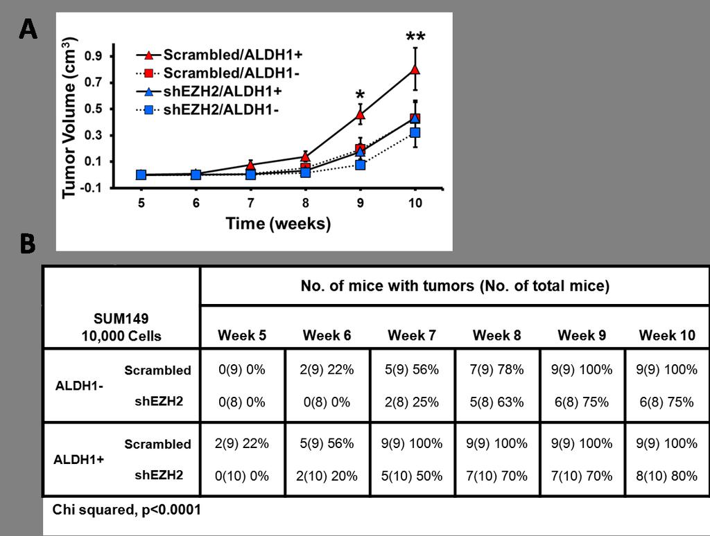

124 112
125 Figure 3-3 EZH2 knockdown in SUM149 ALDH1 + cells decreased the growth rate and time to tumor initiation of in vivo breast tumors. (A) EZH2 knockdown significantly decreased the tumorigenicity of ALDH1 + SUM149 cells compared to controls [mixed effects regression model, p<0.05]. Tumorigenicty assay using ALDH1 + and ALDH1 - sorted populations of SUM149 cells expressing either scrambled shrna or shezh2. After flow cytometry sorting, 10,000 cells were injected into the cleared mammary fat pad of female NOD/SCID mice [n=10 mice per condition] and mice were monitored for tumor growth. Average tumor volume ± SEM for weeks 5-10 post-injection for the four conditions is graphed [Student s t-test, *p=0.004, **p=0.04]. (B) EZH2 knockdown significantly increased the time to tumor initiation in SUM149 ALDH1 + cells compared to controls [Chi-squared, p<0.0001]. The table shows the number of mice with tumors for weeks 5-10 post-injection. (Figure on previous page) Figure 3-4 EZH2 downregulation reduces Notch1 and downstream signaling in breast cancer cells. (A) Heatmap of quantitative RT-PCR microarray (SABiosciences Stem Cell PCR Array) performed using mrna isolated from sorted ALDH1 + SUM149 cells expressing scrambled shrna or shezh2. Upon EZH2 knockdown, Notch1 gene expression was one of the most significantly reduced [Student s t-test, p<0.0001]. (B) mrna from cells described in (A) were used in further quantitative RT-PCR assays analyzing expression of genes within the Notch signaling family. Bar graph shows that EZH2 knockdown in stem cells leads to deregulation of genes involved in Notch signaling. (C) Immunoblots of MDA-MB-231 and SUM149 breast cancer cells show that EZH2 downregulation reduces the expression of the intracellular, activated form of Notch1 [NICD] and NOTHC1 downstream targets CCND1 and CTNNB compared to controls. (D) Immunoblots of patient primary breast cancer cells show that EZH2 downregulation reduces the expression of NICD and Notch1 targets Hes1 and phosphorylated-stat3 [p-stat3]. (E) Immunoblots of MDA-MB-231 and SUM149 cells show that transient adenoviral re-expression of EZH2 in shezh2 cells [+Ad-EZH2] rescues NICD protein levels compared to adenoviral control cells [+Ad-Control]. (Figure on following page) 113


126 114


127 115

![6] were identified between EZH2 and Notch1 mrna levels in publicly available datasets of human breast](/docs-images/74/70224539/images/128-1.jpg "cancer tissues, first page, and human breast cancer cell lines, second page, utilizing Oncomine.")
128 Figure 3-5 EZH2 expression is associated with Notch1 expression in independent datasets of human invasive breast carcinomas. Significant associations [correlation coefficient > 0.6] were identified between EZH2 and Notch1 mrna levels in publicly available datasets of human breast cancer tissues, first page, and human breast cancer cell lines, second page, utilizing Oncomine. (Figure includes previous page) 116
129 Figure 3-6 Notch1 is required for EZH2-induced expansion of MCF10A breast stem cells, part 1. (A) Real-time RT-PCR of MCF10A plvx [control] and plvx-ezh2 [conditional EZH2 TetOn system] cells either untreated or treated with doxycycline [DOX] for 24 hours at 1µg/ml and sorted into ALDH1 - and ALDH1 + populations; mrna expression is relative to GAPDH mrna levels ± SD. Upon DOX treatment, a significant increase in EZH2 mrna expression is seen in both ALDH1 - and ALDH1 + plvx-ezh2 cells. Between these two groups specifically, EZH2 mrna is significantly increased in the ALDH1 + population compared to the ALDH1 - population [Student s t-test, *p<0.0007]. (B) Realtime RT-PCR of MCF10A plvx-ezh2 cells either untreated or treated with DOX for 24 hours and sorted into ALDH1 - and ALDH1 + populations; mrna expression is relative to GAPDH mrna levels ± SD. Upon DOX treatment and EZH2 overexpression, Notch1 mrna levels significantly increase in the ALDH1 + population, but not in the ALDH1 - population [Student s t-test, *p=0.0001]. (C) Immunoblots of MCF10A plvx-ezh2 cells show that DOX treatment results in an increase in EZH2 and NICD protein levels. Upon Notch1 inhibition with the γ-secretase inhibitor [GSI] for 3 days at 17nM in EZH2 overexpressing cells, NICD protein levels are reduced. (D) Representative images of MCF10A plvx and plvx-ezh2 mammospheres formed after 7 days in culture [200X magnification]. (E) DOX-induced overexpression of EZH2 in MCF10A plvx-ezh2 cells leads to a significant increase in mammosphere sizes and numbers compared to controls, which is blocked with Notch1 inhibition. For mammosphere assays, GSI treatment was done for each 7-day generation at 1.7nM. Top, average mammosphere sizes ± SD in the primary generation [Student s t-test, *p<0.002]. Middle and Bottom, average number of mammospheres ± SD per 5x10 4 plated cells in the primary and secondary generations, respectively [Student s t-test, 1⁰ & 2⁰ Gen. *p<0.0001]. (Figure on following page) 117


130 118
 DOX-induced overexpression of EZH2 in MCF10A plvx-ezh2 cells leads to a significant increase in the")
: *p=0.")
131 Figure 3-7 Notch1 is required for EZH2-induced expansion of MCF10A breast stem cells, part 2. (A&B) DOX-induced overexpression of EZH2 in MCF10A plvx-ezh2 cells leads to a significant increase in the percentage of ALDH1 + (A) and CD44 + /CD24 -/low (B) cells compared to controls, which is blocked with Notch1 inhibition. GSI was added for 3 days at 17nM; percentages are expressed ± SD [Student s t-test, (A): *p=0.005, **p=0.002; (B): *p=0.008, **p=0.02]. 119


132 120
133 Figure 3-8. Full length EZH2 is required for Notch1 promoter activation and expansion of mammosphere sizes and numbers. (A) EZH2 overexpression in MCF10A plvx-ezh2 cells leads to a significant increase in Notch1 promoter transcriptional activation. MCF10A plvx and plvx-ezh2 cells with and without DOX treatment were transduced with a pgreenfire lentivirus containing a GFP reporter gene driven by the Notch1 promoter. Transcriptional activation was measured as the percentage of GFP-expressing cells ± SD by flow cytometry [Student s t-test, *p=0.02]. (B) Top, diagram of adenoviral, myc-tagged, EZH2 deletion mutants: ΔSET, ΔHI (Homology Domain I), ΔHII (Homology Domain II), and ΔNLS (Nuclear Localization Signal). Below, immunoblot demonstrates ectopic expression of full length and mutant EZH2 proteins in parental MCF10A cells and their effect on NICD protein levels. (C) Full length EZH2, but not any of the deletion mutants, is required for Notch1 promoter transcriptional activation as measured by the percentage of GFP-expressing cells ± SD [Student s t-test, p=0.0004]. (D) Full length EZH2, but not any of the deletion mutants, is required for the EZH2-induced expansion in mammosphere sizes and numbers. Top and Middle, average number of mammospheres ± SD per 5x10 4 plated cells in the primary and secondary generations [Student s t-test, 1⁰ & 2⁰ Gen., *p<0.0001]. Bottom, average mammosphere sizes ± SD in the primary generation [Student s t-test, *p=0.005]. (Figure on previous page) Figure 3-9 EZH2 binds to the Notch1 gene promoter in benign breast cells, breast cancer cell lines and in patient primary breast cancer cells. (A) Diagram of the Notch1 promoter regions analyzed for EZH2 binding in chromatin immunoprecipitation [ChIP] assays. EZH2 was found to bind region B at -1.2kb; only ChIP real-time PCR data using primers designed for site B are shown. (B) ChIP assays were performed using nontumorigenic primary breast cells expressing control, full length EZH2, EZH2ΔSET or EZH2ΔHII. Primers flanking the GAPDH promoter were used as negative binding controls, and primers flanking the MYT1 promoter, a known H3K27me3-repression target of EZH2, were used as positive binding controls. EZH2 binds the Notch1 promoter independent of H3K27me3 and SUZ12, but coincides with RNA Polymerase II binding and enrichment for the transcriptional activating mark H3K4me2. The N-terminal HII domain is required for EZH2 binding whereas the C- terminal SET domain is dispensable. (C) ChIP assays as described in (B) were perfomed using MDA-MB-231 breast cancer cells expressing scrambled control shrna or shezh2. Endogenous EZH2 binds the Notch1 promoter with similar associations as observed in (B), and binding is blocked with shezh2. (D) ChIP assays as described in (B) were performed using patient primary breast cancer cells. Endogenous EZH2 binds to the Notch1 promoter with similar associations as observed in (B). [Student s t-test, *p <0.05} (Figure on following page) 121
134 122

135 123
136 Figure 3-10 Characterization of EZH2 + /neu and EZH2 wt /neu transgenic mice. (A) Left, representative images of whole mounts of mammary glands from 16 weeks old virgin female mice show that EZH2 + /neu glands exhibit ductal hyperbranching compared to EZH2 wt /neu glands controls. [100X magnification]. Right, graph shows the average number of tertiary branches ± SD in whole mounts preparations described in (A) [Student s t-test, *p<0.05]. (B) Representative histological sections at 8 and 16 weeks of age [400X magnification]; EZH2 + /neu mammary glands show upregulation of EZH2 protein in the nuclei of breast epithelial cells. At 8 weeks, EZH2 + /neu mice exhibit ductal hyperplasia, and at 16 weeks atypical intraductal hyperplasia becomes evident (arrow); changes were not seen in controls for the indicated timepoints. EZH2 + /neu mammary glands also show increased expression of NICD, phosphorylated-stat3 and Ki67 compared to EZH2 wt /neu mammary glands. (Figure on previous page) 124
 Kaplan Meier curve shows that EZH2 + /neu mice formed mammary carcinomas significantly earlier than")
![EZH2 wt /neu mice [Log-rank test, p<0.0001].](/docs-images/74/70224539/images/137-1.jpg "(B) Representative photomicrographs of breast carcinomas from EZH2 + /neu and control EZH2 wt /neu mice")
137 Figure 3-11 Transgenic EZH2 overexpression accelerates tumor initiation and upregulates Notch1 in MMTV-neu mice. (A) Kaplan Meier curve shows that EZH2 + /neu mice formed mammary carcinomas significantly earlier than EZH2 wt /neu mice [Log-rank test, p<0.0001]. (B) Representative photomicrographs of breast carcinomas from EZH2 + /neu and control EZH2 wt /neu mice stained for H&E and immunostained for EZH2 and NICD [400X magnification]. 125
138 Figure 3-12 EZH2 expression is associated with NICD and CD44 + /CD24 - expression in human invasive breast cancer tissues. (A) Representative images of three primary human invasive breast carcinomas (n=107 patients) stained for H&E and immunostained for EZH2, NICD and CD44/CD24 [400X magnification] (B) The table shows the distribution of EZH2 and NICD protein expression in 107 primary invasive breast carcinomas scored from immunostained tissue microarrays; 43.9% of tumors exhibited high expression of both EZH2 and NICD [Fisher s Exact Test, p<0.0001, 2-tailed]. (C) The table shows the distribution of EZH2, NICD and CD44/CD24 protein expression in 70 primary invasive breast carcinomas; 25.7% of tumors exhibited high expression of EZH2, NICD and CD44 + /CD24 - [Fisher s Exact Test, p=0.0004, 2-tailed]. (Figure on following page) 126

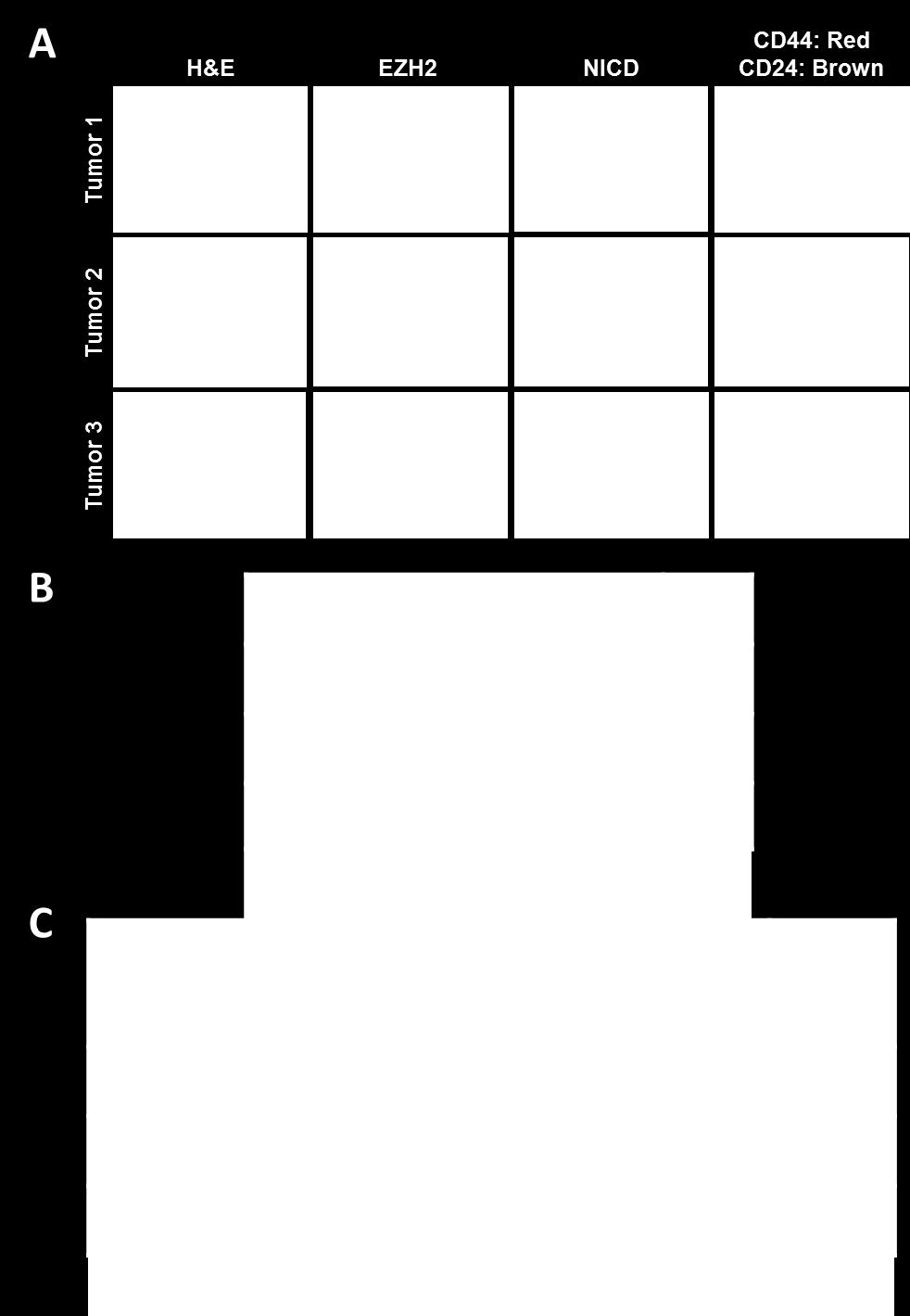
139 127
Contents 1 The Windows of Susceptibility to Breast Cancer 2 The So Called Pre-Neoplastic Lesions and Carcinoma In Situ
 Contents 1 The Windows of Susceptibility to Breast Cancer... 1 1.1 Introduction... 1 1.2 Risk Factor and Etiological Agents... 2 1.3 The Concept of the Windows of Susceptibility to Carcinogenesis... 5
Neoplasia 18 lecture 8. Dr Heyam Awad MD, FRCPath
 Neoplasia 18 lecture 8 Dr Heyam Awad MD, FRCPath ILOS 1. understand the angiogenic switch in tumors and factors that stimulate and inhibit angiogenesis. 2. list the steps important for tumor metastasis
Cancer Biology Course. Invasion and Metastasis
 Cancer Biology Course Invasion and Metastasis 2016 Lu-Hai Wang NHRI Cancer metastasis Major problem: main reason for killing cancer patients, without it cancer can be cured or controlled. Challenging questions:
1. The metastatic cascade. 3. Pathologic features of metastasis. 4. Therapeutic ramifications. Which malignant cells will metastasize?
 1. The metastatic cascade 3. Pathologic features of metastasis 4. Therapeutic ramifications Sir James Paget (1814-1899) British Surgeon/ Pathologist Paget s disease of Paget s disease of the nipple (intraductal
1.The metastatic cascade. 2.Pathologic features of metastasis. 3.Therapeutic ramifications
 Metastasis 1.The metastatic cascade 2.Pathologic features of metastasis 3.Therapeutic ramifications Sir James Paget (1814-1899) British Surgeon/ Pathologist Paget s disease of bone Paget s disease of the
Basement membrane in lobule.
 Bahram Memar, MD Basement membrane in lobule. Normal lobule-luteal phase Normal lobule-follicular phase Lactating breast Greater than 95% are adenocarcinomas in situ carcinomas and invasive carcinomas.
CHAPTER VII CONCLUDING REMARKS AND FUTURE DIRECTION. Androgen deprivation therapy is the most used treatment of de novo or recurrent
 CHAPTER VII CONCLUDING REMARKS AND FUTURE DIRECTION Stathmin in Prostate Cancer Development and Progression Androgen deprivation therapy is the most used treatment of de novo or recurrent metastatic PCa.
SIBLINGs, cancer's multifunctional weapons
 SIBLINGs, cancer's multifunctional weapons 6/18/08 Akeila Bellahcène and Vincent Castronovo of the Metastasis Research laboratory of the University of Liège are among the first researchers to have discovered
CLINICAL SIGNIFICANCE OF BENIGN EPITHELIAL CHANGES
 Papillomas. Papillomas are composed of multiple branching fibrovascular cores, each having a connective tissue axis lined by luminal and myoepithelial cells ( Fig. 23-11 ). Growth occurs within a dilated
Fundamental research on breast cancer in Belgium. Rosita Winkler
 Fundamental research on breast cancer in Belgium Rosita Winkler Medline search for «breast cancer» and Belgium limits: english, posted in the last 5 years. Result: 484 papers - fundamental / clinical -
Biochemistry of Carcinogenesis. Lecture # 35 Alexander N. Koval
 Biochemistry of Carcinogenesis Lecture # 35 Alexander N. Koval What is Cancer? The term "cancer" refers to a group of diseases in which cells grow and spread unrestrained throughout the body. It is difficult
Neoplasia part I. Dr. Mohsen Dashti. Clinical Medicine & Pathology nd Lecture
 Neoplasia part I By Dr. Mohsen Dashti Clinical Medicine & Pathology 316 2 nd Lecture Lecture outline Review of structure & function. Basic definitions. Classification of neoplasms. Morphologic features.
Mousa. Israa Ayed. Abdullah AlZibdeh. 0 P a g e
 1 Mousa Israa Ayed Abdullah AlZibdeh 0 P a g e Breast pathology The basic histological units of the breast are called lobules, which are composed of glandular epithelial cells (luminal cells) resting on
Computational Systems Biology: Biology X
 Bud Mishra Room 1002, 715 Broadway, Courant Institute, NYU, New York, USA L#5:(October-18-2010) Cancer and Signals Outline 1 2 Outline 1 2 Cancer is a disease of malfunctioning cells. Cell Lineage: Adult
- is a common disease - 1 person in 3 can expect to contract cancer at some stage in their life -1 person in 5 can expect to die from it
 MBB157 Dr D Mangnall The Molecular Basis of Disease CANCER Lecture 1 One of the simpler (and better) definitions of cancer comes from the American Cancer Society, who define cancer as; 'Cancer is a group
Diseases of the breast (2 of 2) Breast cancer
 Breast cancer") Diseases of the breast (2 of 2) Breast cancer Epidemiology & etiology The most common type of cancer & the 2 nd most common cause of cancer death in women 1 of 8 women in USA Affects 7% of women Peak at
Diseases of the breast (2 of 2) Breast cancer Epidemiology & etiology The most common type of cancer & the 2 nd most common cause of cancer death in women 1 of 8 women in USA Affects 7% of women Peak at
Cancer and Oncogenes Bioscience in the 21 st Century. Linda Lowe-Krentz
 Cancer and Oncogenes Bioscience in the 21 st Century Linda Lowe-Krentz December 1, 2010 Just a Few Numbers Becoming Cancer Genetic Defects Drugs Our friends and family 25 More mutations as 20 you get older
number Done by Corrected by Doctor Maha Shomaf
 number 21 Done by Ahmad Rawajbeh Corrected by Omar Sami Doctor Maha Shomaf Ability to Invade and Metastasize The metastatic cascade can be subdivided into two phases: 1-invasion of ECM and vascular dissemination:
Maram Abdaljaleel, MD Dermatopathologist and Neuropathologist University of Jordan, School of Medicine
 Maram Abdaljaleel, MD Dermatopathologist and Neuropathologist University of Jordan, School of Medicine The most common non-skin malignancy of women 2 nd most common cause of cancer deaths in women, following
Does EMT Contribute to Radiation Resistance in Human Breast Cancer?
 AD Award Number: W81XWH-10-1-0592 TITLE: Does EMT Contribute to Radiation Resistance in Human Breast Cancer? PRINCIPAL INVESTIGATOR: Anupama Munshi, Ph.D CONTRACTING ORGANIZATION: University of Oklahoma
Oncolytic Virotherapy: Targeting Cancer Stem Cells
 Oncolytic Virotherapy: Targeting Cancer Stem Cells Cancer Stem Cells (CSCs) or Cancer Initiating Cells (CICs) A consensus of five defining criteria has been established to affirm the existence of CICs:
Breast Pathology. Breast Development
 Breast Pathology Lecturer: Hanina Hibshoosh, M.D. Reading: Kumar, Cotran, Robbins, Basic Pathology, 6th Edition, pages 623-635 Breast Development 5th week - thickening of the epidermis - milk line 5th
Inflammatory Cells and Metastasis
 Inflammatory Cells and Metastasis Experimentelle Krebsforschung SS 07 Gerhard Christofori Institute of Biochemistry and Genetics Department of Clinical-Biological Sciences Center of Biomedicine University
BREAST PATHOLOGY. Fibrocystic Changes
 BREAST PATHOLOGY Lesions of the breast are very common, and they present as palpable, sometimes painful, nodules or masses. Most of these lesions are benign. Breast cancer is the 2 nd most common cause
VIII Curso Internacional del PIRRECV. Some molecular mechanisms of cancer
 VIII Curso Internacional del PIRRECV Some molecular mechanisms of cancer Laboratorio de Comunicaciones Celulares, Centro FONDAP Estudios Moleculares de la Celula (CEMC), ICBM, Facultad de Medicina, Universidad
Cancer. October is National Breast Cancer Awareness Month
 Cancer October is National Breast Cancer Awareness Month Objectives 1: Gene regulation Explain how cells in all the different parts of your body develop such different characteristics and functions. Contrast
Signaling Vascular Morphogenesis and Maintenance
 Signaling Vascular Morphogenesis and Maintenance Douglas Hanahan Science 277: 48-50, in Perspectives (1997) Blood vessels are constructed by two processes: vasculogenesis, whereby a primitive vascular
Breast pathology. 2nd Department of Pathology Semmelweis University
 Breast pathology 2nd Department of Pathology Semmelweis University Breast pathology - Summary - Benign lesions - Acute mastitis - Plasma cell mastitis / duct ectasia - Fat necrosis - Fibrocystic change/
In vitro scratch assay: method for analysis of cell migration in vitro labeled fluorodeoxyglucose (FDG)
") In vitro scratch assay: method for analysis of cell migration in vitro labeled fluorodeoxyglucose (FDG) 1 Dr Saeb Aliwaini 13/11/2015 Migration in vivo Primary tumors are responsible for only about 10%
In vitro scratch assay: method for analysis of cell migration in vitro labeled fluorodeoxyglucose (FDG) 1 Dr Saeb Aliwaini 13/11/2015 Migration in vivo Primary tumors are responsible for only about 10%
September 20, Submitted electronically to: Cc: To Whom It May Concern:
 History Study (NOT-HL-12-147), p. 1 September 20, 2012 Re: Request for Information (RFI): Building a National Resource to Study Myelodysplastic Syndromes (MDS) The MDS Cohort Natural History Study (NOT-HL-12-147).
Supplementary Figures
 Supplementary Figures Supplementary Figure 1. Confirmation of Dnmt1 conditional knockout out mice. a, Representative images of sorted stem (Lin - CD49f high CD24 + ), luminal (Lin - CD49f low CD24 + )
Aberrant cell Growth. Younas Masih New Life College of Nursing Karachi. 3/4/2016 Younas Masih ( NLCON)
") Aberrant cell Growth Younas Masih New Life College of Nursing Karachi 1 Objectives By the end of this session the learners will be able to, Define the characteristics of the normal cell Describe the characteristics
Aberrant cell Growth Younas Masih New Life College of Nursing Karachi 1 Objectives By the end of this session the learners will be able to, Define the characteristics of the normal cell Describe the characteristics
EMT: Epithelial Mesenchimal Transition
 EMT: Epithelial Mesenchimal Transition A phenotypic change that is characteristic of some developing tissues and certain forms of cancer. This is a multistep, key process in embryonic development and metastasis
mirna Dr. S Hosseini-Asl
 mirna Dr. S Hosseini-Asl 1 2 MicroRNAs (mirnas) are small noncoding RNAs which enhance the cleavage or translational repression of specific mrna with recognition site(s) in the 3 - untranslated region
Disclosure of Relevant Financial Relationships. Breast Pathology Evening Specialty Conference Case #4. Clinical Case: Pathologic Features
 Breast Pathology Evening Specialty Conference Case #4 K.P. Siziopikou, MD, PhD Professor of Pathology Director of Breast Pathology and Breast Pathology Fellowship Program Northwestern University Feinberg
Early Embryonic Development
 Early Embryonic Development Maternal effect gene products set the stage by controlling the expression of the first embryonic genes. 1. Transcription factors 2. Receptors 3. Regulatory proteins Maternal
Your Guide to the Breast Cancer Pathology. Report. Key Questions. Here are important questions to be sure you understand, with your doctor s help:
 Your Guide to the Breast Cancer Pathology Report Key Questions Here are important questions to be sure you understand, with your doctor s help: Your Guide to the Breast Cancer Pathology Report 1. Is this
CELL BIOLOGY - CLUTCH CH CANCER.
 !! www.clutchprep.com CONCEPT: OVERVIEW OF CANCER Cancer is a disease which is primarily caused from misregulated cell division, which form There are two types of tumors - Benign tumors remain confined
Disorders of Cell Growth & Neoplasia. Lecture 4 Molecular basis of cancer
 General Pathology VPM 152 Disorders of Cell Growth & Neoplasia Lecture 4 Molecular basis of cancer Enrique Aburto Apr 2010 Skin tumor in a 10-year-old Rottweiler. Considering the external appearance and
The Hallmarks of Cancer
 The Hallmarks of Cancer Theresa L. Hodin, Ph.D. Clinical Research Services Theresa.Hodin@RoswellPark.org Hippocrates Cancer surgery, circa 1689 Cancer Surgery Today 1971: Nixon declares War on Cancer
Seeds and soil theory by Stephen Paget at the end of the XIX century.
 Seeds and soil theory by Stephen Paget at the end of the XIX century. In The Distribution Of Secondary Growths In Cancer Of The Breast Paget presents and analyzes 735 fatal cases of breast cancer, complete
The Angiopoietin Axis in Cancer
 Ang2 Ang1 The Angiopoietin Axis in Cancer Tie2 An Overview: The Angiopoietin Axis Plays an Essential Role in the Regulation of Tumor Angiogenesis Growth of a tumor beyond a limiting size is dependent upon
Differential Connexin Function Enhances Self-Renewal in Glioblastoma
 CSU ID: 2558414 Name: Sonal Patel Differential Connexin Function Enhances Self-Renewal in Glioblastoma Cancer is one of the most common disease in the US with more than a million people affected every
number Done by Corrected by Doctor Maha Shomaf
 number 19 Done by Waseem Abo-Obeida Corrected by Abdullah Zreiqat Doctor Maha Shomaf Carcinogenesis: the molecular basis of cancer. Non-lethal genetic damage lies at the heart of carcinogenesis and leads
Papillary Lesions of the Breast A Practical Approach to Diagnosis. (Arch Pathol Lab Med. 2016;140: ; doi: /arpa.
 Papillary Lesions of the Breast A Practical Approach to Diagnosis (Arch Pathol Lab Med. 2016;140:1052 1059; doi: 10.5858/arpa.2016-0219-RA) Papillary lesions of the breast Span the spectrum of benign,
A Genetic Program for Embryonic Development
 Concept 18.4: A program of differential gene expression leads to the different cell types in a multicellular organism During embryonic development, a fertilized egg gives rise to many different cell types
PhD THESIS Epigenetic mechanisms involved in stem cell differentiation
 Romanian Academy Institute of Cellular Biology and Pathology "Nicolae Simionescu" PhD THESIS Epigenetic mechanisms involved in stem cell differentiation Coordinator: Acad. Maya Simionescu PhD Student:
Breast Cancer Diagnosis, Treatment and Follow-up
 Breast Cancer Diagnosis, Treatment and Follow-up What is breast cancer? Each of the body s organs, including the breast, is made up of many types of cells. Normally, healthy cells grow and divide to produce
Negative Regulation of c-myc Oncogenic Activity Through the Tumor Suppressor PP2A-B56α
 Negative Regulation of c-myc Oncogenic Activity Through the Tumor Suppressor PP2A-B56α Mahnaz Janghorban, PhD Dr. Rosalie Sears lab 2/8/2015 Zanjan University Content 1. Background (keywords: c-myc, PP2A,
Cell Polarity and Cancer
 Cell Polarity and Cancer Pr Jean-Paul Borg Email: jean-paul.borg@inserm.fr Features of malignant cells Steps in Malignant Progression Cell polarity, cell adhesion, morphogenesis and tumorigenesis pathways
- A cancer is an uncontrolled, independent proliferation of robust, healthy cells.
 1 Cancer A. What is it? - A cancer is an uncontrolled, independent proliferation of robust, healthy cells. * In some the rate is fast; in others, slow; but in all cancers the cells never stop dividing.
Supplementary Figures
 Supplementary Figures Supplementary Figure 1 DOT1L regulates the expression of epithelial and mesenchymal markers. (a) The expression levels and cellular localizations of EMT markers were confirmed by
LYMPHATIC DRAINAGE AXILLARY (MOSTLY) INTERNAL MAMMARY SUPRACLAVICULAR
 INTERNAL MAMMARY SUPRACLAVICULAR") BREAST LYMPHATIC DRAINAGE AXILLARY (MOSTLY) INTERNAL MAMMARY SUPRACLAVICULAR HISTOLOGY LOBE: (10 in whole breast) LOBULE: (many per lobe) ACINUS/I, aka ALVEOLUS/I: (many per lobule) DUCT(S): INTRA- or
BREAST LYMPHATIC DRAINAGE AXILLARY (MOSTLY) INTERNAL MAMMARY SUPRACLAVICULAR HISTOLOGY LOBE: (10 in whole breast) LOBULE: (many per lobe) ACINUS/I, aka ALVEOLUS/I: (many per lobule) DUCT(S): INTRA- or
CHAPTER 6 SUMMARIZING DISCUSSION
 CHAPTER 6 SUMMARIZING DISCUSSION More than 20 years ago the founding member of the Wnt gene family, Wnt-1/Int1, was discovered as a proto-oncogene activated in mammary gland tumors by the mouse mammary
Acute: Symptoms that start and worsen quickly but do not last over a long period of time.
 Cancer Glossary Acute: Symptoms that start and worsen quickly but do not last over a long period of time. Adjuvant therapy: Treatment given after the main treatment. It usually refers to chemotherapy,
Characteristics of Cancer Stem Cells (CSCs)
") GENReports: Market & Tech Analysis Characteristics of Cancer Stem Cells (CSCs) > Enal Razvi, Ph.D. Biotechnology Analyst, Managing Director Select Biosciences, Inc. enal@selectbio.us! Topic,IntroducEon,and,Scope!
GENReports: Market & Tech Analysis Characteristics of Cancer Stem Cells (CSCs) > Enal Razvi, Ph.D. Biotechnology Analyst, Managing Director Select Biosciences, Inc. enal@selectbio.us! Topic,IntroducEon,and,Scope!
Recent advances in breast cancers
 Recent advances in breast cancers Breast cancer is a hetrogenous disease due to distinct genetic alterations. Similar morphological subtypes show variation in clinical behaviour especially in response
MicroRNA expression profiling and functional analysis in prostate cancer. Marco Folini s.c. Ricerca Traslazionale DOSL
 MicroRNA expression profiling and functional analysis in prostate cancer Marco Folini s.c. Ricerca Traslazionale DOSL What are micrornas? For almost three decades, the alteration of protein-coding genes
Neoplasia literally means "new growth.
 NEOPLASIA Neoplasia literally means "new growth. A neoplasm, defined as "an abnormal mass of tissue the growth of which exceeds and is uncoordinated with that of the normal tissues and persists in the
Cancer and Oncogenes Bioscience in the 21 st Century. Linda Lowe-Krentz October 11, 2013
 Cancer and Oncogenes Bioscience in the 21 st Century Linda Lowe-Krentz October 11, 2013 Just a Few Numbers Becoming Cancer Genetic Defects Drugs Our friends and family 200 180 160 140 120 100 80 60 40
CRIPTO-1 A POSSIBLE NEW BIOMARKER IN GLIOBLASTOMA MULTIFORME PIA OLESEN, MD, PHD STUDENT
 CRIPTO-1 A POSSIBLE NEW BIOMARKER IN GLIOBLASTOMA MULTIFORME PIA OLESEN, MD, PHD STUDENT Glioblastoma WHO Grade IV Glioma Heterogenic Undiffenrentiated phenotype 50% of all Gliomas Around 600 patients
Award Number: W81XWH TITLE: Characterizing an EMT Signature in Breast Cancer. PRINCIPAL INVESTIGATOR: Melanie C.
 AD Award Number: W81XWH-08-1-0306 TITLE: Characterizing an EMT Signature in Breast Cancer PRINCIPAL INVESTIGATOR: Melanie C. Bocanegra CONTRACTING ORGANIZATION: Leland Stanford Junior University Stanford,
Case study 1. Rie Horii, M.D., Ph.D. Division of Pathology Cancer Institute Hospital, Japanese Foundation for Cancer Research
 NCCN/JCCNB Seminar in Japan April 15, 2012 Case study 1 Rie Horii, M.D., Ph.D. Division of Pathology Cancer Institute Hospital, Japanese Foundation for Cancer Research Present illness: A 50y.o.premenopausal
Transformation of Normal HMECs (Human Mammary Epithelial Cells) into Metastatic Breast Cancer Cells: Introduction - The Broad Picture:
 into Metastatic Breast Cancer Cells: Introduction - The Broad Picture:") Transformation of Normal HMECs (Human Mammary Epithelial Cells) into Metastatic Breast Cancer Cells: Introduction - The Broad Picture: Spandana Baruah December, 2016 Cancer is defined as: «A disease caused
Transformation of Normal HMECs (Human Mammary Epithelial Cells) into Metastatic Breast Cancer Cells: Introduction - The Broad Picture: Spandana Baruah December, 2016 Cancer is defined as: «A disease caused
stem cell products Basement Membrane Matrix Products Rat Mesenchymal Stem Cell Growth and Differentiation Products
 stem cell products Basement Membrane Matrix Products Rat Mesenchymal Stem Cell Growth and Differentiation Products Stem Cell Qualified Extracellular Matrix Proteins Stem cell research requires the finest
Tumor microenvironment Interactions and Lung Cancer Invasiveness. Pulmonary Grand Rounds Philippe Montgrain, M.D.
 Tumor microenvironment Interactions and Lung Cancer Invasiveness Pulmonary Grand Rounds Philippe Montgrain, M.D. February 26, 2009 Objectives Review epithelial mesenchymal transition (EMT), and its implications
Disorders of Cell Growth & Neoplasia
 General Pathology VPM 152 Disorders of Cell Growth & Neoplasia Lecture 3 Rate of growth, local invasion, and metastasis. Molecular basis of cancer (normal cell-cycle and cellular proliferation). Enrique
Part I. An Introduction to Cancer
 Part I An Introduction to Cancer 2 Chapter 1 Cancer: Descriptive Overview Cancer is a disease in which cells propagate uncontrollably. These cells can come from many different parts of the body and the
Genetics and Cancer Ch 20
 Genetics and Cancer Ch 20 Cancer is genetic Hereditary cancers Predisposition genes Ex. some forms of colon cancer Sporadic cancers ~90% of cancers Descendants of cancerous cells all cancerous (clonal)
Dr. Heba Kalbouneh. Dr. Heba Kalbouneh. Dr. Heba Kalbouneh
 Dr. Heba Kalbouneh Dr. Heba Kalbouneh Dr. Heba Kalbouneh Basement membrane: What is the basement membrane? - It is a layer of ECM separating the epithelial cells from the underlying connective tissue Basement
04/10/2018 HIGH RISK BREAST LESIONS. Pathology Perspectives of High Risk Breast Lesions ELEVATED RISK OF BREAST CANCER HISTORICAL PERSPECTIVES
 Pathology Perspectives of High Risk Breast Lesions Savitri Krishnamurthy MD Professor of Pathology Deputy Division Head Director of Clinical Trials, Research and Development The University of Texas MD
Neoplasia 2018 Lecture 2. Dr Heyam Awad MD, FRCPath
 Neoplasia 2018 Lecture 2 Dr Heyam Awad MD, FRCPath ILOS 1. List the differences between benign and malignant tumors. 2. Recognize the histological features of malignancy. 3. Define dysplasia and understand
肿瘤信息学 徐鹰佐治亚大学生化系 吉林大学计算机学院
 肿瘤信息学 徐鹰佐治亚大学生化系 吉林大学计算机学院 2 Lecture V Biology of cancer metastasis and metastatic cancers 3 Metastatic Cancer Metastatic cancers are responsible for 90% of cancer related death Currently metastatic cancer
Tumor Associated Macrophages as a Novel Target for Cancer Therapy
 Tumor mass Tumor Associated Macrophage Tumor Associated Macrophages as a Novel Target for Cancer Therapy This booklet contains forward-looking statements that are based on Amgen s current expectations
Chapter 10 Breast Cancer Liver Metastasis
 Chapter 10 Breast Cancer Liver Metastasis Sébastien Tabariès and Peter M. Siegel Abstract The emergence of metastatic breast cancer is the most deadly aspect of this disease and once it has spread from
Molecular factors influencing epithelial-mesenchymal transition in breast cancer
 Molecular factors influencing epithelial-mesenchymal transition in breast cancer Gisela Nilsson Department of Medical Biochemistry and Cell Biology Institute of Biomedicine Sahlgrenska Academy at University
Src-INACTIVE / Src-INACTIVE
 Biology 169 -- Exam 1 February 2003 Answer each question, noting carefully the instructions for each. Repeat- Read the instructions for each question before answering!!! Be as specific as possible in each
Effect of a nutrient mixture on the localization of extracellular matrix proteins in HeLa human cervical cancer xenografts in female nude mice
 Effect of a nutrient mixture on the localization of extracellular matrix proteins in HeLa human cervical cancer xenografts in female nude mice Publication from the Dr. Rath Research Institute Experimental
Tissue renewal and Repair. Nisamanee Charoenchon, PhD Department of Pathobiology, Faculty of Science
 Tissue renewal and Repair Nisamanee Charoenchon, PhD Email: nisamanee.cha@mahidol.ac.th Department of Pathobiology, Faculty of Science Topic Objectives 1. Describe processes of tissue repair, regeneration
Breast Cancer. Most common cancer among women in the US. 2nd leading cause of death in women. Mortality rates though have declined
 Breast Cancer Most common cancer among women in the US 2nd leading cause of death in women Mortality rates though have declined 1 in 8 women will develop breast cancer Breast Cancer Breast cancer increases
Breast cancer as a systemic disease: a view of metastasis
 Review Click here for more articles from the symposium doi: 10.1111/joim.12084 Breast cancer as a systemic disease: a view of metastasis A. J. Redig 1 & S. S. McAllister 1,2,3 From the 1 Division of Hematology,
Papillary Lesions of the Breast
 Papillary Lesions of the Breast Laura C. Collins, M.D. Associate Professor of Pathology Associate Director, Division of Anatomic Pathology Beth Israel Deaconess Medical Center and Harvard Medical School
Breast Cancer. Saima Saeed MD
 Breast Cancer Saima Saeed MD Breast Cancer Most common cancer among women in the US 2nd leading cause of death in women 1 in 8 women will develop breast cancer Incidence/mortality rates have declined Breast
Cell Death and Cancer. SNC 2D Ms. Papaiconomou
 Cell Death and Cancer SNC 2D Ms. Papaiconomou How do cells die? Necrosis Death due to unexpected and accidental cell damage. This is an unregulated cell death. Causes: toxins, radiation, trauma, lack of
Chapter 6. Villous Growth
 Core Curriculum in Perinatal Pathology Chapter 6 Villous Growth Overview of vasculogenesis and angiogenesis Vasculogenesis Extraembryonic Vasculogenesis Angiogenesis Branching angiogenesis Sprouting angiogenesis
LAMININ-1 MEDIATES EPIGENETIC CONTROL OF THE EPITHELIAL CADHERIN FOR BREAST CANCER CELLS IN THREE-DIMENSIONAL CULTURE
 LAMININ-1 MEDIATES EPIGENETIC CONTROL OF THE EPITHELIAL CADHERIN FOR BREAST CANCER CELLS IN THREE-DIMENSIONAL CULTURE A Dissertation submitted to the Faculty of the Graduate School of Arts and Sciences
Understanding Your Pathology Report
 Understanding Your Pathology Report Because every person s breast cancer is unique, it s important to understand the underlying biology of your tumor to personalize your treatment plan. Your physicians
Type of file: PDF Size of file: 0 KB Title of file for HTML: Supplementary Information Description: Supplementary Figures
 Type of file: PDF Size of file: 0 KB Title of file for HTML: Supplementary Information Description: Supplementary Figures Supplementary Figure 1 mir-128-3p is highly expressed in chemoresistant, metastatic
PBS Class #2 Introduction to the Immune System part II Suggested reading: Abbas, pgs , 27-30
 PBS 803 - Class #2 Introduction to the Immune System part II Suggested reading: Abbas, pgs. 15-25, 27-30 Learning Objectives Compare and contrast the maturation of B and T lymphocytes Compare and contrast
Understanding and Optimizing Treatment of Triple Negative Breast Cancer
 Understanding and Optimizing Treatment of Triple Negative Breast Cancer Edith Peterson Mitchell, MD, FACP Clinical Professor of Medicine and Medical Oncology Program Leader, Gastrointestinal Oncology Department
Dominic J Smiraglia, PhD Department of Cancer Genetics. DNA methylation in prostate cancer
 Dominic J Smiraglia, PhD Department of Cancer Genetics DNA methylation in prostate cancer Overarching theme Epigenetic regulation allows the genome to be responsive to the environment Sets the tone for
Overview: Conducting the Genetic Orchestra Prokaryotes and eukaryotes alter gene expression in response to their changing environment
 Overview: Conducting the Genetic Orchestra Prokaryotes and eukaryotes alter gene expression in response to their changing environment In multicellular eukaryotes, gene expression regulates development
Histology = the study of tissues. Tissue = a complex of cells that have a common function
 { EPITHELIAL TISSUE Histology = the study of tissues Tissue = a complex of cells that have a common function The Four Primary Tissue Types: Epithelium (epithelial tissue) covers body surfaces, lines body
SUPPLEMENTARY INFORMATION
 DOI: 10.1038/ncb3021 Supplementary figure 1 Characterisation of TIMPless fibroblasts. a) Relative gene expression of TIMPs1-4 by real time quantitative PCR (RT-qPCR) in WT or ΔTimp fibroblasts (mean ±
Yanrong Su *, Nathan R. Hopfinger, Theresa D. Nguyen, Thomas J. Pogash, Julia Santucci-Pereira and Jose Russo *
 Su et al. Journal of Experimental & Clinical Cancer Research (2018) 37:314 https://doi.org/10.1186/s13046-018-0988-8 RESEARCH Open Access Epigenetic reprogramming of epithelial mesenchymal transition in
Cancer Cells. It would take another 20 years and a revolution in the techniques of biological research to answer these questions.
 Cancer Cells Cancer, then, is a disease in which a single normal body cell undergoes a genetic transformation into a cancer cell. This cell and its descendants, proliferating across many years, produce
Chapter 9, Part 1: Biology of Cancer and Tumor Spread
 PATHOPHYSIOLOGY Name Chapter 9, Part 1: Biology of Cancer and Tumor Spread I. Cancer Characteristics and Terminology Neoplasm new growth, involves the overgrowth of tissue to form a neoplastic mass (tumor).
UNIVERSITY OF MEDICINE AND PHARMACY CRAIOVA PhD SCHOOL. PhD THESIS
 UNIVERSITY OF MEDICINE AND PHARMACY CRAIOVA PhD SCHOOL PhD THESIS THE IMPORTANCE OF TUMOR ANGIOGENESIS IN CEREBRAL TUMOR DIAGNOSIS AND THERAPY ABSTRACT PhD COORDINATOR: Prof. univ. dr. DRICU Anica PhD
PREPARED FOR: U.S. Army Medical Research and Materiel Command Fort Detrick, Maryland
 AD Award Number: W81XWH-04-1-0618 TITLE: Are Breast Tumor Stem Cells Responsible for Metastasis and Angiogenesis PRINCIPAL INVESTIGATOR: Quintin Pan, Ph.D. CONTRACTING ORGANIZATION: University of Michigan