Challenges in Diagnostic Cytomorphology 2
|
|
|
- Rolf Kennedy
- 6 years ago
- Views:
Transcription

1 Challenges in Diagnostic Cytomorphology 2 Dina Mody, MD Richard DeMay, MD David Wilbur, MD Eva Wojick, MD There are no disclosures necessary.
2 10/29/2012 Update for Cervical Cytology Atypical Glandular Cells Unusual HSIL Presentations Biomarkers David C. Wilbur, M.D. Massachusetts General Hospital Harvard Medical School Boston, Massachusetts MASSACHUSETTS GENERAL PHYSICIANS ORGANIZATION HARVARD MEDICAL SCHOOL Topics to be discussed 1) Endocervical neoplasia and mimics 2) High risk variants of HSIL 3) Biomarkers and their utility Incidence of Endocervical Carcinoma 9 US SEER databases 27,016 invasive cervical carcinomas ( ) 19,703 squamous cell carcinoma ( 20%, CIS 1.4x) 3,895 adenocarcinoma ( 2x, AIS 7x) 956 adenosquamous 2,457 type unspecified Worldwide Some countries show declines in ACA Finland, France, Italy 1
3 10/29/2012 Atypical Endocervical Cells Follow Up Studies Rule of Thumb Benign/Reactive 40% Squamous Neoplasia 50% Endocervical Neoplasia 10% Topic 2 Adenocarcinoma in situ/invasive AIS prototypical WD EC neoplasia Invasive EC ACA well to poor differentiation Architectural features Cytologic features Cellularity Background EC Adenocarcinoma in Situ 1) Hyperchromatic crowded groups 2) Increased nucleus to cytoplasmic ratio 3) Nuclei large (75 um2) 4) Even chromatin with coarse granularity 5) Micronucleoli 6) Rosettes 7) Feathering 8) Strips with pseudostratification 9) Mitoses, apoptotic bodies 10) Amphophilic granular cytoplasm 11) Clean or inflammatory background 2


4 10/29/2012 3


5 10/29/2012 p16 Ki67 4


 for pure Gr1 lesions with minimal")
6 10/29/2012 Endometrioid AIS Smaller cells than usual type AIS Absence of EM stroma and stromal fragments Most commonly missed AIS AGC is acceptable Endometrioid AIS Villoglandular Adenocarcinoma Usually occur in younger population (35 y mean) 62% history of oral contraceptive use Usually well-circumscribed, minimally invasive Behavior favorable LN metastases rare Conservative management (cone only) for pure Gr1 lesions with minimal invasion, LVI-, margins- 5


7 10/29/2012 Topic 3 Adenoma Malignum Rare Often not HPV associated Presents with mass Histologically bland Cytologic atypia common 6


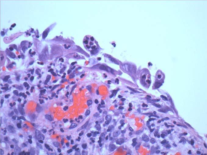
8 10/29/2012 Adenoma Malignum too many EC cells 7


9 10/29/2012 Adenoma Malignum too many EC cells Topic 4 Tubal Metaplasia Benign metaplasia of EM and upper EC canal Architectural mimicker of AIS May have mitotic activity Bland nuclear features P16/Ki67 neg or spotty + TBS


10 10/29/2012 9

")
 3)")
")
")
11 10/29/2012 p16 Ki67 Tubal Metaplasia - Cytologic Features 1) Incomplete features of AIS 2) Chromatin (texture/distribution) 3) Nuclei washed-out 4) Nuclear pleomorphism 5) N/C increased 6) Mitoses/rare apoptosis 7) Large stripped nuclei 8) Cilia/terminal bars 10
 Common 2) Bleeding 3)")

")

12 10/29/2012 Topic 5 Endocervical Polyps 1) Common 2) Bleeding 3) Surface ulceration 4) EC proliferation - abundant cellularity - HCG s 5) Reactive changes - enlargement, nucleoli, mitoses 6) Inflammation - reparative changes 7) Can mimic either EC or EM neoplasia AEC associated with biopsy site 11


13 10/29/2012 EC Polyp EC Polyp EM CA 12
 Wispy granular cytoplasm 5) Some cytoplasmic vacuoles 6) Tubule and gland formation 7) Mitoses/apoptosis")

14 10/29/2012 Topic 6 Directly Sampled Endometrium 1) tight groups or 2-dimensional sheets 2) Small reniform nuclei 3) Small nucleoli/chromocenters 4) Wispy granular cytoplasm 5) Some cytoplasmic vacuoles 6) Tubule and gland formation 7) Mitoses/apoptosis common Directly Sampled Endometrium Present as pseudostratified strips of cells Nuclei are generally coarsely granular Good look-alike for AIS Stromal fragments can be HCG s Stromal fragments attached to EM epithelium Caveat small cells with small nuclei Direct Sampling Exfoliated 13


15 10/29/2012 AIS Direct-sampled EM Size Matters!! ASCCP Flowchart 14
16 10/29/2012 Atypical Glandular Cells Quality Control/Quality Assurance 1) Prevalence of AGC (<1%) 2) Clinical history 3) Communication 4) Criteria Application 5) Retrospective Review 6) Cytology/Histology Correlation AGC Prevalence Range % (mean = 0.48, median = 0.27) CAP Data - mean = 0.35%, median = 0.3% Rule of thumb < 1% Variant Presentations of HSIL 15
 Stripped")

17 10/29/2012 Patterns of HSIL/CA Classic single cell, syncytia Involving glands In atrophy Stripped nucleus EM stroma-like/repair-like Hypochromatic Keratinizing Papanicolaou & Traut ) Stripped Nucleus Pattern Individual stripped nuclei Small groups of stripped nuclei loosely aggregated Nuclei may be enlarged, irregular, and hyperchromatic Nuclei may be relatively bland Mimic is cytolysis 16


18 10/29/2012 Stripped Nucleus Pattern vs. Cytolysis HSIL Cytolysis 5) EM stroma/repair-like Bloody background simulating menses Small tightly packed groups of cells Small isolated cells with high N:C Prominent spindle cell component Cytoplasm spindles from group margins Taffypull cytoplasm +/- nucleoli May be associated with classic HSIL 17


19 10/29/


20 10/29/

21 10/29/2012 Biomarker Resolution 20


22 10/29/2012 P16/Ki67 uses: Squamous lesions To reduce interobserver variability in cervical biopsy interpretation To differentiate HSIL from Atrophy Immature squamous metaplasia Reactive epithelial changes To differentiate AIS/EC dysplasia from benign EC To evaluate cauterized margins To evaluate thin dysplasias To evaluate the presence of invasion To screen putatively negative biopsies after HSIL Paps Screening Pap/Anal Pap tests p16 Ki67 P16 Thin dysplasia Ki67 21


23 10/29/2012 p16/proexc in Pap tests.. P16 ProExC HSIL - Positive staining Sweeney et al, ASC Meeting 2009 p16/ki67 in Anal Specimens p16 ki67 p16 p16 Thank You!! Questions?? 22


24 Body Cavity Fluids Richard M. DeMay, MD Professor of Pathology Director of Cytopathology The University of Chicago Body Cavity Fluids Effusions: Can be benign or malignant, but are always pathologic Transudates: Usually benign Exudates: Can be malignant Mesothelial Cells Mesodermally derived epithelial cells Line body cavities: Pleura, pericardium, peritoneum Normal: Single layer, flat (squamous) cells Effusions: Reactive Cells: plump, cuboidal Nuclei: active DDx: Cancer Common dilemma Page 1


25 Mesothelial Cells Mostly single Small lobulated groups Histiocytes Windows Hugging Mesothelial Cells Cells: Rounded, variable size Nuclei: Reactive changes common Cytoplasm: Dense center, lacy skirts Vacuoles Common, though often nonspecific Degenerative Degenerative Vacuoles Lipid Glycogen Clear, glassy Mucin => adenocarcinoma Page 2
")


26 Marked Reactive Changes Pulmonary embolism or infarct Active cirrhosis or hepatitis Uremia Note blebs (degenerative) Pancreatitis Long term dialysis ChemoRx Radiation, chemo Rx, surgery Also: Pericardial effusions Repeat tap can be helpful! Hepatitis Uremia Pancreatitis Histiocytes Hemosiderin Practically ubiquitous Nuclei: beans, mitoses! Cytoplasm: lacy throughout, phagocytosis typical DDx: Mesothelial Cells No: Molding, hugging, windows, tight groups Other Inflammatory Cells Eosinophils Usually => prior tap Infx, infarct, allergy, idiopathic Not: Cancer, Hodgkin Lymphocytes Common, usually nonspecific Neutrophils Charcot Leyden crystal Nonspecific, but often infectious markedly reactive meso s Rarely numerous in malignant effusions Page 3


27 Psammoma Bodies Not diagnostic of cancer! Mesothelial hyperplasia Endosalpingiosis Cancer: Ovary most common in women Other, eg, papillary adenocarcinomas Collagen Balls Usually peritoneal washes, females Also, other sources, men Associated w/ mesothelial cells Benign proliferation Malignant mesothelioma Rare in adenocarcinoma Odds & Ends Pick ups Curschmann spiral Megakaryocyte Hematoxylin Body LE Cell Sickle cell Ciliocytophthoria Page 4
 Clusters: Benign clue!")

28 Tuberculosis Turbid, yellow or metallic green (pseudochylous effusion) Classic cytologic findings: Lymphs abundant Mesothelials rare Histiocytes present, giant cells rare TB, T cells; Lymphoma, B cells => flow cytometry (TGFF) Clusters: Benign clue! Rheumatoid Effusion Pseudochylous (metallic) classic Cyto: Rheumatoid Granuloma Necrotic debris, ~diagnostic Epithelioid histiocytes Multinucleated giant cells Mesothelial cells sparse Bizarre cells, yet benign! Malignant Effusions Any cancer can cause an effusion Common: Lung, Breast, Ovary, GI tract In oncology pts, most are malignant Can be benign (eg, CHF, Cirrhosis, Rx) Site of effusion clue to primary site Lung & breast usually ipsilateral Abd/pelvic Ascites before pleural Page 5


29 Diagnosis of Malignant Effusions Key: Foreign Cells Diagnosis of Malignant Effusions When cytology is easy, it is very, very easy But when it s difficult Some malignancies mimic benign cells, like Invasion of the Body Snatchers Page 6

30 Mesothelial Pattern Carcinoma When it s difficult Two helpful suggestions Mucicarmine stain Submit more material, if fluid reaccumulates If doesn t, probably benign Improves cellular preservation Benign atypia tends to resolve Page 7


 Breast or Gastric CA [Sarcoma, Melanoma, Mesothelioma] Carcinoma")
31 General Features Malignant Effusions Key: Foreign cells with foreign features ie, discrete or extra population of cells Some possible exceptions: Mesothelioma: Native cells, can be bland Tumor mimics reactive mesothelial cells eg, breast cancer, serous ovarian tumors Tumor Diagnosis: No single feature Cell Groups Group Contours Cell Shape Nuclei Cytoplasm Cell Surface Background + clues to primary Cell Groups Large groups: Suspicious Cell balls, papillae, glands Single tumor cells: Lymphoma (classic) Breast or Gastric CA [Sarcoma, Melanoma, Mesothelioma] Carcinoma Lymphoma Page 1


,")
32 Cannonballs Suggest Breast CA in a woman Papillae Pleural: Lung, Breast Ascites: Ovary, GI tract [Mesothelial hyper/neoplasia] Caterpillars Single file chains Breast, Pancreas, Gastric, Uterus [Lung (small cell), Mesothelioma] Page 2
 Bizarre shapes => malignancy except rheumatoid")


 Suggests")
33 Cell Shape All cells tend to round up in fluids (makes differential dx difficult) Bizarre shapes => malignancy except rheumatoid effusion Nuclei Malignant nuclei = sine qua non Classic cytologic criteria: Enlarged, pleomorphic High N/C ratios Irregular membranes Abnormal chromatin Large/abn nucleoli Hyperchromasia variable May not be prominent due to proliferation Mitoses not helpful: Unless numerous or abnormal Intracytoplasmic Lumens (ICLs) Targetoid Structure Sharp outline Clear Space Bull s eye (mucin) Suggests breast cancer in women Page 3


 Cilia: Strongly")

34 Signet Ring Cells Stomach or Breast Other Adenocarcinomas Extreme Vacuolization Large, glassy, empty looking vacuoles Ovary, Pancreas Cell Surface Cilia Well defined cell borders: Favor malignancy Ill defined cell borders: Favor benign (Mesos or histiocytes) Cilia: Strongly favor benign Exclude prominent microvilli Ciliated carcinoma very rare Endosalpingiosis Ciliated Carcinoma Page 4
 Cigar shaped nuclei Dirty")


 Source:")
 Neoplastic glands in mucous lakes")

35 Colorectal Carcinoma Terminal Bar Terminal bars (Striated border) Cigar shaped nuclei Dirty necrosis Small Cell Carcinoma Caterpillars Clusters Caterpillars: Usually easy diagnosis Clusters: Can be difficult to detect in busy inflammatory background Also: Single cell pattern, mimics lymphoma Pseudomyxoma Peritonei Mucus in peritoneum ( jelly belly ) Source: Mucinous GI Tract lesions, esp appendix (not ovary) Neoplastic glands in mucous lakes Adenomucinosis (bland) Mucinous carcinomatosis Other cells sparse / absent (ie, meso s, histiocytes, WBCs) Cell blocks can be helpful in prep Page 5
 Dense")


, usually elderly men Confined to")

36 Squamous Cell Carcinoma Common cancer, rarely sheds <5% malignant effusions More keratinization => easier Dx Most = NK SCC (DDx Rx mesos, AdCA) Dense cytoplasm, pearls, bizarre shapes DDx: Sarcomas, rheumatoid effusion Also benign squames: cyst, fistula, contamination Lymphoma/Leukemia Effusions usually late, dx established Often chylous effusion Single cells; no tissue aggregate Larger cells => easier dx Clues to look for: Irregular membranes (eg, cleaves, knobs) Massive karyorrhexis (apoptosis) Flow cytometry for difficult cases (TGFF!) Mercury droplets Primary Effusion Lymphoma Immunocompromised pts Mostly homosexual men with AIDS Also, HIV ( ), usually elderly men Confined to body cavities without solid tumor formation Particularly big & ugly lymphoma cells Bridges immunoblastic & anaplastic NHL plasmacytoid ± RS like cells HHV 8, EBV, germ line c myc characteristic CD30 frequently (+) Page 6


 Spindle (eg, Leiomyosarcoma)")
37 Hodgkin Lymphoma Effusions common, diagnostic cells rare Reed Sternberg cells Hodgkin cells Eosinophils rare! DDx: Large Cell NHL; Metastatic Malignancy Clinical history! Melanoma Dark brown/black fluid Big, ugly tumor cells Binucleation common Macronucleoli; INCI Melanin often sparse Problems: Mimics many tumors, late recurrence, unknown 1 Sarcoma Effusions: Primary usually well known Pleomorphic (eg, TTFKA MFH) Round (eg, Neuroblastoma) Spindle (eg, Leiomyosarcoma) DDx: Mesothelioma, SCC, Rheumatoid All cells round up in fluid => DDx wide Page 7

38 Germ Cell Tumors Suspect diagnosis: Big Ugly Tumor cells in young patient Important diagnosis, may respond to Rx Page 8
 Exudate with high cell count remains high after repeated taps")
39 Mesothelioma Epithelioid Sarcomatoid Biphasic Other epithelial subtypes: clear cell, oncocytoid or granular cell, tubulopapillary, large polygonal cell, polyhedral stromal mucin producing, medullary, small cell, lymphohistiocytoid Mesothelioma: Fluid Cytology Often first diagnostic specimen Fluid highly viscous (hyaluronic acid) Exudate with high cell count remains high after repeated taps Combination: High hyaluronic acid (>8mg/dL) plus Atypical mesothelial cells Suggests diagnosis of mesothelioma Cytodiagnosis of Mesothelioma In theory, easy Malignant cells ~ mesothelial Morphologic kinship No foreign cells In practice, difficult WD looks benign PD difficult to classify Page 1


40 Cytodiagnostic Features Cellularity Cell groups Cells Nucleus Cytoplasm Vacuoles Cell borders Background Not every feature is present in every case! Cellularity Important diagnostic clue: More and bigger cells in more and bigger clusters Knobby Cell Groups Papillae Complex, irregular Single Cells Caterpillars Page 2


41 Cell Arrangements Hugging Collagen cores Windows Cells Tumor cells look mesothelial: Key diagnostic feature! Cells Spindle Spindle + Epithelioid Histiocytoid Gigantocyte Mesothelioid Page 3


42 Nucleus N/C ratio maintained Uniformity Macronucleoli Mitoses Not so much! Cytoplasm Vacuoles Lipid Glycogen Hyaluronic Acid Page 4


43 Cell Borders Blebs Microvilli Skirts Background Mesothelioma: Ultrastructure Bushy (vs stubby) microvilli Page 5
 *Does not exclude carcinoma, but helps detect Calretinin, CK 5/6 ( ) cases, often sarcomatoid 2nd Part: Epithelial Markers ( ) CEA, Ber Ep4, BG 8, MOC 31 to exclude adenocarcinoma")
44 2 Part Mesothelioma Immunopanel 1 1st Part: Mesothelial Markers (+) Calretinin, WT 1 (front line) CK 5/6, CAM 5.2* (secondary) *Does not exclude carcinoma, but helps detect Calretinin, CK 5/6 ( ) cases, often sarcomatoid 2nd Part: Epithelial Markers ( ) CEA, Ber Ep4, BG 8, MOC 31 to exclude adenocarcinoma Strongly recommend cell blocks!!! Primary Peritoneal Carcinoma Usually elderly women, normal ovaries Origin: Mesothelium vs Fallopian tube Most: Papillary serous morphology DDx: Mesothelioma Epithelial markers, eg, MOC 31 vs Mesothelial markers, eg, calretinin Ovarian carcinoma Gross ovarian disease, invasion Two take home points Page 6


45 Metastases Foreign cells with foreign features One of these things is not like the other Mesothelioma Malignant cells that look mesothelial in origin More & bigger cells, in more & bigger clusters g{tç~ çéâ Page 7
46 All images copyright ASCP Press Page 8


47


48


49


50


51


52


53


54


55


56


57


58


59

60

61


62


63


64


65


66


67

68
Gynecologic Cytopathology: Glandular lesions
 Gynecologic Cytopathology: Glandular lesions Lin Wai Fung (MSc, MPH, CMIAC) 17/4/2014 Glandular lesions of the uterus Endocervix Endometrium Normal endocervical cells Sheets, strips well-preserved architecture:
Introduction. 23 rd Annual Seminar in Pathology. FLUIDS, Part 1. Pittsburgh, PA Gladwyn Leiman UVMMC, VT
 23 rd Annual Seminar in Pathology Pittsburgh, PA Gladwyn Leiman UVMMC, VT FLUIDS, Part 1 "Blue walls", Claudia Hansen, 2009 Introduction o Challenging to everyone o Almost any benign or malignant process
Hyperchromatic Crowded Groups: What is Your Diagnosis? Session 3000
 Hyperchromatic Crowded Groups: What is Your Diagnosis? Session 3000 Thomas A. Bonfiglio, M.D. Professor Emeritus, Pathology and Laboratory Medicine University of Rochester Disclosures In the past 12 months,
Objectives. Atypical Glandular Cells. Atypical Endocervical Cells. Reactive Endocervical Cells
 2013 California Society of Pathologists 66 th Annual Meeting San Francisco, CA Atypical Glandular Cells to Early Invasive Adenocarcinoma: Cervical Cytology and Histology Christina S. Kong, MD Associate
Case 3 - GYN. History: 66 year old, routine Pap test. Dr. Stelow
 Case 3 - GYN History: 66 year old, routine Pap test Dr. Stelow Case 3 66 year year old woman Routine Pap Test Cytologic Features 3 dimensional clusters of cells with small to moderate amount of
Effusion Cytology: Diagnostic Challenges
 Effusion Cytology: Diagnostic Challenges Tarik M. Elsheikh, MD Professor and Medical Director, Anatomic Pathology Cleveland Clinic Outside Consult Case 45 year old woman, presented with nausea, dyspnea,
BOSNIAN-TURKISH CYTOPATHOLOGY SCHOOL June 18-19, 2016 Sarajevo. Case Discussions. 60 year old woman Routine gynecologic control LBC
 BOSNIAN-TURKISH CYTOPATHOLOGY SCHOOL June 18-19, 2016 Sarajevo Case Discussions Prof Dr Sıtkı Tuzlalı Tuzlalı Pathology Laboratory 60 year old woman Routine gynecologic control LBC 1 2 Endometrial thickening
Case 1. Slide 1 History: 65 year old male presents with bilateral pleural effusions, a 40 pack year smoking history and peripheral and hilar lung
 Case 1. Slide 1 History: 65 year old male presents with bilateral pleural effusions, a 40 pack year smoking history and peripheral and hilar lung masses. Specimen shown is from a tap of the pleural effusion.
Case year female. Routine Pap smear
 Case 1 57 year female Routine Pap smear Diagnosis? 1. Atypical glandular cells of unknown significance (AGUS) 2. Endocervical AIS 3. Endocervical adenocarcinoma 4. Endometrial adenocarcinoma 5. Adenocarcinoma
Ascitic Fluid and Use of Immunocytochemistry. Mercè Jordà, University of Miami
 Ascitic Fluid and Use of Immunocytochemistry Mercè Jordà, University of Miami Is It Malignant? Yes? No Ascitic Fluid Cytomorphologic Useful Findings Tight clusters with smooth borders Cellular and nuclear
Prepared By Jocelyn Palao and Layla Faqih
 Prepared By Jocelyn Palao and Layla Faqih The structure of the suspected atypical cell should always be compared to the structure of other similar, benign, cells which are present in the smears. The diagnosis
Mody. AIS vs. Invasive Adenocarcinoma of the Cervix
 Common Problems in Gynecologic Pathology Michael T. Deavers, M.D. Houston Methodist Hospital, Houston, Texas Common Problems in Gynecologic Pathology Adenocarcinoma in-situ (AIS) of the Cervix vs. Invasive
Serous effusion Objectives. Cytology of Serous Effusions From basics to challenges
 Cytology of Serous Effusions From basics to challenges Cytology of Serous Effusions From basics to challenges Pınar Fırat, MD, MIAC Department of Pathology, İstanbul University, İstanbul Faculty of Medicine,
SQUAMOUS CELLS: Atypical squamous cells (ASC) - of undetermined significance (ASC-US) - cannot exclude HSIL (ASC-H)
 - of undetermined significance (ASC-US) - cannot exclude HSIL (ASC-H)") SQUAMOUS CELLS: Atypical squamous cells (ASC) - of undetermined significance (ASC-US) - cannot exclude HSIL (ASC-H) ASC refers to cytologic changes suggestive of SIL, which are qualitativley or quantitatively
SQUAMOUS CELLS: Atypical squamous cells (ASC) - of undetermined significance (ASC-US) - cannot exclude HSIL (ASC-H) ASC refers to cytologic changes suggestive of SIL, which are qualitativley or quantitatively
CINtec p16 INK4a Staining Atlas
 CINtec p16 INK4a Staining Atlas Rating Rating Positive The rating positive will be assigned if the p16 INK4a -stained slide shows a continuous staining of cells of the basal and parabasal cell layers of
SELECTED DILEMMAS IN RESPIRATORY CYTOPATHOLOGY (2 CASES)
") SELECTED DILEMMAS IN RESPIRATORY CYTOPATHOLOGY (2 CASES) Dr. Mariamma Joseph Professor of Pathology Division Head Cytopathology Department of Pathology and Laboratory Medicine LHSC and Western University
SELECTED DILEMMAS IN RESPIRATORY CYTOPATHOLOGY (2 CASES) Dr. Mariamma Joseph Professor of Pathology Division Head Cytopathology Department of Pathology and Laboratory Medicine LHSC and Western University
Morphologic Clues and Pitfalls for High Grade Lesions in Cervical Cytology
 Morphologic Clues and Pitfalls for High Grade Lesions in Cervical Cytology Ritu Nayar, MD Northwestern University, Feinberg School of Medicine Chicago, IL, USA Disclosures Editor, Cervical Cytology Bethesda
Outline 11/2/2017. Pancreatic EUS-FNA general aspects. Cytomorphologic features of solid neoplasms/lesions of the pancreas
 ENDOSCOPIC ULTRASOUND GUIDED-FINE NEEDLE ASPIRATION CYTOLOGY OF PANCREAS Khalid Amin M.D. Assistant Professor Department of Laboratory Medicine and Pathology University of Minnesota Outline Pancreatic
Diagnostically Challenging Cases in Gynecologic Pathology
 Diagnostically Challenging Cases in Gynecologic Pathology Eric C. Huang, M.D., Ph.D. Department of Pathology and Laboratory Medicine University of California, Davis Medical Center Case 1 Presentation 38
Cytyc Corporation - Case Presentation Archive - July 2002
 ThinPrep Pap Test History: 34 Year Old Female LMP: Day 20 Specimen Type: Cervical/Vaginal Case provided by Mark Tulecke, M.D. and Gabrielle Trawinski CT (ASCP), Mount Auburn Hospital, Cambridge, Massachusetts.
FNA of Thyroid. Toward a Uniform Terminology With Management Guidelines. NCI NCI Thyroid FNA State of the Science Conference
 FNA of Thyroid NCI NCI Thyroid FNA State of the Science Conference Toward a Uniform Terminology With Management Guidelines Thyroid Thyroid FNA Cytomorphology NCI Thyroid FNA State of the Science Conference
Serous Effusions. Spasenija Savic Prince, MD Pathology, University Hospital Basel, Switzerland
 Serous Effusions Spasenija Savic Prince, MD Pathology, University Hospital Basel, Switzerland Serous membrane Body cavities: Pleural Pericardial Peritoneal Effusion = Excess of fluid 80% Benign 20% Malignant
GYN (Glandulars) Still Difficult After All These Years! Dina R Mody, MD Director of Cytology Laboratories and fellowship Program Methodist Hospital
 Still Difficult After All These Years! Dina R Mody, MD Director of Cytology Laboratories and fellowship Program Methodist Hospital") GYN (Glandulars) Still Difficult After All These Years! Dina R Mody, MD Director of Cytology Laboratories and fellowship Program Methodist Hospital and Bioreference Labs (Houston) Department of Pathology
GYN (Glandulars) Still Difficult After All These Years! Dina R Mody, MD Director of Cytology Laboratories and fellowship Program Methodist Hospital and Bioreference Labs (Houston) Department of Pathology
Salivary Glands 3/7/2017
 Salivary Glands 3/7/2017 Goals and objectives Focus on the entities unique to H&N Common board type facts Information for your future practice Salivary Glands Salivary Glands Major gland. Paratid. Submandibular.
Cytyc Corporation - Case Presentation Archive - October 2001
 ThinPrep Pap Test History: 82 Year Old Female Specimen Type: Peritoneal Washings Case provided by Dr. Berle Stratton, Southwest Washington Medical Center, Vancouver, Washington. *The images, analysis and
Cytyc Corporation - Case Presentation Archive - March 2002
 FirstCyte Ductal Lavage History: 68 Year Old Female Gail Index: Unknown Clinical History: Negative Mammogram in 1995 6 yrs. later presents with bloody nipple discharge Subsequent suspicious mammogram Suspicious
Cytology Report Format
 Squamous Precursor Lesions and Malignancies In Pap Test Dina R. Mody, MD, FCAP Director of Cytology The Methodist Hospital, Houston, TX Professor of Pathology and Laboratory Medicine Weill Medical College
Oncocytic-Appearing Salivary Gland Tumors. Oncocytic, Cystic, Mucinous, and High Grade Salivary Gland Tumors SALIVARY GLAND FNA: PART II
 William C. Faquin, MD, PhD Professor of Pathology Harvard Medical School Director of Head and Neck Pathology Massachusetts Eye and Ear Massachusetts General Hospital SALIVARY GLAND FNA: PART II Oncocytic,
Conflict of Interest 9/7/2018. Dr. Mody 1. None with vendors of cytology equipment/testing/vaccines Amirsys (now Elsevier)
") Glandular Lesions in Cervicovaginal Cytology: Patterns, Pitfalls and Bethesda Updates Dina R Mody, MD Director of Cytology Laboratories Houston s Methodist Hospital and Bioreference Laboratory The Ibrahim
Glandular Lesions in Cervicovaginal Cytology: Patterns, Pitfalls and Bethesda Updates Dina R Mody, MD Director of Cytology Laboratories Houston s Methodist Hospital and Bioreference Laboratory The Ibrahim
6/5/2010. Outline of Talk. Endometrial Alterations That Mimic Cancer & Vice Versa: Metaplastic / reactive changes. Problems in Biopsies/Curettages
 Outline of Talk Endometrial Alterations That Mimic Cancer & Vice Versa: Problems in Biopsies/Curettages Metaplastic / reactive changes Mucinous change Microglandular hyperplasia-like change Squamous metaplasia
Salivary Gland Cytology
 Salivary Gland Cytology Diagnostic challenges and potential pitfalls Tarik M. Elsheikh, MD Professor and Medical Director Anatomic Pathology Cleveland Clinic FNA Salivary Gland Lesions Indications Distinguish
New Diagnoses Need New Approaches: A Glimpse into the Near Future of Gynecologic Pathology
 New Diagnoses Need New Approaches: A Glimpse into the Near Future of Gynecologic Pathology United States and Canadian Academy of Pathology 102 nd Annual Meeting Baltimore, Maryland Christina S. Kong, M.D.
Workshop for O& G trainees and paramedics 17 Dec 2011 Cytological Interpretation
 Workshop for O& G trainees and paramedics 17 Dec 2011 Cytological Interpretation May Yu Director of Cytology Laboratory Service Department of Anatomical & Cellular Pathology Prince of Wales Hospital Cervical
LGM International, Inc.
 Liqui-PREP TM Cytology Atlas Preface The following pictures are examples with descriptions of cytology slides processed with the Liqui-PREP TM System.. The descriptions are reviewed by Pathologists. It
Normal endometrium: A, proliferative. B, secretory.
 Normal endometrium: A, proliferative. B, secretory. Nội mạc tử cung Nội mạc tử cung Cyclic changes in endometrium.. Approximate relationship of useful microscopic changes. Arias-Stella reaction in endometrial
Endometrial Metaplasia, Hyperplasia & Other Cancer Mimics: a Consultant s Experience
 Endometrial Metaplasia, Hyperplasia & Other Cancer Mimics: a Consultant s Experience Pacific Northwest Society of Pathologists Vancouver, B.C. September 26, 2015 Teri A. Longacre, M.D. longacre@stanford.edu
Mody. Atypical Glandular Cells(TBS 2001) Adenocarcinoma In Situ(TBS 2001)
 Adenocarcinoma In Situ(TBS 2001)") Glandular Lesions in Cervicovaginal Cytology Dina R. Mody, MD, FCAP Director of Cytology The Methodist Hospital, Houston, TX Professor of Pathology and Laboratory Medicine Weill Medical College of Cornell
Glandular Lesions in Cervicovaginal Cytology Dina R. Mody, MD, FCAP Director of Cytology The Methodist Hospital, Houston, TX Professor of Pathology and Laboratory Medicine Weill Medical College of Cornell
Cytological Sub-classification of Lung Cancer: Morphologic and Molecular Characteristics. Mercè Jordà, University of Miami
 Cytological Sub-classification of Lung Cancer: Morphologic and Molecular Characteristics Mercè Jordà, University of Miami Mortality Lung cancer is the most frequent cause of cancer incidence and mortality
Neoplasia 2018 Lecture 2. Dr Heyam Awad MD, FRCPath
 Neoplasia 2018 Lecture 2 Dr Heyam Awad MD, FRCPath ILOS 1. List the differences between benign and malignant tumors. 2. Recognize the histological features of malignancy. 3. Define dysplasia and understand
In situ and Invasive Endocervical Carcinoma: Problems and Pitfalls in Diagnosis
 In situ and Invasive Endocervical Carcinoma: Problems and Pitfalls in Diagnosis Rouba Ali-Fehmi,MD The Karmanos Cancer Institute, Wayne State University School of Medicine Global incidence of cervical
1.Acute and Chronic Cervicitis - At the onset of menarche, the production of estrogens by the ovary stimulates maturation of the cervical and vaginal
 Diseases of cervix I. Inflammations 1.Acute and Chronic Cervicitis - At the onset of menarche, the production of estrogens by the ovary stimulates maturation of the cervical and vaginal squamous mucosa
How to Recognize Gynecologic Cancer Cells from Pelvic Washing and Ascetic Specimens
 How to Recognize Gynecologic Cancer Cells from Pelvic Washing and Ascetic Specimens Wenxin Zheng, M.D. Professor of Pathology and Gynecology University of Arizona zhengw@email.arizona.edu http://www.zheng.gynpath.medicine.arizona.edu/index.html
Maturation Index 3/29/2017. Disclosure of Relevant Financial Relationships. Gynecologic Cytology. Normal Maturation of Squamous Epithelium : :
 Gynecologic Cytology Fadi W. Abdul Karim, MD MEd Department of Anatomic Pathology Vice Chair Education RT PLMI Professor of Pathology Cleveland Clinic. Cleveland Ohio Disclosure of Relevant Financial Relationships
Morphology I Slide: 1
 Morphology I Slide: 1 Morphology I Slide: 2 ThinPrep Morphology Normal Cytology Morphology I Slide: 3 CT & Pathologist Training Training program begins with ThinPrep morphology presentation Microscopic
Pancreatitis: A Potential Pitfall in Endoscopic Ultrasound Guided Pancreatic FNA
 Pancreatitis: A Potential Pitfall in Endoscopic Ultrasound Guided Pancreatic FNA Jack Yang, MD Department of Pathology, Medical University of South Carolina Objectives Understand the indication of EUS
Urinary Bladder: WHO Classification and AJCC Staging Update 2017
 Urinary Bladder: WHO Classification and AJCC Staging Update 2017 Houston Society of Clinical Pathologists 58 th Annual Spring Symposium Houston, TX April 8, 2017 Jesse K. McKenney, MD Classification
Mesothelioma: diagnostic challenges from a pathological perspective. Naseema Vorajee August 2016
 Mesothelioma: diagnostic challenges from a pathological perspective Naseema Vorajee August 2016 Naseema.vorajee@nhls.ac.za Pleural diseases (whether neoplastic, reactive or infective) may have similar
DETERMINATION OF A LYMPHOID PROCESS
 Chapter 2 Applications of Touch Preparation Cytology to Intraoperative Consultations: Lymph Nodes and Extranodal Tissues for Evaluation of Hematolymphoid Disorders INTRODUCTION As discussed in Chap. 1,
Diagnostic Cytology of Cancer Cases
 Diagnostic Cytology of Cancer Cases Somporn Techangamsuwan Companion Animal Cancer Research Unit (CAC-RU) Department of Pathology, Faculty of Veterinary Science, Chulalongkorn University 1 Tumor or Non-tumor
QUALITY ASSURANCE PROGRAM CYTOLOGY CYCLE 01/2018 (TRIAL)
") [Pick the Date] FINAL REPORT QUALITY ASSURANCE PROGRAM CYTOLOGY CYCLE 01/2018 (TRIAL) NOTES FROM THE COORDINATOR 1. For this cycle 01/2018, a total of 32 pen drives had been circulated. Twenty-eight institutions
[Pick the Date] FINAL REPORT QUALITY ASSURANCE PROGRAM CYTOLOGY CYCLE 01/2018 (TRIAL) NOTES FROM THE COORDINATOR 1. For this cycle 01/2018, a total of 32 pen drives had been circulated. Twenty-eight institutions
Dr Sanjiv Manek Oxford. Oxford Pathology Course 2010 for FRCPath Illustration-Cellular Pathology. Oxford Radcliffe NHS Trust
 Dr Sanjiv Manek Oxford Oxford Pathology Course 2010 for FRCPath Illustration-Cellular Pathology. Oxford Radcliffe NHS Trust Ovarian Endometrial Vulvo-vaginal Cervical Illustration-Cellular Pathology. Oxford
Histopathology: Cervical HPV and neoplasia
 Histopathology: Cervical HPV and neoplasia These presentations are to help you identify basic histopathological features. They do not contain the additional factual information that you need to learn about
ENODMETRIAL CARCINOMA: SPECIAL & NOT SO SPECIAL VARIANTS
 ENODMETRIAL CARCINOMA: SPECIAL & NOT SO SPECIAL VARIANTS Pacific Northwest Society of Pathologists Vancouver, B.C. September 26, 2015 Teri A. Longacre, M.D. longacre@stanford.edu Stanford University, Stanford,
Gross appearance of peritoneal cysts. They have a thin, translucent wall and contain a clear fluid.
 Gross appearance of peritoneal cysts. They have a thin, translucent wall and contain a clear fluid. So-called multicystic benign mesothelioma. A, Gross appearance. So-called multicystic benign mesothelioma.
When Immunostains Can Get You in Trouble: Gynecologic Pathology p16: Panacea or Pandora s Box?
 When Immunostains Can Get You in Trouble: Gynecologic Pathology p16: Panacea or Pandora s Box? Teri A. Longacre, MD Stanford Medicine Stanford California pi6 in Gynecologic Pathology: Panacea or Pandora
Glandular lesions in cervical cytology. Margareta Strojan Fležar Institute of Pathology Faculty of Medicine University of Ljubljana Slovenia
 Glandular lesions in cervical cytology Margareta Strojan Fležar Institute of Pathology Faculty of Medicine University of Ljubljana Slovenia 2nd PANNONIA CONGRESS OF PATHOLOGY, SIÓFOK, HUNGARY, 17-19 MAY
FNA OF SALIVARY GLANDS: A PRACTICAL APPROACH
 FNA OF SALIVARY GLANDS: A PRACTICAL APPROACH FNA of Salivary Glands: Challenges Wide range of neoplastic and non-neoplastic lesions Cytological overlap between the different benign and malignant tumors
The 24 th Congress Of The IAP-Arab Division KHARTOUM - SUDAN Arab School of Pathology Cytology workshop December 5-6, 2012
 International Academy of Pathology - Arab Division The 24 th Congress Of The IAP-Arab Division KHARTOUM - SUDAN Arab School of Pathology Cytology workshop December 5-6, 2012 Body Fluid Cytology Mousa Al-Abbadi,
From Morphology to Molecular Pathology: A Practical Approach for Cytopathologists Part 1-Cytomorphology. Songlin Zhang, MD, PhD LSUHSC-Shreveport
 From Morphology to Molecular Pathology: A Practical Approach for Cytopathologists Part 1-Cytomorphology Songlin Zhang, MD, PhD LSUHSC-Shreveport I have no Conflict of Interest. FNA on Lymphoproliferative
Index. Cytoplasm, nonepithelial malignant tumor features 70
 Accurette device 23 Adenosarcoma, differential diagnosis 80, 81 Arias-Stella reaction 65 Atypical endocervical cells 8 Atypical endometrial cells 8 Atypical glandular cells (AGC) 8, 9 Atypical glandular
ACCME/Disclosures. Diagnosing Mesothelioma in Limited Tissue Samples. Papanicolaou Society of Cytopathology Companion Meeting March 12 th, 2016
 Diagnosing Mesothelioma in Limited Tissue Samples Papanicolaou Society of Cytopathology Companion Meeting March 12 th, 2016 Sanja Dacic, MD, PhD University of Pittsburgh ACCME/Disclosures GENERAL RULES
Synonyms. Nephrogenic metaplasia Mesonephric adenoma
 Nephrogenic Adenoma Synonyms Nephrogenic metaplasia Mesonephric adenoma Definition Benign epithelial lesion of urinary tract with tubular, glandular, papillary growth pattern Most frequently in the urinary
Ben Witt, MD University of Utah/ARUP Laboratories Assistant Professor of Anatomic Pathology
 Ben Witt, MD University of Utah/ARUP Laboratories Assistant Professor of Anatomic Pathology Review some of the more common cytodiagnoses of the Head and Neck Establish an approach to some of the diagnostic
number Done by Corrected by Doctor Maha Shomaf
 number 16 Done by Waseem Abo-Obeida Corrected by Zeina Assaf Doctor Maha Shomaf MALIGNANT NEOPLASMS The four fundamental features by which benign and malignant tumors can be distinguished are: 1- differentiation
05/07/2018. Types of challenges. Challenging cases in uterine pathology. Case 1 ` 65 year old female Post menopausal bleeding Uterine Polyp
 Types of challenges Challenging cases in uterine pathology Nafisa Wilkinson Gynaecological Pathologist UCLH London Lack of complete history often, NO clinical history at all! Cases from other centres often
Thyroid master class. Thyroid Fine needle aspiration cytology and liquid-based techniques: Hologic and Becton Dickinson
 Thyroid master class Thyroid Fine needle aspiration cytology and liquid-based techniques: Hologic and Becton Dickinson Principle of LBC Collection of cells in liquid medium Immediate fixation Processor-prepared
Atypical Hyperplasia/EIN
 EIN Atypical Hyperplasia/EIN Based on scientific and diagnostic advances, in 2014 the WHO moved that the precursor lesion for endometrioid carcinoma be atypical hyperplasia/ein, rather than what was previously
Proceeding of the SEVC Southern European Veterinary Conference
 www.ivis.org Proceeding of the SEVC Southern European Veterinary Conference Oct. 17-19, 2008 Barcelona, Spain http://www.sevc.info Reprinted in the IVIS website with the permission of the SEVC www.ivis.org
Epithelial tumors. Dr. F.F. Khuzin, PhD Dr. M.O. Mavlikeev
 Epithelial tumors Dr. F.F. Khuzin, PhD Dr. M.O. Mavlikeev Epithelial tumors Tumors from the epithelium are the most frequent among tumors. There are 2 group features of these tumors: The presence in most
Ovarian Clear Cell Carcinoma
 Ovarian Clear Cell Carcinoma Rouba Ali-Fehmi, MD Professor of Pathology The Karmanos Cancer Institute, Wayne State University School of Medicine 50 year old woman with chief complaint of shortness of breath
Respiratory Tract Cytology
 Respiratory Tract Cytology 40 th European Congress of Cytology Liverpool, UK Momin T. Siddiqui M.D. Professor of Pathology and Laboratory Medicine Director of Cytopathology Emory University Hospital, Atlanta,
Essentials of Fluid Cytology
 Essentials of Fluid Cytology Gia-Khanh Nguyen 2018 Essentials of Fluid Cytology Gia-Khanh Nguyen, MD, FRCPC Professor Emeritus Department of Laboratory Medicine and Pathology Faculty of Medicine and Dentistry
Tinh hoàn
 Tinh hoàn Tinh hoàn Tinh hoàn Tiền liệt tuyến Tiền liệt tuyến Mào tinh hoàn Mào tinh hoàn Túi tinh Túi tinh Túi tinh Túi tinh So-called cystadenoma of seminal vesicle. Gross appearance of granulomatous
International Journal of Health Sciences and Research ISSN:
 International Journal of Health Sciences and Research www.ijhsr.org ISSN: 2249-9571 Original Research Article Utility of Modified Cell Block Technique in Cases of Pleural Effusion Suspected of Malignancy
Presentation material is for education purposes only. All rights reserved URMC Radiology Page 1 of 98
 Presentation material is for education purposes only. All rights reserved. 2011 URMC Radiology Page 1 of 98 Radiology / Pathology Conference February 2011 Brooke Koltz, Cytopathology Resident Presentation
6/3/2010. Outline of Talk. Lobular Breast Cancer: Definition of lobular differentiation. Common Problems in Diagnosing LCIS in Core Biopsies
 Outline of Talk Lobular Breast Cancer: Common Problems in Diagnosing LCIS in Core Biopsies Definition of lobular differentiation Variants of LCIS that: carry risk for unsampled invasive cancer mimic DCIS
Lesions Mimicking Adenoid Cystic Carcinoma. Diagnostic Problems in Salivary Gland Pathology An Update 5/29/2009
 Diagnostic Problems in Salivary Gland Pathology An Update Lesions Mimicking Adenoid Cystic Carcinoma Stacey E. Mills, M.D. W.S. Royster Professor of Pathology Director of Surgical and Cytopathology University
Villoglandular adenocarcinoma of cervix a tumour with bland cytological features: report of a case missed on cytology
 Malaysian J Pathol 2003; 25(2) : CERVICAL 139 143 VILLOGLANDULAR ADENOCARCINOMA CYTOLOGY CASE REPORT Villoglandular adenocarcinoma of cervix a tumour with bland cytological features: report of a case missed
Problem 1: Differential of Neuroendocrine Carcinoma 3/23/2017. Disclosure of Relevant Financial Relationships
 Differential of Neuroendocrine Carcinoma Alain C. Borczuk,MD Weill Cornell Medicine Disclosure of Relevant Financial Relationships USCAP requires that all faculty in a position to influence or control
Index 179. Genital tract contaminants, 17, 20, 22, 150 papilloma virus-infected cells, 47 squamous cells, sources of, 7
 Index Accuracy of urinary cytology, 166 Acute inflammatory cells, 38 catheter sample, 39 herpes simplex infections, 44 carcinomas, 104, 105 non-viral inclusions, 52, 53 voided urine, 17 Adenocarcinoma
40th European Congress of Cytology Liverpool, UK, 2-5 th October 2016
 40th European Congress of Cytology Liverpool, UK, 2-5 th October 2016 EUS FNA of abdominal organs: An approach to reporting and triage for ancillary testing Date and time: Sunday 2 nd October 2016 15.00-16.30
Intraductal carcinoma of the prostate on needle biopsy: histologic features and clinical significance
 & 2006 USCAP, Inc All rights reserved 0893-3952/06 $30.00 www.modernpathology.org Intraductal carcinoma of the prostate on needle biopsy: histologic features and clinical significance Charles C Guo 1 and
Prostate Pathology: Prostate Carcinoma, variants and Gleason Grading (Part 1)
") Prostate Pathology: Prostate Carcinoma, variants and Gleason Grading (Part 1) Jae Y. Ro, MD, PhD June 7, 2012 Ten Leading Cancer Types for the Estimated New Cancer Cases and Deaths By Sex, United States,
Prostate Pathology: Prostate Carcinoma, variants and Gleason Grading (Part 1) Jae Y. Ro, MD, PhD June 7, 2012 Ten Leading Cancer Types for the Estimated New Cancer Cases and Deaths By Sex, United States,
04/09/2018. Squamous Cell Neoplasia and Precursor Lesions. Agenda. Squamous Dysplasia. Squamo-proliferative lesions. Architectural features
 Squamous Cell Neoplasia and Precursor Lesions Jennifer L. Hunt, MD, MEd Aubrey J. Hough Jr, MD, Endowed Professor of Pathology Chair of Pathology and Laboratory Medicine University of Arkansas for Medical
NEOPLASIA-I CANCER. Nam Deuk Kim, Ph.D.
 NEOPLASIA-I CANCER Nam Deuk Kim, Ph.D. 1 2 Tumor in the hieroglyphics of the Edwin Smith papyrus (1,600 B.C., Breasted s translation 1930) 3 War on Cancer (National Cancer Act, 1971) 4 Cancer Acts in Korea
4/12/2018. MUSC Pathology Symposium Kiawah Island April 18, Jesse K. McKenney, MD
 MUSC Pathology Symposium Kiawah Island April 18, 2018 Jesse K. McKenney, MD 1 Urothelial Carcinoma with Alternative Differentiation 2 Urothelial Carcinoma with Alternative Differentiation Recognition as
Pathology Slides. [Pathology]
![Pathology Slides. [Pathology]](/thumbs/94/120604575.jpg "Pathology Slides. [Pathology]") Pathology Slides MedicoNotes provides real laboratory pathological slides to aid you to differentiate between different pathological structures under microscope. www.mediconotes.com Histology slides example
Pathology Slides MedicoNotes provides real laboratory pathological slides to aid you to differentiate between different pathological structures under microscope. www.mediconotes.com Histology slides example
Biliary tract tumors
 Short Course 2010 Annual Fall Meeting of the Korean Society for Pathologists Biliary tract tumors Joon Hyuk Choi, M.D., Ph.D. Professor, Department of Pathology, Yeungnam Univ. College of Medicine, Daegu,
Award Top Quizzes For Residents
 Award Top Quizzes For Residents Giovanni Negri, MD, Bolzano (Italy) Eva M. Wojcik MD, Department of Pathology, Loyola University, Chicago (USA) Esther D. Rossi MD, PhD, MIAC, Division of Anatomic Pathology
Diseases of the breast (1 of 2)
") Diseases of the breast (1 of 2) Introduction A histology introduction Normal ducts and lobules of the breast are lined by two layers of cells a layer of luminal cells overlying a second layer of myoepithelial
Diseases of the breast (1 of 2) Introduction A histology introduction Normal ducts and lobules of the breast are lined by two layers of cells a layer of luminal cells overlying a second layer of myoepithelial
Dr. Issraa Ali Hussein
 CLINICAL 09888888;rCYTOLOGY Dr. Issraa Ali Hussein objectives Define diagnostic cytology (clinical cytology). Explain the differences between histopathology and cytopathology. Recognize the methods for
5/2/2018. Low Grade Dysplasia of GI Tract. High Grade Dysplasia of GI Tract. Dysplasia in Gastrointestinal Tract: Practical Pearls and Issues
 Dysplasia in Gastrointestinal Tract: Practical Pearls and Issues Arief Suriawinata, M.D. Professor of Pathology and Laboratory Medicine Geisel School of Medicine at Dartmouth Department of Pathology and
Page # 1. Endometrium. Cellular Components. Anatomical Regions. Management of SIL Thomas C. Wright, Jr. Most common diseases:
 Endometrium Pathology of the Endometrium Thomas C. Wright Columbia University, New York, NY Most common diseases: Abnormal uterine bleeding Inflammatory conditions Benign neoplasms Endometrial cancer Anatomical
NEOPLASIA. 3. Which of the following tumour is benign a. Chondrosarcoma b. Osteochondroma c. Chondroblastoma d. Ewing s tumour e.
 NEOPLASIA 1. malignant neoplasms a. are independent of hormonal influence b. are always composed of homogenous cell lines c. arise from differentiated cells by a process of anaplasia d. display abnormal
Benign and malignant epithelial lesions: Seborrheic keratosis: A common benign pigmented epidermal tumor occur in middle-aged or older persons more
 Benign and malignant epithelial lesions: Seborrheic keratosis: A common benign pigmented epidermal tumor occur in middle-aged or older persons more common on the trunk; but extremities, head and neck are
Guidelines for Pathologic Diagnosis of Malignant Mesothelioma. A Consensus Statement from the International Mesothelioma Interest Group
 Guidelines for Pathologic Diagnosis of Malignant Mesothelioma A Consensus Statement from the International Mesothelioma Interest Group Aliya N. Husain, MD; Thomas V. Colby, MD; Nelson G. Ordóñez, MD; Thomas
Biopsy Interpretation of Spindle cell proliferations of the Serosa
 Biopsy Interpretation of Spindle cell proliferations of the Serosa Richard Attanoos, Cardiff. U.K. Disclosure of Relevant Financial Relationships USCAP requires that all planners (Education Committee)
Urinary Cytology. Spasenija Savic Prince, MD Pathology, University Hospital Basel, Switzerland
 Urinary Cytology Spasenija Savic Prince, MD Pathology, University Hospital Basel, Switzerland Outline Pre-analytics The Paris System (TPS): Background Diagnostic categories Morphologic criteria for each
Note: The cause of testicular neoplasms remains unknown
 - In the 15- to 34-year-old age group, they are the most common tumors of men. - Tumors of the testis are a heterogeneous group of neoplasms that include: I. Germ cell tumors : 95%; all are malignant.
Pathology of the female genital tract
 Pathology of the female genital tract Common illnesses of the female genital tract Before menarche Developmental anomalies Tumors (ovarial teratoma) Amenorrhea Fertile years PCOS, ovarian cysts Endometriosis