Essentials of Fluid Cytology
|
|
|
- Emory Stanley
- 5 years ago
- Views:
Transcription
1 Essentials of Fluid Cytology Gia-Khanh Nguyen 2018
2 Essentials of Fluid Cytology Gia-Khanh Nguyen, MD, FRCPC Professor Emeritus Department of Laboratory Medicine and Pathology Faculty of Medicine and Dentistry University of Alberta Edmonton, Alberta, Canada Revised first edition, All rights reserved. Legally deposited at Library and Archives Canada. ISBN:
3 Table of contents Remarks 5 Contributors 6 Abbreviations and Related materials by the same author 7 Chapter 1: Serous effusions 8 Collection and preparation of cell samples Transudate and exudate Benign mesothelial cell proliferation 10 Neoplastic diseases A. Mesothelioma 13 - Histopathology, Immunohistochemistry and Ultrastructure - Cytopathology - Immunocytochemistry and Molecular features - Diagnostic accuracy B. Primary effusion lymphoma C. Metastatic cancers 28 -Epithelial tumors -Non-epithelial tumors Miscellaneous effusions Diagnostic accuracy Bibliography Chapter 2: Peritoneal and Pelvic washings 64 Indications and goals Collection and preparation of cell samples Cytologic findings A. Normal peritoneal and pelvic washings B. Gynecologic malignant tumors C. Second-look laparotomy D. Non-gynecologic malignancies Diagnostic accuracy and pitfalls Bibliography 3
4 Chapter 3: Cerebrospinal fluid 75 Indications and goals Collection and preparation of cell samples Normal CSF cytology and contaminants Inflammatory diseases Neoplasms A. Primary CNS tumors B. Metastatic tumors Diagnostic accuracy and pitfalls Bibliography Chapter 4: Urine in urinary tract lesions 89 Indications and goals Collection of cell samples Specimen processing Normal urinary tract Benign conditions Neoplasms A. Urothelial neoplasms 100 B. Other neoplasms Diagnostic accuracy and pitfalls The Paris System for Reporting Urine Cytology 117 Bibliography Chapter 5: Urine in non-neoplastic renal parenchymal diseases 124 Collection and preparation of cell samples Normal urine Glomerular diseases 125 Acute tubulointerstitial nephritis 130 Chronic tubulointerstitial nephritis Acute renal failure 133 A. Acute tubular necrosis B. Acute renal transplant rejection Renal tubular injury and Repair renal tubular cells 136 Other conditions Bibliography 4
5 Remarks This book is the second revision of the monograph Essentials of Fluid Cytology that was originally written in 2009 for pathologists practicing in community hospitals, residents in pathology and cytotechnologists who wanted to have a quick review of the cytology of serous effusions, peritoneal and pelvic washings, cerebrospinal fluid and urine in neoplastic and non-neoplastic diseases of the kidney and lower urinary tract. In this revised edition, the goals of the book remain unchanged and the text is updated with important, practical and relevant information. Gia-Khanh Nguyen, MD, FRCPC Vancouver, British Columbia, Canada March
6 Contributors Catherine M. Ceballos, MD, FRCPC Clinical Assistant Professor Department of Pathology and Laboratory Medicine Faculty of Medicine University of British Columbia Pathologist, British Columbia Cancer Agency Vancouver, BC, Canada Dianna N. Ionescu, MD, FRCPC Clinical Professor Department of Pathology and Laboratory Medicine Faculty of Medicine University of British Columbia Pathologist, British Columbia Cancer Agency Vancouver, BC, Canada 6
7 Commonly used abbreviations HE: hematoxylin and eosin Pap: Papanicolaou stain MGG: May-Grünwald-Giemsa stain ICC: Immunocytochemistry/Immunohistochemistry ABC: Avidin-biotin complex technique Related material by the same author Essentials of Needle Aspiration Biopsy Cytology, Igaku-Shoin, New York, 1991 Essentials of Exfoliative Cytology, Igaku-Shoin, New York, 1992 Essentials of Cytology. An Atlas, Igaku-Shoin, New York, 1993 Critical Issues in Cytopathology, Igaku-Shoin, New York, 1996 Essentials of Lung Tumor Cytology, UBC Pathology, Vancouver, 2008 Essentials of Abdominal Fine Needle Aspiration Cytology, UBC Pathology, 2008 Essentials of Head and Neck Cytology, UBC Pathology, Vancouver, 2009, 2012 Essentials of Fluid Cytology, UBC Pathology, 2009, 2012 Essentials of Gynecologic Cytology, UBC Pathology, 2011 Essentials of Pap smear and Breast Cytology, UBC Pathology, 2012 Atlas & Text of Lung Cytology, UBC Pathology, 2012, 2014 Cytodiagnosis of Breast Lesions, UBC Pathology,
8 Chapter 1 Serous effusions Gia-Khanh Nguyen The history of serous effusion cytology can be traced back to the 19 th century. Lucke and Klebs were apparently the first investigators who recognized the presence of malignant cells in an ascitic fluid in In 1882 Quincke was credited for detailed descriptions of ovarian and lung cancer cells in serous effusions. Since that time reports on effusion cytology have started to appear in the medical literature, and serous effusion cytology now is a routine diagnostic procedure worldwide. In recent years, with the availability of several commercially available antibodies, diagnosis and typing of malignant cells in serous fluids has become more reliable, obviating the time-consuming and expensive electron microscopic examination of effusion cellblocks. Collection and preparation of cell samples For effusion cytology a proper collection and preparation of cell samples are the prerequisites for a reliable cytodiagnosis. Serosal fluid samples are obtained by needle aspiration or evacuation of symptomatic pleural, pericardial or peritoneal effusions to relieve dyspnea or discomfort. A minimum sample of 20 ml and larger volumes are desirable for cytologic study. A liter of effusion can yield 0.5-1mL of sediment for cellblock (CB) preparation. Routine preparation Fixative is not necessary and there is no significant alteration of cell morphology noted if the specimen is processed within 12 hr or kept refrigerated at 4 0 C up to 72 hr. When a longer delay is anticipated, addition of an equal volume of 50-95% ethanol or Saccomanno fixative (50% ethanol and 2% carbowax) is recommended. Addition of a vial of heparin to fluid a sample will prevent protein precipitation by ethanol, as clotting of a protein-rich effusion interferes with specimen processing. Routinely, 4 cytologic preparations (usually called smears) are made by direct smearing of fluid sediment or by cytocentrifugation. The smears are either fixed in 95% ethanol or air-dried. Fixed smears are stained by the Papanicolaou technique, with hematoxylin and eosin, and air-dried smears are stained with the Romanowsky technique or one of its modified methods (Wright, MGG or Diff-Quik methods). Red blood cells in a bloody smear may be lysed by fixing in Carnoy solution for 3-5 min. A Ficoll-Hypaque solution may be used to separate 8
9 red blood cells from nucleated cells in a markedly bloody specimen. Liquid-based preparations are stained by the Papanicolaou technique. The CB obtained by centrifugation is fixed in formalin and processed as a tissue sample. CB sections are routinely stained with hematoxylin and eosin. Immunocytochemistry ICC staining may be performed on air-dried, ethanol-fixed or formalin-fixed smears. For smears already stained by the Papanicolaou method, destaining with an acid-alcohol solution is not necessary prior to ICC staining. Histologic sections from a formalin-fixed CB are the most suitable specimens for ICC study by the ABC technique. Electron microscopy A small portion of about 2 cubic mm of effusion sediment obtained by centrifugation of a fresh and unfixed fluid sample is fixed in 2% glutaraldehyde and processed as a tissue fragment for transmission electron microscopic (TEM) examination. Unfixed specimens kept refrigerated at 4 o C usually preserve cell morphology for TEM study for about 48 hr. Formalin-fixed and paraffin-embedded CB can be de-waxed and processed for TEM study. Ethanol is not a suitable fixative for TEM as it destroys cellular ultrastructures. Delayed processing Delay in processing of fresh fluid samples kept in a refrigerator at 4 0 C up to 14 days does not cause any remarkable alterations in morphology and ICC or molecular characteristic features of suspended tumor cells, according to Manoska et al. Therefore, residual fluid samples should be stored in refrigerator for additional diagnostic procedures, if indicated. Transudate and exudate In healthy individuals the pleural, peritoneal and pericardial cavities are lined by a single layer of mesothelial cells and contain a small amount of serous fluid. Serous effusions occur when an excessive amount of fluid accumulates in these cavities. Serous effusions are traditionally classified into two types: transudate and exudate. Transudate is caused by changes in osmotic pressure. The most common causes of transudate are congestive heart failure and hypoproteinemia (cirrhosis, renal failure). A transudate has a straw-colored appearance, a specific gravity < 1.010, protein contents < 30g/L and a fluid LDH: serum LDH ratio <0.6. It shows only a few mesothelial cells and lymphocytes in routinely stained cytologic preparations. 9

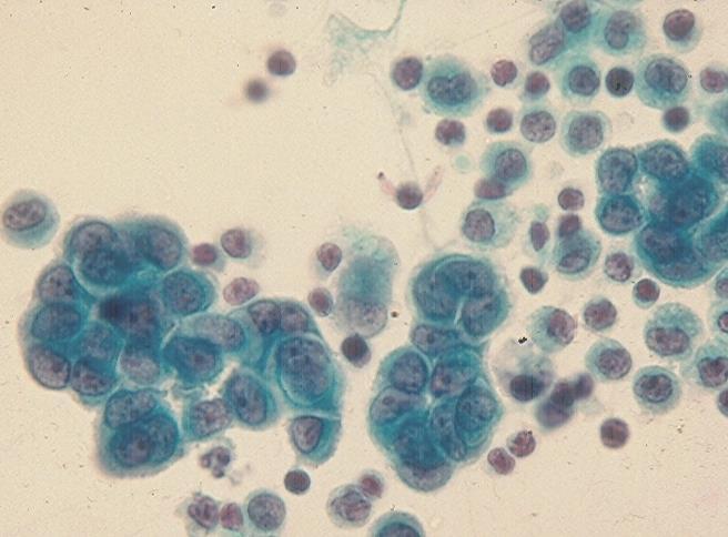
10 Exudate is caused by damage to capillary blood vessel walls. The most common etiologies of exudate include malignancy or inflammation due to infection, acute pancreatitis, pulmonary infarction, chemotherapy and radiotherapy. An exudate is turbid, purulent or bloody in appearance and has a specific gravity >1.010, protein contents >30g/L and a fluid LDH: serum LDH ratio > 0.6. Cytologically, an exudate contains polymorphonuclear leukocytes, lymphocytes and mesothelial cells. In patients with malignant tumor involving a serosal cavity the associated effusion usually contains numerous cancer cells. Benign mesothelial proliferation Benign mesothelial cell proliferation is common in a chronic or long-standing serous effusion. This may be secondary to cirrhosis, chronic inflammatory lung diseases, pulmonary infarction, radiotherapy, chemotherapy, collagen vascular diseases or trauma (e.g. from chest injury or thoracotomy). The fluid sample usually contains a large number of single and clustered hyperplastic mesothelial cells. Rarely, these mesothelial cells form large tridimensional, ball-like clusters with some cellular atypia mimicking malignant cells. (Fig.1.1). A 10
11 B C 11

12 D Fig.1.1. Benign mesothelial cell proliferation. (Pap): A. Hyperplastic mesothelial cells with slightly enlarged nuclei, micronucleoli are present singly and in small clusters. A clear space or window between adjacent cells may be seen. B. Larger clusters of hyperplastic mesothelial cells showing mildly nuclear atypia with small nucleoli. Small clear spaces between adjacent cells are present. C. A tight cluster of atypical mesothelial cells with prominent nucleoli. D. A cluster of highly atypical mesothelial cells showing pleomorphic nuclei, prominent nucleoli and slight nuclear molding. The presence of large tridimensional clusters of epithelial-like cells in a pleural effusion is suggestive of either a reactive mesothelium or a malignant disease (metastatic adenocarcinoma or mesothelioma). Reactive mesothelial cells (RMC) with nuclear atypia may mimic cancer cells from an epithelioid mesothelioma or adenocarcinoma. Clustered RMCs commonly show tight cell junctions and a clear space or "window" between adjacent cells. RMCs rarely form large ball-like clusters in pleural effusions but large cell clusters are more commonly observed in peritoneal and pericardial effusions. The differential diagnosis between RMCs and mesothelioma cells or malignant glandular cells is problematic in routinely stained smears. ICC study is helpful in distinguishing RMCs from adenocarcinoma cells but has no value in separating RMCs from mesothelioma cells as these 2 cell types share a common phenotype (positive for calretinin, CK5/6, mesothelin and Wilms tumor gene product 1 (WT1) and negative with CEA, MOC31 and Ber-Ep4 antibodies). Malignant glandular cells usually have the inverse phenotype: 12
13 positive for CEA, MOC31 and Ber-Ep4 and negative for calretinin, CK5/6, mesothelin and WT1. According to some studies staining with p53 antibody may be useful in distinguishing RMCs from mesothelioma cells as malignant cells may express p53 and reactive cells may not. Some investigators have found that RMCs react weakly or negatively with EMA antibody while mesothelioma cells show a strong membranous reaction. According to Saleh et al. malignant effusions have a Ki-67 (MIB1) immunostain labeling index value > 20% in 82% of cases while the index value of benign effusions is <5%. RMCs display, in addition, no homozygous deletion of p16 by fluorescence in situ hybridization and no loss of BRCA1-associated protein 1 by ICC. In equivocal cases, biopsy of serosal lesions for histologic evaluation is necessary. Neoplastic diseases The serosa lining a body cavity can harbor an epithelial or non-epithelial malignancy. The most common cancer involving a serosal cavity is a metastatic cancer. Among primary malignant tumors of the serosa, mesothelioma is the most important one. A. Mesothelioma Malignant mesothelioma, also known as mesothelioma, is a rare malignant serosal neoplasm that is commonly related to occupational asbestos exposure. Other potential etiologies include chronic inflammation, organic chemicals, non-asbestos mineral fibers, irradiation and genetic factors. About 75% of patients are men between years of ages, and almost all patients die of the disease within 6-12 months after the diagnosis. Mesothelioma most commonly arises in the pleural cavity. Peritoneal mesothelioma accounts for 1-10% of all cases, and the tumor rarely arises in the pericardium or tunica vaginalis. Histopathology, Immunohistochemistry and Ultrastructure Three main histologic types of mesothelioma are encountered: epithelioid, sarcomatoid and mixed tumors. Epithelioid mesothelioma (EM) is the most common tumor type, accounting for about 50% of all cases while sarcomatoid and mixed mesotheliomas account for 15-20% and 25-30% of cases, respectively. Epithelioid and mixed mesotheliomas account for about 90% of all pleural and peritoneal primary tumors that are commonly associated with a serous effusion. Sarcomatoid mesotheliomas usually present as mass lesions and are not associated with a significant serosal effusion. Histologically, an EM consists of tumor cells with variable degrees of anaplasia arranged in tubulopapillary, adenomatoid (microglandular) or solid pattern. Four rare histologic subtypes of EM are adenomatoid, deciduoid, small cell and clear cell mesotheliomas. A sarcomatoid mesothelioma is characterized by spindle malignant cells arranged in a nonspecific pattern. A mixed mesothelioma is composed of epithelioid and sarcomatous 13

 A B 14")
14 elements and areas showing a transition between these two cellular elements may be observed. (Fig.1.2 and Fig.1.3) A B 14


15 Fig.1.2. Main histologic types of mesothelioma. (HE). A. Epithelioid mesothelioma. B. Sarcomatoid mesothelioma. C. Mixed mesothelioma. C Fig.1.3. Adenomatoid mesothelioma showing tumor cells with extensive cytoplasmic vacuolar change. (HE). 15
16 By ICC study, EM cells usually stain positively for pan cytokeratins, CK5/6, vimentin, calretinin, podoplanin (D2-40), HBME, EMA and WT1 and react negatively with epithelial antibodies such as CEA, MOC31 and Ber-Ep4. Sarcomatoid mesothelioma cells usually express vimentin and cytokeratin. By electron microscopy, cells of an EM show desmosomes, intracytoplasmic and perinuclear bundles of intermediate filaments and numerous filamentous microvilli without dense core rootlets and with a length: diameter ratio >12, and intracytoplasmic mucous granules are not identified. Cytology of mesothelioma Serous effusions in patients with epithelioid and mixed mesotheliomas are usually cellular and show numerous EM cells that often display a wide range of nuclear changes, ranging from mild to marked atypia to frank malignancy. In about 10% of cases the effusions are acellular or contain only rare benign reactive mesothelial cells. Sarcomatoid cells in a mixed mesothelioma and cells of a sarcomatoid mesothelioma do not usually and spontaneously exfoliate into associated effusions. In about 50% of cases, cells from an epithelioid or mixed mesothelioma occur singly, in small groups and in large tridimensional ball-like clusters consisting of up to several hundreds cells. In about 25% of cases the tumor cells occur predominantly in tridimensional clusters with very few cells present singly and in small clusters. In the remaining 25% of cases the tumor cells occur predominantly singly. From the cytodiagnostic point of view an EM can be suspected in about 60% of cases by examination of routinely stained cytologic preparations. Classic cytologic manifestations of an EM consist of malignant cells showing the following features. (Fig Fig. 1.7): Tumor cells occur singly, in small groups or clusters, as well as in large tridimensional clusters (>50 cells). Large cell clusters have smooth and lobulated contours. Tumor cells are usually large and resemble normal mesothelial cells except they have larger nuclei, prominent nucleoli and show a spectrum of nuclear changes ranging from benign to atypical to malignant. The presence of two distinct cell populations, one benign and the other malignant, as seen in metastatic cancers, is not obviously present. Small tumor cell clusters commonly show cell-embracing-cell, "push-in" cell junctions and a clear space or window between two adjacent cells. Thick papillary tumor tissue fragments with or without fibrovascular cores may be seen and are highly suggestive of an EM. Tumor cells have a thick endoplasm and a fuzzy ectoplasm that is due to the presence of long filamentous microvilli on free cell surfaces. 16


17 An adenomatoid mesothelioma may exfoliate cells with extensive cytoplasmic vacuolization, mimicking cells from a mucus-secreting adenocarcinoma or signet-ring cell carcinoma. (Fig.1.8). A B 17


18 C Fig.1.4. Serous effusion in a pleural EM of showing: A. Tumor cells present singly, in small clusters and in large ball-like clusters. B and C. Tumor cells showing abundant, granular cytoplasm, intercellular windows and cell-embracing-cell arrangement. (A-C,Pap). B 18
19 B C 19

 and in thick")
20 D Fig.1.5. EM cells present predominantly in large ball-like clusters (A), papillary structures (B and C) and in thick lobulated cohesive clusters (D). (Pap). A 20


21 Fig.1.6. EM cells present predominantly singly in A and B. (Pap) B A 21


22 B Fig.1.7. A and B. Small clusters of pleomorphic malignant tumor cells showing thick endoplasm and fuzzy ectoplasm. Long filamentous microvilli are seen on free cell surfaces. A small window is present between 2 adjacent cells in A. (Pap). A 22


23 B C Fig.1.8. A-C. Single and clustered tumor cells with extensive cytoplasmic vacuolization from 2 cases of adenomatoid mesothelioma. (Pap). 23
. Fig.1.9. Papillary tumor tissue fragments in a CB section prepared from serous effusion associated with a pleural EM. (HE).")
24 Cellblock CB from an effusion secondary to an EM may reveal papillary tumor tissue fragments with fibrovascular cores covered with a single layer of tumor cells. This rare and interesting finding is highly suggestive of an EM. (Fig.1.9). Fig.1.9. Papillary tumor tissue fragments in a CB section prepared from serous effusion associated with a pleural EM. (HE). Immunocytochemical and Molecular features Because in the majority of cases the cytologic manifestations of EM mimic those of a metastatic adenocarcinoma to the serosa, it is important to rule out, by ICC studies, an adenocarcinoma. Important ICC characteristic features of EM cells consist of a lack of expression of epithelial antigens such as CEA, MOC31, Ber-Ep4 and the presence of mesothelioma antigens such as HBME-1, calretinin, CK5/6, podoplanin and WT1. According to Ordonez, a combination of 2 positive markers (calretinin, CK5/6, WT1) and 2 negative markers (CEA, MOC31, Ber-Ep4) is adequate for a firm diagnosis of EM. (Fig.1.10 and Fig.1.11). EM cells commonly display a homozygous deletion of p16 by fluorescence in situ hybridization (p16 FISH) and a loss of BRCA1- associated protein 1 (BAP1) by ICC. However, reactive (benign) mesothelial cells show no deletion of p16 FISH and no loss of BAP1; and cells from a bronchogenic adenocarcinoma commonly show a deletion of p16 FISH and rarely a loss of BAP1. It is important to note that deletions of p16 FISH and BAP1 are highly specific for pleural EMs but they are less specific for peritoneal EMs as some of them display no loss of BAP1. 24


25 Fig Tumor cell cytoplasm reacts negatively with CEA antibody. (ABC). A B 25

26 C Fig ICC of effusion CB in EM. (ABC): A. Tumor cells show strong cytoplasmic reaction to calretinin antibody. B. Strong, thick, membranous positive staining with spiking pattern with EMA antibody, reflecting the presence of long microvilli on tumor cell surfaces. C. EM cells showing positive nuclear staining to WT1 antibody. Diagnostic accuracy Cytodiagnosis of EM in a serous effusion consists of 2 stages: 1. Cytodiagnosis malignancy 2. Identification of mesothelial features of the cancer cells present and rule out reactive mesothelial cells and metastatic carcinoma by appropriate ICC and/or molecular tests Important comparative cytologic, ICC and molecular features of EM, reactive mesothelium and metastatic bronchogenic adenocarcinoma in pleural serous effusions are tabulated in Table 1.1. The cytodiagnostic accuracy of EM in reported series varies widely, ranging from 32% to 93%, according to the literature review by Whitaker et al. In Whitaker s experience, with adequate ICC studies, a cytodiagnostic accuracy of mesothelioma as high as 80% may be reached, and the predictive value of a positive diagnosis of mesothelioma in serous effusions is 100%. With reliable commercial antibodies, electron microscopic study of effusion CB now is rarely necessary to identify mesothelioma cells. However, when ICC results are equivocal, ultrastructural study of the tumor cells is needed for further confirmation. For a more comprehensive discussion on value and limitations of different antibodies used in the ICC diagnosis of mesothelioma the reader is referred to the articles by Attanoos, Ordonez, Leong and Vernon-Roberts, Suster and Moran, Husain et al. and Churg. 26
27 Table 1.1: Comparative Cytologic, Immunocytochemical and Molecular Features of Reactive Mesothelium, Epithelioid Mesothelioma and Bronchogenic Adenocarcinoma in Pleural Effusions* CELLULAR FEATURES Architecture: Cells: -Configuration: REACTIVE MESOTHELIUM -Singly, common -Monolayered sheets -Loose groups with windows -Polygonal, round EPITHELIOID MESOTHELIOMA -Large cohesive clusters with lobulated borders -Small tight clusters with windows and push-in junctions -Polygonal, round BRONCHOGENIC ADENOCARCINOMA -Singly, rare -Tight multilayered, 3-dimensional clusters with smooth borders -Polygonal, round -Cytoplasm: -Well-defined -Foamy or homogenous -Well-defined -Dense ectoplasm -Fuzzy periphery -Ill-defined -Vacuolated -Nucleus: -Anisonucleosis -Irregular contours -Molding -Chromatin Fine Fine or coarse Coarse -Nucleolus Small Small or large Staining characteristics: - Mucin Calretinin, CK5/6, D2-40, Mesothelin, WT CEA, MOC-31, Ber-Ep4, TTF Desmin + +/- - - EMA +, periphery ++, fuzzy, periphery +, periphery - GLUT /- Molecular features: - p16 FISH - BAP Large, single, multiple - + * Adapted from Nguyen GK, Kline TK. Essentials of Cytology. An atlas. Igaku-Shoin, New York, 1993, p.88. * p16 FISH: p16 fluorescence in situ hybridization * BAP1: BRCA1-associated protein 1 immunohistochemistry. 27
28 B. Primary effusion lymphoma This large B-cell neoplasm presents as a serous effusion without detectable tumor masses. It is associated with human herpes virus 8(HHV8)/Kaposi sarcoma herpes virus (KSHV) and it usually occurs in patients with immunodeficiency. Most patients are young to middle-aged homosexual males with HIV infection. However, cases with HIV-negative allograft recipients, particularly after cardiac transplantation, have been documented. The most common sites of tumor involvement are pleural, peritoneal and pericardial serosal cavities. Typically only one cavity is affected. The disease is highly aggressive and most patients died within one year. Cytologically, the tumor cells in the effusion consist of dyshesive, large malignant lymphoid cells with immunoblastic features, prominent nucleoli and mitosis. Markedly pleomorphic tumor cells may be observed in some cases. The cells typically express leukocyte common antigen (CD45), CD30 and plasma cell-related markers (CD38 and CD138), and they are usually negative for pan-b-cell markers such as CD19, CD20 and CD79a. The nuclei are positive for HHV8/KSHV-associated latent protein, and this expression confirms the diagnosis. C. Metastatic cancers The difficulty in cytodiagnosis of malignant effusions varies from case to case. If the primary cancer is known the diagnosis is usually straightforward. However, diagnosis of a metastatic adenocarcinoma of unknown primary by effusion cytology is often challenging and requires a careful clinicopathological correlation and extensive ICC studies. In men, malignant pleural effusions are most commonly caused by lung cancer, followed by lymphoma and gastrointestinal malignancies. In women, they are most frequently caused by metastatic breast cancer, followed in decreasing order by lung cancer, ovarian cancer and gastrointestinal cancer. In the United States, about 1% of malignant pleural effusions are caused by mesothelioma. In men, malignant ascites is most commonly caused by gastrointestinal cancer followed by lung cancer and lymphoma. In women ovarian cancer is the commonest cause followed by breast and gastrointestinal malignancies. Malignant pleural effusion of unknown primary is most commonly secondary to a lung cancer in both men and women. In about 15% of malignant ascites the primary cancer is occult at initial investigation. Of these over 50% are caused by a clinically occult ovarian cancer in women. Cancers arising from the pancreas, gallbladder, prostate and urinary bladder are rarely associated with a malignant ascites. Breast cancer is the most common malignancy in women and it rarely manifests initially as a pleural malignant effusion of 28
29 unknown primary. Lymphoma, melanoma and mesothelioma may also present initially as a malignant pleural effusion of unknown primary. Regarding the histologic types of malignant effusions, adenocarcinoma is the most common tumor followed by lymphoma, large cell carcinoma, squamous cell carcinoma, small cell carcinoma, mesothelioma and sarcoma. Diagnosis of a malignant effusion of unknown or occult primary is challenging but the identification of the primary tumor is now more successful thanks to advances in diagnostic imaging techniques and commercially available high-quality antibodies. Cytology of Metastatic Cancers Common cytologic features of most metastatic malignancies in serous effusions include increased cellularity and a distinct non-mesothelial population of enlarged cells that have enlarged nuclei with irregular nuclear contours, high nuclear : cytoplasmic ratio, coarse chromatin, multiple prominent nucleoli, increased mitosis, atypical mitosis and presence of tridimensional tumor cell clusters. Routinely stained smears of malignant effusions with well-differentiated cancers may permit a proper classification of the cancer cells into 4 broad categories: epithelial, lymphomatous, melanocytic and sarcomatous malignancies. Among the carcinomas, tumor cells with squamous or glandular differentiation and anaplastic cancers of small and large cell types can be cytologically typed with confidence in the majority of cases. In the case of a poorly differentiated or undifferentiated cancer, ICC studies are needed for a more accurate tumor typing. By staining with antibodies to S-100, HMB-45, AE1/AE3, calretinin, MOC31, CEA, LCA and vimentin, poorly differentiated cancer cells may be classified into 5 cell lines: lymphomatous, melanocytic, epithelial, mesothelial and sarcomatous. According to Dabbs, a coordinate staining of epithelial cells with CK7/CK20 antibodies will further divide them into 4 different categories, each with only a few cancer types. 1. CK7+ and CK20+: Urothelial carcinoma and ovarian mucinous carcinoma. 2. CK7+ and CK20-: Carcinomas of lung (small cell, non-small cell and non-squamous), breast, ovary (serous type), endometrium and thyroid; germ cell tumors and epithelioid mesothelioma. 3. CK7- and CK20-: Squamous cell, prostatic, renal cell and hepatocellular carcinomas. 4. CK7- and CK20+: Colorectal and Merkel cell carcinomas. Additional ICC expressions of some cell markers by metastatic carcinoma cells may further confirm the anatomic sites of their primary cancers. These specific antigenic expressions will be mentioned in each carcinoma discussed below. 29

30 Carcinomas arising from certain anatomic sites may have some specific cytologic manifestations in serous effusions. However, cells exfoliated from metastatic adenocarcinomas often display considerable overlapping features. Therefore, ICC studies are extremely important in these situations. Malignant Epithelial Tumors Bronchogenic Carcinoma Bronchogenic cancers account for about 30% of all pleural malignant effusions, and adenocarcinoma is the most common tumor. Lung adenocarcinoma cells express CK7, CEA and TTF1. Cells derived from a squamous cell carcinoma are an exception to those ICC features although a small number of squamous cell carcinomas are CK7 positive. 1. Adenocarcinoma. Cells exfoliated from a bronchogenic adenocarcinoma tend to occur singly and in irregular clusters and show prominent nucleoli, cytoplasmic vacuolization; and multinucleation may be seen. The cancer cell cytoplasm is CEA positive and the tumor cell nuclei express TTF1. (Fig.1.12 and Fig.1.13) A 30


31 B Fig Pleural effusion in a bronchogenic adenocarcinoma showing single and clustered malignant glandular cells. (A, Pap; B, Diff-Quik). A 31
: A: Tumor cells in CB showing a strong cytoplasmic reaction to CEA antibody.")

32 B Fig ICC of bronchogenic adenocarcinoma. (ABC): A: Tumor cells in CB showing a strong cytoplasmic reaction to CEA antibody. B: Tumor cell nuclei in CB stain positively with TTF1 antibody. A bronchioloalveolar carcinoma invading the overlaying pleura exfoliates single and clustered malignant glandular cells in associated effusion. Tumor cells with cellembracing-cell arrangement may be present. (Fig.1.14). A 32


33 B Fig A bronchioloalveolar carcinoma showing single and clustered malignant glandular cells. A cell-embracing-cell arrangement is present in a small cell cluster at the center of figure B and 2 reactive mesothelial cells with a window between them cells are also seen. (Pap, A and B). Bronchioloalveolar cell carcinoma arising from type II pneumocytes shows clustered pleomorphic malignant glandular cells with vacuolated cytoplasm that react negatively with mucicarmine and PAS reagent. Ultrastructural study of the effusion CB revealed intracytoplasmic inclusions with myelin figures suggesting surfactant and confirming pneumocyte type II origin. (Fig ) A 33

")
34 B Fig.1.15: A: A cluster of pleomorphic malignant glandular cells in pleural effusion associated with a bronchioloalveolar cell carcinoma. (Diff-Quik). B: Intracytoplasmic inclusion with concentric myelin figure suggesting that the tumor arises from pneumocytes type II. (Uranyl acetate and lead citrate stain, x 18,000). 2. Large cell carcinoma yields single, large pleomorphic cells. The tumor cells display enlarged nuclei, coarse chromatin and prominent single or multiple nucleoli. (Fig.1.16). Fig Isolated pleomorphic malignant cells with prominent nucleoli from a bronchogenic large cell carcinoma. (Pap). 34
.")
35 3. Squamous cell carcinoma rarely exfoliates cells into an associated effusion. Cells from a well-differentiated or keratinizing tumor are usually present singly and in small clusters, and show a dense, hard, eosinophilic cytoplasm suggesting keratin formation. (Fig. 1.17). Cells from a non-keratinizing or poorly differentiated tumor are commonly seen in large syncytial clusters and show a thin, ill-defined cytoplasm. However, single tumor cells with dense and thick or thin cytoplasm are noted in some cases of the poorly differentiated tumor. (Fig to 1.19). In some patients with a poorly differentiated tumor the exfoliated cells are indistinguishable from those of a lung adenocarcinoma. The neoplastic squamous cells are usually negative for CK7/CK20 and TTF1 and positive for CK5/6, p40 and p63. Fig Metastatic well-differentiated squamous cell carcinoma showing in pleural effusion single and clustered malignant squamous cells with keratinized cytoplasm. (Pap). 35


36 Fig Metastatic poorly differentiated squamous cell carcinoma showing in pleural effusion large irregular syncytial clusters of non-keratinizing cells. (Pap). Fig Metastatic poorly differentiated squamous cell carcinoma, small cell type, showing in pleural effusion small tumor cells with hyperchromatic nuclei present singly and in small clusters. (Pap). 36
.")
.")
37 4. Small cell carcinoma (SCC) cells are small, cuboidal cells with scant cytoplasm and small, round nuclei with stippled ( salt and pepper ) chromatin and inconspicuous nucleoli. They are seen arranged in chains or clusters with nuclear molding. (Fig.1.20). In some cases the tumor cells are larger (SCC of intermediate cell type) and show conspicuous nucleoli, mimicking those of a non-small cell lung carcinoma. Rarely, cells from an SCC are present in large tridimensional clusters and loosely large aggregates, readily mistaken for those of a non-scc. (Fig.1.21). Cells derived from a lung SCC commonly express neuroendocrine markers (neuron-specific enolase, chromogranin, synaptophysin, CD56) and TTF1. A B Fig Small cell carcinoma showing clustered small tumor cells with scant cytoplasm, hyperchromatic nuclei and nuclear molding. (Pap, A and B). 37
 Breast Carcinoma Metastatic breast carcinoma is responsible for about 25% of all malignant pleural effusions.")
 and")
38 A B Fig Pleural effusion from a small cell carcinoma showing: A. abundant tumor cells singly and in large ball-like clusters. B. Aggregated tumor cells with nuclear features of a small-cell lung cancer. (Pap, A and B) Breast Carcinoma Metastatic breast carcinoma is responsible for about 25% of all malignant pleural effusions. Tumor cells from a ductal carcinoma are usually monomorphic, have irregular nuclei, multiple nucleoli and a non-vacuolated cytoplasm. They may resemble reactive and atypical mesothelial cells, and they are typically seen in abundant threedimensional, large cell balls or morulae (>50 cells) and papillae. (Fig.1.22 and Fig. 1.23). 38


39 In some cases the tumor cells are present singly. (Fig.1.24). These cells are CK7 positive, CK20 negative and often express ER, PR and Gross cystic disease fluid proteins. A B Fig Pleural effusion from metastatic mammary duct carcinoma showing single and clustered monomorphic glandular cells. Tumor cells in linear arrangement are seen in A. (Pap, A and B). 39


40 A B Fig Pleural effusion from a metastatic mammary duct carcinoma showing in A tumor cells with prominent nucleoli in a tight ball-like cluster and in a linear row of three cells, and in B a tumor ball in a CB section. (A, Pap; B, HE). 40


41 A B C Fig A: Malignant glandular cells from a metastatic mammary duct carcinoma showing predominantly single cells. Clear spaces or windows between adjacent tumor cells are noted. (Pap). B: CB section stained with Ber-Ep4 antibody showing a membranous pattern. (ABC). C: Positive reaction of tumor cell nuclei with Estrogen receptor antibody. (ABC). 41

42 Metastatic lobular carcinoma of the breast usually shows small epithelial tumor cells with hyperchromatic nuclei that are present singly and in small chains. Single intracytoplasmic vacuoles containing mucinous droplets compressing tumor cell nuclei may be seen. Their ICC expressions are similar to those of a mammary ductal carcinoma. Gastrointestinal carcinoma Well- and moderately differentiated gastrointestinal adenocarcinomas usually show in effusions cohesive clusters of malignant glandular cells with intracytoplasmic vacuoles admixed with single tumor cells with similar features. A poorly differentiated adenocarcinoma yields large cells with vacuolated cytoplasm, pleomorphic nuclei and prominent nucleoli dispersed singly and in small clusters. (Fig.1.25). Fig Poorly differentiated gastric adenocarcinoma showing in associated ascites single and clustered malignant glandular cells with prominent nucleoli. Some tumor cells have a large intracytoplasmic vacuole pushing their nuclei to the cell periphery, creating malignant signet-ring cells. (Pap). Single malignant glandular cells with signet-ring configuration are most commonly derived from a diffuse carcinoma or signet-ring cell carcinoma of the stomach. (Fig. 1.26). 42
. (Courtesy of Dr. T.")
.")
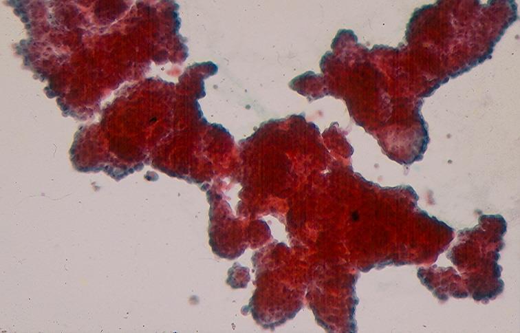
43 A B Fig Diffuse gastric carcinoma showing: A. A loose cluster of tumor cells with intracytoplasmic mucous vacuoles. (Pap). B. Positive staining with CEA-monoclonal antibody of tumor cell cytoplasm. (ABC). (Courtesy of Dr. T. Thomson, Vancouver, BC, Canada) Adenocarcinoma of the appendix is the most common cause of mucinous ascitis (pseudomyxoma peritonei). The peritoneal effusion displays thick mucinous material, clustered malignant epithelial cells and proliferated capillary blood vessels. (Fig.1.27). 43


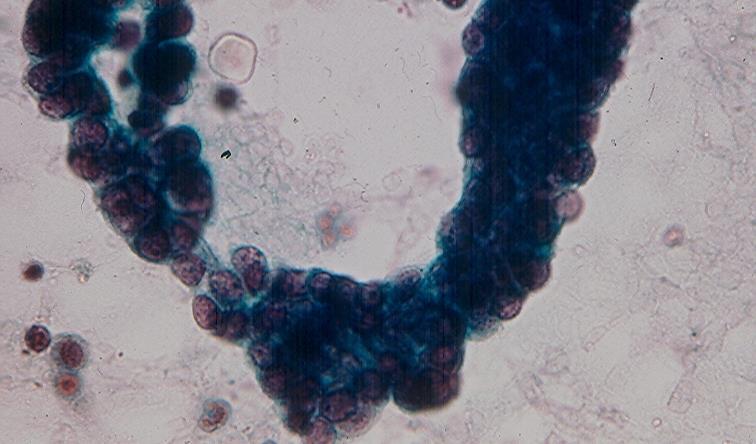
44 A B Fig Pseudomyxoma peritonei secondary to an appendiceal adenocarcinoma: A. A small cluster of malignant epithelial cells in a mucinous background. B. Proliferated capillary blood vessels. (Pap, A and B). Ovarian carcinoma Tumor cells derived from a serous carcinoma of the ovary are typically seen in papillary clusters, and psammoma bodies are often present. (Fig.1.28). It should be born in mind that cells from a papillary carcinoma of the lung and thyroid may also show psammoma bodies. 44

 A mucinous carcinoma of the ovary shows clustered")
45 A B Fig.1.28 Metastatic ovarian serous carcinoma showing papillary clusters of tumor cells containing a laminated psammoma body. (Pap, A and B) A mucinous carcinoma of the ovary shows clustered tumor cells with vacuolated cytoplasm in a background of mucin. (Fig.1.29). 45


46 Cells derived from a serous ovarian carcinoma are Ber-Ep4, WT1 and CK7 positive and CK20 negative while those of a mucinous ovarian carcinoma are CK7 and CK20 positive and rarely express WT1. A B Fig Mucinous ovarian carcinoma metastatic to the pericardium showing: A. Large tridimensional clusters of tumor cells in a mucinous background. B. Tumor cells with intracytoplasmic mucus are seen at higher magnification. (Pap, A and B). 46

47 Other cancers Urothelial carcinoma, high-grade of the urinary tract exfoliates its cells singly and in cohesive clusters. The tumor cells show granular, well-defined cytoplasm, oval nuclei, granular chromatin and prominent nucleoli. (Fig.1.30). These cells express CK7, CK20, uroplakin III, thrombomodulin and p63. Fig.1.30 A cohesive cluster of malignant epithelial cells with thick, granular cytoplasm from a metastatic high-grade urothelial carcinoma. (Pap). Hepatocellular Carcinoma exfoliates single or clustered large, polygonal malignant cells with abundant, granular cytoplasm and large, oval nuclei with macronucleoli. These cells may show intracytoplasmic bile pigment granules, intercellular bile plugs and intracytoplasmic globular inclusions. The tumor cell cytoplasm stains negatively with CK7/CK20 and positively with Hepar1 and alpha-fetoprotein antibodies. Neuroendocrine Carcinoma may arise from the bronchial tree, pancreas, bowels and other anatomic sites. It exfoliates medium-sized polygonal cells with oval nuclei with stipple chromatin, conspicuous nucleoli and granular cytoplasm. They are commonly present in groups and large clusters with moderate cohesiveness. Nuclear molding may be observed. The tumor cell cytoplasm is CK7 positive, CK20 negative and expresses NSE, synaptophysin and chromogranin. Renal Cell Carcinoma of clear cell type yields cohesive tumor cell clusters with clear or granular cytoplasm and oval, large nuclei with prominent nucleoli. The tumor cells express pan-cytokeratin, EMA, vimentin, renal cell carcinoma antigen and PAX-2 but are CK7/CK20 negative. (Fig.1.31). 47

 cells show, in addition, a positive reaction to alpha-fetoprotein antibody.")
48 A B Fig.1.31 Metastatic renal cell carcinoma to the pleura showing in associated effusion clustered malignant cells with clear cytoplasm and prominent nucleoli. (A,Pap;B,CB,HE). Germ Cell Neoplasms arising from the ovary are a group of tumors of different histology and can be benign or malignant. Cells of malignant germ cell tumors such as endodermal sinus tumor, choriocarcinoma, embryonal carcinoma and dysgerminoma are generally positive for CK7 and placental alkaline phosphatase and negative for CK20 and EMA. Endodermal sinus tumor (yolk sac tumor) cells show, in addition, a positive reaction to alpha-fetoprotein antibody. Choriocarcinoma cells express human chorionic gonadotrophin and embryonal carcinoma cells are positive for pancytokeratins, CD30 and OCT4. Dysgerminoma cells are OCT4 positive and keratin negative. 48
. Fig.1.32 Dysgerminoma metastatic to the lung showing in pleural effusion single and loosely clustered malignant tumor cells with prominent nucleoli.")
 reveals superficial and anucleate squamous cells. (Diff-Quik). (Courtesy of Dr. T.")
49 A metastatic ovarian dysgerminoma to the lung shows in pleural effusion single and loosely clustered large malignant cells with variable, defined cytoplasm and large, round or oval nuclei with prominent nucleoli. (Fig.1.32). Fig.1.32 Dysgerminoma metastatic to the lung showing in pleural effusion single and loosely clustered malignant tumor cells with prominent nucleoli. (Pap). A ruptured benign ovarian mature teratoma (dermoid cyst) yields in associated ascites mature benign squamous cells. (Fig.1.33). Fig Peritoneal fluid from a young woman with a ruptured ovarian dermoid cyst (mature teratoma) reveals superficial and anucleate squamous cells. (Diff-Quik). (Courtesy of Dr. T. Thomson, Vancouver, BC, Canada). 49

50 Malignant Non-epithelial Tumors Hematologic malignancies These tumors are the commonest non-epithelial malignant tumors associated with a positive effusion. Hodgkin lymphoma, depending on its histologic variant, may exfoliate lymphocytes and eosinophils in addition to its diagnostic Reed-Sternberg cells. (Fig.1.34). Fig.1.34 Hodgkin disease involving the pleura showing in associated effusion numerous benign lymphoid cells and 2 Reed-Sternberg cells. (Pap). Non-Hodgkin lymphomas shed tumor cells varying from benign-appearing lymphoid cells similar to matures lymphocytes in small lymphocytic lymphoma/chronic lymphocytic leukemia to atypical enlarged lymphoid cells with nuclear indentations and protrusions in higher grade lymphomas. (Fig.1.35). In acute leukemia blast cells may be present. Cells of a chronic myelogenous leukemia consist of mature and immature myelogenous cells without a "leukemic hiatus", as seen in an acute myelogenous leukemia. 50

. (Courtesy of Dr. T.")
51 A B Fig Non-Hodgkin lymphoma: A. Low-grade tumor showing cells with mildly nuclear atypia. B. High-grade tumor showing large cells with pleomorphic nuclei and vacuolated cytoplasm. (Diff-Quik, A and B). (Courtesy of Dr. T. Thomson, Vancouver, BC, Canada) 51
. A B Fig.1.36. A. Neoplastic plasma cells with eccentrically located nuclei. (Pap). B. Neoplastic plasmablasts in a pleural fluid.")

52 A multiple myeloma involving the pleura shows single cancer cells with eccentrically located nuclei with cart-wheel chromatin clumping. (Fig.1.36). A B Fig A. Neoplastic plasma cells with eccentrically located nuclei. (Pap). B. Neoplastic plasmablasts in a pleural fluid. (MGG). (Courtesy of Dr. T. Thomson, Vancouver, BC, Canada) Melanoma Melanoma cells may occur singly and in cohesive clusters. Intracytoplasmic melanin pigment granules, intranuclear cytoplasmic inclusions and macronucleoli are commonly 52

. Bone and Soft Tissue sarcomas These tumors show single and loosely clustered malignant cells.")
53 present. (Fig.1.37). A positive cytoplasmic reaction with S-100 protein, HMB-45 or MART1 antibodies is diagnostic of the tumor. A B Fig Pleural effusion from a cutaneous melanoma metastatic to the lung: A & B: single and clustered tumor cells showing a large intranuclear cytoplasmic inclusion. (Pap, A and B). Bone and Soft Tissue sarcomas These tumors show single and loosely clustered malignant cells. The tumor cells tend to have a round configuration and loose their original shapes in tissue sections. Typing of sarcoma cells is difficult without clinical data, cytochemical, ICC and cytogenetic studies. 53
. Ewing sarcoma yields small polygonal cells with oval nuclei and glycogen-rich cytoplasm singly and in rosettes. (Fig.1.39).")
 Fig.1.")
54 Osteogenic sarcoma usually shows rounded large malignant cells with macronucleoli. Cells from a chondrosarcoma display abundant, well-defined cytoplasm and macronucleoli. (Fig.1.38). Ewing sarcoma yields small polygonal cells with oval nuclei and glycogen-rich cytoplasm singly and in rosettes. (Fig.1.39). The tumor cell cytoplasm stains positively with PAS and negatively with PAS with prior diastase digestion and reacts positively with CD99 antibody. Fig Pleural effusion from a metastatic chondrosarcoma showing single malignant cells with basophilic cytoplasm and double conspicuous nucleoli. (Pap) Fig.1.39 Metastatic Ewing sarcoma cells present singly and in a small rosette. (Pap). 54


55 Biphasic synovial sarcoma may show in associated effusions epithelial-like and spindle-shaped tumor cells in loose aggregates. (Fig.1.40). Epithelial-like tumor cells with focal gland-like arrangement may be seen, and a transition between these two types of cells may be observed. The tumor cells express cytokeratin and vimentin. Fig Spindle malignant cells with scant cytoplasm in pleural effusion associated with a metastatic synovial sarcoma to the lung. (Pap). Leiomyosarcoma. A high-grade uterine leiomyosarcoma metastatic to the lung shows in associated effusion large single pleomorphic malignant cells with bizarre nuclei and dense, granular cytoplasm that stains positively with vimentin, desmin and smooth muscle cell antibodies. (Fig.1.41). The metastatic cancer cells are rounded up and show no resemblance to the cells in the primary tumor. Fig Bizarre large malignant cells in a pleural effusion secondary to a uterine leiomyosarcoma metastatic to the lung. (Pap). 55

. 56")
56 Melanoma of soft parts (clear cell sarcoma of soft parts) metastatic to the lung yields in associated pleural effusion single and loosely clustered polygonal cells with variably abundant cytoplasm. (Fig.1.42). The tumor cell cytoplasm expresses S-100 protein, Melan A and Mart 1. A B Fig Cells derived from a melanoma of soft parts present singly and in loose aggregates. (A, Diff-Quik and B, Pap). 56
. Fig.1.43 Clustered malignant cells from a metastatic high-grade glioma. (Pap). Miscellaneous serous effusions 1.")
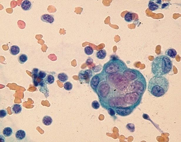
57 Glial tumors High-grade glioma metastatic to the lung yields in pleural effusion clusters of pleomorphic malignant cells that react positively with Glial fibrillary acidic protein antibody. (Fig.1.43). Fig.1.43 Clustered malignant cells from a metastatic high-grade glioma. (Pap). Miscellaneous serous effusions 1. Pleural effusion caused by lung tuberculosis shows an increased number of lymphoid cells with many displaying atypical nuclei. Epithelioid and multinucleated giant cells of Langhans are rarely observed. Mesothelial cells tend to be scanty. Culture is the best way to identify Mycobacterium tuberculosis. 2. Eosinophil-rich effusions, usually pleural, are relatively common. (Fig.1.44). They result most often from air in the serosal cavity, as seen in pneumothorax, post surgery or repeated thoracentesis. They may also be related to infections (bacterial, viral, fungal, or parasitic), pulmonary infarcts, hypersensitivity reactions, or as part of a systemic eosinophilic disorder. 57


58 Fig Eosinophil-rich effusion. (Pap) 3. Extramedullary hematopoiesis may be identified in effusions in patients with myelofibrosis. Immature red blood cells and megakaryocytes are identified. (Fig.1.45). A 58

 may be observed in serous effusions of individuals")
59 B Fig Benign bone marrow cells including a multinucleate megakaryocyte in ascitis of a woman developing extramedullary hematopoiesis secondary to myelofibrosis with anemia. (Pap, A and B) 4. Collagen vascular diseases such as Rheumatoid arthritis and Systemic lupus erythematosus may show in effusions some interesting cytologic findings: - Histiocytes, neutrophils and granular necrotic debris may be seen in effusions of patients with rheumatoid arthritis. - LE cells (polymorphonuclear leukocytes with large intracytoplasmic inclusions of degenerated and antinuclear antibody-coated nuclear material) may be observed in serous effusions of individuals with systemic lupus erythematosus. (Fig. 1.46). The LE cells may also be seen in patients with rheumatoid arthritis and in individuals taking drugs (procainamide, hydralazine) or having multiple myeloma and Hodgkin disease. Fig LE cells in pleural effusion of a patient with systemic lupus erythematosus. (Pap). 59
60 4. Drug reaction-related effusions contain abundant eosinophils. Chemotherapy may produce atypical reactive mesothelial cells mimicking cancer cells. 5. Endometriosis may show in associated ascites, morulae of endometrial epithelial cells and aggregates of hemosiderin laden macrophages. 6. Lung asbestosis may also associate with a nonspecific benign pleural transudate. Diagnostic accuracy Cytodiagnosis of malignant effusions is challenging and usually requires experience and ICC. The diagnostic accuracy rate of malignant effusions is about 85% when multiple samples are evaluated. False-negative diagnostic rates vary widely among different reported series, and their main reasons consist of inadequate sampling, scantiness of cancer cells, faulty preparatory technique and erroneous interpretation. False-positive diagnostic rates up to 3% have been reported, and the most common error is the misinterpretation of reactive atypical mesothelial cells in long-standing benign effusions as cancer cells. Bibliography Andrici J, et al. Loss of expression of BAP1 is very rare in non-small cell lung carcinoma. Pathology. 2016; 48: 336. Attanoos RL, et al. The use of immunohistochemistry in distinguishing reactive from neoplastic mesothelium: a novel use for desmin and comparative evaluation with epithelial membrane antigen, p53, platelet-derived growth factor-receptor, P-glycoprotein and Bcl2. Histopathology. 2003: 43: 231. Beasley MB. Immunohistochemistry of pulmonary and pleural neoplasia. Arch Pathol Lab Med. 2008; 132: 23. Bedrossian CWM. Malignant effusions. A multimodal approach. New York, Igaku-Shoin, Betta P-G, et al. Immunohistochemistry and molecular diagnosis of pleural malignant mesothelioma. Arch Pathol Lab Med. 2012; 136: 253. Boerner SL. Mimicry and pitfalls in effusion cytology. Pathology Case Reviews. 2006; 11:85. 60
61 Caraway NP, Stewart J. Primary effusion lymphoma. Pathology Case Reviews. 2006; 11:78. Churg A, et al. Tumors of the serosal membranes. In AFIP Atlas of tumor pathology, 4 th series, Washington DC, Armed Forces Institute of Pathology. Churg A, et al. New markers for separating benign from malignant mesothelial proliferations. Are we there yet? Arch Pathol Lab Med. 2016; 140:318. Dabbs DJ. Immunohistology of metastatic carcinoma of unknown primary. In Diagnostic Immunohistochemistry, 2 nd ed, 2006, Dabbs D, ed. Philadelphia, Churchill Livingstone Elsevier, p Delahaye M, et al. Complementary value of 5 carcinoma markers for the diagnosis of malignant mesothelioma, adenocarcinoma metastasis and reactive mesothelium in serous effusions. Diagn Cytopathol 1997; 17:115. DeMay R. Fluids. In Arts & Science of Cytopathology. Exfoliative cytology. Chicago, ASCP, 1996, p DiBonito L, et al. The positive pleural effusion. A retrospective study of cytopathologic diagnosis with autopsy confirmation. Acta Cytol. 1992; 36:329. Fletsch PA, Abati A. Immunocytochemistry in effusion cytology. A contemporary review. Cancer (Cancer Cytopathol). 2001;93:293. Giesinger KR, et al. Effusions. In Modern Cytopathology, Giesinger KR, et al, eds. Philadelphia, Churchill Livingstone. 2004, p 257. Garcia L W, et al. The value of multiple fluid specimens in the cytodiagnosis of malignancy. Mod Pathol 1994; 7:665. Hiroshima K, et al. Cytologic differential diagnosis of malignant mesothelioma and reactive mesothelial cells with FISH analysis of p16. Diagn Cytopathol. 2016; 44:591. Hjerpe A, et al. Guidelines for the cytopathologic diagnosis of epithelioid and mixed-type malignant mesotheliomas. A complementary statement from the International Mesothelioma Interest Group. Also endorsed by the International Academy of Cytology and the Papanicolaou Society of Cytopathology. Acta Cytol. 2015; 59: 2. Husain AN, et al. Guidelines for pathologic diagnosis of malignant mesothelioma Update of the consensus statement from the International Mesothelioma Interest Group. Arch Pathol Lab Med. 2013; 137:
62 Krishna M. Diagnosis of metastatic neoplasms. An immunohistochemical approach. Arch Pathol Lab Med. 2010; 134: 207. Leong AS. Immunostaining of cytologic specimens. Am J Clin Pathol.1996; 105:139. Leong A S-Y, Vernon-Roberts E. The immunohistochemistry of malignant mesothelioma. Pathol Annu. 1994; 29(2):157. Lyons-Bourdreaux V, et al. Cytologic malignancy versus benignancy. How useful are the newer markers in body fluid cytology. Arch Pathol Lab Med. 2008; 132:23. Manoska F, et al. Diagnostic effects of prolonged storage on fresh effusion samples. Diagn Cytopathol. 2007; 35:6. Mallonee MM, et al. A morphologic analysis of the cells of ductal carcinoma of the breast and adenocarcinoma of the ovary in pleural and abdominal effusions. Acta Cytol.1987; 31:441. Mullick BB, et al. P53 gene product in pleural effusions. Practical use in distinguishing benign from malignant cells. Acta Cytol. 1996; 40:855. Naylor B. The pathognomonic cytologic pictures of rheumatoid arthritis. Acta Cytol. 1990; 34: 465. Naylor B. Cytological aspects of pleural, peritoneal and pericardial fluids in patients with systemic lupus erythematosus. Cytopathology. 1992; 3:1. Naylor B. Pleural, peritoneal and pericardial effusions. In Comprehensive Cytopathology, Bibbo M, ed. Philadelphia, Saunders Ng WK, et al. Thyroid transcription factor-1 is highly sensitive and specific in differentiating metastatic pulmonary from extrapulmonary adenocarcinoma in effusion fluid cytology specimens. Cancer. 2002; 96:43. Nguyen GK, Jeannot A. Cytology of synovial sarcoma metastases in pleural fluid. Acta Cytol. 1982; 26:517. Nguyen GK. Cytopathology of pleural mesothelioma. Am J Clin Pathol 2000; 114 (Suppl 1): S68. Ordonez NG. Immunohistochemical diagnosis of epithelioid mesotheliomas: a critical review of old markers, new markers. Hum Pathol.2002; 33:935. Ordonez NG. Applicability of immunohistochemistry in the diagnosis of epithelioid mesothelioma: a review and update: Hum Pathol. 2013; 44:1. Panani AD, et al. Numerical abnormalities of chromosome 9 and p16cdkn2a gene deletion detected by FISH in non-small cell lung cancer. Anticancer.Res.2009;29:
63 Saleh H, et al. Value of Ki-67 immunostain in identification of malignancy in serous effusions. Diagn Cytopathol. 1999; 20:24. Sears D, Hajdu S. The cytodiagnosis of malignant neoplasms in pleural and peritoneal effusions. Acta Cytol. 1995; 31:85. Sherman ME, Mark E. Effusion cytology in the diagnosis of malignant epithelioid and biphasic pleural mesothelioma. Arch Pathol Lab Med 1990: 114:845. Shield PW, et al. Markers for metastatic adenocarcinoma in serous effusion specimens. Diagn Cytopathol 1994; 11:237. Smyth-Pareslow MJ, et al. Cells of squamous cell carcinoma in pleural, peritoneal and pericardial fluids. Origin and morphology. Acta Cytol. 1989; 33:245. Suster S, Moran CA. Applications and limitations of immunohistochemistry in the diagnosis of malignant mesothelioma. Adv Anat Pathol. 2006; 13:316. Travis WD, et al. Pathology and genetics of tumors of the lung, pleura, thymus and heart. World Health Organization Classification of Tumours; Lyon: IARC, van Niekerk CC, et al. Marker profile of mesothelial cells versus ovarian carcinoma cells. Int J Cancer.1989; 43:1065. Walts AE, et al. BAP1 immunostain and CDKN2A (p16) FISH analysis: clinical applicability for the diagnosis of malignant mesothelioma in effusions. Diagn Cytopathol. 2016; 44:599. Whitaker D. The cytology of malignant mesothelioma. Cytopathology.2000; 11:139. Wick MR, et al. Immunohistochemical differential diagnosis of pleural effusions, with emphasis on malignant mesothelioma. Curr Opin Pul Med. 2001; 7:187. Zhang K, et al. Utility of immunohistochemistry in the diagnosis of pleuropulmonary and mediastinal cancers. A review and update. Arch Pathol Lab Med. 2014; 138:
64 Chapter 2 Peritoneal and pelvic washings Dianna Ionescu and Gia-Khanh Nguyen Peritoneal and pelvic washings were first used by Keettel and Elkins in 1956 to detect early spreads of ovarian cancers that were not grossly identifiable on the peritoneal surface at laparotomy. This method of investigation has rapidly and increasingly gained a wide acceptance in evaluating cancers arising from the female genital tract and it is included in the FIGO staging systems for ovarian and endometrial cancers. Indications and goals 1. To assist in the initial staging of primary ovarian and endometrial cancers. 2. To exclude intraabdominal occult cancers in patients undergoing surgery for benign pelvic diseases. 3. To monitor the effectiveness of chemotherapy for advanced ovarian cancers (second-look laparotomy). 4. To assist in staging of some non-gynecologic abdominal cancers (gastric and pancreas) Collection and preparation of cell samples Any ascitic fluid present at laparotomy is collected and submitted as a separated sample for cytologic evaluation. A syringe with a long rubber catheter is used to instill ml of sterile normal saline in 4 following anatomic sites: the inferior aspect of the diaphragm, right and left paracolic gutters and pelvic cavity. One option is to aspirate each of these sites and submit separate samples for cytologic evaluation. However, submitting a single sample specimen combining samples from all 4 sites has no inferior diagnostic value. Usually an equal volume of 50% ethanol is added to each cell sample to preserve cell morphology. As peritoneal washing (PW) specimens are usually heavily contaminated with blood that may obscure diagnostic cells, hemolysis of bloody specimens is necessary. Adding ethanol to PW samples has the advantage of destroying most red blood cells. Ficoll-Hypaque solution may be also used to separate red blood cells from nucleated cells. 64

65 The fluid sample is then thoroughly mixed and a 100 ml aliquot is centrifuged at 1800 rpm for 10 min. The supernatant is discarded and the sediment is used to prepare 2-4 direct smears. It may be re-suspended in an appropriate amount of normal saline for cytospin preparation. If the smears/preparations obtained are bloody they may be fixed in Carnoy solution for 3-5 min to lyse red blood cells. A liquid-based preparation method may also be used. The remaining sediment, if present, is fixed in formalin for preparation of a CB that may be used for ICC staining, if necessary. The cell preparations or smears obtained are fixed in 95% ethanol and stained by the Papanicolaou method. CB sections are stained with hematoxylin and eosin. Cytologic findings 1. Normal Peritoneal Washing A PW cell sample from a healthy woman is usually cellular and contains large sheets of mesothelial cells that are exfoliated by washing. Single and clustered histiocytes are commonly present as well as polymorphonuclear leukocytes from the contaminated blood. Minute fragments of benign adipose tissue and skeletal muscle from the abdominal wall incision are also observed, mainly in PW cellblocks. The sheets of mesothelial cells are large and usually folded. These cell sheets consist of cohesive polygonal mesothelial cells with oval nuclei, thin and regular nuclear contours, fine chromatin and small nucleoli. Nuclear grooves are seen in many cells and focal nuclear crowding may also be observed. (Fig.2.1). On rare occasions papillary clusters of benign mesothelial cells containing psammoma bodies and masses of collagen surrounded by mesothelial cells ( collagen balls ) may be seen. (Fig.2.2). A 65


. A B Fig.2.")
66 B Fig.2.1. A large and irregular sheet of mesothelial cells exfoliated by peritoneal jet washing showing evenly spaced mesothelial cells that have ill-defined cytoplasm, oval or bean-shaped nuclei, small conspicuous nucleoli. (Pap, A and B). A B Fig.2.2. Collagene ball in peritoneal washing cell film (A) and in cellblock (B). (Pap, A; HE, B). 66

67 2. Gynecologic Malignant Tumors Ovarian cancer Ovarian carcinoma is a common neoplasm in women over 65 years of age. Most women with ovarian cancer are diagnosed at stage III or IV. Evaluation of PW is not required for advanced ovarian cancer but it is important for staging stage I or II tumors. A positive PW in these two situations will change the tumor stages to IC and IIC, respectively. With the exception of serous borderline tumors, patients with stages IC and IIC ovarian carcinomas receive postoperative chemotherapy to delay recurrences. Presently, there is no convincing evidence that a positive PW predict a poor outcome, independent of other prognostic factors. Serous Carcinoma is the commonest histologic type of ovarian cancers and the one that is often associated with a positive PW. Depending on its histologic grade the cytologic findings are different. A low-grade serous carcinoma will yield fairly monomorphic, small tumor cells organized predominantly in large tridimensional clusters. (Fig.2.3). Fig Low-grade malignant epithelial cells attached to one psammoma body in PW of a woman with an ovarian low-grade serous carcinoma. (Pap). A high-grade tumor is characterized by single and clustered pleomorphic malignant cells. (Fig. 2.4). Single and loosely clustered tumor cells with some cells showing vacuolated cytoplasm are seen. Psammoma bodies are often present within tumor cell clusters from low- as well as high-grade tumors. 67
 is a low-grade neoplasm with significantly better prognosis than an ovarian low-grade serous carcinoma.")

68 A B Fig High-grade ovarian serous carcinoma showing a thick irregular cluster of pleomorphic malignant epithelial cells with prominent nucleoli. (Pap, A and B). Serous borderline tumor (also known as Serous tumor of low-malignant potential) is a low-grade neoplasm with significantly better prognosis than an ovarian low-grade serous carcinoma. The cytologic manifestations of a serous borderline ovarian tumor are similar to those of a low-grade serous carcinoma. A serous borderline tumor is distinguished histologically from a low-grade serous carcinoma by the absence of stromal invasion in the former. This important finding is readily identified by extensive sampling and histologic evaluation of the resected tumor. Mucinous carcinoma of the ovary is less common than serous carcinoma and accounts for about 10-15% of all ovarian tumors. The majority of ovarian mucinous 68
69 tumors are benign (75%) while 10% are borderlines and 15% are carcinomas. Most ovarian mucinous tumors are of intestinal type (85%) and present as unilateral tumors. The other histologic subtype, endocervical-like mucinous carcinomas are bilateral in up to 40% of cases. Regardless of their subtypes, these ovarian tumors yield in PW malignant cells with large cytoplasmic vacuoles arranged in loose clusters and in linear arrangements or cell strips or singly. Other ovarian cancers such as germ cell and sex-cord tumors rarely involve the peritoneum or omentum. Endometrial cancer Endometrial carcinoma is the most common gynecologic malignancy and accounts for about 6% of all cancers in women. According to the old FIGO staging system for endometrial cancer a positive PW constitutes one of the criteria for stage IIIA. Recently, the revised FIGO staging system for endometrial states, with regards to stages IIIA and IIIB, that positive cytology has to be reported separately without changing the stages. However, a positive PW in this situation does not necessary indicate a poor prognosis, as endometrial cancer cells may spread onto peritoneal surface by transtubal retrograde dissemination. This may be facilitated by a prior endometrial biopsy and curettage, by a preoperative intracavitary placement of radium implants or hysteroscopy. Patients with endometrial cancer and positive PW usually have a high-grade tumor with deeper myometrial invasion and lymph node metastasis. Endometroid adenocarcinoma is the commonest histologic type of endometrial carcinomas. Papillary serous and Clear cell carcinomas and Malignant mixed müllerian tumor (MMMT) are rare neoplasms. When these tumors involve the peritonenum or omentum malignant cells may be detected in PWs. A low-grade endometrioid carcinoma is characterized by single and clustered small cuboidal malignant epithelial cells with scant cytoplasm, enlarged but monomorphic nuclei, coarse chromatin and conspicuous nucleoli. (Fig. 2.5). A high-grade tumor yields more pleomorphic malignant cells that may be present in loose aggregates or clusters. A papillary serous tumor is characterized by clustered small cuboidal cells with scant cytoplasm and enlarged hyperchromatic nuclei with inconspicuous nucleoli. A clear cell carcinoma shows single and clustered pleomorphic malignant cells with clear cytoplasm. A MMMT usually shows clusters of malignant glandular cells only, as malignant stromal cells seldom exfoliate. 69
.")

70 A B Fig Low-grade endometrioid adenocarcinoma of the uterus showing clustered monomorphic cells with oval nuclei and small, conspicuous nucleoli. A few tumor cells with irregular nuclear contours are also observed. (Pap, A and B). Cervical cancer The incidence of positive PW in cervical cancer of all stages is low, about 8%, and it is about 1% in stage IB cervical cancer. PW finding is not included in FIGO staging system for cervical cancer. Adenocarcinoma of the cervix more commonly spreads to the peritoneal cavity than its squamous cell counterpart. Adenocarcinoma cells are seen in clusters, as well as those of a non-keratinizing squamous cell carcinoma. Cells derived from a keratizing squamous cell carcinoma are often present singly. 70

71 3. Second-look laparotomy Patients who had chemotherapy or radiotherapy for advanced ovarian cancer and who show no clinical or diagnostic imaging evidence of residual tumor may undergo a laparotomy to evaluate the response to therapy. In about 50% of cases residual tumor is identified grossly, and in 20% of patients an evidence of residual tumor is confirmed by tissue biopsy and/or PW. PW is this situation has a low sensitivity, ranging between 31 and 86%, according to several reported series. 4. Non-gynecologic malignancies Peritoneal washing has been used to assess peritoneal spreading of gastric, pancreatic or colonic cancer at laparotomy. A positive PW, in absence of grossly visible peritoneal deposits, indicates an advanced stage disease in the above-mentioned conditions. (Fig.2.6). Fig.2.6. Isolated malignant cells with signet-ring cell configuration are seen in PW of a patient with an inoperable gastric linitis plastica. (Pap). Diagnostic accuracy and pitfalls In general, PW has a low sensitivity rate, as 23-52% of patients with biopsy proven peritoneal metastases have a negative PW, according to the review of several reported large series by Cibas. On the other hand a positive PW in patients with a gynecologic malignant neoplasm changes the tumor surgical stage in only about 3% of cases. As a positive diagnosis may have an impact on patient management, therefore, only an unequivocally positive diagnosis is generally used for cancer staging while all other diagnostic interpretations (atypical or suspicious) are regarded as negative. 71

72 Cytologic interpretation of PW has diagnostic pitfalls. Hyperplastic mesothelium may occur in a number of benign conditions such as chronic pelvic inflammatory disease, endometriosis, endosalpingiosis, tuboovarian abscess, benign pelvic tumor, after chemotherapy and radiotherapy and it may yield cells with atypia that may mimic malignant glandular cells. Hyperplastic mesothelial cells are usually seen in small clusters and sheets with variable degrees of nuclear atypia. In most cases the clusters and sheets of reactive mesothelial cells display enlarged, hyperchromatic nuclei with prominent nucleoli and focal nuclear crowding. (Fig. 2.7). A cluster of hyperplastic mesothelial cells may also mimic a group of nonmucus secreting malignant epithelial cells. Fig.2.7. A cluster of atypically hyperplastic mesothelial cells in PW of a woman who had radical surgery for her ovarian serous carcinoma. (Pap). The differential diagnosis between hyperplastic mesothelial cells with marked nuclear atypia and malignant glandular cells is challenging. ICC studies of PW CBs are useful in this case. Reactive mesothelial cells are immunoreactive with calretinin, CK5/6, WT1 and D2-40 antibodies and negative for CEA, MOC31 and Ber-Ep4. As ovarian surface epithelium is calretinin positive, this stain should be interpreted with caution and always in conjunction with clinical and diagnostic imaging findings. Ovarian serous carcinoma cells express CK7 and negative for CK20, and they may also express mesothelial markers, especially WT1. Therefore, their immunoprofiles may not be useful to distinguish them from reactive mesothelial cells and cells from a diffuse peritoneal mesothelioma. Mucinous tumor cells stain positively with PAS reagent with prior diastase digestion, and the common intestinal type tumor cells often show immunoreactivity with CEA, CK20 and CDX2 antibodies. Endometrial carcinoma cells are positive for estrogen receptor and vimentin and cells derived from an advanced stage endocervical adenocarcinoma are positive for CEA and p16. 72
73 In the case of peritoneal endometriosis a few sheets of endometrial epithelial cells and clusters of reactive mesothelial cells admixed with hemosiderin-laden macrophages may be observed. A PW CB may reveal fragments of endometrial tissue consisting of both endometrial glandular cells and stromal cells. Salpingiosis may yield atypical glandular cells in sheets or clusters with some cells displaying ciliae, and psammoma bodies may be observed. Detection of malignant cells in PW is important. While high-grade carcinoma cells are easily identified, in difficult cases, cytohistologic correlation with the corresponding surgical specimens is necessary and strongly recommended to avoid a false-positive or false-negative cytodiagnosis. Bibliography Carlson GL, et al. Cytologic diagnosis of florid peritoneal endosalpingiosis: a case report. Acta Cytol. 1986; 30:494. Cibas ES. Peritoneal washings. In Cytology. Diagnostic principles and clinical correlates. 2 nd, 2003, Cibas ES, Ducatman BS, eds. Edingburgh, Saunders, p Damiani D, et al. Young investigator challenge: Cytomorphologic analysis of cerebrospinal fluid in 70 pediatric patients with medulloblastoma and review of the literature focusing on novel diagnostic and prognostic tests. Cancer Cytopathol. 2015; 123:644. Ditto A, et al. Peritoneal cytology as prognostic factor in cervical cancer. Diagn Cytopathol. 2015;43:705. Ho CY, et al. Cytomorphologic and clinicoradiologic analysis of primary nonhematologic central nervous system tumors with positive cerebrospinal fluid. Cancer Cytopathol. 2015; 123:123. Erratum in: Cancer Cytopathol. 2015;123:443. Jain R. Pelvic washings and staging of gynecologic cancers. Pathology Case Reviews. 2006; 11: 92. Johnson TL, et al. Cytologic features of ovarian tumors of low malignant potential in peritoneal fluids. Acta Cytol.1988; 32:513. Jorns JM, Knoepp SM. Occult fallopian tube carcinoma detected in routine pelvic washing specimens submitted for staging: another justification for pelvic washing cytology? Diagn Cytopathol. 2009;37:
74 Kadar N, et al. Positive peritoneal cytology is an adverse factor in endometrial carcinoma only if there is other evidence of extrautrine disease. Gynecol Oncol. 1992;46:145. Keettel WC, Elkins HG. Experience with radioactive colloidal gold in the treatment of ovarian carcinoma. Am J Obstet Gynecol. 1956; 71: 533. Laurain JR. The significance of positive peritoneal cytology in endometrial cancer. Gynecol Oncol. 1992; 46:143. Mathews S, Erozan YS. Significance of peritoneal washings in gynecologic oncology: the experiences with 901 intraoperative washings at an academic medical center. Arch Pathol Lab Med.1997; 121:604. Mulvany NJ, et al. Fallopian tube cytology: a histocorrelative study of 150 washings. Diagn Cytopathol. 1997;16:483. Ordonez NG. The role of immunohistochemistry in distinguishing epithelial peritoneal mesotheliomas from peritoneal and ovarian serous carcinomas. Am J Surg Pathol. 1998; 22: Pecorelli S. Revised FIGO staging for carcinoma of the vulva, cervix, and endometrium. Int J Gynaecol Obstet. 2009; 105:103. Selvaggi SM. Diagnostic pitfalls of peritoneal washing cytology and the role of cell blocks in their diagnosis. Diagn Cytopathol. 2003;28:335. Shield P. Peritoneal washing cytology. Cytopathology :131 Slade AJ, et al. Eosinophilic metaplastic atypia in exfoliated cells of ovarian endometriosis: a potential cytodiagnostic pitfall in peritoneal fluids. Diagn Cytopathol. 2004;31:123. Sneige N, et al. Mullerian inclusions in peritoneal washings. Potential source of error in cytologic diagnosis. Acta Cytol. 1986; 30: 271. Turner DA, et al. The prognostic significance of peritoneal cytology for stage I endometrial cancer. Obstet Gynecol. 1989:74:775. Wojcik EM, Naylor B. Collagen balls in peritoneal washings: prevalence, morphology, origin and significance. Acta Cytol. 1992; 36:466. Zuna RE, et al. Peritoneal washing cytology in cervical carcinoma: analysis of 109 patients. Acta Cytol. 1990;34:
75 Chapter 3 Cerebrospinal fluid Catherine Ceballos and Gia-Khanh Nguyen Cerebrospinal fluid (CSF) cytologic evaluation was developed following the introduction of lumbar puncture in 1891 in Germany, and in the beginning, several cases of meningitis were diagnosed cytologically. Tumor cells were first reported in the CSF in 1904 and metastatic cancer cells were increasingly identified after CSF cytology is now a routine method of investigation of central nervous system (CNS) diseases, worldwide. Indications and goals The main indications and goals of CSF cytology include: - Investigation of patients suspected to have an infection, primary neoplasm, metastatic tumor or degenerative disorder involving the CNS. - Assessing the treatment response of brain tumors. Collection and preparation of cell samples CSF is usually collected by aseptic lumbar puncture. This procedure is contraindicated in patients with increased intracranial pressure as it may cause uncal or tonsillar herniation with fatal compression of the brain stem. CSF may also be obtained from shunt drainage, from the ventricles during craniotomy, via a burr hole or by transfontanelle puncture in infants. It may be aspirated from a plastic reservoir that is connected to one of the lateral ventricles by a cannula via a burr hole. The reservoir is implanted in the subcutaneous tissue of the scalp and it is used for injection of chemotherapeutic agents to treat brain cancers. As cells in the CSF tend to degenerate quickly, the fluid samples must be processed within 30 minutes of procurement. Usually, a 3-tube technique is used. Tube # 1 is frequently contaminated with blood and is therefore used for serology and biochemical tests, tube # 2 is used for microbiological cultures, if an infection of the central nervous system is suspected, and tube # 3 is used for cytologic evaluation. Refrigeration at 4 0 C may preserve cell details up to 48 hr. If a delay in processing CSF is anticipated, the addition of an equal amount of 50% ethanol is recommended. Fresh and unfixed specimens are usually prepared by the usual cytospin technique using a relatively slow speed: 500 rpm for 5 min. The smears obtained are air-dried or fixed in 95% ethanol for staining with the Wright method or the Papanicolaou technique, respectively. Usually, 3-4 ml of CSF is required for a confident diagnosis. A repeat lumbar puncture should be made if the first CSF sample is negative and there is strong clinical suspicion of disease. According to some studies, the false-negative rate of CSF 75
76 cytology decreases with an increased amount of CSF obtained and a decrease in time from specimen procurement to specimen processing. Normal cytology and contaminants Normal CSF is scanty in cellularity. In a routinely prepared specimen, rare benign lymphocytes and macrophages are noted. Choroid plexus and ependymal cells are rarely seen in specimens obtained by lumbar puncture. They are present in large numbers in cell samples aspirated directly from the cerebral ventricles. Normal ependymal cells are round or oval in shape with a moderate amount of granular cytoplasm and round or oval, centrally located nuclei with inconspicuous nucleoli. These cells are either seen singly or in small sheets or clusters. Minute normal brain tissue fragments may be present in specimens aspirated from cerebral ventricles. Specimens obtained by lumbar puncture may be contaminated with bone marrow cells aspirated from a vertebral body or cartilaginous tissue fragments and chondrocytes from an intervertebral disc. Inflammatory diseases 1. Bacterial meningitis is an acute infection commonly caused by Neisseria meningitides (meningococcus), Hemophilus influenza, Streptococcus pneumoniae (pneumococcus) and Listeria monocytogenes. CSF samples show numerous polymorphonuclear leukocytes and necrotic debris. A Gram stain may reveal the causative organisms, but culture of the fluid is required for proper bacterial identification. 2. Aseptic meningitis is a misnomer but the term has been used extensively in medical practice. Most cases are caused by an infectious microorganism, most commonly a virus. The CSF findings are non-specific and consist of increased numbers of lymphocytes and macrophages with occasional atypical lymphoid cells showing larger nuclei with irregular nuclear contours, dispersed chromatin and conspicuous nucleoli. Cells with virally morphologic changes, as seen in herpetic and cytomegalovirus infections of the lung or female genital tract, are not usually observed. This form of meningitis may also be caused by some bacteria (Borrelia burgdorferi, Treponema pallidum, Mycobacterium tuberculosis, Mycoplasma pneumoniae), some fungi (Cryptococcus neoformans, Histoplasma capsulatum, Coccidioides immitis), a variety of drugs, systemic inflammatory diseases (Behcet disease, sarcoidosis), and degenerative CNS diseases. Similar nonspecific cytologic changes may be seen in patients with headaches, seizures, multiple sclerosis or Guillain-Barre syndrome. 3. Cryptococccal meningitis is caused by Cryptococcus neoformans. This infection can occur in both healthy and immunocompromised hosts. The CSF shows an increased number of lymphoid cells and Cryptococcus neoformans may be seen in routinely stained CSF preparations. This microorganism has a round yeast form, 5-15 microns in greatest dimension and may show asymmetric, narrow-based budding. (Fig.3.1). It can be better visualized in CSF stained with Gomori methenamin-silver or PAS reagent. 76

77 Fig.3.1. Cryptococcal meningitis showing in CSF thick-walled yeasts with budding. (Pap). Neoplasms Of the tumors involving the CSF, metastatic cancers are more common than primary tumors. Malignant neoplasms arising from virtually any anatomic site in the body may metastasize to the brain, spinal cord and leptomeninges. Cancers of the lung, breast, stomach, as well as cutaneous melanoma, lymphoma and leukemia are tumors that most frequently spread to the CNS. Tumor cells will not be present in the CSF unless the leptomeninges or ventricular system is invaded. In clinical practice, the primary cancer is known in over 90% of patients with malignant cells in the CSF. However, in the remaining cases, positive CSF is the presenting feature of a clinically occult cancer. A. Primary CNS Tumors Primary CNS tumors are much less common than metastatic tumors. Cells from an intracerebral tumor are only present in the CSF if the ventricular system or the arachnoid space is invaded. In adults, cells from a high-grade astrocytoma such as anaplastic astrocytoma or glioblastoma multiforme and cells from primary lymphoma are commonly seen in the CSF. In children, medulloblastoma is the most common tumor associated with a positive CSF. Astrocytoma is the most common intracerebral glial tumor. The tumor is histologically classified into 3 histologic types/grades: well-differentiated astrocytoma, anaplastic astrocytoma and glioblastoma multiforme, depending on the presence of cellular pleomorphism, necrosis and vascular proliferation. Well-differentiated astrocytoma is a slow-growing tumor and rarely exfoliates its cells in the CSF. In cases with positive CSF a small number of single large tumor cells with enlarged nuclei and small nucleoli may be observed. 77

78 Anaplastic astrocytoma may shed pleomorphic malignant cells into the CSF, either singly or in loose aggregates. (Fig.3.2). The tumor cell cytoplasm is positive for Glial fibrillary acidic protein and negative for epithelial markers such as cytokeratin. Fig.3.2. Anaplastic astrocytoma showing in CSF a tight cluster of malignant cells with scant cytoplasm and pleomorphic nuclei. (Pap). Glioblastoma multiforme involving CSF shows pleomorphic malignant cells with bizarre single or multiple nuclei and macronucleoli. These cells may express Glial fibrillary acidic protein. Medulloblastoma is a small blue cell tumor and is the most common intracranial tumor in children. Cytologically, involved CSF contains single and clustered small tumor cells with nuclear molding. (Fig.3.3). 78
. Ependymoma and Choroid plexus tumors arise in the ventricular system with the 4 th ventricle being the most common location.")

79 A B Fig.3.3. Medulloblastoma showing in CSF small cancer cells with scant cytoplasm, round or irregular nuclei in a tight cluster with nuclear molding. (A, Pap; B, MGG). Ependymoma and Choroid plexus tumors arise in the ventricular system with the 4 th ventricle being the most common location. These neoplasms are more common in children and adolescents than in adults. Ependymoma and choroid plexus papilloma are cytologically similar and exfoliate single and clustered cells into the CSF. These are cuboidal cells with round nuclei and scant cytoplasm. Choroid plexus carcinomas exfoliate cells indistinguishable from those of a metastatic adenocarcinoma. (Fig.3.4). 79

80 Fig.3.4. Choroid plexus carcinoma showing in CSF single and clustered malignant epithelial cells. (MGG). Other Tumors Germ cell tumors can rarely arise intracranially and when they do are usually located in the pineal region. Histologic types include embryonal carcinoma, yolk sac tumor, seminoma/dysgerminoma, teratoma and choriocarcinoma. The tumors may be of pure or mixed histologic type. Primary non-hodgkin lymphoma, a rare CNS tumor that usually arises in an immunocompromised patient. The above-mentioned neoplasms may invade the ventricular system and exfoliate its cells in the CSF. Meningioma, pituitary adenoma and sarcomas rarely shed their cells into the CSF. B. Metastatic tumors Bronchogenic carcinoma of all types can metastasize to the CNS and exfoliate their cells in CSF. Adenocarcinoma and small cell carcinoma are the most common tumors associated with a positive CSF. Malignant glandular cells are usually seen singly and have well-defined cytoplasm and prominent nucleoli. (Fig.3.5). Cells derived from a small cell carcinoma may be single or in small cohesive clusters with nuclear molding. (Fig. 3.6). 80


81 A B Fig.3.5. Metastatic bronchogenic adenocarcinoma showing in CSF malignant, pleomorphic epithelial cells with prominent nucleoli singly and in cluster. (A, Pap and B, MGG) 81
 Mammary carcinoma of ductal or lobular origin may metastasize to the CNS.")

82 Fig.3.6. A cluster of metastatic small cell carcinoma cells in CSF showing nuclear molding. (MGG) Mammary carcinoma of ductal or lobular origin may metastasize to the CNS. It exfoliates in CSF predominantly single, monomorphic malignant glandular cells with conspicuous nucleoli. Tumor cells in cohesive clusters and in linear rows are rarely observed. Cells derived from a lobular carcinoma are smaller than those of a ductal carcinoma. (Fig.3.7 and Fig.3.8). A 82
")
. Fig.3. 8.")
83 B Fig.3.7. Single and loosely clustered monomorphic malignant glandular cells with conspicuous nucleoli (A) and a tumor cell with a tripolar mitotic figure (B) from a metastatic mammary duct carcinoma. (Pap). Fig Metastatic mammary lobular carcinoma showing in CSF small tumor cells present singly and in a linear arrangement. A conspicuous nucleolus is noted in one cell. (Pap). 83
. Melanoma.")
84 Gastrointestinal carcinomas rarely metastasize to the CNS. An example of a metastatic signet-ring cell carcinoma of the stomach involving the CSF is pictured below. (Fig.3.9). Fig.3.9. Metastatic gastric carcinoma showing in CSF isolated cancer cells with signet-ring cell morphology. (MGG). Melanoma. Metastatic melanoma to the CNS, regardless of its primary site, typically shows in CSF single, large malignant cells with defined cytoplasm and enlarged, oval nuclei with macronucleoli. Intracytoplasmic melanin pigment granules and intranuclear cytoplasmic inclusions may be observed. Melanoma is a great mimic of other tumors and can have various cytologic appearances, including a spindle-cell morphology. A positive cytoplasmic reaction with HMB-45 or Melan A antibodies will practically support a melanocytic differentiation. Non-Hodgkin Lymphoma may involve the CNS. The cytologic appearance of the tumor cells in CSF varies with the type of lymphoma but the cells are usually present singly and show monomorphic nuclei with nuclear indentation and protrusion. Hodgkin disease rarely involves the CNS. (Fig.3.10) Leukemia of all types may spread to the leptomeninges. As in non-hodgkin lymphoma, leukemic cells are more easily identified in Wright-stained preparations. Acute lymphoblastic leukemia is the most common malignancy in children but it may affect adult patients. CNS involvement is common and often clinically silent. It is present at initial leukemic diagnosis in about 5% of cases. The disease responds well to chemotherapy and has a favorable prognosis. Numerous single monomorphic lymphoblasts with prominent nucleoli are present in the CSF. (Fig.3.10). 84
. Acute myeloid leukemia is more common in adults than in children. Its leptomeningeal involvement is less common than in acute lymphoblastic leukemia.")
. Fig.3.11. Acute myelogenous leukemia showing in CSF numerous myeloblasts present singly and in aggregates.")
85 Fig Numerous monomorphic lymphoblasts with nuclear clefts and prominent nucleoli are present in CSF of a patient with acute lymphoblastic leukemia. (Wright stain). (Courtesy of Dr. J. Ford, Vancouver, BC, Canada). Acute myeloid leukemia is more common in adults than in children. Its leptomeningeal involvement is less common than in acute lymphoblastic leukemia. Cells derived from an acute myeloid leukemia cells are, in general, large and show round or highly irregular nuclei, fine chromatin and prominent nucleoli. Intracytoplasmic azurophilic granules may be observed in MGG-stained cells. (Fig.3.11). Fig Acute myelogenous leukemia showing in CSF numerous myeloblasts present singly and in aggregates. Intracytoplasmic azurophil granules are seen in one of the myeloblast. (Wright stain). 85
86 Chronic lymphocytic leukemia is predominantly an adult disease. It has a protracted clinical course and it rarely involves the CNS. The tumor cells are B-cells and morphologically similar to small lymphocytes. They should be distinguished from reactive lymphoid cells (T-cells) by cell marker studies. Chronic myelogenous leukemia apparently does not involve the CNS. However, some patients develop a transformation to acute myelogenous leukemia with leptomeningeal spread. Diagnostic sensitivity and accuracy 1. CSF cytology is regarded as a test with moderate sensitivity and high specificity. The sensitivity of CSF cytology in detecting cancer cells is substantially higher when two samples are evaluated. The sensitivity of one sample is 54% while the combined sensitivity of two samples is 84%. Additional samples do not significantly increase the diagnostic sensitivity. 2. For primary brain tumors CSF cytology has a sensitivity of about 32%. The diagnostic accuracy rate varies widely, with medulloblastoma being the most common tumor that is correctly diagnosed (80% accuracy rate). 3. For metastatic cancers the diagnostic sensitivity of CSF cytology is about 53.3%, with lung and breast carcinomas being the most commonly diagnosed malignancies. 4. For lymphoma and leukemia the diagnostic accuracy rate of CSF cytology is about 65%. 5. False-positive diagnosis is about 2-3%, and the most common cause of a falsepositive diagnosis is the overdiagnosis of lymphoma in cases of aseptic meningitis. Diagnostic pitfalls CSF cytology has a few diagnostic pitfalls: Benign cells of choroid plexus may be mistaken for those of a metastatic adenocarcinoma or choroid plexus carcinoma. This occurs mainly in cell samples aspirated from the cerebral ventricular system and rarely in samples taken by lumbar puncture. Bone marrow cells may incidentally be sampled by needling the body of a lumbar vertebra. These cells may be misinterpreted as those of a chronic myelogenous leukemia by a novice. However, the presence of immature red blood cells and megakaryocytes is a strong evidence in favor of normal bone marrow cells. 86
87 Chondrocytes from an intervertebral disk may be sampled by lumbar puncture. They may be mistaken for metastatic glandular cells by an inexperienced observer. The bland nuclei and absence of macronucleoli of chondrocytes are helpful features to rule out a metastatic adenocarcinoma. Bibliography Andrews JM, Schumann GB. Neurocytology. Baltimore, Williams & Wilkins, Ann-Foraker SH. Cytodiagnosis of malignant lesions in cerebrospinal fluid. Review and cytohistologic correlation,. Acta Cytol1985; 29:286. Argon A, et al. The effectiveness of the liquid-based preparation method in cerebrospinal fluid cytology. Acta Cytol. 2013;57:266. Arif SH, et al. Epithelioid cell cluster in cerebrospinal fluid: an unusual finding in tubercular meningitis. Diagn Cytopathol. 2009;37:596. Bigner SH. CSF cytology: current status and diagnostic application. J Neuropathol Exp Neurol 1992; 51:235 Bigner SH, Johnston WW. Cytopathology of the central nervous system. Chicago, ASCP, Billingham ME, et al. The cytodiagnosis of malignant lymphomas and Hodgkin's disease in cerebrospinal, pleural and ascitic fluids. Acta Cytol1975; 19:547 Chhieng DC, et al. Cytology of primary central nervous system neoplasms in cerebrospinal fluid specimens. Diagn Cytopathol. 2002;26:209. Cibas ES. Cerebrospinal fluid. In Cytology. Diagnostic principles and clinical correlates. 2 nd ed, 2003, Cibas ES, Ducatman BS, eds, Edingburgh, Saunders, p.163. Ehya H, et al. Cytopathology of non-lymphoreticular neoplasms metastatic to the central nervous system. Acta Cytol1981; 25:591 Glass JP, et al. Malignant cells in cerebrospinal fluid (CSF): the meaning of a positive CSF cytology. Neurology. 1979; 29: Gondos B, King EB. Cerebrospinal fluid cytology: diagnostic accuracy and comparison of different techniques. Acta Cytol. 1976; 20:542. Health and Public Policy Committee. American College of Physicians. Ann Intern Med. 1986; 104:880. Nguyen GK, Kline TS. Essentials of exfoliative cytology. New York, Igaku-Shoin, 1992, p
88 Perez-Jaffe LA, et al. Cerebral spinal fluid involvement by Hodgkin's disease diagnosed by CSF cytology and immunocytochemistry. Diagn Cytopathol. 1999;20:219. Renshaw SA,et al. The addition of RPMI significantly improves the cellularity of cerebrospinal fluid cytology specimens over time. Cancer Cytopathol. 2013;121:271. Sioutopoulou DO, et al. Diagnosis of metastatic tumors in cerebrospinal fluid samples using thin-layer cytology. Acta Cytol. 2008;52:304. Timperley WR. Cerebrospinal fluid examination and direct brain preparations. In Diagnostic cytopathology, 2 nd ed, 2003, Gray W and McKee GT, eds. Philadelphia, Churchill Livingstone, p Walts AE. Cerebrospinal fluid selective issues. Diagn Cytopathol.1992; 8:394. Wasserstrom WR, et al. Diagnosis and treatment of leptomeningeal metastases from solid tumors: experience with 90 cases. Cancer.1982; 49: 759. Wu JM, et al. Lymphoma, leukemia, and pleiocytosis in cerebrospinal fluid: is accurate cytopathologic diagnosis possible based on morphology alone? Diagn Cytopathol ;37:
89 Chapter 4 Urine in urinary tract lesions Gia-Khanh Nguyen Urine cytology has a long history of development that can be traced back to the 19 th century. In 1856, Lamb of Prague reported on the microscopic findings in urine sediments of a few cases of bladder and urethral tumors. Subsequently, Beale of England published in 1864 a book on urine cytology in which cancer cells were described in detail. Since that time reports on cytology of urinary tract cancers were sporadically encountered in the medical literature. In 1945 Papanicolaou and Marshall published in Science a paper reporting on cytologic examination of urine sediments in 83 patients and in the 1950s Crabbe published extensively on the application of voided urine cytology to the surveillance of workers in dyestuff industries in England. In the 1960s, Koss and his coworkers had several publications on the diagnostic value and limitations of voided urine and also introduced the concept of nonpapillary carcinoma in situ as the principal precursor lesion of invasive carcinoma of the bladder. Currently, several hundreds papers on urine cytology of have appeared in the medical literature each year. Indications and goals 1. Investigation of hematuria 2. Follow-up of patients treated for urothelial cancer 3. Screening for bladder cancer in high-risk workers Collection of cell samples Five types of fluid cell sample are currently used for investigation of urinary tract lesions. 1. Voided urine samples cells from the whole urinary tract. Early morning voided urine specimens are not optimal for cytologic evaluation as exfoliated urothelial cells tend to undergo degenerative changes in a low ph and hypertonic environment. A mid-stream and freshly voided urine sample should be collected in a clean container 3 to 4 hr after the patient has last urinated. In women, to minimize the contamination with vaginal secretions a voided midstream, clean-catch urine sample should be collected after washing the perineum. Usually, ml of urine are required for a proper cytologic study. For detection of cancer cells a randomly voided urine sample is used, with at least 3 samples collected over a 2-week period. 89
90 2. Catheterized urine has no better diagnostic value than a voided urine sample. Catheterized urine sample is usually collected from an indwelling bladder catheter and is often pooled at room temperature for several hours. Therefore, cell degeneration may be marked. Catheterized urine usually contains large fragments of urothelium that are mechanically exfoliated by the catheter tip. 3. Bladder washing is obtained by irrigating the urinary bladder with 5 to 10 pulses of 50 ml of sterile normal saline via a catheter. It is collected before cystoscopy and biopsy of any suspicious bladder lesions. This type of specimen is highly cellular with well-preserved cells. 4. Renal pelvis or ureter washing is performed if a tumor is suspected in these locations. This procedure is done during cystoscopy. Usually cell samples are taken from both right and left ureters and renal pelves. The specimens are usually cellular and contain large fragments of urothelium. 5. Intestinal conduits are created in patients who undergo a radical cystectomy for bladder cancer. Urine samples from intestinal conduits are cellular and full of degenerated intestinal epithelial cells. Screening these cell samples is important because patients with bladder cancer have an increased risk for developing new cancers in their ureters and renal pelves. Specimen processing Epithelial cell morphology is well preserved in unfixed urine kept at room temperature for a long period of time. If the urine specimen is processed within 12 hr no preservative is needed. Refrigeration of urine samples is required if the specimens are processed between 12 and 24 hr. If a longer delay in specimen processing is anticipated urine samples should be mixed with an equal volume of 50% ethanol or Saccomanno fixative (50% ethanol and 2% carbowax). Usually, a vial of 10 ml is centrifuged at 1800 rpm for 10 min. The supernatant is discarded and the remaining 1 ml sediment is used for the preparation of 2-4 cell films/smears by the routine cytospin technique. If the cell button is judged to be too thick, it can be gently smeared using a thin coverslip. Washing the sediment with normal saline may be necessary to remove necrotic debris before smear preparation. Bloody smears may be fixed in Carnoy solution for 3 to 5 min to lyse red blood cells. In addition to the widely available cytospin technique, direct smears prepared from the urine/fluid sample sediment, membrane filtration technique or liquid-based method may also be used for cytologic evaluation. The prepared smears obtained are fixed in 95% ethanol and stained by the Papanicolaou method. A liquid-based preparation is stained by the Papanicolaou technique. 90

. In urinary tract fluid samples both urothelial and squamous cells are seen.")
91 Normal urinary tract Renal pelvis, ureter, urinary bladder and urethra are lined by a single layer of urothelium consisting of 5 to 7 layers of urothelial or transitional cells: basal, intermediate and superficial or umbrella cells. The bladder trigone is covered by a nonkeratinizing squamous epithelium in 50% of women and in a smaller percentage in men. In premenopausal women the trigone squamous epithelium may show cyclic changes. (Fig. 4.1). A B Fig A. Histology of normal urothelium. (HE) B. Histology of normal squamous epithelium in bladder trigone. (HE). In urinary tract fluid samples both urothelial and squamous cells are seen. The intermediate urothelial cells are commonly present singly. They are polygonal in shape with abundant, dense, homogenous, basophilic, well-defined and slightly 91
 are large, multinucleated and show abundant cytoplasm that is commonly vacuolated.")
92 condensed at periphery and measure about 229 sq. microns on average, and their nuclei have a mean area of about 40 sq. microns. The basal cells are small and cuboidal in shape with a dense cytoplasm and monomorphic and slightly hyperchromatic nuclei. The cells measure 82 sq. microns on average and their nuclei have a mean surface area of 24 sq. microns. Umbrella cells (superficial cells) are large, multinucleated and show abundant cytoplasm that is commonly vacuolated. They may show one flat or concave surface, corresponding to the bladder lumen. Squamous cells are large polygonal cells with well-defined and thin cytoplasm without evidence of keratinization. They derive from the bladder trigone or distal part of the urethra. (Fig. 4.2). Urothelial cells with columnar configuration may be observed and these cells are seen singly, in small clusters or rows. (Fig. 4.3). Voided urine is usually less cellular than specimens obtained by bladder catheterization and washing. In about 20% of normal voided urine samples urothelial cell clusters are present. Rarely, prostatic cells, seminal vesicle cells, spermatozoas and corpora amylacea are seen in male urine. Prostatic cells are cuboidal in shape with round nuclei and occasional small nucleoli. They usually occur in small sheets or clusters. Seminal vesicle cells may rarely be seen and they display atypical nuclei and they usually show intracytoplasmic brown pigment granules. As no criteria for adequacy or satisfactory urine cell samples have been defined for cytologic evaluation, a specimen is regarded as unsatisfactory or nondiagnostic if urothelial cells show marked degenerative change or are largely obscured by blood, inflammatory exudate and abundant squamous cells from vaginal secretion. A 92

. A.")

93 B C D Fig Normal urine cells. (Pap: A to D). A. Intermediate urothelial cells showing thick, well-defined cytoplasm and oval nuclei. 93

.")
94 B. A small monolayered sheet of polygonal basal urothelial cells. C. A large multinucleated umbrella cell with a concave cytoplasmic border. D. An intermediate squamous cell with oval nucleus and chromatin clumping, a superficial squamous cell with a small pyknotic nucleus and a small parabasal cell. Fig Benign columnar urothelial cells present singly, in row or loose cluster. (Pap). In specimens collected by bladder catheterization or washing several large tridimensional clusters and sheets of urothelial cells are present. (Fig. 4.4). These cell clusters and sheets are large urothelial fragments that are mechanically exfoliated by the catheter tip. A 94
 have no diagnostic value in urine cytology.")
95 B Fig Urothelial cells in small and large tridimensional clusters with smooth lobulated contours. (Pap, A and B). Degenerating urothelial cells with round, eosinophilic cytoplasmic inclusions of varying sizes (Melamed-Wolinska bodies) have no diagnostic value in urine cytology. Renal tubular cells exfoliate regularly in small number and they appear as small cuboidal cells with small, oval and bland nuclei. These cells are usually seen singly or in small groups consisting of 3 to 5 cells. They are usually ignored by an observer and they may be mistaken for basal urothelial cells. Cells from renal proximal convoluted tubules are larger than those of from distal convoluted or collecting tubules and have a granular cytoplasm. The reader is referred to Chapter 5 for illustrations of renal tubular cells. Benign conditions 1. Casts are formed by precipitation of protein in renal tubular lumens. Casts without entrapped cells are called hyaline casts. When leukocytes, erythrocytes or epithelial cells are present, the cast will be named as leukocytic, erythrocytic or epithelial cast, respectively. The reader is referred to Chapter 5 for illustrations of urinary casts. 2. Crystals are also formed by precipitation of some chemical materials in urine. Crystals are generally identified on morphologic basis, and they are more easily seen in unstained urine sediments under bright-field microscope and are classified as nonpathologic and pathologic. Specific identification of urine crystals is not required in diagnostic cytology and it is generally done by the Routine Urinalysis Service. The most commonly encountered crystals are uric acid crystals which are pleomorphic. Other crystals may 95
 3. Infections of the urinary tract can be caused by bacteria, viruses and fungi.")
96 retain their characteristic features in fixed urine, such as triple phosphate crystals with coffin lid configurations. (Fig.4.5). Fig.4.5. Triple phosphate crystals with coffin lid configuration. (Pap) 3. Infections of the urinary tract can be caused by bacteria, viruses and fungi. Acute bacterial infection is characterized by the presence of numerous polymorphonuclear leukocytes in urine sediments. Atypical urothelial may display cellular atypia with slight nuclear enlargement but the nuclei remain oval in shape with bland chromatin and preserved N/C ratio. Nuclear membrane may become prominent, and single or multiple conspicuous nucleoli may be observed. Repair urothelial cells may be seen in sheets or singly with enlarged and prominent nucleoli. However, their N/C ratio is preserved. Fungal infection of the urinary tract is most commonly caused by monilia. The urine sediment in this case shows easily identifiable pseudohyphae and yeasts. Fugal casts may be observed in patients with renal fungal infection. Viral infections of the urinary tract are rare and may be caused by cytomegalovirus, human polyomavirus and herpes simplex virus, and cells with characteristic cellular changes may be observed in urine sediments: - Cytomegalovirus infection is characterized by cells with large intranuclear basophilic inclusions with halos. - Human polyomavirus infection is characterized by cells with large homogenous, dark nuclear inclusions with or without halos. (Fig. 4.6). 96

97 - Herpes simplex virus infection shows giant multinucleate cells with molded, homogenous, ground-glass nuclei. In doubtful cases immunostaining with commercial Herpes antibody will be helpful to confirm or rule out a herpetic infection. Fig Human polyomavirus infection showing cells with dark, homogenous structureless nuclei without nucleoli. (Pap). Parasitic infection of the lower urinary tract caused by Schistosoma hematobium is endemic in Egypt, but it may be seen in South East Asian countries. The disease is not encountered in North America, but it may be encountered in immigrants from the aboved-mentioned regions. Ovum of Schistosoma hematobium has a terminal spine and can be easily identified in Papanicolaou-stained urine sediments. Infection caused by Schistosoma mansoni is uncommon and it may occur in conjunction with Schistosoma hematobium infection. Ovum of Schistosoma mansoni shows a lateral spine. Cystitis caused by Trichomonas vaginalis is usually secondary to urethral infection. Pear-shaped or ovoid parasites with or without flagellae and intracytoplasmic minute eosinophilic granules may be observed. Trichomonas organisms seen in voided urine samples in women most commonly represent a contamination from the genital tract infection. (Fig. 4.7). 97
. Fig.4.8. A macrophage with Michealis-Gutmann inclusion.")

98 Fig.4.7. Trichomonas vaginalis organisms with intracytoplasmic eosinophilic granules. (Pap). 4. Malakoplakia is a rare chronic granulomatous inflammation of the urinary tract. Macrophages with characteristic intracytoplasmic, round Michealis-Gutmann bodies may be seen in urine sediments. (Fig. 4.8). Fig.4.8. A macrophage with Michealis-Gutmann inclusion. (Pap). 5. Lithiasis. Lithiasis in urinary tract causes an increased exfoliation of urothelial cells by mechanical trauma. The exfoliated cells are seen singly, in clusters and sheets. Cellular atypia may be present and requires careful evaluation. Bladder inflammatory ulcer caused by lithiasis may result in urothelial reparative process that exfoliates repair 98
. Cytologic atypia was noted in 6.")
99 urothelial cells. (Fig.4.9). In a study of voided urine samples in patients with urinary lithiasis, Highmann and Wilson have found that in over 52.6% of cases no cytologic changes were noted. In 40.9% of cases the exfoliated cells were present in round clusters with smooth-bordered contours as seen in instrumentation artifacts. (Fig.4.4). Cytologic atypia was noted in 6.5% of cases, and in some cases an urothelial carcinoma cannot be ruled out on morphologic basis alone. As lithiasis and carcinoma may coexsist in the urinary tract, follow-up is needed after stones are removed. Fig.4.9. Reactive/repair urothelial cells with conspicuous nucleoli from a patient with bladder lithiasis. (Pap). (Courtesy of Dr. G. B. Schumann, Hartford, CT, USA). 6. Radiation-induced cellular changes. Cellular changes in urothelium secondary to radiation may be observed in patients who have received pelvic radiation. The affected cells are seen singly and in clusters and a cellular enlargement with maintained N/C ratio is observed. The nuclei are generally pale but can be hyperchromatic and cytoplasmic vacuolization, multiple nuclei and prominent nucleoli may be present. 7. Chemotherapy-induced cellular changes. - Cyclophosphamide may produce cellular changes as seen in cells with radiation-induced changes. The changes may be so marked that the affected cells may mimic cancer cells. As bladder carcinoma may occur in patients on long-term therapy with cyclophosphamide, these patients should be monitored by urine cytology at the beginning of the treatment. - Bisulfan may produce urothelial cell atypia. - Thiotepa, Mitomycin C and adriamycin are used as topical chemotherapeutic agents for treating superficial urothelial carcinoma. They may also produce cellular atypias, but the cellular changes are less severe as seen in patients treated with cyclophosphamide. 99
. Urothelial neoplasms Urothelial carcinoma is the most common type of cancers arising from the urothelial lining of the urinary tract.")
100 8. Immunotherapy with BCG may produce mild urothelial atypia and formation of granulomas in the bladder wall that may exfoliate epithelioid cells with carrot-shaped nuclei. (Fig.4.10). Fig A cluster of epithelioid cells. (Pap). Urothelial neoplasms Urothelial carcinoma is the most common type of cancers arising from the urothelial lining of the urinary tract. It arises most commonly in the urinary bladder with 76, 960 new cases expected to be diagnosed in 2016 in the United States. The tumor incidence is 4 times higher in men than in women and it occurs mainly in patients over 50 years of age. Welldocumented risk factors include smoking, occupational exposure to some chemical compounds used in dye, leather, paint, organic chemical and rubber industries, Schistosoma hematobium infection and paralysis. Urothelial carcinoma arises most commonly from the urinary bladder and rarely from the renal pelvis and ureters. Of bladder cancers, 90% are urothelial carcinomas, 3% are squamous cell cancers and 7% are glandular in type. Urinary bladder tumors usually present with a painless hematuria. However, an increased urinary frequency or urgency or pain or irritation during urination may be noted. Low-grade urothelial carcinomas of the bladder display a wide range of genomic alterations and the most common one is a mutation of fibroblast growth factor receptor 3. High-grade urothelial cancers are also associated with several genomic abnormalities, and a deletion or mutation of p53 is present in about 70% of tumors. 100
101 About 70% of bladder urothelial carcinomas are papillary, non-invasive and histologically low-grade tumors when detected. They may recur in about 45% of patients and progress to high-grade carcinomas in about 10% of cases. The remaining 30% bladder cancers are muscular invasive and histologically high-grade at diagnosis. The 5-year relative survival of non-invasive urothelial cancers of the bladder is about 96%. For invasive urothelial carcinomas of the bladder the relative 5-year survival rates vary with the tumor stages: 88% in stage I tumor, 63% in stage II tumor, 46% in stage III tumor and 15% in stage IV tumor. For all stages combined, the 5-year relative survival rate is 77%, and the survival rate declines to 70% at 10 years and 65% at 15 years after diagnosis. Table WHO Classification of Urothelial Neoplasms Non-invasive urothelial neoplasms Urothelial carcinoma in situ Non-invasive papillary urothelial carcinoma, low grade Non-invasive papillary urothelial carcinoma, high grade Papillary urothelial neoplasm of low malignant potential Urothelial papilloma Inverted urothelial papilloma Urothelial proliferation of uncertain malignant potential (hyperplasia) Urothelial dysplasia Infiltrating urothelial carcinoma Nested, including large nested Microcystic Micropapillary Lymphoepithelioma-like Plasmacytoid/Signet-ring cell/diffuse Sarcomatoid Giant cell Poorly differentiated Lipid rich Clear cell Cytologic Findings Urothelial Dysplasia is a poorly defined lesion and its histologic diagnosis is subjective and irreproducible. Its cytologic manifestations in urine have not been defined. 101


102 Urothelial Papilloma, Inverted Urothelial Papilloma and Urothelial Hyperplasia exfoliate cells without remarkable cytologic atypia. Papillary Urothelial Neoplasm of Low Malignant Potential cannot be diagnosed by urine cytology as its tumor cells are almost identical to those of normal intermediate urothelium and to urothelial cells with minimal nuclear atypia. The tumor cells are seen singly and in small urothelial fragments with ragged cytoplasmic borders. A slight degree of irregularity of nuclear contours and inconspicuous nucleoli may be observed. (Fig and Fig. 4.12). A B 102
. Fig. 4.12.")
. Low-grade urothelial carcinomas are papillary neoplasms and cannot be diagnosed cytologically unless papillary tumor tissue fragments are present.")
103 C Fig Papillary urothelial neoplasm of low malignant potential: A. Histology of tumor epithelium. (HE). B & C. Small fragments of urothelium with ragged cytoplasmic borders and minimal nuclear atypia in a bladder washing. (Pap). Fig Papillary urothelial tumor of low malignant potential showing in bladder washing a minute urothelial fragment displaying minimally atypical nuclei with or without inconspicuous nucleoli. (Pap). Low-grade urothelial carcinomas are papillary neoplasms and cannot be diagnosed cytologically unless papillary tumor tissue fragments are present. These neoplasms exfoliate irregular syncytial clusters of urothelial cells with ragged cytoplasmic borders and single 103


104 atypical urothelial cells with enlarged, angulated hyperchromatic nuclei, high N/C ratio and cytoplasmic homogeneity. (Fig and Fig. 4.14). The single atypical urothelial cells fulfill the criteria of Atypical Urothelial Cells in The Paris System for Reporting Urine Cytology. Similar atypical urothelial cells have been found in some patients with no urothelial lesions or tumors. Fig Papillary urothelial carcinoma, low-grade showing in bladder washing two syncytial clusters of tumor cells with ragged cytoplasmic borders and oval, slightly hyperchromatic nuclei, inconspicuous nucleoli and a loss of nuclear polarity. (Pap). A 104
.")

105 B C Fig Papillary urothelial carcinoma, low-grade showing in voided urine isolated tumor cells with hyperchromatic, enlarged nuclei and high N/C ratio. Two intracytoplasmic eosinophilic inclusions are noted in a degenerated urothelial cell in C. (Pap, A to C). Non-Invasive Papillary Urothelial Carcinoma, High-Grade, Infiltrating Urothelial Carcinoma and Urothelial Carcinoma In Situ. (Fig.4.15). Carcinoma in situ and highgrade urothelial carcinoma are aneuploid and composed of similar tumor cells showing enlarged, hyperchromatic oval nuclei with irregular nuclear contours and prominent nucleoli. These lesions have established cytodiagnostic criteria: - malignant epithelial cells present singly and in loose clusters. - tumor cells showing a high N/C ratio, marked nuclear hyperchromasia, coarsely granular chromatin, irregular nuclear contours and prominent nucleoli. (Fig and Fig. 4.17). 105
.")

106 The tumor cells described above fulfill most of the criteria of High-grade Urothelial Carcinoma Cells in The Paris System for Reporting Urine Cytology. A B Fig A: Histology of urothelial carcinoma in situ. (HE). B: Histology of a high-grade infiltrating urothelial carcinoma. (HE). In contrast to infiltrating high-grade urothelial carcinoma, necrotic debris and inflammatory exudates are not seen in the smear background of carcinoma in situ cases. As these two lesions may co-exist in the same urinary bladder, it is not important to separate them cytologically. The patient will undergo a cystoscopic evaluation with tissue biopsy for confirmation before a treatment is initiated. As high-grade urothelial carcinomas commonly show focal areas with squamous and glandular differentiations, malignant squamous or glandular cells may be observed in urine sediments of patients with these high-grade neoplasms. 106
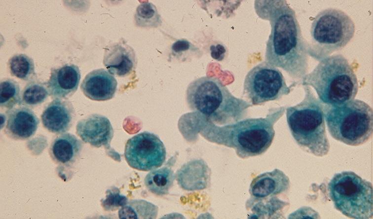
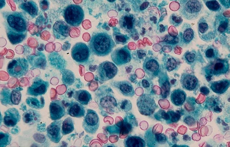
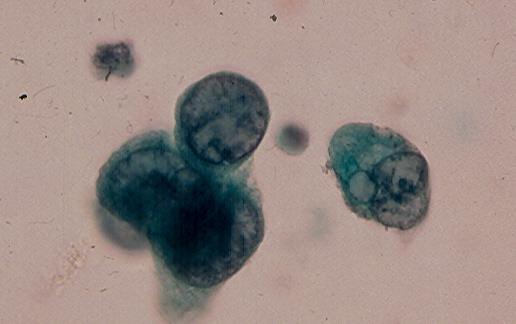
107 A B C 107


108 Fig A-C: High-grade urothelial carcinoma of the bladder showing. (Pap). A, B: Numerous single pleomorphic malignant cells with enlarged, hyperchromatic nuclei, well-defined, basophilic cytoplasm and prominent nucleoli. C: Four large malignant epithelial cells with hypochromatic nuclei with irregular nuclear contours and prominent nucleoli. Sarcomatous urothelial carcinoma, a morphologic variant of high-grade urothelial carcinoma, is rarely encountered in practice. It yields in urine single spindle malignant cells with prominent nucleoli and scant, wispy cytoplasm. (Fig.4.18). A B 108
.")

109 Fig Bladder sarcomatoid urothelial carcinoma: A: Tumor consisting of spindle cells with prominent nucleoli. (HE). B: Urine showing isolated malignant spindle tumor cells with prominent single or double nucleoli and wispy cytoplasm. (Pap). Other tumors Squamous cell carcinoma is usually well-differentiated and commonly seen in patients with Schistosoma hematobium cystitis. It is a rare neoplasm in the United States and accounts for about 3% of all primary bladder cancers. Cytologically, it is characterized by single and clustered malignant keratinizing squamous cells with angulated and hyperchromatic nuclei. (Fig.4.19). A B 109

 A B Fig.4.20. Bladder verrucous squamous cell carcinoma: A. Histology of the tumor. (HE) B.")
110 Fig Bladder well-differentiated squamous cell carcinoma: A. Histology of the tumor. (HE). B. Keratinizing squamous cells with hyperchromatic, angulated or oval nuclei in voided urine. (Pap). Verrucous squamous cell carcinoma is a rare variant of squamous cell carcinoma, accounting for 3-4.6% of bladder squamous cell carcinomas in patients with Schistosomiasis cystitis. It is rare in non-endemic areas. Verrucous carcinoma in patients with long-stading anogenital condyloma acuminata and bladder condyloma acumunatum may be related to HPV infection. The tumor exfoliates in urine abundant benign appearing squamous cells. (Fig.4.20) A B Fig Bladder verrucous squamous cell carcinoma: A. Histology of the tumor. (HE) B. Urine showing abundant benign-appearing squamous cells. (Pap). 110
.")
. B: Urine showing clustered malignant epithelial cells with clear, vacuolated cytoplasm and prominent, eosinophilic nucleoli. (Pap).")
111 Adenocarcinoma accounts for about 7% of all bladder cancers. It occurs most commonly in patients with bladder exstrophy and is usually of intestinal type. A welldifferentiated adenocarcinoma exfoliates in urine single and clustered malignant glandular cells with prominent nucleoli. (Fig.4.21). Signet-ring malignant cells with prominent nucleoli may be seen in patients with a poorly differentiated tumor. A B Fig Well-differentiated adenocarcinoma, intestinal type: A. Histology of the tumor. (HE). B: Urine showing clustered malignant epithelial cells with clear, vacuolated cytoplasm and prominent, eosinophilic nucleoli. (Pap). Small cell carcinoma is a very rare and aggressive tumor of the urinary tract. It shows in urine single and clustered malignant small cells with scant cytoplasm, hyperchromatic 111
. B: Urine showing small malignant cells with small, round, hyperchromatic nuclei and necrotic debris. (Pap).")

112 nuclei with salt and pepper chromatin. (Fig. 4.22). Nuclear molding may be seen in some tumor cell clusters. A B Fig Small cell carcinoma of the bladder: A: Histology of the tumor. (HE). B: Urine showing small malignant cells with small, round, hyperchromatic nuclei and necrotic debris. (Pap). Renal cell carcinoma consists of 3 main histologic types: conventional or clear cell, papillary and chromophobe carcinomas. The tumor may invade the renal pelvis and exfoliates its cells in urine. The neoplastic cells usually occur in clusters and show a clear or granular cytoplasm and prominent nucleoli. (Fig.4.23). Sarcomatoid renal cell carcinoma is a rare histologic variant. It may exist in pure histologic form or as a 112

. Renal collecting duct carcinoma is an uncommon tumor.")
113 component of a conventional renal cell carcinoma. The tumor shows in urine spindle malignant cells that are present predominantly singly. ICC staining of the tumor cells with CK7, CK20, p63 and renal cell carcinoma antibodies is necessary to differentiate them from those of a sarcomatoid urothelial carcinoma. A B Fig Renal cell carcinoma (RCC): A: Histology of a conventional or clear cell RCC, grade I nuclei. (HE). B: Bloody urine showing clustered tumor cells with clear cytoplasm, round or irregular nuclei and inconspicuous nucleoli. (Pap). Renal collecting duct carcinoma is an uncommon tumor. It yields in urine single and clustered small malignant glandular cells with granular cytoplasm, oval nuclei and conspicuous nucleoli. Tumor epithelial fragments may also be observed. (Fig.4.24). The tumor cell cytoplasm, as in renal cell carcinoma, expresses cytokeratin and vimentin. 113
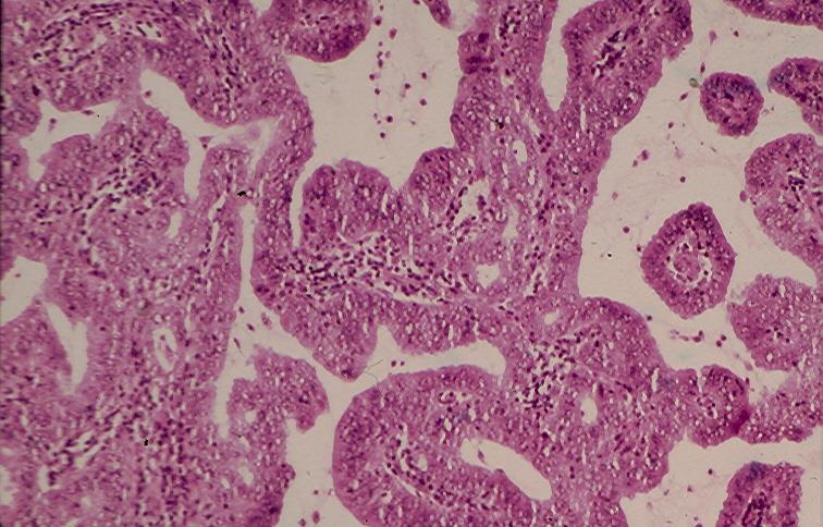
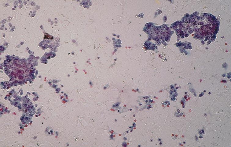
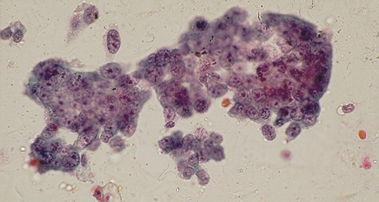
114 A B C 114
.")
.")
115 Fig Renal collecting duct carcinoma. A. Histology of the tumor. (HE). B and C. Urine showing malignant glandular cells singly and in irregular epithelial fragments with oval nuclei and prominent nucleoli. (Pap, B and C). Prostatic adenocarcinoma invading the bladder base may exfoliate its cells in urine. The tumor is usually a high-grade adenocarcinoma that yields small clusters of malignant glandular cells with conspicuous nucleoli. (Fig.4.25). The tumor cell cytoplasm expresses prostatic specific antigen. A B Fig Prostatic adenocarcinoma: A. Histology of the tumor. (HE). B. Urine showing clustered malignant glandular cells with scant, clear cytoplasm and oval or round nuclei with prominent nucleoli. (Pap). 115
116 Diagnostic accuracy and pitfalls Diagnostic accuracy Depending on the tumor grade, the diagnostic accuracy of urothelial carcinomas by urine cytology varies widely. For low-grade tumors a sensitivity and specificity rates of 30% and 80%, respectively, have been reported. For carcinoma in situ and high-grade tumors the cytodiagnosis is almost always straightforward and has a sensitivity and specificity rates of 80% and 95%, respectively. Washing of ureter and renal pelvis has been reported to have a sensitivity of 50% for high-grade tumors. A number of ancillary tests have been developed to improve the cytodiagnostic accuracy of low-grade urothelial carcinoma and/or to determine the risk of tumor progression. These tests include flow cytometry and image analysis for aneuploidy determination, Bard bladder tumor antigen test, nuclear matrix protein NMP22 test, telomemase assays, microsatellite instability assays, hyaluronidase and hyaluronic acid assays, growth factors assays, cell adhesion molecules, fibrinogen degradation products assays, tumor-associated and blood group antigens and fluorescence in-situ hybridization (FISH). One multitarget FISH assay using a combination of centrometric probes with chromosomes 3, 7 and 17 with a locus-specific probe to band 9p21 was reported to have a sensitivity of 81% and a specificity of 96% for patients with urothelial cancer. The UroVysion FISH Test is the most popular one. Diagnostic pitfalls 1- Degenerated urothelial cells: Urothelial cells lying in hypertonic urine may show remarkable degenerative changes. The cells become smaller and show pyknotic nuclei. Repeat examination of a freshly voided urine sample, taken after hydratation, and prepared without delay will be of diagnostic help in this case. 2- Endometrial cells: The presence of small hyperchromatic endometrial cells in a urine sample contaminated with menstruation may cause a diagnostic pitfall, as these cells may be mistaken for cancer cells. Therefore, the date of last menstrual period in a female patient should be provided by the physician who orders the test. 3- Instrumentation may produce cellular changes leading to misinterpretation. An indwelling bladder catheter may exfoliate tightly arranged atypical/reactive/repair urothelial cells mimicking cytologic findings in a low-grade urothelial carcinoma. 116

117 4- Infection, lithiasis, radiation and chemotherapy may also cause some cellular alterations (nuclear enlargement and hyperchromasia, conspicuous nucleoli) mimicking cells of a low-grade urothelial carcinoma. 5- Repair renal tubular cells may show severe cellular atypias, mimicking malignant glandular cells. The reader is referred to Chapter 5 for illustration of repair renal tubular cells. 6- Washing samples of the ureter and renal pelvis may be compounded with diagnostic errors, as normal urothelial cells at these two anatomic sites normally display larger and hyperchromatic nuclei, readily mistaken for malignant cells by an inexperienced observer. (Fig.4.26). The advantage is that the cell samples are usually bilateral and one of them is presumably benign, for comparison. Fig Ureteral washing showing a sheet of basal urothelial cells with monomorphic, enlarged, hyperchromatic nuclei and high N/C ratio in honeycomb pattern. (Pap). The Paris system for reporting urine cytology A few systems for reporting urine cytology have been proposed since the first one by Papanicolaou. However, none of them has gained a wide acceptance by urologists and pathologists. At the 2013 International Congress of Cytology in Paris, France a working group consisting of cytopathologists, surgical pathologists and a urologist was formed. This group has proposed and published a standardized reporting system that included specific diagnostic categories and cytomorphologic criteria for a reliable diagnosis of high-grade urothelial carcinoma in urine samples. The Paris System (TPS) for Reporting Urine 117
118 Cytology was reviewed in June 2016 and its important information are summarized below. For a more comprehensive review of TPS, the reader is advised to consult the article written by Barkan et al. listed in the attached bibliography. This system has 7 diagnostic categories, and each category has specific morphologic criteria. The validity of TPS in cytopathology practice and patient care needs to be evaluated widely. Diagnostic Categories of TPS for Reporting Urine Cytology 1. Nondiagnostic/Unsatisfactory 2. Negative for High-Grade Urothelial Carcinoma 3. Atypical Urothelial Cells 4. Suspicious for High-Grade Urothelial Carcinoma 5. High-Grade Urothelial Carcinoma 6. Low-Grade Urothelial Neoplasms 7. Other: primary and secondary malignancies and miscellaneous lesions Diagnostic Categories and Morphologic Criteria of TPS 1. Adequacy. There is no specific number of cells for an adequate specimen. Any specimens that contain atypical urothelial cells, no matter how few, are regarded as adequate for diagnosis. A specimen is regarded as unsatisfactory for cytologic evaluation if the urothelial cells are completely obscured by lubricant or inflammatory cells. 2. Negative for High-Grade Urothelial Carcinoma. In this category only benign epithelial cells are present. Specimens containing cells with changes associated with urolithiasis, treatment-induced changes and polyomavirus cytopathic changed are classified as Negative for High-Grade Urothelial Carcinoma. 3. Atypical Urothelial Cells. This is an ill-defined category with wide interobserver and intraobserver variabilities. It consists of urothelial cells with N/C >0.5 (required) and at least one of the following features: hyperchromasia, coarse chromatin and irregular chromatinic rim. 4. Suspicious for High-Grade Urothelial Carcinoma. Atypical urothelial cells in this category are similar to the cells seen in High-Grade Urothelial Carcinoma. However, they are very few in number, less than 5 to 10 cells. 5. High-Grade Urothelial Carcinoma. The diagnostic cells in this category are welldefined and characterized by N/C>0.7 (required), hyperchromasia (required) and at least one of these features: coarse chromatin and irregular chromatin rim. 118
119 6. Low-Grade Urothelial Neoplasms. There are no cells with specific morphologic changes permitting a reliable identification of these tumors in the majority of cases. On rare occasions papillary tumor fragments with fibrovascular cores are seen in instrumented urine samples. These tumor tissue fragments are usually not present in void urine sample. They permit a reliable cytodiagnosis of a low-grade urothelial neoplasm. 7. Other Malignancies: Primary, Metastatic and Miscellaneous Lesions. Other primary non-urothelial cancers are rare and account for about 5% of all bladder carcinomas. Secondary cancers of the bladder account for about 10% of all bladder carcinomas. These neoplasms display in urine cancer cells with rather specific features. Risks of Malignancy and Recommendations According to Barkan et al., the relative risks of malignancy in different diagnostic categories of TPS and recommended further investigations in different studies are as follows: 1. Unsatisfactory/Nondiagnostic. The risk of malignancy in this category is <5-10%. The patient should have repeat urine cytology and cystoscopy in 3 months, if clinical suspicion is increased. 2. Negative for High-Grade Urothelial Carcinoma. The risk of malignancy in this category is 0-10% and clinical follow-up is needed. 3. Atypical Urothelial Cells. The risk of malignancy in this category is 8-35%. Clinical follow-up is recommended and potential use of ancillary testing for further diagnosis may be necessary. 4, Supicious for High-Grade Urothelial Carcinoma. The risk of malignancy in this category is 50-90% and more aggressive follow-up and cystoscopy with biopsy are recommended. 5. Low-Grade Urothelial Neoplasm. The risk of malignancy in this category is about 10% and cystoscopy with biopsy to further evaluate grade and stage is recommended. 6. High-Grade Urothelial Carcinoma. The risk of malignancy is > 90% and cystoscopy with biopsy and staging are recommended. 7. Other malignancies. The risk of malignancy is >90% and cystoscopy with biopsy and staging are recommended. 119
120 Bibliography American Cancer Society. Cancer Facts and Figures. Atlanta: American Cancer Society, Arentsen HC, et al. Fluorescence in situ hybridization: a multitarget approach in diagnosis and management of urothelial cancer. Expert Rev Mol Diagn. 2007; 7: 11. Barkan GA, et al. The Paris system for reporting urinary cytology: The quest to develop a standard terminology. Acta Cytol. 2016; 60:185. Barkan GA. Enough is enough: Adequacy of voided urine cytology. Cancer Cytopathol. 2016;124:163. Bastacky S, et al. The accuracy of urine cytology in daily practice. Cancer (Cancer Cytopathol). 1999; 87:118. Caraway NP, et al. Fluorescence in situ hybridization for detecting urothelial carcinoma: a clinicopathologic study. Cancer Cytopathol. 2010;118:259. Cheng L, et al. Bladder cancer: translating molecular genetic insights into clinical practice. Hum Pathol. 2011; 42: 455. Cimino-Mathews A, Ali SZ. The clinicopathologic correlates of cellular atypia in urinary cytology of ileal neobladders. Acta Cytol. 2011;55:449. Comploj E, et al. ucyt+/immunocyt and cytology in the detection of urothelial carcinoma: an update on 7422 analyses. Cancer Cytopathol. 2013;121:392. DeMay RM. The Art and Science of Cytopathology. Volume 1. Chicago, ASCP Press, Deshpande V, McKee GT. Analysis of atypical urine cytology in a tertiary care center. Cancer (Cancer Cytopathol) 2005; 105: Dodd LG, et al. Endoscopic brush cytology of the upper urinary tract: evaluation of its efficacy and potential limitations in diagnosis. Acta Cytol. 1997; 41:377. Eldidi MM, Patten SF. New cytologic classification of normal urothelial cells: an analytical and morphometric study. Acta Cytol.1982;26:725. Ferra S, et al. Reflex UroVysion testing in suspicious urine cytology cases. Cancer.2009; 117:7. Forni AM, et al. Cytological study of effects of cyclophosphamide on the epithelium of the urinary bladder in man. Cancer. 1964;17:
121 Goldstein ML, et al. Significance of cell groups in voided urine. Acta Cytol. 1998;42:290. Granados R, et al. Applying the Paris System for reporting urine cytology increases the rate of atypical urothelial cells in benign cases: a need for patient management recommendations. Acta Cytol. 2017;61:77. Halling KC, Kipp BR. Bladder cancer detection using FISH. Adv Anat Pathol 2008; 15: 279. Humphrey PA, et al. The 2016 WHO Classification of tumours of the urinary system and male genital organs. Part B: prostate and Bladder tumours. Eur Urol. 2016; 70: 106. Kannan V, Bose S. Low grade transitional cell carcinoma and instrument artifacts. A challenge in urinary cytology. Acta Cytol. 1993; 37:899. Koss LG. Diagnostic cytology of the urinary tract. Philadelphia, Lippincott-Raven, Lotan Y, Roehrborn CG. Sensitivity and specificity of commonly available bladder tumor markers versus cytology: results of a comprehensive literature review and meta-analyses. Urology.2003; 61:109. McCroskey Z, et al. Accuray and interobserver variability of cytologic diagnosis of low-grade urothelial carcinoma in instrumental urinary tract cytology specimens. Am J Clin Pathol 2015; 144: 902. McKee G. Urinary tract cytology. In Diagnostic cytopathology. 2 nd ed, 2003, Gray W, McKee GT, eds, Edinburgh, Churchill Livingstone, p.471. Melamed MR, Wolinska WH. On the significance of intracytoplasmic inclusions in the urine sediment. Am J Pathol. 1961; 38:711. Miki Y, et al. Application of the Paris System to atypical cytology urine samples: correlation with histology and Urovision@FISH. Cytopathology. 2017;28:88. Morrow JF, et al. Urine cytology. In Urologic surgical pathology, 2 nd edition, 2008, Bostwick DG and Cheng L, eds. Mosby Elsevier, p Murphy WM. Current status of urinary cytology in the evaluation of bladder neoplasms. Hum Pathol. 1990; 21:886. Murphy WM, et al. Urinary cytology and bladder cancer. The cellular features of transitional cell neoplasms. Cancer. 1984; 53:1555. Murphy WM, et al. Pathological changes associated with topical chemotherapy for superficial bladder cancer. J Urol. 1981; 126:461. Murphy WM, et al. Tumors of the kidney, bladder, and related urinary structures. In AFIP Atlas of tumor pathology. 4 th series, 2004, Washinton DC, American Registry of Pathology, p
122 Muus Ubago J, et al. Evaluation of atypical urine cytology progression to malignancy. Cancer Cytopathol. 2013;121:387. Nguyen GK, Schumann GB. Cytopathology of renal collecting duct carcinoma. Diagn Cytopathol. 1997; 16:446. Ohsaki H, et al. Cytological features of carcinoma of the collecting ducts of Bellini in voided urine cytology. Diagn Cytopathol. 2009; 37: 676. Owens CL, et al. A review of reporting systems and terminology for urine cytology. Cancer Cytopathol. 2013;121:9. Ohsaki H, et al. Can cytological features differentiate reactive renal tubular cells from low-grade urothelial carcinoma cells? Cytopathology. 2010;21:326. Owens CL, et al. A review of reporting systems and terminology for urine cytology. Cancer Cytopathol. 2013; 121: 9. Potts SA, et al. Diagnostic accuracy and key cytologic features of high-grade transitional cell carcinomas in upper urinary tract. Mod Pathol. 1997; 10:657 Rabb S, et al. Low grade transitional cell carcinoma of the bladder. Cytodiagnosis by key features as identified by logistic regression analysis. Cancer. 1994; 74: Ranzi AD, et al. The role of urine cytology for 'decoy cells' as a screening tool in renal transplant recipients. Acta Cytol. 2012; 56:543. Renshaw AA. Subclassifying atypical urinary cytology specimens. Cancer (Cancer Cytopathol). 2000; 90:222. Renshaw AA. Urine and bladder washings. In Cytology. Diagnostic Principles and Clinical Correlates, 2 nd ed, 2003, Cibas E, Ducatman B, eds, p. 97. Schumann GB, et al. Cytodiagnostic urinalysis of renal and lower urinary tract disorders. New York, Igaku-Shoin, Shenoy UA, et al. Reliability of urinary cytodiagnosis in urothelial neoplasms. Cancer.1985; 56:2041. Sojitra P, et al. Dysplastic squamous cells are frequently present in urine cytology specimens of patients with high-grade urothelial carcinoma. Acta Cytol.2012;56:408. Stella F, et al. Urothelial cell changes due to Bisulfan and Cyclophosphamide treatment in bone marrow transplantation. Acta Cytol. 1990; 34:
123 Straccia P, et al. Comparison between cytospin and liquid-based cytology in urine specimens classified according to the Paris System for Reporting Urine Cytology. Cancer Cytopathol. 2016; 124: 519. Sullivan PS, et al. Comparison of ImmunoCyt, UroVysion, and urine cytology in detection of recurrent urothelial carcinoma: a "split-sample" study. Cancer. 2009;117: Thompson A. Crystallinuria. In Urinary sediments:a textbook and atlas, MH Haber, ed. Chicago, ASCP press, 1981, 33. Toll AD, Ali SZ. Urinary cytomorphology and clinical correlates of prostatic small cell neuroendocrine carcinoma. Acta Cytol. 2013;57:495. Wall RL, Claussen KP. Carcinoma of the urinary bladder in patients receiving cyclophosphamide, NEJM.1975; 293:271. Yang CS, et al. High apoptotic index in urine cytology is associated with high-grade urothelial carcinoma. Cancer Cytopathol. 2016; 124:546. Zhang ML, et al. The cytomorphological features of low-grade urothelial neoplasms vary by specimen type. Cancer Cytopathol. 2016;124:
124 Chapter 5 Urine in non-neoplastic renal parenchymal diseases Gia-Khanh Nguyen Urinalysis has been the cornerstone in evaluating renal parenchymal diseases since the 19 th century. It is traditionally performed by nephrologists who use a bright-field or phase-contrast microscope to evaluate wet and unfixed urine sediments. In this method of clinical investigation cells exfoliated from the renal parenchyma and lower urinary tract and non-cellular elements do not show well-defined microscopic features and the cell samples cannot be kept indefinitely for retrospective studies. Cytologic evaluation of fixed and stained urine sediments in the investigation of non-neoplastic lesions of the renal parenchyma is not routinely performed in diagnostic cytology, and reports on this topic have been rarely encountered in the literature. This chapter presents the cytologic findings in Papanicolaou-stained urine sediments in renal glomerular and tubular diseases. Collection and preparation of cell samples To investigate non-neoplastic renal parenchymal diseases urine samples should be prepared according to the technique used for semiquantitative cytologic urinalysis (SCU) A sample of 100 to 200 ml of mid-stream, freshly voided urine without ethanol fixatives is immediately submitted to the hospital cytology laboratory for processing. An aliquot of 10 ml is centrifuged at 1800 rpm for 10 min. 9 ml of the supernatant is discarded, and 4 cytospin preparations or smears are prepared from the remaining 1 ml of the sediment by cytospin technique, with a centrifugation speed of 900 rpm. If the cell button is judged to be too thick, it is gently smeared with a thin coverslip. The smears obtained are immediately fixed in 95% ethanol for 5 minutes and then stained by the Papanicolaou method. A longer fixation in 95% ethanol will severely destroy or lyse almost all red blood cells, making the identification of dysmorphic red blood cells impossible. All four smears are screened with a x10 objective and selected cellular areas are carefully evaluated at a higher magnification with a x 40 objective. Different cellular elements and casts are evaluated and counted. G1 cells are defined as dysmorphic red blood cells (RBC) with a doughnut-like shape, membranous protrusions or blebs and target configuration. Distorted RBCs without target configuration, doughnut-like shape and membranous blebs or protrusions are collectively grouped with morphologically normal RBCs as RBCs. A G1 cell: total RBC ratio is calculated by evaluating 200 RBCs including G1 cells. 124

125 Normal urine In healthy individuals 0-2 renal tubular cells, 3-5 RBCs and 3-5 polymorphonuclear leukocytes are present in each high-power field. Rare hyaline or granular casts may be observed. (Fig.5.1). There are, in addition, a variable number of urothelial and squamous cells. Proximal convoluted tubular cells are cuboidal in shape, measure microns in greatest dimension and have a well-defined, coarsely granular cytoplasm and slightly eccentrically located, single, oval nuclei. Distal convoluted tubular cells are smaller, measure microns in greatest dimension and have a well-defined, finely granular cytoplasm and centrally located, single, oval nuclei. Cells derived from collecting tubules are small and cuboidal in shape with well-defined granular cytoplasm and centrally located, single, round nuclei. Cells from the loops of Henle are rarely seen. These are smaller cuboidal cells with round nuclei and scant cytoplasm. Fig.5.1. A hyaline cast (above) and a granular cast (below) in normal urine. (Pap). Renal glomerular diseases Microscopic hematuria is a common clinical problem and has numerous etiologies, including renal glomerular and tubular diseases, urolithiasis, renal tumor, urothelial neoplasm, infection of the kidney and lower urinary tract and rupture of suburothelial capillary blood vessels. Since the clinical investigation of microscopic hematuria is costly, time-consuming and inconvenient to the patient, identification of patients with glomerular bleeding or lower urinary tract hematuria is desirable for further nephrologic or urologic investigation. 125
126 The most common manifestations of renal glomerular diseases are microscopic hematuria and proteinuria. Usually the presence of red blood cell (RBC) casts in urine sediments is regarded as an evidence of glomerular hematuria or disease. However, 38% of patients with histologically confirmed renal glomerulopathy in Nguyen s 2003 study showed only an increased number of RBCs and no identifiable RBC casts in their urine sediments. Birch and Fairley were apparently the first investigators who reported that RBCs leaking from renal glomeruli were dysmorphic, in contrast to normal RBCs of non-glomerular origin. This finding proved to be of diagnostic value in some studies. However, other studies were unable to confirm the diagnostic value the above-mentioned observation. In 1992 Tomita et al. have reported that urinary RBCs with target configuration, doughnut-like shape and membranous protrusions or blebs or G1 cells constituted a reliable marker for renal glomerular hematuria. This finding was subsequently supported by the work of other investigators. The observation of Tomita and his associates was actually not original, as urinary RBCs with similar morphological changes had been previously observed in urine samples from patients with acute glomerulonephritis by Addis in 1948 and by Kohler et al. in The latter investigators had named those RBCs acanthocytes and found that an acanthocyte/total RBC ratio equal or greater than 5% was associated with a glomerulonephritis in over 50% of cases. This finding was supported by the work of Kitamoto et al. who found that a G1 cell/total RBC ratio greater than 5% was an evidence for glomerular bleeding/disease. Despite several studies conducted in the past two decades, morphologic criteria of dysmorphic RBCs had not been well-defined, and the required percentage of these cells for making a firm diagnosis of glomerular bleeding had not been uniformly agreed upon. The main reasons for the lack of well-defined morphologic criteria of dysmorphic RBCs were that in all of those studies unfixed urine sediments and bright-field and phase-contrast microscopes were used and that the wet-mounted slides could not be kept permanently for review. Because of the aforementioned reasons, there were significant discrepancies in observer interpretations of dysmorphic RBCs. The pathogenesis of dysmorphic RBCs is largely unknown. In two studies, RBCs leaking through diseased glomeruli were normal, and these cells acquired dysmorphic changes by osmotic injury while passing through hypotonic renal tubules and by exposure to a concentrated acidic urine. In another study the dysmorphic changes, in certain cases, could be attributed to 2 consecutive injuries: mechanical injury to the cell membrane during passage through damaged glomerular basement membranes and osmotic injuries during passage through hypotonic renal tubules. The absence of G1 cells in urine sediments in patients with severe renal tubular injury has indicated that G1 cells could not be formed in the absence of a glomerulopathy, and this finding supported the work of Ye and Mao who have suggested that a mechanical injury to RBC membranes was needed, in addition to intratubular osmotic injuries, to produce dysmorphic changes of RBCs infiltrated through diseased renal glomeruli. G1 cells were not seen in renal tubular or lower urinary tract bleeding, according to Nguyen s 126

127 2008 study. A G1/total RBC ratio varying from % indicates a renal glomerular bleeding. If G1 cells are not identified, at least 50% of renal glomerular diseases will be missed by urine cytology in patients with renal glomerular diseases showing no RBC casts in urine. In the absence of RBC casts the presence of G1 cells alone in urine sediments has correctly predicted the presence of a glomerular lesion in at least 86% of the cases, according to Nguyen s 2008 report. As G1 cells are formed in acidic urine with high osmolarity, first morning urine samples are more suitable for detection of G1 cells. Random or spot urine that is often diluted and alkaline is less suitable for G1 cell detection, and diluted urine samples from patients with impaired renal function and from individuals with excessive water intake are suboptimal for G1 cell detection. Urine samples contaminated with vaginal secretion may obscure G1 cells. Therefore, perineum washing in women is required prior collection of voided urine samples for SCU. Urine collected by bladder catheterization is suboptimal for SCU as it contains abundant RBCs and urothelial cells. Faulty collection or preparation techniques are usually the main reasons for a false-negative result. Freshly voided urine without ethanol fixatives is the best sample for identifying G1 cells, as a long fixation in ethanol will destroy RBCs and RBC casts. Effort should be made to identify pseudo-g1 cells that are RBCs with artifactual changes. These cells are more commonly seen in important urinary tract bleedings of nonglomerular origin and they are not a marker for renal glomerular bleeding. Urine sediments containing pseudo-g1 cells always show only RBCs with target configuration that have a thick hemoglobin rim with smooth outer and inner contours and no membranous fragmentation and blebs or protrusion, as seen in G1 cells. (Fig.5.2). The morphogenesis of pseudo-g1 cells is unknown. Fig Pseudo-G1 cells. (Pap). 127
. 2.")
128 Cytologic findings In Nguyen s 2003 study of 178 patients with histologically confirmed renal glomerular diseases 4 smear patterns were identified, and each pattern was associated with different types of renal glomerular lesion/disease. 1. G1 Cells only: This pattern was the most common one and present in 54% of patients. (Fig.5.3). 2. G1 Cells and RBC Casts: This pattern was present in 20% of patients. (Fig.5.4). 3. RBC Casts and No G1 Cells. This pattern was present in about 18% of patients. 4. Normal RBCs only. This finding was seen in less than 10% of patients. Fig.5.3. A. Numerous G1 cells in urine sediment of a patient with IgA nephropathy: G1 cell with target configuration (arrow head), G1 cell with doughnut-like shape (short arrow) and G1 cell with membranous blebs (long arrow). (Pap). From: Nguyen GK: Renal glomerular disease screening: practical value of G1 cell. Russian News in Clinical Cytology. 2008; 12:13, with permission. 128

129 Fig Numerous RBCs including G1 cells and RBC casts. (Pap). Patients with renal glomerular disease usually present with either a nephritic syndrome or nephrotic syndrome. Nephretic syndrome is characterized by the presence of numerous RBCs and RBC casts in urine sediment, and it is commonly found in patients with acute glomerulonephritis, IgA nephropathy and rapidly progressive glomerulonephritis. Nephrotic syndrome is usually present in patients with minimal change glomerular disease, focal glomerularsclerosis, membranous nephropathy and membranoproliferative glomerulonephritis. Some conditions such as HIV-associated nephropathy, systemic lupus erythematosus and Hepatitis C can be associated with either a nephrotic or nephritic syndrome. However, exceptions do exist. A nephrotic syndrome is characterized cytologically by the presence of renal tubular cells with large intracytoplasmic fat droplets, fatty casts, a few RBCs and no RBC casts. (Fig.5.5). 129
.")

130 A B Fig.5.5. A patient with focal glomerular sclerosis with nephrotic syndrome showing in urine: A. A cluster of renal tubular cells with intracytoplasmic fat droplets. (Pap). B. A tubular fatty cast, a tubular epithelial cell and a few polymorphonuclear leukocytes. (Pap). Acute tubulointerstitial nephritis The disease has several etiologies including drug reaction (antibiotics, nonsteroid antiinflammatory drugs, diuretics), systemic bacterial infection or viral infection. Most cases are drug related, and when no cause is found it is classified as idiopathic. Clinically, the disease is most commonly characterized by fever, flank pain, microscopic hematuria and eosinophilia. The urine sediment shows numerous polymorphonuclear leukocytes, white blood cell casts, renal tubular cells and abundant or rare eosinophils. (Fig.5.6). 130
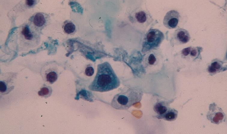
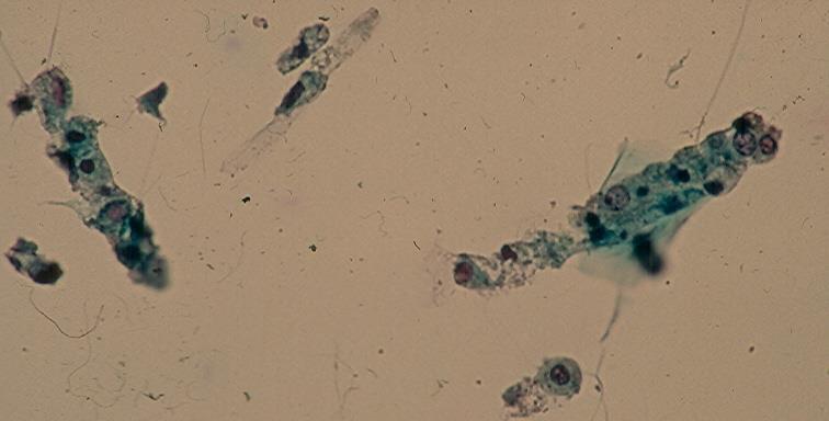
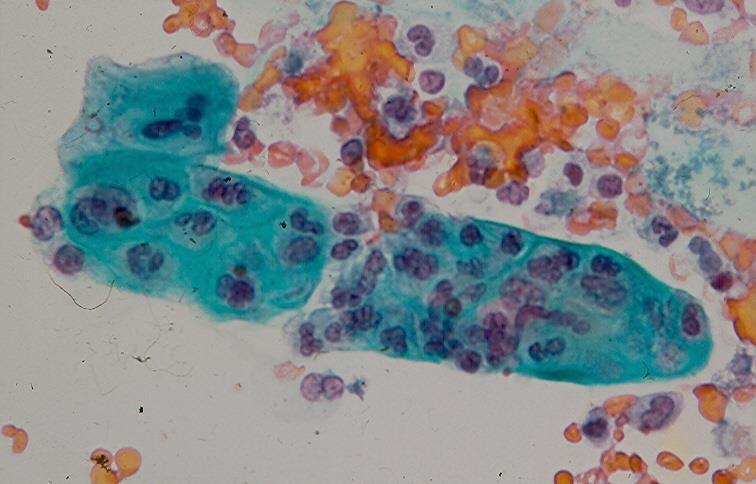
131 A B C 131


132 D Fig Acute tubulointerstitial nephritis showing in urine. (Pap, A to D). A. An increased number of renal tubular cells (RTC). B. A RTC cast. C. A white blood cell cast containing polymorphonuclear leukocytes. D. Several eosinophils. Chronic tubulointerstitial nephritis The main etiologies of this disease are obstructive uropathy, reflux nephropathy, heavy metal exposure (lead and cadmium) and prolonged ingestion of acetaminophen. It is characterized by a decreased urine output, metabolic hyperchloremic acidosis and hyperkalemia. The urine sediment shows a few thick broad hyaline/granular casts and a number of inflammatory cells. (Fig.5.7). Fig Thick broad hyaline casts and polymorphonuclear leukocytes. (Pap). 132

133 Acute renal failure Acute renal failure (ARF) is characterized by a sudden increase of BUN and serum creatinine and oliguria. However, polyuria is present in some cases. It is classified into 3 types: prerenal, post renal and renal. 1. Prerenal ARF is caused by poor renal perfusion of different reasons. The urine sediment is normal and shows only a few hyaline casts. 2. Postrenal ARF is caused by an obstruction of urinary tract by urinary stone, enlarged prostate or cancer invasion obstructing the urine flow. The urine sediment is also normal in this case and some crystals may be observed. 3. Renal ARF is caused by intrinsic renal diseases such as acute tubular necrosis, acute glomerulonephritis and acute tubulointerstitial nephritis. Acute tubular necrosis (ATN) is caused by hypotension and endogenous or exogenous nephrotoxins. It can be oliguric or polyuric. Endogenous nephrotoxins include myoglobin, crystals and Bence Jone proteins. Myoglobinuria is secondary to rhabdomyolysis that may occur in patients with alcoholic abuse, crush injury, prolonged coma or seizures. The patients have a high serum CK level and hyperkalemia and show in urine sediments numerous myoglobin pigment casts, in addition to renal tubular cells (RTC), RTC casts and RBCs, and no G1 cells are identified. (Fig. 5.8). Fig.5.8. Dark brown myoglobin casts and single renal tubular cells are present. (Pap). Uric acid crystals are seen in patients with hyperuremia secondary to chemotherapy for leukemia, lymphoma and some solid cancers. Bence Jones proteins are detected in 133


134 patients with multiple myeloma. Exogenous nephrotoxins include antibiotics, radiographic contrast material and cyclosporine. Antibiotics include gentamycin, tobramycin and amykacin. Aminoglycoside-induced ATN is nonoliguric. An ATN is characterized by the presence of numerous RTCs and a few RTC casts in urine sediments. RBC casts may be seen if a severe tubular bleeding is present but no G1 cells are identified. The RTCs stain positively with vimentin antibody while other benign epithelial cells in urine do not. (Fig.5.9). A B Fig Severe renal tubular injury/atn showing numerous renal tubular cells in urine sediment (A). The tubular cell cytoplasm expresses vimentin (B). (A, Pap; B, ABC). 134

. A B Fig. 5.10. Acute renal transplant rejection: A. Histopathology of a lymphocytic tubulitis. (HE). B. Urine showing numerous lymphocytes, RBCs and a RBC cast. (Pap).")
135 Acute glomerulonephritis and Acute interstitial nephritis. The urine cytologic findings in these two conditions have been previously discussed. Acute renal transplant rejection may occur any time in patients with renal transplant. The urine sediment in this situation shows evidence of a renal tubular injury that is characterized by an increased number of RTCs, several CD4 and CD8 positive T-cells, RBCs and RBC casts. (Fig.5.10). A B Fig Acute renal transplant rejection: A. Histopathology of a lymphocytic tubulitis. (HE). B. Urine showing numerous lymphocytes, RBCs and a RBC cast. (Pap). An acute renal transplant rejection may mimic a polyomavirus nephritis clinically and histologically. A polyomavirus nephritis may show renal tubular cells with characteristic 135
136 nuclear features of polyomavirus infection. The diagnosis may be further confirmed by immunostaining of the abnormal tubular cells with a commercially available polyomavirus antibody or by identification of nm icosahedral virus particles in the sediment of a negative urine (negative for cells with changes characteristic for polyomavirus infection). A rapid and accurate diagnosis of acute renal transplant rejection is very important, as the therapeutic approaches are different: increased immunosuppression for rejection and decreased immunosuppression for polyomavirus nephritis. Chronic renal failure Chronic renal failure is characterized by a progressive azotemia over weeks or months. The patient usually has isosthenuria and hypertension. Both kidneys are small on ultrasonography or CT scan, and radiologic evidence of renal osteodystrophy is present. It is secondary to renal glomerulopathies, tubulointerstitial nephritis, polycystic kidneys, Alport syndrome, obstructive nephropathy and hypertension. The patient s urine sediment may show a few broad casts, and RBCs and RBC casts may also be seen. Dysmorphic RBCs are usually not seen even in patient with a prior history of renal glomerulonephritis. Renal tubular injury and repair renal tubular cells Renal tubular injury of mild and moderate degrees is common and usually asymptomatic. It is caused by numerous reasons including ingestion of some foods, chemical substances or drugs with nephrotoxic effects. Examination of the patient s urine sediment will reveal an increased number of RTCs. Renal tubular injury has been arbitrarily classified as mild, moderate and severe by Schumann based on the number of RTCs present in 10 high power fields (x 40 objective). A diagnosis of a mild or moderate renal tubular injury is made when the number of RTCs is greater than 50 or between 100 and 200, respectively. When the number of RTCs is greater than 200 a severe renal tubular injury is present. Injured RTCs are replaced by repair RTCs that develop from residual, viable RTCs within renal tubules. Repair RTCs are large and measure up to 100 microns in greatest dimension and display regenerative changes that are characterized by cytoplasmic basophilia, enlarged and hyperchromatic nuclei, prominent nucleoli and occasional mitoses; and intracytoplasmic vacuoles may be observed. Repair RTCs may occur singly but more often in small clusters of 10 to 20 cells with acinar arrangement. Nuclear pleomorphism may be present. Repair RTCs may be mistaken for malignant glandular cells by an inexperienced observer. Usually, repair RTCs in the same specimen display a spectrum of nuclear changes ranging from mild to severe atypia with a variable degree of nuclear pleomorphism, hyperchromasia and chromatin clumping. Conspicuous or prominent nucleoli are usually present. (Fig. 5.11). 136
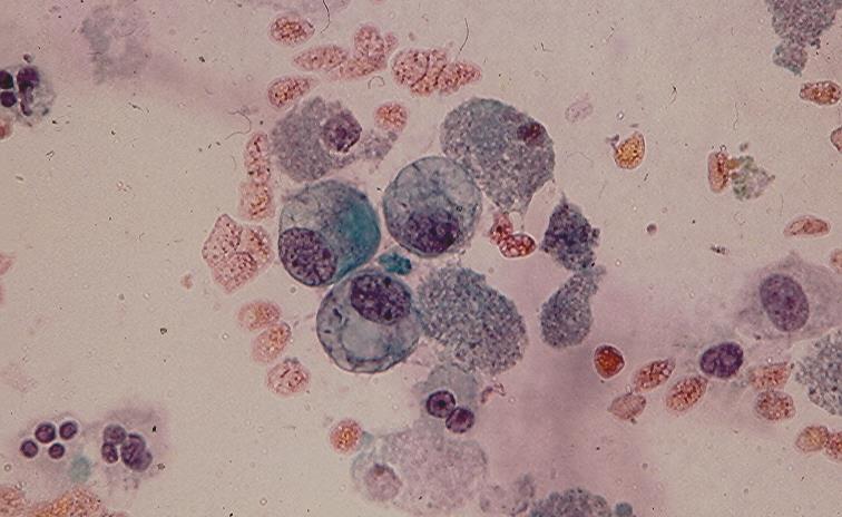
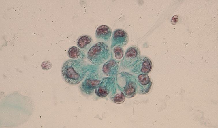
137 A B 137

.")
138 C Fig Repair renal tubular cells (RRTC). (Pap, A to C). A. An aggregate of repair RRTCs showing basophilic or vacuolated cytoplasm, oval nuclei and conspicuous nucleoli. B. A group of RRTCs with prominent nucleoli forming a rosette. C. A cluster of RRTCs with some having a columnar configuration with tapered cytoplasm. Damaged and repair RTCs express vimentin and are negative for thrombomoludin and Uroplakin III while urothelial cells are positive for thrombomoludin and Uroplakin III and negative for vimentin. (Fig.5.12). RTCs should also be differentiated from those of a renal cell carcinoma that express vimentin and renal cell carcinoma antigen. A 138
 Other conditions Fungal casts may be found in urine sediments of patients with renal candidiasis. (Fig.5.13).")

139 B C Fig A to C: Clustered pleomorphic RRTCs with prominent nucleoli showing a positive cytoplasmic staining reaction to vimentin antibody. (ABC stain on cells previously stained with Pap) Other conditions Fungal casts may be found in urine sediments of patients with renal candidiasis. (Fig.5.13). Clusters of renal tubular cells with yellow bile-stained cytoplasm may be found urine of patients with severe jaundice and hepatorenal syndrome. (Fig.5.14). Malakoplakia is a rare chronic granulomatous disease that may affect the lower urinary tract and kidney. Cases with malakoplakia involving the renal parenchyma presenting with acute renal failure and fever of unknown origin, have been reported. Macrophages with intracytoplasmic round Michealis-Gutmann bodies may be identified in those patients urine sediments. (Fig.5.15). 139


140 Fig A large fungal cast in urine sediment of a patient with renal candidiasis. (Pap). Fig A cluster of renal tubular cells showing yellow bile-stained cytoplasm in urine sediment of a patient with hepatorenal syndrome and jaundice. (Pap). 140
: 845. Dinda AK, et al.")
141 Fig A degenerated macrophage with intracytoplasmic Michealis-Gutmann body and one degenerated renal tubular cell in urine sediment of a patient with renal malakoplakia. (Pap). Bibliography Addis T. Glomerular nephritis. New York: The Mac Millan Co, 1948: 42. Birch DF, Fairley KF. Hematuria: glomerular or non-glomerular. Lancet 1979; 2(8147): 845. Dinda AK, et al. Diagnosis of glomerular haematuria: role of dysmorphic red cell, G1 cell and bright-field microscopy. Scand J Clin Lab Invest 1997; 57:203. Fassett RG, et al. Detection of glomerular bleeding by phase-contrast microscopy. Lancet 1982; 1:1432. Favaro S, et al. Is the red cell morphology really useful to detect the source of hematuria? Am J Nephrol 1997; 17:172. Funfstuck R, et al. The significance of erythrocyte morphology in glomerular and nonglomerular hematuria. Z Urol Nephrol 1989; 82:85. Grone HJ, et al. Coexpression of keratin and vimentin in damaged and regenerating tubular epithelia of the kidney. Am J Pathol.1987;129:1. 141
ESSENTIALS OF FLUID CYTOLOGY
 ESSENTIALS OF FLUID CYTOLOGY Gia-Khanh Nguyen 2010 ESSENTIALS OF FLUID CYTOLOGY Gia-Khanh Nguyen, M.D. Professor Emeritus Laboratory Medicine and Pathology University of Alberta Edmonton, Alberta, Canada
Introduction. 23 rd Annual Seminar in Pathology. FLUIDS, Part 1. Pittsburgh, PA Gladwyn Leiman UVMMC, VT
 23 rd Annual Seminar in Pathology Pittsburgh, PA Gladwyn Leiman UVMMC, VT FLUIDS, Part 1 "Blue walls", Claudia Hansen, 2009 Introduction o Challenging to everyone o Almost any benign or malignant process
ACCME/Disclosures. Diagnosing Mesothelioma in Limited Tissue Samples. Papanicolaou Society of Cytopathology Companion Meeting March 12 th, 2016
 Diagnosing Mesothelioma in Limited Tissue Samples Papanicolaou Society of Cytopathology Companion Meeting March 12 th, 2016 Sanja Dacic, MD, PhD University of Pittsburgh ACCME/Disclosures GENERAL RULES
Ascitic Fluid and Use of Immunocytochemistry. Mercè Jordà, University of Miami
 Ascitic Fluid and Use of Immunocytochemistry Mercè Jordà, University of Miami Is It Malignant? Yes? No Ascitic Fluid Cytomorphologic Useful Findings Tight clusters with smooth borders Cellular and nuclear
Effusion Cytology: Diagnostic Challenges
 Effusion Cytology: Diagnostic Challenges Tarik M. Elsheikh, MD Professor and Medical Director, Anatomic Pathology Cleveland Clinic Outside Consult Case 45 year old woman, presented with nausea, dyspnea,
Mesothelioma: diagnostic challenges from a pathological perspective. Naseema Vorajee August 2016
 Mesothelioma: diagnostic challenges from a pathological perspective Naseema Vorajee August 2016 Naseema.vorajee@nhls.ac.za Pleural diseases (whether neoplastic, reactive or infective) may have similar
Case 1. Slide 1 History: 65 year old male presents with bilateral pleural effusions, a 40 pack year smoking history and peripheral and hilar lung
 Case 1. Slide 1 History: 65 year old male presents with bilateral pleural effusions, a 40 pack year smoking history and peripheral and hilar lung masses. Specimen shown is from a tap of the pleural effusion.
LGM International, Inc.
 Liqui-PREP TM Cytology Atlas Preface The following pictures are examples with descriptions of cytology slides processed with the Liqui-PREP TM System.. The descriptions are reviewed by Pathologists. It
Salivary Gland Cytology
 Salivary Gland Cytology Diagnostic challenges and potential pitfalls Tarik M. Elsheikh, MD Professor and Medical Director Anatomic Pathology Cleveland Clinic FNA Salivary Gland Lesions Indications Distinguish
Pancreatitis: A Potential Pitfall in Endoscopic Ultrasound Guided Pancreatic FNA
 Pancreatitis: A Potential Pitfall in Endoscopic Ultrasound Guided Pancreatic FNA Jack Yang, MD Department of Pathology, Medical University of South Carolina Objectives Understand the indication of EUS
Serous Effusions. Spasenija Savic Prince, MD Pathology, University Hospital Basel, Switzerland
 Serous Effusions Spasenija Savic Prince, MD Pathology, University Hospital Basel, Switzerland Serous membrane Body cavities: Pleural Pericardial Peritoneal Effusion = Excess of fluid 80% Benign 20% Malignant
Presentation material is for education purposes only. All rights reserved URMC Radiology Page 1 of 98
 Presentation material is for education purposes only. All rights reserved. 2011 URMC Radiology Page 1 of 98 Radiology / Pathology Conference February 2011 Brooke Koltz, Cytopathology Resident Presentation
Prepared By Jocelyn Palao and Layla Faqih
 Prepared By Jocelyn Palao and Layla Faqih The structure of the suspected atypical cell should always be compared to the structure of other similar, benign, cells which are present in the smears. The diagnosis
FNA of Thyroid. Toward a Uniform Terminology With Management Guidelines. NCI NCI Thyroid FNA State of the Science Conference
 FNA of Thyroid NCI NCI Thyroid FNA State of the Science Conference Toward a Uniform Terminology With Management Guidelines Thyroid Thyroid FNA Cytomorphology NCI Thyroid FNA State of the Science Conference
Dr. Issraa Ali Hussein
 CLINICAL 09888888;rCYTOLOGY Dr. Issraa Ali Hussein objectives Define diagnostic cytology (clinical cytology). Explain the differences between histopathology and cytopathology. Recognize the methods for
Case year female. Routine Pap smear
 Case 1 57 year female Routine Pap smear Diagnosis? 1. Atypical glandular cells of unknown significance (AGUS) 2. Endocervical AIS 3. Endocervical adenocarcinoma 4. Endometrial adenocarcinoma 5. Adenocarcinoma
International Journal of Health Sciences and Research ISSN:
 International Journal of Health Sciences and Research www.ijhsr.org ISSN: 2249-9571 Original Research Article Utility of Modified Cell Block Technique in Cases of Pleural Effusion Suspected of Malignancy
Gross appearance of peritoneal cysts. They have a thin, translucent wall and contain a clear fluid.
 Gross appearance of peritoneal cysts. They have a thin, translucent wall and contain a clear fluid. So-called multicystic benign mesothelioma. A, Gross appearance. So-called multicystic benign mesothelioma.
From Morphology to Molecular Pathology: A Practical Approach for Cytopathologists Part 1-Cytomorphology. Songlin Zhang, MD, PhD LSUHSC-Shreveport
 From Morphology to Molecular Pathology: A Practical Approach for Cytopathologists Part 1-Cytomorphology Songlin Zhang, MD, PhD LSUHSC-Shreveport I have no Conflict of Interest. FNA on Lymphoproliferative
Serous effusion Objectives. Cytology of Serous Effusions From basics to challenges
 Cytology of Serous Effusions From basics to challenges Cytology of Serous Effusions From basics to challenges Pınar Fırat, MD, MIAC Department of Pathology, İstanbul University, İstanbul Faculty of Medicine,
DOWNLOAD ENTIRE DOCUMENT FROM
 PREVIEW ONLY 1 Atlas on Bethesda system for reporting Thyroid Cytology PREVIEW ONLY 2 OVERVIEW 1. Indications and goal of thyroid FNA 2. Contraindications 3. Procurement of cell sample 4. Staining methods
Respiratory Tract Cytology
 Respiratory Tract Cytology 40 th European Congress of Cytology Liverpool, UK Momin T. Siddiqui M.D. Professor of Pathology and Laboratory Medicine Director of Cytopathology Emory University Hospital, Atlanta,
Almost any suspected tumor can be aspirated easily and safely. Some masses are more risky to aspirate including:
 DOES THIS PATIENT HAVE CANCER? USING IN-HOUSE CYTOLOGY TO HELP YOU MAKE THIS DIAGNOSIS. Joyce Obradovich, DVM, Diplomate, ACVIM (Oncology) Animal Cancer & Imaging Center, Canton, Michigan Almost every
Applications of IHC. Determination of the primary site in metastatic tumors of unknown origin
 Applications of IHC Determination of the primary site in metastatic tumors of unknown origin Classification of tumors that appear 'undifferentiated' by standard light microscopy Precise classification
Normal Morphology. Anatomic Considerations. Normal Urothelial Histology and Cytology
 1 Normal Morphology Anatomic Considerations The urinary tract can be divided into three regions: the kidney; the calyces, pelves and ureters (upper collecting system or upper tract); and the bladder and
Cutaneous metastases. Thaddeus Mully. University of California, San Francisco Professor, Departments of Pathology and Dermatology
 Cutaneous metastases Thaddeus Mully University of California, San Francisco Professor, Departments of Pathology and Dermatology DISCLOSURE OF RELATIONSHIPS WITH INDUSTRY Thaddeus Mully Course C005 Essential
Cytyc Corporation - Case Presentation Archive - October 2001
 ThinPrep Pap Test History: 82 Year Old Female Specimen Type: Peritoneal Washings Case provided by Dr. Berle Stratton, Southwest Washington Medical Center, Vancouver, Washington. *The images, analysis and
Cytological evaluation of effusion fluid with cell block technique and cytology smears among Sudanese patients
 EUROPEAN ACADEMIC RESEARCH Vol. IV, Issue 3/ June 2016 ISSN 2286-4822 www.euacademic.org Impact Factor: 3.4546 (UIF) DRJI Value: 5.9 (B+) Cytological evaluation of effusion fluid with cell block technique
DIAGNOSTIC DILEMMA. Case Reports Clinical history. Materials and Methods
 DIAGNOSTIC DILEMMA A Metastatic Renal Carcinoid Tumor Presenting as Breast Mass: A Diagnostic Dilemma Farnaz Hasteh, M.D., 1 Robert Pu, M.D., Ph.D., 2 and Claire W. Michael, M.D. 2 * We present clinicopathological
INTRODUCTION TO PATHOLOGICAL TECHNIQUES. 1. Types of routine biopsy procedures 2. Special exams (IHC, FISH)
") INTRODUCTION TO PATHOLOGICAL TECHNIQUES 1. Types of routine biopsy procedures 2. Special exams (IHC, FISH) Biopsy-Indications Diffuse/multifocal lesions (neoplastic, inflammatory, etc) Etiology of the
INTRODUCTION TO PATHOLOGICAL TECHNIQUES 1. Types of routine biopsy procedures 2. Special exams (IHC, FISH) Biopsy-Indications Diffuse/multifocal lesions (neoplastic, inflammatory, etc) Etiology of the
Cytological Sub-classification of Lung Cancer: Morphologic and Molecular Characteristics. Mercè Jordà, University of Miami
 Cytological Sub-classification of Lung Cancer: Morphologic and Molecular Characteristics Mercè Jordà, University of Miami Mortality Lung cancer is the most frequent cause of cancer incidence and mortality
A 53 year-old woman with a lung mass, right hilar mass and mediastinal adenopathy.
 November 2015 Case of the Month A 53 year-old woman with a lung mass, right hilar mass and mediastinal adenopathy. Contributed by: Rasha Salama, M.D., IU Department of Pathology and Laboratory Medicine
The role of Electron Microscopy in the study of cytologic specimens. Elba A. Turbat-Herrera, MD
 The role of Electron Microscopy in the study of cytologic specimens. Elba A. Turbat-Herrera, MD Louisiana State University Health Sciences Center Shreveport, LA, USA Introduction The field of Cytology
Gynecologic Cytopathology: Glandular lesions
 Gynecologic Cytopathology: Glandular lesions Lin Wai Fung (MSc, MPH, CMIAC) 17/4/2014 Glandular lesions of the uterus Endocervix Endometrium Normal endocervical cells Sheets, strips well-preserved architecture:
Biopsy Interpretation of Spindle cell proliferations of the Serosa
 Biopsy Interpretation of Spindle cell proliferations of the Serosa Richard Attanoos, Cardiff. U.K. Disclosure of Relevant Financial Relationships USCAP requires that all planners (Education Committee)
ACCME/Disclosures. Case 4 USCAP Pulmonary Panel Case 4 History
 Case 4 USCAP Pulmonary Panel 2016 Andrew Churg, MD Department of Pathology Vancouver General Hospital & University of British Columbia Vancouver, BC achurg@mail.ubc.ca. ACCME/Disclosures The USCAP requires
CINtec p16 INK4a Staining Atlas
 CINtec p16 INK4a Staining Atlas Rating Rating Positive The rating positive will be assigned if the p16 INK4a -stained slide shows a continuous staining of cells of the basal and parabasal cell layers of
Case 2. Dr. Sathima Natarajan M.D. Kaiser Permanente Medical Center Sunset
 Case 2 Dr. Sathima Natarajan M.D. Kaiser Permanente Medical Center Sunset History 24 year old male presented with a 3 day history of right flank pain, sharp in nature Denies fever, chills, hematuria or
Salivary Glands 3/7/2017
 Salivary Glands 3/7/2017 Goals and objectives Focus on the entities unique to H&N Common board type facts Information for your future practice Salivary Glands Salivary Glands Major gland. Paratid. Submandibular.
Value of antimesothelioma HBME 1 in the diagnosis of inflammatory and malignant pleural effusions
 Romanian Journal of Morphology and Embryology 2006, 47(4):351 355 ORIGINAL PAPER Value of antimesothelioma HBME 1 in the diagnosis of inflammatory and malignant pleural effusions LILIANA MOCANU 1), ANCA
Oncocytic-Appearing Salivary Gland Tumors. Oncocytic, Cystic, Mucinous, and High Grade Salivary Gland Tumors SALIVARY GLAND FNA: PART II
 William C. Faquin, MD, PhD Professor of Pathology Harvard Medical School Director of Head and Neck Pathology Massachusetts Eye and Ear Massachusetts General Hospital SALIVARY GLAND FNA: PART II Oncocytic,
SELECTED DILEMMAS IN RESPIRATORY CYTOPATHOLOGY (2 CASES)
") SELECTED DILEMMAS IN RESPIRATORY CYTOPATHOLOGY (2 CASES) Dr. Mariamma Joseph Professor of Pathology Division Head Cytopathology Department of Pathology and Laboratory Medicine LHSC and Western University
SELECTED DILEMMAS IN RESPIRATORY CYTOPATHOLOGY (2 CASES) Dr. Mariamma Joseph Professor of Pathology Division Head Cytopathology Department of Pathology and Laboratory Medicine LHSC and Western University
Outline 11/2/2017. Pancreatic EUS-FNA general aspects. Cytomorphologic features of solid neoplasms/lesions of the pancreas
 ENDOSCOPIC ULTRASOUND GUIDED-FINE NEEDLE ASPIRATION CYTOLOGY OF PANCREAS Khalid Amin M.D. Assistant Professor Department of Laboratory Medicine and Pathology University of Minnesota Outline Pancreatic
Case 3 - GYN. History: 66 year old, routine Pap test. Dr. Stelow
 Case 3 - GYN History: 66 year old, routine Pap test Dr. Stelow Case 3 66 year year old woman Routine Pap Test Cytologic Features 3 dimensional clusters of cells with small to moderate amount of
Expression of Cytokeratin 5/6 in Epithelial Neoplasms: An Immunohistochemical Study of 509 Cases
 Expression of Cytokeratin 5/6 in Epithelial Neoplasms: An Immunohistochemical Study of 509 Peiguo G. Chu, M.D., Ph.D., Lawrence M. Weiss, M.D. Department of Pathology, City of Hope National Medical Center,
Thyroid master class. Thyroid Fine needle aspiration cytology and liquid-based techniques: Hologic and Becton Dickinson
 Thyroid master class Thyroid Fine needle aspiration cytology and liquid-based techniques: Hologic and Becton Dickinson Principle of LBC Collection of cells in liquid medium Immediate fixation Processor-prepared
Proceeding of the SEVC Southern European Veterinary Conference
 www.ivis.org Proceeding of the SEVC Southern European Veterinary Conference Oct. 17-19, 2008 Barcelona, Spain http://www.sevc.info Reprinted in the IVIS website with the permission of the SEVC www.ivis.org
Cancers of unknown primary : Knowing the unknown. Prof. Ahmed Hossain Professor of Medicine SSMC
 Cancers of unknown primary : Knowing the unknown Prof. Ahmed Hossain Professor of Medicine SSMC Definition Cancers of unknown primary site (CUPs) Represent a heterogeneous group of metastatic tumours,
Unit I Problem 9 Histology: Basic Tissues of The Body
 Unit I Problem 9 Histology: Basic Tissues of The Body - What is the difference between cytology and histology? Cytology: it is the study of the structure and functions of cells and their contents. Histology:
Tumor Markers & Cytopathology
 Tumor Markers & Cytopathology Objectives: After learning, student should be able to 1. Describe the basic concepts of tumor markers and Asst. Prof. Prasit Suwannalert, Ph.D. (Email: prasit.suw@mahidol.ac.th)
Diagnostic Cytology of Cancer Cases
 Diagnostic Cytology of Cancer Cases Somporn Techangamsuwan Companion Animal Cancer Research Unit (CAC-RU) Department of Pathology, Faculty of Veterinary Science, Chulalongkorn University 1 Tumor or Non-tumor
Immunohistochemistry on Fluid Specimens: Technical Considerations
 Immunohistochemistry on Fluid Specimens: Technical Considerations Blake Gilks Dept of Pathology University of British Columbia, Vancouver, BC, Canada Disclosures None Learning Objectives At the end of
Note: The cause of testicular neoplasms remains unknown
 - In the 15- to 34-year-old age group, they are the most common tumors of men. - Tumors of the testis are a heterogeneous group of neoplasms that include: I. Germ cell tumors : 95%; all are malignant.
FNA OF SALIVARY GLANDS: A PRACTICAL APPROACH
 FNA OF SALIVARY GLANDS: A PRACTICAL APPROACH FNA of Salivary Glands: Challenges Wide range of neoplastic and non-neoplastic lesions Cytological overlap between the different benign and malignant tumors
ACCURACY OF IMMUNOHISTOCHEMISTRY IN EVALUATION
 POL J PATHOL 2011; 2: 95-100 ACCURACY OF IMMUNOHISTOCHEMISTRY IN EVALUATION OF MALIGNANT PLEURAL AND PERITONEAL EFFUSIONS FERESHTEH ENSANI, FARNAZ NEMATIZADEH, GITI IRVANLOU Department of Cytology, Cancer
A neoplasm is defined as "an abnormal tissue proliferation, which exceeds that of adjacent normal tissue. This proliferation continues even after
 NEOPLASIA Neoplasia is a very important topic in pathology because neoplasms are both common and serious diseases. A neoplasm literally means a new growth, and this term is used interchangeably with a
Objectives. Atypical Glandular Cells. Atypical Endocervical Cells. Reactive Endocervical Cells
 2013 California Society of Pathologists 66 th Annual Meeting San Francisco, CA Atypical Glandular Cells to Early Invasive Adenocarcinoma: Cervical Cytology and Histology Christina S. Kong, MD Associate
Mody. AIS vs. Invasive Adenocarcinoma of the Cervix
 Common Problems in Gynecologic Pathology Michael T. Deavers, M.D. Houston Methodist Hospital, Houston, Texas Common Problems in Gynecologic Pathology Adenocarcinoma in-situ (AIS) of the Cervix vs. Invasive
Papillary Lesions of the Breast A Practical Approach to Diagnosis. (Arch Pathol Lab Med. 2016;140: ; doi: /arpa.
 Papillary Lesions of the Breast A Practical Approach to Diagnosis (Arch Pathol Lab Med. 2016;140:1052 1059; doi: 10.5858/arpa.2016-0219-RA) Papillary lesions of the breast Span the spectrum of benign,
Fellowship in Cytopathology Department of Pathology. All India Institute of Medical Sciences (AIIMS) Jodhpur, Rajasthan, India
 Jodhpur, Rajasthan, India") Fellowship in Cytopathology Department of Pathology All India Institute of Medical Sciences (AIIMS) Jodhpur, Rajasthan, India Syllabus for Fellowship in Cytopathology: FNAC Direct, Guided, EUS Exfoliative
Fellowship in Cytopathology Department of Pathology All India Institute of Medical Sciences (AIIMS) Jodhpur, Rajasthan, India Syllabus for Fellowship in Cytopathology: FNAC Direct, Guided, EUS Exfoliative
Gross appearance of nodular hyperplasia in material obtained from suprapubic prostatectomy. Note the multinodular appearance and the admixture of
 Tiền liệt tuyến Tiền liệt tuyến Gross appearance of nodular hyperplasia in material obtained from suprapubic prostatectomy. Note the multinodular appearance and the admixture of solid and microcystic areas.
Guidelines for Pathologic Diagnosis of Malignant Mesothelioma. A Consensus Statement from the International Mesothelioma Interest Group
 Guidelines for Pathologic Diagnosis of Malignant Mesothelioma A Consensus Statement from the International Mesothelioma Interest Group Aliya N. Husain, MD; Thomas V. Colby, MD; Nelson G. Ordóñez, MD; Thomas
QUALITY ASSURANCE PROGRAM CYTOLOGY CYCLE 01/2018 (TRIAL)
") [Pick the Date] FINAL REPORT QUALITY ASSURANCE PROGRAM CYTOLOGY CYCLE 01/2018 (TRIAL) NOTES FROM THE COORDINATOR 1. For this cycle 01/2018, a total of 32 pen drives had been circulated. Twenty-eight institutions
[Pick the Date] FINAL REPORT QUALITY ASSURANCE PROGRAM CYTOLOGY CYCLE 01/2018 (TRIAL) NOTES FROM THE COORDINATOR 1. For this cycle 01/2018, a total of 32 pen drives had been circulated. Twenty-eight institutions
SQUAMOUS CELLS: Atypical squamous cells (ASC) - of undetermined significance (ASC-US) - cannot exclude HSIL (ASC-H)
 - of undetermined significance (ASC-US) - cannot exclude HSIL (ASC-H)") SQUAMOUS CELLS: Atypical squamous cells (ASC) - of undetermined significance (ASC-US) - cannot exclude HSIL (ASC-H) ASC refers to cytologic changes suggestive of SIL, which are qualitativley or quantitatively
SQUAMOUS CELLS: Atypical squamous cells (ASC) - of undetermined significance (ASC-US) - cannot exclude HSIL (ASC-H) ASC refers to cytologic changes suggestive of SIL, which are qualitativley or quantitatively
Case Report Diagnosis of Thymic Clear Cell Carcinoma by Cytology
 Case Reports in Pathology Volume 2013, Article ID 617810, 4 pages http://dx.doi.org/10.1155/2013/617810 Case Report Diagnosis of Thymic Clear Cell Carcinoma by Cytology Seema A. Lale, 1 Patricia G. Tiscornia-Wasserman,
Hyperchromatic Crowded Groups: What is Your Diagnosis? Session 3000
 Hyperchromatic Crowded Groups: What is Your Diagnosis? Session 3000 Thomas A. Bonfiglio, M.D. Professor Emeritus, Pathology and Laboratory Medicine University of Rochester Disclosures In the past 12 months,
Differentiation of Renal Tubular Epithelium in Renal Transplantation Cytology
 Differentiation of Renal Tubular Epithelium in Renal Transplantation Cytology G. BERRY SCHUMANN, M.D., LAWRENCE J. PALMIERI, B.S., C.T.(ASCP), AND DAVID B. JONES, M.D. Schumann, G. Berry, Palmieri, Lawrence
Approaches to the Interpretation of Transabdominal Fine-Needle Aspiration Biopsy
 CHAPTER 3 Approaches to the Interpretation of Transabdominal Fine-Needle Aspiration Biopsy The interpretation of fine-needle aspiration biopsy specimens is very different from that of tissue sections.
Radiology Pathology Conference
 Radiology Pathology Conference Sharlin Johnykutty,, MD, Cytopathology Fellow Sara Majewski, MD, Radiology Resident Friday, August 28, 2009 Presentation material is for education purposes only. All rights
Cytyc Corporation - Case Presentation Archive - March 2002
 FirstCyte Ductal Lavage History: 68 Year Old Female Gail Index: Unknown Clinical History: Negative Mammogram in 1995 6 yrs. later presents with bloody nipple discharge Subsequent suspicious mammogram Suspicious
number Done by Corrected by Doctor Maha Shomaf
 number 16 Done by Waseem Abo-Obeida Corrected by Zeina Assaf Doctor Maha Shomaf MALIGNANT NEOPLASMS The four fundamental features by which benign and malignant tumors can be distinguished are: 1- differentiation
40th European Congress of Cytology Liverpool, UK, 2-5 th October 2016
 40th European Congress of Cytology Liverpool, UK, 2-5 th October 2016 EUS FNA of abdominal organs: An approach to reporting and triage for ancillary testing Date and time: Sunday 2 nd October 2016 15.00-16.30
SCOPE OF PRACTICE PGY-5
 Recognize normal cytomorphology of cells derived from the respiratory, gastrointestinal, and genitourinary tracts, and body fluid (Cerebrospinal fluid, pleural and peritoneal fluid) Recognize normal cytomorphology
International Journal of Pharma and Bio Sciences CHROMOPHOBE VARIANT OF RENAL CELL CARCINOMA MASQUARDING AS RENAL ONCOCYTOMA ON CYTOLOGY.
 Case Report Pathology International Journal of Pharma and Bio Sciences ISSN 0975-6299 CHROMOPHOBE VARIANT OF RENAL CELL CARCINOMA MASQUARDING AS RENAL ONCOCYTOMA ON CYTOLOGY. DR.MAMATHA K*, DR. ARAKERI
Diagnostic IHC in lung and pleura pathology
 Diagnostic IHC in lung and pleura pathology Mogens Vyberg Professor of Clinical Pathology Director of NordiQC Aalborg University Hospital, Aalborg, Denmark WHO 2004 and Web Malignant mesothelioma Epithelioid
LOOK-ALIKES IN SPINDLE AND EPITHELIOID TUMORS: Immunohistochemistry. Cytogenetics Flow cytometry Molecular diagnostics
 LOOK-ALIKES IN SPINDLE AND EPITHELIOID TUMORS: Ultrastructural value and pitfalls in diagnosis Guillermo A Herrera Department of Pathology and Translational Pathobiology Louisiana State University Health
Synonyms. Nephrogenic metaplasia Mesonephric adenoma
 Nephrogenic Adenoma Synonyms Nephrogenic metaplasia Mesonephric adenoma Definition Benign epithelial lesion of urinary tract with tubular, glandular, papillary growth pattern Most frequently in the urinary
Tumors of the Lungs and Pleura
 5 Tumors of the Lungs and Pleura Saul Suster and Cesar Moran Introduction Carcinoma of the lung is a common malignant tumor. Histological typing of lung tumors has long been a source of controversy in
Lung Cytology: Lessons Learned from Errors in Practice
 Lung Cytology: Lessons Learned from Errors in Practice Stephen S. Raab, M.D. Department of Laboratory Medicine Eastern Health and Memorial University of Newfoundland, St. John s, NL and University of Washington,
Tumour Markers. For these reasons, only a handful of tumour markers are commonly used by most doctors.
 Tumour Markers What are Tumour Markers? Tumour markers are substances that can be found in the body when cancer is present. They are usually found in the blood or urine. They can be products of cancer
Tinh hoàn
 Tinh hoàn Tinh hoàn Tinh hoàn Tiền liệt tuyến Tiền liệt tuyến Mào tinh hoàn Mào tinh hoàn Túi tinh Túi tinh Túi tinh Túi tinh So-called cystadenoma of seminal vesicle. Gross appearance of granulomatous
NEW IHC A n t i b o d i e s
 NEW IHC Antibodies TABLE OF CONTENTS NEW IHC ANTIBODIES from Cell Marque CITED1 (5H6).... 1 Claudin 7 (5D10F3).... 1 GATA1 (4F5).... 1 Transgelin (2A10C2).... 1 NEW IHC ANTIBODIES using RabMAb Technology
Problem 1: Differential of Neuroendocrine Carcinoma 3/23/2017. Disclosure of Relevant Financial Relationships
 Differential of Neuroendocrine Carcinoma Alain C. Borczuk,MD Weill Cornell Medicine Disclosure of Relevant Financial Relationships USCAP requires that all faculty in a position to influence or control
, , 2011 HODGKIN LYMPHOMA
 European Federation of Cytology Societies 4tu Annual Tutorial in Cytopathology Trieste, June 6-10, 2011 HODGKIN LYMPHOMA Classification The World Health Organization Classification of Lymphomas (2001)
CYTOMORPHOLOGY MODULE 28.1 INTRODUCTION OBJECTIVES 28.2 GENERAL GUIDELINES. Notes
 28 CYTOMORPHOLOGY 28.1 INTRODUCTION Light microscopic examination of stained cells in smears is the method of choice of diagnostic cytology. It allows classification of most normal cells as to type and
Normal endometrium: A, proliferative. B, secretory.
 Normal endometrium: A, proliferative. B, secretory. Nội mạc tử cung Nội mạc tử cung Cyclic changes in endometrium.. Approximate relationship of useful microscopic changes. Arias-Stella reaction in endometrial
Case of the month. Dr Charles Bénière, Institut universitaire de pathologie, Lausanne
 Case of the month Dr Charles Bénière, Institut universitaire de pathologie, Lausanne Clinical history 39 years old male, smoker (19 pack-year) without any prior medical record nor professional exposure.
HISTOPATHOLOGY. Shannon Martinson
 HISTOPATHOLOGY Shannon Martinson March 2013 Case #1 History: 8 year old beagle Neck pain for the past couple of weeks Paresis, followed by paralysis developed over the past few days Gross Description courtesy
Mesothelioma Pathobasic. Lukas Bubendorf Pathology
 Mesothelioma Pathobasic Lukas Bubendorf Pathology Mechanisms of Asbestos Carcinogenesis in Mesothelioma Asprin High-mobility group protein B1 master switch HMGB1 Initiation/ perpetuation of inflamm. response
Chapter 2 Normal Components
 Chapter 2 Normal Components Epithelial Elements Tracheal and Bronchial Respiratory Epithelium Normal bronchial respiratory epithelium usually appears as monolayer tissue fragments and strips in bronchoscopic
Nasal Cavity and Paranasal Sinuses
 Chapter 2 Nasal Cavity and Paranasal Sinuses Introduction Included in this chapter are nasal cavities, frontal sinus, ethmoid complex, sphenoid sinus, and maxillary sinuses. These cavities and sinuses
Exfoliative cytology of diffuse mesothelioma
 Exfoliative cytology of diffuse mesothelioma G. HEFIN ROBERTS AND G. M. CAMPBELL From the Pathology Department, Southern General Hospital, Glasgow J. clin. Path., 1972, 25, 577-582 SYNOPSIS The exfoliative
Dr Sanjiv Manek Oxford. Oxford Pathology Course 2010 for FRCPath Illustration-Cellular Pathology. Oxford Radcliffe NHS Trust
 Dr Sanjiv Manek Oxford Oxford Pathology Course 2010 for FRCPath Illustration-Cellular Pathology. Oxford Radcliffe NHS Trust Ovarian Endometrial Vulvo-vaginal Cervical Illustration-Cellular Pathology. Oxford
Anaplastic Large Cell Lymphoma (of T cell lineage)
") Anaplastic Large Cell Lymphoma (of T cell lineage) Definition T-cell lymphoma comprised of large cells with abundant cytoplasm and pleomorphic, often horseshoe-shaped nuclei CD30+ Most express cytotoxic
Anaplastic Large Cell Lymphoma (of T cell lineage) Definition T-cell lymphoma comprised of large cells with abundant cytoplasm and pleomorphic, often horseshoe-shaped nuclei CD30+ Most express cytotoxic
Cytology for the Endocrinologist. Nicole Massoll M.D
 Cytology for the Endocrinologist Nicole Massoll M.D Objectives Discuss slide preperation Definitions of adequacy ROSE (Rapid On-Site Evaluation) Thyroid Cytology Adequacy Nicole Massoll M.D. University
Predictors of Malignancy in Thyroid Fine-Needle Aspirates Cyst Fluid Only Cases
 Predictors of Malignancy in Thyroid Fine-Needle Aspirates Cyst Fluid Only Cases Can Potential Clues of Malignancy Be Identified? Mohammad Jaragh, MD 1 ; V. Bessie Carydis, MMedSci (Cytol) 1 ; Christina
Follow up of the Guidelines for Cytopathologic Diagnosis of Malignant Mesothelioma
 Follow up of the Guidelines for Cytopathologic Diagnosis of Malignant Mesothelioma Assoc. Prof. Katalin Dobra, Senior Lecturer in Molecular Pathology Karolinska University Hospital Stockholm, Sweden Disclosure
Cerebral Parenchymal Lesions: I. Metastatic Neoplasms
 Chapter 4 Cerebral Parenchymal Lesions: I. Metastatic Neoplasms After one has reasonably ruled out the possibility of a nonneoplastic diagnosis (see Chap. 3), one is left with considering a diagnosis of
EDUCATIONAL COMMENTARY DIFFERENTIATING IMMATURE PERIPHERAL BLOOD CELLS
 Educational commentary is provided through our affiliation with the American Society for Clinical Pathology (ASCP). To obtain FREE CME/CMLE credits click on Continuing Education on the left side of the
The 24 th Congress Of The IAP-Arab Division KHARTOUM - SUDAN Arab School of Pathology Cytology workshop December 5-6, 2012
 International Academy of Pathology - Arab Division The 24 th Congress Of The IAP-Arab Division KHARTOUM - SUDAN Arab School of Pathology Cytology workshop December 5-6, 2012 Body Fluid Cytology Mousa Al-Abbadi,