Lung Cancer Associated with Cystic Airspaces: Don t Let This Lesion Fool You!
|
|
|
- Laureen Jefferson
- 5 years ago
- Views:
Transcription


1 Lung Cancer Associated with Cystic Airspaces: Don t Let This Lesion Fool You! Annemie Snoeckx Antwerp University Hospital, University of Antwerp, Belgium Head of Department: Prof. dr. Paul M. Parizel June 18-21, 2017 Boston, MA Co-authors: Pieter Reyntiens, Maarten J. Spinhoven, Laurens Carp, Paul E. Van Schil, Patrick Pauwels, Jan P. van Meerbeeck, Paul M. Parizel Thoracic Oncology UZA
2 The authors do not have any conflict of interest to disclose for this presentation
3 Overview 1. Learning objectives 2. Background 3. Imaging findings 4. Cases 5. Differential diagnosis 6. To remember 7. Suggested readings
4 1. Learning objectives - To illustrate the spectrum of imaging findings of lung cancer associated with cystic airspaces - To define key imaging features - To discuss and illustrate differential diagnoses
5 2. Background - Lung cancer is the leading cause of cancer deaths world wide - It typically presents as a mass or nodule, round or oval in shape. Recognition of these typical cases is often straightforward, whereas diagnosis of uncommon manifestations is more challenging - Lung cancer associated with cystic airspaces is an uncommon entity becoming more frequently recognized - It comprises cancer arising or abutting the wall of a pulmonary cystic airspace - These cystlike lesions have been called cysts, bullae, blebs, Mostly the generic term cystic airspaces is used: it indicates discrete thinwalled air-containing spaces in the lung regardless of the pathologic findings
6 2. Background - Patients are mostly smokers - Incidence - Represented 3.6% of screen-detected tumors in I-ELCAP lung cancer screening trial - Kaneda et al. reported an incidence in the same range: 3.5% in a group of 545 surgical cases - These cancers are generally aggressive - Possible mechanism: check-valve obstruction at the terminal bronchiolar level by an inflammatory or neoplastic process that leads to formation of the cystic airspace
7 2. Background - Histopathology - The cystic airspace can have different histologic features - Lung cancer component of the lesion can be any histology, but mainly is adenocarcinoma and squamous carcinoma to a lesser extent - Lung Cancer Screening % (5/22) of missed carcinomas in the NELSON lung cancer screening trial presented as bulla with wall thickening - Lung cancer screening (trials) will give more insight into the progression of these cancers and might help in answering questions
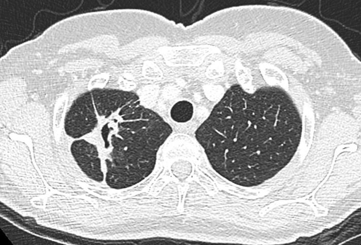
8 3. Imaging findings - Mascalchi et al. described 4 types of morphologic patterns on CT - Type 1: presence of a nodule extruding from the wall of the cystic airspace - Type II: nodule confined within the lumen of the cystic airspace - Type III: soft tissue density extending along the wall of the cystic airspace - Type IV: solid or nonsolid tissue intermixed within cluster of multiple cystic airspaces Type 1 Type II Type III Type IV
9 3. Imaging findings - Evolution: uniformly thin wall of the airspace, becoming thicker with increased circumferential involvement and nodule formation between 12 and 118 months after initial CT scan - No histologic features identified that suggested preexisting congenital cystic lung disease nor presence of a preexisting cavity - Associated emphysema is often present - When the solid part of the lesion increases, the diameter of the cystic airspace can decrease, increase or remain stable
10 3. Imaging findings - 18F-FDG-uptake is variable, ranging from absent or mild to moderate or marked - This is related to two facts - When the predominant tumor component presents as lepidic growth, FDG-uptake can be mild or absent. These lesions often present on CT as ground-glass - The cystic component of the lesion itself does not show 18F-FDG-uptake


 was")
11 4. Cases CASE 1 72-y-old man with a previous history of head and neck cancer who presented during follow-up with an irregular thickening of the wall of a cystic airspace in the right lower lobe. Within an extremely short time period of 3 months, the lesion had grown, filling the cystic airspace and presenting as a solid nodule. Diagnosis of primary lung cancer (squamous cell carcinoma) was confirmed after transthoracic time of diagnosis 3 months earlier 6 months earlier 5 years before


12 CASE 2 57-y-old woman who presented with a cystic airspace in the left upper lobe. In the periphery of the lesion there were two irregular nodules: one at the top and one at the bottom of the cystic airspace. Both nodules showed a high uptake on 18F-FDG-PET. Lobectomy was performed and histopathology confirmed the diagnosis of a cystic airspace with two nodular foci of adenocarcinoma.


13 CASE 3 72-y-old woman in whom as incidental finding a subpleural cystic airspace was seen in the left lower lobe. The medial wall of the cystic airspace showed a bandlike thickening. Follow-up CT 6 months later showed an increase of the exophytic component with more nodular aspect as well as a change of the internal structure of the cystic airspace. Lobectomy was performed and histopathology showed an invasive adenocarcinoma. Initial examination 6 months later


14 CASE 4 76-y-old man who presented with a subpleural nodule with associated cystic airspace in the left lower lobe. The lesion showed a very high uptake of 18F-FDG PET. Transthoracic biopsy confirmed the diagnosis of squamous cell carcinoma. Follow-up after radiotherapy treatment of the malignant lesion in the left lower lobe showed a nodule abutting the wall of a cystic airspace in the right lower lobe. Growth of this nodule was demonstrated after a second follow-up examination another 3 months later; findings were suspicious for a second primary. Initial examination 6 months later Another 3 months later


, part-solid nodules,")


15 CASE 5 68-y-old woman who presented with numerous nodules in both lungs: large spiculated solid nodules (perihilar region right upper lobe), part-solid nodules, pure ground glass nodules as well as 3 lesions presenting as lung cancer associated with cystic airspace (blue arrows). Diagnosis of adenocarcinoma was histopathologically proven in the large spiculated solid lesion in the right upper lobe and in the cystic airspace lesion with solid component in the left lower lobe.


16 CASE 6 74-y-old man who presented with a large cystic airspace surrounded and interspersed with ground glass. The lesion remained stable during a 3 month period of follow-up making an infectious lesion unlikely and the morphology of the lesion was found to be suspicious for lung cancer. 18F-FDG-PET showed only very minor uptake. Lobectomy was performed. Histopathology confirmed the diagnosis of lung cancer and showed a NSCLC with predominant lepidic pattern. These findings are consistent on imaging with the ground glass aspect of the abnormalities and are an explanation for the very low 18F-FDG-uptake on PET.


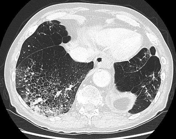
17 CASE 7 78-y-old man who presented with a persistent triangular area of cystic changes surrounded by an area of consolidation. Over time the area changed in morphology with decrease of the cystic airspaces and increase of the consolidation. Lobectomy was performed and histopathology showed an invasive adenocarcinoma. Initial examination 6 months later Another 6 months later


18 CASE 8 Area of consolidation with central foci of cystic airspaces in the right lower lobe in a 69-year-old man. During follow-up the lesion showed an increase in size and density. Diagnosis of adenocarcinoma was confirmed on histopathology. Initial examination 6 months later
19 5. Differential diagnosis - Depends on the type (I-IV) of lung cancer associated with cystic airspace - Differential diagnosis is broad and includes a variety of solitary cavitary lung lesions: tumor, infection, granulomatous disease, bronchogenic cyst,... - Entities that may mimic type IV are diffuse centrilobular emphysema with superimposed ground glass or consolidation, large pulmonary infarct,... - Look for associated findings to narrow down a differential diagnosis list - Clinical information, evolution and previous medical history are crucial for assessing the right diagnosis


20 Old tuberculous cavern with small fungus ball (Candida) Infected bronchogenic cyst Cavitated lung infarct Squamous cell arcinoma


21 Emphysema with superimposed infectious consolidation Large pulmonary infarct Adenosquamous carcinoma
22 6. To remember - Lung cancer associated with cystic airspaces is a relative new entity, getting more attention - Closely look at the wall of cystic airspaces, especially when they are new - Nodules, consolidation or ground-glass surrounding or abutting cystic airspaces should be closely monitored - Early recognition is important to avoid delay in diagnosis and treatment - Lots of questions remain and further research is needed When to consider malignancy? What regimen of follow-up? Are all 4 types of the same concern?

23 You only see what you look for and you only look for what you know Don t forget to think about lung cancer associated with cystic airspaces when you read chest CT s
24 7. Suggested readings 1. Farooqi AO, Cham M, Zhang L, Beasley MB, Austin JHM, Miller A, et al. Lung cancer associated with cystic airspaces. AJR Am J Roentgenol Oct;199(4): Mascalchi M, Attinà D, Bertelli E, Falchini M, Vella A, Pegna AL, et al. Lung cancer associated with cystic airspaces. J Comput Assist Tomogr Jan;39(1): Maki D, Takahashi M, Murata K, Sawai S, Fujino S, Inoue S. Computed tomography appearances of bronchogenic carcinoma associated with bullous lung disease. J Comput Assist Tomogr May;30(3): Kaneda M, Tarukawa T, Watanabe F, Adachi K, Sakai T, Nakabayashi H. Clinical features of primary lung cancer adjoining pulmonary bulla. Interactive CardioVascular and Thoracic Surgery May 17;10(6): Rampinelli C, Calloni SF, Minotti M, Bellomi M. Spectrum of early lung cancer presentation in low-dose screening CT: a pictorial review. Insights into Imaging. Insights into Imaging; 2016 May 14;: Scholten ET, Horeweg N, de Koning HJ, Vliegenthart R, Oudkerk M, Mali WPTM, et al. Computed tomographic characteristics of interval and post screen carcinomas in lung cancer screening. Eur Radiol Sep 4;25(1): Devaraj A. Missed cancers in lung cancer screening--more than meets the eye. Eur Radiol Jan;25(1): Yoshida T, Harada T, Fuke S, Konishi J, Yamazaki K, Kaji M, et al. Lung adenocarcinoma presenting with enlarged and multiloculated cystic lesions over 2 years. Respir Care Dec;49(12): Guo J, Liang C, Sun Y, Zhou N, Liu Y, Chu X. Lung cancer presenting as thin-walled cysts: An analysis of 15 cases and review of literature. Asia-Pac J Clin Oncol Dec 19;12(1):e
25 Author contact information Annemie Snoeckx, MD Antwerp University Hospital Radiology Department Wilrijkstraat Edegem, Belgium
Thoracic CT pattern in lung cancer: correlation of CT and pathologic diagnosis
 19 th Congress of APSR PG of Lung Cancer (ESAP): Update of Lung Cancer Thoracic CT pattern in lung cancer: correlation of CT and pathologic diagnosis Kazuma Kishi, M.D. Department of Respiratory Medicine,
Pulmonary Nodules & Masses
 Pulmonary Nodules & Masses A Diagnostic Approach Heber MacMahon The University of Chicago Department of Radiology Disclosure Information Consultant for Riverain Technology Minor equity in Hologic Royalties
PULMONARY NODULES AND MASSES : DIAGNOSTIC APPROACH AND NEW MANAGEMENT GUIDELINES. https://tinyurl.com/hmpn2018
 PULMONARY NODULES AND MASSES : DIAGNOSTIC APPROACH AND NEW MANAGEMENT GUIDELINES Heber MacMahon MB, BCh Department of Radiology The University of Chicago https://tinyurl.com/hmpn2018 Disclosures Consultant
Cavitation in primary lung cancer:
 Cavitation in primary lung cancer: characteristic features and mechanisms Yoshie Kunihiro 1,2), Taiga Kobayashi 2), Nobuyuki Tanaka 3), Tsuneo Matsumoto 1), Naofumi Matsunaga 2) 1) National Hospital Organization,
Excavated pulmonary nodule: steps to diagnosis?
 Excavated pulmonary nodule: steps to diagnosis? Poster No.: C-1044 Congress: ECR 2014 Type: Authors: Keywords: DOI: Educational Exhibit W. Mnari, M. MAATOUK, A. Zrig, B. Hmida, M. GOLLI; Monastir/ TN Metastases,
GUIDELINES FOR PULMONARY NODULE MANAGEMENT : RECENT CHANGES AND UPDATES
 Venice 2017 GUIDELINES FOR PULMONARY NODULE MANAGEMENT : RECENT CHANGES AND UPDATES Heber MacMahon MB, BCh Department of Radiology The University of Chicago Disclosures Consultant for Riverain Medical
Impact of the favorable prognosis of patients with lung cancer adjoining bullae
 Original Article Impact of the favorable prognosis of patients with lung cancer adjoining bullae Shuichi Shinohara 1, Masakazu Sugaya 1, Takamitsu Onitsuka 1, Kazuhiko Machida 2, Masaki Matsuo 2, Kazuo
OBJECTIVES. Solitary Solid Spiculated Nodule. What would you do next? Case Based Discussion: State of the Art Management of Lung Nodules.
 Organ Imaging : September 25 2015 OBJECTIVES Case Based Discussion: State of the Art Management of Lung Nodules Dr. Elsie T. Nguyen Dr. Kazuhiro Yasufuku 1. To review guidelines for follow up and management
LUNG NODULES: MODERN MANAGEMENT STRATEGIES
 Department of Radiology LUNG NODULES: MODERN MANAGEMENT STRATEGIES Christian J. Herold M.D. Department of Biomedical Imaging and Image-guided Therapy Medical University of Vienna Vienna, Austria Pulmonary
Approach to Pulmonary Nodules
 Approach to Pulmonary Nodules Edwin Jackson, Jr., DO Assistant Professor-Clinical Director, James Early Detection Clinic Department of Internal Medicine Division of Pulmonary, Allergy, Critical Care and
Chest Radiology Interpretation: Findings of Tuberculosis
 Chest Radiology Interpretation: Findings of Tuberculosis Get out your laptops, smart phones or other devices pollev.com/chestradiology Case #1 1 Plombage Pneumonia Cancer 2 Reading the TB CXR Be systematic!
How to Analyse Difficult Chest CT
 How to Analyse Difficult Chest CT Complex diseases are:- - Large lesion - Unusual or atypical pattern - Multiple discordant findings Diffuse diseases are:- - Numerous findings in both sides 3 basic steps
CT Screening for Lung Cancer for High Risk Patients
 CT Screening for Lung Cancer for High Risk Patients The recently published National Lung Cancer Screening Trial (NLST) showed that low-dose CT screening for lung cancer reduces mortality in high-risk patients
Xiaohuan Pan 1,2 *, Xinguan Yang 1,2 *, Jingxu Li 1,2, Xiao Dong 1,2, Jianxing He 2,3, Yubao Guan 1,2. Original Article
 Original Article Is a 5-mm diameter an appropriate cut-off value for the diagnosis of atypical adenomatous hyperplasia and adenocarcinoma in situ on chest computed tomography and pathological examination?
Update on 2015 WHO Classification of Lung Adenocarcinoma 1/3/ Mayo Foundation for Medical Education and Research. All rights reserved.
 1 Our speaker for this program is Dr. Anja Roden, an associate professor of Laboratory Medicine and Pathology at Mayo Clinic as well as consultant in the Anatomic Pathology Laboratory and co-director of
Spectrum of Radiological Findings in Bronchogenic Carcinoma A Retrospective Study
 IOSR Journal of Dental and Medical Sciences (IOSR-JDMS) e-issn: 2279-0853, p-issn: 2279-0861.Volume 17, Issue 01 Ver. VIII January. (2018), PP 43-59 www.iosrjournals.org Spectrum of Radiological Findings
The Spectrum of Management of Pulmonary Ground Glass Nodules
 The Spectrum of Management of Pulmonary Ground Glass Nodules Stanley S Siegelman CT Society 10/26/2011 No financial disclosures. Noguchi M et al. Cancer 75: 2844-2852, 1995. 236 surgically resected peripheral
Radiology Pathology Conference
 Radiology Pathology Conference Sharlin Johnykutty,, MD, Cytopathology Fellow Sara Majewski, MD, Radiology Resident Friday, August 28, 2009 Presentation material is for education purposes only. All rights
Web Chapter 3. Image Gallery: Lesion detection on low dose chest CT
 Web Chapter 3 Image Gallery: Lesion detection on low dose chest CT Sarabjeet Singh, MD Mannudeep K. Kalra, MD *Eugene J. Mark, MD *James Stone, MD James H. Thrall, MD Department of Radiology and *Department
PET/CT in lung cancer
 PET/CT in lung cancer Andrei Šamarin North Estonia Medical Centre 3 rd Baltic Congress of Radiology 08.10.2010 Imaging in lung cancer Why do we need PET/CT? CT is routine imaging modality for staging of
The small subsolid pulmonary nodules. What radiologists need to know.
 The small subsolid pulmonary nodules. What radiologists need to know. Poster No.: C-1250 Congress: ECR 2016 Type: Educational Exhibit Authors: L. Fernandez Rodriguez, A. Martín Díaz, A. Linares Beltrán,
Worse survival after curative resection in patients with pathological stage I non-small cell lung cancer adjoining pulmonary cavity formation
 Original Article Worse survival after curative resection in patients with pathological stage I non-small cell lung cancer adjoining pulmonary cavity formation Hiroyuki Kimura 1, Hisashi Saji 1,2, Tomoyuki
Characteristic Radiographic Features of Pulmonary Carcinoma Associated with Large Bulla
 Characteristic Radiographic Features of Pulmonary Carcinoma Associated with Large Bulla Masayoshi Tsutsui, M.D., Yasuo Araki, M.D., Takayuki Shirakusa, M.D., and Sadamitsu Inutsuka, M.D. ABSTRACT Primary
September 2014 Imaging Case of the Month. Michael B. Gotway, MD. Department of Radiology Mayo Clinic Arizona Scottsdale, AZ
 September 2014 Imaging Case of the Month Michael B. Gotway, MD Department of Radiology Mayo Clinic Arizona Scottsdale, AZ Clinical History: A 57-year-old non-smoking woman presented to her physician as
CT Screening for Lung Cancer: Frequency and Significance of Part-Solid and Nonsolid Nodules
 Claudia I. Henschke 1 David F. Yankelevitz 1 Rosna Mirtcheva 1 Georgeann McGuinness 2 Dorothy McCauley 1 0lli S. Miettinen 3 for the ELCAP Group Received June 19, 2001; accepted after revision November
I appreciate the courtesy of Kusumoto at NCC for this presentation. What is Early Lung Cancers. Early Lung Cancers. Early Lung Cancers 18/10/55
 I appreciate the courtesy of Kusumoto at NCC for this presentation. Dr. What is Early Lung Cancers DEATH Early period in its lifetime Curative period in its lifetime Early Lung Cancers Early Lung Cancers
Imaging: how to recognise idiopathic pulmonary fibrosis
 REVIEW IDIOPATHIC PULMONARY FIBROSIS Imaging: how to recognise idiopathic pulmonary fibrosis Anand Devaraj Affiliations: Dept of Radiology, St George s Hospital, London, UK. Correspondence: Anand Devaraj,
RADIOLOGIC EVALUATION OF PULMONARY NTM INFECTION. Tilman Koelsch, MD National Jewish Health - Department of Radiology
 Pr N op ot e fo rty rr o f ep Pr ro es du en ct te io r n RADIOLOGIC EVALUATION OF PULMONARY NTM INFECTION Tilman Koelsch, MD National Jewish Health - Department of Radiology Disclosures None Goals Identify
Rodney C Richie MD FACP FCCP DBIM Texas Life and EMSI
 Rodney C Richie MD FACP FCCP DBIM Texas Life and EMSI Pulmonary Nodules Well-circumscribed, radiographic opacities measuring 3 cm in diameter Surrounded by aerated lung Not associated with atelectesis
Single Lung Cyst Caused by Metastatic Bladder Cancer -A Case Report
 Single Lung Cyst Caused by Metastatic Bladder Cancer -A Case Report *& Jin-Duo ng, Chih-Bin Lin, Min-Shin Kuo', Jen-Jyh Lee The lungs are one of the most common sites of metastases from transitional cell
CT findings in multifocal or diffuse non-mucinous bronchioloalveolar carcinoma (BAC)
") CT findings in multifocal or diffuse non-mucinous bronchioloalveolar carcinoma (BAC) Poster No.: C-2192 Congress: ECR 2014 Type: Educational Exhibit Authors: I. Sandu, A. R. Popita, I.-A. Brumboiu; Cluj-Napoca/RO
CT findings in multifocal or diffuse non-mucinous bronchioloalveolar carcinoma (BAC) Poster No.: C-2192 Congress: ECR 2014 Type: Educational Exhibit Authors: I. Sandu, A. R. Popita, I.-A. Brumboiu; Cluj-Napoca/RO
Published Pulmonary Nodule Guidelines A Synthesis
 Published Pulmonary Nodule Guidelines A Synthesis Dr A Devaraj Royal Brompton Hospital London 4/28/2015 1 And very soon to be published Published ^ Pulmonary Nodule Guidelines A Synthesis Dr A Devaraj
Steering Committee. Waiting on photo. Paul A. Bunn, Jr., MD Kavita Garg, MD Kim Geisinger, MD Fred R. Hirsch, Gregory Riely, MD, PhD.
 Steering Committee Paul A. Bunn, Jr., MD Kavita Garg, MD Kim Geisinger, MD Fred R. Hirsch, Gregory Riely, MD, PhD MD, PhD Waiting on photo Paul Van Schil, MD, PhD William D. Travis, MD Ming-Sound Tsao,
CT findings in multifocal or diffuse non-mucinous bronchioloalveolar carcinoma (BAC)
") CT findings in multifocal or diffuse non-mucinous bronchioloalveolar carcinoma (BAC) Poster No.: C-2192 Congress: ECR 2014 Type: Educational Exhibit Authors: I. Sandu, A. R. Popita, I.-A. Brumboiu; Cluj-Napoca/RO
CT findings in multifocal or diffuse non-mucinous bronchioloalveolar carcinoma (BAC) Poster No.: C-2192 Congress: ECR 2014 Type: Educational Exhibit Authors: I. Sandu, A. R. Popita, I.-A. Brumboiu; Cluj-Napoca/RO
Acute and Chronic Lung Disease
 KATHOLIEKE UNIVERSITEIT LEUVEN Faculty of Medicine Acute and Chronic Lung Disease W De Wever, JA Verschakelen Department of Radiology, University Hospitals Leuven, Belgium Clinical utility of HRCT To detect
Financial disclosure COMMON DIAGNOSES IN HRCT. High Res Chest HRCT. HRCT Pre test. I have no financial relationships to disclose. Anatomy Nomenclature
 Financial disclosure I have no financial relationships to disclose. Douglas Johnson D.O. Cardiothoracic Imaging Gaston Radiology COMMON DIAGNOSES IN HRCT High Res Chest Anatomy Nomenclature HRCT Sampling
Radiological staging of lung cancer. Shukri Loutfi,MD,FRCR Consultant Thoracic Radiologist KAMC-Riyadh
 Radiological staging of lung cancer Shukri Loutfi,MD,FRCR Consultant Thoracic Radiologist KAMC-Riyadh Bronchogenic Carcinoma Accounts for 14% of new cancer diagnoses in 2012. Estimated to kill ~150,000
Purpose. Methods and Materials
 Thin-section CT findings in peripheral lung cancer of 3 cm or smaller: are there any characteristic features for predicting tumor histology or do they depend only on tumor size? Poster No.: C-1893 Congress:
Hypothesis on the Evolution of Cavitary Lesions in Nontuberculous Mycobacterial Pulmonary Infection: Thin-Section CT and Histopathologic Correlation
 CT of Nontuberculous Mycobacterial Pulmonary Infection Tae Sung Kim 1 Won-Jung Koh 2 Joungho Han 3 Myung Jin Chung 1 Ju Hyun Lee 1 Kyung Soo Lee 1 O Jung Kwon 2 Kim TS, Koh W-J, Han J, et al. Received
HRCT in Diffuse Interstitial Lung Disease Steps in High Resolution CT Diagnosis. Where are the lymphatics? Anatomic distribution
 Steps in High Resolution CT Diagnosis Pattern of abnormality Distribution of disease Associated findings Clinical history Tomás Franquet MD What is the diagnosis? Hospital de Sant Pau. Barcelona Secondary
Evidence based approach to incidentally detected subsolid pulmonary nodule. DM SEMINAR July 27, 2018 Harshith Rao
 Evidence based approach to incidentally detected subsolid pulmonary nodule DM SEMINAR July 27, 2018 Harshith Rao Outline Definitions Etiologies Risk evaluation Clinical features Radiology Approach Modifications:
Small Pulmonary Nodules: Our Preliminary Experience in Volumetric Analysis of Doubling Times
 Small Pulmonary Nodules: Our Preliminary Experience in Volumetric Analysis of Doubling Times Andrea Borghesi, MD Davide Farina, MD Roberto Maroldi, MD Department of Radiology University of Brescia Brescia,
11/10/2014. Multi-disciplinary Approach to Diffuse Lung Disease: The Imager s Perspective. Radiology
 Multi-disciplinary Approach to Diffuse Lung Disease: The Imager s Perspective Radiology Pathology Clinical 1 Role of HRCT Diagnosis Fibrosis vs. inflammation Next step in management Response to treatment
Bronchioloalveolar Carcinoma Mimicking DILD:
 Bronchioloalveolar Carcinoma Mimicking DILD: A Case Report 1 Ju Young Lee, M.D., In Jae Lee, M.D., Dong Gyu Kim, M.D. 2, Soo Kee Min, M.D. 3, Min-Jeong Kim, M.D., Sung Il Hwang, M.D., Yul Lee, M.D., Sang
PET CT for Staging Lung Cancer
 PET CT for Staging Lung Cancer Rohit Kochhar Consultant Radiologist Disclosures Neither I nor my immediate family members have financial relationships with commercial organizations that may have a direct
DENOMINATOR: All final reports for CT imaging studies with a finding of an incidental pulmonary nodule for patients aged 35 years and older
 Quality ID #364: Optimizing Patient Exposure to Ionizing Radiation: Appropriateness: Follow-up CT Imaging for Incidentally Detected Pulmonary Nodules According to Recommended Guidelines National Quality
Spectrum of Cystic Lung Disease and its Mimics. Kathleen Jacobs MD and Elizabeth Weihe MD UC San Diego Medical Center, Department of Radiology
 Spectrum of Cystic Lung Disease and its Mimics Kathleen Jacobs MD and Elizabeth Weihe MD UC San Diego Medical Center, Department of Radiology No Financial Disclosures Learning Objectives 1. Review the
2018 OPTIONS FOR INDIVIDUAL MEASURES: REGISTRY ONLY. MEASURE TYPE: Process
 Quality ID #364: Optimizing Patient Exposure to Ionizing Radiation: Appropriateness: Follow-up CT Imaging for Incidentally Detected Pulmonary Nodules According to Recommended Guidelines National Quality
With recent advances in diagnostic imaging technologies,
 ORIGINAL ARTICLE Management of Ground-Glass Opacity Lesions Detected in Patients with Otherwise Operable Non-small Cell Lung Cancer Hong Kwan Kim, MD,* Yong Soo Choi, MD,* Kwhanmien Kim, MD,* Young Mog
RADIOLOGIC EVALUATION OF PULMONARY NTM INFECTION. Tilman Koelsch, MD National Jewish Health - Department of Radiology
 Pr N op ot er fo ty r R of ep Pr ro es du en ct te io r n RADIOLOGIC EVALUATION OF PULMONARY NTM INFECTION Tilman Koelsch, MD National Jewish Health - Department of Radiology Disclosures No relevant financial
Use of Integrated PET CT in the Clinical Staging of Non Small Cell Lung Cancer
 November 2010 Use of Integrated PET CT in the Clinical Staging of Non Small Cell Lung Cancer Laura Myers, Harvard Medical School, Year III Clinical Presentation 79yo woman with cough productive of green
Expert Round Table with Drs. Anne Tsao and Alex Farivar Part 1: Elderly Man with Indolent Bronchioloalveolar Carcinoma
 Expert Round Table with Drs. Anne Tsao and Alex Farivar Part 1: Elderly Man with Indolent Bronchioloalveolar Carcinoma February 2010 I d like to welcome everyone, thanks for coming out to our lunch with
Learning Objectives. 1. Identify which patients meet criteria for annual lung cancer screening
 Disclosure I, Taylor Rowlett, DO NOT have a financial interest /arrangement or affiliation with one or more organizations that could be perceived as a real or apparent conflict of interest in the context
Lung Cancer Risk Associated With New Solid Nodules in the National Lung Screening Trial
 Cardiopulmonary Imaging Original Research Pinsky et al. Lung Cancer Risk Associated With New Nodules Cardiopulmonary Imaging Original Research Paul F. Pinsky 1 David S. Gierada 2 P. Hrudaya Nath 3 Reginald
42 yr old male with h/o Graves disease and prior I 131 treatment presents with hyperthyroidism and undetectable TSH. 2 hr uptake 20%, 24 hr uptake 50%
 Pinhole images of the neck are acquired in multiple projections, 24hrs after the oral administration of approximately 200 µci of I123. Usually, 24hr uptake value if also calculated (normal 24 hr uptake
October 2012 Imaging Case of the Month. Michael B. Gotway, MD Associate Editor Imaging. Department of Radiology Mayo Clinic Arizona Scottsdale, AZ
 October 2012 Imaging Case of the Month Michael B. Gotway, MD Associate Editor Imaging Department of Radiology Mayo Clinic Arizona Scottsdale, AZ Clinical History: A 65-year-old non-smoking woman presented
An Image Repository for Chest CT
 An Image Repository for Chest CT Francesco Frajoli for the Chest CT in Antibody Deficiency Group An Image Repository for Chest CT he Chest CT in Antibody Deficiency Group is an international and interdisciplinary
Lung Cancer Screening: To Screen or Not to Screen?
 Lung Cancer Screening: To Screen or Not to Screen? Lorriana Leard, MD Co-Director of UCSF Lung Cancer Screening Program Vice Chief of Clinical Activities UCSF Pulmonary, Critical Care, Allergy & Sleep
August 2018 Imaging Case of the Month: Dyspnea in a 55-Year-Old Smoker. Michael B. Gotway, MD
 August 2018 Imaging Case of the Month: Dyspnea in a 55-Year-Old Smoker Michael B. Gotway, MD Department of Radiology Mayo Clinic Arizona Scottsdale, AZ USA Clinical History: A 55 year old woman presented
Usual Interstitial pneumonia and Nonspecific Interstitial Pneumonia. Nitra and the Gangs.
 Usual Interstitial pneumonia and Nonspecific Interstitial Pneumonia Nitra and the Gangs. บทน ำและบทท ๓, ๑๐, ๑๒, ๑๓, ๑๔, ๑๕, ๑๗ Usual Interstitial Pneumonia (UIP) Most common & basic pathologic pattern
Management of Multiple Pure Ground-Glass Opacity Lesions in Patients with Bronchioloalveolar Carcinoma
 ORIGINAL ARTICLE Management of Multiple Pure Ground-Glass Opacity Lesions in Patients with Bronchioloalveolar Carcinoma Hong Kwan Kim, MD,* Yong Soo Choi, MD,* Jhingook Kim, MD, PhD,* Young Mog Shim, MD,
I9 COMPLETION INSTRUCTIONS
 The I9 Form is completed for each screening exam at T0, T1, and T2. At T0 (baseline), the I9 documents comparison review of the baseline screen (C2 Form) with any historical images available. At T1 and
Is it really honeycombing? Limitations and pitfalls in radiological diagnosis of honeycombing.
 Is it really honeycombing? Limitations and pitfalls in radiological diagnosis of honeycombing. J. Arenas-Jiménez, E. García-Garrigós, M. Sirera-Matilla; M.C. Planells-Alduvin, F.I. Aranda* Hospital General
Lung. 10/24/13 Chest X-ray: 2.9 cm mass like density in the inferior lingular segment worrisome for neoplasm. Malignancy cannot be excluded.
 Lung Case Scenario 1 A 54 year white male presents with a recent abnormal CT of the chest. The patient has a history of melanoma, kidney, and prostate cancers. 10/24/13 Chest X-ray: 2.9 cm mass like density
Guidelines for the Management of Pulmonary Nodules Detected by Low-dose CT Lung Cancer Screening
 Guidelines for the Management of Pulmonary Nodules Detected by Low-dose CT Lung Cancer Screening 1. Introduction In January 2005, the Committee for Preparation of Clinical Practice Guidelines for the Management
Primary pulmonary cancer colliding with metastatic choriocarcinoma
 Case Report Primary pulmonary cancer colliding with metastatic choriocarcinoma Zheng Wang 1, Jingwen Si 2, Jingwei Liu 1 1 Department of Thoracic Surgery, 2 Department of Pathology, Peking University First
Lung Cancers Manifesting as Part-Solid Nodules in the National Lung Screening Trial
 Cardiopulmonary Imaging Original Research Yip et al. Lung Cancers Manifesting as Part-Solid Nodules Cardiopulmonary Imaging Original Research Rowena Yip 1 Claudia I. Henschke 1 Dong Ming Xu 1 Kunwei Li
Common Blind Spots on Chest CT: Where Are They All Hiding? Part 1 Airways, Lungs, and Pleura
 Residents Section Structured Review Wu et al. Common lind Spots on Chest CT Residents Section Structured Review Carol C. Wu 1 Leila Khorashadi 2 Gerald F. bbott 1 Matthew D. Gilman 1 Wu CC, Khorashadi
A Chronology of Advancements in the Diagnosing of Lung Nodules
 November 17, 2017 A Chronology of Advancements in the Diagnosing of Lung Nodules Presenter: Daniel P. Harley, MD, MSB, FACS Surgical Director of the Angelos Center for Lung Diseases 1 Pulmonary Nodules
MANAGEMENT RECOMMENDATIONS
 1 MANAGEMENT RECOMMENDATIONS 1. Adrenal masses!!!!!!! page 2 2. Liver Masses!!!!!!! page 3 3. Obstetric US Soft Markers for Aneuploidy!! pages 4-6 4. Ovarian and Adnexal Cysts!!!!! pages 7-10 5. Pancreatic
INTERACTIVE SESSION 2
 INTERACTIVE SESSION 2 2 patients with lung metastases, with complete response after oncologic treatment - Clinical Case Presentation: Dr. Esther Casado Dr. Sergi Call - Expert Opinion: Dr. Raúl Embún Dr.
Stage I synchronous multiple primary non-small cell lung cancer: CT findings and the effect of TNM staging with the 7th and 8th editions on prognosis
 Original Article Stage I synchronous multiple primary non-small cell lung cancer: CT findings and the effect of TNM staging with the 7th and 8th editions on prognosis Jingxu Li, Xinguan Yang, Tingting
CT Signs of Solitary Pulmonary Lesions: Revisited
 CT Signs of Solitary Pulmonary Lesions: Revisited Poster No.: C-1764 Congress: ECR 2015 Type: Educational Exhibit Authors: H. Hayashi, K. Ashizawa, Y. Ogihara, A. Nishida, T. Tanaka, 1 1 2 1 1 1 1 1 2
Diagnostic challenge: Sclerosing Hemangioma of the Lung. Department of Medicine, Division of Pulmonary and Critical Care, Lincoln Medical and
 Diagnostic challenge: Sclerosing Hemangioma of the Lung. S. Arias M.D, R. Loganathan M.D, FCCP Department of Medicine, Division of Pulmonary and Critical Care, Lincoln Medical and Mental Health Center/Weill
TB Radiology for Nurses Garold O. Minns, MD
 TB Nurse Case Management Salina, Kansas March 31-April 1, 2010 TB Radiology for Nurses Garold O. Minns, MD April 1, 2010 TB Radiology for Nurses Highway Patrol Training Center Salina, KS April 1, 2010
CT Findings of Surgically Resected Pleomorphic Carcinoma of the Lung in 30 Patients
 Kim et al. CT of Pleomorphic Carcinoma of the Lung Chest Imaging Clinical Observations Tae Sung Kim 1 Joungho Han 2 Kyung Soo Lee 1 Yeon Joo Jeong 1 Seo Hyun Kwak 1 Hong Sik Byun 1 Myung Jin Chung 1 Hojoong
University of Groningen
 University of Groningen Morphological characteristics of potentially malignant pulmonary nodules in high-risk male smokers detected in lung cancer screening trial in Cracow, Poland Kiszka, Kinga; Rudnicka-Sosin,
An Introduction to Radiology for TB Nurses
 An Introduction to Radiology for TB Nurses Garold O. Minns, MD September 14, 2017 TB Nurse Case Management September 12 14, 2017 EXCELLENCE EXPERTISE INNOVATION Garold O. Minns, MD has the following disclosures
Lung Cancer Diagnosis for Primary Care
 Lung Cancer Diagnosis for Primary Care Daniel Nader, DO, FCCP Cancer Treatment Center of America Case 1 In which of the following situations would the U.S. Preventive Services Task Force (USPSTF) recommend
Surgical indications: Non-malignant pulmonary diseases. Punnarerk Thongcharoen
 Surgical indications: Non-malignant pulmonary diseases Punnarerk Thongcharoen Non-malignant Malignant as a pathological term: Cancer Non-malignant = not cancer Malignant as an adjective: Disposed to cause
Case of the Day Chest
 Case of the Day Chest Darin White MDCM FRCPC Department of Radiology, Mayo Clinic 76 th Annual Scientific Meeting Canadian Association of Radiologists Montreal, QC April 26, 2013 2013 MFMER slide-1 Disclosures
Lung Cancer in Chronic Interstitial Pneumonia: Early Manifestation From Serial CT Observations
 Cardiopulmonary Imaging Original Research Yoshida et al. CT of Lung Cancer Cardiopulmonary Imaging Original Research Rika Yoshida 1 Hiroaki rakawa Yasushi Kaji Yoshida R, rakawa H, Kaji Y Keywords: CT,
Intraductal papillary mucinous neoplasm of the bile ducts: a rare form of premalignant lesion of invasive cholangiocarcinoma
 Intraductal papillary mucinous neoplasm of the bile ducts: a rare form of premalignant lesion of invasive cholangiocarcinoma Authors: R. Revert Espí, Y. Fernandez Nuñez, I. Carbonell, D. P. Gómez valencia,
GROUP 1: Peripheral tumour with normal hilar and mediastinum on staging CT with no disant metastases. Including: Excluding:
 GROUP 1: Including: Excluding: Peripheral tumour with normal hilar and mediastinum on staging CT with no disant metastases Solid pulmonary nodules 8mm diameter / 300mm3 volume and BROCK risk of malignancy
Consolidations in Nodular Bronchiectatic Mycobacterium Avium Complex Lung Disease: Mycobacterium Avium Complex or Other Infection?
 Original Article DOI 10.3349/ymj.2010.51.4.546 pissn: 0513-5796, eissn: 1976-2437 Yonsei Med J 51(4):546-551, 2010 Consolidations in Nodular Bronchiectatic Mycobacterium Avium Complex Lung Disease: Mycobacterium
Nanda Horeweg, Carlijn M. van der Aalst, Erik Thunnissen, Kristiaan Nackaerts, Carla Weenink, Harry J.M. Groen, Jan-Willem J.
 Characteristics of lung cancers detected in the randomized NELSON lung cancer screening trial Nanda Horeweg, Carlijn M. van der Aalst, Erik Thunnissen, Kristiaan Nackaerts, Carla Weenink, Harry J.M. Groen,
Lung Tumor Cases: Common Problems and Helpful Hints
 Lung Tumor Cases: Common Problems and Helpful Hints Brandon T. Larsen, MD, PhD Senior Associate Consultant Department of Laboratory Medicine and Pathology Mayo Clinic Arizona Arizona Society of Pathologists
THE BENEFITS OF BIG DATA
 THE BENEFITS OF BIG DATA Disclosures I am a named inventor on a number of patents and patent applications relating to the evaluation of pulmonary nodules on CT scans of the chest which are owned by Cornell
Role of CT imaging to evaluate solitary pulmonary nodule with extrapulmonary neoplasms
 Original Research Article Role of CT imaging to evaluate solitary pulmonary nodule with extrapulmonary neoplasms Anand Vachhani 1, Shashvat Modia 1*, Varun Garasia 1, Deepak Bhimani 1, C. Raychaudhuri
Eun-Young Kang, M.D., Jae Wook Lee, M.D., Ji Yung Choo, M.D., Hwan Seok Yong, M.D., Ki Yeol Lee, M.D., Yu-Whan Oh, M.D.
 Eun-Young Kang, M.D., Jae Wook Lee, M.D., Ji Yung Choo, M.D., Hwan Seok Yong, M.D., Ki Yeol Lee, M.D., Yu-Whan Oh, M.D. Department of Radiology, Korea University Guro Hospital, College of Medicine, Korea
What to Do with Small Lung Nodules Hanh Vu Nghiem, MD William Beaumont Hospital Royal Oak, Michigan
 What to Do with Small Lung Nodules Hanh Vu Nghiem, MD William Beaumont Hospital Royal Oak, Michigan Small Lung Nodules What to do with small lung nodules? We biopsy them when requested What are our accuracy
Pictorial essay of unusual radiologic manifestations of pulmonary and airway metastasis at initial presentation of lung cancer
 Pictorial essay of unusual radiologic manifestations of pulmonary and airway metastasis at initial presentation of lung cancer Poster No.: C-2297 Congress: ECR 2012 Type: Educational Exhibit Authors: Y.
Invasive Pulmonary Adenocarcinomas versus Preinvasive Lesions Appearing as Ground-Glass Nodules: Differentiation by Using CT Features 1
 Note: This copy is for your personal non-commercial use only. To order presentation-ready copies for distribution to your colleagues or clients, contact us at www.rsna.org/rsnarights. Sang Min Lee, MD
Do you want to be an excellent Radiologist? - Focus on the thoracic aorta on lateral chest image!!!
 The lateral chest radiograph: Challenging area around the thoracic aorta!!! Do you want to be an excellent Radiologist? - Focus on the thoracic aorta on lateral chest image!!! Dong Yoon Han 1, So Youn
Tuberculosis: The Essentials
 Tuberculosis: The Essentials Kendra L. Fisher, MD, PhD THORACIC TUBERCULOSIS: THE BARE ESSENTIALS Kendra Fisher MD, FRCP (C) Department of Radiology Loma Linda University Medical Center TUBERCULOSIS ()
objectives Pitfalls and Pearls in PET/CT imaging Kevin Robinson, DO Assistant Professor Department of Radiology Michigan State University
 objectives Pitfalls and Pearls in PET/CT imaging Kevin Robinson, DO Assistant Professor Department of Radiology Michigan State University To determine the regions of physiologic activity To understand
Hypersensitivity Pneumonitis: Spectrum of High-Resolution CT and Pathologic Findings
 CT of Hypersensitivity Pneumonitis Chest Imaging Pictorial Essay C. Isabela S. Silva 1 ndrew Churg 2 Nestor L. Müller 1 Silva CIS, Churg, Müller NL Keywords: high-resolution CT, hypersensitivity pneumonitis,
Pulmonary aspergilloma: Imaging findings with pathologic correlation
 Pulmonary aspergilloma: Imaging findings with pathologic correlation Poster No.: E-0017 Congress: ESTI 2012 Type: Scientific Exhibit Authors: K. Odev, K. Ödev, S. Demirtas, O. Ozbek, O. K. Aribas, A. Kucukapan,
A Case of Pediatric Plasma Cell Granuloma
 August 2001 A Case of Pediatric Plasma Cell Granuloma Nii Tetteh, Harvard Medical School Year IV Our Patient 8 year old male with history of recurrent left lower lobe and lingular pneumonias since 1994.