How to Reduce Missed Diagnosis of Breast Cancer
|
|
|
- Ashley Marshall
- 5 years ago
- Views:
Transcription
1 How to Reduce Missed Diagnosis of Breast Cancer Heeboong Park M.D., Ph.D. Park Surgical Clinic Medical Park Co., Ltd.
2 Definition of Missed Diagnosis Misdiagnosis means that a doctor has diagnosed a person with the wrong medical condition. Missed Diagnosis: Failure to diagnose or delayed diagnosis, occurs when a doctor fails to identify a medical condition at the time they re presented with it. Objective, whole breast examination - mammography After multimodal diagnosis of breast cancer - diagnostic sensitivity of examination modality Breast cancer detected after recent examination is suspected to missed diagnosis.
3 Missed Diagnosis PE Mammography US MRI Biopsy
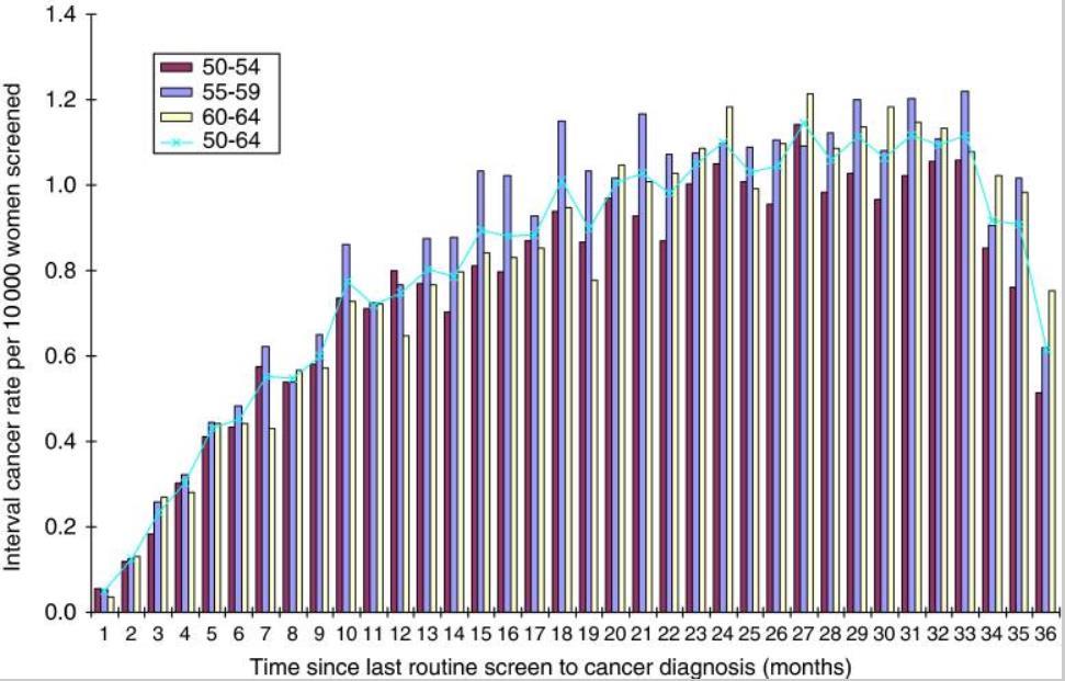
4 Interval breast cancer The definition of interval cancers in the NHSBSP (NHS Breast Screening Radiologists Quality Assurance Committee, 2005) is consistent with that in the European guidelines (Perry et al, 2006); breast cancers diagnosed in the interval between scheduled screening episodes in women screened and given a normal' screening result that is, the previous screening episode was negative. In our analyses we defined core interval cancers as those occurring within 36 months of a woman's last negative-screening episode in women aged between 50 and 64 at their last routine screen; Bennett et al. Br J Cancer Feb 15; 104(4):
5
6 Breast cancer tumor growth estimated through mammography screening data Cancer incidence and tumor measurement data from 395,188 women aged 50 to 69 years. Tumor growth varied considerably between subjects, with 5% of tumors taking less than 1.2 months to grow from 10 mm to 20 mm in diameter, and another 5% taking more than 6.3 years. The mean time a tumor needed to grow from 10 mm to 20 mm in diameter was estimated as 1.7 years, increasing with age. The screen test sensitivity was estimated to increase sharply with tumor size, rising from 26% at 5 mm to 91% at 10 mm. Harald Weedon-Fekjæ et. al Breast Cancer Research :R41
7 ESTIMATES OF BREAST CANCER GROWTH RATE FROM MAMMOGRAMS AND ITS RELATION TO TUMOUR CHARACTERISTICS. This study aimed to investigate the growth rate of 31 consecutive invasive breast cancers based on volume measures on at least two serial mammograms and its relation to histopathological findings. The average tumor volume-doubling time in all invasive breast cancer subtypes was 282 d (range d). Grade III breast cancers had a significantly shorter average tumour volumedoubling time of 105 d (range d) compared with Grade I and II tumors (average of 296 d, range d and average of 353 d, range d, respectively) (p = 0.002). Multiple linear regression identified that tumor volume-doubling time was positively associated with patient age, histological grade and progesterone receptor expression and inversely associated with axillary lymph node involvement, human epidermal growth factor receptor 2 and Ki-67 expression (p < 0.001). Förnvik D et al. Radiat Prot Dosimetry Jun;169(1-4):151-7.
8 A Review of Interval Breast Cancers Diagnosed among Participants of the Nova Scotia Breast Screening Program The NSBSP maintains databases that contain information on women participating in the program. From 1991 to 2004, the NSBSP collected information for 302,234 screening examinations performed in 115,433 women. The rate of missed cancers per 1000 women screened was one-half of the true interval cancer rate among women screened annually (for ages years, 0.45 vs 0.93; for ages years, 1.08 vs 2.22). Among women aged years who were screened biennially, the rate of missed cancers per 1000 women screened was one-third of the true interval cancer rate (0.90 vs 3.15). Similarly, the rate of missed cancers per screening examinations was one-half of the true interval rate among those years old (1.95 vs 3.99) and one-third of the true interval cancer rate among those years old (3.34 vs 10.44). Jennifer I. Payne et al. Radiology Jan;266(1):
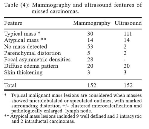
9 Missed Breast Carcinoma; Why and How to Avoid? Study over a two-year interval 152 cases revealed 121 infiltrating ductal carcinomas, 2 lobular, 4 mucinous, 14 inflammatory carcinomas, 6 carcinomas in situ (3 of which were intracystic), 2 intraductal papillary carcinomas and 3 cases with Paget's disease of the nipple. 4 causative factors; patient, tumor, technical or provider factors Tumor factors were the most commonly encountered, accounting for 44.1%, while provider factors were the least commonly encountered in 14.5% Kamal et al. J Egyptian NCI, 19(3): , 2007
10 44 carcinomas were detected on double and re-reading by more experienced radiologists. Additional mammographic views were recommended in 35 (23%) cases. Complementary ultrasound examination was performed for all 152 cases (100%) and showed a higher sensitivity than mammography in carcinoma detection. It was diagnostic in 138 (90.8%) cases only. In the remaining 14 cases, further MRI and biopsy were performed.
11
12 Happy eye syndrome The observation of an obvious finding (benign or malignant) may cause the "happy eye syndrome," misleading the radiologist into not looking carefully for other lesions. Small cancer with big benign tumor- two cases Multicentric and multifocal carcinomas were missed in 4.6% of cases (7 patients) and contralateral carcinomas in 3 other patients (2% of cases).
13 How to avoid missing a breast carcinoma? Review clinical data and use US and other adjunct techniques as MRI and biopsy to assess a palpable or mammographically detected mass. Be strict about positioning and technical factors. Try to optimize image quality. Be alert to subtle features of breast cancers. Always consider the well defined carcinoma. Compare current images with multiple prior studies to look for subtle increases in lesion size. Look for other lesions when one abnormality is seen. Judge a lesion by its most malignant features. Double reading and the use of computer aided diagnosis (CAD) and finally FFDM (Full Field Digital Mammography). Close cooperation between the oncologist, radiologist and pathologist is essential to avoid missing any case of breast carcinoma.
14 Detection of breast cancer in asymptomatic and symptomatic groups using computer-aided detection with full-field digital mammography. Analyzed digital mammography and CAD images from 210 patients diagnosed with breast cancer. The detection rate of the CAD system was 87.8% in the asymptomatic group. The sensitivity in different tissue densities was 100% in P1, 88.9% in P2, 94.4% P3, and 66.7% in extremely dense breasts (P4). The detection rate of the CAD system in the symptomatic group was 87.2%, and the sensitivity was 90.5%, 90%, 86.6%, and 75% in P1-P4 breasts, respectively. In the asymptomatic group, the CAD system detected 90.3% of invasive ductal carcinomas, not otherwise specified (IDC-NOS) and 88.9% of ductal carcinomas in situ (DCIS), but did not detect other types of malignancy. In the symptomatic group, the CAD system detected 88.2% of IDC-NOS, 88.9% of DCIS and 75% of other types of malignancy. When analyzed according to tumor size, the sensitivity of CAD in the asymptomatic and symptomatic groups was 82.6% and 83.3% for tumors <1 cm, 76.5% and 82.4% for tumors between 1 and 2 cm, and 91.7% and 89% in tumors >2 cm. Park et al J Breast Cancer Sep; 16(3):
 2600 2000 target: -lesion: lesion classification (benign vs. malignant), -mass: mass detection -MC: microcalcification detection. Geraset et al arxiv:1703.")
15 High-Resolution Breast Cancer Screening with Multi-View Deep Convolutional Neural Networks 129,208 patients, 201,698 exams and 886,437 images in the data set input image: 829k (57k) target: -lesion: lesion classification (benign vs. malignant), -mass: mass detection -MC: microcalcification detection. Geraset et al arxiv: v2 2017
16

 30 epoch, minibatch=64(16 pt) 8 GPU Kim et al.")
17 Applying Data-driven Imaging Biomarker in Mammography for Breast Cancer Screening: Preliminary Study 29,107 digital mammograms from five institutions (4,339 cancer cases and 24,768 normal cases) were included Input size 1600x1600, SGD(lr=0.001, decay=0.0001, momentum=0.9) 30 epoch, minibatch=64(16 pt) 8 GPU Kim et al. Scientiic Reports 8:2762, 2018
18
19
20 Malignancy Detection on Mammography Using Dual Deep Convolutional Neural Networks and Genetically Discovered False Color Input Enhancement. Mammography sensitivity ranging from 62 to 87% and specificity from 75 to 91%. Inception v3, preprocessing and computer vision methods. The Digital Database for Screening Mammography (DDSM) 의 6,000 mammographic image and Zebra Mammography Dataset image 1,739. We also utilize dual deep convolutional neural networks at different scales, for classification of full mammogram images and derivative patches combined with a random forest gating network as a novel architectural solution capable of discerning malignancy with a sensitivity of 0.91 and a specificity of Teare et al. J Digit Imaging Aug;30(4):


21 CAD Impact iview SonoEye TM Typical Benign Case Typical Malignant Case - ROI image vs. full size image - Biopsy-proven DB : about 1,000 cases - Malignant cases : about Benign cases : about In situations in which the radiologist and the computer disagree, this is useful solutions. - Computer-generated malignant score is calculated based on the retrieved images. - User can infer what's the basis of computer's malignancy score.

22 SonoEye 를이용한유방초음파검사상종괴에대한컴퓨터보조진단 (CAD) 의유용성 윤태일, 박신혜, 변종석, 박희붕 J. Breast Cancer (2) 110pp 2004 May ~ 2005 Apr. Breast cancer images Benign mass images US-CAD : SonoEye, Cadimpact Sensitivity: 83.6 ~ 96.4% Specificity: 53.2 ~ 73.4 %

23 삼성메디슨유방과갑상선에서 AI 이용자동진단프로그램
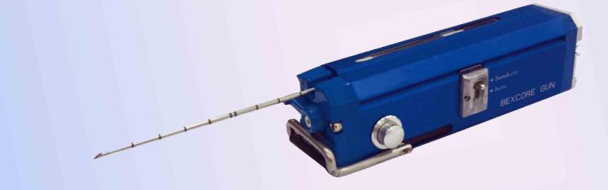
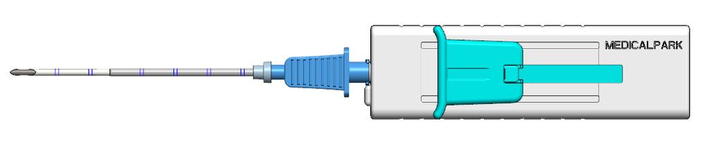
24 A deep learning framework for supporting the classification of breast lesions in ultrasound images 5,151 patients cases containing a total of 7,408 ultrasound breast images. 4,254 benign and 3,154 malignant lesions. GoogleLeNet (22 layers, input 255x255, grey -> color) Data augmentation: 147 times(553,455 vs 413,658) SGD(lr=0.0001, decay=0.0002, momentum=0.9) 1 GPU, minibatch size =32 Accuracy of about 0.9 (90%), a sensitivity of 0.86 and a specificity of Area under ROC curve AUC 0.9 Han et al. Phys Med Biol Sep 15;62(19):
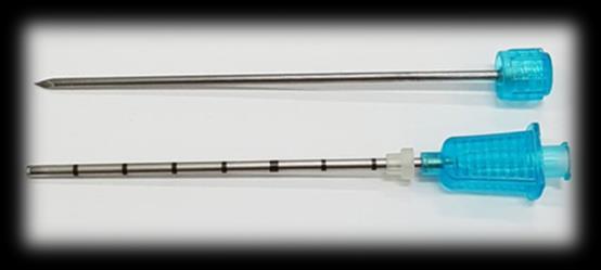
25 Deep learning breast US(I) Malignant(invasive) vs benign neoplasm Training 5,095 validation 1,100 images from Park Surgical Clinic Keras with Tensorflow backend. Various models and accuracy -Inception v3: 87% -VGG19: 87% -ResNet50: 82% -Xception: 92% Sensitivity 84% and Specificity 97% Sensitivity 91% and Specificity 86%
26 Deep learning US(II) Xception +Top layer, 2 GPU minibatch20, 50 epoch Images from Park Surgical Clinic -training: 54,748(malignant(Invasive+ DCIS): 6,964) benign( FA, FCD, ADH, Normal) -validation13,600(20%) Accuracy: 92% Sensitivity: 68% Specificity: 95%
27 Quantitative shear wave ultrasound elastography: initial experience in solid breast masses Using the Aixplorer ultrasound system (SuperSonic Imagine, Aix en Provence, France), 53 solid breast lesions were identified in 52 consecutive patients. Mean elasticity cut off value of 50 kilopascals (kpa) was selected for benign/malignant differentiation. Shear wave elastography versus greyscale BI-RADS performance figures were sensitivity: 97% vs 87%, specificity: 83% vs 78%, positive predictive value (PPV): 88% vs 84%, negative predictive value (NPV): 95% vs 82% and accuracy: 91% vs 83% respectively. These differences were not statistically significant. Evans et al. Breast Cancer Research 2010, 12:R104
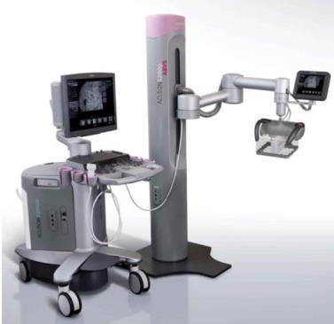
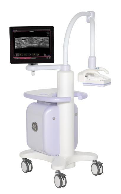
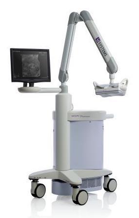
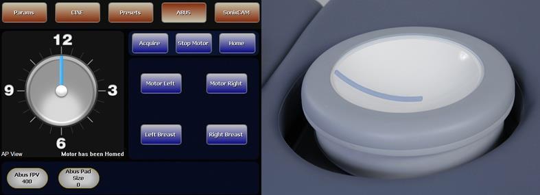
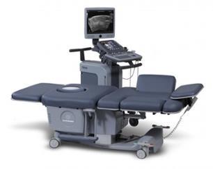
28 SonixEmbrace U system Somo-V Siemens ABVS GE invenia Techniscan Warmbath


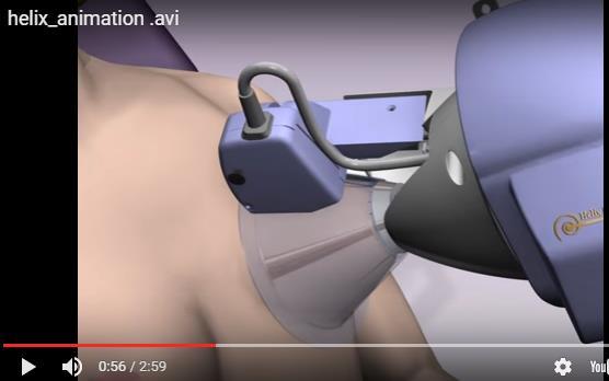
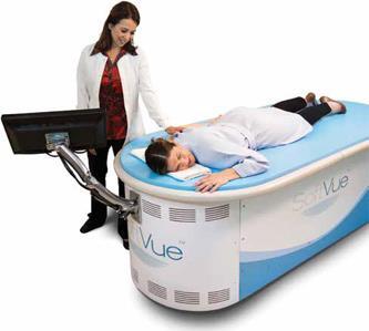
29 Hitachi Sofia Delphinus SoftVue Reflection Attenuation Transmission Caperay US & x-ray 고해상도고조도상화술


30 MammouS

31 MammouS CAD -Segmentation
32 MRI Cat 4 lesion : Korean Health Insurance reimbuse. Multiple cystic and mass in both breast Cancer patients.


33 6 mm in MRI and Second look US Path: 3 mm DCIS with Bexcore
34 Breast Biopsy Options Invasive Open Surgical Breast Biopsy Minimally Invasive FNAB Breast cytology Core Needle Breast Biopsy Vacuum Assisted Breast Biopsy


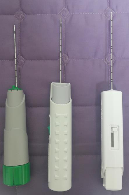
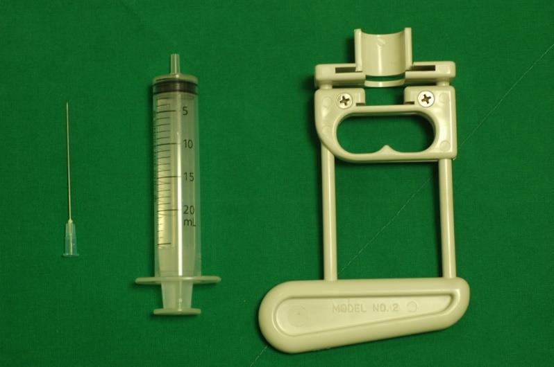
35 FNAB Core Biopsy Gun Biopsy
36 Rebiopsy(surgical excision) after Percutaneous Biopsy - ADH - Phyllodes tumor - Discordance between imaging and histologic findings - Inadequate specimens - Controversy in papillary lesion radial scar atypical lobular hyperplasia LCIS - Larger volume of tissue or more contiguous sampling : lower the rebiopsy rate 14G core biopsy: 15 % 11G Mammotome: 9 % Philpotts LE, AJR 1999

37 Shortcomings of standard automated core biopsy 29% of breast lesions are heterogeneous yielding different histologic results from targeted center and periphery, thus sampling only part of a heterogeneous mass result in a misdiagnosis Morris et al Breast J 2002 Core biopsy findings of ADH underestimate the diagnosis of malignancy by 18 88% Joshi et al Breast J 2001
38
39


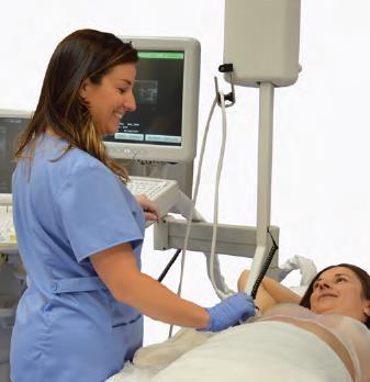
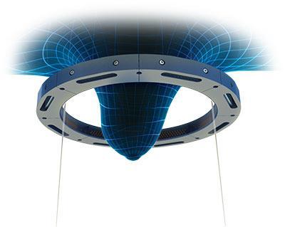
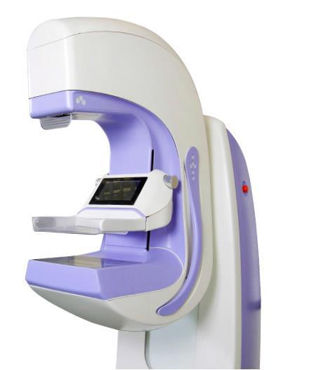
40 Breast related instruments and equipment


41 Thank You. Park Surgical Clinic Medical Park Ltd.
Diagnostic benefits of ultrasound-guided. CNB) versus mammograph-guided biopsy for suspicious microcalcifications. without definite breast mass
 versus mammograph-guided biopsy for suspicious microcalcifications. without definite breast mass") Volume 118 No. 19 2018, 531-543 ISSN: 1311-8080 (printed version); ISSN: 1314-3395 (on-line version) url: http://www.ijpam.eu ijpam.eu Diagnostic benefits of ultrasound-guided biopsy versus mammography-guided
Volume 118 No. 19 2018, 531-543 ISSN: 1311-8080 (printed version); ISSN: 1314-3395 (on-line version) url: http://www.ijpam.eu ijpam.eu Diagnostic benefits of ultrasound-guided biopsy versus mammography-guided
Mammographic imaging of nonpalpable breast lesions. Malai Muttarak, MD Department of Radiology Chiang Mai University Chiang Mai, Thailand
 Mammographic imaging of nonpalpable breast lesions Malai Muttarak, MD Department of Radiology Chiang Mai University Chiang Mai, Thailand Introduction Contents Mammographic signs of nonpalpable breast cancer
Pitfalls and Limitations of Breast MRI. Susan Orel Roth, MD Professor of Radiology University of Pennsylvania
 Pitfalls and Limitations of Breast MRI Susan Orel Roth, MD Professor of Radiology University of Pennsylvania Objectives Review the etiologies of false negative breast MRI examinations Discuss the limitations
Atypical Ductal Hyperplasia and Papillomas: A Comparison of Ultrasound Guided Breast Biopsy and Stereotactic Guided Breast Biopsy
 Atypical Ductal Hyperplasia and Papillomas: A Comparison of Ultrasound Guided Breast Biopsy and Stereotactic Guided Breast Biopsy Breast Cancer is the most common cancer diagnosed in women in the United
Does elastography change the indication to biopsy? IBDC
 Does elastography change the indication to biopsy? A LEXANDRA A THANASIOU, M D DEPARTMENT OF RADIOLOGY CURIE INSTITUTE PARIS, FRANCE IBDC Ultrasound Detected Cancers Physician-performed ultrasound increases
Diagnostic Dilemmas of Breast Imaging
 Diagnostic Dilemmas of Breast Imaging Common Causes of Error in Breast Cancer Detection By: Jason Cord, M.D. Mammography: Initial Imaging The standard for detection of breast cancer Screening mammography
CNB vs Surgical Excision
 Update on Core Needle Biopsy of Non-palpable Breast Lesions Nour Sneige, M.D. UT MD Anderson Cancer Center Houston, Tx Image-Guided CNB of Breast Lesions An alternative to surgical biospy CNB vs Surgical
Treatment options for the precancerous Atypical Breast lesions. Prof. YOUNG-JIN SUH The Catholic University of Korea
 Treatment options for the precancerous Atypical Breast lesions Prof. YOUNG-JIN SUH The Catholic University of Korea Not so benign lesions? Imaging abnormalities(10% recall) lead to diagnostic evaluation,
Vacuum-assisted breast biopsy using computer-aided 3.0 T- MRI guidance: diagnostic performance in 173 lesions
 Vacuum-assisted breast biopsy using computer-aided 3.0 T- MRI guidance: diagnostic performance in 173 lesions Poster No.: C-2870 Congress: ECR 2017 Type: Scientific Exhibit Authors: A. Pozzetto, L. Camera,
Atypical ductal hyperplasia diagnosed at ultrasound guided biopsy of breast mass
 Atypical ductal hyperplasia diagnosed at ultrasound guided biopsy of breast mass Poster No.: C-1483 Congress: ECR 2014 Type: Authors: Keywords: DOI: Scientific Exhibit J. Cho, J. Chung, E. S. Cha, J. E.
Imaging in breast cancer. Mammography and Ultrasound Donya Farrokh.MD Radiologist Mashhad University of Medical Since
 Imaging in breast cancer Mammography and Ultrasound Donya Farrokh.MD Radiologist Mashhad University of Medical Since A mammogram report is a key component of the breast cancer diagnostic process. A mammogram
EARLY DETECTION: MAMMOGRAPHY AND SONOGRAPHY
 EARLY DETECTION: MAMMOGRAPHY AND SONOGRAPHY Elizabeth A. Rafferty, M.D. Avon Comprehensive Breast Center Massachusetts General Hospital Harvard Medical School Breast Cancer Screening Early detection of
Excisional biopsy or long term follow-up results in breast high-risk lesions diagnosed at core needle biopsy
 Excisional biopsy or long term follow-up results in breast high-risk lesions diagnosed at core needle biopsy Poster No.: C-2515 Congress: ECR 2015 Type: Authors: Scientific Exhibit Ö. S. Okcu 1, A. Oktay
EARLY DETECTION: MAMMOGRAPHY AND SONOGRAPHY
 EARLY DETECTION: MAMMOGRAPHY AND SONOGRAPHY Elizabeth A. Rafferty, M.D. Avon Comprehensive Breast Center Massachusetts General Hospital Harvard Medical School Breast Cancer Screening Early detection of
Mammographic features and correlation with biopsy findings using 11-gauge stereotactic vacuum-assisted breast biopsy (SVABB)
") Original article Annals of Oncology 14: 450 454, 2003 DOI: 10.1093/annonc/mdh088 Mammographic features and correlation with biopsy findings using 11-gauge stereotactic vacuum-assisted breast biopsy (SVABB)
Original article Annals of Oncology 14: 450 454, 2003 DOI: 10.1093/annonc/mdh088 Mammographic features and correlation with biopsy findings using 11-gauge stereotactic vacuum-assisted breast biopsy (SVABB)
Case study 1. Rie Horii, M.D., Ph.D. Division of Pathology Cancer Institute Hospital, Japanese Foundation for Cancer Research
 NCCN/JCCNB Seminar in Japan April 15, 2012 Case study 1 Rie Horii, M.D., Ph.D. Division of Pathology Cancer Institute Hospital, Japanese Foundation for Cancer Research Present illness: A 50y.o.premenopausal
PURPOSE IMAGE-GUIDANCE MODALITIES IMAGE-GUIDED BREAST BIOPSY. US-Techniques. Ultrasound. US guided NLOBB. TH. Helbich
 IMAGE-GUIDED BREAST BIOPSY PURPOSE TH. Helbich Department of Radiology Division of Molecular & Gender Imaging Medical University of Vienna Imaging techniques Interventional procedures Quality management
Lesion Imaging Characteristics Mass, Favoring Benign Circumscribed Margins Intramammary Lymph Node
 Lesion Imaging Characteristics Mass, Favoring Benign Circumscribed Margins Intramammary Lymph Node Oil Cyst Mass, Intermediate Concern Microlobulated Margins Obscured Margins Mass, Favoring Malignant Indistinct
Underestimation of Atypical Ductal Hyperplasia at Sonographically Guided Core Biopsy of the Breast
 Women s Imaging Original Research Jang et al. Sonographic Breast Biopsy Women s Imaging Original Research WOMEN S IMAGING Underestimation of Atypical Ductal Hyperplasia at Sonographically Guided Core Biopsy
Breast pathology. 2nd Department of Pathology Semmelweis University
 Breast pathology 2nd Department of Pathology Semmelweis University Breast pathology - Summary - Benign lesions - Acute mastitis - Plasma cell mastitis / duct ectasia - Fat necrosis - Fibrocystic change/
Current Status of Supplementary Screening With Breast Ultrasound
 Current Status of Supplementary Screening With Breast Ultrasound Stephen A. Feig, M.D., FACR Fong and Jean Tsai Professor of Women s Imaging Department of Radiologic Sciences University of California,
Since its introduction in 2000, digital mammography has become
 Review Article Smith A, PhD email : Andrew.smith@hologic.com Since its introduction in 2000, digital mammography has become an accepted standard of care in breast cancer screening and has paved the way
Breast Cancer Imaging
 Breast Cancer Imaging I. Policy University Health Alliance (UHA) will cover breast imaging when such services meet the medical criteria guidelines (subject to limitations and exclusions) indicated below.
BI-RADS CATEGORIZATION AND BREAST BIOPSY categorization in the selection of appropriate breast biopsy technique is also discussed. Patients and method
 Original Article Positive Predictive Value of BI-RADS Categorization in an Asian Population Yah-Yuen Tan, Siew-Bock Wee, Mona P.C. Tan and Bee-Kiang Chong, 1 Departments of General Surgery and 1Diagnostic
Non-mass Enhancement on Breast MRI. Aditi A. Desai, MD Margaret Ann Mays, MD
 Non-mass Enhancement on Breast MRI Aditi A. Desai, MD Margaret Ann Mays, MD Breast MRI Important screening and diagnostic tool, given its high sensitivity for breast cancer detection Breast MRI - Indications
Utility of Adequate Core Biopsy Samples from Ultrasound Biopsies Needed for Today s Breast Pathology
 Utility of Adequate Core Biopsy Samples from Ultrasound Biopsies Needed for Today s Breast Pathology Ugur Ozerdem, M.D. 1 Abstract Background: There is a paradigm shift in breast biopsy philosophy. In
Sonographically-Guided 14-Gauge Core Needle Biopsy for Papillary Lesions of the Breast
 Sonographically-Guided 14-Gauge Core Needle Biopsy for Papillary Lesions of the Breast Eun Sook Ko, MD Nariya Cho, MD Joo Hee Cha, MD Jeong Seon Park, MD Sun Mi Kim, MD Woo Kyung Moon, MD Index terms:
Ge elastography cpt codes
 Ge elastography cpt codes Aetna considers digital mammography a medically necessary acceptable alternative to film mammography. Currently, there are no guideline recommendations from leading medical professional
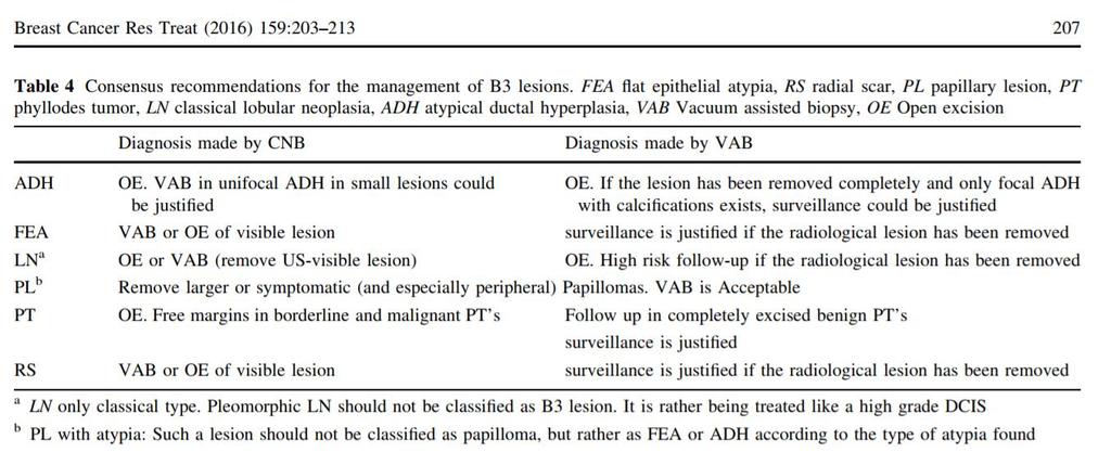
Guidance on the management of B3 lesions
 Guidance on the management of B3 lesions Lesion diagnosed on 14g or vacuumassisted biopsy (VAB) Risk of upgrade Recommended investigation Suggested approach for follow-up if no malignancy on VAE awaiting
Stereotactic 11-Gauge Vacuum- Assisted Breast Biopsy: A Validation Study
 Georg Pfarl 1 Thomas H. Helbich 1 Christopher C. Riedl 1 Teresa Wagner 2 Michael Gnant 3 Margaretha Rudas 4 Laura Liberman 5 Received March 11, 2002; accepted after revision May 17, 2002. 1 Department
Breast Lesion Excision System-Intact (BLES): A Stereotactic Method of Biopsy of Suspicius Non-Palpable Mammographic Lesions.
: A Stereotactic Method of Biopsy of Suspicius Non-Palpable Mammographic Lesions.") Breast Lesion Excision System-Intact (BLES): A Stereotactic Method of Biopsy of Suspicius Non-Palpable Mammographic Lesions. Poster No.: C-1595 Congress: ECR 2014 Type: Authors: Scientific Exhibit I. Georgiou
Breast Lesion Excision System-Intact (BLES): A Stereotactic Method of Biopsy of Suspicius Non-Palpable Mammographic Lesions. Poster No.: C-1595 Congress: ECR 2014 Type: Authors: Scientific Exhibit I. Georgiou
IBCM 2, April 2009, Sarajevo, Bosnia and Herzegovina
 Preoperative diagnosis and treatment planning in breast cancer The pathologist s perspective L. Mazzucchelli Istituto Cantonale di Patologia Locarno, Switzerland IBCM 2, 23-25 April 2009, Sarajevo, Bosnia
Radiologic and pathologic correlation of non-mass like breast lesions on US and MRI: Benign, high risk, versus malignant
 Radiologic and pathologic correlation of non-mass like breast lesions on US and MRI: Benign, high risk, versus malignant Poster No.: C-1161 Congress: ECR 2013 Type: Educational Exhibit Authors: J. Kwak,
CURRENT METHODS IN IMAGE GUIDED BREAST BIOPSY
 CURRENT METHODS IN IMAGE GUIDED BREAST BIOPSY Stuart Silver April 24, 2004 OBJECTIVES Review development of current techniques Discuss stereotactic breast biopsy Discuss US guided breast biopsy 1 OBJECTIVES
Compressive Re-Sampling for Speckle Reduction in Medical Ultrasound
 Compressive Re-Sampling for Speckle Reduction in Medical Ultrasound Professor Richard Mammone Rutgers University Email Phone Number Christine Podilchuk, Lev Barinov, Ajit Jairaj and William Hulbert ClearView
New Imaging Modalities for better Screening and Diagnosis
 New Imaging Modalities for better Screening and Diagnosis Miri Sklair-Levy, MD Department of Diagnostic Imaging Sheba Medical Center, Sackler School of Medicine, Tel Aviv University Department of Diagnostic
ACRIN 6666 Therapeutic Surgery Form
 S1 ACRIN 6666 Therapeutic Surgery Form 6666 Instructions: Complete a separate S1 form for each separate area of each breast excised with the intent to treat a cancer (e.g. each lumpectomy or mastectomy).
BREAST MRI. Elizabeth A. Rafferty, M.D. Avon Comprehensive Breast Center Massachusetts General Hospital Harvard Medical School
 BREAST MRI Elizabeth A. Rafferty, M.D. Avon Comprehensive Breast Center Massachusetts General Hospital Harvard Medical School BREAST MRI Any assessment of the breast parenchyma requires the administration
BREAST PATHOLOGY. Fibrocystic Changes
 BREAST PATHOLOGY Lesions of the breast are very common, and they present as palpable, sometimes painful, nodules or masses. Most of these lesions are benign. Breast cancer is the 2 nd most common cause
Emerging Techniques in Breast Imaging: Contrast-Enhanced Mammography and Fast MRI
 Emerging Techniques in Breast Imaging: Contrast-Enhanced Mammography and Fast MRI Lilian Wang, M.D. Breast Imaging Section Department of Radiology Northwestern Medicine Overview Rationale for new imaging
Breast Imaging Update: Old Dog New Tricks
 Breast Imaging Update: Old Dog New Tricks Claire McKay, DO M&S Imaging Assoc. San Antonio, TX cmckayhart@juno.com Goals Describe modalities available, old and new Provide understanding of pros and cons
BARC/2013/E/019 BARC/2013/E/019. AUDIT OF MAMMOGRAPHY PERFORMED IN OUR HOSPITAL by Surita Kantharia Medical Division
 BARC/2013/E/019 BARC/2013/E/019 AUDIT OF MAMMOGRAPHY PERFORMED IN OUR HOSPITAL by Surita Kantharia Medical Division BARC/2013/E/019 GOVERNMENT OF INDIA ATOMIC ENERGY COMMISSION BARC/2013/E/019 AUDIT OF
Mammographic evaluation of palpable breast masses with pathological correlation: a tertiary care centre study in Nepal
 Original article 21 Mammographic evaluation of palpable breast masses with pathological correlation: a tertiary care centre study in Nepal G. Gurung, R. K. Ghimire, B. Lohani Department of Radiology and
Case #1: 75 y/o Male (treated and followed by prostate cancer oncology specialist ).
.") SOLID TUMORS WORKSHOP Cases for review Prostate Cancer Case #1: 75 y/o Male (treated and followed by prostate cancer oncology specialist ). January 2009 PSA 4.4, 20% free; August 2009 PSA 5.2; Sept 2009
SOLID TUMORS WORKSHOP Cases for review Prostate Cancer Case #1: 75 y/o Male (treated and followed by prostate cancer oncology specialist ). January 2009 PSA 4.4, 20% free; August 2009 PSA 5.2; Sept 2009
Can magnetic resonance imaging obviate the need for biopsy for microcalcifications?
 Original Article Can magnetic resonance imaging obviate the need for biopsy for microcalcifications? Shinya Yamamoto, Takashi Chishima Department of Breast Surgery, Yokohama Rosai Hospital, Yokohama 222-0036,
ROLE OF MRI IN SCREENING, DIAGNOSIS AND MANAGEMENT OF BREAST CANCER. B.Zandi Professor of Radiology
 ROLE OF MRI IN SCREENING, DIAGNOSIS AND MANAGEMENT OF BREAST CANCER B.Zandi Professor of Radiology Introduction In the USA, Breast Cancer is : The Most Common Non-Skin Cancer The Second Leading cause of
3/27/2017. Disclosure of Relevant Financial Relationships. Papilloma???
 Management of Papillary Lesions Diagnosed at Rad Path Concordant Core Biopsy (CNB) Disclosure of Relevant Financial Relationships USCAP requires that all planners (Education Committee) in a position to
Mammography is a most effective imaging modality in early breast cancer detection. The radiographs are searched for signs of abnormality by expert
 Abstract Methodologies for early detection of breast cancer still remain an open problem in the Research community. Breast cancer continues to be a significant problem in the contemporary world. Nearly
Diagnostic accuracy of ultrasonography-guided core needle biopsy for breast lesions
 Singapore Med J 01; 5(1) 40 Diagnostic accuracy of ultrasonography-guided core needle biopsy for breast lesions Wiratkapun Cl, MD, Treesit T1, MD, Wibulpolprasert E1, MD, Lertsithichai P, MD, MSc INTRODUCTION
Effective Health Care Program
 Comparative Effectiveness Review Number 19 Effective Health Care Program Comparative Effectiveness of Core-Needle and Open Surgical Biopsy for the Diagnosis of Breast Lesions Executive Summary Background
Imaging-Guided Core Needle Biopsy of Papillary Lesions of the Breast
 Eric L. Rosen 1 Rex C. Bentley 2 Jay A. Baker 1 Mary Scott Soo 1 Received January 30, 2002; accepted after revision April 12, 2002. 1 Department of Radiology, Breast Imaging Division, Duke University Medical
Breast Evaluation & Management Guidelines
 Breast Evaluation & Management Guidelines Pamela L. Kurtzhals, M.D. F.A.C.S. Head, Dept. of General Surgery Scripps Clinic, La Jolla Objective Review screening & diagnostic guidelines Focused patient complaints
Breast Cancer Screening and Diagnosis
 Breast Cancer Screening and Diagnosis Priya Thomas, MD Assistant Professor Clinical Cancer Prevention and Breast Medical Oncology University of Texas MD Anderson Cancer Center Disclosures Dr. Thomas has
Here are examples of bilateral analog mammograms from the same patient including CC and MLO projections.
 Good afternoon. It s my pleasure to be discussing Diagnostic Breast Imaging over the next half hour. I m Wei Yang, Professor of Diagnostic Radiology and Chief, the Section of Breast Imaging as well as
Breast Cancer. Most common cancer among women in the US. 2nd leading cause of death in women. Mortality rates though have declined
 Breast Cancer Most common cancer among women in the US 2nd leading cause of death in women Mortality rates though have declined 1 in 8 women will develop breast cancer Breast Cancer Breast cancer increases
Image guided core biopsies:
 Recommendations on the Surgical, Radiologic and Pathologic Approaches to Breast Disease: Using best practices based on multidisciplinary methodologies developed through the Allina Breast Committee. Image
04/10/2018. Intraductal Papillary Neoplasms Of Breast INTRADUCTAL PAPILLOMA
 Intraductal Papillary Neoplasms Of Breast Savitri Krishnamurthy MD Professor of Pathology Deputy Division Head The University of Texas MD Anderson Cancer Center 25 th Annual Seminar in Pathology Pittsburgh,
Breast Imaging! Ravi Adhikary, MD!
 Breast Imaging! Ravi Adhikary, MD! ACS Estimated Cancers Statistics 2014! Breast! New Cases in Women! 232,670 (+67,570 in situ)! Deaths in Women! 40,000! Colon! 48,380! 24,040! Cervical! 12,360! 4,020!
ShearWave elastography in lymph nodes
 ShearWave elastography in lymph nodes Poster No.: B-0158 Congress: ECR 2015 Type: Authors: Keywords: DOI: Scientific Paper F. Houari, O. Lucidarme, J. Gabarre, F. Charlotte, C. Pellot- Barakat, M. Lefort,
Macrobiopsy under X-Ray Guidance
 Macrobiopsy under X-Ray Guidance C. Balleyguier, B. Boyer Radiology Gustave Roussy, Villejuif, France Breast Intervention Imaging Major domain in breast imaging European guidelines recommend a pre surgical
BREAST MRI. Elizabeth A. Rafferty, M.D. Avon Comprehensive Breast Center Massachusetts General Hospital Harvard Medical School
 BREAST MRI Elizabeth A. Rafferty, M.D. Avon Comprehensive Breast Center Massachusetts General Hospital Harvard Medical School BREAST MRI Any assessment of the breast parenchyma requires the administration
Mammography and Other Screening Tests. for Breast Problems
 301.681.3400 OBGYNCWC.COM Mammography and Other Screening Tests What is a screening test? for Breast Problems A screening test is used to find diseases, such as cancer, in people who do not have signs
BREAST MRI. VASILIKI FILIPPI RADIOLOGIST CT MRI & PET/CT Departments Hygeia Hospital, Athens, Greece
 BREAST MRI VASILIKI FILIPPI RADIOLOGIST CT MRI & PET/CT Departments Hygeia Hospital, Athens, Greece Breast ΜR Imaging (MRM) Breast MR imaging is an extremely powerful diagnostic tool, that when used in
Minimizing Errors in Diagnostic Pathology
 Shahla Masood, M.D. Professor and Chair Department of Pathology and Laboratory Medicine University of Florida College of Medicine-Jacksonville Medical Director, Shands Jacksonville Breast Health Center
Surgical Pathology Issues of Practical Importance
 Surgical Pathology Issues of Practical Importance Anne Moore, MD Medical Oncology Syed Hoda, MD Surgical Pathology The pathologist is central to the team approach needed to manage the patient with breast
AMSER Case of the Month: November 2018
 AMSER Case of the Month: November 2018 52 year old female with an abnormal screening mammogram Areeg Rehman, MS 4 Nova Southeastern University Rebecca T. Sivarajah, MD Penn State University College of
CPC 4 Breast Cancer. Rochelle Harwood, a 35 year old sales assistant, presents to her GP because she has noticed a painless lump in her left breast.
 CPC 4 Breast Cancer Rochelle Harwood, a 35 year old sales assistant, presents to her GP because she has noticed a painless lump in her left breast. 1. What are the most likely diagnoses of this lump? Fibroadenoma
Detailed Program of the second BREAST IMAGING AND INTERVENTIONS PROGRAM am am : Clinician s requirements from breast imaging
 Detailed Program of the second BREAST IMAGING AND INTERVENTIONS PROGRAM 2012 Day one, 2 nd November BREAST IMAGING AND INTERVENTIONS PROGRAM 2012 9.00 AM 9.10 am Introduction 9.10 am - 9.30 am : Clinician
Deep-Learning Based Semantic Labeling for 2D Mammography & Comparison of Complexity for Machine Learning Tasks
 Deep-Learning Based Semantic Labeling for 2D Mammography & Comparison of Complexity for Machine Learning Tasks Paul H. Yi, MD, Abigail Lin, BSE, Jinchi Wei, BSE, Haris I. Sair, MD, Ferdinand K. Hui, MD,
DIAGNOSIS. Biopsy, Pathology and Subtypes. Knowledge Summary
 DIAGNOSIS Biopsy, Pathology and Subtypes Knowledge Summary DIAGNOSIS Biopsy, Pathology and Subtypes INTRODUCTION The success of an effective breast health care program is directly related to the availability
Quality ID #263: Preoperative Diagnosis of Breast Cancer National Quality Strategy Domain: Effective Clinical Care
 Quality ID #263: Preoperative Diagnosis of Breast Cancer National Quality Strategy Domain: Effective Clinical Care 2018 OPTIONS FOR INDIVIDUAL MEASURES: REGISTRY ONLY MEASURE TYPE: Process DESCRIPTION:
MR sin plass i brystkreftdiagnostikk, dagens anbefalinger og fremtidsperspektiver
 MR sin plass i brystkreftdiagnostikk, dagens anbefalinger og fremtidsperspektiver Kathinka Kurz, MD, PhD, seksjonsoverlege SUS, kathinka.dehli.kurz@sus.no Technique - Subtraction Without contrast agent
Interpretation of Breast Pathology in the Era of Minimally Invasive Procedures
 Shahla Masood, M.D. Professor and Chair Department of Pathology and Laboratory Medicine University of Florida College of Medicine Jacksonville Medical Director, UF Health Breast Center Chief of Pathology
National Diagnostic Imaging Symposium 2013 SAM - Breast MRI 1
 National Diagnostic Imaging Symposium 2013 December 8-12, 2013 Disney s Yacht Club Resort Lake Buena Vista, Florida Self Assessment Module Questions, Answers and References Day SAM Title - Each SAM title
Sonographically Guided Core Biopsy of the Breast: Comparison of 14-Gauge Automated Gun and 11-Gauge Directional Vacuum-Assisted Biopsy Methods
 Sonographically Guided Core Biopsy of the Breast: Comparison of 14-Gauge Automated Gun and 11-Gauge Directional Vacuum-Assisted Biopsy Methods Nariya Cho, MD 1 Woo Kyung Moon, MD 1 Joo Hee Cha, MD 1 Sun
Screening with Abbreviated Breast MRI (AB-MR)
") Screening with Abbreviated Breast MRI (AB-MR) Christopher Comstock, MD, FACR, FSBI Department of Radiology Memorial Sloan-Kettering Cancer Center New York, NY Outline History of our approach to screening
Screening with Abbreviated Breast MRI (AB-MR) Christopher Comstock, MD, FACR, FSBI Department of Radiology Memorial Sloan-Kettering Cancer Center New York, NY Outline History of our approach to screening
04/10/2018 HIGH RISK BREAST LESIONS. Pathology Perspectives of High Risk Breast Lesions ELEVATED RISK OF BREAST CANCER HISTORICAL PERSPECTIVES
 Pathology Perspectives of High Risk Breast Lesions Savitri Krishnamurthy MD Professor of Pathology Deputy Division Head Director of Clinical Trials, Research and Development The University of Texas MD
Artificial Intelligence in Breast Imaging
 Artificial Intelligence in Breast Imaging Manisha Bahl, MD, MPH Director of Breast Imaging Fellowship Program, Massachusetts General Hospital Assistant Professor of Radiology, Harvard Medical School Outline
Case Scenario 1: This case has been slightly modified from the case presented during the live session to add clarity.
 Case Scenario 1: This case has been slightly modified from the case presented during the live session to add clarity. Background: 46 year old married premenopausal female with dense breasts has noticed
Breast Cancer. Saima Saeed MD
 Breast Cancer Saima Saeed MD Breast Cancer Most common cancer among women in the US 2nd leading cause of death in women 1 in 8 women will develop breast cancer Incidence/mortality rates have declined Breast
Mammography Education, Inc.
 Hands-on Breast Screening and Diagnosis Course * Screening of 315 full field digital mammography cases. * Reading a mixture of normals and proven abnormals at high resolution viewing stations. * Immediate
Breast Pathology. Breast Development
 Breast Pathology Lecturer: Hanina Hibshoosh, M.D. Reading: Kumar, Cotran, Robbins, Basic Pathology, 6th Edition, pages 623-635 Breast Development 5th week - thickening of the epidermis - milk line 5th
STEREOTACTIC BREAST BIOPSY: CORRELATION WITH HISTOLOGY
 3-rd Baltic Congress of Radiology, October 8-9, 2010 Riga Rūta Briedienė, Rūta Grigienė, Raimundas Meškauskas Institute of Oncology Vilnius University, National Centre of Pathology STEREOTACTIC BREAST
BCSC Glossary of Terms (Last updated 09/16/2009) DEFINITIONS
 DEFINITIONS") Screening mammography scrmam_c BCSC Glossary of Terms (Last updated 09/16/2009) DEFINITIONS The radiologist s indication for exam is the primary determinant of whether a mammogram is screening or diagnostic.
Screening mammography scrmam_c BCSC Glossary of Terms (Last updated 09/16/2009) DEFINITIONS The radiologist s indication for exam is the primary determinant of whether a mammogram is screening or diagnostic.
Cystic Hypersecretory Carcinoma of the Breast:
 J Korean Soc Radiol 2010;62:287-294 Cystic Hypersecretory Carcinoma of the Breast: Sonographic Features with a Histological Correlation 1 Sang Yu Nam, M.D., Boo-Kyung Han, M.D., Jung Hee Shin, M.D., Eun
Ana Sofia Preto 19/06/2013
 Ana Sofia Preto 19/06/2013 Understanding the underlying pathophysiologic processes leading to the various types of calcifications Description and illustration of the several types of calcifications, according
Papillary lesions of the breast - Imaging findings and diagnostic challenges
 Papillary lesions of the breast - Imaging findings and diagnostic challenges Poster No.: R-0146 Congress: RANZCR-AOCR 2012 Type: Educational Exhibit Authors: P. Jagmohan, F. J. Pool Keywords: Breast, Mammography,
Breast Health and Imaging Glossary
 Contact: Lorna Vaughan HerSpace Breast Imaging & Biopsy Associates 300 State Route 35 South W. Long Branch, NJ 07764 732-571-9100, ext. 104 lorna@breast-imaging.com Breast Health and Imaging Glossary Women
Case Scenario 1: This case has been slightly modified from the case presented during the live session to add clarity.
 Case Scenario 1: This case has been slightly modified from the case presented during the live session to add clarity. Background: 46 year old married premenopausal female with dense breasts has noticed
TMIST: Frequently Asked Questions
 TMIST: Frequently Asked Questions Key Topics for Site Investigators and Staff This document answers frequently asked questions about the Tomosynthesis Mammographic Imaging Screening Trial (TMIST/EA1151);
MEDICAL POLICY SUBJECT: MAMMOGRAPHY: COMPUTER- AIDED DETECTION (CAD) POLICY NUMBER: CATEGORY: Technology Assessment
 POLICY NUMBER: CATEGORY: Technology Assessment") MEDICAL POLICY SUBJECT: MAMMOGRAPHY: COMPUTER- PAGE: 1 OF: 5 If a product excludes coverage for a service, it is not covered, and medical policy criteria do not apply. If a commercial product, including
MEDICAL POLICY SUBJECT: MAMMOGRAPHY: COMPUTER- PAGE: 1 OF: 5 If a product excludes coverage for a service, it is not covered, and medical policy criteria do not apply. If a commercial product, including
Ultrasound of the Breast BASICS FOR THE ORDERING CLINICIAN
 Ultrasound of the Breast BASICS FOR THE ORDERING CLINICIAN Breast Ultrasound Anatomy Skin Breast Parenchyma Pectoralis Fascia Pectoralis Breast Ultrasound Anatomy Indications for Breast Ultrasound Palpable
BreastScreen Victoria Annual Statistical Report
 BreastScreen Victoria Annual Statistical Report 005 Produced by: BreastScreen Victoria Coordination Unit Level, Pelham Street, Carlton South Victoria 05 PH 0 9660 6888 FX 0 966 88 EM info@breastscreen.org.au
Screening Mammograms: Questions and Answers
 CANCER FACTS N a t i o n a l C a n c e r I n s t i t u t e N a t i o n a l I n s t i t u t e s o f H e a l t h D e p a r t m e n t o f H e a l t h a n d H u m a n S e r v i c e s Screening Mammograms:
Radiologic Findings of Mucocele-like Tumors of the breast: Can we differentiate pure benign from associated with high risk lesions?
 Radiologic Findings of Mucocele-like Tumors of the breast: Can we differentiate pure benign from associated with high risk lesions? Poster No.: C-0332 Congress: ECR 2014 Type: Educational Exhibit Authors:
The Radiology Aspects
 REQUIREMENTS FOR INTERNATIONAL ACCREDITATION OF BREAST CENTERS/UNITS The Radiology Aspects Miri Sklair-Levy, Israel RADIOLOGY GUIDELINES FOR QUALITY ASSURANCE IN BREAST CANCER SCREENING AND DIAGNOSIS Radiologists
A comparison of the accuracy of film-screen mammography, full-field digital mammography, and digital breast tomosynthesis
 Clinical Radiology xxx (2012) 1e6 Contents lists available at SciVerse ScienceDirect Clinical Radiology journal homepage: www.clinicalradiologyonline.net A comparison of the accuracy of film-screen mammography,
CLINICAL SIGNIFICANCE OF BENIGN EPITHELIAL CHANGES
 Papillomas. Papillomas are composed of multiple branching fibrovascular cores, each having a connective tissue axis lined by luminal and myoepithelial cells ( Fig. 23-11 ). Growth occurs within a dilated
Look differently. Invenia ABUS. Automated Breast Ultrasound
 Look differently. Invenia ABUS Automated Breast Ultrasound InveniaTM ABUS from GE Healthcare offers a view beyond mammography, with breast screening technology that looks differently. 40 % The unseen risk.