MR sin plass i brystkreftdiagnostikk, dagens anbefalinger og fremtidsperspektiver
|
|
|
- Joella Underwood
- 5 years ago
- Views:
Transcription

1 MR sin plass i brystkreftdiagnostikk, dagens anbefalinger og fremtidsperspektiver Kathinka Kurz, MD, PhD, seksjonsoverlege SUS, kathinka.dehli.kurz@sus.no


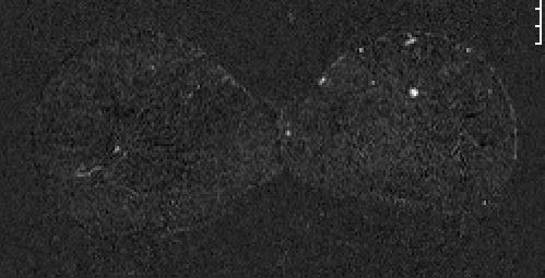
2 Technique - Subtraction Without contrast agent 2 min after i.v. injection of gadolinium containing contrast agent Subtraction (contrast series minus unenhanced series, pixel by pixel)


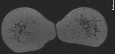
3 Technique Fat suppression Without contrast agent 2 min after i.v. injection of gadolinium containing contrast agent Subtraction (contrast series minus unenhanced series, pixel by pixel)
4 Chocolate Hills in Bohol, Philippines
5 Signal intensity [%] Initial phase Delayed phase Fast Persistent (I) Plateau (II) Medium Wash out (III) Slow Initial Phase: First two minutes after contrast agent injection or when curve starts to change. Time [min]

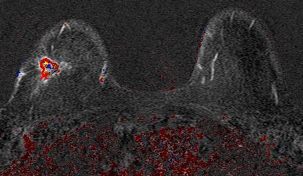
6 Irregular shaped mass with spiculated margins and inhomogeneous enhancement.
: Persistent")
7 Type I Time Intensity curve (TIC): Persistent rise
8 Type II TIC: Rapid rise and plateau
9 Type III TIC: Rapid rise and wash out
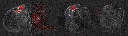
10 Why performe dynamic series?


11 Why performe dynamic series? 16 mm IDC G3
12 Crystal Hills, Hohenems, Australia
13 Indications - Preoperative: Rule out multifocal / multicentric growth of carcinoma prior to planned breast conserving therapy (especially by dense breasts and invasive lobular carcinoma) - Differentiate between scar and recurrent disease after breast conserving therapy - Control the tumor response by neoadjuvant chemoterapy - Carcinoma of unknown region - High risk
14 Indications - Preoperative: Rule out multifocal / multicentric growth of carcinoma prior to planned breast conserving therapy (especially by dense breasts and invasive lobular carcinoma) - Differentiate between scar and recurrent disease after breast conserving therapy - Control the tumor response by neoadjuvant chemoterapy - Carcinoma of unknown region - High risk


15 - Palpable lump 10 o clock right breast - Mammography normal - Sonography mass suspect of malignancy correlative to palpable lump - MRI mamma: Another mass suspect of malignancy at 4 o clock right breast - Histology: Multicentric invasiv lobular carcinoma
16 Local staging preoperative Main indication for breast conserving therapy: Small carcinomas compared to the size of the breast Main contraindication: Multicentric or multifocal growth of tumor MRI of the breast is more accurate than the combination of clinical examination, mammography and sonography
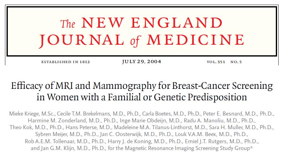
17 Local staging preoperative - 16% additional carcinomas in the ipsilateral breast (range 6-34%). 52% TP - 6% additional contralateral carcinomas (range 3-24%) - Invasive lobular carcinomas - Positive family history of breast cancer After clinical examination MG US Libermann L. Breast MR imaging in assessing extent of disease. Magn Reson Imaging Clin N Am Aug;14(3):339-49, vi (Review)
18 Local staging preoperative Especially important by lobular carcinoma patients with dense breasts high risk patients BUT: Discussion in the literature because there is not such a high recurrence rate (16%).
19 Indications - Preoperative: Rule out multifocal / multicentric growth of carcinoma prior to planned breast conserving therapy (especially by dense breasts and invasive lobular carcinoma) - Differentiate between scar and recurrent disease after breast conserving therapy - Control the tumor response by neoadjuvant chemoterapy - Carcinoma of unknown region - High risk
20 Scar Recurrent disease 1% local recurrence by breast conserving therapy per year. MRI of the breast diagnoses the recurrent carcinomas earlier than mammography and ultrasound On average 2-3 years after the operation, 5-15 mm size (Rieber 1997, Krämer 1998) BUT: The advantage of MRI is the high negative predictive value of 98.8%. Biopsy is not needed by negative MRI (93 patients) Preda L et al. Breast Cancer Res. 2006;8(5):R53


21 BCT 8 years ago. Aftercare. No signs of local recurrence.
22 BCT 14 years ago. Mammography slightly larger scar. Ultrasound: Lots of shadows.

23 BCT 14 years ago. Mammography slightly larger scar. Ultrasound: Lots of shadows. Local recurrence of invasive ductal carcinoma.
24 Indications - Preoperative: Rule out multifocal / multicentric growth of carcinoma prior to planned breast conserving therapy (especially by dense breasts and invasive lobular carcinoma) - Differentiate between scar and recurrent disease after breast conserving therapy - Control the tumor response by neoadjuvant chemoterapy - Carcinoma of unknown region - High risk
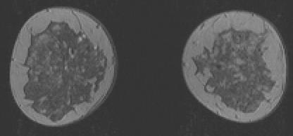
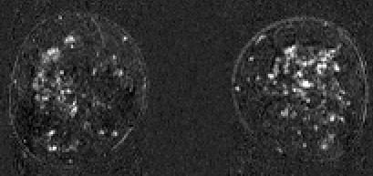
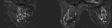
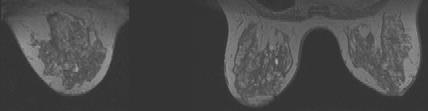
25 Neoadjuvant chemotherapy - The size can be measured clinically, mammographically and by ultrasound - MRI can be used to control the effect of the therapy - MRI of the breast shows the best correlation between preoperative measured and histologically measured size


26 46 yo patient with IDC G3 infiltrating the skin (ct4bn1biv M1).
27 Indications - Preoperative: Rule out multifocal / multicentric growth of carcinoma prior to planned breast conserving therapy (especially by dense breasts and invasive lobular carcinoma) - Differentiate between scar and recurrent disease after breast conserving therapy - Control the tumor response by neoadjuvant chemoterapy - Carcinoma of unknown region - High risk
28 Carcinoma of unknown primary Usually axillary lymph node metastasis, but also i.e. bone- or liver metastasis Clinical examination, mammography, ultrasound > 99% of the carcinomas in the breast are found MRI of the breast will find around 80% of the remaining breast carcinomas

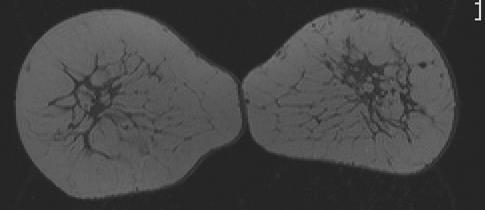
29 Axillary lymph node metastasis on the left side. No signs of malignancy by clinical examination, mammography and ultrasound.

30 Axillary lymph node metastasis on the left side. No signs of malignancy by clinical examination, mammography and ultrasound.
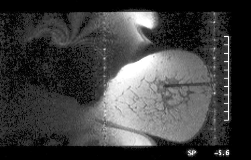
31 MRI-guided needle localization. 5 mm invasive ductal carcinoma
32 One of the few times one gets happy by diagnosing a carcinoma of the breast
33 Indications - Preoperative: Rule out multifocal / multicentric growth of carcinoma prior to planned breast conserving therapy (especially by dense breasts and invasive lobular carcinoma) - Differentiate between scar and recurrent disease after breast conserving therapy - Control the tumor response by neoadjuvant chemoterapy - Carcinoma of unknown region - High risk
34
35 Study overview Study n Ca. IVC n [%] Sensitivity [%] MX US MR PPV [%] MX US MR Kriege / Warner 2 236*/ Leach 3 649/ Kuhl 4 529/ Hagen 5 491*/ ** Riedl 6 327/ Kuhl 7 687/ Sardanelli 8 501/ * Mutation carriers only **Combined with ultrasound if clinical indicated 1) NEJM 2004; 351: ) Breast 2007; 16: ) JAMA 2004; 292: ) Clin Cancer Res 2007; 15: ) Lancet 2005; 365: ) J Clin Oncol 2010; 28: ) J Clin Oncol 2005;23: ) Invest Radiol 2011; 46:94-105
36 Study-differences Sample size Number of screening events Number of detected cancers Lifetime risk Age Single- or multicenter setting Clinical breast examination and ultrasound Exclusion of high-risk women with previous breast cancer BI-RADS 3: Positive or negative finding Study n Ca. IVC n [%] Sensitivity[%] MX US MR PPV [%] MX US MR Kriege / Warner 2 236*/ Leach 3 649/ Kuhl 4 529/ Hagen 5 491*/ ** Riedl 6 327/ Kuhl 7 687/ Sardanell 8 501/
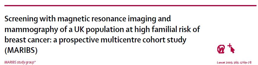
37 High risk screening studies Despite heterogeneity in the studies, results have been remarkably consistent with sensitivity of MRI between 71% 93% and 33% - 50% for mammography Study n Ca. IVC n [%] Sensitivity[%] MX US MR PPV [%] MX US MR Kriege / Warner 2 236*/ Leach 3 649/ Kuhl 4 529/ Hagen 5 491*/ ** Riedl 6 327/ Kuhl 7 687/ Sardanell 8 501/
38 Rapaälv, Sverige


39 Optional indications - Discordant findings by imaging and minimal invasive biopsy results - Screening of women with silicone implants and implant rupture evaluation

40 Rio negro, Brasil
 - First line examination of palpable masses - By hormone replacement treatment - In the")
41 When is MRI of the breast not expedient? - Without good indication (to expensive, to many false positives) - First line examination of palpable masses - By hormone replacement treatment - In the wrong phase of the menstrual cycle - Soon after operations, by or soon after radiation treatment only in special cases

42 Starry sky

43 Contraindications / difficulties - Contraindications against gadolinium containing contrast agent or MR - i.e. renal insufficiency, allergy, pregnancy - Pacemaker, cochlea implant - Magnetic expanders - Claustrophobia, difficulty with prone positioning - Adipositas per magna
44 Fremtidsperspektiver Diffusjonsvektet serie. Kontrastløs MR mamma. Screening? DCIS er allerede vist at en ser det bedre på MR mamma enn ved Mammografi og ultralyd (Kuhl CK, Lancet 2007) Enda bedre oppløsning => Bedre spesifisitet Mer av det!!

45 Summary MRI is a useful diagnostic tool by special indications. TAKK FOR OPPMERKSOMHETEN
BREAST MRI. Elizabeth A. Rafferty, M.D. Avon Comprehensive Breast Center Massachusetts General Hospital Harvard Medical School
 BREAST MRI Elizabeth A. Rafferty, M.D. Avon Comprehensive Breast Center Massachusetts General Hospital Harvard Medical School BREAST MRI Any assessment of the breast parenchyma requires the administration
Angela Gilliam, MD University of Colorado Surgical Grand Rounds November 3, 2008
 Angela Gilliam, MD University of Colorado Surgical Grand Rounds November 3, 2008 Breast Cancer Most common cancer in American women 180,000 new cases per year Second most common cause of cancer death 44,000
BREAST MRI. VASILIKI FILIPPI RADIOLOGIST CT MRI & PET/CT Departments Hygeia Hospital, Athens, Greece
 BREAST MRI VASILIKI FILIPPI RADIOLOGIST CT MRI & PET/CT Departments Hygeia Hospital, Athens, Greece Breast ΜR Imaging (MRM) Breast MR imaging is an extremely powerful diagnostic tool, that when used in
Breast MRI: Friend or Foe?
 Breast MRI: Friend or Foe? UCSF Postgraduate Course May 18, 2013 Cheryl Ewing, MD Clinical Professor of Surgery UCSF Department of Surgery APPLEGATE HAS DOUBLE MASTECTOMY IN CANCER SCARE DIAGNOSED WITH
Pitfalls and Limitations of Breast MRI. Susan Orel Roth, MD Professor of Radiology University of Pennsylvania
 Pitfalls and Limitations of Breast MRI Susan Orel Roth, MD Professor of Radiology University of Pennsylvania Objectives Review the etiologies of false negative breast MRI examinations Discuss the limitations
Diagnosis and Treatment of Patients with Primary and Metastatic Breast Cancer
 Diagnosis and Treatment of Patients with Primary and Metastatic Breast Cancer Early Detection and Diagnosis Early Detection and Diagnosis Version 2005: Junkermann Version 2006 2009: Schreer / Albert Version
ROLE OF MRI IN SCREENING, DIAGNOSIS AND MANAGEMENT OF BREAST CANCER. B.Zandi Professor of Radiology
 ROLE OF MRI IN SCREENING, DIAGNOSIS AND MANAGEMENT OF BREAST CANCER B.Zandi Professor of Radiology Introduction In the USA, Breast Cancer is : The Most Common Non-Skin Cancer The Second Leading cause of
Breast MRI: Friend or Foe?
 Breast : Friend or Foe? APPLEGATE HAS DOUBLE MASTECTOMY IN CANCER SCARE DIAGNOSED WITH CANCER IN ONE BREAST Comments: 0 ASSOCIATED PRESS 8/19/2008 UCSF Postgraduate Course March 19, 2009 E. Shelley Hwang
Contrast-enhanced Breast MRI RSSA 2013
 Contrast-enhanced Breast MRI RSSA 2013 Prof. dr. Maurice van den Bosch University Medical Center Utrecht, the Netherlands Index 1) Breast cancer 2) Why MRI of the breast 3) Technique 4) Interpretation
National Diagnostic Imaging Symposium 2013 SAM - Breast MRI 1
 National Diagnostic Imaging Symposium 2013 December 8-12, 2013 Disney s Yacht Club Resort Lake Buena Vista, Florida Self Assessment Module Questions, Answers and References Day SAM Title - Each SAM title
MRI in breast cancer: diagnosis and intervention. Dr Sue Barter Addenbrookes Hospital, Cambridge UK
 MRI in breast cancer: diagnosis and intervention Dr Sue Barter Addenbrookes Hospital, Cambridge UK Intervention will be discussed in High Risk Screening! Indications UK and Europe: Breast MRI is well established
WHICH INDICATION FOR BREAST MRI?
 WHICH INDICATION FOR BREAST MRI? Dr. P. De Visschere, Prof. Dr. G. Villeirs Genitourinary Radiology and Mammography University Hospital Gent Symposium Belgian Menopause Society 13/03/2010 Which Indication
Mammographic imaging of nonpalpable breast lesions. Malai Muttarak, MD Department of Radiology Chiang Mai University Chiang Mai, Thailand
 Mammographic imaging of nonpalpable breast lesions Malai Muttarak, MD Department of Radiology Chiang Mai University Chiang Mai, Thailand Introduction Contents Mammographic signs of nonpalpable breast cancer
Breast Cancer Imaging
 Breast Cancer Imaging I. Policy University Health Alliance (UHA) will cover breast imaging when such services meet the medical criteria guidelines (subject to limitations and exclusions) indicated below.
Armed Forces Institute of Pathology.
 Armed Forces Institute of Pathology www.radpath.com Armed Forces Institute of Pathology Breast Disease www.radpath.org Armed Forces Institute of Pathology Interpretation of Breast MRI Leonard M. Glassman
Breast MRI Update. Jeffrey C. Weinreb, MD, FACR Yale University School of Medicine
 Breast MRI Update Jeffrey C. Weinreb, MD, FACR jeffrey.weinreb@yale.edu Yale University School of Medicine I disclose the following financial relationships with relevant commercial interests: Bracco Bayer
Clinical Practice Guideline for the Indications for Use of Breast Magnetic Resonance Imaging (MRI)
") CIHRT Exhibit P-2595 Page 1 Question: Clinical Practice Guideline for the Indications for Use of Breast Magnetic Resonance Imaging (MRI) Eastern Health Breast Disease Site Group What are the current indications
CIHRT Exhibit P-2595 Page 1 Question: Clinical Practice Guideline for the Indications for Use of Breast Magnetic Resonance Imaging (MRI) Eastern Health Breast Disease Site Group What are the current indications
Breast Imaging! Ravi Adhikary, MD!
 Breast Imaging! Ravi Adhikary, MD! ACS Estimated Cancers Statistics 2014! Breast! New Cases in Women! 232,670 (+67,570 in situ)! Deaths in Women! 40,000! Colon! 48,380! 24,040! Cervical! 12,360! 4,020!
Standard Breast Imaging Modalities. Lilian Wang, M.D. Breast Imaging Section Department of Radiology Northwestern Medicine
 Standard Breast Imaging Modalities Lilian Wang, M.D. Breast Imaging Section Department of Radiology Northwestern Medicine Overview Standard breast imaging modalities Mammography Ultrasound MRI Imaging
Breast Cancer. Most common cancer among women in the US. 2nd leading cause of death in women. Mortality rates though have declined
 Breast Cancer Most common cancer among women in the US 2nd leading cause of death in women Mortality rates though have declined 1 in 8 women will develop breast cancer Breast Cancer Breast cancer increases
BREAST IMAGING and NEW IMAGING MODALITIES- A Surgeons view
 BREAST IMAGING and NEW IMAGING MODALITIES- A Surgeons view DR CHANTEL THORNTON SPECIALIST BREAST CANCER SURGEON BMSc (hons) MBBS (hons) FRACS Epworth Hospital, Richmond- Agora Centre for Women s Health
Breast Cancer. Saima Saeed MD
 Breast Cancer Saima Saeed MD Breast Cancer Most common cancer among women in the US 2nd leading cause of death in women 1 in 8 women will develop breast cancer Incidence/mortality rates have declined Breast
Emerging Techniques in Breast Imaging: Contrast-Enhanced Mammography and Fast MRI
 Emerging Techniques in Breast Imaging: Contrast-Enhanced Mammography and Fast MRI Lilian Wang, M.D. Breast Imaging Section Department of Radiology Northwestern Medicine Overview Rationale for new imaging
MEDICAL POLICY SUBJECT: MAGNETIC RESONANCE IMAGING (MRI) OF THE BREAST. POLICY NUMBER: CATEGORY: Technology Assessment
 OF THE BREAST. POLICY NUMBER: CATEGORY: Technology Assessment") MEDICAL POLICY SUBJECT: MAGNETIC RESONANCE IMAGING (MRI) OF THE BREAST PAGE: 1 OF: 9 If the member's subscriber contract excludes coverage for a specific service it is not covered under that contract.
MEDICAL POLICY SUBJECT: MAGNETIC RESONANCE IMAGING (MRI) OF THE BREAST PAGE: 1 OF: 9 If the member's subscriber contract excludes coverage for a specific service it is not covered under that contract.
MP Magnetic Resonance Imaging for Detection and Diagnosis of Breast Cancer
 Medical Policy MP 6.01.29 BCBSA Ref. Policy: 6.01.29 Last Review: 09/19/2018 Effective Date: 09/19/2018 Section: Radiology Related Policies 6.01.45 Computer-Aided Evaluation of Malignancy With Magnetic
BI-RADS and Breast MRI. Kathy Borovicka, M.D. Thursday February 15, 2018
 BI-RADS and Breast MRI Kathy Borovicka, M.D. Thursday February 15, 2018 Learning Objectives Be familiar with the Breast Imaging Reporting and Data System (BI-RADS) Understand the components of a breast
Throughout this policy, bracketed numbers link topics across multiple sections according to the indication numbers in the following list.
 Subject: Magnetic Resonance Imaging of the Breast Page: 1 of 33 Last Review Status/Date: September 2015 Magnetic Resonance Imaging of the Breast Description Magnetic resonance imaging (MRI) of the breast
Successful Breast MRI Program : The ingredients
 Successful Breast MRI Program : The ingredients Dr. Smriti Hari Associate Professor Deptt. Of Radiology All India Institute of Medical Sciences New Delhi How to perform Breast MRI Breast MRI descriptors
Current Status of Supplementary Screening With Breast Ultrasound
 Current Status of Supplementary Screening With Breast Ultrasound Stephen A. Feig, M.D., FACR Fong and Jean Tsai Professor of Women s Imaging Department of Radiologic Sciences University of California,
Non-mass Enhancement on Breast MRI. Aditi A. Desai, MD Margaret Ann Mays, MD
 Non-mass Enhancement on Breast MRI Aditi A. Desai, MD Margaret Ann Mays, MD Breast MRI Important screening and diagnostic tool, given its high sensitivity for breast cancer detection Breast MRI - Indications
Medical Policy An independent licensee of the Blue Cross Blue Shield Association
 Magnetic Resonance Imaging (MRI) of the Breast Page 1 of 51 Medical Policy An independent licensee of the Blue Cross Blue Shield Association Title: Magnetic Resonance Imaging (MRI) of the Breast Professional
Advances in Breast Surgery. Catherine Campo, D.O. Breast Surgeon Meridian Health System April 17, 2015
 Advances in Breast Surgery Catherine Campo, D.O. Breast Surgeon Meridian Health System April 17, 2015 Objectives Understand the surgical treatment of breast cancer Be able to determine when a lumpectomy
Imaging in breast cancer. Mammography and Ultrasound Donya Farrokh.MD Radiologist Mashhad University of Medical Since
 Imaging in breast cancer Mammography and Ultrasound Donya Farrokh.MD Radiologist Mashhad University of Medical Since A mammogram report is a key component of the breast cancer diagnostic process. A mammogram
Current Strategies in the Detection of Breast Cancer. Karla Kerlikowske, M.D. Professor of Medicine & Epidemiology and Biostatistics, UCSF
 Current Strategies in the Detection of Breast Cancer Karla Kerlikowske, M.D. Professor of Medicine & Epidemiology and Biostatistics, UCSF Outline ν Screening Film Mammography ν Film ν Digital ν Screening
Magnetic resonance imaging (MRI) in high risk women: benefits and problems
 in high risk women: benefits and problems") Magnetic resonance imaging (MRI) in high risk women: benefits and problems Poster No.: C-2466 Congress: ECR 2013 Type: Scientific Exhibit Authors: A. Pecchi, V. Marchesi, R. Battista, B. Canossi, L. Cortesi,
Magnetic resonance imaging (MRI) in high risk women: benefits and problems Poster No.: C-2466 Congress: ECR 2013 Type: Scientific Exhibit Authors: A. Pecchi, V. Marchesi, R. Battista, B. Canossi, L. Cortesi,
Case Scenario 1: This case has been slightly modified from the case presented during the live session to add clarity.
 Case Scenario 1: This case has been slightly modified from the case presented during the live session to add clarity. Background: 46 year old married premenopausal female with dense breasts has noticed
Case 1. BREAST CANCER From Diagnosis to Treatment: The Role of Primary Care
 BREAST CANCER From Diagnosis to Treatment: The Role of Primary Care Leah Karliner, MD MAS University of California San Francisco Primary Care Medicine Update 2009 April 2009 Case 1 AR, a 60 year old African
Case Scenario 1: This case has been slightly modified from the case presented during the live session to add clarity.
 Case Scenario 1: This case has been slightly modified from the case presented during the live session to add clarity. Background: 46 year old married premenopausal female with dense breasts has noticed
Dr Robin Wilson, The Royal Marsden
 Screening: State of the Art High risk and dense breasts Robin Wilson Smart Breast Screening? 1 in 8 women in the will get breast cancer 8 in 9 will not 55% of breast cancers are not screen detected One
Newly Diagnosed Breast Cancer: Preoperative Imaging and Localization
 Newly Diagnosed Breast Cancer: Preoperative Imaging and Localization Debra Monticciolo, MD Professor of Radiology Texas A&M University no disclosures Debra Monticciolo, MD Professor of Radiology Texas
Lesion Imaging Characteristics Mass, Favoring Benign Circumscribed Margins Intramammary Lymph Node
 Lesion Imaging Characteristics Mass, Favoring Benign Circumscribed Margins Intramammary Lymph Node Oil Cyst Mass, Intermediate Concern Microlobulated Margins Obscured Margins Mass, Favoring Malignant Indistinct
We are IntechOpen, the world s leading publisher of Open Access books Built by scientists, for scientists. International authors and editors
 We are IntechOpen, the world s leading publisher of Open Access books Built by scientists, for scientists 4,000 116,000 120M Open access books available International authors and editors Downloads Our
Breast Imaging Update: Old Dog New Tricks
 Breast Imaging Update: Old Dog New Tricks Claire McKay, DO M&S Imaging Assoc. San Antonio, TX cmckayhart@juno.com Goals Describe modalities available, old and new Provide understanding of pros and cons
The best way of detection of and screening for breast cancer in women with genetic or hereditary risk
 The best way of detection of and screening for breast cancer in women with genetic or hereditary risk Ingrid Vogelaar Introduction Each year almost 1.2 million women are diagnosed with breast cancer worldwide.
Breast cancer reconstruction surgery (immediate and delayed) across Ontario: Patient indications and appropriate surgical options
 across Ontario: Patient indications and appropriate surgical options") A Quality Initiative of the Program in Evidence-Based Care (PEBC), Cancer Care Ontario (CCO) Breast cancer reconstruction surgery (immediate and delayed) across Ontario: Patient indications and appropriate
A Quality Initiative of the Program in Evidence-Based Care (PEBC), Cancer Care Ontario (CCO) Breast cancer reconstruction surgery (immediate and delayed) across Ontario: Patient indications and appropriate
ANNEX 1 OBJECTIVES. At the completion of the training period, the fellow should be able to:
 1 ANNEX 1 OBJECTIVES At the completion of the training period, the fellow should be able to: 1. Breast Surgery Evaluate and manage common benign and malignant breast conditions. Assess the indications
Breast Imaging: Multidisciplinary Approach. Madelene Lewis, MD Assistant Professor Associate Program Director Medical University of South Carolina
 Breast Imaging: Multidisciplinary Approach Madelene Lewis, MD Assistant Professor Associate Program Director Medical University of South Carolina No Disclosures Objectives Discuss a multidisciplinary breast
When do you need PET/CT or MRI in early breast cancer?
 When do you need PET/CT or MRI in early breast cancer? Elizabeth A. Morris MD FACR Chief, Breast Imaging Service Memorial Sloan-Kettering Cancer Center NY, NY Objectives What is the role of MRI in initial
Monitoring neo-adjuvant chemotherapy: comparison of contrast-enhanced spectral mammography (CESM) and MRI versus breast cancer characteristics
 and MRI versus breast cancer characteristics") Monitoring neo-adjuvant chemotherapy: comparison of contrast-enhanced spectral mammography (CESM) and MRI versus breast cancer characteristics Poster No.: B-1062 Congress: ECR 2016 Type: Scientific Paper
Monitoring neo-adjuvant chemotherapy: comparison of contrast-enhanced spectral mammography (CESM) and MRI versus breast cancer characteristics Poster No.: B-1062 Congress: ECR 2016 Type: Scientific Paper
Here are examples of bilateral analog mammograms from the same patient including CC and MLO projections.
 Good afternoon. It s my pleasure to be discussing Diagnostic Breast Imaging over the next half hour. I m Wei Yang, Professor of Diagnostic Radiology and Chief, the Section of Breast Imaging as well as
Low Dose Molecular Breast Imaging
 Low Dose Molecular Breast Imaging Dr. M.K. O Connor Conflict of Interest Royalties - Gamma Medica Research funding GE Healthcare Research support MTTI Michael O Connor, Ph.D Dept. of Radiology Mayo Clinic
Medical Policy An independent licensee of the Blue Cross Blue Shield Association
 CAE of Malignancy with MRI of the Breast Page 1 of 9 Medical Policy An independent licensee of the Blue Cross Blue Shield Association Title: See also: Computer-Aided Evaluation of Malignancy with Magnetic
The radiologic workup of a palpable breast mass
 Imaging in Practice CME CREDIT EDUCTIONL OJECTIVE: The reader will consider which breast masses require further workup and which imaging study is most appropriate Lauren Stein, MD Imaging Institute, Cleveland
University of Washington Radiology Review Course: Strange and Specific Diagnoses. Case #1
 University of Washington Radiology Review Course: Strange and Specific Diagnoses Katherine E. Dee, MD Seattle Breast Center Via Radiology 2014 Case #1 37 year old presents with bilateral palpable lumps.
Index. C Calcifications fat necrosis 1, 61 fat necrosis 4, 69 nipple/peri-areolar involvement 1, 165
 A ADH. See Atypical ductal hyperplasia (ADH) American College of Radiology (ACR), BI-RADS background parenchymal enhancement, 8, 9, 81, 82 fibroglandular tissue guidelines, 6 American Joint Committee on
Maram Abdaljaleel, MD Dermatopathologist and Neuropathologist University of Jordan, School of Medicine
 Maram Abdaljaleel, MD Dermatopathologist and Neuropathologist University of Jordan, School of Medicine The most common non-skin malignancy of women 2 nd most common cause of cancer deaths in women, following
Presented by: Lillian Erdahl, MD
 Presented by: Lillian Erdahl, MD Learning Objectives What is Breast Cancer Types of Breast Cancer Risk Factors Warning Signs Diagnosis Treatment Options Prognosis What is Breast Cancer? A disease that
Case Scenario 1 History and Physical 3/15/13 Imaging Pathology
 Case Scenario 1 History and Physical 3/15/13 The patient is an 84 year old white female who presented with an abnormal mammogram. The patient has a five year history of refractory anemia with ringed sideroblasts
Breast Cancer Diagnosis, Treatment and Follow-up
 Breast Cancer Diagnosis, Treatment and Follow-up What is breast cancer? Each of the body s organs, including the breast, is made up of many types of cells. Normally, healthy cells grow and divide to produce
Imaging Surveillance in Women with a History of Treated Breast Cancer. Wei Tse Yang, M.D.
 Imaging Surveillance in Women with a History of Treated Breast Cancer Wei Tse Yang, M.D. Breast Cancer 1. Extent 2. Response 3. Recurrence Surveillance Breast Cancer 1. Extent 2. Response Surveillance
High Risk Screening: A Multimodality Approach
 High Risk Screening: A Multimodality Approach John Lewin, M.D., FACR, FSBI The Women s Imaging Center Denver, Colorado Disclosures Consultant to Hologic Previously received research funds from Hologic
Case Scenario 1. 2/15/2011 The patient received IMRT 45 Gy at 1.8 Gy per fraction for 25 fractions.
 Case Scenario 1 1/3/11 A 57 year old white female presents for her annual mammogram and is found to have a suspicious area of calcification, spread out over at least 4 centimeters. She is scheduled to
Triple Negative Breast Cancer
 Triple Negative Breast Cancer Prof. Dr. Pornchai O-charoenrat Division of Head-Neck & Breast Surgery Department of Surgery Faculty of Medicine Siriraj Hospital Breast Cancer Classification Traditional
Evaluation of surgical margins by specimen in impalpable breast carcinoma: a radiopathological correlation
 Evaluation of surgical margins by specimen in impalpable breast carcinoma: a radiopathological correlation Poster No.: C-1146 Congress: ECR 2014 Type: Scientific Exhibit Authors: D. Mandich, L. Koren,
Breast Cancer Update 2018 The Latest in Diagnosis and Treatment SARATH K, PALAKODETI, DO, FAACS GENERAL, BREAST, AND COSMETIC SURGEON TOLEDO CLINIC
 Breast Cancer Update 2018 The Latest in Diagnosis and Treatment SARATH K, PALAKODETI, DO, FAACS GENERAL, BREAST, AND COSMETIC SURGEON TOLEDO CLINIC Objectives Identify breast lesions and masses, and know
Dense Breasts, Get Educated
 Dense Breasts, Get Educated What are Dense Breasts? The normal appearances to breasts, both visually and on mammography, varies greatly. On mammography, one of the important ways breasts differ is breast
What is Cancer? Petra Ketterl, MD Medical Oncology and Functional Medicine
 What is Cancer? Petra Ketterl, MD Medical Oncology and Functional Medicine What is Cancer? Layman s terms: cancer starts when cells grow out of control (in any place in the body) and crowd out normal cells
Evaluation of BI-RADS 3 lesions in women with a high risk of hereditary breast cancer.
 Evaluation of BI-RADS 3 lesions in women with a high risk of hereditary breast cancer. Poster No.: C-0346 Congress: ECR 2014 Type: Scientific Exhibit Authors: A. Thomas 1, R. Dominguez Oronoz 1, S. Roche
Value of the BI-RADS classification in MR-Mammography for diagnosis of benign and malignant breast tumors
 Eur Radiol (2011) 21:2475 2483 DOI 10.1007/s00330-011-2210-7 BREAST Value of the BI-RADS classification in MR-Mammography for diagnosis of benign and malignant breast tumors Christian Sohns & Martin Scherrer
Computer-Aided Evaluation of Malignancy with Magnetic Resonance Imaging of the Breast. Original Policy Date
 MP 6.01.36 Computer-Aided Evaluation of Malignancy with Magnetic Resonance Imaging of the Breast Medical Policy Section Radiology Issue 12:2013 Original Policy Date 12:2013 Last Review Status/Date Reviewed
PAAF vs Core Biopsy en Lesiones Mamarias Case #1
 5/19/2014 PAAF vs Core Biopsy en Lesiones Mamarias Case #1 Fine Needle Aspiration Cytology of Breast: Correlation with Needle Core Biopsy 64-year-old woman Mass in breast Syed Hoda, MD CD31 Post-Radiation
Non-Discrimination Statement and Multi-Language Interpreter Services information are located at the end of this document.
 MRI OF THE BREAST Non-Discrimination Statement and Multi-Language Interpreter Services information are located at the end of this document. Coverage for services, procedures, medical devices and drugs
AMSER Case of the Month: September 2018
 AMSER Case of the Month: September 2018 60-year-old woman with a left breast mass noted on screening mammography. Catherine McNulty, MS4 Tulane University School of Medicine Dr. Robin Sobolewski Breast
New Imaging Modalities for better Screening and Diagnosis
 New Imaging Modalities for better Screening and Diagnosis Miri Sklair-Levy, MD Department of Diagnostic Imaging Sheba Medical Center, Sackler School of Medicine, Tel Aviv University Department of Diagnostic
Breast Cancer: Selected Topics for the Primary Care Clinician
 Breast Cancer: Selected Topics for the Primary Care Clinician Leah Karliner, MD MAS October 2009 Primary Care Medicine: Principles and Practice OUTLINE Incidence and Mortality Risk Factors and Risk Reduction/Prevention
CPC 4 Breast Cancer. Rochelle Harwood, a 35 year old sales assistant, presents to her GP because she has noticed a painless lump in her left breast.
 CPC 4 Breast Cancer Rochelle Harwood, a 35 year old sales assistant, presents to her GP because she has noticed a painless lump in her left breast. 1. What are the most likely diagnoses of this lump? Fibroadenoma
Wellness Along the Cancer Journey: Cancer Types Revised October 2015 Chapter 2: Breast Cancer
 Wellness Along the Cancer Journey: Cancer Types Revised October 2015 Chapter 2: Breast Cancer Cancer Types Rev. 10.20.15 Page 19 Breast Cancer Group Discussion True False Not Sure 1. Breast cancer is not
MEDICAL IMAGING AND BREAST DISEASE HOW CAN WE HELP YOU?
 MEDICAL IMAGING AND BREAST DISEASE HOW CAN WE HELP YOU? Barbara M. Preston, M.D. SCREENING MAMMOGRAPHY AVERAGE RISK PATIENTS KAISER RECOMMENDATION: ALL WOMEN (INCLUDING TRANSGENDER FEMALES) Every 1-21
It is a malignancy originating from breast tissue
 59 Breast cancer 1 It is a malignancy originating from breast tissue including both early stages which are potentially curable, and metastatic breast cancer (MBC) which is usually incurable. Most breast
Triple-negative breast cancer: which typical features can we identify on conventional and MRI imaging?
 Triple-negative breast cancer: which typical features can we identify on conventional and MRI imaging? Poster No.: C-1862 Congress: ECR 2013 Type: Educational Exhibit Authors: V. Bertani 1, A. Gualano
CDIS: what's beyond microcalcifications? - Pictorial essay
 CDIS: what's beyond microcalcifications? - Pictorial essay Poster No.: C-1096 Congress: ECR 2014 Type: Educational Exhibit Authors: R. N. Lucas, C. A. S. Ruano, I. Oliveira, J. M. G. Lourenco, Z. 1 1 1
Breast Cancer. Dr. Andres Wiernik 2017
 Breast Cancer Dr. Andres Wiernik 2017 Agenda: The Facts! (Epidemiology/Risk Factors) Biological Classification/Phenotypes of Breast Cancer Treatment approach Local Systemic Agenda: The Facts! (Epidemiology/Risk
Ultrasonography. Methods. Brief Description. Indications. Device-related Prerequisites. Technical Requirements. Evaluation Criteria
 1 Ultrasonography Brief Description Imaging modality using sound waves Tissue-specific wave reflection. Indications Evaluation of palpable breast nodules Evaluation of clinically occult mammographic findings
Contralateral Prophylactic Mastectomy with Immediate Reconstruction: Added Benefits, Added Risks
 Contralateral Prophylactic Mastectomy with Immediate Reconstruction: Added Benefits, Added Risks Grant W. Carlson Wadley R. Glenn Professor of Surgery Divisions of Plastic Surgery & Surgical Oncology Emory
Indications for Breast MRI: Case-Based Review
 AJR Integrative Imaging LIFELONG LEARNING FOR RADIOLOGY Indications for Breast MRI: Case-Based Review Amy Argus 1, Mary C. Mahoney Objective The educational objectives of this continuing medical education
MRI BI-RADS: How to make it out?
 MRI BI-RADS: How to make it out? Poster No.: C-1850 Congress: ECR 2016 Type: Educational Exhibit Authors: M. Ben Ammar, A. Ben Miled, O. Ghdes, S. Harguem, A. Gaja, N. Mnif; Tunis/TN Keywords: Breast,
What s New in Breast Imaging. Jennifer A. Harvey, M.D., FACR Professor of Radiology University of Virginia
 What s New in Breast Imaging Jennifer A. Harvey, M.D., FACR Professor of Radiology University of Virginia Disclosure Hologic, Inc. Shareholder and research agreement. Volpara Solutions, Ltd. Shareholder
COPE Library Sample
 Breast Anatomy LOBULE LOBE ACINI (MILK PRODUCING UNITS) NIPPLE AREOLA COMPLEX ENLARGEMENT OF DUCT AND LOBE LOBULE SUPRACLAVICULAR NODES INFRACLAVICULAR NODES DUCT DUCT ACINI (MILK PRODUCING UNITS) 8420
What the surgeon wants from the radiologist before breast cancer surgery. Erica Patocskai Isabelle Trop
 What the surgeon wants from the radiologist before breast cancer surgery Erica Patocskai Isabelle Trop Centre Hospitalier de l université de Montréal CAR, April 2013 Plan What is the role of MRI for breast
Balancing Evidence and Clinical Practice in the Treatment of Localized Breast Cancer May 5, 2006
 Balancing Evidence and Clinical Practice in the Treatment of Localized Breast Cancer May 5, 2006 Deborah Hamolsky MS, RN : DCIS Carol Franc Buck Breast Care Center UCSF Comprehensive Cancer Center Jane
ACRIN 6666 Therapeutic Surgery Form
 S1 ACRIN 6666 Therapeutic Surgery Form 6666 Instructions: Complete a separate S1 form for each separate area of each breast excised with the intent to treat a cancer (e.g. each lumpectomy or mastectomy).
Prediction of Postoperative Tumor Size in Breast Cancer Patients by Clinical Assessment, Mammography and Ultrasonography
 Prediction of Postoperative Tumor Size in Breast Cancer Patients by Clinical Assessment, Mammography and Ultrasonography Eyad Fawzi AlSaeed 1 and Mutahir A. Tunio 2* 1 Consultant Radiation Oncology, Chairman
BREAST SURGERY PROGRESS TEST Name:
 General Surgery Residency Program Excellent surgeons BREAST SURGERY PROGRESS TEST Name: Choose the BEST answer for the following questions. 1. All of the following factors are associated with an increased
Impact of value based breast cancer care pathway implementation on pre-operative breast magnetic resonance imaging utilization
 Original Article Impact of value based breast cancer care pathway implementation on pre-operative breast magnetic resonance imaging utilization Devina K. S. McCray, Stephen R. Grobmyer, Holly J. Pederson
DR AISHA A UMAR CHIEF CONSULTANT RADIOLOGIST NATIONAL HOSPITAL ABUJA.
 DR AISHA A UMAR CHIEF CONSULTANT RADIOLOGIST NATIONAL HOSPITAL ABUJA. OUTLINE WHY DO WE IMAGE WHOM TO IMAGE WHEN TO IMAGE HOW TO IMAGE WHAT TO IMAGE WITH PERSONAL EXPERIENCE CONCLUSION/RECOMMENDATIONS
Contrast-enhanced magnetic resonance imaging (MRI) is a complementary
 is a complementary") Diagn Interv Radiol 2012; 18:460 467 Turkish Society of Radiology 2012 BREAST IMAGING ORIGINAL ARTICLE The role of breast MRI in planning the surgical treatment of breast cancer Gökhan Duygulu, Ayşenur
Diagn Interv Radiol 2012; 18:460 467 Turkish Society of Radiology 2012 BREAST IMAGING ORIGINAL ARTICLE The role of breast MRI in planning the surgical treatment of breast cancer Gökhan Duygulu, Ayşenur
Breast Health. Program Objectives. Facts About Breast Cancer in the United States
 Breast Health Meridian Cancer Care Yolanda Tammaro, M.D. Meridian Medical Group- Specialty Care Breast Surgery, Ocean Medical Center Program Objectives Participants will: Learn some basic breast cancer
Surgical Therapy: Sentinel Node Biopsy and Breast Conservation
 Surgical Therapy: Sentinel Node Biopsy and Breast Conservation Stephen B. Edge, MD Professor of Surgery and Oncology Roswell Park Cancer Institute University at Buffalo Dr. Roswell Park: Tradition in Cancer
Treatment options for the precancerous Atypical Breast lesions. Prof. YOUNG-JIN SUH The Catholic University of Korea
 Treatment options for the precancerous Atypical Breast lesions Prof. YOUNG-JIN SUH The Catholic University of Korea Not so benign lesions? Imaging abnormalities(10% recall) lead to diagnostic evaluation,