IDENTIFICATION OF MICROORGANISMS YUSRON SUGIARTO
|
|
|
- Scott Williamson
- 5 years ago
- Views:
Transcription
1 IDENTIFICATION OF MICROORGANISMS YUSRON SUGIARTO
2 BACKGROUND One of the most fundamental tasks that someone working with micro-organisms must perform is to identify microorganisms. There are a number of ways that this can be done. Firstly, the micro-organism can be stained and viewed under the microscope. This gives information regarding the shape (morphology) of the micro-organism. Another way of identifying micro-organisms is to find out the reactions that the micro-organism carries out. This is known as biochemical testing
3 BACKGROUND Once the morphology and biochemistry of the microorganism is known, identification keys and tables can be consulted to work out the identity of the micro-organism being investigated.
4 STAINING Staining is any procedure that applies colored chemicals called dyes to specimens. Dyes impart a color to cells or cell parts by becoming affixed to them through a chemical reaction. In general, they are classified as basic (cationic) dyes, which have a positive charge, or acidic (anionic) dyes, which have a negative charge.
.")
5 NEGATIVE VERSUS POSITIVE STAINING Two basic types of staining technique are used, depending upon how a dye reacts with the specimen Most procedures involve a positive stain, in which the dye actually sticks to cells and gives them color. A negative stain, on the other hand, is just the reverse (like a photographic negative). The dye does not stick to the specimen but dries around its outer boundary, forming a silhouette. A quick assessment can thus be made regarding cellular size, shape, and arrangement.
6 SIMPLE VERSUS DIFFERENTIAL STAINING Positive staining methods are classified as simple, differential, or structural (figure 3.16). Simple stains require only a single dye and an uncomplicated procedure Differential stains use two different-colored dyes, called the primary dye and the counter stain, to distinguish between cell types or parts. These staining techniques tend to be more complex and sometimes require additional chemical reagents to produce the desired reaction. Most simple staining techniques take advantage of the ready binding of bacterial cells to dyes like malachite green, crystal violet, basic fuchsin, and safranin. Simple stains cause all cells in a smear to appear more or less the same color, regardless of type, but they can still reveal bacterial characteristics such as shape, size, and arrangement. A simple stain with methylene blue is often used to stain granules in bacteria such as Coryne bacterium ( figure 3.16 a), which can be a factor in identification.
7 GRAM STAIN In 1884, Hans Christian Gram discovered a staining technique that could be used to make bacteria in infectious specimens more visible. His technique consisted of timed, sequential applications of crystal violet (the primary dye), Gram s iodine (IKI, the mordant), an alcohol rinse (decolorizer), and a contrasting counterstain. The initial counterstain used was yellow or brown and was later replaced by the red dye, safranin. Since that substitution, bacteria that stained purple are called gram-positive, and those that stained red are called gram-negative.
8 GRAM STAIN Although these staining reactions involve an attraction of the cell to a charged dye, it is important to note that the terms gram-positive and gram-negative are not used to indicate the electrical charge of cells or dyes but whether or not a cell retains the primary dye-iodine complex after decolorization. There is nothing specific in the reaction of gram-positive cells to the primary dye or in the reaction of gram negative cells to the counterstain. The different results in the Gram stain are due to differences in the structure of the cell wall and how it reacts to the series of reagents applied to the cells.
9 GRAM STAIN 1. In the first step, crystal violet stains cells in a smear all the same purple color. 2. The second and key differentiating step is the mordant Gram s iodine. The mordant is a stabilizer that causes the dye to form large crystals that get trapped by the thick meshwork of the cell wall. Because this layer in gram-positive cells is thicker, the entrapment of the dye is far more extensive in them than in gramnegative cells. 3. Application of alcohol in the third step dissolves lipids in the outer membrane and removes the dye from the gram-negative cells. By contrast, the crystals of dye tightly embedded in the gram-positive bacteria are relatively inaccessible and resistant to removal. Because gram-negative bacteria are colorless after decolorization, their presence is demonstrated by applying the counterstain safranin in the final step.
10 GRAM STAIN This staining method remains an important basis for bacterial classification and identification. It permits differentiation of four major categories based upon color reaction and shape: gram-positive rods, grampositive cocci, gram-negative rods, and gram-negative cocci (see table 4.4). The Gram stain can also be a practical aid in diagnosing infection and in guiding drug treatment. For example, Gram staining a fresh urine or throat specimen can help pinpoint the possible cause of infection, and in some cases, it is possible to begin drug therapy on the basis of this stain. Even in this day of elaborate and expensive medical technology, the Gram stain remains an important and unbeatable first tool in diagnosis.

11 GRAM STAIN
12 Gram Staining Procedure Crystal Violet sec. Rinse, Iodine sec. Rinse, Decolorize 5 sec or flash Rinse, Saffranin sec. Rinse, blot dry Why do we bother staining anything?
13 Gram + and - Organisms Gram + ( are what color?) Cocci in clusters: staphylococcus Cocci in chains: streptococcus Bacillus:Bacillus anthracis Short rods:mycobacterium Gram (are what color?) Rods: E. coli Diplococci:Neisseria Coccobacillus:Haemophilus, Bordetella

14 Outer Membrane Gram negative bacteria major permeability barrier space between inner and outer membrane periplasmic space store degradative enzymes Gram positive bacteria no periplasmic space
15 Prokaryotic cell Gram + Cell membrane Nucleoid Flagellum Cell wall Gram - Pili Capsule Granule Cell (inner) membrane Outer membrane Ribosomes Cell wall
16 Terms Normal flora Nosocomial Opportunistic Pleomorphic Resistance Sensitivity Inhibition Broad Spectrum

17 Microscope Types Compound Light Microscope-2 lenses Brightfield-used in clinical labs, dark object/light background Darkfield-used in clinical labs, light object/dark background Phase contrast-better resolution, can see organelles and internal structures

18 Microscope Types Fluorescent-designed to detect fluorescent light, specimen must be dyed, ultraviolet light used Electron-can magnify 100,000 times, can see viruses
19 Staining Allows for: Observing bacterial morphology and arrangements Other critical information such as cell wall structure
20 Staining Types of Stains Simple-look at morphology and arrangement Differential-structure Special-specific structures of bacteria Before staining you must fix specimen to slide Procedure: Place specimen on slide Dry Fix on slide with heat-not too hot should be able to touch back of hand
21 Staining Simple stain Good for observing morphology Result-all bacterial cells stain the same color Stains might include methylene blue, basic fuchsin, crystal violet Differential stain Used to examine morphological features Involves exposing cells to more than on stain Ex. Gram Stain, Acid-Fast stain, Capsule stain, Endospore stain Gram stain Separates bacteria into two groups Gram Positive (G+)-deep violet, thick cell wall Gram Negative (G-)-red, thin cell wall

22 Staining Acid-Fast Stain Type of differential stain Used to detect organisms with a waxy substance in the cell wall Useful for detecting Mycobacterium Mycobacterium tuberculosis Mycobacterium leprae
 and other important Mycobacterium species.")
23 Acid-fast stain (Ziehl-Neelsen stain ) It is a special bacteriological stain used to identify acid-fast organisms, mainly Mycobacteria. Mycobacterium tuberculosis is the most important of this group because it is responsible for tuberculosis (TB) and other important Mycobacterium species. Acid fast organisms like Mycobacterium contain large amounts of waxy lipid substances within their cell walls called mycolic acids. These acids resist staining by ordinary methods such as a Gram stain. It can also be used to stain a few other bacteria, such as Nocardia. The reagents used are Ziehl Neelsen carbolfuchsin, acid alcohol, and methylene blue. Acid-fast bacilli will be bright red after staining.
24 Principle of acid fast stain Cell wall of M.tuberculosis is impermeability to stains and dyes. But M.tuberculosis can be stained by acid-fast stain with long time heating, this mean that carbolfuchsin which is a phenolic stain is soluble in in the lipids of mycobacterial cell wall and the heating process o adding the targitol, increase the pentration of the carbolfuchsin. Bacteria except M.tuberculosis can be decolorized by 3% acid alcohol. So the color of M.tuberculosis is red and that of other Mycolic acid negtive bacteria is blue after Counterstain with methylene blue.
25 Acid-Fast Organisms Primary stain binds cell wall mycolic acids Intense decolorization does not release primary stain from the cell wall of AFB, since the carbolfuchsin is more soluble in mycolic acid than in the decolrizer. Color of AFB-based on primary stain Counterstain provides contrasting background.

26 Acid fast bacteria
27 Preparation of AFB Smears Make a smear of the sputum, dry and fix it. Ziehl-Neelsen acid-fast stain: (1) Flood the slide with carbolfuchsin. Heat the slide to steaming for 5mins. Do not boil or allow the smear to dry. As stain evaporated from the slide, replenish with additional carbolfuchsin. Allow the slide to cool and rinse it thoroughly with water. (2) Decolorize the slide with acid alcohol until the red color no longer comes off in the decolorizer. It takes about 30secs. Rinse the slide with water. (3) Counterstain with methylene blue, allow the stain to react for 1min, rinse as above. (4) Bolt the slide carefully. Examine under microscope.
28 Fixation of AFB Smears Fixation may kill some bacilli makes smear stick to slide by heat or alcohol do not overheat safe smear?
29 Primary Staining-ZN Carbol fuchsin staining Uses higher fuchsin concentration Dissolve well!!! IUATLD/WHO : 0.3% references?? Heat well-apply long enough Cold staining prevents phenol s toxicity!
30 Decolorization of Smears Decolorization must be complete not possible to de-stain too much repeat as needed use strong acids alcohol not absolutely needed
31 Counter-staining Provides good contrast for observation of AFB background for focusing, not too strong methylene blue 0.3%? diluted or < 1 min use of malachite green?
32 Microscopic Reading: Red slender rods on blue background accept only typical shape, at least some depends condition of microscope! light! binocular, mechanical stage, good optics 100x oil immersion objective, 10x eyepieces Requires: patience, sincerity AFB microscopy is not difficult but tough.

33

34
35 Spore Staining
36 Endospore The name "endospore" is suggestive of a spore or seed-like form (endo means within). Endospore formation is usually triggered by a lack of nutrients, and usually occurs in Gram-positive bacteria. Endospores enable bacteria to lie dormant for extended periods, even centuries. Revival of spores millions of years old has been claimed. When the environment becomes more favorable, the endospore can reactivate itself to the vegetative state.
37 Endospore Most types of bacteria cannot change to the endospore form. Examples of bacteria that can form endospores include Bacillus and Clostridium. Endospore formation process called sporogenesis. An endospore is a dormant, tough, and nonreproductive structure produced by certain bacteria from the Firmicute phylum.
38 Endospore The endospore consists of the bacterium's DNA and part of its cytoplasm, surrounded by a very tough outer coating. Endospores can survive without nutrients. They are resistant to ultraviolet radiation, desiccation, high temperature, extreme freezing and chemical disinfectants. According to scientist Dr. Steinn Sigurdsson, "There are viable bacterial spores that have been found that are 40 million years old on Earth - and we know they're very hardened to radiation. Common anti-bacterial agents that work by destroying vegetative cell walls do not affect endospores. Endospores are commonly found in soil and water, where they may survive for long periods of time
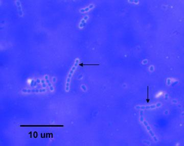
39 Endospore Location The position of the endospore differs among bacterial species and is useful in identification. The main types within the cell are terminal, subterminal, and centrally placed endospores. Terminal endospores are seen at the poles of cells, whereas central endospores are more or less in the middle. Subterminal endospores are those between these two extremes, usually seen far enough towards the poles but close enough to the center so as not to be considered either terminal or central. Lateral endospores are seen occasionally.
40 Endospore staining (Schaeffer-fulton Method) Prepare a smear of the bacteria Bacillus megatatium (a spore-producing organism) Flood the smear with malachite green Do not allow the stain to evaporate or completely evaporate. Remove from heat and allow slides to cool Once the slides are cool (important) rinse with water Flood the sample with safranin (30-60 seconds) Rinse the slide blot dry observe under microscopy. 10x, 40x, 100x (oil immersion).

41 Endospore stain

42 Endospore stain


43
44 Capsules are structures that lay outside of an organism's cell wall and thus are in direct contact with the environment. Many, perhaps most, bacteria produce capsules under the right conditions Some capsules are composed of Carbohydrates or Glycoprotein.
45
46 1. Protect the cell from desiccation (drying) ) 2. Protect the cell from phagocytes (being engulfed by white blood cells) 3. Provide a food reserve when certain organic compounds are in excess. 4. A virulence determinant of pathogenic microbes 5. They serve as binding or adhesion agents for sticking cells together and/or to a surface such as a rock in flowing stream or a tooth
47 Bacterial Heat fixation cause capsule shrinkage Because most capsule materials are water soluble, simple stains will not adhere to them
 Streptococcus lactis Escherichia")
48 The capsule is a major virulence factor in the major disease-causing bacteria, such as:- Klebsiella pneumoniae or Enterobacter aerogenes (slant) Streptococcus lactis Escherichia coli

49 1. Detection of dental plaque Light micrograph of S. mutans


50 2.Detection of Anthrax B. anthracis A lesion on the 10the day of anthrax infection
51 . In this stain we use acidic and basic acidic dye as India Ink and Nigrosen use to stain the background of the slide but basic dye as methylene blue and crystal violet use to stain the cell

52 Older cultures are more likely to exhibit capsule production. When performing a capsule stain on your unknown, be sure the culture you take your sample from is at least five days old

53 Clean slid microscope India Ink and Methylene Wash bottle Culture of bacteria.sterile loop


54

55

56 Capsule stain of Streptococcus lactis.

57 Capsule stain of Enterobacter aerogenes

58 Endospore Stain Difficult to stain but once stained they resist decolorizing Intense heating causes the Endospores to be penetrated by the malachite green Safranin counterstain stains all material other than the endospores Spore stains are typically performed on older cultures
59 THANK YOU YUSRON SUGIARTO
Staining Technology and Bright- Field Microscope Use
 Staining Technology and Bright- Field Microscope Use 2 Abstract We will introduce bright-field microscope use, practice Gram staining with foodborne pathogens, and practice endospore staining with Bacillus
Ch 3: Observing Microorganisms Through a Microscope
 Ch 3: Observing Microorganisms Through a Microscope SLOs Review the metric units of measurement Define total magnification and resolution Explain how electron and light microscopy differ Differentiate
Fig. LPS in Gram negative bacteria
 Structure of bacterial cell Dentistry college - first class Medical biology- Lec.3 Lecturer D. Hanan S A- Cell wall ***Chemical composition of the cell wall Bacteria are divided into two separated groups
Bacterial Structure and Function
 Bacterial Structure and Function Charles Okolie, PhD. Room 311 (on level 4), First College Building, Landmark University okolie.charles@lmu.edu.ng Tel: Ext: Mobile: 08060241166 Structure of Bacteria The
MULTIPLE CHOICE. Choose the one alternative that best completes the statement or answers the question.
 Exam Name MULTIPLE CHOICE. Choose the one alternative that best completes the statement or answers the question. 1) A nanometer would be a suitable unit of measurement for which of the following? 1) A)
Ch 4. Functional Anatomy of Prokaryotic and Eukaryotic Cells
 Ch 4 Functional Anatomy of Prokaryotic and Eukaryotic Cells Objectives Compare and contrast the overall cell structure of prokaryotes and eukaryotes. Identify the three basic shapes of bacteria. Describe
chapter one: the history of microbiology
 chapter one: the history of microbiology Revised 8/29/2016 microbes microscopic (small) organisms, viruses, prions prefix sci. notation frac. equivalent dec. equivalent kilo- (k) 1 10 3 1000/1 = 1000 1000
Characteristics of Mycobacterium
 Mycobacterium Characteristics of Mycobacterium Very thin, rod shape. Culture: Aerobic, need high levels of oxygen to grow. Very slow in grow compared to other bacteria (colonies may be visible in up to
Prokaryotic Cell Structure
 Prokaryotic Cell Structure Chapter 3 Prokaryotes vs Eukaryotes DNA Prokaryotes Eukaryotes Organelles Size & Organization Kingdoms 1 Where do viruses fit in? Acellular microorganisms Cannot reproduce outside
Prokaryotic Cell Structure
 Prokaryotic Cell Structure Chapter 3 Prokaryotes vs Eukaryotes DNA Prokaryotes Eukaryotes Organelles Size & Organization Kingdoms Where do viruses fit in? Acellular microorganisms Cannot reproduce outside
Medical Microbiology. Microscopic Techniques :
 ! Lecture 2 Dr. Ismail I. Daood Medical Microbiology Microscopic Techniques : Several types of microscopes are used in study of microbiology one of the most important tools for studying microorganisms
Microbiology: A Systems Approach
 Microbiology: A Systems Approach First Edition Cowan & Talaro Chapter 4 Prokaryotic Profiles: the Bacteria and the Archaea Chapter 4 Fig. 4.1 3 3 parts flagella filament long, thin, helical structure composed
A.Kavitha Assistant professor Department of Botany RBVRR Womens college
 A.Kavitha Assistant professor Department of Botany RBVRR Womens college The Ultrastructure Of A Typical Bacterial Cell The Bacterial Cell This is a diagram of a typical bacterial cell, displaying all of
MYCOBACTERIA. Pulmonary T.B. (infect bird)
") MYCOBACTERIA SPP. Reservoir Clinical Manifestation Mycobacterium tuberculosis Human Pulmonary and dissem. T.B. M. lepra Human Leprosy M. bovis Human & cattle T.B. like infection M. avium Soil, water, birds,
MYCOBACTERIA SPP. Reservoir Clinical Manifestation Mycobacterium tuberculosis Human Pulmonary and dissem. T.B. M. lepra Human Leprosy M. bovis Human & cattle T.B. like infection M. avium Soil, water, birds,
Cell Structure and Function
 Cell Structure and Function Cell Structure and Function Topics External Structures Cell Envelope Internal Structures Cell Shapes, Arrangement, and Sizes Classification An Infectious Exam Patient with Tuberculosis
Evaluation of a Cold Staining Method for Detecting Acid Fast Bacilli in the Sputum
 International Journal of Current Microbiology and Applied Sciences ISSN: 2319-7706 Volume 5 Number 6 (2016) pp. 125-129 Journal homepage: http://www.ijcmas.com Original Research Article http://dx.doi.org/10.20546/ijcmas.2016.506.015
Biology Multiple Choice, 2 pt each.
 Biology 3340 Spring 2007 Name Exam 1, Version A Write your name on both the exam booklet and the mark sense sheet. On the upper left corner of the mark sense sheet in the Key ID box, mark the version letter
Chapter 4 Prokaryotic Profiles
 Chapter 4 Prokaryotic Profiles Topics: External Structures Cell Envelope Internal Structures Cell Shapes, Arrangement, and Sizes Prokaryotes are unicellular organisms Prokaryotes include two small groups
Bacterial Cell Structures. Stijn van der Veen
 Bacterial Cell Structures Stijn van der Veen How do I know what bacterium makes my patient ill? Bacterial species can be differentiated by: Morphology (shape) Composition (cell envelope and other structures)
Classification of Infectious Agents. Dr W. D. Colby
 Classification of Infectious Agents Dr W. D. Colby Nonliving Infectious Agents PRIONS: abnormally configured self-replicating protein templates VIRUSES: nucleic acid (DNA or RNA) genes packaged in protein
Cell Structure. Morphology of Prokaryotic Cell. Cytoplasmic Membrane 4/6/2011. Chapter 3. Cytoplasmic membrane
 Cell Structure Chapter 3 Morphology of Prokaryotic Cell Cytoplasmic membrane Delicate thin fluid structure Surrounds cytoplasm of cell Defines boundary Defines boundary Serves as a selectively permeable
Bacterial Structures. Capsule or Glycocalyx TYPES OF FLAGELLA FLAGELLA. Average size: µm 2-8 µm Basic shapes:
 PROKARYOTIC One circular chromosome, not in a membrane No histones No organelles Peptidoglycan cell walls Binary fission EUKARYOTIC Paired chromosomes, in nuclear membrane Histones Organelles Polysaccharide
number Done by Corrected by Doctor Dr. Hamed Al Zoubi
 number Done by Corrected by 46 2017/9/20 Doctor Dr. Hamed Al Zoubi 66 /8486535 مركز الرائد للخدمات الطالبية 66 /8486535 مركز الرائد للخدمات الطالبية 2 nd year Medical Students JU Bacterial Structure and
Microbiology. Morphology & Ultra-Structure of Microorganism. Prof. Dr. Batool Hassan Al-Ghurabi
 Microbiology Morphology & Ultra-Structure of Microorganism Prof. Dr. Batool Hassan Al-Ghurabi Microbiology: the study of organisms too small to be seen without magnification. Micro - too small to be seen
CHAPTER 4 CELL STRUCTURE/FUNCTION. 2. The uses the visible light to illuminate cell. 3. How is the magnification of a compound microscope calculated?
 CHAPTER 4 CELL STRUCTURE/FUNCTION 1. Define magnification and the term resolution. 2. The uses the visible light to illuminate cell. 3. How is the magnification of a compound microscope calculated? 4.
ILOs. 10/10/2016 Maha Fathy 2
 ILOs 1- List different components of bacterial cell. 2-Describe structure of cell wall of Gram +ve and ve bacteria 3-Recognize role of cell wall and cytoplasmic membrane in survival and growth of bacterial
S-LAYER ;- Protoplasts, Spheroplasts, and L Forms The Mycoplasmas 1
 S-LAYER ;- A paracrystalline protein or glycolprotein layer has been demonstrated in some bacteria (both G+ and G- bacteria as well as archae bacteria). This layer can be shown by electron microscopy.
Small living organism Not visible to the naked eye Must be viewed under a microscope Found everywhere in the environment, including on and in the
 Small living organism Not visible to the naked eye Must be viewed under a microscope Found everywhere in the environment, including on and in the human body Many Microorganisms are part of normal flora
Objective 3 Viruses & Bacteria genetic material capsule Pili DNA
 Objective 3 Viruses & Bacteria 1. Compare the structure and functions of viruses to cells and describe the role of viruses in causing diseases and conditions such as acquired immune deficiency syndrome,
Medical Bacteriology- lecture 13. Mycobacterium Actinomycetes
 Medical Bacteriology- lecture 13 Mycobacterium Actinomycetes Mycobacterium tuberculosis Large, very weakly gram positive rods, Obligate aerobes, related to Actinomycetes, non spore forming, non motile
Unchalee Tansuphasiri 1 and Booncherd Kladphuang 2
 SOUTHEAST ASIAN J TROP MED PUBLIC HEALTH EVALUATION OF SPUTUM STAINING BY MODIFIED COLD METHOD AND COMPARISON WITH ZIEHL-NEELSEN AND FLUOROCHROME METHODS FOR THE PRIMARY DIAGNOSIS OF TUBERCULOSIS Unchalee
ethylene glycol. The latter was regarded as the more suitable solvent, by Smith and Clark (1937) one of the important differential points
 one of the important differential points") STUDIES OF THE COMMON AEROBIC SPORE-FORMING BACILLI, I. STAINING FOR FAT WITH SUDAN BLACK B-SAFRANIN KENNETH L. BURDON,2 Consultant, JULIA C. STOKES, Junior Bacteriologist, AND CECIL E. KIMBROUGH, Assistant
STUDIES OF THE COMMON AEROBIC SPORE-FORMING BACILLI, I. STAINING FOR FAT WITH SUDAN BLACK B-SAFRANIN KENNETH L. BURDON,2 Consultant, JULIA C. STOKES, Junior Bacteriologist, AND CECIL E. KIMBROUGH, Assistant
Micro lab notes. Dana alsulaibi
 Micro lab notes Dana alsulaibi Respiratory system Microbiology laboratory section Gram Positive Coccus These are the most common microorganisms to appear on a throat swab Staphylococcus Spp. Streptococcus
Medical Bacteriology- Lecture 10. Mycobacterium. Actinomycetes. Nocardia
 Medical Bacteriology- Lecture 10 Mycobacterium Actinomycetes Nocardia 1 Mycobacterium Characteristics - Large, very weakly gram positive rods - Obligate aerobes, related to Actinomycetes - Catalase positive
Prokaryotic Profiles The Bacteria and Archaea. Chapter 3
 Prokaryotic Profiles The Bacteria and Archaea Chapter 3 Prokaryotic Form and Function Prokaryotes can be distinguished from eukaryotes by: the way their DNA is packaged (lack of nucleus and histones) the
Mahon: Textbook of Diagnostic Microbiology, 4 th Edition
 Mahon: Textbook of Diagnostic Microbiology, 4 th Edition Chapter 01: Bacterial Cell Structure, Physiology, Metabolism, and Genetics Test Bank MULTIPLE CHOICE 1. To survive, microbial inhabitants have learned
Bacteria. Bacteria and Archaea are both: unicelluar (single-celled) prokaryotes (lacking a nucleus and membrane bound organelles)
 prokaryotes (lacking a nucleus and membrane bound organelles)") Bacteria Bacteria and Archaea are both: unicelluar (single-celled) prokaryotes (lacking a nucleus and membrane bound organelles) 1 Grouped by their need for oxygen obligate anaerobes are poisoned by oxygen
Bacteria Bacteria and Archaea are both: unicelluar (single-celled) prokaryotes (lacking a nucleus and membrane bound organelles) 1 Grouped by their need for oxygen obligate anaerobes are poisoned by oxygen
Multi-Site Performance Summary of. AEROSPRAY TB SLIDE STAINER/CYTOCENTRIFUGE (Model 7722)
") Multi-Site Performance Summary of AEROSPRAY TB SLIDE STAINER/CYTOCENTRIFUGE (Model 7722) Abstract The new Aerospray TB Slide Stainer/Cytocentrifuge - Model 7722 (ELITechGroup Inc., www.elitechgroup.com)
Multi-Site Performance Summary of AEROSPRAY TB SLIDE STAINER/CYTOCENTRIFUGE (Model 7722) Abstract The new Aerospray TB Slide Stainer/Cytocentrifuge - Model 7722 (ELITechGroup Inc., www.elitechgroup.com)
Microbiology for Environmental Health Officers. EHL0033 Prokaryotes 3
 Microbiology for Environmental Health Officers EHL0033 Prokaryotes 3 Mutualism: bacterial headlights. The glowing oval below the eye of the flashlight fish (Photoblepharon palpebratus) is an organ harboring
PATHOPHYSIOLOGY. DEFINED Involves the study of function that results from disease processes.
 DEFINED Involves the study of function that results from disease processes. What is pathology? Pathology is the branch of medical sciences that treats the essential nature of disease, especially the changes
BACTERIAL EXAMINATION OF WATER
 BACTERIAL EXAMINATION OF WATER The bacteriological examination of water is performed routinely by water utilities and many governmental agencies to ensure a safe supply of water for drinking, bathing,
Made by :aseel al-waked corrected by : sarah awaisheh
 Microbiology sheet (2) Made by :aseel al-waked corrected by : sarah awaisheh DATE :25-9-2016 "Anatomy" and Function of Prokaryotes: Last lecture we talked about the shape of the bacteria. Now the shape
Order: Actinomycetales. Family: Mycobactericeae. Some are parasitic to cold blooded animal, others are saprophytic in nature.
 Order: Actinomycetales Family: Mycobactericeae They are widely distributed in nature. Few no is pathogenic for man & animal. Some are parasitic to cold blooded animal, others are saprophytic in nature.
Topic 03 Prokaryotes (3.3)
") Topic 03 Prokaryotes (3.3) Topics Characteristics (comparison) External Structures Cell Envelope Internal Structures Cell Shapes, Arrangement, and Sizes Classification 1 Relative size of bacterial cell
Topic 03 Prokaryotes (3.3) Topics Characteristics (comparison) External Structures Cell Envelope Internal Structures Cell Shapes, Arrangement, and Sizes Classification 1 Relative size of bacterial cell
320 MBIO Microbial Diagnosis. Aljawharah F. Alabbad Noorah A. Alkubaisi 2017
 320 MBIO Microbial Diagnosis Aljawharah F. Alabbad Noorah A. Alkubaisi 2017 Pathogens of the Urinary tract The urinary system is composed of organs that regulate the chemical composition and volume of
Chapter 4 M I C R O B I O L O G Y. The Anatomy of the Cell. a n i n t r o d u c t i o n
 ninth edition TORTORA FUNKE CASE M I C R O B I O L O G Y a n i n t r o d u c t i o n Chapter 4 The Anatomy of the Cell PowerPoint Lecture Slide Presentation prepared by Christine L. Case! Copyright 2006
HELMINTHS IMAGE DISEASE STAGE SOURCE SYMPTOMS FOUND LEN TAENIA SAGINATA (BEEF) TAENIA SOLIUM (PORK) TAENIASIS (TAPEWORM)
 TAENIA SOLIUM (PORK) TAENIASIS (TAPEWORM)") HELMINTHS IMAGE DISEASE STAGE SOURCE SYMPTOMS FOUND LEN TAENIA SAGINATA (BEEF) TAENIA SOLIUM (PORK) TAENIASIS (TAPEWORM) HOOKS /AND /OR/SUCKERS SCOLEX (ADULT) INGESTION OF CONTAMINATED PORK OR BEEF DIARRHEA
HELMINTHS IMAGE DISEASE STAGE SOURCE SYMPTOMS FOUND LEN TAENIA SAGINATA (BEEF) TAENIA SOLIUM (PORK) TAENIASIS (TAPEWORM) HOOKS /AND /OR/SUCKERS SCOLEX (ADULT) INGESTION OF CONTAMINATED PORK OR BEEF DIARRHEA
Bacterial Mechanisms of Pathogenicity. 2 nd Lecture
 Bacterial Mechanisms of Pathogenicity 2 nd Lecture Preferred Portal of Entry Just because a pathogen enters your body it does not mean it s going to cause disease. pathogens - preferred portal of entry
Introduction. Biochemistry: It is the chemistry of living things (matters).
.") Introduction Biochemistry: It is the chemistry of living things (matters). Biochemistry provides fundamental understanding of the molecular basis for the function and malfunction of living things. Biochemistry
Introduction Biochemistry: It is the chemistry of living things (matters). Biochemistry provides fundamental understanding of the molecular basis for the function and malfunction of living things. Biochemistry
! gives mechanical strength to the cell and protects it from exploding due to osmotic lysis (shape and strength due to the peptidoglycan)
") Cell Wall! The cell wall is a rigid structure that surrounds the bacterial cell just outside of the plasma membrane Functions to:! gives the bacterium its shape! gives mechanical strength to the cell and
Cell Wall! The cell wall is a rigid structure that surrounds the bacterial cell just outside of the plasma membrane Functions to:! gives the bacterium its shape! gives mechanical strength to the cell and
Cell Structure and Function
 Cell Structure and Function Chapter 4 4.1 What is a Cell? Each cell has a plasma membrane, cytoplasm, and a nucleus (in eukaryotic cells) or a nucleoid (in prokaryotic cells) 1 Nucleoid concentrated DNA
Structure of Prokaryotic & Eukaryotic Cells
 Structure of Prokaryotic & Eukaryotic Cells Review of Prokaryotic & Eukaryotic Cells Nucleus vs nucleoid DNA : circular vs linear, presence of histones Membranous organelles Cell wall-peptidoglycan Cell
Weds. Date. Aug. 26. Sept. 2
 Mt.SanAntonioCollege Microbiology 22 Lab Schedule for Fall 2015 Mon./ Split Lab Sections ONLY Wk. Mon. 1 Aug. 24 Orientation with Introductions & Safety Rules/Regulations 2 Aug. 31 Exercise #1: The Microscope
BACTERIAL EXAMINATION OF WATER
 BACTERIAL EXAMINATION OF WATER The bacteriological examination of water is performed routinely by water utilities and many governmental agencies to ensure a safe supply of water for drinking, bathing,
Mt. San Antonio College Microbiology 22 Lab Schedule for Spring 2018 Mon/Weds. Split Lab Sections ONLY
 Mt. San Antonio College Microbiology 22 Lab Schedule for Spring 2018 Mon/ Split Lab Sections ONLY Wk 1 Feb. 26 Orientation with Introductions & Safety Rules/Regulations Feb. 28 Orientation with Pathogen
Mt. San Antonio College Microbiology 22 Lab Schedule for Spring 2018 Tues/Thurs. Split Lab Sections ONLY
 Mt. San Antonio College Microbiology 22 Lab Schedule for Spring 2018 Tues/ Split Lab Sections ONLY Wk 1 Feb. 27 Orientation with Introductions & Safety Rules/Regulations March 1 Orientation with Pathogen
Mt. San Antonio College Microbiology 22 Lab Schedule for Fall 2017 Tues/Thurs. Split Lab Sections ONLY
 Mt. San Antonio College Microbiology 22 Lab Schedule for Fall 2017 Tues/ Split Lab Sections ONLY Wk 1 Aug. 29 Orientation with Introductions & Safety Rules/Regulations Aug. 31 Orientation with Pathogen
2018 Science Olympiad: Microbe Mission - Sample Tournament Div C
 2018 Science Olympiad: Microbe Mission - Sample Tournament Div C Section A: Types of cells and their parts 1. Please state if the cell is prokaryotic or eukaryotic. Then label the following molecular components
alcohol-soluble dyes, such as Spirit Blue and Night Blue, if we dye-iodine molecule, and postulate that the essential difference
 THE GRAM STAIN AND DIFFERENTIAL STAINING M. L. KAPLAN AND LEAH KAPLAN Bureau of Laboratories, Department of Health, New York City Received for publication, June 22, 1932 The Gram test is the most commonly
EQUIVALENCE OF ACID ALONE OR ACID-ALCOHOL AS DECOLOURIZING AGENT IN ZIEHL - NEELSEN METHOD
 Original Article 219 EQUIVALENCE OF ACID ALONE OR ACID-ALCOHOL AS DECOLOURIZING AGENT IN ZIEHL - NEELSEN METHOD M. Gomathi Sekar, Fathima Rehman, Vanaja Kumar and N. Selvakumar (Received on 12.4.2012;
Mycobacterial cell wall. Cell Cycle Lengths. Outline of Laboratory Methods. Laboratory Methods
 Laboratory Methods Cell Cycle Lengths Generation time (hrs) Days needed for 26 generations (colony) E. coli 0.33 0.36 Nancy Connell, PhD Professor, nfectious Disease Department of Medicine Center for Emerging
50% of those. TB, if left untreated, kills more than. infected. The Global Tuberculosis Epidemic. What Exactly is TB?
 The Global Tuberculosis Epidemic What Exactly is TB? One third of the world's population is thought to be infected with M. Tuberculosis (better known as TB), and new infections occur at a rate of about
LESSON 2.6 WORKBOOK Diagnosing infections, and, what s up your nose?
 Staphylococcus aureus Morphology: The physical form or structure of a microbe.. LESSON 2.6 WORKBOOK Diagnosing infections, and, what s up your nose? Now we have discussed the different requirements that
Starch grains - excess sugars
 (a) Membrane system - site of light reactions (photosynthesis) - chlorpophyll pigments - enzymes - electron carriers - flattened, fluid-filled sacs (called thylakoids which are stacked to form grana) -
Name Class Date. Infection in which a virus inserts its nucleic acid into the DNA of the host cell and is duplicated with the cell s DNA
 Name Class Date 20.1 Viruses Lesson Objectives Explain how viruses reproduce. Explain how viruses cause infection. BUILD Vocabulary A. The chart below shows key terms from the lesson with their definitions.
done by: mohammad haitham
 done by: mohammad haitham "Anatomy" and Function of Prokaryotes: The pictures are very important, go back to the slide2! Last lecture we talked about the shape of the bacteria. Now the shape of the bacteria
What s in a Cell? From Ch. 4
 What s in a Cell? From Ch. 4 Plant cell walls Amit1b.files.wordpress.com genomebiology.com Figure 4.1 Arrangements of cocci. Plane of division Diplococci Streptococci Tetrad Sarcinae Staphylococci. Figure
Chapter 4 Inflammation and Infection
 Chapter 4 Inflammation and Infection Defense Mechanisms Three lines of defense protect the body against foreign invasion: Physical or surface barriers Inflammation Immune response Inflammation Non-specific
Medical Microbiology
 Lecture 5!!!!!!ƒš!!Œ!!! š!!œ!! Œ!!!! Dr. Ismail I. Daood Medical Microbiology!! Systematic Bacteriology Gram-Positive Cocci : GENUS : Staphylococcus : The general properties of Staphylococcus are Gram-
be formulated. It is agreed that disruption of the cell membrane by chemical carbol-fuchsin and for the artifacts that have been observed.
 THE NATURE OF THE ACID-FAST STAIN CARL LAMANNA Camp Detrick, Frederick, Maryland, Received for publication April 1, 1946 A satisfactory explanation of the nature of the Ziehl-Neelsen stain remains to be
Microscopy. Münchenwiler, March 25, Hans L Rieder. Picture drawings: Koch R. Mittheil Kaiserl Gesundheitsamt 1884;2:1 88
 Microscopy Münchenwiler, March 25, 2010 Hans L Rieder Picture drawings: Koch R. Mittheil Kaiserl Gesundheitsamt 1884;2:1 88 Sensitivity of Direct Sputum Smear Examination in Identifying Pulmonary Tuberculosis
2) In what two ways do Archaea differ from true bacteria?
 In what two ways do Archaea differ from true bacteria?") STATION 1: 1) Explain the benefit of saprotrophic bacteria/fungi. 2) In what two ways do Archaea differ from true bacteria? 3) When transitioning from a low power to a higher power on a microscope, describe
STATION 1: 1) Explain the benefit of saprotrophic bacteria/fungi. 2) In what two ways do Archaea differ from true bacteria? 3) When transitioning from a low power to a higher power on a microscope, describe
Phases of the bacterial growth:
 L3: Physiology of Bacteria: Bacterial growth Growth is the orderly increase in the sum of all the components of an organism. Cell multiplication is a consequence of growth, in unicellular organism, growth
PRESENTER: DENNIS NYACHAE MOSE KENYATTA UNIVERSITY
 18/8/2016 SOURCES OF MICROBIAL CONTAMINANTS IN BIOSAFETY LABORATORIES IN KENYA PRESENTER: DENNIS NYACHAE MOSE KENYATTA UNIVERSITY 1 INTRODUCTION Contamination occurs through avoidable procedural errors
Chapter 21: Prokaryotes & Viruses
 Chapter 21: Prokaryotes & Viruses Microorganisms Single-celled organisms that are too small to be seen without a microscope Bacteria are the smallest living organisms Viruses are smaller but are not alive
Mycobacteriaceae
 Mycobacteriaceae 9.04.2018 1 Classification Kingdom: Bacteria Phylum: Actinobacteria Order: Actinomycetales Family: Mycobacteriaceae Genus: Mycobacterium 9.04.2018 2 The properties of Mycobacterium genus
Shirin Abadi, B.Sc.(Pharm.), ACPR, Pharm.D. Clinical Pharmacy Specialist & Pharmacy Education Coordinator, BC Cancer Agency Clinical Associate
, ACPR, Pharm.D. Clinical Pharmacy Specialist & Pharmacy Education Coordinator, BC Cancer Agency Clinical Associate") Shirin Abadi, B.Sc.(Pharm.), ACPR, Pharm.D. Clinical Pharmacy Specialist & Pharmacy Education Coordinator, BC Cancer Agency Clinical Associate Professor of Pharmacy & Associate Member of Medicine, UBC
Shirin Abadi, B.Sc.(Pharm.), ACPR, Pharm.D. Clinical Pharmacy Specialist & Pharmacy Education Coordinator, BC Cancer Agency Clinical Associate Professor of Pharmacy & Associate Member of Medicine, UBC
it selectively allows some molecules to pass into the organism
 Multiple Choice Quiz Procaryotic Cell Structure and Function Eucaryotic Cell Structure and Function Choose the best answer 1. The significance of the plasma membrane is that it selectively allows some
Module No. # 01 Lecture No. # 02 Glimpses of Microbial World-Bacteria. Good morning students. (Refer Slide Time: 00:29)
") Biochemical Engineering Prof. Dr. Rintu Banerjee Department of Agricultural and Food Engineering Assistant Prof. Dr. Saikat Chakraborty Department of Chemical Engineering Indian Institute of Technology,
Biochemical Engineering Prof. Dr. Rintu Banerjee Department of Agricultural and Food Engineering Assistant Prof. Dr. Saikat Chakraborty Department of Chemical Engineering Indian Institute of Technology,
Early scientists who observed cells made detailed sketches of what they saw.
 Early scientists who observed cells made detailed sketches of what they saw. Early scientists who observed cells made detailed sketches of what they saw. CORK Early scientists who observed cells made detailed
Identification & Control of Filamentous Bacteria
 Identification & Control of Filamentous Bacteria Toni Glymph, Wastewater Microbiologist ToniGlymph@msn.com ARWA-Lonoke Filamentous Bacteria Sludge Bulking Filamentous Bacteria Sludge Foaming FILAMENTOUS
Host Parasite Relationship. Prof. Hanan Habib Department of Pathology, College of Medicine,KSU
 Host Parasite Relationship Prof. Hanan Habib Department of Pathology, College of Medicine,KSU OBJECTIVES Define core terms important in host-parasite relationship. Know host response to parasite invasion
Diagnosis of Pulmonary Tuberculosis Using Conventional Smear Microscopy and Culture Methods in a Tertiary Care Hospital
 Diagnosis of Pulmonary Tuberculosis Using Conventional Smear Microscopy and Culture Methods in a Tertiary Care Hospital 1 SM.Nachammai, 2 Dr.Mangayarkarasi.V (MD) Abstract: Tuberculosis, one of the oldest
What does your body do to protect itself from invading microbes?
 46 Disease Fighters i n v e s t i g at i o n What does your body do to protect itself from invading microbes? Even before an organism can enter your body, your skin provides a protective barrier. But foreign
Cell Boundaries Section 7-3
 Cell Boundaries Section 7-3 The most important parts of a cell are its borders, which separate the cell from its surroundings. The cell membrane is a thin, flexible barrier that surrounds all cells. The
BACTERIAL PATHOGENESIS
 BACTERIAL PATHOGENESIS A pathogen is a microorganism that is able to cause disease. Pathogenicity is the ability to produce disease in a host organism. Virulence a term which refers to the degree of pathogenicity
MODULE ONE" TB Basic Science" Treatment Action Group TB/HIV Advocacy Toolkit
 MODULE ONE" TB Basic Science" Treatment Action Group TB/HIV Advocacy Toolkit Topics to be covered What is Tuberculosis? TB bacteria and what is unique about it. How is TB different from HIV? How is TB
Cytology I Study of Cells
 Cytology I Study of Cells Biology 20 Which cell type has organelles such as mitochondria, nuclues, Golgi bodies, etc? A) prokaryotic B) eukaryotic C) bacterial D) viral E) none of these Cellular Basis
4. The most common cause of traveller s diarrheoa is a. Rotavirus b. E coli c. Shigella d. Giardia e. Salmonella
 INFECTIOUS DISEASE 1. Mumps virus is a a. Adenovirus b. Herpes virus c. Paramyxovirus d. Pox virus e. Picornavirus 2. All of the following cause a clinical effect via the production of exotoxin except
Pathogenesis of Infectious Diseases. CLS 212: Medical Microbiology
 Pathogenesis of Infectious Diseases CLS 212: Medical Microbiology Definitions Path- means disease. Pathogenesis The steps or mechanisms involved in the development of a disease. Infection The presence
Principles of Infectious Disease Lecture #13 Dr. Gary Mumaugh
 Principles of Infectious Disease Lecture #13 Dr. Gary Mumaugh Terminology Pathology study of disease Etiology cause of disease Pathogenesis disease process Infection colonization by microbes Disease illness
Course Learning Outcomes for Unit III. Reading Assignment. Unit Lesson. UNIT III STUDY GUIDE Essential Parts: Cells
 UNIT III STUDY GUIDE Essential Parts: Cells Course Learning Outcomes for Unit III Upon completion of this unit, students should be able to: 1. Evaluate concepts and theories of basic biological sciences
O.k., Now Starts the Good Stuff (Part I) Prokaryotic Cell Structure and Function
 Prokaryotic Cell Structure and Function") O.k., Now Starts the Good Stuff (Part I) Prokaryotic Cell Structure and Function Prokaryotic Characteristics DNA not enclosed in membrane. No histone proteins associated with DNA. Lack membrane-bound organelles
O.k., Now Starts the Good Stuff (Part I) Prokaryotic Cell Structure and Function Prokaryotic Characteristics DNA not enclosed in membrane. No histone proteins associated with DNA. Lack membrane-bound organelles
Dairy Microbiology. Key Terms. Mastitis. Somatic Cells. Microorganisms. Bacteria. Psychrotrophic. Thermoduric. Thermophilic. Endospores.
 Dairy Microbiology Key Terms Mastitis Somatic Cells Microorganisms Bacteria Psychrotrophic Thermoduric Thermophilic Endospores Coliform Indicator Organisms Pasteurization Dairy animals, including: cows,
3/24/2016. Unit 2 From the Atom to the Cell. Organisms + Chemistry. What are Ions? Chemical Bonds Covalent. Chemical Bonds Ionic
 Organisms + Chemistry Organic chemistry: the study of carbon-containing compounds (help make up our bodies) Biochemistry: the study of chemical reactions that occur in living systems Unit 2 From the Atom
Bacteria and Viruses. Chapter 20 Biology II
 Bacteria and Viruses Chapter 20 Biology II 3 Domains of Living Organisms Section 1 - Bacteria Prokaryotes Oldest living things on Earth Date back 3.5 mya Single-celled organisms No membrane bound organelles
Chapter 4. Prokaryotic Cells. Prokaryotic cells. Prokaryotic vs. Eukaryotic Cells. Comparing Prokaryotic and. Eukaryotic cells.
 Chapter 4 Comparing Prokaryotic and Eukaryotic Cells Prokaryotic vs. Eukaryotic Cells Prokaryotic cells No nucleus No organelles Cell walls composed of peptidoglycan Reproduce asexually via binary fission
Veterinary Bacteriology and Mycology
 Veterinary Bacteriology and Mycology PJL:2011 Bacterial Overview: Morphology, Structure, Jargon General Features Domain Bacteria Proteobacteria Spirochaetes Firmicutes Actinobacteria No nuclear membrane
Microbiology sheet ()
") Microbiology sheet () Made by : Majd abu-fares corrected by : Abd. salman Note: (slide "18" min: "6:15-9:50") DATE :28-9-2016 Acid-fast Cell Walls Means: bacteria that don't pigment to gram stain Acid-fast
Microbiology sheet () Made by : Majd abu-fares corrected by : Abd. salman Note: (slide "18" min: "6:15-9:50") DATE :28-9-2016 Acid-fast Cell Walls Means: bacteria that don't pigment to gram stain Acid-fast