Lecture 2 I. Membrane Proteins II. Intracellular Compartments
|
|
|
- Milo Crawford
- 6 years ago
- Views:
Transcription
1 Lecture 2 I. Membrane Proteins II. Intracellular Compartments Ref: MBoC (5th Edition), Alberts Johnson Lewis Raff Roberts Walter Chapter 10 Membrane Structure Chapter 12 Intracellular Compartments and Protein Sorting Hualin Zhong 2/01/2012 1

2 Three Views of a Cell Membrane Figure 10-1 The lipid bilayer provides the basic structure of biological membranes; The membrane proteins perform most of the membrane s specific tasks and therefore give each type of cell membrane its characteristic functional properties. 2
 a single α helix, (2) multiple α helices, (3) a rolled-up β sheet (a β barrel).")
3 Membrane Proteins Can Be Associated with the Lipid Bilayer in Various Ways Figure A. Transmembrane proteins extend across the bilayer as (1) a single α helix, (2) multiple α helices, (3) a rolled-up β sheet (a β barrel). Some of these single-pass and multipass proteins have a covalently attached fatty acid chain inserted in the cytosolic lipid monolayer (1). B. Membrane proteins are exposed at only one side of the membrane as (4) anchored to the cytosolic surface by an amphiphilic α helix that partitions into the cytosolic monolayer of the lipid bilayer through the hydrophobic face of the helix, (5) attached to the bilayer solely by a covalently attached lipid chain-either a fatty acid chain or a prenyl group-in the cytosolic monolayer or, (6) via an oligosaccharide linker, to phosphatidylinositol in the noncytosolic monolayer-called a glycosylphosphatidylinositol (GPI) anchor. C. Peripheral proteins are attached to the membrane only by noncovalent interactions with other membrane proteins (7, 8). 3
 A myristic acid is attached via an amide linkage to an N-terminal glycine. (B) A palmitic acid is attached via a thioester linkage to a cysteine.")
4 Membrane Proteins Attach to the Membrane via a Fatty Acid Chain or a Prenyl Group Figure The covalent attachment of lipid can help localize a water-soluble protein to a membrane after its synthesis in the cytosol. (A) A myristic acid is attached via an amide linkage to an N-terminal glycine. (B) A palmitic acid is attached via a thioester linkage to a cysteine. (C) A prenyl group (either farnesyl or a longer geranylgeranyl group) is attached via a thioether linkage to a cysteine residue that is initially located four residues from the protein s C-terminus. After prenylation, the terminal three amino acids are cleaved off, and the new C-terminus is methylated before insertion of the anchor into the membrane (not shown). 4

5 In Most Transmembrane Proteins the Polypeptide Chain Crosses the Lipid Bilayer in an α-helical Conformation A segment of a transmembrane polypeptide chain of the bacterial photosynthetic reaction center crossing the lipid bilayer as an α helix. Only the α-carbon backbone of the polypeptide chain is shown, with the hydrophobic amino acids in green and yellow. Figure
6 Use Hydropathy Plots to Localize Potential α-helical Membrane-Spanning Segments in a Polypeptide Chain +84 kj mol -1 (+ 20 kcal mol -1 ) 6

7 Locating the Membrane-Spanning Helix of Glycophorin Glycophorin A from the red blood cell membrane! 15 O-linked carbohydrate units shown in diamond shape N-linked carbohydrate units shown in a lozenge shape 7

8 An Example of Helices that Extend only Part Way Across the Lipid Bilayer Two α helices in the aquaporin water channel, each of which spans only halfway through the lipid bilayer. In the membrane, the protein forms a tetramer of four such two-helix segments, such that the colored surface shown here is buried at an interface formed by protein protein interactions. Figure
 Proteolytic cleavage of one loop to create two fragments that stay together and function normally.")
9 Transmembrane α Helices Often Interact with One Another Figure Converting a single-chain multipass protein into a two-chain multipass protein. (A) Proteolytic cleavage of one loop to create two fragments that stay together and function normally. (B) Expression of the same two fragments from separate genes gives rise to a similar protein that functions normally. 9

10 Steps in the Folding of a Multipass Transmembrane Protein Figure When the newly synthesized transmembrane α helices are released into the lipid bilayer, they are initially surrounded by lipid molecules. As the protein folds, contacts between the helices displace some of the lipid molecules surrounding the helices. 10
 that hydrolyzes lipid molecules. The amino acids that catalyze the enzymatic reaction (shown in red) protrude from the outside surface of the barrel.")
11 Some β Barrels Form Large Transmembrane Channels Figure β barrels formed from different numbers of β strands: (1) The E. coli OmpA protein serves as a receptor for a bacterial virus. (2) The E.coli OMPLA protein is an enzyme (a lipase) that hydrolyzes lipid molecules. The amino acids that catalyze the enzymatic reaction (shown in red) protrude from the outside surface of the barrel. (3) A porin from the bacterium Rhodobacter capsulatus forms a waterfilled pore across the outer membrane. The diameter of the channel is restricted by loops (shown in blue) that protrude into the channel. (4) The E. coli FepA protein transports iron ions. The inside of the barrel is completely filled by a globular protein domain (shown in blue) that contains an iron-binding site (not shown). This domain is thought to change its conformation to transport the bound iron, but the molecular details of the changes are not known. 11

12 Many Membrane Proteins Are Glycosylated As in glycolipids, the sugar residues of glycoproteins are added in the lumen of the ER and the Golgi apparatus. The oligosaccharide chains are always present on the noncytosolic side of the membrane. The polypeptide chain traverses the lipid bilayer as a right-handed α helix. The oligosaccharide chains and disulfide bonds are all on the noncytosolic surface of the membrane. The sulfhydryl groups in the cytosolic domain of the protein do not normally form disulfide bonds because the reducing environment in the cytosol maintains these groups in their reduced ( SH) form. Figure


13 Many Membrane Proteins Are Glycosylated (A) EM: surface of a lymphocyte stained with ruthenium red emphasizes the thick carbohydrate-rich layer surrounding the cell. (B) The carbohydrate layer is made up of the oligosaccharide side chains of glycolipids and integral membrane glycoproteins and the polysaccharide chains on integral membrane proteoglycans. Figure *All of the carbohydrate is on the noncytosolic surface of the membrane. 13

14 Membrane Proteins Often Function as Large Complexes The 3D structure of the photosynthetic reaction center of the bacterium Rhodopseudomonas viridis. The complex consists of four subunits L, M, H, and a cytochrome. The L and M subunits form the core of the reaction center, and each contains five α helices that span the lipid bilayer. The locations of the various electron carrier coenzymes are shown in black. The coenzymes are arranged in the spaces between the helices. Figure
 At low concentration, detergent molecules are monomeric in solution.")
 Detergent molecules are amphiphilic; and because they are cone-shaped, they form micelles rather than bilayers.")

15 The Structure of Detergent Micelles (B) (A) (C) Figure (A) SDS, an anionic detergent, and Triton X-100 and β-octylglucoside, two nonionic detergents. (B) At low concentration, detergent molecules are monomeric in solution. As their concentration is increased beyond the critical micelle concentration (CMC), some of the detergent molecules form micelles. (C) Detergent molecules are amphiphilic; and because they are cone-shaped, they form micelles rather than bilayers. Detergent micelles have irregular shapes. Due to packing constraints, the hydrophobic tails are partially exposed to water. The spacefilling model shows the structure of a micelle composed of 20 β-octylglucoside molecules, predicted by molecular dynamics calculations. 15
16 Membrane Proteins Can Be Solubilized and Purified in Detergents Figure A mild nonionic detergent solubilizes membrane proteins. The detergent disrupts the lipid bilayer and brings the proteins into solution as protein-lipid-detergent complexes. The phospholipids in the membrane are also solubilized by the detergent. 16

17 An example of using mild nonionic detergents for solubilizing, purifying, and reconstituting functional membrane protein systems. Functional Na + -K + pump molecules are purified and incorporated into phospholipid vesicles. The Na + -K + pump is an ion pump that is present in the plasma membrane of most animal cells; it uses the energy of ATP hydrolysis to pump Na + out of the cell and K + in. Figure

18 Many Membrane Proteins Diffuse in the Plane of the Membrane An experiment demonstrating the diffusion of proteins in the plasma membrane of mouse human hybrid cells. The mouse and human proteins are initially confined to their own halves of the newly formed heterocaryon plasma membrane, but they intermix over time. The two antibodies used to visualize the proteins can be distinguished in a fluorescence microscope because fluorescein is green and rhodamine is red. Figure

19 Fluorescence Recovery After Photobleaching (FRAP) Figure 10-36a A specific protein of interest can be expressed as a fusion protein with green fluorescent protein (GFP). In the FRAP technique, fluorescent molecules are bleached in a small area using a laser beam. The fluorescence intensity recovers as the bleached molecules diffuse away and unbleached molecules diffuse into the irradiated area. The diffusion coefficient is calculated from a graph of the rate of recovery: the greater the diffusion coefficient (mobility) of the membrane protein, the faster the recovery. 19

20 Fluorescence Loss In Photobleaching (FLIP) Figure 10-36b In the FLIP technique, a small area of membrane is irradiated continuously, and fluorescence is measured in a separate area. Fluorescence in the second area progressively decreases as fluorescent proteins diffuse out and bleached molecules diffuse in; eventually, all of the fluorescent protein molecules are bleached, as long as they are mobile and not permanently anchored to the cytoskeleton or extracellular matrix. 20

.")
21 Cells Can Confine Proteins and Lipids to Specific Domains Within a Membrane In an epithelial cell, protein A and protein B can diffuse laterally in their own domains but are prevented from entering the other domain, at least partly by the specialized cell junction called a tight junction. Figure Lipid molecules in the outer (noncytosolic) monolayer of the plasma membrane are likewise unable to diffuse between the two domains; lipids in the inner (cytosolic) monolayer, however, are able to do so (not shown). The basal lamina is a thin mat of extracellular matrix that separates epithelial sheets from other tissues Four ways of restricting the lateral mobility of specific plasma membrane proteins: (A) The proteins can self-assemble into large aggregates (e.g. bacteriorhodopsin in the purple membrane of Halobacterium); they can be tethered by interactions with assemblies of macromolecules (B) outside or (C) inside the cell; or they can interact with proteins on the surface of another cell (D). Figure
 (B) Spectrin dimers are linked together into a netlike meshwork by junctional complexes.")
.")
22 The Cortical Cytoskeleton Gives Membrane Mechanical Strength and Restricts Membrane Protein Figure 10-41a Figure 10-41b The Spectrin-based cytoskeleton on the cytosolic side of the human red blood cell plasma membrane (A) (B) Spectrin dimers are linked together into a netlike meshwork by junctional complexes. The junctional complexes are composed of short actin filaments, band 4.1, adducin, and a tropomyosin molecule. The cytoskeleton is linked to the membrane through two transmembrane (band 3 & glycophorin). The EM shows the cytoskeleton on the cytosolic side of a red blood cell membrane after fixation and negative staining. 22

23 The Major Intracellular Compartments of an Animal Cell Figure 12-1 The cytoplasm consists of the cytosol and the cytoplasmic organelles. 23
24 24

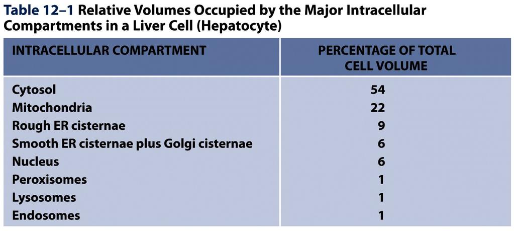
25 Table

26 An Electron Micrograph of Part of a Liver Cell Seen in Cross Section Figure
 (A) A possible pathway for the evolution of the cell nucleus and the ER.")

27 Evolutionary Origins Explain the Topological Relationships of Organelles The origins of mitochondria, chloroplasts, ER, and the cell nucleus might explain the topological relationships of these compartments in eucaryotic cells. (A) (A) A possible pathway for the evolution of the cell nucleus and the ER. (B) Figure 12-4 (B) Mitochondria (and plastids) are thought to have originated when a bacterium was engulfed by a larger preeucaryotic cell. This could explain why they contain their own genomes and why the lumens of these organelles remain isolated from the membrane traffic that interconnects the lumens of many other intracellular compartments. 27

28 Topological Relationships Between Compartments of the Secretory and Endocytic Pathways in a Eucaryotic Cell Figure 12-5 Topologically equivalent spaces are shown in red. In principle, cycles of membrane budding and fusion permit the lumen of any of these organelles to communicate with any other and with the cell exterior by means of transport vesicles. Blue arrows indicate the extensive outbound and inbound vesicular traffic. Some organelles, most notably mitochondria and (in plant cells) plastids, do not take part in this communication and are isolated from the traffic between organelles shown here. 28
I. Membrane Proteins II. Intracellular Compartments III. Protein Translocation
 Lecture 3 I. Membrane Proteins II. Intracellular Compartments III. Protein Translocation Ref: MBoC (5th Edition), Alberts Johnson Lewis Raff Roberts Walter Chapter 10 Membrane Structure Chapter 12 Intracellular
Rama Abbady. Odai Bani-Monia. Diala Abu-Hassan
 5 Rama Abbady Odai Bani-Monia Diala Abu-Hassan Lipid Rafts Lipid rafts are aggregates (accumulations) of sphingolipids. They re semisolid clusters (10-200 nm) of cholesterol and sphingolipids (sphingomyelin
Lecture 15. Membrane Proteins I
 Lecture 15 Membrane Proteins I Introduction What are membrane proteins and where do they exist? Proteins consist of three main classes which are classified as globular, fibrous and membrane proteins. A
Cell Membranes. Dr. Diala Abu-Hassan School of Medicine Cell and Molecular Biology
 Cell Membranes Dr. Diala Abu-Hassan School of Medicine Dr.abuhassand@gmail.com Cell and Molecular Biology Organelles 2Dr. Diala Abu-Hassan Membrane proteins Major components of cells Nucleic acids DNA
Zool 3200: Cell Biology Exam 4 Part I 2/3/15
 Name: Key Trask Zool 3200: Cell Biology Exam 4 Part I 2/3/15 Answer each of the following questions in the space provided, explaining your answers when asked to do so; circle the correct answer or answers
Biology 4410 First Examination Version B
 Biology 4410 Spring 2006 Name First Examination Version B This examination consists of two parts, a multiple-choice section and an essay section. Be sure to put your name on both the mark-sense sheet and
Biology 4410 First Examination Version B
 Biology 4410 Spring 2006 Name First Examination Version B This examination consists of two parts, a multiple-choice section and an essay section. Be sure to put your name on both the mark-sense sheet and
Intracellular Compartments and Protein Sorting
 Intracellular Compartments and Protein Sorting Intracellular Compartments A eukaryotic cell is elaborately subdivided into functionally distinct, membrane-enclosed compartments. Each compartment, or organelle,
Chapter 12. Part II. Biological Membrane
 Chapter 12 Part II. Biological Membrane Single-tailed lipids tend to form micelles Critical micelle concentration (cmc): minimum concentration that forms micelles e.g.) cmc for SDS 1mM; cmc for phospholipids
TRANSPORT PROCESSES. 1b. moving proteins into membranes and organelles
 1b. moving proteins into membranes and organelles SLIDE 1 A typical mammalian cell contains up to 10,000 different kinds of proteins. The vast majority of these proteins are synthesized by cytosolic ribosomes,
Chapt. 11, Membrane Structure. Chapt. 11, Membrane Structure. Chapt. 11, Membrane Structure. Functions of cell membrane. Functions of cell membrane
 Chapt. 11, Membrane Structure Functions of cell membrane 1 Chapt. 11, Membrane Structure Functions of cell membrane As a container/ barrier to movement of small molecules. Figure 11 2 Chapt. 11, Membrane
Protein Trafficking in the Secretory and Endocytic Pathways
 Protein Trafficking in the Secretory and Endocytic Pathways The compartmentalization of eukaryotic cells has considerable functional advantages for the cell, but requires elaborate mechanisms to ensure
Lecture Series 4 Cellular Membranes. Reading Assignments. Selective and Semi-permeable Barriers
 Lecture Series 4 Cellular Membranes Reading Assignments Read Chapter 11 Membrane Structure Review Chapter 12 Membrane Transport Review Chapter 15 regarding Endocytosis and Exocytosis Read Chapter 20 (Cell
Name: Multiple choice questions. Pick the BEST answer (2 pts ea)
") Exam 1 202 Oct. 5, 1999 Multiple choice questions. Pick the BEST answer (2 pts ea) 1. The lipids of a red blood cell membrane are all a. phospholipids b. amphipathic c. glycolipids d. unsaturated 2. The
Exam 1 202 Oct. 5, 1999 Multiple choice questions. Pick the BEST answer (2 pts ea) 1. The lipids of a red blood cell membrane are all a. phospholipids b. amphipathic c. glycolipids d. unsaturated 2. The
Biomembranes structure and function. B. Balen
 Biomembranes structure and function B. Balen All cells are surrounded by membranes Selective barrier But also important for: 1. Compartmentalization 2. Biochemical activities 3. Transport of dissolved
The Cell Membrane (Ch. 7)
") The Cell Membrane (Ch. 7) Phospholipids Phosphate head hydrophilic Fatty acid tails hydrophobic Arranged as a bilayer Phosphate attracted to water Fatty acid repelled by water Aaaah, one of those structure
The Cell Membrane (Ch. 7) Phospholipids Phosphate head hydrophilic Fatty acid tails hydrophobic Arranged as a bilayer Phosphate attracted to water Fatty acid repelled by water Aaaah, one of those structure
Lecture Series 4 Cellular Membranes
 Lecture Series 4 Cellular Membranes Reading Assignments Read Chapter 11 Membrane Structure Review Chapter 12 Membrane Transport Review Chapter 15 regarding Endocytosis and Exocytosis Read Chapter 20 (Cell
Practice Exam 2 MCBII
 1. Which feature is true for signal sequences and for stop transfer transmembrane domains (4 pts)? A. They are both 20 hydrophobic amino acids long. B. They are both found at the N-terminus of the protein.
Life Sciences 1a. Practice Problems 4
 Life Sciences 1a Practice Problems 4 1. KcsA, a channel that allows K + ions to pass through the membrane, is a protein with four identical subunits that form a channel through the center of the tetramer.
MEMBRANE STRUCTURE. Lecture 8. Biology Department Concordia University. Dr. S. Azam BIOL 266/
 1 MEMBRANE STRUCTURE Lecture 8 BIOL 266/4 2014-15 Dr. S. Azam Biology Department Concordia University Plasma Membrane 2 Plasma membrane: The outer boundary of the cell that separates it from the world
Zool 3200: Cell Biology Exam 4 Part I 2/3/15
 Name: Trask Zool 3200: Cell Biology Exam 4 Part I 2/3/15 Answer each of the following questions in the space provided, explaining your answers when asked to do so; circle the correct answer or answers
A. Major parts 1. Nucleus 2. Cytoplasm a. Contain organelles (see below) 3. Plasma membrane (To be discussed in Cellular Transport Lecture)
 3. Plasma membrane (To be discussed in Cellular Transport Lecture)") Lecture 5: Cellular Biology I. Cell Theory Concepts: 1. Cells are the functional and structural units of living organisms 2. The activity of an organism is dependent on both the individual and collective
Lecture 5: Cellular Biology I. Cell Theory Concepts: 1. Cells are the functional and structural units of living organisms 2. The activity of an organism is dependent on both the individual and collective
4 A Tour of the Cell CAMPBELL BIOLOGY IN FOCUS. Urry Cain Wasserman Minorsky Jackson Reece
 CAMPBELL BIOLOGY IN FOCUS Urry Cain Wasserman Minorsky Jackson Reece 4 A Tour of the Cell Lecture Presentations by Kathleen Fitzpatrick and Nicole Tunbridge Overview: The Fundamental Units of Life All
10/13/11. Cell Theory. Cell Structure
 Cell Structure Grade 12 Biology Cell Theory All organisms are composed of one or more cells. Cells are the smallest living units of all living organisms. Cells arise only by division of a previously existing
Essential Cell Biology
 Alberts Bray Hopkin Johnson Lewis Raff Roberts Walter Essential Cell Biology FOURTH EDITION Chapter 15 Intracellular Compartments and Protein Transport Copyright Garland Science 2014 CHAPTER CONTENTS MEMBRANE-ENCLOSED
Chapter 9 - Biological Membranes. Membranes form a semi-permeable boundary between a cell and its environment.
 Chapter 9 - Biological Membranes www.gsbs.utmb.edu/ microbook/ch037.htmmycoplasma Membranes form a semi-permeable boundary between a cell and its environment. Membranes also permit subcellular organization
Ch. 7 Cell Membrane BIOL 222
 Ch. 7 Cell Membrane BIOL 222 Overview: Plasma Membrane Plasma membrane boundary that separates the living cell from its surroundings Selec4ve permeability Allowance of some substances to cross more easily
Cellular Biochemistry
 Cellular Biochemistry Fall Semester 2013 Sept. 23 Benoit Kornmann Institute of Biochemistry Introduction to biological membranes General functions and properties Membrane lipids Physical properties Distribution/asymmetry
Phospholipids. Extracellular fluid. Polar hydrophilic heads. Nonpolar hydrophobic tails. Polar hydrophilic heads. Intracellular fluid (cytosol)
") Module 2C Membranes and Cell Transport All cells are surrounded by a plasma membrane. Eukaryotic cells also contain internal membranes and membrane- bound organelles. In this module, we will examine the
Module 2C Membranes and Cell Transport All cells are surrounded by a plasma membrane. Eukaryotic cells also contain internal membranes and membrane- bound organelles. In this module, we will examine the
I. Fluid Mosaic Model A. Biological membranes are lipid bilayers with associated proteins
 Lecture 6: Membranes and Cell Transport Biological Membranes I. Fluid Mosaic Model A. Biological membranes are lipid bilayers with associated proteins 1. Characteristics a. Phospholipids form bilayers
Membrane Structure and Function
 Chapter 7 Membrane Structure and Function PowerPoint Lecture Presentations for Biology Eighth Edition Neil Campbell and Jane Reece Lectures by Chris Romero, updated by Erin Barley with contributions from
Chapter 4: Cell Structure and Function
 Chapter 4: Cell Structure and Function Robert Hooke Fig. 4-2, p.51 The Cell Smallest unit of life Can survive on its own or has potential to do so Is highly organized for metabolism Senses and responds
BIOLOGICAL CHEMISTRY Prof. J.H.P. Bayley, Dr. R.M. Adlington and Dr. L. Smith Trinity Term First Year. Lecture 2 Hagan Bayley
 BIOLOGICAL CHEMISTRY Prof. J.H.P. Bayley, Dr. R.M. Adlington and Dr. L. Smith Trinity Term 2007 - First Year Lecture 2 Hagan Bayley Introduction to the macromolecules of life and cell structures. Introduction
Cytosol the fluid Cytoplasm cell interior, everything outside the nucleus but within the cell membrane, includes the organelles, cytosol, and
 Cell Organelles Plasma Membrane comprised of a phospholipid bilayer and embedded proteins Outer surface has oligosaccharides separates the cells s contents from its surroundings Cytosol the fluid Cytoplasm
Boundary Lipid bilayer Selectively Permeable Fluid mosaic of lipids and proteins Contains embedded proteins
 1 Boundary Lipid bilayer Selectively Permeable Fluid mosaic of lipids and proteins Contains embedded proteins 2 Phosphate head hydrophilic Fatty acid tails hydrophobic Amphipathic Phosphate attracted to
The Cell Membrane AP Biology
 The Cell Membrane AP Biology! 2007-2008 Overview! Cell membrane separates living cell from nonliving surroundings " thin barrier = 8nm thick! Controls traffic in & out of the cell " selectively permeable
Membrane Structure and Membrane Transport of Small Molecules. Assist. Prof. Pinar Tulay Faculty of Medicine
 Membrane Structure and Membrane Transport of Small Molecules Assist. Prof. Pinar Tulay Faculty of Medicine Introduction Cell membranes define compartments of different compositions. Membranes are composed
Lecture Series 4 Cellular Membranes
 Lecture Series 4 Cellular Membranes Reading Assignments Read Chapter 11 Membrane Structure Review Chapter 21 pages 709-717 717 (Animal( Cell Adhesion) Review Chapter 12 Membrane Transport Review Chapter
Diffusion across cell membrane
 The Cell Membrane and Cellular Transport Diffusion across cell membrane Cell membrane is the boundary between inside & outside separates cell from its environment Can it be an impenetrable boundary? NO!
GCD3033:Cell Biology. Plasma Membrane Dynamics
 Plasma Membrane Dynamics Membrane Structure I) Lipid Bilayer A) Membrane Lipids B) Membrane Flexibility & Composition C) Phospholipids II) Membrane Proteins A) association with membranes B) membrane solubilization
Molecular Cell Biology Problem Drill 16: Intracellular Compartment and Protein Sorting
 Molecular Cell Biology Problem Drill 16: Intracellular Compartment and Protein Sorting Question No. 1 of 10 Question 1. Which of the following statements about the nucleus is correct? Question #01 A. The
Methods of studying membrane structure
 King Saud University College of Science Department of Biochemistry Biomembranes and Cell Signaling (BCH 452) Chapter 2 Methods of studying membrane structure Prepared by Dr. Farid Ataya http://fac.ksu.edu.sa/fataya
Week 5 Section. Junaid Malek, M.D.
 Week 5 Section Junaid Malek, M.D. HIV: Anatomy Membrane (partiallystolen from host cell) 2 Glycoproteins (proteins modified by added sugar) 2 copies of RNA Capsid HIV Genome Encodes: Structural Proteins
Plasma Membrane. comprised of a phospholipid bilayer and embedded proteins separates the cells s contents from its surroundings
 Cell Organelles Plasma Membrane comprised of a phospholipid bilayer and embedded proteins separates the cells s contents from its surroundings Cytosol the fluid Cytoplasm cell interior, everything outside
BIOL 158: BIOLOGICAL CHEMISTRY II
 BIOL 158: BIOLOGICAL CHEMISTRY II Lecture 1: Membranes Lecturer: Christopher Larbie, PhD Introduction Introduction Cells and Organelles have membranes Membranes contain lipids, proteins and polysaccharides
Human height. Length of some nerve and muscle cells. Chicken egg. Frog egg. Most plant and animal cells Nucleus Most bacteria Mitochondrion
 10 m 1 m 0.1 m 1 cm Human height Length of some nerve and muscle cells Chicken egg Unaided eye 1 mm Frog egg 100 µm 10 µm 1 µm 100 nm 10 nm Most plant and animal cells Nucleus Most bacteria Mitochondrion
Cells. Variation and Function of Cells
 Cells Variation and Function of Cells Cell Theory states that: 1. All living things are made of cells 2. Cells are the basic unit of structure and function in living things 3. New cells are produced from
CWDHS Mr. Winch Grade 12 Biology
 The Cell Membrane Overview Cell separates living cell from nonliving surroundings thin barrier = 8nm thick Controls traffic in & out of the cell selectively permeable allows some substances to cross more
The Cell. Biology 105 Lecture 4 Reading: Chapter 3 (pages 47 62)
") The Cell Biology 105 Lecture 4 Reading: Chapter 3 (pages 47 62) Outline I. Prokaryotic vs. Eukaryotic II. Eukaryotic A. Plasma membrane transport across B. Main features of animal cells and their functions
The Cell Biology 105 Lecture 4 Reading: Chapter 3 (pages 47 62) Outline I. Prokaryotic vs. Eukaryotic II. Eukaryotic A. Plasma membrane transport across B. Main features of animal cells and their functions
Lecture Series 5 Cellular Membranes
 Lecture Series 5 Cellular Membranes Cellular Membranes A. Membrane Composition and Structure B. Animal Cell Adhesion C. Passive Processes of Membrane Transport D. Active Transport E. Endocytosis and Exocytosis
A. Membrane Composition and Structure. B. Animal Cell Adhesion. C. Passive Processes of Membrane Transport. D. Active Transport
 Cellular Membranes A. Membrane Composition and Structure Lecture Series 5 Cellular Membranes B. Animal Cell Adhesion E. Endocytosis and Exocytosis A. Membrane Composition and Structure The Fluid Mosaic
A Tour of the Cell. Chapter 6. Biology Eighth Edition Neil Campbell and Jane Reece. PowerPoint Lecture Presentations for
 Chapter 6 A Tour of the Cell PowerPoint Lecture Presentations for Biology Eighth Edition Neil Campbell and Jane Reece Lectures by Chris Romero, updated by Erin Barley with contributions from Joan Sharp
Bio Microbiology - Spring 2012 Learning Guide 03.
 Bio 230 - Microbiology - Spring 2012 Learning Guide 03 http://seemikedraw.files.wordpress.com/2007/07/biology-final.jpg Walsby's Square Bacteria Images of cells from 12 different colonies on blood agar
Protein sorting (endoplasmic reticulum) Dr. Diala Abu-Hsasan School of Medicine
 Dr. Diala Abu-Hsasan School of Medicine") Protein sorting (endoplasmic reticulum) Dr. Diala Abu-Hsasan School of Medicine dr.abuhassand@gmail.com An overview of cellular components Endoplasmic reticulum (ER) It is a network of membrane-enclosed
Protein sorting (endoplasmic reticulum) Dr. Diala Abu-Hsasan School of Medicine dr.abuhassand@gmail.com An overview of cellular components Endoplasmic reticulum (ER) It is a network of membrane-enclosed
Structure and function of cell membranes
 Paper Module : 15 : 01 Development Team Principal Investigator : Prof. Neeta Sehgal Department of Zoology, University of Delhi Co-Principal Investigator : Prof. D.K. Singh Department of Zoology, University
Nucleic acids. Nucleic acids are information-rich polymers of nucleotides
 Nucleic acids Nucleic acids are information-rich polymers of nucleotides DNA and RNA Serve as the blueprints for proteins and thus control the life of a cell RNA and DNA are made up of very similar nucleotides.
(a) TEM of a plasma. Fimbriae. Nucleoid. Ribosomes. Plasma membrane. Cell wall Capsule. Bacterial chromosome
 TEM of a plasma. Fimbriae. Nucleoid. Ribosomes. Plasma membrane. Cell wall Capsule. Bacterial chromosome") 0 m m 0. m cm mm 00 µm 0 µm 00 nm 0 nm Human height Length of some nerve and muscle cells Chicken egg Frog egg Most plant and animal cells Most bacteria Smallest bacteria Viruses Proteins Unaided eye Light
0 m m 0. m cm mm 00 µm 0 µm 00 nm 0 nm Human height Length of some nerve and muscle cells Chicken egg Frog egg Most plant and animal cells Most bacteria Smallest bacteria Viruses Proteins Unaided eye Light
Phospholipids. Phosphate head. Fatty acid tails. Arranged as a bilayer. hydrophilic. hydrophobic. Phosphate. Fatty acid. attracted to water
 The Cell Membrane Phospholipids Phosphate head hydrophilic Fatty acid tails hydrophobic Arranged as a bilayer Phosphate attracted to water Fatty acid repelled by water I want you to remember: Structure
The Cell Membrane. Cell membrane separates living cell from nonliving surroundings. Controls traffic in & out of the cell
 The Cell Membrane 1 Overview Cell membrane separates living cell from nonliving surroundings thin barrier = 8nm thick Controls traffic in & out of the cell selectively permeable allows some substances
Main Functions maintain homeostasis
 The Cell Membrane Main Functions The main goal is to maintain homeostasis. Regulates materials moving in and out of the cell. Provides a large surface area on which specific chemical reactions can occur.
COR 011 Lecture 9: ell membrane structure ept 19, 2005
 COR 011 Lecture 9: ell membrane structure ept 19, 2005 Cell membranes 1. What are the functions of cell membranes? 2. What is the current model of membrane structure? 3. Evidence supporting the fluid mosaic
Lipids and Membranes
 Lipids and Membranes Presented by Dr. Mohammad Saadeh The requirements for the Pharmaceutical Biochemistry I Philadelphia University Faculty of pharmacy Biological membranes are composed of lipid bilayers
Chapter 7: Membranes
 Chapter 7: Membranes Roles of Biological Membranes The Lipid Bilayer and the Fluid Mosaic Model Transport and Transfer Across Cell Membranes Specialized contacts (junctions) between cells What are the
Chapter 5 Cell Membrane Structure and Organelles
 Part II Principles of Individual Cell Function Chapter 5 Cell structures consist of biological membranes essentially mobile lipid bilayers to which many membrane proteins attach. The cell membrane separates
Posttranslational Modification and Targeting of Proteins
 Posttranslational Modification and Targeting of Proteins Graduate Biochemistry Term 2/2016 Assist. Prof. Dr. Panida Khunkaewla School of Chemistry, Institute of Science Suranaree University of Technology
Cellular membranes are fluid mosaics of lipids and proteins.
 Study Guide e Plasma Membrane You should be able to write out the definitions to each of the following terms in your own words: plasma membrane fluid mosaic integral proteins peripheral proteins receptor
Early scientists who observed cells made detailed sketches of what they saw.
 Early scientists who observed cells made detailed sketches of what they saw. Early scientists who observed cells made detailed sketches of what they saw. CORK Early scientists who observed cells made detailed
Advanced Cell Biology. Lecture 28
 Advanced Cell Biology. Lecture 28 Alexey Shipunov Minot State University April 8, 2013 Shipunov (MSU) Advanced Cell Biology. Lecture 28 April 8, 2013 1 / 41 Outline Questions and answers Shipunov (MSU)
The Cell Membrane. Usman Sumo Friend Tambunan Arli Aditya Parikesit. Bioinformatics Group Faculty of Mathematics and Science University of Indonesia
 The Cell Membrane Usman Sumo Friend Tambunan Arli Aditya Parikesit Bioinformatics Group Faculty of Mathematics and Science University of Indonesia Overview Cell membrane separates living cell from nonliving
ME 411 / ME 511. Biological Frameworks for Engineers
 ME 411 / ME 511 Biological Frameworks for Engineers Class Organization HW 1 due on Friday HW 2 available online Grad project available online What are Cells? Sea Urchin Mouse Seaweed Robert Hooke Prokaryotic
Study Guide for Biology Chapter 5
 Class: Date: Study Guide for Biology Chapter 5 Multiple Choice Identify the choice that best completes the statement or answers the question. 1. Which of the following led to the discovery of cells? a.
Chapter 7. (7-1 and 7-2) A Tour of the Cell
 A Tour of the Cell") Chapter 7 (7-1 and 7-2) A Tour of the Cell Microscopes as Windows to the World of Cells Cells were first described in 1665 by Robert Hooke. By the mid-1800s, the accumulation of scientific evidence led
Chapter 7 (7-1 and 7-2) A Tour of the Cell Microscopes as Windows to the World of Cells Cells were first described in 1665 by Robert Hooke. By the mid-1800s, the accumulation of scientific evidence led
Monday, September 30 th :
 Monday, September 30 th : QUESTION TO PONDER: Differentiate between a pro- and eukaryotic organism. List 4 organelles that each type of organism has in common. The Cell Membrane Modified from Kim Foglia
Lecture Series 2 Macromolecules: Their Structure and Function
 Lecture Series 2 Macromolecules: Their Structure and Function Reading Assignments Read Chapter 4 (Protein structure & Function) Biological Substances found in Living Tissues The big four in terms of macromolecules
Cell Membranes and Signaling
 5 Cell Membranes and Signaling Concept 5.1 Biological Membranes Have a Common Structure and Are Fluid A membrane s structure and functions are determined by its constituents: lipids, proteins, and carbohydrates.
Lecture Series 2 Macromolecules: Their Structure and Function
 Lecture Series 2 Macromolecules: Their Structure and Function Reading Assignments Read Chapter 4 (Protein structure & Function) Biological Substances found in Living Tissues The big four in terms of macromolecules
Structure & Function of Cells
 Anatomy & Physiology 101-805 Unit 4 Structure & Function of Cells Paul Anderson 2011 Anatomy of a Generalised Cell Attached or bound ribosomes Cilia Cytosol Centriole Mitochondrion Rough endoplasmic reticulum
The Cell Organelles. Eukaryotic cell. The plasma membrane separates the cell from the environment. Plasma membrane: a cell s boundary
 Eukaryotic cell The Cell Organelles Enclosed by plasma membrane Subdivided into membrane bound compartments - organelles One of the organelles is membrane bound nucleus Cytoplasm contains supporting matrix
A. Lipids: Water-Insoluble Molecules
 Biological Substances found in Living Tissues Lecture Series 3 Macromolecules: Their Structure and Function A. Lipids: Water-Insoluble Lipids can form large biological molecules, but these aggregations
AP Biology Cells: Chapters 4 & 5
 AP Biology Cells: Chapters 4 & 5 Multiple Choice Identify the choice that best completes the statement or answers the question. 1. The was the first unifying principle of biology. a. spontaneous generation
AP Biology. Overview. The Cell Membrane. Phospholipids. Phospholipid bilayer. More than lipids. Fatty acid tails. Phosphate group head
 Overview The Cell Membrane Cell separates living cell from nonliving surroundings thin barrier = 8nm thick Controls traffic in & out of the cell selectively permeable allows some substances to cross more
Cytoskeleton. Provide shape and support for the cell. Other functions of the cytoskeleton. Nucleolus. Nucleus
 Chapter 4: Cell Structure and Function Cytoskeleton The cytoskeleton is a network of fibers that organizes structures and activities in the cell. Microtubules (the largest) Intermediate fibers Microfilaments
(d) are made mainly of lipids and of proteins that lie like thin sheets on the membrane surface
 are made mainly of lipids and of proteins that lie like thin sheets on the membrane surface") Which of the following statements is no true? Biological membranes (a) are composed partly of amphipathic lipids (b) have hydrophobic and hydrophilic regions (c) are typically in a fluid state (d) are
Which of the following statements is no true? Biological membranes (a) are composed partly of amphipathic lipids (b) have hydrophobic and hydrophilic regions (c) are typically in a fluid state (d) are
Endomembrane system, *Chloroplasts, *Mitochondria. *Learn these from text/connect1. Fertilization of a human cell
 Key Concepts: - Cells are the Basic Unit of Life Cell Theory, Surface to Volume - 2 Cell Types Prokaryotic, Eukaryotic - Cell Membrane Membrane Structure - Cell Organelles Endomembrane system, *Chloroplasts,
Main differences between plant and animal cells: Plant cells have: cell walls, a large central vacuole, plastids and turgor pressure.
 Main differences between plant and animal cells: Plant cells have: cell walls, a large central vacuole, plastids and turgor pressure. Animal cells have a lysosome (related to vacuole) and centrioles (function
Cell wall components:
 Main differences between plant and animal cells: Plant cells have: cell walls, a large central vacuole, plastids and turgor pressure. The Cell Wall The primary cell wall is capable of rapid expansion during
A TOUR OF THE CELL 10/1/2012
 A TOUR OF THE CELL Chapter 6 KEY CONCEPTS: Eukaryotic cells have internal membranes that compartmentalize their functions The eukaryotic cell s genetic instructions are housed in the nucleus and carried
Membrane Structure. Membrane Structure. Membrane Structure. Membranes
 Membrane Structure Membranes Chapter 5 The fluid mosaic model of membrane structure contends that membranes consist of: -phospholipids arranged in a bilayer -globular proteins inserted in the lipid bilayer
Advanced Cell Biology. Lecture 28
 Alexey Shipunov Minot State University March 30, 2012 Outline Questions and answers Outline Questions and answers Questions and answers Previous final question: the answer How to make a transgenic organism
Lecture 4. Protein Translocation & Nucleocytoplasmic Transport
 Lecture 4 Protein Translocation & Nucleocytoplasmic Transport Chapter 12 MBoC (5th Edition) Alberts et al. Reference paper: Tran and Wente, Cell 125, 1041-1053, 2006 2/6/2013 1 A Model for How a Soluble
CHAPTER 8 MEMBRANE STUCTURE AND FUNCTION
 CHAPTER 8 MEMBRANE STUCTURE AND FUNCTION Plasma Membrane Plasma membrane is selectively permeable, (allowing some substances to cross more easily than others) PM is flexible bends and changes shape
Summary of Endomembrane-system
 Summary of Endomembrane-system 1. Endomembrane System: The structural and functional relationship organelles including ER,Golgi complex, lysosome, endosomes, secretory vesicles. 2. Membrane-bound structures
Division Ave High School Ms. Foglia AP Biology
 The Cell Membrane Phospholipids Phosphate head hydrophilic Fatty acid tails hydrophobic Arranged as a bilayer Phosphate attracted to water Fatty acid repelled by water 2007-2008 Aaaah, one of those structure
Cell morphology. Cell organelles structure and function. Chapter 1: UNIT 1. Dr. Charushila Rukadikar
 UNIT 1 Cell morphology Cell organelles structure and function Chapter 1: Dr. Charushila Rukadikar Assistant Professor Department Of Physiology ZMCH, Dahod Physiology The science that is concerned with
Chapter 12: Membranes. Voet & Voet: Pages
 Chapter 12: Membranes Voet & Voet: Pages 390-415 Slide 1 Membranes Essential components of all living cells (define boundry of cells) exclude toxic ions and compounds; accumulation of nutrients energy
CELLS. Cells. Basic unit of life (except virus)
") Basic unit of life (except virus) CELLS Prokaryotic, w/o nucleus, bacteria Eukaryotic, w/ nucleus Various cell types specialized for particular function. Differentiation. Over 200 human cell types 56%
Basic unit of life (except virus) CELLS Prokaryotic, w/o nucleus, bacteria Eukaryotic, w/ nucleus Various cell types specialized for particular function. Differentiation. Over 200 human cell types 56%
MCB II MCDB 3451 Exam 1 Spring, minutes, close everything and be concise!
 MCB II MCDB 3451 Exam 1 Spring, 2016 50 minutes, close everything and be concise! Name ID NOTE: QUESTIONS ARE NOT ALL WORTH THE SAME POINTS Total (100) Grade EXAM 1, 2016 MCBII Name 1. Which is UNIQUE
Cell Membranes Valencia college
 6 Cell Membranes Valencia college 6 Cell Membranes Chapter objectives: The Structure of a Biological Membrane The Plasma Membrane Involved in Cell Adhesion and Recognition Passive Processes of Membrane
Don t Freak Out. Test on cell organelle on Friday!
 Cell Structure 1 Don t Freak Out Test on cell organelle on Friday! This test should be a buffer test and help raise your overall test score. All information will come from this week! 2 Cells Provide Compartments
Cell membranes. Stef Elorriaga 4/11/2016 BIO102
 Cell membranes Stef Elorriaga 4/11/2016 BIO102 Announcements Lab report 2 is due now Quiz 2 is on Wednesday on cells, part of the cells, plasma membrane, and enzymes Outline of the day Activity on the