Bell Work: What is the fundamental unit of life? 2014 Pearson Education, Inc.
|
|
|
- Ezra Gibbs
- 5 years ago
- Views:
Transcription
1 Bell Work: What is the fundamental unit of life?

2 All organisms are made of cells The cell is the simplest collection of matter that can be alive All cells are related by their descent from earlier cells Cells can differ substantially from one another but share common features


3 Cells are usually too small to be seen by the naked eye
 the light, so that the image is")
4 Microscopes are used to visualize cells In a light microscope (LM), visible light is passed through a specimen and then through glass lenses Lenses refract (bend) the light, so that the image is magnified

5 EM LM Unaided eye Figure m 1 m 0.1 m 1 cm 1 mm Human height Length of some nerve and muscle cells Chicken egg Frog egg 100 µm 10 µm 1 µm Human egg Most plant and animal cells Nucleus Most bacteria Mitochondrion 100 nm 10 nm 1 nm Smallest bacteria Viruses Ribosomes Proteins Lipids Small molecules Superresolution microscopy 0.1 nm Atoms
6 Light microscopes can magnify effectively to about 1,000 times the size of the actual specimen Various techniques enhance contrast and enable cell components to be stained or labeled The resolution of standard light microscopy is too low to study organelles, the membrane-enclosed structures in eukaryotic cells
 Brightfield (unstained specimen) Brightfield (stained")
7 50 µm Figure 6.3a Light Microscopy (LM) Brightfield (unstained specimen) Brightfield (stained specimen) Phase-contrast Differentialinterference-contrast (Nomarski)
 Fluorescence 10 µm")
8 Figure 6.3b 50 µm 10 µm Light Microscopy (LM) Fluorescence 10 µm Deconvolution Confocal (without) Confocal (with)
 Super-resolution (without) Super-resolution (with) Electron Microscopy")
9 1 µm Figure 6.3c Light Microscopy (LM) Super-resolution (without) Super-resolution (with) Electron Microscopy (EM) Scanning electron microscopy (SEM) 2 µm Transmission electron microscopy (TEM) 2 µm
10 Two basic types of electron microscopes (EMs) are used to study subcellular structures Scanning electron microscopes (SEMs) focus a beam of electrons onto the surface of a specimen, providing images that look 3-D Transmission electron microscopes (TEMs) focus a beam of electrons through a specimen TEMs are used mainly to study the internal structure of cells Link to classzone
11 Cell fractionation takes cells apart and separates the major organelles from one another Centrifuges fractionate cells into their component parts Cell fractionation enables scientists to determine the functions of organelles Biochemistry and cytology help correlate cell function with structure
12 Figure 6.4 Homogenization Tissue cells Homogenate Centrifugation 1,000 g 10 min Supernatant poured into next tube 20,000 g 20 min Pellet rich in nuclei and cellular debris Pellet rich in mitochondria and chloroplasts 80,000 g 60 min 150,000 g 3 hr Differential centrifugation Pellet rich in microsomes Pellet rich in ribosomes
13 Figure 6.4b 1,000 g 10 min Supernatant poured into next tube 20,000 g 20 min Pellet rich in nuclei and cellular debris Pellet rich in mitochondria and chloroplasts 80,000 g 60 min 150,000 g 3 hr Differential centrifugation Pellet rich in microsomes Pellet rich in ribosomes

14 The basic structural and functional unit of every organism is one of two types of cells: prokaryotic or eukaryotic Only organisms of the domains Bacteria and Archaea consist of prokaryotic cells Protists, fungi, animals, and plants all consist of eukaryotic cells
15 Basic features of all cells Plasma membrane Semifluid substance called cytosol Chromosomes (carry genes) Ribosomes (make proteins)
16 Prokaryotic cells are characterized by having No nucleus DNA in an unbound region called the nucleoid No membrane-bound organelles Cytoplasm bound by the plasma membrane
17 Figure 6.5 Fimbriae Nucleoid Ribosomes Plasma membrane Bacterial chromosome (a) A typical rod-shaped bacterium Cell wall Capsule Flagella (b) 0.5 µm A thin section through the bacterium Bacillus coagulans (TEM)
18 Eukaryotic cells are characterized by having DNA in a nucleus that is bounded by a membranous nuclear envelope Membrane-bound organelles Cytoplasm in the region between the plasma membrane and nucleus Eukaryotic cells are generally much larger than prokaryotic cells
19 Metabolic requirements set upper limits on the size of cells The surface area to volume ratio of a cell is critical As a cell increases in size, its volume grows proportionately more than its surface area
 of all box sides number")
20 Figure 6.7 Surface area increases while total volume remains constant Total surface area [sum of the surface areas (height width) of all box sides number of boxes] Total volume [height width length number of boxes] Surface-to-volume (S-to-V) ratio [surface area volume]
21 Friday, September 19 Bell Work: Check your winogradsky columns. Get a computer out and log in.

22 A eukaryotic cell has internal membranes that partition the cell into organelles The basic fabric of biological membranes is a double layer of phospholipids and other lipids Plant and animal cells have most of the same organelles
23 The plasma membrane is a selective barrier that allows sufficient passage of oxygen, nutrients, and waste to service the volume of every cell

24 Figure 6.6 Outside of cell (a) TEM of a plasma membrane Inside of cell 0.1 µm Carbohydrate side chains Hydrophilic region Hydrophobic region Hydrophilic region Phospholipid Proteins (b) Structure of the plasma membrane
 Rough ER Smooth ER Nuclear envelope Nucleolus NUCLEUS Flagellum Chromatin Centrosome Plasma membrane")
25 Figure 6.8a ENDOPLASMIC RETICULUM (ER) Rough ER Smooth ER Nuclear envelope Nucleolus NUCLEUS Flagellum Chromatin Centrosome Plasma membrane CYTOSKELETON: Microfilaments Intermediate filaments Microtubules Ribosomes Microvilli Golgi apparatus Peroxisome Mitochondrion Lysosome

26 Figure 6.8b NUCLEUS Nuclear envelope Nucleolus Chromatin Rough ER Smooth ER Golgi apparatus Ribosomes Central vacuole Microfilaments Microtubules CYTOSKELETON Mitochondrion Peroxisome Plasma membrane Wall of adjacent cell Cell wall Plasmodesmata Chloroplast
 Nucleus")
27 Figure 6.8c Animal Cells Plant Cells 5 μm 10 μm Fungal Cells Unicellular Eukaryotes 8 μm 5 μm 1 μm Cell Parent cell Buds 1 μm Cell wall Vacuole Human cells from lining of uterus (colorized TEM) Nucleus Nucleolus Yeast cells budding (colorized SEM) A single yeast cell (colorized TEM) Nucleus Mitochondrion Cells from duckweed (colorized TEM) Cell Cell wall Chloroplast Mitochondrion Nucleus Nucleolus Chlamydomonas (colorized SEM) Chlamydomonas (colorized TEM) Flagella Nucleus Nucleolus Vacuole Chloroplast Cell wall
28
29
30 The nucleus contains most of the cell s genes and is usually the most conspicuous organelle The nuclear envelope encloses the nucleus, separating it from the cytoplasm The nuclear membrane is a double membrane; each membrane consists of a lipid bilayer
 Nuclear envelope: Inner membrane Outer membrane Nuclear pore Pore complex Ribosome Nucleus")
31 0.5 μm 0.25 μm Figure μm Surface of nuclear envelope (TEM) Nuclear envelope: Inner membrane Outer membrane Nuclear pore Pore complex Ribosome Nucleus Nucleolus Chromatin Nucleus Rough ER Close-up of nuclear envelope Chromatin Pore complexes (TEM) Nuclear lamina (TEM)
32 Pores regulate the entry and exit of molecules from the nucleus The nuclear size of the envelope is lined by the nuclear lamina, which is composed of proteins and maintains the shape of the nucleus
33 In the nucleus, DNA is organized into discrete units called chromosomes Each chromosome is composed of a single DNA molecule associated with proteins The DNA and proteins of chromosomes are together called chromatin Chromatin condenses to form discrete chromosomes as a cell prepares to divide The nucleolus is located within the nucleus and is the site of ribosomal RNA (rrna) synthesis
34 Ribosomes are complexes made of ribosomal RNA and protein Ribosomes carry out protein synthesis in two locations In the cytosol (free ribosomes) On the outside of the endoplasmic reticulum or the nuclear envelope (bound ribosomes)
 Ribosomes bound to ER Large subunit TEM showing")
35 Figure 6.10 Ribosomes ER 0.25 μm Free ribosomes in cytosol Endoplasmic reticulum (ER) Ribosomes bound to ER Large subunit TEM showing ER and ribosomes Diagram of a ribosome Small subunit Computer model of a ribosome

36 The endomembrane system consists of Nuclear envelope Endoplasmic reticulum Golgi apparatus Lysosomes Vacuoles Plasma membrane These components are either continuous or connected via transfer by vesicles

37 The endoplasmic reticulum (ER) accounts for more than half of the total membrane in many eukaryotic cells The ER membrane is continuous with the nuclear envelope There are two distinct regions of ER Smooth ER, which lacks ribosomes Rough ER, whose surface is studded with ribosomes

38 Figure 6.11 Smooth ER Rough ER Nuclear envelope Smooth ER Rough ER ER lumen Cisternae Ribosomes Transport vesicle Transitional ER 0.20 μm
39 The smooth ER Synthesizes lipids Metabolizes carbohydrates Detoxifies drugs and poisons Stores calcium ions
40 The rough ER Has bound ribosomes, which secrete glycoproteins (proteins covalently bonded to carbohydrates) Distributes transport vesicles, secretory proteins surrounded by membranes Is a membrane factory for the cell
41 The Golgi apparatus consists of flattened membranous sacs called cisternae Functions of the Golgi apparatus Modifies products of the ER Manufactures certain macromolecules Sorts and packages materials into transport vesicles
 Cisternae 0.")
42 Figure 6.12 Golgi apparatus cis face ( receiving side of Golgi apparatus) Cisternae 0.1 μm trans face ( shipping side of Golgi apparatus) TEM of Golgi apparatus
43 A lysosome is a membranous sac of hydrolytic enzymes that can digest macromolecules Lysosomal enzymes work best in the acidic environment inside the lysosome Hydrolytic enzymes and lysosomal membranes are made by rough ER and then transferred to the Golgi apparatus for further processing
44 Some types of cell can engulf another cell by phagocytosis; this forms a food vacuole A lysosome fuses with the food vacuole and digests the molecules Lysosomes also use enzymes to recycle the cell s own organelles and macromolecules, a process called autophagy

45 Figure 6.13 Nucleus 1 μm Vesicle containing two damaged organelles 1 μm Lysosome Digestive enzymes Lysosome Mitochondrion fragment Peroxisome fragment Lysosome Plasma membrane Food vacuole (a) Phagocytosis: lysosome digesting food Digestion Peroxisome Mitochondrion Vesicle (b) Autophagy: lysosome breaking down damaged organelles Digestion
46
47

48 Vacuoles are large vesicles derived from the ER and Golgi apparatus Vacuoles perform a variety of functions in different kinds of cells
49 Food vacuoles are formed by phagocytosis Contractile vacuoles, found in many freshwater protists, pump excess water out of cells Central vacuoles, found in many mature plant cells, hold organic compounds and water

50 Figure 6.14 Central vacuole Cytosol Nucleus Central vacuole Cell wall Chloroplast 5 μm

51 Figure 6.14a Cytosol Nucleus Cell wall Chloroplast Central vacuole 5 μm
52 The endomembrane system is a complex and dynamic player in the cell s compartmental organization

53 Figure 6.15 Nucleus Rough ER Smooth ER cis Golgi trans Golgi Plasma membrane

54 Mitochondria are the sites of cellular respiration, a metabolic process that uses oxygen to generate ATP Chloroplasts, found in plants and algae, are the sites of photosynthesis Peroxisomes are oxidative organelles

55 Mitochondria and chloroplasts have similarities with bacteria Enveloped by a double membrane Contain free ribosomes and circular DNA molecules Grow and reproduce somewhat independently in cells These similarities led to the endosymbiont theory
56 The endosymbiont theory suggests that an early ancestor of eukaryotes engulfed an oxygen-using nonphotosynthetic prokaryotic cell The engulfed cell formed a relationship with the host cell, becoming an endosymbiont The endosymbionts evolved into mitochondria At least one of these cells may have then taken up a photosynthetic prokaryote, which evolved into a chloroplast
 Engulfing of oxygenusing nonphotosynthetic prokaryote, which becomes a")
57 Figure 6.16 Endoplasmic reticulum Nucleus Nuclear envelope Ancestor of eukaryotic cells (host cell) Engulfing of oxygenusing nonphotosynthetic prokaryote, which becomes a mitochondrion Engulfing of Mitochondrion photosynthetic prokaryote Chloroplast Mitochondrion At least one cell Nonphotosynthetic eukaryote Photosynthetic eukaryote
58 Mitochondria are in nearly all eukaryotic cells They have a smooth outer membrane and an inner membrane folded into cristae The inner membrane creates two compartments: intermembrane space and mitochondrial matrix Some metabolic steps of cellular respiration are catalyzed in the mitochondrial matrix Cristae present a large surface area for enzymes that synthesize ATP

59 Figure 6.17 Mitochondrion Intermembrane space Outer membrane Mitochondria 10 μm Free ribosomes in the mitochondrial matrix DNA Inner membrane Cristae Matrix (a) Diagram and TEM of mitochondrion 0.1 μm Mitochondrial DNA (b) Nuclear DNA Network of mitochondria in Euglena (LM)
60 Chloroplasts contain the green pigment chlorophyll, as well as enzymes and other molecules that function in photosynthesis Chloroplasts are found in leaves and other green organs of plants and in algae

61 Figure 6.18 Chloroplast Ribosomes Stroma Inner and outer membranes Granum 50 μm DNA Thylakoid Intermembrane space (a) Diagram and TEM of chloroplast 1 μm Chloroplasts (red) (b) Chloroplasts in an algal cell
62 Chloroplast structure includes Thylakoids, membranous sacs, stacked to form a granum Stroma, the internal fluid The chloroplast is one of a group of plant organelles, called plastids
63 Peroxisomes are specialized metabolic compartments bounded by a single membrane Peroxisomes produce hydrogen peroxide and convert it to water Peroxisomes perform reactions with many different functions How peroxisomes are related to other organelles is still unknown

64 Figure 6.19 Peroxisome Mitochondrion Chloroplasts 1 μm
65 Bell Work: The smooth ER carries out all of the functions below EXCEPT: Lipid production Detoxification Connecting rough ER to Golgi RNA synthesis

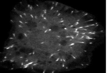
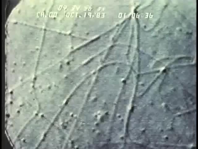
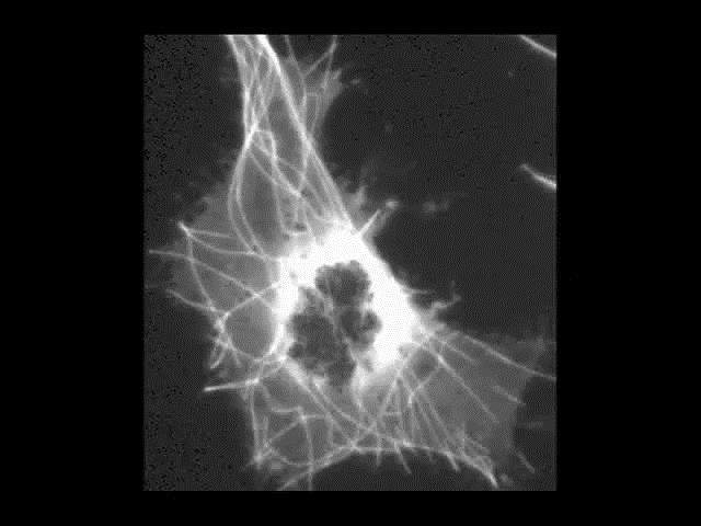
66 The cytoskeleton is a network of fibers extending throughout the cytoplasm It organizes the cell s structures and activities, anchoring many organelles It is composed of three types of molecular structures Microtubules Microfilaments Intermediate filaments

67 Figure μm
68

69
70
71 The cytoskeleton helps to support the cell and maintain its shape It interacts with motor proteins to produce motility Inside the cell, vesicles can travel along tracks provided by the cytoskeleton
 (a) Motor proteins walk")
72 Figure 6.21 ATP Vesicle Receptor for motor protein Microtubule Motor protein (ATP powered) (a) Motor proteins walk vesicles along cytoskeletal fibers. Vesicles Microtubule of cytoskeleton 0.25 μm (b) SEM of a squid giant axon
73
74

75
76 Three main types of fibers make up the cytoskeleton Microtubules are the thickest of the three components of the cytoskeleton Microfilaments, also called actin filaments, are the thinnest components Intermediate filaments are fibers with diameters in a middle range
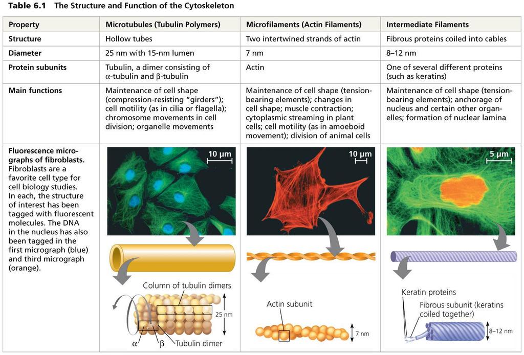
77 Table 6.1
78 Microtubules are hollow rods about 25 nm in diameter and about 200 nm to 25 microns long Functions of microtubules Shaping the cell Guiding movement of organelles Separating chromosomes during cell division
79 Centrosomes and Centrioles In animal cells, microtubules grow out from a centrosome near the nucleus In animal cells, the centrosome has a pair of centrioles, each with nine triplets of microtubules arranged in a ring

80 Figure 6.22 Centrosome Microtubule Centrioles 0.25 μm Longitudinal section of one centriole Microtubules Cross section of the other centriole
81 Cilia and Flagella Microtubules control the beating of flagella and cilia, microtubule-containing extensions that project from some cells Cilia and flagella differ in their beating patterns
 Motion of flagella Direction of swimming 5 μm (b) Motion of cilia")
82 Figure 6.23 (a) Motion of flagella Direction of swimming 5 μm (b) Motion of cilia Direction of organism s movement Power stroke Recovery stroke 15 μm
83
84 Microfilaments are solid rods about 7 nm in diameter, built as a twisted double chain of actin subunits The structural role of microfilaments is to bear tension, resisting pulling forces within the cell They form a 3-D network called the cortex just inside the plasma membrane to help support the cell s shape Bundles of microfilaments make up the core of microvilli of intestinal cells

85 0.25 µm Figure 6.25 Microvillus Plasma membrane Microfilaments (actin filaments) Intermediate filaments
86 Microfilaments that function in cellular motility contain the protein myosin in addition to actin In muscle cells, thousands of actin filaments are arranged parallel to one another Thicker filaments composed of myosin interdigitate with the thinner actin fibers
 Myosin motors in muscle cell contraction Chloroplast (c) Cytoplasmic streaming in plant cells 30 µm")
87 Figure 6.26 Muscle cell 0.5 µm Actin filament Myosin filament Myosin head (a) Myosin motors in muscle cell contraction Chloroplast (c) Cytoplasmic streaming in plant cells 30 µm Cortex (outer cytoplasm): gel with actin network 100 µm Inner cytoplasm (more fluid) (b) Amoeboid movement Extending pseudopodium
88
89 Cells crawl along a surface by extending pseudopodia (cellular extensions) and moving toward them Cytoplasmic streaming is a circular flow of cytoplasm within cells This streaming speeds distribution of materials within the cell
90 Intermediate filaments range in diameter from 8 12 nanometers, larger than microfilaments but smaller than microtubules They support cell shape and fix organelles in place Intermediate filaments are more permanent cytoskeleton fixtures than the other two classes

91 Most cells synthesize and secrete materials that are external to the plasma membrane These extracellular structures are involved in a great many cellular functions
92 The cell wall is an extracellular structure that distinguishes plant cells from animal cells Prokaryotes, fungi, and some unicellular eukaryotes also have cell walls The cell wall protects the plant cell, maintains its shape, and prevents excessive uptake of water Plant cell walls are made of cellulose fibers embedded in other polysaccharides and protein
93 Plant cell walls may have multiple layers Primary cell wall: Relatively thin and flexible Middle lamella: Thin layer between primary walls of adjacent cells Secondary cell wall (in some cells): Added between the plasma membrane and the primary cell wall Plasmodesmata are channels between adjacent plant cells

94 Figure 6.27 Secondary cell wall Primary cell wall Middle lamella 1 μm Central vacuole Cytosol Plasma membrane Plant cell walls Plasmodesmata
95 Animal cells lack cell walls but are covered by an elaborate extracellular matrix (ECM) The ECM is made up of glycoproteins such as collagen, proteoglycans, and fibronectin ECM proteins bind to receptor proteins in the plasma membrane called integrins

96 Figure 6.28 EXTRACELLULAR FLUID A proteoglycan complex Collagen Fibronectin Polysaccharide molecule Carbohydrates Core protein Plasma membrane Microfilaments CYTOPLASM Integrins Proteoglycan molecule
97 The ECM has an influential role in the lives of cells ECM can regulate a cell s behavior by communicating with a cell through integrins The ECM around a cell can influence the activity of gene in the nucleus Mechanical signaling may occur through cytoskeletal changes, that trigger chemical signals in the cell
98 Neighboring cells in tissues, organs, or organ systems often adhere, interact, and communicate through direct physical contact
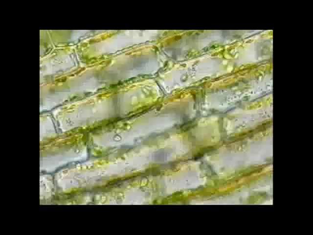
99 Plasmodesmata are channels that perforate plant cell walls Through plasmodesmata, water and small solutes (and sometimes proteins and RNA) can pass from cell to cell

100 Figure 6.29 Cell walls Interior of cell Interior of cell 0.5 μm Plasmodesmata Plasma membranes
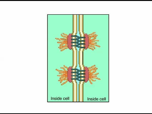
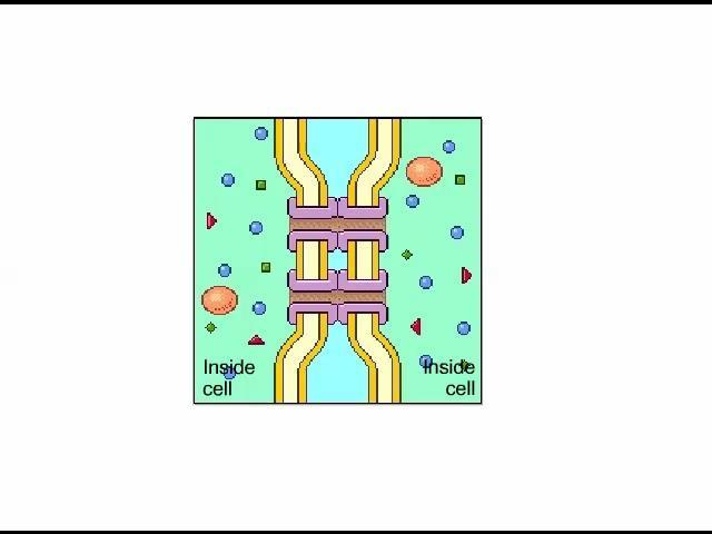
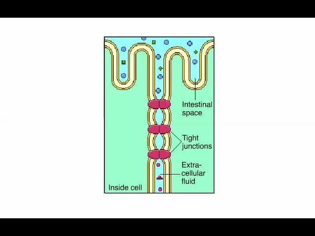
101 Three types of cell junctions are common in epithelial tissues At tight junctions, membranes of neighboring cells are pressed together, preventing leakage of extracellular fluid Desmosomes (anchoring junctions) fasten cells together into strong sheets Gap junctions (communicating junctions) provide cytoplasmic channels between adjacent cells
 1 μm Ions or small molecules Plasma membranes of adjacent cells Space")
102 TEM Figure 6.30 Tight junctions prevent fluid from moving across a layer of cells. Tight junction TEM 0.5 μm Tight junction Intermediate filaments Desmosome Gap junction Desmosome (TEM) 1 μm Ions or small molecules Plasma membranes of adjacent cells Space between cells Extracellular matrix 0.1 μm Gap junctions
103
104
105
106 Cells rely on the integration of structures and organelles in order to function For example, a macrophage s ability to destroy bacteria involves the whole cell, coordinating components such as the cytoskeleton, lysosomes, and plasma membrane

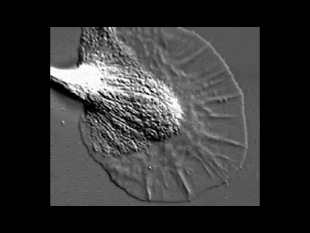
107 Figure μm
4 A Tour of the Cell CAMPBELL BIOLOGY IN FOCUS. Urry Cain Wasserman Minorsky Jackson Reece
 CAMPBELL BIOLOGY IN FOCUS Urry Cain Wasserman Minorsky Jackson Reece 4 A Tour of the Cell Lecture Presentations by Kathleen Fitzpatrick and Nicole Tunbridge Overview: The Fundamental Units of Life All
Human height. Length of some nerve and muscle cells. Chicken egg. Frog egg. Most plant and animal cells Nucleus Most bacteria Mitochondrion
 10 m 1 m 0.1 m 1 cm Human height Length of some nerve and muscle cells Chicken egg Unaided eye 1 mm Frog egg 100 µm 10 µm 1 µm 100 nm 10 nm Most plant and animal cells Nucleus Most bacteria Mitochondrion
A Tour of the Cell. Chapter 6. Biology. Edited by Shawn Lester. Inner Life of Cell. Eighth Edition Neil Campbell and Jane Reece
 Chapter 6 A Tour of the Cell Inner Life of Cell Edited by Shawn Lester PowerPoint Lecture Presentations for Biology Eighth Edition Neil Campbell and Jane Reece Lectures by Chris Romero, updated by Erin
(a) TEM of a plasma. Fimbriae. Nucleoid. Ribosomes. Plasma membrane. Cell wall Capsule. Bacterial chromosome
 TEM of a plasma. Fimbriae. Nucleoid. Ribosomes. Plasma membrane. Cell wall Capsule. Bacterial chromosome") 0 m m 0. m cm mm 00 µm 0 µm 00 nm 0 nm Human height Length of some nerve and muscle cells Chicken egg Frog egg Most plant and animal cells Most bacteria Smallest bacteria Viruses Proteins Unaided eye Light
0 m m 0. m cm mm 00 µm 0 µm 00 nm 0 nm Human height Length of some nerve and muscle cells Chicken egg Frog egg Most plant and animal cells Most bacteria Smallest bacteria Viruses Proteins Unaided eye Light
CH 4: A tour of the cell Overview: The Fundamental Units of Life. Concept 4.1: Biologists use microscopes and the tools of biochemistry to study cells
 CH 4: A tour of the cell Overview: The Fundamental Units of Life All organisms are made of cells The cell is the simplest collection of matter that is alive All cells are related by descent from earlier
A Tour of the Cell. Chapter 6. Biology Eighth Edition Neil Campbell and Jane Reece. PowerPoint Lecture Presentations for
 Chapter 6 A Tour of the Cell PowerPoint Lecture Presentations for Biology Eighth Edition Neil Campbell and Jane Reece Lectures by Chris Romero, updated by Erin Barley with contributions from Joan Sharp
Lecture 5- A Tour of the Cell
 Lecture 5- A Tour of the Cell 1 In this lecture Prokaryotes vs. eukaryotes The organelles of the eukaryotic cell The cytoskeleton Extracellular components 2 What are cells? Cells are the fundamental unit
A Tour of the Cell 4/10/12. Chapter 6. Overview: The Fundamental Units of Life
 Chapter 6 LECTURE PRESENTATIONS For CAMPBELL BIOLOGY, NINTH EDITION Jane B. Reece, Lisa A. Urry, Michael L. Cain, Steven A. Wasserman, Peter V. Minorsky, Robert B. Jackson A Tour of the Cell Lectures by
LECTURE PRESENTATIONS
 LECTURE PRESENTATIONS For CAMPBELL BIOLOGY, NINTH EDITION Jane B. Reece, Lisa A. Urry, Michael L. Cain, Steven A. Wasserman, Peter V. Minorsky, Robert B. Jackson Chapter 6 A Tour of the Cell Lectures by
BIOLOGY. A Tour of the Cell CAMPBELL. Reece Urry Cain Wasserman Minorsky Jackson. Lecture Presentation by Nicole Tunbridge and Kathleen Fitzpatrick
 CAMPBELL BIOLOGY TENTH EDITION Reece Urry Cain Wasserman Minorsky Jackson 6 A Tour of the Cell Lecture Presentation by Nicole Tunbridge and Kathleen Fitzpatrick Concept 6.2: Eukaryotic cells have internal
Lectures by Erin Barley Kathleen Fitzpatrick Pearson Education, Inc Pearson Education, Inc. Figure 6.5. Bacterial chromosome
 Chapter 6 LECTURE PRESENTATIONS For CAMPBELL BIOLOGY, NINTH EDITION Jane B. Reece, Lisa A. Urry, Michael L. Cain, Steven A. Wasserman, Peter V. Minorsky, Robert B. Jackson A Tour of the Cell Overview:
General Biology. The Fundamental Unit of Life The Cell. All organisms are made of cells The cell is the simplest collection of matter that can live
 General Biology Course No: BNG2003 Credits: 3.00 3. A Tour of the Cell Prof. Dr. Klaus Heese The Fundamental Unit of Life The Cell All organisms are made of cells The cell is the simplest collection of
A Tour of the Cell. Chapter 6. Biology Eighth Edition Neil Campbell and Jane Reece. PowerPoint Lecture Presentations for
 Chapter 6 1 A Tour of the Cell PowerPoint Lecture Presentations for Biology Eighth Edition Neil Campbell and Jane Reece Lectures by Chris Romero, updated by Erin Barley with contributions from Joan Sharp
A TOUR OF THE CELL 10/1/2012
 A TOUR OF THE CELL Chapter 6 KEY CONCEPTS: Eukaryotic cells have internal membranes that compartmentalize their functions The eukaryotic cell s genetic instructions are housed in the nucleus and carried
Ch. 6 Tour of the Cell
 Ch. 6 Tour of the Cell 2007-2008 Microscopy Scientists use microscopes to visualize cells too small to see with the naked eye In a light microscope (LM), visible light is passed through a specimen and
CHAPTER 6: A TOUR OF THE CELL AP BIOLOGY 2011
 CHAPTER 6: A TOUR OF THE CELL AP BIOLOGY 2011 1 IMPORTANCE OF CELLS ALL ORGANISMS ARE MADE OF CELLS CELLS ARE THE SMALLEST LIVING UNIT STRUCTURE IS CORRELATED TO FUNCTION ALL CELLS ARE RELATED BY THEIR
A Tour of the Cell. Chapter 7
 A Tour of the Cell Chapter 7 Cytology: Study of Cells Light Microscopes uses light & a set of lenses Magnification ratio of object s image size to its real size Resolution measures the clarity of the image
A Tour of the Cell CAMPBELL BIOLOGY IN FOCUS URRY CAIN WASSERMAN MINORSKY REECE. Overview: The Fundamental Units of Life
 4 A Tour of the Cell CAMPBELL BIOLOGY IN FOCUS URRY CAIN WASSERMAN MINORSKY REECE Lecture Presentations by Kathleen Fitzpatrick and Nicole Tunbridge, Simon Fraser University SECOND EDITION Overview: The
LECTURE PRESENTATIONS
 LECTURE PRESENTATIONS For CAMPBELL BIOLOGY, NINTH EDITION Jane B. Reece, Lisa A. Urry, Michael L. Cain, Steven A. Wasserman, Peter V. Minorsky, Robert B. Jackson Chapter 6 A Tour of the Cell Lectures by
A Tour of the Cell. Chapter 6. PowerPoint Lecture Presentations for Biology Eighth Edition Neil Campbell and Jane Reece
 Chapter 6 A Tour of the Cell PowerPoint Lecture Presentations for Biology Eighth Edition Neil Campbell and Jane Reece Lectures by Chris Romero, updated by Erin Barley with contributions from Joan Sharp
A. Major parts 1. Nucleus 2. Cytoplasm a. Contain organelles (see below) 3. Plasma membrane (To be discussed in Cellular Transport Lecture)
 3. Plasma membrane (To be discussed in Cellular Transport Lecture)") Lecture 5: Cellular Biology I. Cell Theory Concepts: 1. Cells are the functional and structural units of living organisms 2. The activity of an organism is dependent on both the individual and collective
Lecture 5: Cellular Biology I. Cell Theory Concepts: 1. Cells are the functional and structural units of living organisms 2. The activity of an organism is dependent on both the individual and collective
A Tour of the Cell. Chapter 6. Slide 1. Slide 2. Slide 3. Overview: The Fundamental Units of Life
 Slide 1 Chapter 6 A Tour of the Cell PowerPoint Lecture Presentations for Biology Eighth Edition Neil Campbell and Jane Reece Lectures by Chris Romero, updated by Erin Barley with contributions from Joan
Early scientists who observed cells made detailed sketches of what they saw.
 Early scientists who observed cells made detailed sketches of what they saw. Early scientists who observed cells made detailed sketches of what they saw. CORK Early scientists who observed cells made detailed
LECTURE PRESENTATIONS
 LECTURE PRESENTATIONS For CAMPBELL BIOLOGY, NINTH EDITION Jane B. Reece, Lisa A. Urry, Michael L. Cain, Steven A. Wasserman, Peter V. Minorsky, Robert B. Jackson Chapter 6 A Tour of the Cell Lectures by
The Fundamental Unit of Life The Cell. General Biology. All organisms are made of cells. The cell is the simplest collection of matter that can live
 Course No: BNG2003 Credits: 3.00 3. A Tour of the Cell General Biology The Fundamental Unit of Life The Cell All organisms are made of cells The cell is the simplest collection of matter that can live
Chapter 6: A Tour of the Cell. 1. Studying Cells 2. Intracellular Structures 3. The Cytoskeleton 4. Extracellular Structures
 Chapter 6: A Tour of the Cell 1. Studying Cells 2. Intracellular Structures 3. The Cytoskeleton 4. Extracellular Structures 1. Studying Cells Concepts of Microscopy MAGNIFICATION factor by which the image
1. Studying Cells. Concepts of Microscopy 11/7/2016. Chapter 6: A Tour of the Cell
 Electron microscope Light microscope Unaided eye 11/7/2016 Chapter 6: A Tour of the Cell 1. Studying Cells 2. Intracellular Structures 3. The Cytoskeleton 4. Extracellular Structures 1. Studying Cells
A Tour of the Cell. Chapter 6. Biology Eighth Edition Neil Campbell and Jane Reece. PowerPoint Lecture Presentations for
 Chapter 6 A Tour of the Cell PowerPoint Lecture Presentations for Biology Eighth Edition Neil Campbell and Jane Reece Lectures by Chris Romero, updated by Erin Barley with contributions from Joan Sharp
AP Biology Summer Assignment
 AP Biology Summer Assignment 2018-2019 AP Biology is a rigorous course and due to the large amount of material that needs to be covered during the school year, a summer assignment is essential. The first
Cytosol the fluid Cytoplasm cell interior, everything outside the nucleus but within the cell membrane, includes the organelles, cytosol, and
 Cell Organelles Plasma Membrane comprised of a phospholipid bilayer and embedded proteins Outer surface has oligosaccharides separates the cells s contents from its surroundings Cytosol the fluid Cytoplasm
All organisms are made of cells (cells are the basic units of life) Cell structure is highly correlated to cellular function
 Cell structure is highly correlated to cellular function") CELLS CHAPTER 6 I. CELL THEORY - All organisms are made of cells (cells are the basic units of life) Cell structure is highly correlated to cellular function All cells are related by their descent from
CELLS CHAPTER 6 I. CELL THEORY - All organisms are made of cells (cells are the basic units of life) Cell structure is highly correlated to cellular function All cells are related by their descent from
The Golgi Apparatus: Shipping and Receiving Center. The Golgi apparatus. Functions of the Golgi apparatus. Lysosomes: Digestive Compartments
 The Golgi Apparatus: Shipping and Receiving Center The Golgi apparatus Receives (on the cis-side) many of the transport vesicles produced in the rough ER Consists of flattened membranous sacs called cisternae
Cell Theory. Chapter 6. cell. fundamental unit of structure and function for all living organisms. arise only from previously existing cell
 Chapter 6 cell Cell Theory fundamental unit of structure and function for all living organisms arise only from previously existing cell Figure 5.4 The size range of cells WHY are your brain cells the same
Microfilaments. myosin. In muscle cells. Microfilaments. Microfilaments. Video: Cytoplasmic Streaming. amoeboid movement. Pseudopodia.
 Microfilaments Fig, 6-27a myosin Microfilaments protein func3ons in cellular mo3lity in addi3on to ac3n In muscle cells Thousands of ac3n filaments are arranged parallel to one another Thicker myosin filaments
CHAPTER 6 A TOUR OF THE CELL
 Electron microscope Light microscope Unaided eye Overview: The Fundamental Units of Life All organisms are made of cells The cell is the simplest collection of matter that can live Cell structure is correlated
Nucleic acids. Nucleic acids are information-rich polymers of nucleotides
 Nucleic acids Nucleic acids are information-rich polymers of nucleotides DNA and RNA Serve as the blueprints for proteins and thus control the life of a cell RNA and DNA are made up of very similar nucleotides.
A Tour of the Cell. Chapter 6. Biology Eighth Edition Neil Campbell and Jane Reece. PowerPoint Lecture Presentations for
 Chapter 6 A Tour of the Cell PowerPoint Lecture Presentations for Biology Eighth Edition Neil Campbell and Jane Reece Lectures by Chris Romero, updated by Erin Barley with contributions from Joan Sharp
Chapter 6. A Tour of the Cell. Concept 6.1 Biologists use microscopes and the tools of biochemistry to study cells
 Chapter 6 A Tour of the Cell Chapter Outline Concept 6.1 Biologists use microscopes and the tools of biochemistry to study cells In a light microscope (LM), visible light passes through the specimen and
A Tour of the Cell. Ch. 7
 A Tour of the Cell Ch. 7 Cell Theory O All organisms are composed of one or more cells. O The cell is the basic unit of structure and organization of organisms. O All cells come from preexisting cells.
Plasma Membrane. comprised of a phospholipid bilayer and embedded proteins separates the cells s contents from its surroundings
 Cell Organelles Plasma Membrane comprised of a phospholipid bilayer and embedded proteins separates the cells s contents from its surroundings Cytosol the fluid Cytoplasm cell interior, everything outside
Cells. Variation and Function of Cells
 Cells Variation and Function of Cells Cell Theory states that: 1. All living things are made of cells 2. Cells are the basic unit of structure and function in living things 3. New cells are produced from
Thursday, October 16 th
 Thursday, October 16 th Good morning. Those of you needing to take the Enzymes and Energy Quiz will start very soon. Students who took the quiz Wednesday: Please QUIETLY work on the chapter 6 reading guide.
NOTES: CH 6 A Tour of the Cell
 NOTES: CH 6 A Tour of the Cell Overview: The Importance of Cells All organisms are made of cells The cell is the simplest collection of matter that can live Cell structure is correlated to cellular function
Review from Biology A
 Chapter 4 Review from Biology A The Cell Theory All organisms are made of cells Cells come from pre-existing cells The cell is the simplest collection of matter that can live Scientists whose work you
BIOLOGY. A Tour of the Cell CAMPBELL. Robert Hooke (1665) Antoni van Leeuwenhoek (1674) Reece Urry Cain Wasserman Minorsky Jackson
 Antoni van Leeuwenhoek (1674) Reece Urry Cain Wasserman Minorsky Jackson") 6 A Tour of the Cell CAMPBELL BIOLOGY TENTH EDITION Reece Urry Cain Wasserman Minorsky Jackson Lecture Presentation by Nicole Tunbridge and Kathleen Fitzpatrick Robert Hooke (1665) Figure 1.1 The structure
6 A Tour of the Cell CAMPBELL BIOLOGY TENTH EDITION Reece Urry Cain Wasserman Minorsky Jackson Lecture Presentation by Nicole Tunbridge and Kathleen Fitzpatrick Robert Hooke (1665) Figure 1.1 The structure
10 m Human height 1 m Length of some nerve and muscle cells eye 100 mm (10 cm) Chicken egg aid n 10 mm
 Chicken egg aid n 10 mm") Biology 112 Unit Three Chapter Four 1 Cell Sizes Smallest Bacteria Largest Bird egg Longest Giraffe s Nerve Cell Most Cells Diameter of 0.7µm to 105 µm 2 10 m 1 m 100 mm (10 cm) 10 mm (1 cm) Human height
Biology 112 Unit Three Chapter Four 1 Cell Sizes Smallest Bacteria Largest Bird egg Longest Giraffe s Nerve Cell Most Cells Diameter of 0.7µm to 105 µm 2 10 m 1 m 100 mm (10 cm) 10 mm (1 cm) Human height
CHAPTER 4 A TOUR OF THE CELL
 CHAPTER 4 A TOUR OF THE CELL Microscopes Con. 4.1 magnification: size resolution: clarity contrast: differences in parts Light Microscopy Techniques (p.68) a. Brightfield unstained b. Brightfield stained
4/12/17. Cells. Cell Structure. Ch. 2 Cell Structure and Func.on. Range of Cell Sizes BIOL 100
 Ch. 2 Cell Structure and Func.on BIOL 100 Cells Fundamental units of life Cell theory All living things are composed of one or more cells. The cell is the most basic unit of life. All cells come from pre-existing
10/13/11. Cell Theory. Cell Structure
 Cell Structure Grade 12 Biology Cell Theory All organisms are composed of one or more cells. Cells are the smallest living units of all living organisms. Cells arise only by division of a previously existing
A Tour of the Cell. Chapter 4. Most cells are microscopic. Cells vary in size and shape
 Chapter 4 A Tour of the Cell Most cells are microscopic Cells vary in size and shape 10 m Human height 1 m Length of some nerve and muscle cells 100 mm (10 cm) 10 mm (1 cm) Chicken egg Unaided eye 1 mm
Chapter 4. A Tour of the Cell. Lectures by Edward J. Zalisko
 Chapter 4 A Tour of the Cell PowerPoint Lectures for Campbell Essential Biology, Fifth Edition, and Campbell Essential Biology with Physiology, Fourth Edition Eric J. Simon, Jean L. Dickey, and Jane B.
Ch. 6 A Tour of the Cell BIOL 222
 Ch. 6 A Tour of the Cell BIOL 222 Overview: The Fundamental Units of Life All organisms are made of cells The cell is the simplest collec=on of ma>er that can live Cell structure is correlated to cellular
Cytology. Light microscopy resolving power Electron microscopy TEM SEM Cell fractionation Ultracentrifuges
 Chapter 7: A Tour of the Cell Cytology Light microscopy resolving power Electron microscopy TEM SEM Cell fractionation Ultracentrifuges Prokaryotic cells Nucleoid No organelles with membranes Ribosomes
Chapter 7. (7-1 and 7-2) A Tour of the Cell
 A Tour of the Cell") Chapter 7 (7-1 and 7-2) A Tour of the Cell Microscopes as Windows to the World of Cells Cells were first described in 1665 by Robert Hooke. By the mid-1800s, the accumulation of scientific evidence led
Chapter 7 (7-1 and 7-2) A Tour of the Cell Microscopes as Windows to the World of Cells Cells were first described in 1665 by Robert Hooke. By the mid-1800s, the accumulation of scientific evidence led
CELL PARTS TYPICAL ANIMAL CELL
 AP BIOLOGY CText Reference, Campbell v.8, Chapter 6 ACTIVITY1.12 NAME DATE HOUR CELL PARTS TYPICAL ANIMAL CELL ENDOMEMBRANE SYSTEM TYPICAL PLANT CELL QUESTIONS: 1. Write the name of the cell part in the
Cytology = the study of cells. Chapter 4 CELL STRUCTURE
 Cytology = the study of cells Chapter 4 CELL STRUCTURE Cellular basis of life: Basic unit of life Lowest level with all attributes of life Organisms composed of one or more cells Cell structure correlated
ORGANELLES OF THE ENDOMEMBRANE SYSTEM
 Membranes compartmentalize the interior of the cell and facilitate a variety of metabolic activities. Chloroplasts and a rigid cell wall are what distinguish a plant cell from an animal cell. A typical
Lysosomes. Vacuoles. Phagocytosis. One cell engulfing another. forms a food vacuole. fuses with lysosome. Autophagy. Lysosomes use enzymes
 Lysosomes Phagocytosis One cell engulfing another forms a food vacuole fuses with lysosome Autophagy Lysosomes use enzymes to recycle the cell s own organelles and macromolecules Fig. 6-14 Nucleus 1 µm
A Tour of the Cell Lecture 2, Part 1 Fall 2008
 Cell Theory 1 A Tour of the Cell Lecture 2, Part 1 Fall 2008 Cells are the basic unit of structure and function The lowest level of structure that can perform all activities required for life Reproduction
Chapter 4 A Tour of the Cell
 Chapter 4 A Tour of the Cell PowerPoint Lectures Campbell Biology: Concepts & Connections, Eighth Edition REECE TAYLOR SIMON DICKEY HOGAN Lecture by Edward J. Zalisko Introduction Cells have a cytoskeleton
Chapter 6. A Tour of the Cell
 Chapter 6 A Tour of the Cell PowerPoint lectures are originally from Campbell / Reece Media Manager and Instructor Resources for BIOLOGY, 7 th & 8 th Edition by N. A. Campbell & J. B. Reece Copyright 2005
Chapter 4. A Tour of the Cell. Lecture by Richard L. Myers
 Chapter 4 A Tour of the Cell PowerPoint Lectures for Biology: Concepts & Connections, Sixth Edition Campbell, Reece, Taylor, Simon, and Dickey Lecture by Richard L. Myers Introduction: Cells on the Move
Chapter 4. A Tour of the Cell. Lecture by Richard L. Myers
 Chapter 4 A Tour of the Cell PowerPoint Lectures for Biology: Concepts & Connections, Sixth Edition Campbell, Reece, Taylor, Simon, and Dickey Lecture by Richard L. Myers Introduction: Cells on the Move
Organelles. copyright cmassengale 1
 Organelles copyright cmassengale 1 Organelles Very small (Microscopic) Perform various functions for a cell Found in the cytoplasm May or may not be membrane-bound 2 Animal Cell Organelles Nucleolus Nucleus
Cell Structure & Function. Source:
 Cell Structure & Function Source: http://koning.ecsu.ctstateu.edu/cell/cell.html Definition of Cell A cell is the smallest unit that is capable of performing life functions. http://web.jjay.cuny.edu/~acarpi/nsc/images/cell.gif
Cell Structure. Cells. Why are cells so small? 9/15/2016. Schleiden and Schwann proposed Cell Theory in
 Cell Structure Cells Cells are sacs of fluid that are reinforced by proteins and surrounded by membranes. Inside the fluid float organelles. Organelles: structures inside the cell that are used for metabolic
Organelles of the Cell & How They Work Together. Packet #7
 Organelles of the Cell & How They Work Together Packet #7 Introduction Introduction Organization of cells is basically similar in all cells. Additionally, most cells are tiny Ranging from 1 1000 cubic
Cytology II Study of Cells
 Cytology II Study of Cells Biology 20 Cellular Basis of Life 1. Basic unit of Life 2. Composed of one or more cells 3. Arises from pre-existing cells Asexual (Mitosis)/Sexual (Meiosis) 4. Surrounded by
A Tour of the Cell Chapter 4. Outline. Early contributors to Understanding Cells. Cell Theory. Cell Size s Matt Schleiden & Ted Schann
 A Tour of the Cell Chapter 4 Outline History of the science behind cells Cell theory & its importance Why are cells small? Microscopes Cell structure and function Prokaryotic cells Eukaryotic cells Early
Chapter 4: Cell Structure and Function
 Chapter 4: Cell Structure and Function Robert Hooke Fig. 4-2, p.51 The Cell Smallest unit of life Can survive on its own or has potential to do so Is highly organized for metabolism Senses and responds
Eukaryotic cell. Premedical IV Biology
 Eukaryotic cell Premedical IV Biology The size range of organisms Light microscopes visible light is passed through the specimen and glass lenses the resolution is limited by the wavelength of the visible
Chapter 4 A Tour of the Cell
 Chapter 4 A Tour of the Cell PowerPoint Lectures for Campbell Biology: Concepts & Connections, Seventh Edition Reece, Taylor, Simon, and Dickey Lecture by Edward J. Zalisko The image The Introduction Cells
Ch. 7 Inside the Cell BIOL 222
 Ch. 7 Inside the Cell BIOL 222 Overview: The Fundamental Units of Life All organisms are made of cells The cell is the simplest collec=on of ma>er that can live Cell structure is correlated to cellular
Chapter 4. A Tour of the Cell. RPTSE Biology Fall 2015, Dr. Jong B. Lee 1. Biology and Society: Antibiotics: Drugs that Target Bacterial Cells
 Chapter 4 A Tour of the Cell Biology and Society: Antibiotics: Drugs that Target Bacterial Cells Antibiotics were first isolated from mold in 1928. The widespread use of antibiotics drastically decreased
Cell Structure and Function
 Cell Theory Cell Structure and Function Chapter 6 Pg. 94-124 What is a cell? The basic functional unit of all living things. The Cell Theory states All organisms are made of one or more cells. Cells are
Organelles of the Cell & How They Work Together. Packet #5
 Organelles of the Cell & How They Work Together Packet #5 Developed by Mr. Barrow 2018 1 Introduction Organization of cells is basically similar in all cells. Additionally, most cells are tiny Ranging
A Tour of the cell. 2- Eukaryotic cells have internal membranes that compartmentalize their functions
 A Tour of the cell 1- To study cells, biologists use microscopes and the tools of biochemistry 2- Eukaryotic cells have internal membranes that compartmentalize their functions 3- The eukaryotic cell s
Chapter 4. A Tour of the Cell. Lectures by Chris C. Romero, updated by Edward J. Zalisko
 Chapter 4 A Tour of the Cell Lectures by Chris C. Romero, updated by Edward J. Zalisko PowerPoint Lectures for Campbell Essential Biology, Fourth Edition Eric Simon, Jane Reece, and Jean Dickey Campbell
A Tour of the Cell. reference: Chapter 6. Reference: Chapter 2
 A Tour of the Cell reference: Chapter 6 Reference: Chapter 2 Monkey Fibroblast Cells stained with fluorescent dyes to show the nucleus (blue) and cytoskeleton (yellow and red fibers), image courtesy of
Unit A: Cells. Ch. 4 A Tour of the Cell
 Unit A: Cells Ch. 4 A Tour of the Cell Standards By the end of this unit you should be able to: Recognize and explain the function of each organelle Look at micrographs/diagrams/pictures and correctly
Chapter 4. A Tour of the Cell. Lectures by Edward J. Zalisko
 Chapter 4 A Tour of the Cell PowerPoint Lectures for Campbell Essential Biology, Fifth Edition, and Campbell Essential Biology with Physiology, Fourth Edition Eric J. Simon, Jean L. Dickey, and Jane B.
Organelles of the Cell & How They Work Together. Packet #5
 Organelles of the Cell & How They Work Together Packet #5 Developed by Mr. Barrow 2018 1 Introduction Organization of cells is basically similar in all cells. Additionally, most cells are tiny Ranging
2011 Pearson Education, Inc.
 Overview: The Fundamental Units of Life All organisms are made of cells The cell is the simplest collec=on of ma>er that can be alive Cell structure is correlated to cellular func=on All cells are related
2011 Pearson Education, Inc.
 Overview: The Fundamental Units of Life All organisms are made of cells The cell is the simplest collec=on of ma>er that can be alive Cell structure is correlated to cellular func=on All cells are related
Name 4 A Tour of the Cell Test Date Study Guide You must know: The difference between prokaryotic and eukaryotic cells. The structure and function of
 Name _ 4 A Tour of the Cell Test Date Study Guide You must know: The difference between prokaryotic and eukaryotic cells. The structure and function of organelles common to plant and animal cells. The
BIOSC 041. v Today s lecture. v Today s lab. v Note- Monday is a holiday good time to do some reading!
 BIOSC 041 v Today s lecture Review questions Chapter 6, Cells More review questions v Today s lab Quick review of lab safety The Scientific Method start thinking about which environments you might want
BIOLOGY. A Tour of the Cell. Outline. Cell Theory. Cells. Cell Size. Cell Theory
 6 A Tour of the Cell CAMPBELL BIOLOGY TENTH EDITION Reece Urry Cain Wasserman Minorsky Jackson Outline I. Cell Theory II. Studying cells III. Prokaryotic vs Eukaryotic IV. Eukaryotic A. Animal cells B.
Human Epithelial Cells
 The Cell Human Epithelial Cells Plant Cells Cells have an internal structure Eukaryotic cells are organized Protective membrane around them that communicates with other cells Organelles have specific jobs
First to View Cells. copyright cmassengale
 CELL THEORY All living things are made of cells Cells are the basic unit of structure and function in an organism (basic unit of life) Cells come from the reproduction of existing cells (cell division)
Chapter 4. A Tour of the Cell. Lectures by Edward J. Zalisko
 Chapter 4 A Tour of the Cell PowerPoint Lectures for Campbell Essential Biology, Fifth Edition, and Campbell Essential Biology with Physiology, Fourth Edition Eric J. Simon, Jean L. Dickey, and Jane B.
Bio10 Cell Structure SRJC
 3.) Cell Structure and Function Structure of Cell Membranes Fluid mosaic model Mixed composition: Phospholipid bilayer Glycolipids Sterols Proteins Fluid Mosaic Model Phospholipids are not packed tightly
Ch. 6: A Tour of the Cell
 Ch. 6: A Tour of the Cell 1. Compare the 2 Types of Cells PROKARYOTES BOTH EUKARYOTES Domain: Domain: Relative Size & Complexity: Relative Size & Complexity: No DNA in No Examples: Has Has Examples: 2.
Animal & Plant Cells Biology 20
 Animal & Plant Cells Biology 20 Structures in Cells ALL cells start out as fully functional living things They must be able to create and maintain substances (compounds, ATP, ADP) and structures (membranes,
AP Biology Book Notes Chapter 4: Cells v Cell theory implications Ø Studying cell biology is in some sense the same as studying life Ø Life is
 AP Biology Book Notes Chapter 4: Cells v Cell theory implications Ø Studying cell biology is in some sense the same as studying life Ø Life is continuous v Small cell size is becoming more necessary as
Don t Freak Out. Test on cell organelle on Friday!
 Cell Structure 1 Don t Freak Out Test on cell organelle on Friday! This test should be a buffer test and help raise your overall test score. All information will come from this week! 2 Cells Provide Compartments
Biology Structures in Cells. 1.3 Structures in Cells
 Biology 2201 1.3 Structures in Cells Structures in Cells ALL cells start out as fully functional living things They must be able to create and maintain substances (compounds, ATP, ADP) and structures (membranes,
Objectives. To determine the differences between plant and animal cells To discover the structure and function of cellular organelles.
 Cell Organelles 3.2 Objectives To determine the differences between plant and animal cells To discover the structure and function of cellular organelles. Basic Cellular Structures Cell membrane (cytoplasmic
Eukaryotic Cell Structures
 Comparing the Cell to a Factory Eukaryotic Cell Structures Structures within a eukaryotic cell that perform important cellular functions are known as organelles. Cell biologists divide the eukaryotic cell
11/1/2014. accumulate in brain.
 EU 4.A: Interactions within biological systems lead to complex properties. EU 4.B: Competition and cooperation are important aspects of biological systems. EU 4.C: Naturally occurring diversity among and