Diagnostic value of dynamic contrast-enhanced MRI for unilocular cystic-type
|
|
|
- Angela Sutton
- 5 years ago
- Views:
Transcription
1 Diagnostic value of dynamic contrast-enhanced MRI for unilocular cystic-type ameloblastomas with homogeneously bright high signal intensity on T2-weighted or STIR MR images Miki Hisatomi a, Yoshinobu Yanagi b, Hironobu Konouchi b, Hidenobu Matsuzaki b, Toshihiko Takenobu a, Teruhisa Unetsubo a, Junichi Asaumi a,b a Department of Oral and Maxillofacial Radiology, Field of Tumor Biology, Okayama University Graduate School of Medicine, Dentistry and Pharmaceutical Sciences, 5-1, Shikata-cho, 2-chome, Okayama , Japan b Department of Oral Diagnosis and Dentomaxillofacial Radiology, Okayama University Hospital of Medicine and Dentistry, 5-1, Shikata-cho, 2-chome, Okayama , Japan Corresponding Author: Miki Hisatomi address: tomi@md.okayama-u.ac.jp Full postal address: Department of Oral and Maxillofacial Radiology, Field of Tumor Biology, Okayama University Graduate School of Medicine, Dentistry and Pharmaceutical Sciences, 5-1, Shikata-cho, 2-chome, Okayama , Japan Telephone number: FAX number:
2 SUMMARY Typical MR images of ameloblastomas on T2-weighted image (WI) or short inversion time inversion-recovery (STIR) show multiple bright high-signal-intensity loci on a high-signal-intensity background. Unilocular cystic-type ameloblastomas show homogeneously bright high signal intensity on T2WI or STIR as a water-like signal intensity. Therefore, it is difficult to distinguish unilocular cystic-type ameloblastoma from other cystic lesions such as keratocystic odontogenic tumors, radicular cysts (residual cysts) and dentigerous cysts only on the basis of MRI signal intensity. In the present study, we evaluated whether contrast-enhanced (CE)-T1WI and dynamic CE-MRI (DCE-MRI) could provide additional information for differential diagnosis in unilocular cystic-type ameloblastoma. Images from 12 cases of suspected unilocular cystic-type ameloblastoma were evaluated in the present study. Of them, 5 had areas suspected of indicating a solid component on T1WI and T2WI (or STIR). Ten had undergone additional CE-T1WI and DCE-MRI. On 5 of 10 cases of CE-T1WI, a tiny enhancement area was detected. On 6 of 10 DCE-images, a time-course enhanced area which was suspected to be a solid component was detected. CE-T1WI was helpful in the diagnosis of ameloblastoma because the tiny enhanced areas were taken to indicate possible solid components. Moreover, the rim-enhancement area on CE-T1WI could be divided into small regions of interest, and some of these showed slightly increased enhancement on DCE-MRI, which was taken to indicate a solid component and/or intramural nodule with focal invasion of ameloblastoma tissue. DCE-MRIs of the 4 remaining cases, which provided no clues to the diagnosis of ameloblastoma in the manner of the above descriptions, showed thicker rim enhancement than odontogenic cysts. Thus, CE-T1WI and DCE-MRI were helpful in the differential diagnosis of unilocular cystic-type ameloblastomas with homogeneously bright high signal intensity on T2WI or STIR. Keywords: ameloblastoma, unicystic, sold/multicystic, MRI, DCE-MRI, odontogenic tumor, odontogenic cyst
3 Introduction Ameloblastoma is the most common and the most clinically significant odontogenic tumor. 1-4 Ameloblastomas show a remarkable tendency to undergo cystic change; most of them have combinations of cystic and solid features that vary over time. 1 The cystic portion can be present as a large cyst which occupies a sizable portion of the ameloblastoma. Ameloblastomas are divided into solid/multicystic, extraosseous/peripheral, desmoplastic and unicystic types according to the 2005 WHO histological classification of odontogenic tumors. 5 The most typical radiographic feature is multilocular radiolucency. The lesion is often described as having a soap bubble appearance. This feature is a direct clue to the diagnosis of ameloblastomas. On the other hand, it is difficult to diagnose ameloblastomas radiographically when they show unilocular features. Ameloblastomas that show unilocular radiolucency have two possible MR feature types. One is a mixed type with multicystic and solid components. On MRIs of this ameloblastoma type, the solid portions show a predilection for intermediate signal intensity on T1 weighted image (WI) and high signal intensity on T2WI or short inversion time inversion-recovery (STIR) images, and the cystic portions show a predilection for homogeneous intermediate signal intensity on T1WI and homogeneously bright high signal intensity on T2WI or STIR. 6 At that time we can easily diagnose these lesions as ameloblastomas because this type of T2-weighted MR image shows a multiple-locus bright high signal intensity corresponding to cystic fluids intermixed with areas of relatively high signal intensity corresponding to the solid portion. 7-9 Another is the unilocular cystic type, which shows homogeneously bright high signal intensity on T2WI or STIR reflecting the inner part
4 of the water-like content. Therefore, the latter type is difficult to distinguish from cystic lesions such as keratocystic odontogenic tumors, radicular cysts (residual cysts) and dentigerous cysts, which present as unicystic lesions, on the basis of MR signals only. In contrast-enhanced MRI (CE-MRI) the margins are more clearly defined, and secondary mass effects are more clearly shown. In addition, dynamic contrast enhanced MRI (DCE-MRI), which produces functional images, can illustrate the time course of contrast enhancement. DCE-MRI not only aids diagnosis but also provides measures that relate to the histological assessment of vascular density. 10, 11 Some studies have shown the rapid uptake patterns of invasive cancers on DEC-MRI reflect high intratumor angiogenesis. 12, 13 It has been reported that DCE-MRI is useful for the differential diagnosis of some tumors, and many investigators have attempted to identify the differences between benign and malignant tumors by analyzing curve patterns such as rapid uptake or gradual increase from the time course of dynamic contrast enhancement. 14, 15 Therefore, we may be able to differentiate unilocular cystic lesions including ameloblastomas by detecting nodular regions on CE-MRI and/or DEC-MRI, or by detecting areas of high intratumor angiogenesis on DEC-MRI. In the present study, we retrospectively evaluated the DCE-MRI of unilocular cystic-type ameloblastomas suspected of being cystic lesions due to homogeneously bright high signal intensity on T2WI (or STIR) MRI in order to assess whether the DCE-MRI could provide additional information in the differential diagnosis between cystic-type ameloblastomas and other cysts.
5 Materials and methods Patient population Thirty-five patients who were histopathologically diagnosed with ameloblastoma and underwent MR examinations in our university hospital between April, 1998 and December, 2009 were enrolled in this retrospective study. Of 35 cases, 12 that had been suspected of being cystic lesions including unilocular cystic-type ameloblastomas due to homogeneously bright high signal intensity on T2WI (or STIR) MRI were selectively examined. Of these 12 patients, 5 were men and 7 were women (age range: years; mean: 37.1 years) (Table 1). This study was approved by our institutional review board (No. 232). MR imaging The MR examination was performed on a 1.5-T unit (Magnetom Vision; Siemens, Erlangen, Federal Republic of Germany) with a CP head coil or a head-neck coil. Routine T1-weighted axial images were acquired with a spin-echo sequence using a repetition time (TR) of 500 to 660 ms and echo time (TE) of 15 ms. T2-weighted axial images in 5 cases or STIR axial images in 7 cases were acquired with a turbo-spin-echo sequence using 3000/90 ms (TR/TE) for T2WI and 4500/60/140 ms (TR/TE/TI) for STIR. For DCE-images, 14 consecutive data sets were acquired for 210 s (14 s/1 scan) with three-dimensional fast imaging with steady-state precession (repetition time/echo time/flip angle 5/2/25 degrees, 16 slices over 48 mm of slab thickness, resulting in an effective slice thickness of 3 mm). Frequency-selective fat-suppressed T1WI was
6 immediately acquired as CE-T1WI. Intravenous injection of a contrast medium (Omniscan syringe; Daiichi Pharmaceutical Co., Tokyo, Japan or Magnevist syringe; Nihon Schering, Osaka, Japan) was archived manually at a rate of approximately 2 ml/s through a 21-gauge butterfly needle inserted into a vein in the cubital fossa. The injection of contrast medium started 6 s before the initiation of a second scan of 14 DCE-image data sets. Image analysis The MR images in 12 cases were retrospectively examined as shown in Table 1. For the criteria of the signal intensity, the musculature was defined as intermediate signal on T1WI, and the cerebrospinal fluid as a bright high signal on T2WI or STIR. The signal intensity between intermediate and high signal intensities on T1WI was described as slightly high signal intensity. CE-T1WI and DCE-images were obtained in 10 cases. The T1WI, T2WI (or STIR), CE-T1WI and DCE-images were studied for signal intensity, cystic and solid features and marginal features. MR images of ameloblastomas suspected of being cystic lesions with homogeneously bright high signal intensity on T2WI or STIR were evaluated to assess whether T1WI, T2WI (or STIR), CE-T1WI and DCE-images could provide additional diagnostic information. Data analysis Regions of interest (ROI) were drawn in the suspected solid area and rim-enhancement area on DCE-images. The ROI of each lesion was calculated using the Volume Analyzer SYNAPSE VINCENT 3D image analysis system (FUJIFILM Medical, Tokyo, Japan). Time-to-intensity curves were obtained by plotting the signal intensity
7 on a time course.
8 Results Table 1 shows the clinical features and the results of the signal intensity on T1WI, T2WI (or STIR), CE-T1WI and DCE-images in 12 cases. In terms of histopathological diagnosis, 9 of the 12 cases were solid/multicystic type and the remaining 3 unicystic (Table 1). On the basis of histological classification of unicystic ameloblastoma subtypes according to Ackermann et al., 16 the 3 unicystic ameloblastomas were further classified into 1 mural type (invading cyst wall) (Case 1) and 2 intraluminal type (protruding into cyst cavity) (Case 5, 6). Of the 12 cases, five were diagnosed as ameloblastomas because they had an area of suspected solid component on T1WI and T2WI (or STIR) (Case 2-4, 7, 8). CE-MRI was performed on all but 2 cases (Case 7, 8). Of the 10 cases with CE-MRI, 5 revealed tiny nodular enhanced areas and were therefore diagnosed as ameloblastomas (Case 2-6) (Fig. 1; Case 2). Of these, cases 5 and 6 had not been diagnosed as ameloblastoma on the basis of T1 and/or T2WI (or STIR) alone. DCE-MRI was also performed on all but 2 cases (Case 7, 8). Of the 10 cases with DCE-MRI, 6 revealed a time-course enhancement area, which was suspected of indicating a solid component; hence, they were diagnosed as ameloblastomas (Case 1-6). Case 1 had not been previously diagnosed as ameloblastoma because its T1 and STIR showed homogeneous signal intensity like a liquid component of an inner part. However, DCE-MRI allowed a diagnosis of ameloblastoma because it showed a gradual increase of enhancement (Fig. 2). Therefore, DCE-MRI of Case 1 was useful in detecting the region of high intratumor angiogenesis indicative of a solid portion. Of 12 cases, 4 could not be diagnosed as ameloblastoma because no solid component was detected even using CE-T1WI and/or DCE-MRI (Case 9-12) (Case 9,
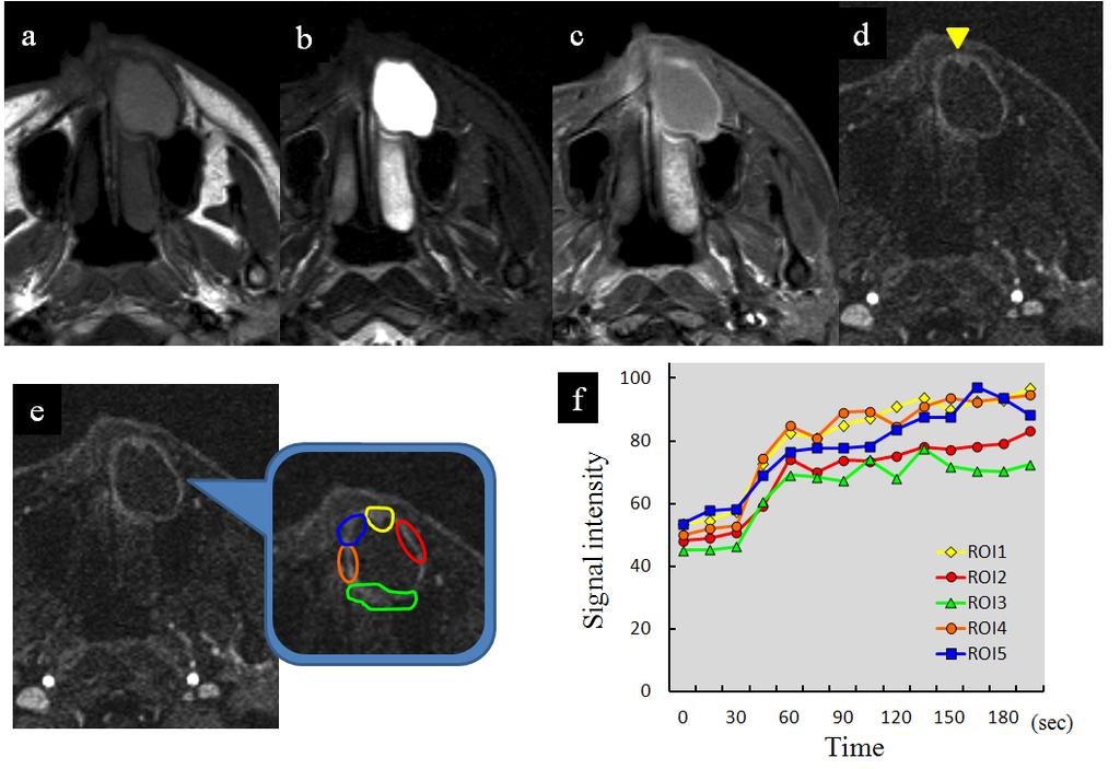
9 Fig. 3). Although in Case 9, a tiny enhanced area was detected on DCE-imaging (Fig. 3d), the area was not confirmed as a solid component in the time-to-intensity curve (Fig. 3e, f). DCE-MRIs of these 4 cases showed thicker rim enhancement than odontogenic cysts without any of the above clues leading to the diagnosis of ameloblastoma (Fig. 3d). This feature was considered to be an important point for differential diagnosis of cystic-type ameloblastoma from other cystic lesions.
10 Discussion Unilocular radiolucent lesions in the jawbone include dentigerous cysts, radicular cysts, keratocystic odontogenic tumors and ameloblastomas. In the present study, we evaluated whether MRI was useful for the differentiation of unilocular cystic-type ameloblastoma from other cystic lesions. Odontogenic cysts such as dentigerous cysts and radicular cysts have the same predilections in terms of signal intensity: homogeneously intermediate signal intensity on T1WI and homogeneously bright high signal intensity on T2WI (or STIR), reflecting the fluid content of the inner part of the lesion. 17 Keratocystic odontogenic tumors are classified as benign tumors of odontogenic origin according to the 2005 WHO histological classification 5 ; specifically, cystic lesions including fluid with a characteristically parakeratinized stratified squamous epithelium. Keratocystic odontogenic tumors have heterogeneous intermediate-to-high signal intensity on T1WI and heterogeneous low-to-high signal intensity on T2WI (or STIR), findings which distinguish them from other cystic lesions. 17 The cystic lumen of keratocystic odontogenic tumors may be filled with a cheesy material that consists of keratinaceous debris on microscopic examination. 1 MR findings of heterogeneous intermediate-to-high signal intensity on T1WI and heterogeneous low-to-high signal intensity on T2WI or STIR would reflect the protein content in the cyst fluid. Therefore, the higher signal intensity on T1WI and the heterogeneous signal intensity on MRI are useful findings for diagnosing keratocystic odontogenic tumor as opposed to other odontogenic cysts. Ameloblastomas are divided into solid/multicystic, extraosseous/peripheral, desmoplastic, and unicystic types according to the 2005 WHO histological classification of odontogenic tumors. 5 Any of these types can show a unilocular radiolucency. In the
11 present study, of 35 cases, 32 were solid/multicystic types, and 3 were unicystic types. Of the 32 solid/multicystic type cases, 9 showed unilocular and 23 multilocular radiolucency. All three unicystic type cases showed unilocular radiolucency. Extraosseous/peripheral types and desmoplastic types were not present. MR images of ameloblastomas can be divided into solid and cystic portions on the basis of signal intensity. Most ameloblastomas have multiple cystic portions of various sizes that show intermediate signal intensity on T1WI, bright high signal intensity on T2WI or STIR, and no enhancement on CE-T1WI. Ameloblastomas show a predilection for intermediate signal intensity on T1WI, high signal intensity on T2WI or STIR, and good enhancement on CE-T1WI in the solid portion. 6 These types of ameloblastoma can be easily differentiated from other odontogenic cystic lesions. However, ameloblastomas sometimes show unilocular cystic lesions that show cyst-like signal intensity on T1WI and T2WI (or STIR). The unilocular cystic type ameloblastoma is difficult to distinguish from other cystic lesions using MRI signal intensity alone. Of the 35 cases of ameloblastomas on which MRI was performed, 12 (34.3%) were suspected of being cystic lesions and 23 (65.7%) were diagnosed as ameloblastomas according to the findings described above. Enhancement solid portions on CE-T1WI and multiple cystic portions on T2WI or STIR are useful in diagnosing ameloblastoma. However, unilocular cystic-type ameloblastomas show homogeneous bright high signal intensity on T2WI or STIR as a water-like signal intensity. Unilocular cystic-type ameloblastoma are difficult to distinguish from odontogenic cysts such as dentigerous cysts and radicular cysts on the basis of MR signals alone. An area of rim enhancement on DCE-MRI was verified in the present study as differentiating unilocular cystic-type ameloblastomas from other cystic lesions.
12 Histopathological variants of unicystic-type ameloblastoma are luminal (ameloblastomatous cyst epithelium), intraluminal (protruding into the cyst cavity) and mural (invading the cyst wall). 5,16,18 In the present study, case 5 and 6 were the intraluminal type, i.e., protruding into the cyst cavity, and case 1 was a mural type, invading the cyst wall. In the intraluminal type, the area which showed an enhancement on CE-T1WI and gradual increased enhancement on DCE-MRI corresponded to the solid component. An enhancement area on MRI appearing to protrude into the cyst cavity defined the intraluminal type. In the mural type, an area which showing a thick rim enhancement on CE-T1WI and gradually increasing enhancement on DCE-MRI corresponded to an intramural nodule with focal invasion of the ameloblastoma tissue (Case 1, Fig. 2). Areas suspected of being intramural nodules with focal invasion of ameloblastoma tissue could be retrospectively detected on CE-T1WI and DCE-MRI. However, it was difficult to prospectively diagnose the mural type of unicystic ameloblastoma even using DCE-MRI. Therefore, clinicians should not overlook the signs of mural-type unicystic ameloblastoma in cases with thick rim enhancement. The luminal type (ameloblastomatous cyst epithelium) of unicystic ameloblastoma would have limitations in being diagnosed as cystic lesion even using CE-T1WI and DCE-MRI, although no luminal cases were observed in the present study. DCE-MRIs of the remaining 4 cases, on which no clues leading to the diagnosis of ameloblastoma could be observed, showed thicker rim enhancement than odontogenic cysts (Fig. 3d). This feature of thicker rim enhancement than odontogenic cysts was considered to be an important point for differentiating cystic-type ameloblastomas from other cystic lesions. Minami et al. 7 have also suggested that 7 out of 19 keratocystic odontogenic tumors (odontogenic keratocysts) had thick walls and unilocular in 10/19; but for
13 ameloblastoma 11/11 had irregular thick walls based on standard MR imaging. In the present study, we gave additional information that small ROIs set on the irregular thick wall of unicystic ameloblastomas on DCE-MRI would show gradually increasing enhancement that proved a proliferative ability of ameloblastoma as we have already shown in our previous report. 6 Thus, DCE-MRI could give useful information to distinct unilocular keratocystic odontogenic tuomors from ameloblastomas. In conclusion, CE-MRI and DCE-MRI could provide additional information in the differential diagnosis of unilocular cystic-type ameloblastoma according to findings as follows. 1) CE-T1WI showed tiny enhanced areas indicating the exact location of the solid component. 2) Some small ROIs set on the rim-enhanced area of CE-T1WI showed gradually increasing enhancement on DCE-MRI; these areas were suspected of being a solid component and/or intramural nodules with focal invasion of ameloblastoma tissue. 3) DCE-MRI of cystic-type ameloblastomas shows thicker rim enhancement than odontogenic cysts. Conflict of interest statement None declared.
14 References 1. Waldron CA, Shafer WG, Gorlin RJ. Oral & Maxollofacial Pathology. 2nd edition Philadelphia: WB Saunders Reichart PA, Philipsen HP, Sonner S. Ameloblastoma: biological profile of 3677 cases. Eur J Cancer B Oral Oncol 1995;31B(2): Gardner DG. Critique of the 1995 review by Reichart et al. of the biologic profile of 3677 ameloblastomas. Oral Oncol 1999;35(4): Philipsen HP, Reichart PA. Unicystic ameloblastoma. A review of 193 cases from the literature. Oral Oncol 1998;34(5): Barnes L, Eveson JW, Reichart P, Sidransky D. World Health Organization Classification of Tumours: Pathology and Genetics of Head and Neck Tumours. Lyon: International Agency for Research on Cancer Press Asaumi J, Hisatomi M, Yanagi Y, Matsuzaki H, Choi YS, Kawai N, et al. Assessment of ameloblastomas using MRI and dynamic contrast-enhanced MRI. Eur J Radiol 2005;56(1): Minami M, Kaneda T, Ozawa K, Yamamoto H, Itai Y, Ozawa M, et al. Cystic lesions of the maxillomandibular region: MR imaging distinction of odontogenic keratocysts and ameloblastomas from other cysts. AJR Am J Roentgenol 1996;166(4): Minami M, Kaneda T, Yamamoto H, Ozawa K, Itai Y, Ozawa M, et al. Ameloblastoma in the maxillomandibular region: MR imaging. Radiology 1992;184(2): Konouchi H, Asaumi J, Yanagi Y, Hisatomi M, Kawai N, Matsuzaki H, et al.
15 Usefulness of contrast enhanced-mri in the diagnosis of unicystic ameloblastoma. Oral Oncol 2006;42(5): Epub 2006 Feb Tuncbilek N, Karakas HM, Okten OO. Dynamic magnetic resonance imaging in determining histopathological prognostic factors of invasive breast cancers. Eur J Radiol 2005;53(2): Tuncbilek N, Karakas HM, Altaner S. Dynamic MRI in indirect estimation of microvessel density, histologic grade, and prognosis in colorectal adenocarcinomas. Abdom Imaging 2004;29(2): Hulka CA, Edmister WB, Smith BL, Tan L, Sgroi DC, Campbell T, et al. Dynamic echo-planar imaging of the breast: experience in diagnosing breast carcinoma and correlation with tumor angiogenesis. Radiology 1997;205(3): Unetsubo T, Konouchi H, Yanagi Y, Murakami J, Fujii M, Matsuzaki H, et al. Dynamic contrast-enhanced magnetic resonance imaging for estimating tumor proliferation and microvessel density of oral squamous cell carcinomas. Oral Oncol 2009;45(7): Joe VQ, Weatesson PL. Tumors of the parotid gland: MR imaging characteristics of various histologic types. AJR Am J Roentgenol. 1994;163(2): Hisatomi M, Asaumi JI, Yanagi Y, Unetsubo T, Maki Y, Murakami J, et al. Diagnostic value of dynamic contrast-enhanced MRI in the salivary gland tumors. Oral Oncol 2007;43(9): Ackermann GL, Altini M, Shear M. The unicystic ameloblastoma: a clinicopathological study of 57 cases. J Oral Pathol 1988;17(9-10): Hisatomi M, Asaumi J, Konouchi H, Shigehara H, Yanagi Y, Kishi K. MR imaging of epithelial cysts of the oral and maxillofacial region. Eur J Radiol 2003;48(2):
16 18. Philipsen HP, Reichart PA. Unicystic ameloblastoma. A review of 193 cases from the literature. Oral Oncol 1998;34(5):
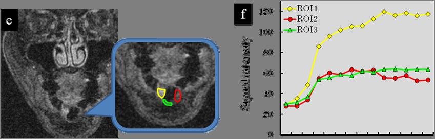
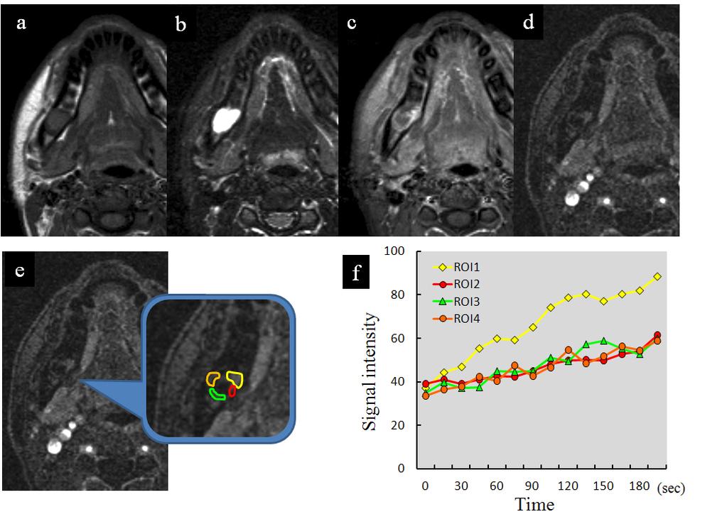
17 Figure legend Figure 1 Case 2 MRI showed homogeneously intermediate signal intensity on T1WI (a), and homogeneously bright high signal intensity on short inversion time inversion-recovery (b). A small area of intermediate signal intensity on short inversion time inversion-recovery showed enhancement on CE-T1WI (b, c). A small area, region of interest (ROI) 1, showed more increased enhancement than ROI 2 and 3 on dynamic CE images (d-f). Figure 2 Case 1 MRI showed homogeneously intermediate signal intensity on T1WI (a), homogeneously bright high signal intensity on short inversion time inversion-recovery (b), and thick rim enhancement on contrast-enhanced (CE)-T1WI (c). Some small ROIs were drawn on the rim-enhancement area on dynamic CE image (d, e). The area of ROI 1 was recognized as a solid component by showing a gradual increase of enhancement on dynamic CE images (f). Figure 3 Case 9 T1WI (a), short inversion time inversion-recovery (b) and contrast-enhanced (CE)-T1WI (c) showed no area indicating a solid component. Although a tiny enhanced area (arrowhead) which was suspected to solid component was detected on dynamic CE image (d), the area was not confirmed as a solid component due to its time-to-intensity curve (e, f).


18 Figure 1
19 Figure 2
20 Figure 3
Diagnostic value of MRI for odontogenic tumours
 Dentomaxillofacial Radiology (2013) 42, 20120265 ª 2013 The British Institute of Radiology http://dmfr.birjournals.org RESEARCH Diagnostic value of MRI for odontogenic tumours M Fujita 1, H Matsuzaki 2,
Ameloblastomatous Gorlin s cyst
 319 Journal of Oral Science, Vol. 49, No. 4, 319-323, 2007 Case Report Ameloblastomatous Gorlin s cyst Mala Kamboj 1) and Manish Juneja 2) 1) Department of Oral Pathology and Microbiology, U.P. King George
Case Report Unicystic Ameloblastoma with Mural Proliferation Managed by Conservative Treatment
 Case Reports in Pathology Volume 2016, Article ID 3089540, 4 pages http://dx.doi.org/10.1155/2016/3089540 Case Report Unicystic Ameloblastoma with Mural Proliferation Managed by Conservative Treatment
Glandular odontogenic cyst associated with ameloblastoma: Case report and review of the literature
 Journal section: Oral Medicine and Pathology Publication Types: Case Report doi:10.4317/jced.53775 http://dx.doi.org/10.4317/jced.53775 : Case report and review of the literature Timothée Cousin 1, Samuel
Imaging findings in a case of Gorlin-Goltz syndrome: a survey using advanced modalities
 Imaging Science in Dentistry 2011; 41 : 171-5 http://dx.doi.org/10.5624/isd.2011.41.4.171 Pegah ronoosh, Ali Reza Shakibafar*, Maneli Houshyar, Shima Nafarzade** Oral Radiology Department, Dental School,
Basal cell adenomas (BCAs), as defined by the World
, as defined by the World") ORIGINAL RESEARCH M. Okahara H. Kiyosue S. Matsumoto Y. Hori S. Tanoue D. Uchida H. Mori Y. Kondo Basal Cell Adenoma of the Parotid Gland: MR Imaging Findings with Pathologic Correlation BACKGROUND AND
ORIGINAL RESEARCH M. Okahara H. Kiyosue S. Matsumoto Y. Hori S. Tanoue D. Uchida H. Mori Y. Kondo Basal Cell Adenoma of the Parotid Gland: MR Imaging Findings with Pathologic Correlation BACKGROUND AND
Mucous and ciliated cell metaplasia in epithelial linings of odontogenic inflammatory and developmental cysts
 77 Journal of Oral Science, Vol. 47, No. 2, 77-81, 2005 Original Mucous and ciliated cell metaplasia in epithelial linings of odontogenic inflammatory and developmental cysts Yasunori Takeda, Yuko Oikawa,
Unicystic Ameloblastoma of mandible. Aggresive treatment A myth or a need. Case report and Extensive review of literature
 IOSR Journal of Dental and Medical Sciences (IOSR-JDMS) e-issn: 2279-0853, p-issn: 2279-0861. Volume 12, Issue 2 (Nov.- Dec. 2013), PP 26-31 Unicystic Ameloblastoma of mandible. Aggresive treatment A myth
A case of orthokeratinizing odontogenic cyst in the mandible
 114 J Meikai Dent Med 44 1, 114 118, 2015 1 1 1 2 1 1 1 1 1 1 1 1 1 1 1 2 1 1 2 1 46 A case of orthokeratinizing odontogenic cyst in the mandible Nobuaki TAMURA 1, Hiroshi TAKESHIMA 1, Kentaro KIKUCHI
Can magnetic resonance imaging obviate the need for biopsy for microcalcifications?
 Original Article Can magnetic resonance imaging obviate the need for biopsy for microcalcifications? Shinya Yamamoto, Takashi Chishima Department of Breast Surgery, Yokohama Rosai Hospital, Yokohama 222-0036,
Magnetic resonance imaging (MRI) and dynamic MRI evaluation of extranodal non-hodgkin lymphoma in oral and maxillofacial regions
 and dynamic MRI evaluation of extranodal non-hodgkin lymphoma in oral and maxillofacial regions") Vol. 113 No. 1 January 2012 Magnetic resonance imaging (MRI) and dynamic MRI evaluation of extranodal non-hodgkin lymphoma in oral and maxillofacial regions Hidenobu Matsuzaki, DDS, a Marina Hara, DDS,
Vol. 113 No. 1 January 2012 Magnetic resonance imaging (MRI) and dynamic MRI evaluation of extranodal non-hodgkin lymphoma in oral and maxillofacial regions Hidenobu Matsuzaki, DDS, a Marina Hara, DDS,
The ameloblastoma is a benign but locally aggressive
 The Unicystic Ameloblastoma: A Clinicopathologically Distinct Entity Tie-Jun LI 1 Abstract: Classification of ameloblastomas into solid or multicystic, unicystic, peripheral and desmoplastic types based
Primary intraosseous squamous cell carcinoma of the maxilla possibly arising from an infected residual cyst: A case report
 ONCOLOGY LETTERS 9: 131-135, 2015 Primary intraosseous squamous cell carcinoma of the maxilla possibly arising from an infected residual cyst: A case report SHINTARO SUKEGAWA 1, HIDENOBU MATSUZAKI 2, NAOKI
JMSCR Vol 05 Issue 06 Page June 2017
 www.jmscr.igmpublication.org Impact Factor 5.84 Index Copernicus Value: 83.27 ISSN (e)-2347-176x ISSN (p) 2455-0450 DOI: https://dx.doi.org/10.18535/jmscr/v5i6.29 MRI in Clinically Suspected Uterine and
Categorical Classification of Spiculated Mass on Breast MRI
 Categorical Classification of Spiculated Mass on Breast MRI Poster No.: C-1974 Congress: ECR 2013 Type: Authors: Scientific Exhibit Y. Kanda 1, S. Kanao 2, M. Kataoka 2, K. Togashi 2 ; 1 Kyoto City/JP,
Preoperative prediction of the malignancy or benignancy of
 ORIGINAL RESEARCH S. Eida M. Sumi N. Sakihama H. Takahashi T. Nakamura Apparent Diffusion Coefficient Mapping of Salivary Gland Tumors: Prediction of the Benignancy and Malignancy BACKGROUND AND PURPOSE:
Origin of Odontogenic Cysts & Tumors
 Origin of Odontogenic Cysts & Tumors Odontogenic Apparatus Origin of Odontogenic Cysts & Tumors Odontogenic Apparatus Remnants of dental lamina Reduced enamel epithelium Odontogenic rests Basal cell layer
MR imaging of FIGO stage I uterine cervical cancer: The diagnostic impact of 3T-MRI
 MR imaging of FIGO stage I uterine cervical cancer: The diagnostic impact of 3T-MRI Poster No.: C-1191 Congress: ECR 2010 Type: Educational Exhibit Topic: Genitourinary Authors: M. Takeuchi, K. Matsuzaki,
Management of Unicystic Plexiform Ameloblastoma in a 12 year Old Female: Report of a Case
 Article ID: WMC002613 2046-1690 Management of Unicystic Plexiform Ameloblastoma in a 12 year Old Female: Report of a Case Corresponding Author: Dr. Sheeraz Badal, Senior Lecturer, Dept of Oral & Maxillofacial
The concept of unicystic ameloblastoma (UA) was
 was") Publication Treatment of Giant Unicystic Ameloblastoma Suction Drainage and Secondary Curettage: a Case Report Bing LIU 1, Wen Feng ZHANG 1, Xin Ming CHEN 2, Zhi Jun SUN 3, Mohd Jamal Alsharif 1, Yi Fang
Publication Treatment of Giant Unicystic Ameloblastoma Suction Drainage and Secondary Curettage: a Case Report Bing LIU 1, Wen Feng ZHANG 1, Xin Ming CHEN 2, Zhi Jun SUN 3, Mohd Jamal Alsharif 1, Yi Fang
A case of orthokeratinized odontoge Title suspected to be a radicular cyst
 A case of orthokeratinized odontoge Title suspected to be a radicular cyst Onuki, M; Saito, A; Hosokawa, S; Oh Author(s) Hayakawa, H; Seta, S; Muramatsu, T; Journal Bulletin of Tokyo Dental College, 5
A COMPARATIVE HISTOPATHOLOGICAL STUDY OF EPITHELIAL linings OF ODONTOGENIC CYSTS AND UNICYSTIC AMElOBlASTOMAS
 A COMPARATIVE HISTOPATHOLOGICAL STUDY OF EPITHELIAL linings OF ODONTOGENIC CYSTS AND UNICYSTIC AMElOBlASTOMAS J Sudiono, RB Zain. A Comparative Histopathological Study of Epithelial Linings of Odontogenic
Unicystic Ameloblastoma: Implant- supported Reconstruction and Long-Term Follow-up
 OLGU RAPORU (Case Report) Hacettepe Dişhekimliği Fakültesi Dergisi Cilt: 31, Sayı: 2, Sayfa: 49-53, 2007 Unicystic Ameloblastoma: Implant- supported Reconstruction and Long-Term Follow-up Unikistik Ameloblastoma:
Case Report Basal Cell Ameloblastoma of Mandible: A Rare Case Report with Review
 Case Reports in Dentistry Volume 2013, Article ID 187820, 4 pages http://dx.doi.org/10.1155/2013/187820 Case Report Basal Cell Ameloblastoma of Mandible: A Rare Case Report with Review Hemant Shakya, 1
Odontogenic Keratocyst Masquerading as a Dentigerous Cyst in the Maxilla: A Case Report of an Unusual Presentation
 European Journal of Therapeutics DOI: 10.5152/EurJTher.2018.496 Case Report Odontogenic Keratocyst Masquerading as a Dentigerous Cyst in the Maxilla: A Case Report of an Unusual Presentation Arvind Karikal
Glandular Odontogenic Cyst Coexisting with a Dentigerous Cyst: Case Report
 SmyrnaMed Case 2018;2(1): 1-5 ISSN (Online): 2564-6869 www.smyrnamed.com Glandular Odontogenic Cyst Coexisting with a Dentigerous Cyst: Case Report Assist.Prof.Dr. Serap Keskin Tunç 1, Dt. Erkan Feslihan
MRI IN THE CHARACTERIZATION OF SEMINOMATOUS AND NONSEMINOMATOUS GERM CELL TUMORS OF THE TESTIS
 MRI IN THE CHARACTERIZATION OF SEMINOMATOUS AND NONSEMINOMATOUS GERM CELL TUMORS OF THE TESTIS Ambesh Deshar *, Gyanendra KC and Zhang Lopsang *Department of Medical Imaging and Nuclear Medicine, First
بسم هللا الرحمن الرحيم. Prof soha Talaat
 بسم هللا الرحمن الرحيم Ovarian tumors The leading indication for gynecologic surgery. Preoperative characterization of complex solid and cystic adnexal masses is crucial for informing patients about possible
Orthokeratinized Odontogenic Cyst: A Rarity
 aijoc AIJOC Case Report 1 Heena Sonawane, 2 Freny R Karjodkar, 3 Kaustubh Sansare, 4 Nimish Prakash ABSTRACT Orthokeratinized odontogenic cyst (OOC) was first identified as the rare variant of keratocystic
Citation American Journal of Surgery, 196(5)
") NAOSITE: Nagasaki University's Ac Title Author(s) Multifocal branch-duct pancreatic i neoplasms Tajima, Yoshitsugu; Kuroki, Tamotsu Amane; Adachi, Tomohiko; Mishima, T Kanematsu, Takashi Citation American
NAOSITE: Nagasaki University's Ac Title Author(s) Multifocal branch-duct pancreatic i neoplasms Tajima, Yoshitsugu; Kuroki, Tamotsu Amane; Adachi, Tomohiko; Mishima, T Kanematsu, Takashi Citation American
Contrast-enhanced Breast MRI RSSA 2013
 Contrast-enhanced Breast MRI RSSA 2013 Prof. dr. Maurice van den Bosch University Medical Center Utrecht, the Netherlands Index 1) Breast cancer 2) Why MRI of the breast 3) Technique 4) Interpretation
International Journal of Pharma and Bio Sciences MUCOEPIDERMOID CARCINOMA OF MINOR SALIVARY GLAND-PALATE: ABSTRACT
 Case report Biosciences International Journal of Pharma and Bio Sciences ISSN 0975-6299 MUCOEPIDERMOID CARCINOMA OF MINOR SALIVARY GLAND-PALATE: SHIVAKUMAR.S 1 AND SUBAIR VC 2 1 Professor, Department of
Management of Plexiform Ameloblastoma in a 12 year old female: A Case Report
 Article ID: ISSN 2046-1690 Management of Plexiform Ameloblastoma in a 12 year old female: A Case Report Corresponding Author: Dr. Sheeraz Badal, Senior Lecturer, Dept of Oral & Maxillofacial Surgery, MIDSR,
SQUAMOUS ODONTOGENIC TUMOUR: REPORT OF FIVE CASES FROM NIGERIA AND REVIEW OF LITERATURE
 African Journal of Oral Health Volume 3 Numbers 1&2, 2006:1-5 REFEREED ARTICLE SQUAMOUS ODONTOGENIC TUMOUR: REPORT OF FIVE CASES FROM NIGERIA AND REVIEW OF LITERATURE Adebiyi K.E., Odukoya O., Taiwo, E.O.
NEW SUBTRACTION ALGORITHMS FOR EVALUATION OF BREAST LESIONS ON DYNAMIC CONTRAST ENHANCED MR MAMMOGRAPHY
 A-056 NEW SUBTRACTION ALGORITHMS FOR EVALUATION OF BREAST LESIONS ON DYNAMIC CONTRAST ENHANCED MR MAMMOGRAPHY So Hee Cho, M.D., Byung Gil Choi, M.D., Hak Hee Kim, M.D., Euy Neyng Kim, M.D., Bum-soo Kim,
International Journal of Health Sciences and Research ISSN:
 International Journal of Health Sciences and Research www.ijhsr.org ISSN: 2249-9571 Case Report Orthokeratinized Odontogenic Cyst: A Case Report- A Milder Variant of OKC or an Independent Entity Mariyam
Proceedings of the 36th World Small Animal Veterinary Congress WSAVA
 www.ivis.org Proceedings of the 36th World Small Animal Veterinary Congress WSAVA Oct. 14-17, 2011 Jeju, Korea Next Congress: http://www.ivis.org October 14(Fri) ~ 17(Mon) 2011 ICC Jeju, Korea 2011 WSAVA
Odontomes and Odontogenic tumours
 Odontomes and Odontogenic tumours Odontomes Developmental hamartoma Hamartoma: normal tissue in abnormal location Any cells to be neoplastic it must be able to replicate, which is not seen in hamartoma
Rong Yang 1*, Zheqi Liu 1*, Sandhya Gokavarapu 2, Canbang Peng 1, Wei Cao 1, Tong Ji 1. Original Article. Abstract
 Original Article Recurrence and cancerization of ameloblastoma: multivariate analysis of 87 recurrent craniofacial ameloblastoma to assess risk factors associated with early recurrence and secondary ameloblastic
Young Nam Park, MD, Hee Young Hwang, MD, Young Sup Shim, MD, Sung Su Byun, MD, Hye-Young Choi, MD, Hyung-Sik Kim, MD
 Original Article pissn 1738-2637 Characteristic Dynamic Enhancement Pattern of Magnetic Resonance Imaging for Malignant Thyroid Tumor: A Preliminary Report 악성갑상선종양의특징적인역동적자기공명영상의조영증강소견 : Preliminary Report
Disclosure. Educational Objectives. Terminology. Odontogenic Cysts. Terminology
 Disclosure Lisa J. Koenig BChD, DDS, MS Professor & Program Director, Oral Medicine and Oral Radiology Marquette University School of Dentistry Consultant to Soredex for the Scanora 3D and 3Dx Author/Editor
MRI features of Triple-negative breast cancer: our experience.
 MRI features of Triple-negative breast cancer: our experience. Poster No.: C-1852 Congress: ECR 2013 Type: Scientific Exhibit Authors: V. Bertani, A. Gualano, V. Londero, A. Dal Col, M. Marcon, P. 1 2
Squamous Cell Carcinoma Arising in a Residual Cyst: A Case Report
 Squamous Cell Carcinoma Arising in a Residual Cyst: A Case Report Abstract Aim: The purpose of this report is to present a case of squamous cell carcinoma (SCC) arising from a mandibular residual cyst.
Pitfalls and Limitations of Breast MRI. Susan Orel Roth, MD Professor of Radiology University of Pennsylvania
 Pitfalls and Limitations of Breast MRI Susan Orel Roth, MD Professor of Radiology University of Pennsylvania Objectives Review the etiologies of false negative breast MRI examinations Discuss the limitations
Problem diagnoses. Current issues in Anatomic pathology. Problem Diagnoses in Tumors of the Oral Cavity 5/29/2009
 Current issues in Anatomic pathology Problem Diagnoses in Tumors of the Oral Cavity Richard Jordan DDS PhD FRCPath Professor of Oral Pathology & Pathology Director, UCSF Oral Pathology Diagnostic Laboratory
Magnetic Resonance Imaging of the Normal Tongue: Qualitative Evaluation of Fat-suppressed Contrast Enhanced Images
 Bulletin of the Osaka Medical College 49 1, 2 21-28, 2003 21 Original Article Magnetic Resonance Imaging of the Normal Tongue: Qualitative Evaluation of Fat-suppressed Contrast Enhanced Images Yasunori
Common Occurrence of Benign Liver Lesions in Patients With Newly Diagnosed Breast Cancer Investigated by MRI for Suspected Liver Metastases
 JOURNAL OF MAGNETIC RESONANCE IMAGING 10:165 169 (1999) Original Research Common Occurrence of Benign Liver Lesions in Patients With Newly Diagnosed Breast Cancer Investigated by MRI for Suspected Liver
The pharyngeal mucosal space is a common primary site for
 ORIGINAL RESEARCH Y. Ichikawa M. Sumi M. Sasaki T. Sumi T. Nakamura Efficacy of Diffusion-Weighted Imaging for the Differentiation between Lymphomas and Carcinomas of the Nasopharynx and Oropharynx: Correlations
Anatomical and Functional MRI of the Pancreas
 Anatomical and Functional MRI of the Pancreas MA Bali, MD, T Metens, PhD Erasme Hospital Free University of Brussels Belgium mbali@ulb.ac.be Introduction The use of MRI to investigate the pancreas has
Epithelial Sources. Rests of Serres Rests of Malassez Reduced Enamel Epithelium Surface Mucosa
 ODONTOGENIC CYSTS Epithelial Sources Rests of Serres Rests of Malassez Reduced Enamel Epithelium Surface Mucosa Epithelial Sources Surface Epithelium Rests of Serres Reduced Enamel Epithelium Rests of
Treatment Modalities of Odontogenic Keratocyst of Maxilla and Mandible: Our Experience
 wjd Parveen Akhter Lone et al Original Research 10.5005/jp-journals-10015-1344 Treatment Modalities of Odontogenic Keratocyst of Maxilla and Mandible: Our Experience 1 Parveen Akhter Lone, 2 Mohan Singh,
Case Report The Onset of a Peripheral Ameloblastoma
 Volume 2012, Article ID 729467, 4 pages doi:10.1155/2012/729467 Case Report The Onset of a Peripheral Ameloblastoma Kellen Cristine Tjioe, 1 José Humberto Damante, 1 anddenisetostesoliveira 2, 3 1 Department
A CASE OF UNICYSTIC AMELOBLASTOMA
 Dentistry Original Article International Journal of Clinical And Diagnostic Research ISSN 2395-3403 Volume 4, Issue 2, Mar-April 2016. Glorigin Lifesciences Private Limited. A CASE OF UNICYSTIC AMELOBLASTOMA
Objective: To evaluate the applicability of the cell block technique as a complementary
 www.scielo.br/jaos Cell block technique as a complementary method in the clinical diagnosis of cyst-like lesions of the jaw Elena Riet Correa RIVERO 1, Liliane Janete GRANDO 1, Fabíola MENEGAT 2, Jonathas
高雄醫學大學 口腔醫學院 口腔病理影像科 牙科 X 光影像判讀 教學範例
 高雄醫學大學 口腔醫學院 口腔病理影像科 牙科 X 光影像判讀 教學範例 Content Image No. 001 Dentigerous cyst over left upper embedded canine--------------------- 頁 1 Image No. 002---------------------------------------------------------------
Role of imaging in RCC. Ultrasonography. Solid lesion. Cystic RCC. Solid RCC 31/08/60. From Diagnosis to Treatment: the Radiologist Perspective
 Role of imaging in RCC From Diagnosis to Treatment: the Radiologist Perspective Diagnosis Staging Follow up Imaging modalities Limitations and pitfalls Duangkamon Prapruttam, MD Department of Therapeutic
Inter-radicular Radiolucencies
 Inter-radicular Radiolucencies Differential Diagnosis Laterally Displaced Radicular Cyst Accessory canals Root fracture Lateral Periodontal Cyst Botryoid variant Odontogenic Keratocyst Incisive Canal Cyst
Morphological Variants of Ameloblastoma and Their Mimickers
 20 Jan 2012 Vol 5 No.1 North American Journal of Medicine and Science Review Morphological Variants of Ameloblastoma and Their Mimickers Khaled Shaikhi, BDS; 1 Mirdza Neiders, DDS, MS; 1 Frank Chen, MD,
Gallium-67 Scintigraphy in Differential Diagnosis of Malignant Tumours from Non-Tumorous Lesions of the Maxilla
 Gallium-67 Scintigraphy in Differential Diagnosis of Malignant Tumours from Non-Tumorous Lesions of the Maxilla Ichiro OGURA 1, Takaaki ODA 1, Mikiko SUE 1, Yoshihiko SASAKI 1, Kazuhide HAYAMA 2 Objective:
Thoracic CT pattern in lung cancer: correlation of CT and pathologic diagnosis
 19 th Congress of APSR PG of Lung Cancer (ESAP): Update of Lung Cancer Thoracic CT pattern in lung cancer: correlation of CT and pathologic diagnosis Kazuma Kishi, M.D. Department of Respiratory Medicine,
Odontogenic tumors: a retrospective clinicopathological study from two Italian centers
 PATHOLOGICA 2017;109:35-46 Review - Study Group GIPaTeC Odontogenic tumors: a retrospective clinicopathological study from two Italian centers C. RUBINI 1, M. MASCITTI 2, A. SANTARELLI 2, A. TEMPESTA 3,
Table 9: Vascularity and Hemorrhage
 Table 9: Vascularity and Hemorrhage Di Ieva (2007) 120 Fractal dimension as a quantitator the microvasculat ure normal and adenomatous tissue. Clinical experience characterizing vascular surface fractal
Case Report Intraosseous Follicular Adenomatoid Odontogenic Tumour A Case Report
 International Dentistry Volume 2009, Article ID 597483, 4 pages doi:10.1155/2009/597483 Case eport Intraosseous Follicular Adenomatoid Odontogenic Tumour A Case eport Farhan Durrani 1 and oyana Singh 2
A Case Report on Surgical Management of Odontogenic Keratocyst
 World Journal of Medical Sciences 10 (2): 212-216, 2014 ISSN 1817-3055 IDOSI Publications, 2014 DOI: 10.5829/idosi.wjms.2014.10.2.82185 A Case Report on Surgical Management of Odontogenic Keratocyst 1
Prevalence of Meniscal Radial Tears of the Knee Revealed by MRI After Surgery
 Downloaded from www.ajronline.org by 46.3.207.114 on 12/22/17 from IP address 46.3.207.114. Copyright RRS. For personal use only; all rights reserved Thomas Magee 1 Marc Shapiro David Williams Received
Soft Tissue Tumour & Sarcoma Imaging Guidelines 2012
 Soft Tissue Tumour & Sarcoma Imaging Guidelines 2012 Version Control This is a controlled document please destroy all previous versions on receipt of a new version. Date Approved: March 2011 reissued April
Title. CitationEuropean journal of radiology, 84(11): Issue Date Doc URL. Rights
: Issue Date Doc URL. Rights") Title Diagnostic value of tumor blood flow and its histogr malignant lymphoma from squamous cell carcinoma Fujima, Noriyuki; Kameda, Hiroyuki; Tsukahara, Akiko Author(s) Tha, Khin Khin; Kudo, Kohsuke;
Title Diagnostic value of tumor blood flow and its histogr malignant lymphoma from squamous cell carcinoma Fujima, Noriyuki; Kameda, Hiroyuki; Tsukahara, Akiko Author(s) Tha, Khin Khin; Kudo, Kohsuke;
A CLINICOPATHOLOGIC STUDY OF ODONTOGENIC KERATOCYST (OKC) AND THE ROLE OF AgNORs IN CELL PROLIFERATION
 AND THE ROLE OF AgNORs IN CELL PROLIFERATION") A CLINICOPATHOLOGIC STUDY OF ODONTOGENIC KERATOCYST (OKC) AND THE ROLE OF AgNORs IN CELL PROLIFERATION * Vindhya Savithri **Sudha S ***Shameena P.M ****Ipe Varghese Abstract : The histologic pattern of
A CLINICOPATHOLOGIC STUDY OF ODONTOGENIC KERATOCYST (OKC) AND THE ROLE OF AgNORs IN CELL PROLIFERATION * Vindhya Savithri **Sudha S ***Shameena P.M ****Ipe Varghese Abstract : The histologic pattern of
Warthin s Tumor of the Parotid Gland:
 J Korean Soc Radiol 2009;61:17-22 Warthin s Tumor of the Parotid Gland: CT and MR Features 1 Yun Hee Lee, M.D., In Kyu Yu, M.D., Moon Hee Han, M.D. 2, Byung-Hee Lee, M.D., Min Sun Kim, M.D., Chang Joon
CME Article Clinics in diagnostic imaging (135)
") Medical Education Singapore Med J 2011; 52(5) : 384 CME Article Clinics in diagnostic imaging (135) Pojchamarnwiputh S, Muttarak M, Sriplakich S H 1a 1b 1c 1d Fig. 1 (a) Axial unenhanced; (b & c) delayed
Medical Education Singapore Med J 2011; 52(5) : 384 CME Article Clinics in diagnostic imaging (135) Pojchamarnwiputh S, Muttarak M, Sriplakich S H 1a 1b 1c 1d Fig. 1 (a) Axial unenhanced; (b & c) delayed
Original Research JOURNAL OF MAGNETIC RESONANCE IMAGING 32: (2010)
") JOURNAL OF MAGNETIC RESONANCE IMAGING 32:1117 1123 (2010) Original Research Peripheral Hyperintense Pattern on T2-Weighted Magnetic Resonance Imaging (MRI) in Breast Carcinoma: Correlation With Early Peripheral
JOURNAL OF MAGNETIC RESONANCE IMAGING 32:1117 1123 (2010) Original Research Peripheral Hyperintense Pattern on T2-Weighted Magnetic Resonance Imaging (MRI) in Breast Carcinoma: Correlation With Early Peripheral
Radiologic Pathologic Correlation of Intraosseous Lipomas. Tim Propeck 1, Mary Anne Bullard 1, John Lin 1, Kei Doi 2, William Martel 1
 Downloaded from www.ajronline.org by 148.251.232.83 on 04/10/18 from IP address 148.251.232.83. opyright RRS. For personal use only; all rights reserved Radiologic Pathologic orrelation of Intraosseous
J of Evolution of Med and Dent Sci/ eissn , pissn / Vol. 3/ Issue 18/May 05, 2014 Page 4859
 CYSTIC DEGENERATION IN FOLLICULAR AMELOBLASTOMA: A CASE REPORT Neeraj Kumar 1, Niharika Rathore 2, Hemant Shakya 3, Anshuman Jamdade 4, Puneet Chitlangia 5 HOW TO CITE THIS ARTICLE: Neeraj Kumar, Niharika
Educational Cases EQA November T.J. Palmer Raigmore Hospital Inverness
 Educational Cases EQA November 2013 T.J. Palmer Raigmore Hospital Inverness Case 2 Clinical Details Dob 11 February 1951 PMH: 1964 Extraction of 45 aet 13 yr 1966 Cyst between 44 and 46 enucleated 1973
Differential Diagnosis of Radiolucent Lesions of the Jaws
 Differential Diagnosis of Radiolucent Lesions of the Jaws Multilocular Multilocular Radiolucencies Odontogenic Keratocyst Botryoid Odontogenic Cyst Glandular odontogenic Cyst Invasive Ameloblastoma Central
Case Report Keratocystic Odontogenic Tumor with an Ectopic Tooth in Maxilla
 Case Reports in Dentistry Volume 2013, Article ID 232096, 4 pages http://dx.doi.org/10.1155/2013/232096 Case Report Keratocystic Odontogenic Tumor with an Ectopic Tooth in Maxilla Basavaraj T. Bhagawati,
MR of Intraparotid Masses
 MR of Intraparotid Masses Bruce N. Schlakman and David M. Yousem PURPOSE: To determine which MR techniques are best for identifying intraparotid masses and to assess the utility of MR for predicting specific
Case Report Mandibular Ameloblastoma in an Elderly Patient: A Case Report
 Volume 2013, Article ID 145282, 4 pages http://dx.doi.org/10.1155/2013/145282 Case Report andibular Ameloblastoma in an Elderly Patient: A Case Report Kokoro Nagata, Kasumi Shimizu, Chu Sato, Hiroshi orita,
Ring-shaped Lateral Ventricular Nodules Detected with Brain MR Imaging
 Magn Reson Med Sci, Vol. 12, No. 2, pp. 105 110, 2013 2013 Japanese Society for Magnetic Resonance in Medicine doi:10.2463/mrms.2012-0044 MAJOR PAPER Ring-shaped Lateral Ventricular Nodules Detected with
Successful Conservative Surgical Treatment of Ameloblastic Fibroma in the Posterior Maxilla : A Case Report
 http://dx.doi.org/10.5933/jkapd.2013.40.4.321 ISSN (print) 1226-8496 Successful Conservative Surgical Treatment of Ameloblastic Fibroma in the Posterior Maxilla : A Case Report Youngeun Lee 1, Hyojung
MRI features of serous oligocystic adenoma of the pancreas: differentiation from mucinous cystic neoplasm of the pancreas
 The British Journal of Radiology, 85 (2012), 571 576 MRI features of serous oligocystic adenoma of the pancreas: differentiation from mucinous cystic neoplasm of the pancreas 1,2 J H LEE, MD, 1 J K KIM,
Keratocystic Odontogenic Tumor (KCOT) in Maxillary Sinus arising from an Infected Dentigerous Cyst
 in Maxillary Sinus arising from an Infected Dentigerous Cyst") Keratocystic Odontogenic Tumor (KCOT) in Maxillary Sinus arising from an Infected Dentigerous Cyst N R Chourasia, Abhishek Singh Payak, Preeti Bhadouria Department of Oral and Maxillofacial Surgery, Rishiraj
Keratocystic Odontogenic Tumor (KCOT) in Maxillary Sinus arising from an Infected Dentigerous Cyst N R Chourasia, Abhishek Singh Payak, Preeti Bhadouria Department of Oral and Maxillofacial Surgery, Rishiraj
doi: /j.anl
 doi: 10.1016/j.anl.2006.07.001 Synchronous unilateral parotid gland neoplasms of three different histological types Shuho Tanaka 1, Keiji Tabuchi 1, Keiko Oikawa 1, Rika Kohanawa 1, Hideki Okubo 1, Dai
Scrotum-like protrusion of lipoma arising from the proximal thigh
 Upsala J Med sci 109: 261 265, 2004 Scrotum-like protrusion of lipoma arising from the proximal thigh Report of two cases Koshi Hattori, 1 Masahito Hatori, 1 Mika Watanabe, 2 Toshihisa Osanai, 3 Shoichi
Spectrum of findings of sclerosing adenosis at breast MRI.
 Spectrum of findings of sclerosing adenosis at breast MRI. Poster No.: C-0738 Congress: ECR 2012 Type: Scientific Exhibit Authors: F. Vasselli 1, F. Pediconi 2, M. Telesca 2, M. Luciani 2, V. Casali 2,
Malignant Transformation of Endometriosis: Magnetic Resonance Imaging Aspects
 Malignant Transformation of Endometriosis: Magnetic Resonance Imaging Aspects Poster No.: C-0084 Congress: ECR 2014 Type: Scientific Exhibit Authors: E. A. Yukhno, I. Trofimenko, G. Trufanov; St. Petersburg/RU
Malignant Transformation of Endometriosis: Magnetic Resonance Imaging Aspects
 Malignant Transformation of Endometriosis: Magnetic Resonance Imaging Aspects Poster No.: C-0084 Congress: ECR 2014 Type: Scientific Exhibit Authors: E. A. Yukhno, I. Trofimenko, G. Trufanov; St. Petersburg/RU
Visualization of Endolymphatic Hydrops after Intratympanic Injection of Gd-DTPA: Comparison of 2D and 3D Real Inversion Recovery Imaging
 Magn Reson Med Sci, Vol. 10, No. 2, pp. 101 106, 2011 MAJOR PAPER Visualization of Endolymphatic Hydrops after Intratympanic Injection of Gd-DTPA: Comparison of 2D and 3D Real Inversion Recovery Imaging
Using lesion washout volume fraction as a biomarker to improve suspicious breast lesion characterization
 JOURNAL OF APPLIED CLINICAL MEDICAL PHYSICS, VOLUME 16, NUMBER 5, 2015 Using lesion washout volume fraction as a biomarker to improve suspicious breast lesion characterization Jie Huang, a Sarah M. Schafer,
Radiologic and pathologic correlation of non-mass like breast lesions on US and MRI: Benign, high risk, versus malignant
 Radiologic and pathologic correlation of non-mass like breast lesions on US and MRI: Benign, high risk, versus malignant Poster No.: C-1161 Congress: ECR 2013 Type: Educational Exhibit Authors: J. Kwak,
Mural Unicystic Ameloblastoma Crossing the Midline: A Rare Case Report
 Int. J. Odontostomat., 6(1):97-103, 2012. Mural Unicystic Ameloblastoma Crossing the Midline: A Rare Case Report Ameloblastoma Uniquístico Mural Traspasando la Línea Mediana: Reporte de un Caso Infrecuente
An unusual case of unicystic intramural ameloblastoma and review of the literature
 An unusual case of unicystic intramural ameloblastoma and review of the literature MASSIMILIANO RICCI, FRANCESCO MANGANO 1, PAOLO TONELLI 2, ANTONIO BARONE 3, CESARE GALLETTI 4, UGO COVANI 5 Abstract Ameloblastoma
RADIOGRAPHIC INTERPRETATION Differential Diagnosis
 RADIOGRAPHIC INTERPRETATION Differential Diagnosis MODULE 1: The Introduction. Chief complaint Demographics Age Sex Race Historical findings Physical findings Clinical Radiographic Location Maxilla/mandible
Large Dentigerous Cyst
 Volume 16.2.1 Feb 2016 This Lecture Series qualifies for 0.5 Informal CPD Learning Hours Large Dentigerous Cyst By Dr Hassem Geha A 55 year-old male presented with a painless swelling in the right mandible.
Interpretation pearls for MR imaging of parotid gland tumor
 European Annals of Otorhinolaryngology, Head and Neck diseases (2013) 130, 30 35 Available online at www.sciencedirect.com TECHNICAL NOTE Interpretation pearls for MR imaging of parotid gland tumor S.
Value of the BI-RADS classification in MR-Mammography for diagnosis of benign and malignant breast tumors
 Eur Radiol (2011) 21:2475 2483 DOI 10.1007/s00330-011-2210-7 BREAST Value of the BI-RADS classification in MR-Mammography for diagnosis of benign and malignant breast tumors Christian Sohns & Martin Scherrer
The Natural History of Cerebellar Hemangioblastomas in von Hippel-Lindau Disease
 AJNR Am J Neuroradiol 24:1570 1574, September 2003 The Natural History of Cerebellar Hemangioblastomas in von Hippel-Lindau Disease Andrew Slater, Niall R. Moore, and Susan M. Huson BACKGROUND AND PURPOSE:
Whole-tumor apparent diffusion coefficient measurements in nephroblastoma: Can it identify blastemal predominance? Abstract Purpose To explore the
 Whole-tumor apparent diffusion coefficient measurements in nephroblastoma: Can it identify blastemal predominance? Abstract Purpose To explore the potential relation between whole-tumor apparent diffusion
An unusual site of Adenomatoid Odontogenic Tumor: A rare case report
 J. Int Oral Health 2010 Case Report All right reserved An unusual site of Adenomatoid Odontogenic Tumor: A rare case report Sapna Panjwani*, Anjana Bagewadi**, Vaishali Keluskar*** *Post Graduate Student