Title. CitationEuropean journal of radiology, 84(11): Issue Date Doc URL. Rights
|
|
|
- Margaret Robertson
- 5 years ago
- Views:
Transcription
1 Title Diagnostic value of tumor blood flow and its histogr malignant lymphoma from squamous cell carcinoma Fujima, Noriyuki; Kameda, Hiroyuki; Tsukahara, Akiko Author(s) Tha, Khin Khin; Kudo, Kohsuke; Shirato, Hiroki CitationEuropean journal of radiology, 84(11): Issue Date Doc URL Rights This manuscript version is made available un Rights(URL) Type article (author version) File Information manuscript.pdf Instructions for use Hokkaido University Collection of Scholarly and Aca
2 ORIGINAL ARTICLE Diagnostic value of tumor blood flow and its histogram analysis obtained with pcasl to differentiate sinonasal lymphoma from squamous cell carcinoma Noriyuki Fujima, MD, PhD 1 ; Hiroyuki Kameda, MD 1 ; Akiko Tsukahara, MD 1 ; Daisuke Yoshida, MD 1 ; Tomohiro Sakashita, MD, PhD 2 ; Akihiro Homma, MD, PhD 2 ; Khin Khin Tha, MD, PhD 3,4 ; Kohsuke Kudo, MD, PhD 1 ; and Hiroki Shirato, MD, PhD 3,4 1 Department of Diagnostic and Interventional Radiology, Hokkaido University Hospital, Sapporo, Japan 2 Department of Otolaryngology-Head and Neck Surgery, Hokkaido University Graduate School of Medicine, Sapporo, Japan 3 Department of Radiation Medicine, Hokkaido University Graduate School of Medicine, Sapporo, Japan 4 The Global Station for Quantum Medical Science and Engineering, Global Institution for collaborative research and education, Sapporo, Japan Corresponding Author Noriyuki Fujima Department of Diagnostic and Interventional Radiology, Hokkaido University Hospital N15, W7, Kita-Ku, Sapporo , Japan Phone: , Fax: Noriyuki.Fujima@mb9.seikyou.ne.jp
3 1 Original Research Diagnostic value of tumor blood flow and its histogram analysis obtained with pcasl to differentiate sinonasal malignant lymphoma from squamous cell carcinoma
4 2 ABSTRACT Objectives: To investigate the diagnostic value of tumor blood flow (TBF) obtained with pseudo-continuous arterial spin labeling (pcasl) for the differentiation of squamous cell carcinoma (SCC) and malignant lymphoma (ML) in the nasal or sinonasal cavity. Methods: Thirty-three patients with SCC and six patients with ML in the nasal or sinonasal cavity were retrospectively analyzed. Quantitative TBF values were obtained using whole-tumor region of interest (ROI) from pcasl data. The histogram analysis of TBF values within the tumor ROI was also performed by calculating the coefficient of variation (CV), kurtosis, and skewness. The mean TBF value, histogram CV, kurtosis and skewness of the patients with SCC were compared with those of the ML patients. The diagnostic accuracy to differentiate SCC from ML was also calculated by receiver operating characteristic (ROC) curve analysis. In addition, multiple logistic regression models were also performed to determine their independent predictive value, and diagnostic accuracy with the combined use of these parameters. Results: Between the SCC and ML groups, significant differences were observed in mean TBF, CV, and kurtosis, but not in skewness. In ROC curve analysis, the diagnostic accuracy values for the differentiation of SCC from ML in mean TBF, CV, and kurtosis
5 3 were all 0.87, respectively. Multiple logistic regression models revealed TBF and CV were respectively independent predictive value. With the combination of these parameters, the diagnostic accuracy was elevated to Conclusions: The TBF value and its histogram analysis obtained with pcasl can help differentiate SCC and ML. Keywords: Tumor blood flow, pseudo-continuous arterial spin labeling, squamous cell carcinoma, malignant lymphoma
6 4 Abbreviations ADC: apparent diffusion coefficient AUC: area under curve CT: computed tomography CV: coefficient of variation DCE: dynamic contrast-enhanced DWI: diffusion-weighted imaging FOV: field of view ML: malignant lymphoma MRI: magnetic resonance imaging pcasl: pseudo-continuous arterial spin labeling ROC: receiver operating characteristic ROI: region of interest SCC: squamous cell carcinoma T1WI: T1-weighted image T2WI: T2-weughted image TBF: tumor blood flow TSE: turbo spin-echo
7 5 INTRODUCTION Squamous cell carcinoma (SCC) is the most common tumor among nasal and sinonasal cavity malignancies [1]. Treatments for SCC include surgical treatment, chemotherapy, radiotherapy, and combinations of these [2]. Malignant lymphoma (ML) is the most common malignancy among non-epithelial sinonasal neoplasms, and it is especially frequent in Asian populations [3, 4]. Unlike SCC, the standard methods of treatment for ML do not include surgical treatment, and the chemotherapy regimen for ML is quite different from that used for SCC, in which cisplatin is usually used [5]. The differentiation of SCC and ML is required in many cases. The histopathological findings are the gold standard for the diagnoses of both SCC and ML. However, a surgical biopsy sometimes misses the diagnosis because tissue containing the tumor cells is not always obtained; peripheral inflammatory tissue (especially concomitant or simultaneous inflammation) is commonly observed in the nasal or sinonasal cavity [6]. For this reason, other methods to assist the diagnosis of SCC or ML are desired. The differentiation of SCC and ML by only conventional computed tomography (CT) or magnetic resonance imaging (MRI) such as T1-weighted images (T1WI), T2-weughted images (T2WI) and post-contrast-enhanced images has been described as
8 6 difficult [7]. Diffusion-weighted imaging (DWI) was reported to be useful in the differentiation of SCC and ML for the evaluation of the primary site or lymph nodes by using the apparent diffusion coefficient (ADC), which is reported to reflect the physiological parameter of tissue cellular density [8-11]. A tendency for lower ADC values in MLs compared to that in SCCs was reported, but the diagnostic performance of the ADCs was not adequate, and several cases overlapped between SCC and ML [8-10]. In contrast, tumor perfusion can be useful for the differential diagnosis of SCC and ML as another physiological parameter. Dynamic contrast-enhanced (DCE) perfusion techniques that were reported to be useful for this differentiation use the pattern of the dynamic curve of the tumor concentration of the contrast agent, or the perfusion-related parameters of k-trans, plasma volume, and the volume of extracellular space [12-14]. A pseudo-continuous arterial spin labeling (pcasl) technique is now widely used for the noninvasive measurement of tissue blood flow by using protons in arterial water as the intrinsic tracer [15]. The reliability of pseudo-continuous arterial spin labeling for use in head and neck tumors was recently described [16]. Unlike DCE perfusion, pcasl can be performed noninvasively without contrast agent, and its blood flow measurements might be more accurate because the tracer in ASL is more diffusible than the contrast agent in MR Perfusion (gadolinium chelate) [17].
9 7 The aim of the present study was to investigate the diagnostic value of tumor blood flow (TBF) obtained with pcasl for the differentiation of SCC and ML in the nasal or sinonasal cavity. MATERIALS AND METHODS Patients The study protocol was approved by our institutional review board. The cases of 33 patients with SCC and six patients with ML in the nasal or sinonasal cavity were evaluated retrospectively with the following inclusion criteria: (1) the patient was first diagnosed (not a recurrent case) histopathologically as having SCC or ML, and (2) MR scanning was performed before any treatment. The SCC patients were 28 males (mean age 61.8 yrs, range yrs) and five females (mean age 59.2 yrs, range yrs). The primary sites of the 33 patients with SCC were the maxillary sinus in 29 patients and the nasal cavity in four patients. The patients with ML were six males (mean age 65.8 yrs, range yrs) and no females. The primary sites were the maxillary sinus in two patients, the ethmoid sinus in one patient, and the nasal cavity in three patients.
10 8 Imaging parameters All MR imaging was performed using a 3.0 Tesla unit (Achieva TX; Philips Medical Systems, Best, the Netherlands) with a 16-channel neurovascular coil. First, conventional MR images were obtained to evaluate the primary tumor. These images included (a) axial T1WI with a spin-echo sequence (TR, 450 msec; TE, 10 msec; field of view (FOV), mm; matrix; slice thickness, 5 mm; inter-slice gap, 30%; scanning time, 2 min 12 s) and (b) axial T2WI with a turbo spin-echo (TSE) sequence with fat suppression (TR, 4500 msec; TE, 70 msec; TSE factor, 9; FOV, mm; matrix; slice thickness, 5 mm; inter-slice gap, 30%; scanning time, 2 min 6 s). In the pcasl scanning, coronal T2WI was performed to obtain anatomical information of the carotid artery for the positioning of the labeling slab. A T1 map was also obtained to measure the longitudinal relaxation in the tumor tissue, and it was used for the TBF quantification. The acquisition of pcasl was performed by using multi-shot spin-echo echo-planar imaging to obtain control and labeled images. The labeling slab was placed just under the bifurcation of the internal and external carotid arteries by using coronal T2WI as a reference. The MR parameters of the pcasl were as follows: labeling duration, 1650 msec;
11 9 post-label delay, 1280 msec; TR, 3619 msec; TE, 18 msec; flip angle, 90 ; number of shots, two; FOV, mm; matrix, 80 80; slice thickness, 5 mm; number of slices, 15; acceleration factor for parallel imaging, two; and scanning time, 5 min 11 s. The patients were instructed not to swallow, move their tongues, open their mouths, or make any other voluntary motion during the pcasl scan. In addition, their heads were fixed firmly with the coil to prevent movement during the scan. The coronal T2WI was obtained by TSE sequence with the following parameters: TR, 4500 msec; TE, 70 msec; TSE factor, 9; FOV, mm; matrix, ; slice thickness, 4 mm; inter-slice gap, 30%; scanning time, 2 min 6 s. For the T1 map, a gradient echo sequence with the Look-Locker readout and a constant flip angle was used with the following parameters: TR, 7 msec; TE, 1.7 msec; flip angle, 7 ; FOV, mm; matrix, ; slice thickness, 10 mm (single slice acquisition); scanning time, 6 s. Data analysis TBF calculation by pcasl We calculated the TBF of the pcasl (f) from the signal difference (ΔM), which was calculated by subtracting the labeled image from the control image, using the
12 10 previously described equation [18]: f ΔMλR1a exp( ωr 2M α 0 1a ) [1 exp( γr 1a )] -1 [1] where R 1a is the longitudinal relaxation rate of blood (0.67 s 1 ), γ is the labeling time (1.65 s), ω is the post-labeling delay time (1.28 s), α is the labeling efficiency (0.85), and λ is the blood/tumor-tissue water partition coefficient (1.0 g/ml) [19, 20]. M 0 is the equilibrium magnetization of the tumor tissue, which was estimated from the signal intensity of the control image and the tumor longitudinal relaxation rate obtained by the T1 map. Using Eq. [1], we created the TBF maps on a pixel-by-pixel basis. We used Mathematical software (MATLAB, ver. 2012a) to calculate the TBF values for the pcasl. Tumor ROI delineation and quantification The primary tumor in both the SCCs and MLs was outlined by a board-certified neuroradiologist with 18 years of experience (A.T.), blinded to the histopathological diagnosis. The delineation was performed on the axial T2WI with a polygonal region of interest (ROI), and the ROI was then copied onto a TBF map (Fig. 1). T1WI was also used as a guide to determine the ROI. To avoid vascular artifacts in the ROI, the area of
13 11 the vessel signal void was also delineated on T2WI, and this area was excluded from the TBF measurement. A strong high signal area with T2WI which suggested necrosis was also excluded. The TBF value of pcasl in each patient was determined as the mean of the TBF values in the delineated ROI. If the tumor extended into two or more slices on the TBF map, the mean TBF of all pixels in all ROIs of the tumor was calculated as the TBF value. A histogram analysis method was performed to assess the distribution of TBF values within each tumor ROI. As with the TBF calculation, if the tumor extended into two or more slices on the TBF map, the TBF values of all pixels in all ROIs of the tumor was used for the histogram analysis. The histogram analysis parameters assessed with this method were the coefficient of variation (CV) and histogram skewness and kurtosis. In this study, CV, skewness and kurtosis were defined as follows: CV / skewness 3 3 1/ n ( ) / 4 4 kurtosis 1/ n ( ) / 3 where n is the number of pixels within the tumor ROI, x is the TBF value in each pixel,
14 12 μ is the mean of x, and σ is the standard deviation of x. The CV describes the normalized measure of dispersion of TBF values. Histogram skewness describes the skew in the shape of the distribution curve of the TBF values. The kurtosis describes the peak and/or flatness of the curve peak; a more acute peak has higher kurtosis, and a more broadened and/or flattened peak has lower kurtosis [21]. Statistical analysis As univariate analysis, Mann-Whitney U-test was used to compare mean TBF values, histogram parameters of the CV, skewness, and kurtosis values between the SCC and ML respectively. If a significant difference was observed in a parameter, receiver operating characteristic (ROC) curve were constructed for the calculation of area under curve (AUC), and for the determination of best diagnostic accuracy by using the closest point to the upper left corner of ROC curve in the division of SCC and ML patient. In addition, if a significant difference was obtained for more than two parameters among TBF, CV, skewness, and kurtosis, these parameters were subsequently analyzed by multiple logistic regression models to determine their independent predictive value. If multiple parameters were determined as independent predictor by multiple logistic regression models, we also determined the sensitivity, specificity, and diagnostic
15 13 accuracy of the combined use of these parameters so that diagnostic accuracy becomes the highest. A p-value of 0.05 was considered significant. All statistics were performed by using the SPSS version 20 for Windows. RESULTS The pcasl scanning, TBF measurement and parameter calculation in the histogram analysis were successfully performed in all 39 patients. In univariate analysis using Mann-Whitney U-test, the mean TBF in the 33 SCC patients (140.6 ± 35.7 ml/100 g/min) was significantly higher than that of the six ML patients (93.8 ± 15.1 ml/100 g/min) (p<0.001) (Fig. 2). In the histogram analysis, the CV of the SCC patients (0.45 ± 0.09) was significantly higher than that of the ML patients (0.33 ± 0.04) (p<0.01) (Fig. 3). The kurtosis of the SCC patients ( 0.47 ± 0.29) was significantly lower than that of the ML patients ( 0.18 ± 0.21) (p<0.01) (Fig. 4). In contrast, there was no significant difference in skewness between the patients with SCC (0.03± 0.29) and those with ML (0.17± 0.37) (p=0.27) (Fig. 5). On ROC curve analysis, AUC, sensitivity, specificity, and accuracy of mean TBF were 0.87, 0.85 (28/33), 0.83 (5/6) and 0.85 (33/39) respectively when the threshold was set at ml/100 g/min. Those of the CV were 0.87, 0.82 (27/33), 0.83 (5/6), 0.82 (32/39) by setting the
16 14 threshold at Those of kurtosis were 0.78, 0.64 (21/33), 0.83 (5/6), and 0.67 (26/39) respectively by setting the threshold at respectively. By the multiple logistic regression models, mean TBF (p<0.05) and histogram parameter of the CV (p<0.05) were respectively determined as independent predictor. With the combination of these parameters, with the threshold of ML set as TBF < , CV > 0.37, the highest accuracy of 0.97 (38/39) was obtained with 0.97 (32/33) sensitivity and 1.00 (6/6) specificity (Figs. 6, 7). DISCUSSION We found that the ML patients had significantly lower mean TBF, higher kurtosis and lower CV by histogram analysis compared to the SCC patients by univariate analysis. Additionally, by the multiple logistic regression models, mean TBF and histogram parameter of the CV were revealed as independent diagnostic parameter for the differentiation of SCC and ML patients. Higher diagnostic accuracy was obtained by combining these two parameters, although the case of only one patient with SCC overlapped in patients group of ML. We were unable to find any report in the extant literature that describes a
17 15 difference in TBF between SCC and ML cases using quantitative values obtained by pcasl. However, several studies investigated tumor perfusion-related parameters by using a DCE technique. Asaumi et al. reported that high peak signal intensity from the dynamic curve after a bolus injection of contrast agent was obtained from most SCCs, whereas 70% of the ML cases did not show such high peak signal intensity in their dynamic curves [14]. Lee et al. reported that the mean plasma volume (which has been related to blood volume) was higher in SCC cases than that in ML cases, although the difference was not significant [12]. These reports indicated that the degree of the tumor perfusion of MLs was lower than that of SCCs. Our present finding of a significant difference in TBF values between SCC and ML shows the same tendency as that described in these earlier reports. In general imaging findings in conventional MR signal intensity, a tendency for homogeneous signal intensity in ML has been reported [22]. However, homogeneity in the tumor perfusion map was not investigated in the past studies. The present study s ML findings of higher kurtosis and lower CV in the histogram analysis showed a narrow range of TBF map pixel values; this result indicates homogeneous perfusion in most cases of ML compared to SCC. We suspect that one of the main reasons for the heterogeneity in the TBF distribution is the intra-tumoral necrosis in SCCs. Lesions
18 16 near tumor necrosis are generally considered hypoxic areas with hypovascularity. In contrast, hypervascularity has been observed frequently in peripheral areas of SCCs [16]. This difference in intra-tumoral vascularity was considered to reflect heterogeneity in the TBF map. Because necrotic areas are less frequently observed in ML, a more homogeneous TBF distribution was observed in the present ML cases compared to the SCC cases. With the combination of the mean TBF and the histogram parameter of CV, the diagnostic accuracy was better than that obtained with the mean TBF or a histogram analysis parameter alone. The mean TBF values of several patients with ML overlapped with those of several patients with SCC. However, such patients can be differentiated by using the parameters in a histogram analysis. In contrast, for several of our patients with SCC, the histogram analysis revealed homogeneity with higher kurtosis and a lower CV value similar to those of our ML patients, and the cases of these SCC patients were thus difficult to differentiate from ML by histogram parameters alone. However, most of such patients with SCC can be successfully differentiated with an additional evaluation by mean TBF. In light of these findings, the incidence of SCCs with both lower TBF and a homogeneous histogram is considered very rare, and thus, in the present study the combined use of the mean TBF and histogram parameters obtained with a TBF map was
19 17 found to be very effective and to have high diagnostic value for the differentiation of SCC and ML. In the present study, only one SCC was completely overlapped in the area of ML, even though both the mean TBF and its histogram value were respectively used for differentiation. The location of this SCC was the right maxillary sinus, and it was moderately differentiated in histopathological findings. There was no particular information that differed from that of the other SCCs. It remains unclear whether other factors such as the p16, p53, Ki-67 expression or human papilloma virus status are related to the difficulty of the differentiation to ML. Further analyses in a multivariate correlation study are necessary to address this question. In patients with nasal or sinonasal malignancy such as SCC or lymphoma, it is very important to determine the treatment method as soon as possible. The pcasl diagnostic technique used in the present study can contribute to the quick assessment for a prompt start of treatment by providing information for the diagnosis. This study has several limitations. First, the number of patients with ML (n=6) was small. However, it may be difficult to match the patient number between ML and SCC cases, since ML patients are less frequently encountered compared to SCC patients. Further studies of larger numbers of ML patients are necessary. In addition, the details
20 18 of the distributions of locations differed between the SCCs and MLs. In particular, there was no patient whose SCC was located in the ethmoid sinus, whereas the tumor of one ML patient was located in the ethmoid sinus. The different locations of the lesions can affect the lesions feeding arteries and may also affect the TBF values. Another subgroup study of a greater number of patients based on the location of the tumors is also needed. Second, a direct comparison of TBF with other parameters was not conducted. As noted above, conventional imaging such as T1WI and T2WI in ML tended to show homogenous intensity [22], and it can be useful for the differentiation of SCC and ML, although it was also reported that this differentiation was difficult by these findings alone [7]. Additionally, an ADC value measured by DWI was reported to be useful to differentiate SCC and ML [8-11]. The determination of additional diagnostic value of TBF can be used to confirm the utility of TBF assessment. Moreover, TBF and ADC are different physiological parameters, because the ADC value reflects mainly the cellular density of tumor tissue. Combining the TBF and the ADC may provide better diagnostic value. Third, it was sometimes difficult to distinguish secondary inflammation tissue around the tumor from the tumor tissue itself, and thus inflammatory tissue may have been present within the tumor ROI in some cases. In such cases, a certain level of error was included in the measured TBF value due to the
21 19 mixture of tumor and inflammatory tissue within the ROI, and this may have influenced the results. We nevertheless speculate that a large error did not occur because the signal intensity was generally different between the tumor tissue and inflammatory tissue, which typically presented clearly high signal intensity on T2WI. Forth, inter-observer agreement was not investigated in the present study. The variations of the shape and/or size of the primary tumors outlined by different researchers may have a significant influence on the histogram analysis. To address this concern, an inter-observer agreement analysis should be performed in a future study. CONCLUSIONS In conclusion, we found that pcasl can be a useful tool for the differentiation of SCC and ML. In particular, the combined use of both the mean TBF and histogram parameter of CV obtained with the intra-tumoral TBF signal will be effective as a diagnostic tool with high diagnostic accuracy.
22 20 REFERENCES 1. Sanghvi S, Khan MN, Patel NR, Yeldandi S, Baredes S, Eloy JA. Epidemiology of sinonasal squamous cell carcinoma: a comprehensive analysis of 4994 patients. Laryngoscope 2014;124(1): Jegoux F, Metreau A, Louvel G, Bedfert C. Paranasal sinus cancer. Eur Ann Otorhinolaryngol Head Neck Dis 2013;130(6): Zylka S, Bien S, Kaminski B, Postula S, Ziolkowska M. [Epidemiology and clinical characteristics of the sinonasal malignancies]. Otolaryngol Pol 2008;62(4): Vidal RW, Devaney K, Ferlito A, Rinaldo A, Carbone A. Sinonasal malignant lymphomas: a distinct clinicopathological category. Ann Otol Rhinol Laryngol 1999;108(4): Logsdon MD, Ha CS, Kavadi VS, Cabanillas F, Hess MA, Cox JD. Lymphoma of the nasal cavity and paranasal sinuses: improved outcome and altered prognostic factors with combined modality therapy. Cancer 1997;80(3): Pal I, Gupta A, Sengupta S. An attempt to define the type of biopsy in a sinonasal lesion showing bony erosion. Indian J Otolaryngol Head Neck Surg
23 ;62(1): Liu XW, Xie CM, Mo YX, Zhang R, Li H, Huang ZL, et al. Magnetic resonance imaging features of nasopharyngeal carcinoma and nasopharyngeal non-hodgkin's lymphoma: are there differences? Eur J Radiol 2012;81(6): Ichikawa Y, Sumi M, Sasaki M, Sumi T, Nakamura T. Efficacy of diffusion-weighted imaging for the differentiation between lymphomas and carcinomas of the nasopharynx and oropharynx: correlations of apparent diffusion coefficients and histologic features. AJNR Am J Neuroradiol 2012;33(4): Sumi M, Nakamura T. Diagnostic importance of focal defects in the apparent diffusion coefficient-based differentiation between lymphoma and squamous cell carcinoma nodes in the neck. Eur Radiol 2009;19(4): Sumi M, Ichikawa Y, Nakamura T. Diagnostic ability of apparent diffusion coefficients for lymphomas and carcinomas in the pharynx. Eur Radiol 2007;17(10): Razek AA. Diffusion-weighted magnetic resonance imaging of head and neck. J Comput Assist Tomogr 2010;34(6):
24 Lee FK, King AD, Ma BB, Yeung DK. Dynamic contrast enhancement magnetic resonance imaging (DCE-MRI) for differential diagnosis in head and neck cancers. Eur J Radiol 2012;81(4): Matsuzaki H, Hara M, Yanagi Y, Asaumi J, Katase N, Unetsubo T, et al. Magnetic resonance imaging (MRI) and dynamic MRI evaluation of extranodal non-hodgkin lymphoma in oral and maxillofacial regions. Oral Surg Oral Med Oral Pathol Oral Radiol 2012;113(1): Asaumi J, Yanagi Y, Konouchi H, Hisatomi M, Matsuzaki H, Kishi K. Application of dynamic contrast-enhanced MRI to differentiate malignant lymphoma from squamous cell carcinoma in the head and neck. Oral Oncol 2004;40(6): Telischak NA, Detre JA, Zaharchuk G. Arterial spin labeling MRI: Clinical applications in the brain. J Magn Reson Imaging 2014; DOI: /jmri Fujima N, Kudo K, Tsukahara A, Yoshida D, Sakashita T, Homma A, et al. Measurement of tumor blood flow in head and neck squamous cell carcinoma by pseudo-continuous arterial spin labeling: Comparison with dynamic contrast-enhanced MRI. J Magn Reson Imaging 2014; DOI: /jmri
25 Fujima N, Kudo K, Yoshida D, Homma A, Sakashita T, Tsukahara A, et al. Arterial spin labeling to determine tumor viability in head and neck cancer before and after treatment. Journal of magnetic resonance imaging. J Magn Reson Imaging 2014;40(4): Wang Z, Aguirre GK, Rao H, Wang J, Fernandez-Seara MA, Childress AR, et al. Empirical optimization of ASL data analysis using an ASL data processing toolbox: ASLtbx. Magn Reson Imaging 2008;26(2): Golay X, Petersen ET, Hui F. Pulsed star labeling of arterial regions (PULSAR): a robust regional perfusion technique for high field imaging. Magn Reson Med 2005;53(1): Wheeler RH, Ziessman HA, Medvec BR, Juni JE, Thrall JH, Keyes JW, et al. Tumor blood flow and systemic shunting in patients receiving intraarterial chemotherapy for head and neck cancer. Cancer Res 1986;46(8): King AD, Chow KK, Yu KH, Mo FK, Yeung DK, Yuan J, et al. Head and neck squamous cell carcinoma: diagnostic performance of diffusion-weighted MR imaging for the prediction of treatment response. Radiology 2013;266(2): Negendank WG, al-katib AM, Karanes C, Smith MR. Lymphomas: MR imaging
26 24 contrast characteristics with clinical-pathologic correlations. Radiology 1990;177(1):
 The tumor was delineated on the axial T2WI with a polygonal ROI.")
27 25 Figure Legends Fig. 1. Tumor ROI delineation. A sample case (61-yr-old male, right maxillary cancer) of ROI delineation. (a) The tumor was delineated on the axial T2WI with a polygonal ROI. (b) After the delineation, the ROI was copied to the corresponding TBF map. (a)
 was significantly higher than that in the six patients with ML (93.8 ± 15.1 ml/100 g/min) (p>0.001) (a, *). The ROC curve analysis revealed the AUC of 0.87 (p<0.")
28 26 (b) Fig. 2. The TBF in the SCC patients and ML patients. (a) TBF in patients with SCC or ML. (b) ROC curve for the determination of diagnostic accuracy. The TBF in the 33 patients with SCC (140.6 ± 35.7 ml/100 g/min) was significantly higher than that in the six patients with ML (93.8 ± 15.1 ml/100 g/min) (p>0.001) (a, *). The ROC curve analysis revealed the AUC of 0.87 (p<0.01) (b), with the best diagnostic accuracy of 0.85 (33/39), sensitivity of 0.85 (28/33), and specificity of 0.83 (5/6), with a threshold value of ml/100g/min (a, **).
29 (a) 27
30 28 (b) Fig. 3. Histogram CV in the SCC and ML patients. (a) Histogram CV in patients with SCC or ML. (b) ROC curve for the determination of diagnostic accuracy. There was a significant difference in the CV between the SCC patients (0.45 ± 0.09) and that of the ML patients (0.33 ± 0.04) (p<0.01) (*).The ROC
31 29 curve analysis revealed the AUC of 0.87 (p<0.01) (b), with the best diagnostic accuracy of 0.83 (32/39), sensitivity of 0.82 (27/33), and specificity of 0.83 (5/6), with a threshold value of 0.37 (a, **). (a)
32 30 (b) Fig. 4. Histogram kurtosis in the SCC and ML patients. (a) Histogram kurtosis in patients with SCC or ML. (b) ROC curve for the determination of diagnostic accuracy. There was a significant difference in the histogram kurtosis between the SCC patients ( 0.47 ± 0.29) and the ML patients ( 0.18
33 31 ± 0.21) (p<0.01) (*).The ROC curve analysis revealed the AUC of 0.78 (p<0.01) (b), with the best diagnostic accuracy of 0.67 (26/33), sensitivity of 0.64 (21/33), and specificity of 0.83 (5/6), with a threshold value of (a, **). (a)
34 32 (b) Fig. 5. Histogram skewness in the SCC and ML patients. No significant difference was observed in the histogram skewness between the patients with SCC and those with ML (p=0.27).
35 33 Fig. 6. Two-dimensional plot graph of TBF and histogram CV values. Two-dimensional plot graph with the percentage change of TV on the vertical axis and the percentage change of TBF on the horizontal axis. When the threshold line was set to for TBF (*), 0.37 for histogram CV (**), the sensitivity, specificity and accuracy for the division of SCC and ML were 0.97 (32/33), 1.00 (6/6), and 0.97 (38/39), respectively. With this setting of the threshold for TBF and CV, kurtosis did not
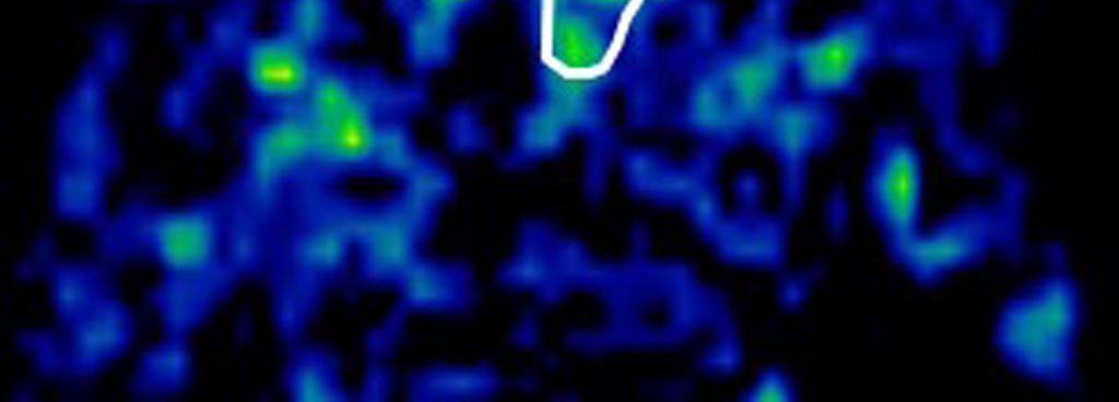
36 34 contribute to the diagnostic accuracy. Fig. 7. Case examples of TBF maps of SCC and ML cases. T2WI and TBF map with the delineated tumor ROI in a patient with SCC (a) and a patient with ML (b). The differentiation was considered difficult from the signal intensity in T2WI only. However, higher TBF and intra-tumoral heterogeneity was clearly observed in the TBF map of the SCC patient (arrow). The measured parameters were: the SCC patient (a), TBF, ml/100 g/min; CV, 0.59; kurtosis, The ML patient (b), TBF, ml/100 g/min; CV, 0.28; kurtosis, 0.04.


37 35 (a)


38 36 (b)
The utility of MRI histogram and texture analysis for the prediction of histological diagnosis in head and neck malignancies
 Fujima et al. Cancer Imaging (2019) 19:5 https://doi.org/10.1186/s40644-019-0193-9 RESEARCH ARTICLE Open Access The utility of MRI histogram and texture analysis for the prediction of histological diagnosis
Disclosures. Diffusion and Perfusion Imaging in the Head and Neck. Learning objectives ???
 Disclosures No relevant financial disclosures Diffusion and Perfusion Imaging in the Head and Neck Ashok Srinivasan, MD Associate Professor Director of Neuroradiology University of Michigan Health System
Title. CitationMagnetic resonance imaging, 36: Issue Date Doc URL. Rights
 Title Advanced diffusion models in head and neck squamous diffusion parameters and comparison with dynamic con Fujima, Noriyuki; Sakashita, Tomohiro; Homma, Akihir Author(s) Khin Khin; Kudo, Kohsuke; Shirato,
Effect of intravenous contrast medium administration on prostate diffusion-weighted imaging
 Effect of intravenous contrast medium administration on prostate diffusion-weighted imaging Poster No.: C-1766 Congress: ECR 2015 Type: Authors: Keywords: DOI: Scientific Exhibit J. Bae, C. K. Kim, S.
Imaging can play an important role in determining benignancy
 ORIGINAL RESEARCH A. Srinivasan C.J. Galbán T.D. Johnson T.L. Chenevert B.D. Ross S.K. Mukherji Utility of the K-Means Clustering Algorithm in Differentiating Apparent Diffusion Coefficient Values of Benign
The pharyngeal mucosal space is a common primary site for
 ORIGINAL RESEARCH Y. Ichikawa M. Sumi M. Sasaki T. Sumi T. Nakamura Efficacy of Diffusion-Weighted Imaging for the Differentiation between Lymphomas and Carcinomas of the Nasopharynx and Oropharynx: Correlations
CT and conventional MR imaging (using spin-echo [SE]
![CT and conventional MR imaging (using spin-echo [SE]](/thumbs/86/94270487.jpg "CT and conventional MR imaging (using spin-echo [SE]") ORIGINAL RESEARCH A. Srinivasan R. Dvorak K. Perni S. Rohrer S.K. Mukherji Differentiation of Benign and Malignant Pathology in the Head and Neck Using 3T Apparent Diffusion Coefficient Values: Early Experience
ORIGINAL RESEARCH A. Srinivasan R. Dvorak K. Perni S. Rohrer S.K. Mukherji Differentiation of Benign and Malignant Pathology in the Head and Neck Using 3T Apparent Diffusion Coefficient Values: Early Experience
Basics of Quantification of ASL
 Basics of Matthias van Osch Contents Thoughts about quantification Some quantification models Bloch equations Buxton model Parameters that need to be estimated Labeling efficiency M Bolus arrival time
Assessment of longer post-labeling delays to measure CBF using pseudo-continuous ASL with background suppression
 Assessment of longer post-labeling delays to measure CBF using pseudo-continuous ASL with background suppression Poster No.: C-2189 Congress: ECR 2013 Type: Authors: Keywords: DOI: Scientific Exhibit Y.
The sinonasal area is affected by benign and malignant tumors
 ORIGINAL RESEARCH M. Sasaki S. Eida M. Sumi T. Nakamura Apparent Diffusion Coefficient Mapping for Sinonasal Diseases: Differentiation of Benign and Malignant Lesions BACKGROUND AND PURPOSE: CT and MR
Perfusion Physics. ICMRI2018 March 29-31, 2018 Grand Hilton Hotel, Seoul, Korea. Asian Forum Ⅱ: Perfusion MRI SY24-1.
 SY24-1 Perfusion Physics Hiroyuki Kabasawa MR Collaborations and Development, GE Healthcare, Tokyo, Japan Perfusion is referred as the blood supply to micro capillary in tissue. Perfusion parameter such
Magnetic Resonance Angiography
 Magnetic Resonance Angiography 1 Magnetic Resonance Angiography exploits flow enhancement of GR sequences saturation of venous flow allows arterial visualization saturation of arterial flow allows venous
Whole-tumor apparent diffusion coefficient measurements in nephroblastoma: Can it identify blastemal predominance? Abstract Purpose To explore the
 Whole-tumor apparent diffusion coefficient measurements in nephroblastoma: Can it identify blastemal predominance? Abstract Purpose To explore the potential relation between whole-tumor apparent diffusion
Osteoradionecrosis of the hyoid bone after intra-arterial chemoradiotherapy for oropharyngeal cancer: MR imaging findings
 Hatakeyama et al. Cancer Imaging (2017) 17:22 DOI 10.1186/s40644-017-0123-7 RESEARCH ARTICLE Osteoradionecrosis of the hyoid bone after intra-arterial chemoradiotherapy for oropharyngeal cancer: MR imaging
Perfusion MRI. Youngkyoo Jung, PhD Associate Professor Radiology, Biomedical Engineering, and Clinical & Translational Science Institute
 Perfusion MRI Youngkyoo Jung, PhD Associate Professor Radiology, Biomedical Engineering, and Clinical & Translational Science Institute Perfusion The delivery of blood to a capillary bed in tissue Perfusion
Functional Magnetic Resonance Imaging with Arterial Spin Labeling: Techniques and Potential Clinical and Research Applications
 pissn 2384-1095 eissn 2384-1109 imri 2017;21:91-96 https://doi.org/10.13104/imri.2017.21.2.91 Functional Magnetic Resonance Imaging with Arterial Spin Labeling: Techniques and Potential Clinical and Research
Cover Page. The handle holds various files of this Leiden University dissertation.
 Cover Page The handle http://hdl.handle.net/1887/35124 holds various files of this Leiden University dissertation. Author: Wokke, Beatrijs Henriette Aleid Title: Muscle MRI in Duchenne and Becker muscular
Title. Author(s)Matsushita, Kazuhiro; Yamamoto, Hidekazu. CitationBritish journal of oral and maxillofacial surgery, 5. Issue Date
Matsushita, Kazuhiro; Yamamoto, Hidekazu. CitationBritish journal of oral and maxillofacial surgery, 5. Issue Date") Title Bilateral hypoplasia of the maxillary sinus : swelli Author(s)Matsushita, Kazuhiro; Yamamoto, Hidekazu CitationBritish journal of oral and maxillofacial surgery, 5 Issue Date 2017-04 Doc URL http://hdl.handle.net/2115/68826
Title Bilateral hypoplasia of the maxillary sinus : swelli Author(s)Matsushita, Kazuhiro; Yamamoto, Hidekazu CitationBritish journal of oral and maxillofacial surgery, 5 Issue Date 2017-04 Doc URL http://hdl.handle.net/2115/68826
Case Reports: Tumor Detection by Diffusion-Weighted MRI and ADC-Mapping with Correlation to PET/CT Results
 Case Reports: Tumor Detection by Diffusion-Weighted MRI and ADC-Mapping with Correlation to PET/CT Results Matthias Philipp Lichy, M.D.; Philip Aschoff, M.D.; Christina Pfannenberg, M.D.; Schlemmer Heinz-Peter,
The added value of DW-MRI in characterization of tissue in treated head and neck tumors
 ORIGINAL ARTICLE The added value of DW-MRI in characterization of tissue in treated head and neck tumors Lobna Rashed a, Shereen Elwan b, Laila Abdurrahman b, Lobna El Fiky c, Mohamed Shehata c, Reem Basyouni
MR Advance Techniques. Vascular Imaging. Class II
 MR Advance Techniques Vascular Imaging Class II 1 Vascular Imaging There are several methods that can be used to evaluate the cardiovascular systems with the use of MRI. MRI will aloud to evaluate morphology
Arterial Spin Labeling to Determine Tumor Viability in Head and Neck Cancer Before and After Treatment
 JOURNAL OF MAGNETIC RESONANCE IMAGING 40:920 928 (2014) Original Research Arterial Spin Labeling to Determine Tumor Viability in Head and Neck Cancer Before and After Treatment Noriyuki Fujima, MD, PhD,
Role of MRI Apparent Diffusion Coefficient Quantification in the Differentiation between Benign and Malignant Mediastinal and Pulmonary Lesions
 Med. J. Cairo Univ., Vol. 82, No. 2, March: 153-158, 2014 www.medicaljournalofcairouniversity.net Role of MRI Apparent Diffusion Coefficient Quantification in the Differentiation between Benign and Malignant
Neuroradiology Case of the Day
 Neuroradiology Case of the Day 76 th CAR Annual Meeting, Montreal, Quebec April 27, 2013 Eugene Yu, MD Assistant Professor of Radiology and Otolaryngology-Head and Neck Surgery Head and Neck Imaging Princess
The Egyptian Journal of Hospital Medicine (April 2018) Vol. 71 (2), Page
 Vol. 71 (2), Page") The Egyptian Journal of Hospital Medicine (April 2018) Vol. 71 (2), Page 2490-2497 Role of ADC Map MR Imaging in Prediction of Local Aggressiveness of Prostate Cancer Asaad Gamal Asaad Sorial, Omar Farouk
The Egyptian Journal of Hospital Medicine (April 2018) Vol. 71 (2), Page 2490-2497 Role of ADC Map MR Imaging in Prediction of Local Aggressiveness of Prostate Cancer Asaad Gamal Asaad Sorial, Omar Farouk
MRI/MRS Biomarkers. Robert E. Lenkinski, Ph.D.
 MRI/MRS Biomarkers Robert E. Lenkinski, Ph.D. Disclosure GE Healthcare-Research Grant Aspect MR-Scientific Advisor Aposense-Scientific Advisor Brainwatch-Scientific Advisor I will be discussing off-label
The role of apparent diffusion coefficient (ADC) and relative ADC in the evaluation of breast masses
 and relative ADC in the evaluation of breast masses") The role of apparent diffusion coefficient (ADC) and relative ADC in the evaluation of breast masses Poster No.: C-1749 Congress: ECR 2014 Type: Scientific Exhibit Authors: U. Aksoy Ozcan 1, A. Öz 2, S.
The role of apparent diffusion coefficient (ADC) and relative ADC in the evaluation of breast masses Poster No.: C-1749 Congress: ECR 2014 Type: Scientific Exhibit Authors: U. Aksoy Ozcan 1, A. Öz 2, S.
Anatomical and Functional MRI of the Pancreas
 Anatomical and Functional MRI of the Pancreas MA Bali, MD, T Metens, PhD Erasme Hospital Free University of Brussels Belgium mbali@ulb.ac.be Introduction The use of MRI to investigate the pancreas has
Magnetic Resonance Imaging of the Normal Tongue: Qualitative Evaluation of Fat-suppressed Contrast Enhanced Images
 Bulletin of the Osaka Medical College 49 1, 2 21-28, 2003 21 Original Article Magnetic Resonance Imaging of the Normal Tongue: Qualitative Evaluation of Fat-suppressed Contrast Enhanced Images Yasunori
MRI Abdomen Protocol Pancreas/MRCP with Contrast
 MRI Abdomen Protocol Pancreas/MRCP with Contrast Reviewed By: Brett Mollard, MD; Anna Ellermeier, MD Last Reviewed: July 2018 Contact: (866) 761-4200 Standard uses: 1. Characterization of cystic and solid
Abdominal applications of DWI
 Postgraduate course, SPR San Antonio (Texas), May 14-15, 2013 Abdominal applications of DWI Rutger A.J. Nievelstein Wilhelmina Children s s Hospital, Utrecht (NL) Outline What is DWI? How to perform? Challenges
Diagnostic value of dynamic contrast-enhanced MRI for unilocular cystic-type
 Diagnostic value of dynamic contrast-enhanced MRI for unilocular cystic-type ameloblastomas with homogeneously bright high signal intensity on T2-weighted or STIR MR images Miki Hisatomi a, Yoshinobu Yanagi
T2, T2*, ute. Yeo Ju Kim. Radiology, Inha University Hospital, Incheon, Korea
 SY28-1 T2, T2*, ute Yeo Ju Kim Radiology, Inha University Hospital, Incheon, Korea T2 relaxation times relate to the rate of transverse magnetization decay, caused by the loss of phase coherence induced
Non Contrast MRA. Mayil Krishnam. Director, Cardiovascular and Thoracic Imaging University of California, Irvine
 Non Contrast MRA Mayil Krishnam Director, Cardiovascular and Thoracic Imaging University of California, Irvine No disclosures Non contrast MRA-Why? Limitations of CTA Radiation exposure Iodinated contrast
Role of MRI Diffusion in Assessment of Mediastinal Lymphadenopathy
 Med. J. Cairo Univ., Vol. 85, No. 3, June: 925-931, 2017 www.medicaljournalofcairouniversity.net Role of MRI Diffusion in Assessment of Mediastinal Lymphadenopathy YOUSSRIAH Y. SABRI, M.D.*; MARIAN FAYEK,
Diffusion weighted MRI in evaluation of transplanted kidney: Preliminary clinical experience
 African Journal of Nephrology (2009) 13: 26-30 Original Article AJN Diffusion weighted MRI in evaluation of transplanted kidney: Preliminary clinical experience Mohamed Abou El-Ghar; M.D, Huda Refaie;
RECENT ADVANCES IN CLINICAL MR OF ARTICULAR CARTILAGE
 In Practice RECENT ADVANCES IN CLINICAL MR OF ARTICULAR CARTILAGE By Atsuya Watanabe, MD, PhD, Director, Advanced Diagnostic Imaging Center and Associate Professor, Department of Orthopedic Surgery, Teikyo
Title. CitationInternational Journal of Clinical Oncology, 20(6): 1. Issue Date Doc URL. Rights. Type. File Information
: 1. Issue Date Doc URL. Rights. Type. File Information") Title Clinical outcomes of weekly cisplatin chemoradiother Sakashita, Tomohiro; Homma, Akihiro; Hatakeyama, Hir Author(s) Takatsugu; Iizuka, Satoshi; Onimaru, Rikiya; Tsuchiy CitationInternational Journal
Title Clinical outcomes of weekly cisplatin chemoradiother Sakashita, Tomohiro; Homma, Akihiro; Hatakeyama, Hir Author(s) Takatsugu; Iizuka, Satoshi; Onimaru, Rikiya; Tsuchiy CitationInternational Journal
Magnetic Resonance Imaging. Basics of MRI in practice. Generation of MR signal. Generation of MR signal. Spin echo imaging. Generation of MR signal
 Magnetic Resonance Imaging Protons aligned with B0 magnetic filed Longitudinal magnetization - T1 relaxation Transverse magnetization - T2 relaxation Signal measured in the transverse plane Basics of MRI
Preoperative prediction of the malignancy or benignancy of
 ORIGINAL RESEARCH S. Eida M. Sumi N. Sakihama H. Takahashi T. Nakamura Apparent Diffusion Coefficient Mapping of Salivary Gland Tumors: Prediction of the Benignancy and Malignancy BACKGROUND AND PURPOSE:
D. J. Margolis 1, S. Natarajan 2, D. Kumar 3, M. Macairan 4, R. Narayanan 3, and L. Marks 4
 Biopsy Tracking and MRI Fusion to Enhance Imaging of Cancer Within the Prostate D. J. Margolis 1, S. Natarajan 2, D. Kumar 3, M. Macairan 4, R. Narayanan 3, and L. Marks 4 1 Dept. of Radiology, UCLA, Los
PTCOG 46. Educational Workshop Session IV. Head & Neck CLINICAL. J. Mizoe (NIRS, Japan)
") PTCOG 46 Educational Workshop Session IV CLINICAL Head & Neck J. Mizoe (NIRS, Japan) Photon X-Ray γ-ray Fast Neutron Non-Charged Radiation Electron Proton Helium Light Ion Heavy Particle Carbon Neon Argon
PTCOG 46 Educational Workshop Session IV CLINICAL Head & Neck J. Mizoe (NIRS, Japan) Photon X-Ray γ-ray Fast Neutron Non-Charged Radiation Electron Proton Helium Light Ion Heavy Particle Carbon Neon Argon
Utility of ADC Measurements in the Discrimination between Benign and Lymphomatous Abdomino-Pelvic Lymph Nodes
 Med. J. Cairo Univ., Vol. 84, No. 2, September: 1-7, 2016 www.medicaljournalofcairouniversity.net Utility of ADC Measurements in the Discrimination between Benign and Lymphomatous Abdomino-Pelvic Lymph
MR imaging of FIGO stage I uterine cervical cancer: The diagnostic impact of 3T-MRI
 MR imaging of FIGO stage I uterine cervical cancer: The diagnostic impact of 3T-MRI Poster No.: C-1191 Congress: ECR 2010 Type: Educational Exhibit Topic: Genitourinary Authors: M. Takeuchi, K. Matsuzaki,
Radiation Technology, Hyogo Ion Beam Medical Center, Tatsuno, Hyogo, JAPAN
 Analysis of Visual Loss Due to Radiation- Induced Optic Neuropathy After Particle Therapy for Head and Neck and Skull Base Tumors Adjacent to Optic Nerves Y. Demizu 1, M. Murakami 1, D. Miyawaki 1, Y.
Impaired Regional Myocardial Function Detection Using the Standard Inter-Segmental Integration SINE Wave Curve On Magnetic Resonance Imaging
 Original Article Impaired Regional Myocardial Function Detection Using the Standard Inter-Segmental Integration Ngam-Maung B, RT email : chaothawee@yahoo.com Busakol Ngam-Maung, RT 1 Lertlak Chaothawee,
ASL BASICS II. Learning Objectives. Outline. Acquisition. M. A. Fernández-Seara, Ph. D. Arterial spin labeled perfusion MRI: basic theory
 Acquisition ASL BASICS II M. A. Fernández-Seara, Ph. D. Neuroimaging Laboratory Center for Applied Medical Research University of Navarra Pamplona, Spain Outline Arterial spin labeled perfusion MRI: basic
1. Resident Doctor, 2. Professor, Geetanjali Medical College & Hospital, Udaipur, Rajasthan.
 International Journal of Medical Science and Education An official Publication of Association for Scientific and Medical Education (ASME) www.ijmse.com Original Research Article pissn- 2348 4438 eissn-2349-3208
Functional Chest MRI in Children Hyun Woo Goo
 Functional Chest MRI in Children Hyun Woo Goo Department of Radiology and Research Institute of Radiology Asan Medical Center, University of Ulsan College of Medicine, Seoul, Korea No ionizing radiation
Developing a Statistical Method of Quantifying Vascular Response after Radiotherapy Co-supervised by Dr. Glenn Bauman and Dr.
 6 Week Project Developing a Statistical Method of Quantifying Vascular Response after Radiotherapy Co-supervised by Dr. Glenn Bauman and Dr. Slav Yartsev Michal Stankiewicz April 9, 2013 Medical Biophysics,
PI-RADS classification: prognostic value for prostate cancer grading
 PI-RADS classification: prognostic value for prostate cancer grading Poster No.: C-1622 Congress: ECR 2014 Type: Scientific Exhibit Authors: I. Platzek, A. Borkowetz, T. Paulus, T. Brauer, M. Wirth, M.
Clinical Applications
 C H A P T E R 16 Clinical Applications In selecting pulse sequences and measurement parameters for a specific application, MRI allows the user tremendous flexibility to produce variations in contrast between
A characteristic feature of acute haematomas in the brain on echo-planar diffusion-weighted imaging
 Neuroradiology (2002) 44: 907 911 DOI 10.1007/s00234-002-0860-5 DIAGNOSTIC NEURORADIOLOGY N. Morita M. Harada K. Yoneda H. Nishitani M. Uno A characteristic feature of acute haematomas in the brain on
Magnetic susceptibility artefact on MRI mimicking lymphadenopathy: description of a nasopharyngeal carcinoma patient
 Case Report on Focused Issue on Translational Imaging in Cancer Patient Care Magnetic susceptibility artefact on MRI mimicking lymphadenopathy: description of a nasopharyngeal carcinoma patient Feng Zhao
MR cholangiopancreatography; Predicting imaging findings for differentiation of malignant bile ductal obstruction versus benign lesion
 Acta Med Kindai Univ Vol.43, No.1 1-8, 2018 1 MR cholangiopancreatography; Predicting imaging findings for differentiation of malignant bile ductal obstruction versus benign lesion Shojiro Hidaka 1,2,
Essentials of Clinical MR, 2 nd edition. 99. MRA Principles and Carotid MRA
 99. MRA Principles and Carotid MRA As described in Chapter 12, time of flight (TOF) magnetic resonance angiography (MRA) is commonly utilized in the evaluation of the circle of Willis. TOF MRA allows depiction
Supplementary Online Content
 Supplementary Online Content Gregg NM, Kim AE, Gurol ME, et al. Incidental cerebral microbleeds and cerebral blood flow in elderly individuals. JAMA Neurol. Published online July 13, 2015. doi:10.1001/jamaneurol.2015.1359.
RADIOLOGY TEACHING CONFERENCE
 RADIOLOGY TEACHING CONFERENCE John Athas, MD Monica Tadros, MD Columbia University, College of Physicians & Surgeons Department of Otolaryngology- Head & Neck Surgery September 27, 2007 CT SCAN IMAGING
Diffusion Weighted Imaging in Prostate Cancer
 Diffusion Weighted Imaging in Prostate Cancer Disclosure Information Vikas Kundra, M.D, Ph.D. No financial relationships to disclose. Education Goals and Objectives To describe the utility of diffusion-weighted
MR Tumor Staging for Treatment Decision in Case of Wilms Tumor
 MR Tumor Staging for Treatment Decision in Case of Wilms Tumor G. Schneider, M.D., Ph.D.; P. Fries, M.D. Dept. of Diagnostic and Interventional Radiology, Saarland University Hospital, Homburg/Saar, Germany
Liver Fat Quantification
 Liver Fat Quantification Jie Deng, PhD, DABMP Department of Medical Imaging May 18 th, 2017 Disclosure Research agreement with Siemens Medical Solutions 2 Background Non-alcoholic fatty liver diseases
The Egyptian Journal of Hospital Medicine (July 2018) Vol. 72 (11), Page
 Vol. 72 (11), Page") The Egyptian Journal of Hospital Medicine (July 2018) Vol. 72 (11), Page 5547-5551 The Role of Dynamic MRI with Diffusion Weighted Images in Evaluation of Portal Vein Thrombosis in Hepatic Patients 1 Suzan
The Egyptian Journal of Hospital Medicine (July 2018) Vol. 72 (11), Page 5547-5551 The Role of Dynamic MRI with Diffusion Weighted Images in Evaluation of Portal Vein Thrombosis in Hepatic Patients 1 Suzan
Diffusion-Weighted Imaging of Prostate Cancer
 ORIGINAL ARTICLE Diffusion-Weighted Imaging of Prostate Cancer Ryota Shimofusa, MD,* Hajime Fujimoto, MD, Hajime Akamata, MD, Ken Motoori, MD,* Seiji Yamamoto, MD,* Takuya Ueda, MD,* and Hisao Ito, MD*
Diffusion Restriction Precedes Contrast Enhancement in Glioblastoma Multiforme
 Diffusion Restriction Precedes Contrast Enhancement in Glioblastoma Multiforme Adil Bata 1, Jai Shankar 2 1 Faculty of Medicine, Class of 2017 2 Department of Diagnostic Radiology, Division of Neuroradiology,
Head & Neck Clinical Sub Group. Network Agreed Imaging Guidelines for UAT and Thyroid Cancer. Measure Nos: 11-1C-105i & 11-1C-106i
 Greater Manchester, Lancashire & South Cumbria Strategic Clinical Network & Senate Head & Neck Clinical Sub Group Network Agreed Imaging Guidelines for UAT and Thyroid Cancer Measure Nos: 11-1C-105i &
NEW SUBTRACTION ALGORITHMS FOR EVALUATION OF BREAST LESIONS ON DYNAMIC CONTRAST ENHANCED MR MAMMOGRAPHY
 A-056 NEW SUBTRACTION ALGORITHMS FOR EVALUATION OF BREAST LESIONS ON DYNAMIC CONTRAST ENHANCED MR MAMMOGRAPHY So Hee Cho, M.D., Byung Gil Choi, M.D., Hak Hee Kim, M.D., Euy Neyng Kim, M.D., Bum-soo Kim,
CARDIAC MRI. Cardiovascular Disease. Cardiovascular Disease. Cardiovascular Disease. Overview
 CARDIAC MRI Dr Yang Faridah A. Aziz Department of Biomedical Imaging University of Malaya Medical Centre Cardiovascular Disease Diseases of the circulatory system, also called cardiovascular disease (CVD),
Time-resolved Magnetic Resonance Angiography for assessment of recanalization after coil embolization of visceral artery aneurysms
 Signature: Pol J Radiol, 2013; 78(1): 64-68 DOI: 10.12659/PJR.883769 CASE REPORT Received: 2012.09.29 Accepted: 2013.01.15 Time-resolved Magnetic Resonance Angiography for assessment of recanalization
What is head and neck cancer? How is head and neck cancer diagnosed and evaluated? How is head and neck cancer treated?
 Scan for mobile link. Head and Neck Cancer Head and neck cancer is a group of cancers that start in the oral cavity, larynx, pharynx, salivary glands, nasal cavity or paranasal sinuses. They usually begin
Clinical analysis of 29 cases of nasal mucosal malignant melanoma
 1166 Clinical analysis of 29 cases of nasal mucosal malignant melanoma HUANXIN YU and GANG LIU Department of Otorhinolaryngology Head and Neck Surgery, Tianjin Huanhu Hospital, Tianjin 300060, P.R. China
Methods of MR Fat Quantification and their Pros and Cons
 Methods of MR Fat Quantification and their Pros and Cons Michael Middleton, MD PhD UCSD Department of Radiology International Workshop on NASH Biomarkers Washington, DC April 29-30, 2016 1 Disclosures
dgemric Effectively Predicts Cartilage Damage Associated with Femoroacetabular Impingement
 Riccardo Lattanzi 1,2 Catherine Petchprapa 2 Daniele Ascani 1 Roy I. Davidovitch 3 Thomas Youm 3 Robert J. Meislin 3 Michael. Recht 2 1 The Bernard and Irene Schwartz Center for Biomedical Imaging, New
3D high-resolution MR imaging can provide reliable information
 Published April 11, 2013 as 10.3174/ajnr.A3472 ORIGINAL RESEARCH HEAD & NECK High-Resolution MRI of the Intraparotid Facial Nerve Based on a Microsurface Coil and a 3D Reversed Fast Imaging with Steady-State
Functional aspects of anatomical imaging techniques
 Functional aspects of anatomical imaging techniques Nilendu Purandare Associate Professor & Consultant Radiologist Tata Memorial Centre Functional/metabolic/molecular imaging (radioisotope scanning) PET
6/23/2009. Inversion Recovery (IR) Techniques and Applications. Variations of IR Technique. STIR, FLAIR, TI and TI Null. Applications of IR
 Techniques and Applications. Variations of IR Technique. STIR, FLAIR, TI and TI Null. Applications of IR") The Anatomy of Basic R Pulse Sequences Inversion Recovery () Techniques and Applications Chen Lin, PhD Indiana University School of edicine & Clarian Health Partners agnetization Preparation Section Chemical
The Anatomy of Basic R Pulse Sequences Inversion Recovery () Techniques and Applications Chen Lin, PhD Indiana University School of edicine & Clarian Health Partners agnetization Preparation Section Chemical
NIH Public Access Author Manuscript Otolaryngol Head Neck Surg. Author manuscript; available in PMC 2011 August 1.
 NIH Public Access Author Manuscript Published in final edited form as: Otolaryngol Head Neck Surg. 2010 August ; 143(2): 304 306. doi:10.1016/j.otohns.2010.03.012. Application of diffusion tensor imaging
Quantitative evaluation of endolymphatic hydrops and acceptance testing in patients with Menière's disease
 Quantitative evaluation of endolymphatic hydrops and acceptance testing in patients with Menière's disease Award: Certificate of Merit Poster No.: C-0185 Congress: ECR 2014 Type: Scientific Exhibit Authors:
Oak foundation for donating the 3T Siemens Verio scanner. Board of directors BBH and Frh Hospitals for supporting the
 Knee pain and inflammation in the infrapatellar fat pad estimated by conventional and dynamic contrast-enhanced magnetic resonance imaging in obese patients with osteoarthritis: a crosssectional study
Assessment of Adipose Tissue from Whole Body 3T MRI Scans
 Assessment of Adipose Tissue from Whole Body 3T MRI Scans Ting Song 1, Jing An 2, Qun Chen 2, Vivian Lee 2, Andrew Laine 1 1 Department of Biomedical Engineering, Columbia University, New York, NY, USA
Field Strength. Regional Perfusion Imaging (RPI) matches cerebral arteries to flow territories
 matches cerebral arteries to flow territories") Field Strength Changing how the world looks at MR. Regional Perfusion Imaging (RPI) matches cerebral arteries to flow territories Research groups in Utrecht, Baltimore and Singapore collaborate on this
Field Strength Changing how the world looks at MR. Regional Perfusion Imaging (RPI) matches cerebral arteries to flow territories Research groups in Utrecht, Baltimore and Singapore collaborate on this
Title. CitationInternational Cancer Conference Journal, 4(1): Issue Date Doc URL. Rights. Type. File Information
: Issue Date Doc URL. Rights. Type. File Information") Title Lymph node metastasis in the suprasternal space from Homma, Akihiro; Hatakeyama, Hiromitsu; Mizumachi, Ta Author(s) Tomohiro; Fukuda, Satoshi CitationInternational Cancer Conference Journal, 4(1):
Title Lymph node metastasis in the suprasternal space from Homma, Akihiro; Hatakeyama, Hiromitsu; Mizumachi, Ta Author(s) Tomohiro; Fukuda, Satoshi CitationInternational Cancer Conference Journal, 4(1):
Tissue-engineered medical products Evaluation of anisotropic structure of articular cartilage using DT (Diffusion Tensor)-MR Imaging
-MR Imaging") Provläsningsexemplar / Preview TECHNICAL REPORT ISO/TR 16379 First edition 2014-03-01 Tissue-engineered medical products Evaluation of anisotropic structure of articular cartilage using DT (Diffusion Tensor)-MR
Provläsningsexemplar / Preview TECHNICAL REPORT ISO/TR 16379 First edition 2014-03-01 Tissue-engineered medical products Evaluation of anisotropic structure of articular cartilage using DT (Diffusion Tensor)-MR
Using lesion washout volume fraction as a biomarker to improve suspicious breast lesion characterization
 JOURNAL OF APPLIED CLINICAL MEDICAL PHYSICS, VOLUME 16, NUMBER 5, 2015 Using lesion washout volume fraction as a biomarker to improve suspicious breast lesion characterization Jie Huang, a Sarah M. Schafer,
Monitoring bony metastases response with diffusion MRI
 Monitoring bony metastases response with diffusion MRI Anwar Padhani MD Mount Vernon Hospital Cancer Centre London, UK Objectives To illustrate the potential of whole body DWI in the therapy response assessment
Xin Ye*, Shuo Chen*, Yaru Tian, Bin You, Wenqian Zhang, Yan Zhao, Tao Jiang, Bin Hu, Hui Li. Original Article
 Original Article A preliminary exploration of the intravoxel incoherent motion applied in the preoperative evaluation of mediastinal lymph node metastasis of lung cancer Xin Ye*, Shuo Chen*, Yaru Tian,
Advances in MRI for Radiation Therapy
 Advances in MRI for Radiation Therapy Jing Cai, PhD, DABR Associate Professor Department of Radiation Oncology Duke University Medical Center, Durham NC Advances in MRI Structural Imaging Fast Imaging
Diagnostic value of MRI for odontogenic tumours
 Dentomaxillofacial Radiology (2013) 42, 20120265 ª 2013 The British Institute of Radiology http://dmfr.birjournals.org RESEARCH Diagnostic value of MRI for odontogenic tumours M Fujita 1, H Matsuzaki 2,
Head and Neck Cancer. What is head and neck cancer?
 Scan for mobile link. Head and Neck Cancer Head and neck cancer is a group of cancers that usually originate in the squamous cells that line the mouth, nose and throat. Typical symptoms include a persistent
Essentials of Clinical MR, 2 nd edition. 73. Urinary Bladder and Male Pelvis
 73. Urinary Bladder and Male Pelvis Urinary bladder carcinoma is best locally staged with MRI. It is important however to note that a thickened wall (> 5 mm) is a non-specific finding seen in an underfilled
1Pulse sequences for non CE MRA
 MRI: Principles and Applications, Friday, 8.30 9.20 am Pulse sequences for non CE MRA S. I. Gonçalves, PhD Radiology Department University Hospital Coimbra Autumn Semester, 2011 1 Magnetic resonance angiography
Evaluation of Intracranial Vasculatures in Healthy Subjects with Arterial-Spin-Labeling-Based 4D-MR Angiography at 3T
 Magn Reson Med Sci, Vol. 15, No. 3, pp. 335 339, 2016 doi:10.2463/mrms.tn.2015-0081 TECHNICAL NOTE Evaluation of Intracranial Vasculatures in Healthy Subjects with Arterial-Spin-Labeling-Based 4D-MR Angiography
Abstract. Introduction. Material and Methods. Results. Conclusion
 Dynamic susceptibility contrast MRI calibrated using T1-based steadystate CBV and vascular space occupancy (VASO): Comparison with model-free arterial spin labelling Emelie Lindgren Supervisors: Linda
Imaging: When to get MRI, CT or PET-CT?
 Imaging: When to get MRI, CT or PET-CT? Alina Uzelac, D.O. Assistant Clinical Professor Neuroradiology UCSF Department of Radiology and Biomedical Imaging San Francisco General Hospital Overview CT MRI
Fracture risk in unicameral bone cyst. Is magnetic resonance imaging a better predictor than plain radiography?
 Acta Orthop. Belg., 2011, 77, 230-238 ORIGINAL STUDY Fracture risk in unicameral bone cyst. Is magnetic resonance imaging a better predictor than plain radiography? Nathalie PiREAU, Antoine DE GHELDERE,
ONCOLOGY LETTERS 10: , 2015
 ONCOLOGY LETTERS 10: 749-753, 2015 Inability of PET/CT to identify a primary sinonasal inverted papilloma with squamous cell carcinoma in a patient with a submandibular lymph node metastasis: A case report
Amide Proton Transfer Imaging: A Novel MR Method for High-grade Brain Tumors.
 Amide Proton Transfer Imaging: A Novel MR Method for High-grade Brain Tumors. Poster No.: C-1732 Congress: ECR 2013 Type: Scientific Exhibit Authors: M. Ida, M. Ishizuka, T. Suzuki, Y. Kubo, K. Hino, S.
Evaluation of adrenal masses using FIESTA-MRI sequence
 Evaluation of adrenal masses using FIESTA-MRI sequence 14.September.2018/Friday / Room 3 / 11.00-1230 GÖKHAN PEKİNDİL, FATMA CAN, CELAL BAYAR UNIVERSITY MEDICAL FACULTY DEP. OF RADIOLOGY MANİSA-TURKEY
Early View Article: Online published version of an accepted article before publication in the final form.
 : Online published version of an accepted article before publication in the final form. Journal Name: International Journal of Case Reports and Images (IJCRI) Type of Article: Case Series Title: Ultrasound
Philips Gemini TF camera (Philips, Cleveland OH), with an 18 cm coronal and a 57 cm axial
, with an 18 cm coronal and a 57 cm axial") Supplement Material 18 F-DCFBC PET/CT Imaging Protocol 18 F-DCFBC PET/CT imaging was performed on a 3D time of flight (TOF) mode Philips Gemini TF camera (Philips, Cleveland OH), with an 18 cm coronal
Supplement Material 18 F-DCFBC PET/CT Imaging Protocol 18 F-DCFBC PET/CT imaging was performed on a 3D time of flight (TOF) mode Philips Gemini TF camera (Philips, Cleveland OH), with an 18 cm coronal
8/4/2016. MRI for Radiotherapy: MRI Basics. Nuclear Magnetic Resonance. Nuclear Magnetic Resonance. Wilson Miller
 MRI for Radiotherap: MRI asics Wilson Miller Universit of Virginia Department of Radiolog & Medical Imaging AAPM 2016 August 4, 2016 Nuclear Magnetic Resonance Magnetic resonance images are created using