Teeth, orofacial development and
|
|
|
- Pearl Rosanna White
- 6 years ago
- Views:
Transcription
1 Teeth, orofacial development and cleft anomalies Miroslav Peterka
 cartilaginous fish shark; (B) an example of a bone fish; (C ) amphibian frog; (D) reptile - turtle; (E) reptile python;")
2 Variability of jaws in vertebrates. (A) cartilaginous fish shark; (B) an example of a bone fish; (C ) amphibian frog; (D) reptile - turtle; (E) reptile python; (F) reptile crocodile; (G) bird goose; (H) mammal - dolphin. (From Seichert et al., 2006).
 opossum; (B) hedgehog; (C) coati; (D) porcupine; (E) baboon; (F) deer, note the atavistic canine (c); (G) armadillo; (H) mandible of elephant with horizontally replacing molars (molars are")
3 Variability of dentition in mammals. (A) opossum; (B) hedgehog; (C) coati; (D) porcupine; (E) baboon; (F) deer, note the atavistic canine (c); (G) armadillo; (H) mandible of elephant with horizontally replacing molars (molars are permanently shifting mesially, where they are finally shed, while a new molar emerges distally; (I) dolphin; (J) toothless ante-eater (tamanoir). (From Seichert et al., 2006).
4 Human tooth pattern. (A) Deciduous dentition (green) in a child. (B) Permanent dentition (orange and blue) in an adult. (C) Relationship between the deciduous and permanent dentition.
5 The tooth formula of a permanent dentition in human. Two incisors (I1, I2); one canine (C); two premolars (P1, P2); three molars (M1, M2, M3). Two terminologies can be used to indicate anatomical directions (A, B).

6 Human adult dentition. Tooth formula is similar in both jaws: two incisors, one canine, two premolars and three molars. C canine; P2, P2 - the second upper and lower premolar, respectively; M3 and M3 the third upper and lower molar, respectively.
7
8

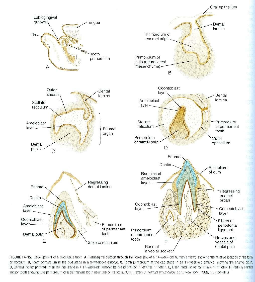
9 The sequence of morphological stages which characterize tooth development. The shape of the dental epithelium on frontal sections is shown in orange, dental mesenchyme in green. The first morphological sign of tooth formation is a thickening of the oral epithelium, which later forms a dental lamina. The dental lamina gives rise to the epithelial tooth buds with the surrounding condensed mesenchyme. Then the main morphological change concerns the development of the cervical loop (asterisk), which characterizes the cap stage. The mesenchyme enclosed in the cap cavity is called tooth papilla (dark green). At the early bell stage, cusps formation is initiated, and the cervical loop progressively elongates and further delimitates the mesenchyme of the tooth papilla. At the late bell stage, the root formation initiates by further extension of the dental epithelium, which forms the Hertwig s epithelial root sheet (arrowhead). Functional differentiation of specific cells is initiated at the epithelium-mesenchyme interface (dashed line). The cells become postmitotic and then start to elongate and polarize. Firstly odontoblasts appear on the mesenchymal side and later ameloblasts become visible on the epithelial side (see in the rectangle).
.")
. Deep color dots demonstrate a large number of cells, and light color ones a small number.")
10 Schematic illustration showing the distribution patterns of neural crest cells originated from the anterior midbrain (A), posterior midbrain (B), and anterior hindbrain (C). Colored dots show the crest cells emigrating by the end of the 5-somite stage (red), at the 6-somite stage (green), and at the 7to 8-somite stage (blue). Deep color dots demonstrate a large number of cells, and light color ones a small number. In general, the early-emigrating crest cells migrate to the more distal region, while those emigrating at the later stage are distributed in the more proximal part.
. (B) Frontal histological sections.")
11 Stages of tooth development shown in the cheek region of the prenatal mouse mandible. (A) 3D reconstructions document the spatial arrangement of the dental and adjacent oral epithelium. A frontal section has been drawn to present the shape of the dental epithelium in the central part of a 3D model (modified according to Peterkova et al, 2002). (B) Frontal histological sections. Bar = 50um.
12
13
14 Classical interpretation of the early development of human dentition. Human teeth develop from a U-shaped dental lamina (DL), which exists in each upper and lower jaw. Externally, another U-shaped structure is located and called a vestibular lamina (VL). The latter structure is presented as the anlage of oral vestibule.
15 A developmental relationship between developing dentition and oral vestibule in human. A scheme of the dental (red) and vestibular (yellow and blue) epithelium in human embryos is based by computer aided 3D reconstructions. The prospective oral vestibule (yellow) has different origins in the lip and cheek regions of both: upper (A) and lower (B) jaws. The remaining vestibular epithelium is in blue.

16 The trigeminal (fifth cranial) nerve consists of large sensory branches and a small motor branch. There are three sensory branches: the ophthalmic to the orbit and forehead, the mandibular to teeth of the lower jaw and tongue, and the maxillary to the maxillary sinus and teeth of the upper jaw.

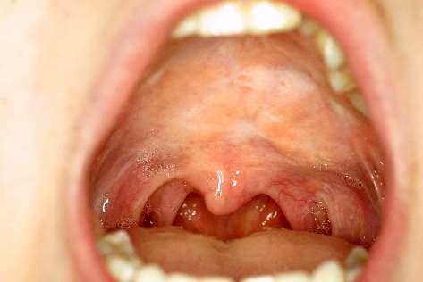
17 Malocclusion is the term used when the occlusion, or bite, is not ideal. crossbite, too much of an over/under bite as pictured below, or other other discrepancies.

18 (A), Normal occlusion; (B), Class I malocclusion; (C), Class II malocclusion; (D), Class III malocclusion. Note the position of the mesial cusp of the maxillary molar relative to the mandibular molar in each type of occlusion.
19
20
21
22
23


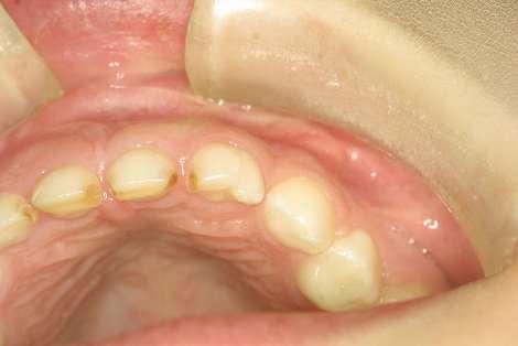
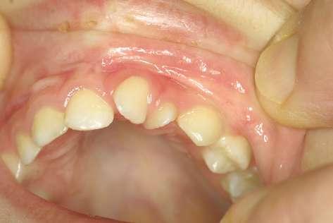
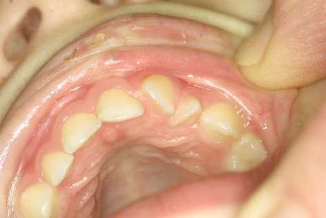
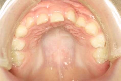
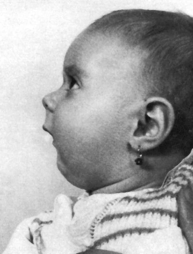
24 Double second upper incisor
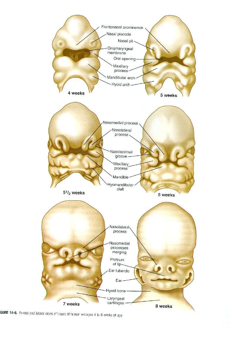
25 Normal development 5th week 6th week yellow thickened dental epithelium of the mn (medial nasal process); red thickened dental epithelium of the mx (maxillary process); ln lateral nasal process md mandibular process
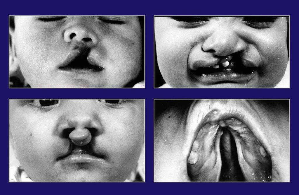
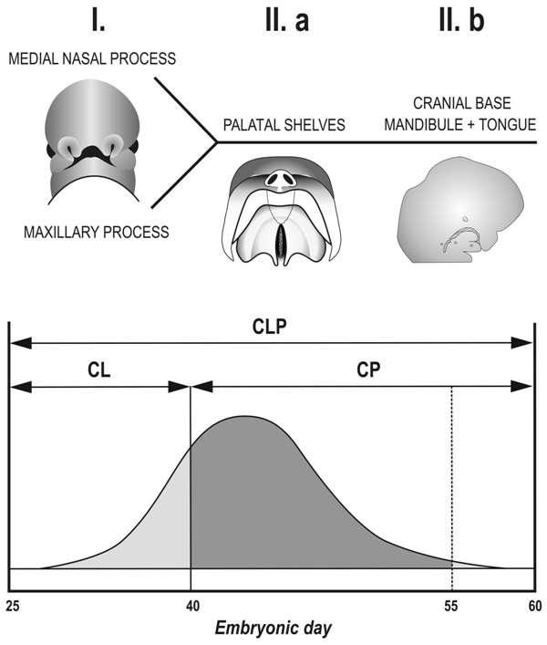
26 The early development of the human upper jaw and dental arch in a scheme. The scheme shows frontal aspect of the embryonic human face and the oral aspect of the upper jaw arch before (A) and after the critical period of the fusion of the facial processes (B) and dental epithelium (C). In the 5th week old embryo (A), the medial nasal (mn) and the maxillary (mx) processes are not yet fused. (B) During normal development (I), the facial processes fuse giving rise to a continuous upper lip and jaw arch. Origin of the upper jaw cleft results from failing fusion of the mn and mx (III). During normal development (I), the fusion of the facial processes is followed by a fusion of their dental epithelia and formation of a continuous dental lamina in week 6-7 human embryo. At the site of the fusion of the dental epithelia, the germ of the lateral incisor (i2) emerges, containing material from both the mn and mx under (C/I). Developmental anomalies of i2 (duplication, hypoplasia, absence) are typically associated with a presence of the upper jaw cleft (C/III). However, incisor anomalies also frequently occur in normally formed upper jaw arch. This can be explained by a defect in fusion of the two incisor components (C/II). Red, yellow and green dental epithelium of the respective mn, mx and mandibular process (md).
 computer-aided 3D reconstructions at ED")
27 Normal development (epithelium from mesenchymal view) computer-aided 3D reconstructions at ED 40-42
28
29
30
31 Pierre-Robin Syndrome
32


33 Mutation in IRF6 (Interferon Regulatory Factor 6) causes VAN der WOUDE Syndrome (Salivary fistula + BCLP)

34 Atypical clefts
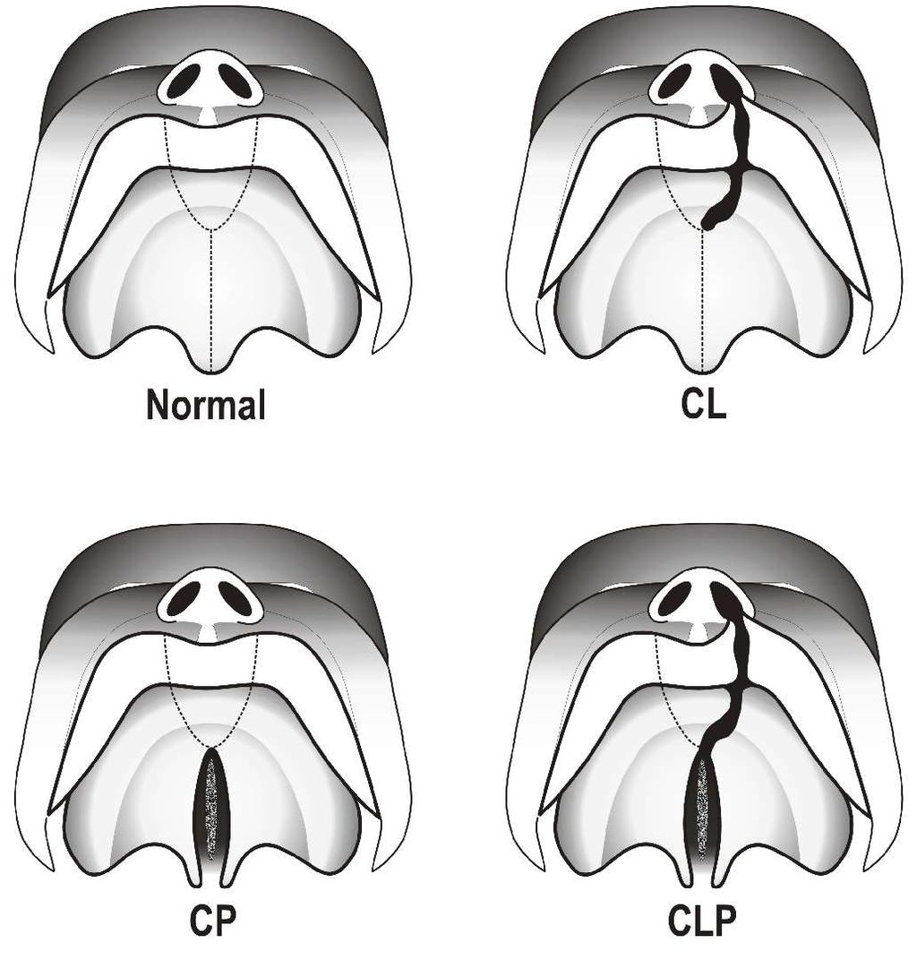
35 INCIDENCE OF OROFACIAL CLEFT
36
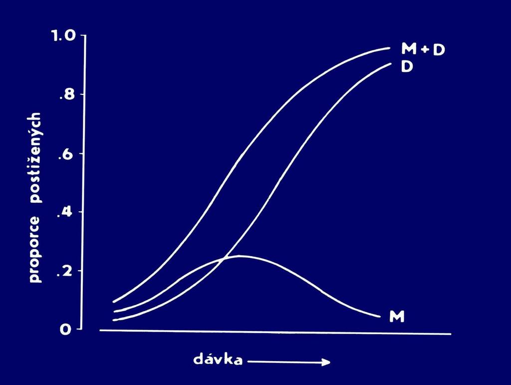
37 dose proportion
38 Spontaneous abortions Czech Republic A official statistics no records of abortions during first month of pregnancy B situation after mathematical extrapolation of data using exponential curve
39 r e b mu N Newborns with an orofacial cleft in Czech Rep. Years
40 r e b mu N Living newborns in Czech Rep. Years
 ec ne di c ni 7. 8.")
41 Incidence of orofacial cleft in Czech Rep. (on 1000 newborns) ec ne di c ni Years
42
43
 Děčín Most Plzeň Plzeň-sever Praha-východ Ústí nad Orlicí Svitavy Teplice Kladno Klatovy Prachatice")
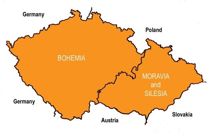
44 Districts with the highest (black) and the lowest (white) incidence of orofacial clefts in the Czech Rep. ( and ) Děčín Most Plzeň Plzeň-sever Praha-východ Ústí nad Orlicí Svitavy Teplice Kladno Klatovy Prachatice Jičín Kolín Pardubice
45 Primary prevention of malformations Planning of pregnancy: avoiding of the exposition to embryotoxic factors by professional employment (chemicals, psychical stress) investigation of mother s health (internal, gynecological, stomatological) optimalization of alimentation and life style (smoking, abusus of alcohol) treatment of chronic diseases (e.g. diabetes, epilepsy, thyroid gland disturbances, asthma) and optimalization of medical treatment (drug doses and scheme of therapy)
46 Secondary prevention of malformations Prenatal diagnostics: Ultra-sonography (detecting of external malformations, heart defects, sex of embryos) Amniocentesis during weeks (chromosomal aberrations, metabolic diseases, sex chromatin patterns, alpha-fetoprotein assay) Chorionic villus sampling (biopsies - several weeks earlier than amniocentesis)

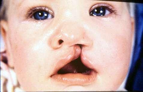
47 Unilateral incomplete cleft lip in fetus at 34 weeks' gestation. Smith A S et al. AJR 2004;183: by American Roentgen Ray Society

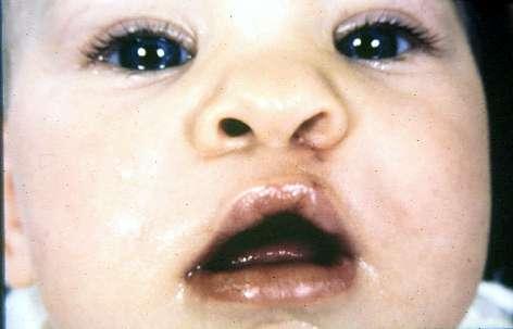
48 Bilateral complete cleft lip in fetus at 18 weeks' gestation. Smith A S et al. AJR 2004;183: by American Roentgen Ray Society

49 Warfarin syndrome Hypoplastic nose, flat face, low nasal bridge, altered calcification (Smith, 1982)
Development of teeth. 5.DM - Pedo
 Development of teeth 5.DM - Pedo Tooth development process of continuous changes in predetermined order starts from dental lamina A band of ectodermal cells growing from the epithelium of the embryonic
Head and Neck Development and Malformations
 Head and Neck Development and Malformations Yang Chai, DDS, PhD Professor George and MaryLou Boone Chair Ostrow School of Dentistry of USC ychai@usc.edu C D E A. B Learning Objectives - Learn cranial neural
Australian Dental Journal
 Australian Dental Journal The official journal of the Australian Dental Association Australian Dental Journal 2014; 59:(1 Suppl): 55 80 doi: 10.1111/adj.12130 Three-dimensional analysis of the early development
Attachment G. Orthodontic Criteria Index Form Comprehensive D8080. ABBREVIATIONS CRITERIA for Permanent Dentition YES NO
 First Review IL HFS Dental Program Models Second Review Ortho cad Attachment G Orthodontic Criteria Index Form Comprehensive D8080 Ceph Film X-Rays Photos Narrative Patient Name: DOB: ABBREVIATIONS CRITERIA
04 Development of the Face and Neck. Development of the Face Development of the neck
 04 Development of the Face and Neck Development of the Face Development of the neck Development of the face Overview of facial development The fourth week ~ the twelfth week of prenatal development Between
Remember from the first year embryology Trilaminar disc has 3 layers: ectoderm, mesoderm, and endoderm
 Development of face Remember from the first year embryology Trilaminar disc has 3 layers: ectoderm, mesoderm, and endoderm The ectoderm forms the neural groove, then tube The neural tube lies in the mesoderm
6610 NE 181st Street, Suite #1, Kenmore, WA
 660 NE 8st Street, Suite #, Kenmore, WA 9808 www.northshoredentalacademy.com.08.900 READ CHAPTER The Professional Dental Assistant (p.-9) No Key Terms Recall Questions:,,,, and 6 CLASS SYLLABUS DAY READ
Oral Embryology and Histology
 Oral Embryology and Histology Chapter 8 Copyright 2018, Elsevier Inc. All Rights Reserved. 1 Learning Objectives Lesson 8.1: Oral Embryology 1. Pronounce, define, and spell the key terms. 2. Define embryology
Fundamental & Preventive Curvatures of Teeth and Tooth Development. Lecture Three Chapter 15 Continued; Chapter 6 (parts) Dr. Margaret L.
 Dr. Margaret L.") Fundamental & Preventive Curvatures of Teeth and Tooth Development Lecture Three Chapter 15 Continued; Chapter 6 (parts) Dr. Margaret L. Dennis Proximal contact areas Contact areas are on the mesial and
Fundamental & Preventive Curvatures of Teeth and Tooth Development Lecture Three Chapter 15 Continued; Chapter 6 (parts) Dr. Margaret L. Dennis Proximal contact areas Contact areas are on the mesial and
Arrangement of the artificial teeth:
 Lecture Prosthodontic Dr. Osama Arrangement of the artificial teeth: It s the placement of the teeth on a denture with definite objective in mind or it s the setting of teeth on temporary bases. Rules
Tooth eruption and movement
 Tooth eruption and movement Dr. Krisztián Nagy Diphydont dentition Deciduous dentition primary dentition Diphydont dentition Permanent dentition secondary dentition Mixed Dentition: Presence of both dentitions
Dental Morphology and Vocabulary
 Dental Morphology and Vocabulary Palate Palate Palate 1 2 Hard Palate Rugae Hard Palate Palate Palate Soft Palate Palate Palate Soft Palate 4 Palate Hard Palate Soft Palate Maxillary Arch (Maxilla) (Uppers)
06 Tooth Development and Eruption
 + 06 Tooth Development and Eruption Tooth development Root development PDL and alveolar bone development Primary tooth eruption and shedding Permanent tooth eruption Q. Where and how tooth starts to form?
Development of occlusion
 Development of occlusion The development of dentition is an important part of craniofacial growth as the formation, eruption, exfoliation and exchange of teeth take place during this period. Term occlusion
 Drawings illustrating the human pharyngeal apparatus. Drawings illustrating the human pharyngeal apparatus. Drawings illustrating the human pharyngeal apparatus. Drawings illustrating the human pharyngeal
Anatomy and Physiology. Bones, Sutures, Teeth, Processes and Foramina of the Human Skull
 Anatomy and Physiology Chapter 6 DRO Bones, Sutures, Teeth, Processes and Foramina of the Human Skull Name: Period: Bones of the Human Skull Bones of the Cranium: Frontal bone: forms the forehead and the
Dental Anatomy and Occlusion
 CHAPTER 53 Dental Anatomy and Occlusion Ma Lou C. Sabino DDS, and Emily G. Smythe, DDS What numerical system is used most commonly in the United States for designating the adult dentition? Pediatric dentition?
NEUROCRANIUM VISCEROCRANIUM VISCEROCRANIUM VISCEROCRANIUM
 LECTURE 4 SKULL NEUROCRANIUM VISCEROCRANIUM VISCEROCRANIUM VISCEROCRANIUM CRANIUM NEUROCRANIUM (protective case around brain) VISCEROCRANIUM (skeleton of face) NASOMAXILLARY COMPLEX MANDIBLE (DESMOCRANIUM)
Preventive Orthodontics
 Semmelweis University Faculty of Dentistry Department in Community Dentistry director: Dr. Kivovics Péter assoc.prof. http://semmelweis-egyetem.hu/fszoi/ https://www.facebook.com/fszoi Preventive Orthodontics
ORTHODONTICS Treatment of malocclusion Assist.Lec.Kasem A.Abeas University of Babylon Faculty of Dentistry 5 th stage
 Lec: Treatment of class I malocclusion Class I occlusion can be defined by Angles, classification as the mesiobuccal cusp of the upper 1 st permanent molar occlude with the developmental groove of the
Morphology of an Anatomic Crown. By: Assistant Professor Dr. Baydaa Ali Al - Rawi
 Morphology of an Anatomic Crown By: Assistant Professor Dr. Baydaa Ali Al - Rawi October 4, 2009 Elevated landmarks Depressed landmarks A) Elevated landmarks : 1. Dental lobe : is one of the primary centers
Pharyngeal Apparatus. Pouches Endoderm Grooves Ectoderm Arch Neural Crest Somitomeres Aortic Arch - Vessel
 Pharyngeal Apparatus Pouches Endoderm Grooves Ectoderm Arch Neural Crest Somitomeres Aortic Arch - Vessel Segmental Organization Humans: Arch 1-4 prominent Arch 5 absent Arch 6 - transient First Arch Face
Arrangement of posterior artificial teeth Standardized parameters Curve of Wilson Curve of Spee
 . Arrangement of posterior artificial teeth Posterior teeth are set up in tight centric occlusion. The mandibular teeth are set in the wax occlusion rim over the residual ridge in their ideal buccolingual
Essentials in Head and Neck Embryology. Part 3 Development of the head, face, and oral cavity
 Essentials in Head and Neck Embryology Part 3 Development of the head, face, and oral cavity Outline General overview of prenatal development Embryonic period phase 1 Formation of bilaminar disk Formation
Treatment planning of nonskeletal problems. in preadolescent children
 In the name of GOD Treatment planning of nonskeletal problems in preadolescent children Presented by: Dr Somayeh Heidari Orthodontist Reference: Contemporary Orthodontics Chapter 7 William R. Proffit,
ALTERNATE OCCLUSAL SCHEMES
 ALTERNATE OCCLUSAL SCHEMES The same basic concepts need to be applied to all occlusal schemes. Some challenges include missing teeth, transposed teeth, crossbites, and anterior open bites. POSTERIOR CROSSBITES
1. What is the highest and sharpest cusp on the lower first deciduous molar? 2. Which of the following is NOT the correct location of an embrasure?
 1 1. What is the highest and sharpest cusp on the lower first deciduous molar? a. mesiobuccal b. distobuccal c. distolingual d.mesiolingual 2. Which of the following is NOT the correct location of an embrasure?
Dr Robert Drummond. BChD, DipOdont Ortho, MChD(Ortho), FDC(SA) Ortho. Canad Inn Polo Park Winnipeg 2015
, FDC(SA) Ortho. Canad Inn Polo Park Winnipeg 2015") Dr Robert Drummond BChD, DipOdont Ortho, MChD(Ortho), FDC(SA) Ortho Canad Inn Polo Park Winnipeg 2015 Severely compromised FPM with poor prognosis Children often present with a developing dentition affected
Dr Robert Drummond BChD, DipOdont Ortho, MChD(Ortho), FDC(SA) Ortho Canad Inn Polo Park Winnipeg 2015 Severely compromised FPM with poor prognosis Children often present with a developing dentition affected
Texas A&M College of Dentistry Caruth School of Dental Hygiene
 Texas A&M College of Dentistry Caruth School of Dental Hygiene Course Number and Name: 3120 Dental Anatomy Course Type: Lecture Laboratory Seminar Academic Year/Semester Offered: 2016/Fall Semester Course
REVIEW OF CLINICAL EMBRYOLOGY OF HEAD AND NECK
 REVIEW OF CLINICAL EMBRYOLOGY OF HEAD AND NECK OUTLINE - EMBRYOLOGY UNDERLYING CLINICAL CONDITIONS I. EARLY DEVELOPMENT OF FACE: CLEFT LIP, CLEFT PALATE, OBSTRUCTED NASOLACRIMAL DUCT II. BRANCHIAL ARCHES
Concepts of occlusion Balanced occlusion. Monoplane occlusion. Lingualized occlusion. Figure (10-1)
") Any contact between teeth of opposing dental arches; usually, referring to contact between the occlusal surface. The static relationship between the incising or masticatory surfaces of the maxillary or
Any contact between teeth of opposing dental arches; usually, referring to contact between the occlusal surface. The static relationship between the incising or masticatory surfaces of the maxillary or
Prosthetic Options in Implant Dentistry. Hakimeh Siadat, DDS, MSc Associate Professor
 Prosthetic Options in Dentistry Hakimeh Siadat, DDS, MSc Associate Professor Dental Research Center, Department of Prosthodontics & Dental s Faculty of Dentistry, Tehran University of Medical Sciences
Dental Health Considerations & Solutions in Patients with Turner Syndrome
 Dental Health Considerations & Solutions in Patients with Turner Syndrome Robert Korwin, D.M.D. Post Graduate Certificate in General Practice Master, Academy of General Dentistry Master, International
Primary Teeth Chapter 18. Dental Anatomy 2016
 Primary Teeth Chapter 18 Dental Anatomy 2016 Primary Teeth - Introduction Synonyms deciduous teeth, baby teeth, temporary teeth, milk teeth. There are 20 primary teeth, designated as A thru T in the Universal
Early treatment. Interceptive orthodontics
 Early treatment Interceptive orthodontics Early treatment Some malocclusion can be prevented or intercepted. Diphasic treatment is sometimes considered more logical and sensible. During the phase one,
Mesial Step Class I or Class III Dependent upon extent of step seen clinically and patient s growth pattern Refer for early evaluation (by 8 years)
") Orthodontics and Dentofacial Development Overview Development of Dentition Treatment Retention and Relapse Growth of Naso-Maxillary Complex Develops postnatally entirely by intramenbranous ossification
Orthodontics and Dentofacial Development Overview Development of Dentition Treatment Retention and Relapse Growth of Naso-Maxillary Complex Develops postnatally entirely by intramenbranous ossification
Supplementary Figure 1: Signaling centers contain few proliferating cells, express p21, and
 Supplementary Figure 1: Signaling centers contain few proliferating cells, express p21, and exclude YAP from the nucleus. (a) Schematic diagram of an E10.5 mouse embryo. (b,c) Sections at B and C in (a)
PH-04A: Clinical Photography Production Checklist With A Small Camera
 PH-04A: Clinical Photography Production Checklist With A Small Camera Operator Name Total 0-49, Passing 39 Your Score Patient Name Date of Series Instructions: Evaluate your Series of photographs first.
EUROPEAN SOCIETY OF LINGUAL ORTHODONTICS
 EUROPEAN SOCIETY OF LINGUAL ORTHODONTICS CANDIDATE NUMBER: Dr. Stefan Blasius Year: 2010 WBLO 01 EUROPEAN SOCIETY OF LINGUAL ORTHODONTICS CANDIDATE NUMBER: Dr. Stefan Blasius Year: 2010 WBLO 01 RÉSUMÉ
Chapter 2. Material and methods
 Chapter 2 Material and methods Material and methods Summary This chapter describes the subjects and methods being used in this study. Between 1986 and 1997 9 expeditions were undertaken in remote areas
Treatment of Angle Class III. Department of Paedodontics and Orthodontics Dr. habil. Melinda Madléna associate professor
 Department of Paedodontics and Orthodontics Dr. habil. Melinda Madléna associate professor Disorders in Angle Class III The position of the lower jaw is foreward regarding to the upper jaw Mesialocclusion
Phylogenetics Lab: Character Descriptions
 Phylogenetics Lab: Character Descriptions 1) Osseous Auditory Canal. 0= absent, 1= present. Does the organism have a bony auditory canal? This will look like a hole or opening in the skull behind the jaw
Trigeminal Nerve (V)
") Trigeminal Nerve (V) Lecture Objectives Discuss briefly how the face is developed. Follow up the course of trigeminal nerve from its point of central connections, exit and down to its target areas. Describe
Trigeminal Nerve (V) Lecture Objectives Discuss briefly how the face is developed. Follow up the course of trigeminal nerve from its point of central connections, exit and down to its target areas. Describe
Supplemental mandibular incisors: a Recherché
 CASE REPORT 34 Supplemental mandibular incisors: a Recherché Sankriti Murthy Introduction: Supernumerary teeth are a developmental disturbance encountered in the dental arches. These teeth are in excess
Brain and spinal nerve. By: shirin Kashfi
 Brain and spinal nerve By: shirin Kashfi Nervous system: central nervous system (CNS) peripheral nervous system (PNS) Brain (cranial) nerves Spinal nerves Ganglions (dorsal root ganglions, sympathetic
Alveolar Growth in Japanese Infants: A Comparison between Now and 40 Years ago
 Bull Tokyo Dent Coll (2017) 58(1): 9 18 Original Article doi:10.2209/tdcpublication.2016-0500 Alveolar Growth in Japanese Infants: A Comparison between Now and 40 Years ago Hiroki Imai 1), Tetsuhide Makiguchi
Corporate Medical Policy
 Corporate Medical Policy File Name: Origination: Last CAP Review: Next CAP Review: Last Review: orthodontics_for_pediatric_patients 2/2014 10/2017 10/2018 10/2017 Description of Procedure or Service Children
An Anterior Tooth Size Comparison in Unilateral and Bilateral Congenitally Absent Maxillary Lateral Incisors
 An Anterior Tooth Size Comparison in Unilateral and Bilateral Congenitally Absent Maxillary Lateral Incisors Abstract The purpose of this study is to compare the anterior tooth size width in patients with
Development of the Pharyngeal Arches
 Development of the Pharyngeal Arches Thomas A. Marino, Ph.D. Temple University School of Medicine Competencies: Upon completion of this section of the course, the student must be able to: 1. Recall the
#45 Ortho-Tain, Inc PREVENTIVE ERUPTION GUIDANCE -- PREVENTIVE OCCLUSAL DEVELOPMENT
 #45 Ortho-Tain, Inc. 1-800-541-6612 PREVENTIVE ERUPTION GUIDANCE -- PREVENTIVE OCCLUSAL DEVELOPMENT Analysis and Diagnosis of Occlusion: The ideal child of 5 y ears of age that probably has the best chance
Sample Case #1. Disclaimer
 ABO Sample Cases Disclaimer Sample Case #1 The following sample questions and answers were composed and vetted by a panel of experts in orthodontics and are intended to provide an example of the types
The Premolars. Chapter 17 Permanent Posterior Teeth (p )
") The Premolars Chapter 17 Permanent Posterior Teeth (p. 230-244) General Information Function: u Hold and grind food u Work with molars in mastication. u Even without molars one may be able to chew well
The Premolars Chapter 17 Permanent Posterior Teeth (p. 230-244) General Information Function: u Hold and grind food u Work with molars in mastication. u Even without molars one may be able to chew well
22q11 deletion syndrome Report from observation charts
 Orofacial function of persons having 22q11 deletion syndrome Report from observation charts The survey comprises 147 observation charts. Synonyms: CATCH 22, Di George syndrome, Velocardiofacial syndrome
Development of the dentition
 4 Development of the dentition 85 Humans have two dentitions, the deciduous (primary) and permanent (secondary). Each dentition is heterodont, meaning that it consists of teeth with different shapes and
Orthodontics. Anomalies
 Orthodontics Anomalies Anomalies of Teeth Groups of teeth Jaws Intermaxilary relations Anomalies of tooth number Hypodontics (hypodontia) the tooth (or teeth) are missing Third molars (if third molars
Basic Anatomy and Physiology of the Lips and Oral Cavity. Dr. Faghih
 Basic Anatomy and Physiology of the Lips and Oral Cavity Dr. Faghih It is divided into seven specific subsites : 1. Lips 2. dentoalveolar ridges 3. oral tongue 4. retromolar trigone 5. floor of mouth 6.
TRAUMA TO THE FACE AND MOUTH
 Dr.Yahya A. Ali 3/10/2012 F.I.C.M.S TRAUMA TO THE FACE AND MOUTH Bailey & Love s 25 th edition Injuries to the orofacial region are common, but the majority are relatively minor in nature. A few are major
Postnatal Growth. The study of growth in growing children is for two reasons : -For health and nutrition assessment
 Growth of The Soft Tissues Postnatal Growth Postnatal growth is defined as the first 20 years of growth after birth krogman 1972 The study of growth in growing children is for two reasons : -For health
Embryo#1. Mohammad Hisham Al-Mohtaseb باشق جهاد. 0 P a g e
 Embryo#1 Mohammad Hisham Al-Mohtaseb باشق جهاد 0 P a g e Before you start, it is important to link what you learn in gross anatomy with developmental stages discussed in embryology. Cells that form organs
Semester Credits: 3 Lecture Hours: 3. Prerequisites:
 Revised: Fall 2015 Semester Credits: 3 Lecture Hours: 3 21THistology DNH 115 Admission into dental hygiene program. Prerequisites: Course Description: Presents a study of the microscopic and macroscopic
Lecture. Permanent maxillary premolars
 Lecture Permanent maxillary premolars Permanent premolars The maxillary premolars are four in number: two in the right and two in the left. They are posterior to the canines and anterior to the molars.
Response Type axium Adult Comprehensive Oral Examination (COE)
") Page 1 1. RADIOGRAPHIC EVALUATION 1001 Panoramic image (PAN) Maxillary sinuses Nasal cavity TMJ complex Mandibular canal visualization?bone anomalies (eg. radiopacity/radiolucency) Soft tissue abnormalities
Page 1 1. RADIOGRAPHIC EVALUATION 1001 Panoramic image (PAN) Maxillary sinuses Nasal cavity TMJ complex Mandibular canal visualization?bone anomalies (eg. radiopacity/radiolucency) Soft tissue abnormalities
Applied Equine Dental Development
 Published in IVIS with the permission of the AAEP Close this window to return to IVIS Applied Equine Dental Development Kirstie Dacre, BVMS, MSc, Cert EM (Int Med), PhD Author s address: Veterinary Teaching
Yokose, T; Sakamoto, T; Sueishi, K; Author(s) Tsujino, K; Kubo, S; Yakushiji, M; Journal Bulletin of Tokyo Dental College, 4
 Tsujino, K; Kubo, S; Yakushiji, M; Journal Bulletin of Tokyo Dental College, 4") Two cases with supernumerary teeth Title region Yokose, T; Sakamoto, T; Sueishi, K; Author(s) Tsujino, K; Kubo, S; Yakushiji, M; Journal Bulletin of Tokyo Dental College, 4 URL http://hdl.handle.net/10130/216
Two cases with supernumerary teeth Title region Yokose, T; Sakamoto, T; Sueishi, K; Author(s) Tsujino, K; Kubo, S; Yakushiji, M; Journal Bulletin of Tokyo Dental College, 4 URL http://hdl.handle.net/10130/216
Temporal fossa Infratemporal fossa Pterygopalatine fossa Terminal branches of external carotid artery Pterygoid venous plexus
 Outline of content Temporal fossa Infratemporal fossa Pterygopalatine fossa Terminal branches of external carotid artery Pterygoid venous plexus Boundary Content Communication Mandibular division of trigeminal
Lec [8]: Mandibular nerve:
![Lec [8]: Mandibular nerve:](/thumbs/94/121295776.jpg "Lec [8]: Mandibular nerve:") Lec [8]: Mandibular nerve: The mandibular branch from the trigeminal ganglion lies in the middle cranial fossa lateral to the cavernous sinus. With the motor root of the trigeminal nerve [motor roots lies
Lec [8]: Mandibular nerve: The mandibular branch from the trigeminal ganglion lies in the middle cranial fossa lateral to the cavernous sinus. With the motor root of the trigeminal nerve [motor roots lies
Permanent 2 nd Maxillary Molars
 Permanent 2 nd Maxillary Molars In comparison to the first max molar First molars appears in the oral cavity at the age of 6 years old.. While 2 nd molar 3 rd molar Max. 2 nd molar have long roots (sometimes
Medical NBDE-II. Dental Board Exams Part I.
 Medical NBDE-II Dental Board Exams Part I http://killexams.com/exam-detail/nbde-ii Question: 149 Anatomically, the term "clinical root" can be defined as which of the following: A. The space in the tooth
Class II Correction with Invisalign Molar rotation.
 Tips from your peers to help you treat with confidence. Class II Correction with Invisalign Molar rotation. Dr. Mazyar Moshiri. Class II Correction with Invisalign Molar Rotation. Dr. Mazyar Moshiri. Orthodontic
Mandibular incisor extraction: indications and long-term evaluation
 European Journal of Orthodontics 18 (1996) 485-489 O 1996 European Orthodontic Society Mandibular incisor extraction: indications and long-term evaluation Jose-Antonio Canut University of Valencia, Spain
Applications in Dermatology, Dentistry and LASIK Eye Surgery using LASERs
 Applications in Dermatology, Dentistry and LASIK Eye Surgery using LASERs http://www.medispainstitute.com/menu_laser_tattoo.html http://www.life123.com/bm.pix/bigstockphoto_close_up_of_eye_surgery_catar_2264267.s600x600.jpg
Treatment of a Rare Bilateral Severe Ectopic Eruption of the Maxillary First Permanent Molar: A Case Report
 Case Report Treatment of a Rare Bilateral Severe Ectopic Eruption of the Maxillary First Permanent Molar: A Case Report MS. Ahmad Akhoundi 1, 2, AH. Sadrhaghighi 3 1 Associate Professor, Dental Research
T O O T H A T L A S C O U R S E G U I D E A S S I S T A N T E D I T I O N
 T O O T H A T L A S C O U R S E G U I D E A S S I S T A N T E D I T I O N The information in this guide was prepared by ehuman with contributions from: Cara Miyasaki, RDHEF, MS, Foothill College Kay Murphy,
Ectopic Eruption of Teeth and their Management in Children: Literature Review and Case Reports
 Cronicon OPEN ACCESS EC DENTAL SCIENCE Case Report Ectopic Eruption of Teeth and their Management in Children: Literature Review and Case Reports Bimal Chandra Kirtaniya 1 *, Sonia Tiwari 2, Satya Prakash
ORAL ANATOMY AND PHYSIOLOGY
 CHAPTER 7 ORAL ANATOMY AND PHYSIOLOGY INTRODUCTION This chapter covers the oral anatomy and physiology of the teeth, the histology of the tissues and supporting structures, and concentrates on the external
NATIONAL EXAMINING BOARD FOR DENTAL NURSES
 NATIONAL EXAMINING BOARD FOR DENTAL NURSES NATIONAL DIPLOMA EXAMINATION DENTAL CHARTING NEBDN is a limited company registered in England & Wales No. 5580200 Registered with the Charity Commisioners No.
ADOLESCENT TREATMENT. Thomas J. Cangialosi. Stella S. Efstratiadis. CHAPTER 18 Pages CLASS II DIVISION 1 WHY NOW?
 ADOLESCENT By Thomas J. Cangialosi and Stella S. Efstratiadis From Riolo, M. and Avery, J. Eds., Essentials for Orthodontic Practice, EFOP Press of EFOP, LLC. Ann Arbor and Grand Haven, Michigan, U.S.A.,
Mixed Dentition Treatment and Habits Therapy
 Interception Mixed Dentition Treatment and Habits Therapy Anterior Crossbites Posterior Crossbites Interference s with Normal Eruption Habit Therapy Tsung-Ju Hsieh, DDS, MSD 1 2 Anterior Crossbites Anterior
PTERYGOPALATINE FOSSA
 PTERYGOPALATINE FOSSA Outline Anatomical Structure and Boundaries Foramina and Communications with other spaces and cavities Contents Pterygopalatine Ganglion Especial emphasis on certain arteries and
The morphological studies of root r maxillary primary canines and their Title the position of successive permanen Micro-CT
 The morphological studies of root r maxillary primary canines and their Title the position of successive permanen Micro-CT Author(s) Saka, H; Koyama, T; Tamatsu, Y; Usa Alternative Journal Pediatric dental
Face. Definition: The area between the two ears and from the chin to the eye brows. The muscles of the face
 Face Definition: The area between the two ears and from the chin to the eye brows. The muscles of the face The muscle of facial expression (include the muscle of the face and the scalp). All are derived
DLT 111 DENTAL ANATOMY/PHYSIOLOGY
 DLT 111 DENTAL ANATOMY/PHYSIOLOGY COURSE DESCRIPTION: Prerequisites: Corequisites: Enrollment in the Dental Laboratory Technology program None This course introduces the anatomy of the individual tooth
Upper arch. 1Prosthodontics. Dr.Bassam Ali Al-Turaihi. Basic anatomy & & landmark of denture & mouth
 1Prosthodontics Lecture 2 Dr.Bassam Ali Al-Turaihi Basic anatomy & & landmark of denture & mouth Upper arch Palatine process of maxilla: it form the anterior three quarter of the hard palate. Horizontal
Dr. Sami Zaqout Faculty of Medicine IUG
 The Nose External Nose Nasal Cavity External Nose Blood and Nerve Supplies of the External Nose Blood Supply of the External Nose The skin of the external nose Branches of the ophthalmic and the maxillary
COURSE OUTLINE AND OBJECTIVES. OFFICE HOURS: Wednesdays and Thursdays, 11:30-12:30, P-201 CUNY PROFICIENCY IN READING, WRITING, AND MATHEMATICS
 NEW YORK CITY COLEGE OF TECHNOLOGY DENTAL HYGIENE DEPARTMENT COURSE OUTLINE AND OBJECTIVES COURSE CODE AND TITLE: DEN 1112 ORAL ANATOMY TERM: FALL 2016 INSTRUCTOR: ANNA MATTHEWS, RDH, MS amatthews@citytech.cuny.edu
Techniques of local anesthesia in the mandible
 Techniques of local anesthesia in the mandible The technique of choice for anesthesia of the mandible is the block injection and this is attributed to the absence of the advantages which are present in
LOCAL ANESTHESIA IN PEDIATRIC DENTISTRY
 Disclaimer This movie is an educational resource only and should not be used to manage your health. All decisions about the management of local anesthesia in pediatric dentistry must be made in conjunction
Human Healed Trauma Skull
 Human Healed Trauma Skull Product Number: Specimen Evaluated: BC-303 Original Specimen Skeletal Inventory: 1 Cranium with full dentition (teeth ##1-16) 1 Mandible with full dentition (teeth ##17-32) Osteological
Wisdom Can Be Painful: Third Molar Impaction In Human Populations and Its Evolutionary Significance
 Wisdom Can Be Painful: Third Molar Impaction In Human Populations and Its Evolutionary Significance Since Charles Darwin published the Origin of Species in 1859, evolution and the mechanisms underlying
Subject Index. AXIN2, cleft defects 24, 26
 Subject Index ADAMTS, mouse mutants and palate development 37, 38 Africa, cleft lip and palate prevalence 6, 7 Alcohol dependence, pregnancy risks for cleft 25, 61 Altitude, pregnancy risks for cleft 25,
Prevalence of Incisors Crowding in Saudi Arabian Female Students
 Prevalence of Incisors Crowding in Saudi Arabian Female Students Fadia M. Al-Hummayani, BDS, MS * Abstract This study was carried out to determine the prevalence of incisor crowding in Saudi Arabian female
Ahtiainen et al., http :// /cgi /content /full /jcb /DC1
 Supplemental material JCB Ahtiainen et al., http ://www.jcb.org /cgi /content /full /jcb.201512074 /DC1 THE JOURNAL OF CELL BIOLOGY Figure S1. Distinct distribution of different cell cycle phases in the
POSTGRADUATE INSTITUTE OF MEDICINE UNIVERSITY OF COLOMBO SELECTION EXAMINATION IN MD (ORAL SURGERY) OCTOBER 2009 PAPER 1.1. Part A (General Anatomy)
 OCTOBER 2009 PAPER 1.1. Part A (General Anatomy)") POSTGRADUATE INSTITUTE OF MEDICINE UNIVERSITY OF COLOMBO SELECTION EXAMINATION IN MD (ORAL SURGERY) OCTOBER 2009 Date : 5 th October 2009 Time : 2.00 p.m. 5.00p.m. PAPER 1.1 Answer three (03) questions
POSTGRADUATE INSTITUTE OF MEDICINE UNIVERSITY OF COLOMBO SELECTION EXAMINATION IN MD (ORAL SURGERY) OCTOBER 2009 Date : 5 th October 2009 Time : 2.00 p.m. 5.00p.m. PAPER 1.1 Answer three (03) questions
Speech/Resonance Disorders due to Clefts and Craniofacial Anomalies
 Speech/Resonance Disorders due to Clefts and Craniofacial Anomalies Ann W. Kummer, PhD, CCC-SLP Cincinnati Children s Hospital Medical Center Royalties: Financial Disclosures Book: Kummer, AW. Cleft Palate
ALABAMA DENTAL HYGIENE PROGRAM 50 QUESTIONS PRE ENTRANCE EXAM
 ALABAMA DENTAL HYGIENE PROGRAM 50 QUESTIONS PRE ENTRANCE EXAM NAME DATE RETURN COMPLETED EXAM WITH APPLICATION You may copy for future reference 1. One cause of decay is the Streptococcus mutans bacteria
-Ibrahim Al-Naser. -Dr Al- Muhtaseb. 1 P a g e
 -1 -Ibrahim Al-Naser - -Dr Al- Muhtaseb 1 P a g e The Digestive System The doctor started the lecture by talking about the class rules. The GI system is an organ system, it is divided into: The Alimentary
Anatomic Relations Summary. Done by: Sohayyla Yasin Dababseh
 Anatomic Relations Summary Done by: Sohayyla Yasin Dababseh Anatomic Relations Lecture 1 Part-1 - The medial wall of the nose is the septum. - The vestibule lies directly inside the nostrils (Nares). -
Structure Location Function
 Frontal Bone Cranium forms the forehead and roof of the orbits Occipital Bone Cranium forms posterior and inferior portions of the cranium Temporal Bone Cranium inferior to the parietal bone forms the
Dr.Sepideh Falah-kooshki
 Dr.Sepideh Falah-kooshki MAXILLA Premaxillary/median palatal suture (radiolucent). Incisive fossa and foramen (radiolucent). Nasal passages (radiolucent). Nasal septum (radiopaque). Anterior nasal spine