NEUROCRANIUM VISCEROCRANIUM VISCEROCRANIUM VISCEROCRANIUM
|
|
|
- Paula Haynes
- 5 years ago
- Views:
Transcription

1 LECTURE 4 SKULL

2 NEUROCRANIUM VISCEROCRANIUM VISCEROCRANIUM VISCEROCRANIUM
 CRANIAL VAULT (CHONDROCRANIUM) CRANIAL")
3 CRANIUM NEUROCRANIUM (protective case around brain) VISCEROCRANIUM (skeleton of face) NASOMAXILLARY COMPLEX MANDIBLE (DESMOCRANIUM) CRANIAL VAULT (CHONDROCRANIUM) CRANIAL BASE
4 NEUROCRANIUM (CRANIAL VAULT) Houses and protect the brain FRONTAL OCCIPITAL SPHENOID UNPAIRED BONES ETHMOID PARIETAL TEMPORAL PAIRED BONES
")
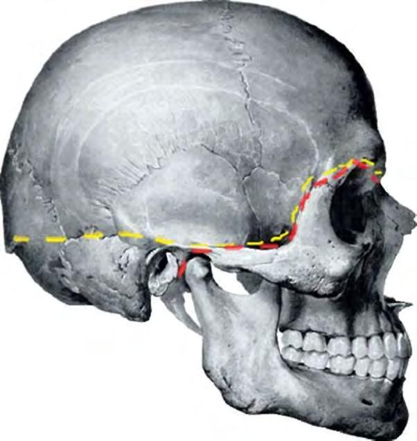
5 Skull base (external) Skull base (internal)

6 VISCEROCRANIUM (FACE & JAW) MANDIBLE VOMER UNPAIRED BONES MAXILLA ZYGOMATIC NASAL LACRIMAL PAIRED BONES PALATINE INFERIOR CONCHA

7 NEUROCRANIUM MEMBRANOUS CARTILAGINOUS NEURAL CREST CELLS & PARAXIAL MESODERM Prechordal chondrocranium NEURAL CREST CELLS Chordal chondrocranium OCCIPITAL SCLEROTOMES PARAXIAL MESODERM
")
 the viscerocranium or facial")
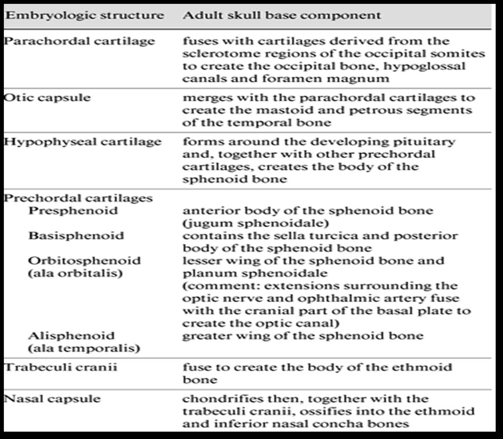
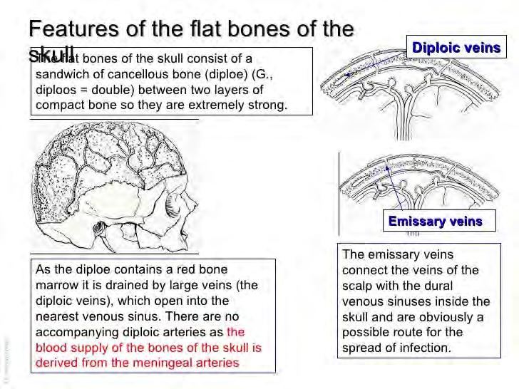
8 The human skull consists of three components: (1) the membranous neurocranium, which constitutes the flat bones of the skull, (2) the cartilaginous neurocranium or chondrocranium which forms the majority of the skull base, and (3) the viscerocranium or facial skeleton. 8


9 The basicranium develops primarily from cartilage precursors, with a small component from membranous bone. The development of the cartilaginous skull base begins around the 40th day of gestation, with the conversion of mesenchyme into cartilage. Occipital sclerotomal mesenchyme concentrates around the notochord and extends cephalically forming the floor of skull

10 The Parachordal Cartilages and Early Chondrogenesis The chondrocranium begins to form when the collections of mesenchyme accumulating around and in front of the notochord condense into cartilage. These chondrification centers, termed the parachordal cartilages, form early in the seventh week adjacent to the rostral end of the notochord and contribute to the creation of the basal plate. The parachordal cartilage fuse with the sclerotomes arising from the occipital somites surrounding the neural tube.
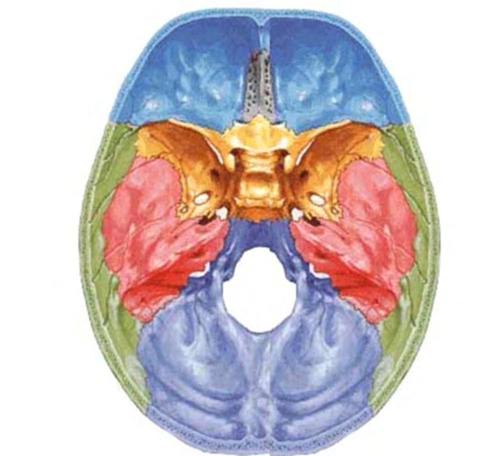
11 CARTILAGINOUS NEUROCRANIUM Cartilages that lie in front of rostral end of notochord arise from neural crest cells forming prechordal chondrocranium Cartilages posterior to this end arise from Occipital sclerotomes forming Chordal chondrocranium
 chondrocranium (blue).")
12 Dorsal view of the chondrocranium, or base of the skull, in the adult showing bones formed by endochondral ossification. Bones that form rostral to the rostral half of the sella turcica arise from neural crest and constitute the prechordal (in front of the notochord) chondrocranium (blue). Those forming posterior to this landmark arise from paraxial mesoderm (chordal chondrocranium) (red).
13 Base of the skull All cartilages fuse Ossify by Endochondral ossification
14

15

16 MEMBRANOUS NEUROCRANIUM Mesenchyme from these sources invests the brain and undergoes membranous ossification Needle like bony spicules radiate from primary ossification centre towards periphery

17 With growth during fetal and postnatal life, membranous bones enlarge Apposition on outer surface Osteoclastic resorption from inside

18 kjjkkjhihihk VISCEROCRANIUM MEMBRANOUS CARTILAGINOUS Intramembranous ossification in maxillary and mandibular prominence of first pharyngeal arch Neural crest cells squamous temporal, maxillary, zygomatic bone, mandible Bones and connective tissue of craniofacial structures
 arches appear along the lateral wall of pharyngeal gut.")
19 Embryo at 4-5 weeks (Lateral view) By 4th week of development, bars of mesenchymal tissue separated by deep clefts. 5 th branchial (pharyngeal) arches appear along the lateral wall of pharyngeal gut. At the end of 4th week center of face is formed by stomodeum surrounded by 1 st pair of branchial (pharyngeal) arches
20 BRANCHIAL (PHARYNGEAL) ARCHES The first pharyngeal arch Consists of: Dorsal portion - maxillary process Ventral portion - mandibular process Mesenchyme of maxillary process gives rise to Premaxilla Maxilla Zygomatic bone Part of temporal bone (through membranous ossification) Mandible is formed by membranous ossification of mesenchyme surrounding Meckel s cartilage. The first pair of pharyngeal arches plays a major role in facial development

21 Face develops from 5 prominences that surround the stomatodeum - Frontonasal - Paired maxillary processes. - Paired mandibular processes.
22 Early in the 4 th week, five primordial swellings consisting primarily of neural crest-derived mesenchyme appear around the stomodeum and play an important role in the development of face 1 Frontonasal prominence 2 Maxillary prominences 2 Mandibular prominences Stomodeum


23 The single frontonasal prominence ventral to the forebrain The paired maxillary prominences develop from the cranial part of first pharyngeal arch The paired mandibular prominences develop from the caudal part of first pharyngeal arch Lateral view

24 By the end of 4 th week, bilateral oval-shaped ectodermal thickenings called nasal placodes appear on each side of the lower part of the frontonasal prominence Nasal placodes are primordia of the nose and nasal cavities. Frontonasal prominence
25 Frontonasal prominence formed by proliferation of mesenchyme ventral to the forebrain. It forms - Lateral optic diverticula eyes - Forehead (between the eyes) - Nasal placodes

26 Mesenchyme proliferates around the placodes producing medial and lateral nasal prominences Lateral nasal prominence separated from maxillary process by nasolacrimal groove

27 Mesenchymal cells proliferate at the margin of the placodes and produce horse-shoe shaped swellings around these. The sides of these swellings are called medial and lateral nasal prominences The placodes now lie in the floor of a depression called nasal pits Each lateral nasal prominence is separated from the maxillary swelling by nasolacrimal groove

28 The maxillary prominences continue to increase in size and: Laterally, merge with the mandibular prominences to form the cheek Medially, compress the medial nasal prominences toward the midline and finally fuses with these to form the upper lip. The upper lip is formed by the two medial nasal prominences & the two maxillary prominences

29 The medial nasal swellings enlarge, grow medially and merge with each other in the midline to form the intermaxillary segment Human embryo: 7 weeks

30 At 42 days embryo Differentiation of structers depends on epithelial and mesenchymal interactions

31 Changes in Face during Fetal period Mainly result from changes in the proportion & relative positioning of facial structures In early fetal period the nose is flat and mandible underdeveloped. They attain their characteristic form during fetal period The enlargement of brain results in the formation of a prominent forehead Eyes initially appear on each side of frontonasal prominence move medially Ears first appear on lower portion of lower jaw, grow in upper direction to the level of the eyes

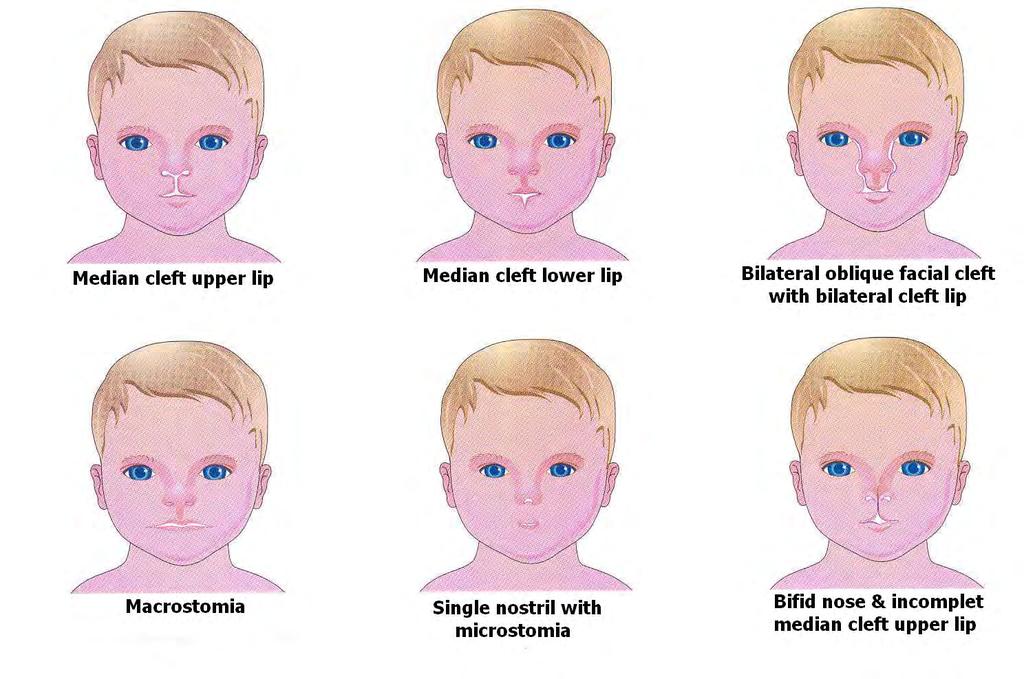
32 Facial clefts Failure of the embryonic facial prominences to fuse properly May be unilateral or bilateral May involve: Lips only: Cleft lip Palate only: Cleft palate Lip & palate: Cleft lip & palate Region of nasolacrimal groove: Facial clefts Lead to difficulty in breathing feeding sucking swallowing & speech
33

34 Median cleft lip: results from failure of the medial nasal prominences to merge and form the intermaxillary segments


35 Bilateral cleft lip: results due to failure of maxillary prominences to meet and unite with the medial nasal prominences on both sides

36 Unilateral cleft lip: result from failure of the maxillary prominence to merge with the medial nasal prominence on the affected side

37 Oblique facial cleft: results from failure of the maxillary prominence to fuse with the lateral nasal prominence

38 Cleft lip coupled with clefts of the anterior palate or entire palate.

39 Oblique facial cleft Cleft palate leaves the nasal and oral cavities connected & results in nursing problem for the new born May be: Anterior/posterior to incisive foramen Unilateral/bilateral Isolated/associated with cleft lips Cleft lip, cleft jaw & cleft palate


40 Absence or hypoplasia of the bone of the cranium

41 PRENATAL CHANGES OF THE SKULL

42 SKULL of the NEWBORN
")
 amortization.")

43 Functions of fontanelles: 1) alignment of fluctuations in intracranial pressure; 2) displacement of the bones; 3) sources of apposition of bone growth; 4) amortization. TIMINGS OF CLOSURE: ANTERIOR FONTANELLE= 18 months of age POSTERIOR FONTANELLE = 1 to 2 months of age

44 Post Natal Development of skull
; sutures are not formed; a number of cartilage at the base of the skull;")
; alveolar bone of the upper jaw and alveolar part of the mandible are not")
45 Features of new born skull proportion (brain / front offices 8/1); incompleteness of ossification (bone fragments); sutures are not formed; a number of cartilage at the base of the skull; fontanelles; the lack of buttresses; pneumatic sinuses are not formed mandibular angle-150 degree (120 in adults); alveolar bone of the upper jaw and alveolar part of the mandible are not developed; large orbit.

46 Factors of formation of the cranial vault: growing brain intracranial pressure growing brain action of soft tissue The growth mechanism of the skull. The arrows show the movement of the bones of the associated with the expansion of the brain capsule. Zone regional bone growth are marked in black. Osteogenesis in sutures is compensatory and increases the surface moving bones.
47 Growth of cranial base Cranial base grows post-natally by complex interaction between the following three growth processes. Extensive cortical drift and remodelling Elongation at synchondrosis Sutural growth


48 Factors of formation of the skull base Inside surface Outer surface: brain development; the development of the sense organs; the formation of dentoalveolar apparatus; dura mater organic interactions muscle traction (support function);

49 Factors of formation of the facial skull support for the visceral organs; development of the sense organs, primarily the nose of the capsule; the formation of dentoalveolar apparatus; action of of soft tissue.
50
51

52 Connections of bones of a skull Sutures

53
54

55 Connections of bones of a skull SYNCHODROSIS
56

57 Connections of bones of a skull Temporomandibular joint

58 PARANASAL SINUSES
59 Functions of paranasal sinuses reduction in weight of the skull to reach equilibrium of the head; means for increasing the volume of the head; participate in breathing; resonator function; participation in the sense of smell (in animals); protection and isolation of the brain and the eye socket from vibration; protection of the nasal cavity by heat leakage; tools for growth and development of the skull;

60

61
Pharyngeal Apparatus. Pouches Endoderm Grooves Ectoderm Arch Neural Crest Somitomeres Aortic Arch - Vessel
 Pharyngeal Apparatus Pouches Endoderm Grooves Ectoderm Arch Neural Crest Somitomeres Aortic Arch - Vessel Segmental Organization Humans: Arch 1-4 prominent Arch 5 absent Arch 6 - transient First Arch Face
Remember from the first year embryology Trilaminar disc has 3 layers: ectoderm, mesoderm, and endoderm
 Development of face Remember from the first year embryology Trilaminar disc has 3 layers: ectoderm, mesoderm, and endoderm The ectoderm forms the neural groove, then tube The neural tube lies in the mesoderm
Skeletal System. Prof. Dr. Malak A. Al-yawer Department of Anatomy/Embryology Section
 Skeletal System Prof. Dr. Malak A. Al-yawer Department of Anatomy/Embryology Section Learning objectives At the end of this lecture, the medical student will be able to: State the embryonic origin of skeletal
Essentials in Head and Neck Embryology. Part 3 Development of the head, face, and oral cavity
 Essentials in Head and Neck Embryology Part 3 Development of the head, face, and oral cavity Outline General overview of prenatal development Embryonic period phase 1 Formation of bilaminar disk Formation
04 Development of the Face and Neck. Development of the Face Development of the neck
 04 Development of the Face and Neck Development of the Face Development of the neck Development of the face Overview of facial development The fourth week ~ the twelfth week of prenatal development Between
Bones of the skull & face
 Bones of the skull & face Cranium= brain case or helmet Copyright The McGraw-Hill Companies, Inc. Permission required for reproduction or display. The cranium is composed of eight bones : frontal Occipital
Chapter 7. Skeletal System
 Chapter 7 Skeletal System 1 Skull A. The skull is made up of 22 bones: 8 cranial bones, 13 facial bones, and the mandible. B. The Cranium encloses and protects the brain, provides attachments for muscles,
Trigeminal Nerve (V)
") Trigeminal Nerve (V) Lecture Objectives Discuss briefly how the face is developed. Follow up the course of trigeminal nerve from its point of central connections, exit and down to its target areas. Describe
Trigeminal Nerve (V) Lecture Objectives Discuss briefly how the face is developed. Follow up the course of trigeminal nerve from its point of central connections, exit and down to its target areas. Describe
Chapter 7 Part A The Skeleton
 Chapter 7 Part A The Skeleton Why This Matters Understanding the anatomy of the skeleton enables you to anticipate problems such as pelvic dimensions that may affect labor and delivery The Skeleton The
AXIAL SKELETON SKULL
 AXIAL SKELETON SKULL CRANIAL BONES (8 total flat bones w/ 2 paired) 1. Frontal forms forehead & upper portion of eyesocket (orbital) 2. Parietal paired bones; form superior & lateral walls of cranium 3.
Skeletal System: Skull.
 Skeletal System: Skull www.fisiokinesiterapia.biz Bones of the Skull SPLANCHNOCRANIUM Nasal (2) Maxilla (2) Lacrimal (2) Zygomatic (2) Palatine (2) Inferior concha (2) Vomer Mandible NEUROCRANIUM Frontal
Chapter 7: Head & Neck
 Chapter 7: Head & Neck Osteology I. Overview A. Skull The cranium is composed of irregularly shaped bones that are fused together at unique joints called sutures The skull provides durable protection from
Skeletal System -Axial System. Chapter 7 Part A
 Skeletal System -Axial System Chapter 7 Part A Skeleton Learn: Names of the s. Identify specific landmarks that allow: Bones to fit into each other, Organs to fit into the cavities, Muscles to attach,
Cranium Facial bones. Sternum Rib
 Figure 7.1 The human skeleton. Skull Thoracic cage (ribs and sternum) Cranium Facial bones Sternum Rib Bones of pectoral girdle Vertebral column Sacrum Vertebra Bones of pelvic girdle (a) Anterior view
Biology 218 Human Anatomy. Adapted from Martini Human Anatomy 7th ed. Chapter 6 The Skeletal System: Axial Division
 Adapted from Martini Human Anatomy 7th ed. Chapter 6 The Skeletal System: Axial Division Introduction The axial skeleton: Composed of bones along the central axis of the body Divided into three regions:
Dr. Sami Zaqout, IUG Medical School
 The skull The skull is composed of several separate bones united at immobile joints called sutures. Exceptions? Frontal bone Occipital bone Vault Cranium Sphenoid bone Zygomatic bones Base Ethmoid bone
ANATOMY & PHYSIOLOGY I Laboratory Version B Name Section. REVIEW SHEET Exercise 10 Axial Skeleton
 ANATOMY & PHYSIOLOGY I Laboratory Version B Name Section REVIEW SHEET Exercise 10 Axial Skeleton 1 POINT EACH. THE SKULL MULTIPLE CHOICE 1. The major components of the axial skeleton include the 7. The
Review Article INTRODUCTION AND BACKGROUND. Abstract
 Review Article Identification of varied Sudanese infants ages (at birth, day 4, and 8 weeks) by determination of ossification pattern in the anterior skull base using computed tomography scan (A qualitative
Skeletal Development Multiple Cellular Origins. Intramembranous Bone. Endochondrial Bone. Cartilage template of the limb in the Chick wing
 Skeletal Development Multiple Cellular Origins 1 - Paraxial Mesoderm Somite, Sclerotome Axial Skeleton (e.g. vertebra) 2 - Lateral Plate Mesoderm Appendicular Skeleton (e.g. limb) 3 - Neural Crest Head
Anatomy and Physiology. Bones, Sutures, Teeth, Processes and Foramina of the Human Skull
 Anatomy and Physiology Chapter 6 DRO Bones, Sutures, Teeth, Processes and Foramina of the Human Skull Name: Period: Bones of the Human Skull Bones of the Cranium: Frontal bone: forms the forehead and the
Skeletal system. Prof. Abdulameer Al-Nuaimi. E. mail:
 Skeletal system Prof. Abdulameer Al-Nuaimi E-mail: a.al-nuaimi@sheffield.ac.uk E. mail: abdulameerh@yahoo.com Functions of Bone and The Skeletal System Support: The skeleton serves as the structural framework
Structure Location Function
 Frontal Bone Cranium forms the forehead and roof of the orbits Occipital Bone Cranium forms posterior and inferior portions of the cranium Temporal Bone Cranium inferior to the parietal bone forms the
Pharyngeal apparatus. - At the third week, it is a 3 layered structure: ectoderm, mesoderm and endoderm. This is called trilaminar disc
 Pharyngeal apparatus Remember from the first year embryology - The embryo was disc shaped in the second week of development (this is called embryonic disc) and it is a 2 layered disc (composed of two layers)---bilaminar
SKULL AS A WHOLE + ANTERIOR CRANIAL FOSSA
 SKULL AS A WHOLE + ANTERIOR CRANIAL FOSSA LEARNING OBJECTIVES At the end of this lecture, the student should be able to know: Parts of skeleton (axial and appendicular) Parts of skull Sutures of skull
Lecture 1: Embryology of the Limbs (Development of Skeletal and Muscular Systems)
") Lecture 1: Embryology of the Limbs (Development of Skeletal and Muscular Systems) Musculoskeletal Block Editing file Colour Index: Red: Important Gray: Notes Green: Dr. s notes After this lecture you should
Lecture 1: Embryology of the Limbs (Development of Skeletal and Muscular Systems) Musculoskeletal Block Editing file Colour Index: Red: Important Gray: Notes Green: Dr. s notes After this lecture you should
Trigeminal nerve. Slide in bold and please go back to see the pictures, if I skipped any part of record that because it wasn t clear to me
 Trigeminal nerve Slide in bold and please go back to see the pictures, if I skipped any part of record that because it wasn t clear to me Hala nsour 2/26/2018 P a g e 1 this lecture contain two topics
Development of the skull
 Development of the skull Ágnes Nemeskéri 2017 Semmelweis University Budapest Department of Anatomy, Histology and Embryology Clinical Anatomy Research Laboratory nemeskeri.agnes@med.semmelweis-univ.hu
Embryo#1. Mohammad Hisham Al-Mohtaseb باشق جهاد. 0 P a g e
 Embryo#1 Mohammad Hisham Al-Mohtaseb باشق جهاد 0 P a g e Before you start, it is important to link what you learn in gross anatomy with developmental stages discussed in embryology. Cells that form organs
REVIEW OF CLINICAL EMBRYOLOGY OF HEAD AND NECK
 REVIEW OF CLINICAL EMBRYOLOGY OF HEAD AND NECK OUTLINE - EMBRYOLOGY UNDERLYING CLINICAL CONDITIONS I. EARLY DEVELOPMENT OF FACE: CLEFT LIP, CLEFT PALATE, OBSTRUCTED NASOLACRIMAL DUCT II. BRANCHIAL ARCHES
Human Anatomy and Physiology - Problem Drill 07: The Skeletal System Axial Skeleton
 Human Anatomy and Physiology - Problem Drill 07: The Skeletal System Axial Skeleton Question No. 1 of 10 Which of the following statements about the axial skeleton is correct? Question #01 A. The axial
Exercise 10. The Axial Skeleton
 Exercise 10 The Axial Skeleton The Axial Skeleton Consists of the skeletal structures found along the midline of the body. Includes the skull, hyoid, vertebrae, ribs, sternum, and sacrum. The cartilages
Bones Ethmoid bone Inferior nasal concha Lacrimal bone Maxilla Nasal bone Palatine bone Vomer Zygomatic bone Mandible
 splanchnocranium - Consists of part of skull that is derived from branchial arches - The facial bones are the bones of the anterior and lower human skull Bones Ethmoid bone Inferior nasal concha Lacrimal
 Drawings illustrating the human pharyngeal apparatus. Drawings illustrating the human pharyngeal apparatus. Drawings illustrating the human pharyngeal apparatus. Drawings illustrating the human pharyngeal
APPENDICULAR SKELETON 126 AXIAL SKELETON SKELETAL SYSTEM. Cranium. Skull. Face. Skull and associated bones. Auditory ossicles. Associated bones.
 SKELETAL SYSTEM 206 AXIAL SKELETON 80 APPENDICULAR SKELETON 26 Skull Skull and associated s 29 Cranium Face Auditory ossicles 8 4 6 Associated s Hyoid Thoracic cage 25 Sternum Ribs 24 Vertebrae 24 column
Head and Neck Development and Malformations
 Head and Neck Development and Malformations Yang Chai, DDS, PhD Professor George and MaryLou Boone Chair Ostrow School of Dentistry of USC ychai@usc.edu C D E A. B Learning Objectives - Learn cranial neural
The SKELETAL System. The framework of bones and cartilage which protect organs, and provides a lever system that allows locomotion.
 The SKELETAL System The framework of bones and cartilage which protect organs, and provides a lever system that allows locomotion. Functions of the Skeletal System Support Protection Movement Facilitation
YOU MUST BRING YOUR OWN GLOVES FOR THIS ACTIVITY.
 ACTIVITY 3: AXIAL SKELETON AND LONG BONE DISSECTION Objectives: 1) How to get ready: Read Chapter 7, McKinley et al., Human Anatomy, 5e. All text references are for this textbook. Learning the meanings
in compact bone, large vertical canals carrying blood vessels and nerves. in compact bone, large horizontal canals carrying blood vessels and nerves.
 Carl Christensen, PhD Skeletal System (Bones`) Bio. 2304 Human Anatomy 1. Identify a term for each of the following: shaft of a long bone ends of a long bone ossified remnant of the "growth plate" connective
BIO 137 AXIAL SKELETON BONE STUDY THE HUMAN SKELETON
 BIO 137 THE AXIAL SKELETON MARY CATHERINE FLATH, Ph.D. THE HUMAN SKELETON AXIAL SKULL HYOID THORACIC CAGE VERTEBRAL COLUMN APPENDICULAR PECTORAL GIRDLE UPPER LIMBS PELVIC GIRDLE LOWER LIMBS AXIAL SKELETON
Biology 340 Comparative Embryology Lecture 11 Dr. Stuart Sumida. Overview of Embryology of the Vertebrate Skull. Emphasis on Amniota
 Biology 340 Comparative Embryology Lecture 11 Dr. Stuart Sumida Overview of Embryology of the Vertebrate Skull Emphasis on Amniota Initial introduction to components parts of a vertebrate head. This lecture
o Diaphysis o Area where red marrow is found o Area where yellow marrow is found o Epiphyseal plate AXIAL SKELETON Skull
 64 Anatomy & Physiology Coloring Workbook 7. Figure 5-2A is a midlevel, cross-sectional view of the diaphysis of the femur. Label the membrane that lines the cavity and the membrane that covers the outside
Anatomic Relations Summary. Done by: Sohayyla Yasin Dababseh
 Anatomic Relations Summary Done by: Sohayyla Yasin Dababseh Anatomic Relations Lecture 1 Part-1 - The medial wall of the nose is the septum. - The vestibule lies directly inside the nostrils (Nares). -
Skull-2. Norma Basalis Interna Norma Basalis Externa. Dr. Heba Kalbouneh Associate Professor of Anatomy and Histology
 Skull-2 Norma Basalis Interna Norma Basalis Externa Dr. Heba Kalbouneh Associate Professor of Anatomy and Histology Norma basalis interna Base of the skull- superior view The interior of the base of the
Infratemporal fossa: Tikrit University college of Dentistry Dr.Ban I.S. head & neck Anatomy 2 nd y.
 Infratemporal fossa: This is a space lying beneath the base of the skull between the lateral wall of the pharynx and the ramus of the mandible. It is also referred to as the parapharyngeal or lateral pharyngeal
Development and Growth of the Normal Cranial Vault : An Embryologic Review
 www.jkns.or.kr http://dx.doi.org/10.3340/jkns.2016.59.3.192 J Korean Neurosurg Soc 59 (3) : 192-196, 2016 Print ISSN 2005-3711 On-line ISSN 1598-7876 Copyright 2016 The Korean Neurosurgical Society Pediatric
Development of the Pharyngeal Arches
 Development of the Pharyngeal Arches Thomas A. Marino, Ph.D. Temple University School of Medicine Competencies: Upon completion of this section of the course, the student must be able to: 1. Recall the
Nasal region. cartilages: septal cartilage (l); lateral nasal cartilage (2); greater alar cartilages (2); lesser alar cartilages (?
; lateral nasal cartilage (2); greater alar cartilages (2); lesser alar cartilages (?") Nasal region skull bones: nasal and frontal processes of maxilla cartilages: septal cartilage (l); lateral nasal cartilage (2); greater alar cartilages (2); lesser alar cartilages (?) 1 Nasal cavity Roof
Nasal region skull bones: nasal and frontal processes of maxilla cartilages: septal cartilage (l); lateral nasal cartilage (2); greater alar cartilages (2); lesser alar cartilages (?) 1 Nasal cavity Roof
Introduction to Local Anesthesia and Review of Anatomy
 5-Sep Introduction and Anatomy Review 12-Sep Neurophysiology and Pain 19-Sep Physiology and Pharmacology part 1 26-Sep Physiology and Pharmacology part 2 Introduction to Local Anesthesia and Review of
ACTIVITY 3: AXIAL SKELETON AND LONG BONE DISSECTION COW BONE DISSECTION
 ACTIVITY 3: AXIAL SKELETON AND LONG BONE DISSECTION Objectives: 1) How to get ready: Read Chapter 7, McKinley et al., Human Anatomy, 4e. All text references are for this textbook. Learning the meanings
Week 14. Development of the Musculoskeletal System
 Week 14 Development of the Musculoskeletal System Skeletal System Derived from: paraxial mesoderm somites and somitomeres sclerotome sclerotome differentiation induced by SHH from notochord and floor plate
Chapter 8A. The Skeletal System: The Axial Skeleton. The Skeletal System: The Axial Skeleton. Types of Bones. Types of Bones
 Chapter 8A The Skeletal System: The Axial Skeleton The Skeletal System: The Axial Skeleton 206 named bones Axial Skeleton 80 bones lie along longitudinal axis skull, hyoid, vertebrae, ribs, sternum, ear
Dr.Noor Hashem Mohammad Lecture (5)
") Dr.Noor Hashem Mohammad Lecture (5) 2016-2017 If the mandible is discarded, the anterior part of this aspect of the skull is seen to be formed by the hard palate. The palatal processes of the maxillae
Dr.Noor Hashem Mohammad Lecture (5) 2016-2017 If the mandible is discarded, the anterior part of this aspect of the skull is seen to be formed by the hard palate. The palatal processes of the maxillae
Skull basic structures. Neurocranium
 Assoc. Prof. Květuše Lovásová, M.V.D., PhD. Skull basic structures Skull consists of two groups of bones: neurocranium (bones forming the brain box) splanchnocranium (bones forming the facial skeleton)
Skull and Axial Skeleton
 Published on Second Faculty of Medicine, Charles University (http://www.lf2.cuni.cz ) Skull and Axial Skeleton Description of the test The examination of the skull skeleton is in oral format. It consists
SKULL / CRANIUM BONES OF THE NEUROCRANIUM (7) Occipital bone (1) Sphenoid bone (1) Temporal bone (2) Frontal bone (1) Parietal bone (2)
 Occipital bone (1) Sphenoid bone (1) Temporal bone (2) Frontal bone (1) Parietal bone (2)") Important! 1. Memorizing these pages only does not guarantee the succesfull passing of the midterm test or the semifinal exam. 2. The handout has not been supervised, and I can not guarantee, that these
Important! 1. Memorizing these pages only does not guarantee the succesfull passing of the midterm test or the semifinal exam. 2. The handout has not been supervised, and I can not guarantee, that these
Skull. Sphenoid and Ethmoid bones
 Skull. Sphenoid and Ethmoid bones PhD., Dr. David Lendvai Department of Anatomy, Histology and Embriology Semmelweis University, Faculty of Medicine 2018. Skeletal system Structure of the skull Border
Development of Spinal Cord & Vertebral Column. Dr. Sanaa Alshaarawi & Prof. Ahmed Fathalla
 Development of Spinal Cord & Vertebral Column Dr. Sanaa Alshaarawi & Prof. Ahmed Fathalla OBJECTIVES At the end of the lecture, students should be able to: q Describe the development of the spinal cord
CHAPTER 7, PART II (BONES)
") Anatomy Name: CHAPTER 7, PART II (BONES) Entry #: INSTRUCTIONS: 1) READ Chapter 7, pg. 140-161. 2) Using the outline, make a note card for each underlined bone name or phrase. 3) On each note card, put
Anatomy Name: CHAPTER 7, PART II (BONES) Entry #: INSTRUCTIONS: 1) READ Chapter 7, pg. 140-161. 2) Using the outline, make a note card for each underlined bone name or phrase. 3) On each note card, put
The Skeletal System: Axial Skeleton
 The Skeletal System: Axial Skeleton The Big Idea The Axial Skeleton & Homeostasis The bones of the axial skeleton contribute to homeostasis by protecting many of the body s organs such as the brain, spinal
Biology 210 Chapter 8: Skeletal Tissues Supplement 1
 Biology 210 Chapter 8: Skeletal Tissues Supplement 1 By John McGill Material contributed by Beth Wyatt & Jack Bagwell DIVISIONS OF THE SKELETAL SYSTEM AXIAL SKELETON (80 BONES) Bones of the Head, Neck,
BY Dr Farooq Khan Aman Ullah khan
 Lecture 01 General Anatomy & Classification of Bone BY Dr Farooq Khan Aman Ullah khan Dated: 22.012.2017 Skeleton The hard, supporting framework of the body is called the skeleton. Skeleton includes bones
University of Palestine. Midterm Exam 2013/2014 Total Grade:
 Course No: DNTS2208 Course Title: Head and Neck Anatomy Date: 09/11/2013 No. of Questions: (50) Time: 1hour Using Calculator (No) University of Palestine Midterm Exam 2013/2014 Total Grade: Instructor
Omran Saeed. Luma Taweel. Mohammad Almohtaseb. 1 P a g e
 2 Omran Saeed Luma Taweel Mohammad Almohtaseb 1 P a g e I didn t include all the photos in this sheet in order to keep it as small as possible so if you need more clarification please refer to slides In
TRANSCRIPT OF NARRATION FOR DEVELOPMENT OF THE FACE
 TRANSCRIPT OF NARRATION FOR DEVELOPMENT OF THE FACE Slide 1 (introduction) In this lecture, I ll be addressing two separate but interrelated topics. First, I ll be summarizing some of the most fundamental
Bio 5/6 5 The Skeletal System Study Guide
 Name: THE SKELETAL SYSTEM: 5 The Skeletal System Study Guide Period: The skeleton is constructed of two of the most supportive tissues found in the human body - cartilage and bone. Besides supporting and
Upper arch. 1Prosthodontics. Dr.Bassam Ali Al-Turaihi. Basic anatomy & & landmark of denture & mouth
 1Prosthodontics Lecture 2 Dr.Bassam Ali Al-Turaihi Basic anatomy & & landmark of denture & mouth Upper arch Palatine process of maxilla: it form the anterior three quarter of the hard palate. Horizontal
UNIT 4 - SKELETAL SYSTEM LECTURE NOTES
 UNIT 4 - SKELETAL SYSTEM LECTURE NOTES 4.01 FUNCTIONS OF THE SKELETAL SYSTEM A. Support 1. Provides a framework for the body 2. Supports soft tissue 3. Serves as a point of attachment for ligaments, tendons,
Anatomy Made Easy MSS
 Anatomy Made Easy MSS part #1 هذا الملف يشمل تفريغ المحاضرة الثانية لعون بدءا من الصفحة 11 وحتى األخير Done By :MohamedA. Diabat Edited by Awn Academic team The Axial Skeleton The axial skeleton consist
BONE CHALLENGE DANIL HAMMOUDI.MD
 BONE CHALLENGE DANIL HAMMOUDI.MD Bone Basic functions? A. support B. protection C. movement assistance in D. RBC formation-hemopoiesis E. mineral homeostasis +importance of calcium F. energy supply -yellow
Musculoskeletal System (Part A-1) Module 7 -Chapter 10 Overview. Functions
 Module 7 -Chapter 10 Overview. Functions") Musculoskeletal System (Part A-1) Module 7 -Chapter 10 Overview Susie Turner, M.D. 1/8/13 Muscles Attachments Bones Bone types Surface features of bones Divisions of the skeletal system Joints or Articulations
Musculoskeletal System (Part A-1) Module 7 -Chapter 10 Overview Susie Turner, M.D. 1/8/13 Muscles Attachments Bones Bone types Surface features of bones Divisions of the skeletal system Joints or Articulations
DEVELOPMENTAL ANATOMY OF THE FACE, JAW AND NECK. O.M. Oluwatosin Department of Surgery
 DEVELOPMENTAL ANATOMY OF THE FACE, JAW AND NECK O.M. Oluwatosin Department of Surgery 1 2 By the end of this lecture, you should be able to: Discuss the embryology of the face Relate congenital anomalies
Warm-Up Activity. Fill in the names of the bones in the skeleton diagram.
 Warm-Up Activity Fill in the names of the bones in the skeleton diagram. Warm-Up 1. What are the 4 types of bones? Give an example of each. 2. Give 3 ways you can tell a female skeleton from a male skeleton.
Chapter 7: Skeletal System: Gross Anatomy
 Chapter 7: Skeletal System: Gross Anatomy I. General Considerations A. How many bones in an average adult skeleton? B. Anatomic features of bones are based on II. Axial Skeleton A. Skull 1. Functionally
Trigeminal Nerve Anatomy. Dr. Mohamed Rahil Ali
 Trigeminal Nerve Anatomy Dr. Mohamed Rahil Ali Trigeminal nerve Largest cranial nerve Mixed nerve Small motor root and large sensory root Motor root Nucleus of motor root present in the pons and medulla
Anatomy and Physiology 1 Chapter 7 self quiz Pro, Dima Darwish,MD.
 Anatomy and Physiology 1 Chapter 7 self quiz Pro, Dima Darwish,MD. 1) How many bones make up the axial skeleton? A) 50 B) 60 C) 70 D) 80 E) 90 2) Which of the following is a function of the axial skeleton?
Ligaments of the vertebral column:
 In the last lecture we started talking about the joints in the vertebral column, and we said that there are two types of joints between adjacent vertebrae: 1. Between the bodies of the vertebrae; which
The Axial Skeleton. C h a p t e r. PowerPoint Lecture Slides prepared by Jason LaPres Lone Star College - North Harris
 C h a p t e r 7 The Axial Skeleton PowerPoint Lecture Slides prepared by Jason LaPres Lone Star College - North Harris Copyright 2009 Pearson Education, Inc., publishing as Pearson Benjamin Cummings An
Crafton Hills College Human Anatomy & Physiology Axial Skeleton
 A. Major Divisions Crafton Hills College Human Anatomy & Physiology Axial keleton 1. Axial: Part of skeleton lies along long axis of body 2. Appendicular: Bones & features of the appendages B. AXIAL KELETON
Temporal fossa Infratemporal fossa Pterygopalatine fossa Terminal branches of external carotid artery Pterygoid venous plexus
 Outline of content Temporal fossa Infratemporal fossa Pterygopalatine fossa Terminal branches of external carotid artery Pterygoid venous plexus Boundary Content Communication Mandibular division of trigeminal
Postnatal Growth. The study of growth in growing children is for two reasons : -For health and nutrition assessment
 Growth of The Soft Tissues Postnatal Growth Postnatal growth is defined as the first 20 years of growth after birth krogman 1972 The study of growth in growing children is for two reasons : -For health
THE SKELETAL SYSTEM. Focus on the Skull
 THE SKELETAL SYSTEM Focus on the Skull Review Anatomical Terms Anterior/Posterior Dorsal/Ventral Medial/Lateral Superior/Inferior Bone Markings - Review Projections for attachment of muscles, ligaments
Labs 9 and 10. Classification of Bones. Bone Shapes 1/05/13. Skeletal system overview. Bone are identified by:
 Labs 9 and 10 Skeletal system overview Classification of Bones Bone are identified by: shape internal tissues bone markings 1. Flat bones 2. Long bones 3. Short bones 4. Irregular bones 5. Sutural bones
3. The Jaw and Related Structures
 Overview and objectives of this dissection 3. The Jaw and Related Structures The goal of this dissection is to observe the muscles of jaw raising. You will also have the opportunity to observe several
Dr.ALI AL BAZZAZ PLASTIC SURGON CLEFT LIP AND PALATE
 Dr.ALI AL BAZZAZ PLASTIC SURGON CLEFT LIP AND PALATE Cleft lip (cheiloschisis) and cleft palate (palatoschisis), which can also occur together as cleft lip and palate, are variations of a type of clefting
Interactions of the endocrine system, bone and oral health
 Interactions of the endocrine system, bone and oral health All bones are not equal! Dense high proportion of cortical bone High proportion of trabecular bone Mandible Functions: mastication, respiration,
An Introduction to the Axial Skeleton. Copyright 2009 Pearson Education, Inc., publishing as Pearson Benjamin Cummings
 An Introduction to the Axial Skeleton Copyright 2009 Pearson Education, Inc., publishing as Pearson Benjamin Cummings Terms: Structures of Bones Articulations: Contacts with other bones Landmarks (Bone
Bio 103 Skeletal System 45
 45 Lecture Outline: SKELETAL SYSTEM [Chapters 7, 8] Introduction A. Components B. Functions 1. 2. 3. 4. Classification and Parts A. Bone Shapes 1. Long: 2. Short: 3. Flat: 4. Irregular: 5. Sesamoid: B.
Cleft Lip and Palate: The Effects on Speech and Resonance
 Ann W. Kummer, PhD, CCC-SLP Cincinnati Children s Cleft lip and/or palate can have a negative impact on both speech and resonance. The following is a summary of normal anatomy, the types and causes of
Bones of the Skull Lateral View
 Bones of the Skull Lateral View Frontal Bone Parietal Bone Occipital Bone Temporal Bone Sphenoid Bone Pterion Sutures of the Skull Lateral View Coronal Suture Lambdoid Suture Squamous Suture Sutures of
DEFINATION Growth was concieved by an anatomist, born to a biologist, delivered by a physician, left on a chemist doorstep, and adopted by a physiolog
 DEFINATION Growth was concieved by an anatomist, born to a biologist, delivered by a physician, left on a chemist doorstep, and adopted by a physiologist.at an early age- she eloped with a statistician,
Part 1 of this 3-part review primarily dealt with the early embryologic
 REVIEW ARTICLE Illustrated Review of the Embryology and Development of the Facial Region, Part 2: Late Development of the Fetal Face and Changes in the Face from the Newborn to Adulthood P.M. Som and T.P.
Skeletal system overview. Classification of Bones
 Skeletal system overview BIOL241 Lab #9 Classification of Bones Bone are identified by: shape internal tissues bone markings 1 1. Flat bones 2. Long bones 3. Short bones 4. Irregular bones 5. Sutural bones
The Skull DANIL HAMMOUDI.MD
 The Skull DANIL HAMMOUDI.MD summary of bones/structures in Chapter 15 of the manual need tp be print as soon as possible http://www.mnsu.edu/emuseum/biology/humananatomy/skeletal/skul l/frontal/frontal.html
University of Palestine. Midterm Exam 2013/2014 Total Grade:
 [ Course No: DNTS2208 Course Title: Head and Neck Anatomy Date: 17/11/1024 No. of Questions: (52) Time: 2hours Using Calculator (No) University of Palestine Midterm Exam 2013/2014 Total Grade: Instructor
Trigeminal Nerve Worksheets, Distributions Page 1
 Trigeminal Nerve Worksheet #1 Distribution by Nerve Dr. Darren Hoffmann Dental Gross Anatomy, Spring 2013 We have drawn out each of the branches of CN V in lecture and you have an idea now for their basic
PTERYGOPALATINE FOSSA
 PTERYGOPALATINE FOSSA Outline Anatomical Structure and Boundaries Foramina and Communications with other spaces and cavities Contents Pterygopalatine Ganglion Especial emphasis on certain arteries and
Anatomy of the Nervous System. Brain Components
 Anatomy of the Nervous System Brain Components NERVOUS SYSTEM INTRODUCTION Is the master system of human body, controlling the functions of rest of the body systems Nervous System CLASSIFICATION A. Anatomical