Head and Neck Development and Malformations
|
|
|
- Britton Barton
- 5 years ago
- Views:
Transcription
1 Head and Neck Development and Malformations Yang Chai, DDS, PhD Professor George and MaryLou Boone Chair Ostrow School of Dentistry of USC

2 C D E A. B
3 Learning Objectives - Learn cranial neural crest cells and their contribution to craniofacial development - Know the different components of the pharyngeal arch and their later fates, be able to recognize the pharyngeal origin of mature structures, and recognize which structures are nonpharyngeal. Developmental terms from this lecture that are of particular importance: pharyngeal arch, pouch, and cleft, frontonasal process. - You should be able to recognize the developmental significance of innervation patterns and transitions in terms of the developmental origins of the innervated structures. - Embryonic basis for cleft lip with or without cleft palate, cleft palate, and other craniofacial malformations.
4 Neural crest cells


5 Cranial neural crest cell migration
6
7 NEURAL CREST CELLS NEURAL CREST CELLS ECTOMESENCHYMAL CELLS MELANOBLAST SCHWANN CELLS CHONDROBLASTS FIBROBLASTS NEUROBLAST CHROMAFFIN CELLS ODONTOBLAST CEMENTOBLASTS SENSORY NEURON SYMPATHETIC NEURON MOTOR NEURON

8 Rhombomeres and neural crest cell migration N. Le Douarin

9 Pharyngeal arches and their associated cranial nerves

10 Neural Crest Cell Fate Analysis using DiI injection

11 Labeling of the cranial nerves

12 Migration of cranial neural crest cells

 Endoderm separating grooves on inside =")
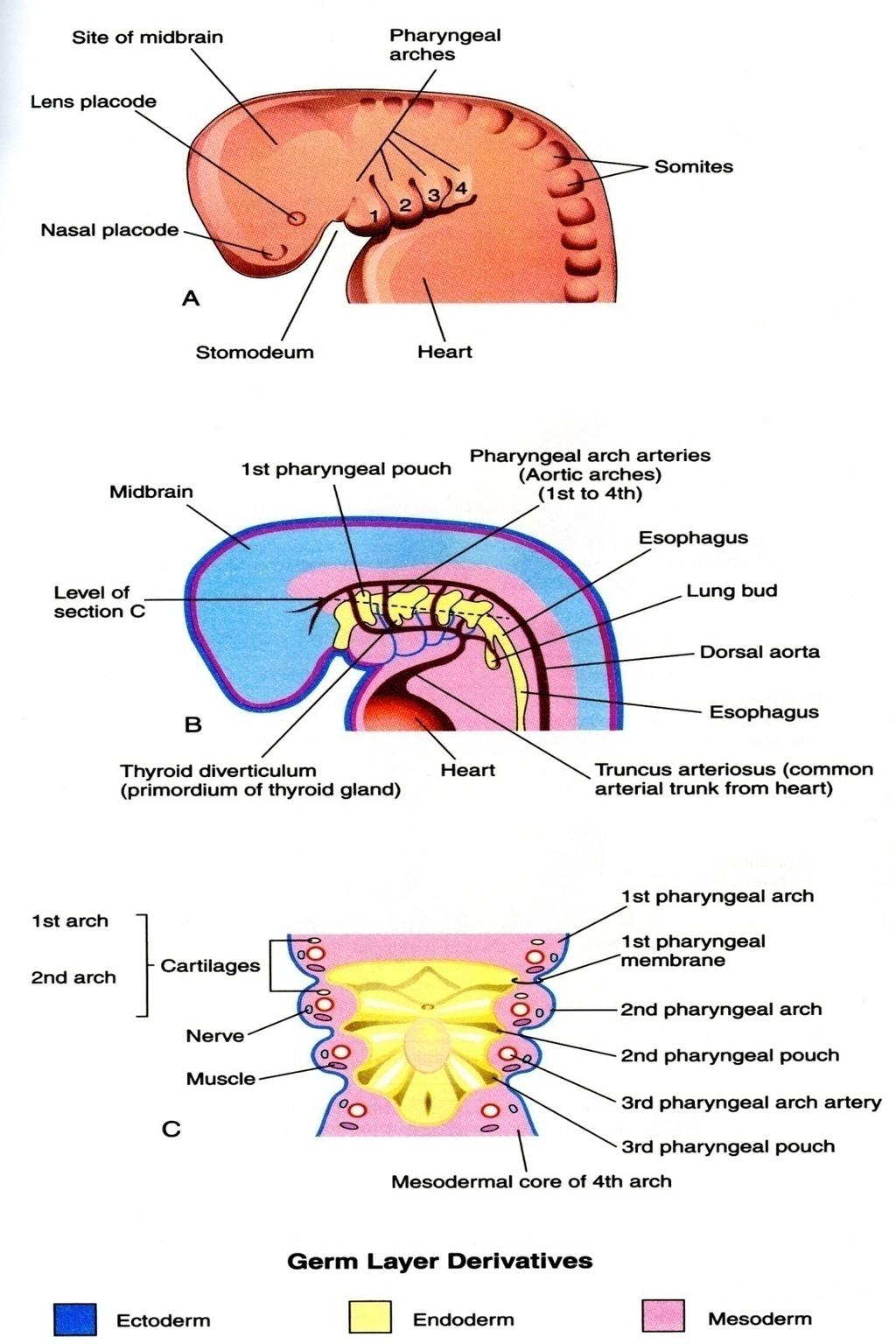
13 Pharyngeal arches In humans = pharyngeal arches In fish = branchial arches FNP Mx Mn Ectoderm separating arches on outside = pharyngeal cleft (or groove) Endoderm separating grooves on inside = pharyngeal pouch E28
14 The pharyngeal arches are fetal structures that constitute most of the nonneural elements of the head and neck. Humans (and other mammals) have five pharyngeal arches (numbered 1, 2, 3, 4, and 6). A pharyngeal cleft (or groove) separates adjacent arches on the outside (= ectoderm), and a pharyngeal pouch separates adjacent arches on the inside (= endoderm). In addition to ectoderm and endoderm, each arch has an artery, a nerve, a cartilagenous element, and mesenchymal cells. The first arch has two subdivisions: the maxillary process and the mandibular process. - The trigeminal nerve (the nerve of the 1st arch) has separate branches that innervate each (CNV2 and CNV3 respectively). The frontonasal process (prominence) (FNP) in the anterior midline is similar to a pharyngeal arch (it has a nerve (CN V1) and mesenchyme and follows a similar developmental strategy) but is somewhat different as well (there is no pouch or cleft that demarks it, and no cartilage).
")

15 Pharyngeal arches (note: 1st arch ectoderm (blue) extends to inside (of what will become the oral cavity))
16
17

18 Structures derived from pharyngeal arch components
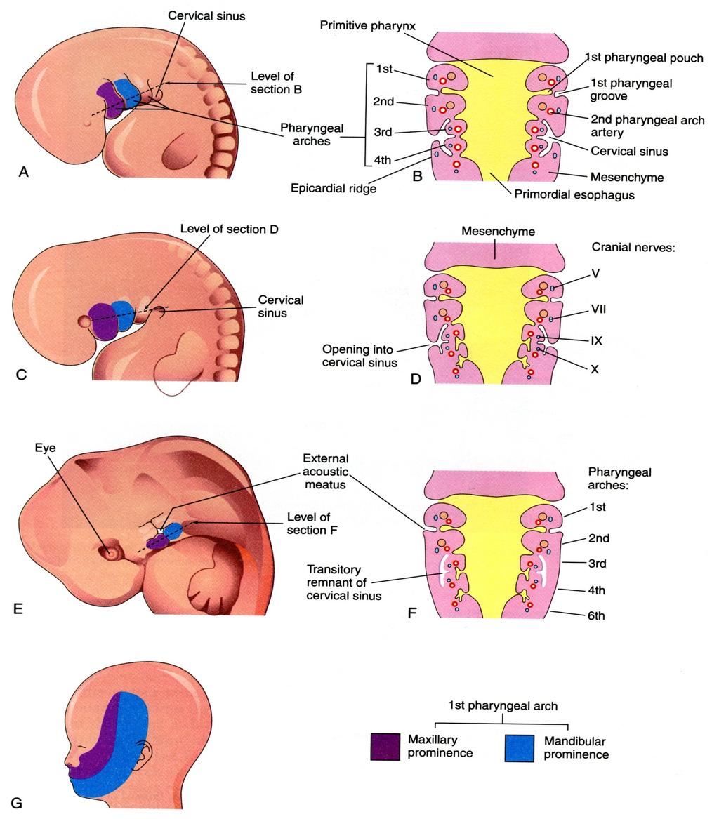
19 4 of the 12 cranial nerves innervate the pharyngeal arches Pharyngeal arch nerves First arch: Trigeminal (CN V) Second: Facial (CN VII) Third: Glossopharyngeal (CN IX) Fourth and sixth: Vagus (CNX) -***Each pharyngeal nerve is a mixed nerve, with a motor component to pharyngeal muscle cells, and a sensory component to pharyngeal epithelium (ectoderm or endoderm).

20 Pharyngeal arch cartilage

21 Pharyngeal arch muscle Posterior belley of digastric muscle
22 Recall the general formation of most body muscles: are derived from paraxial mesoderm -> somites - myotome -> muscle cells. - Muscle cells obtain innervation early, and maintain that innervation as they migrate. - Craniofacial muscles are also derived from paraxial mesoderm, except that somites do not form per se in the head (there is an analogous domain of paraxial mesoderm that is called somitomeres, not a term to know). Craniofacial muscles also maintain their early innervation pattern once it is established. Note that some craniofacial muscles migrate within pharyngeal arches, other do not. Pharyngeal muscles intermingle and migrate widely though the head and neck. However, the origin of each muscle can always be determined by its innervation pattern. The origin and innervation of tongue muscles is described below.
23 Innervation of cranial muscles Innervation Oculomotor (III) Troclear (IV) Trigeminal (V) Abducens (VI) Facial (VII) Glossopharyngeal (IX) Vagus (X) Hypoglossal (XII) Muscles Sup, med, inf. recti Sup. oblique Muscles of mastication Lat. rectus Facial (various) Stylopharyngeus Palate and Intrinsic laryngeal Tongue muscles (Pharyngeal nerves and muscles in red)


 to all tongue muscles except for")
24 General sensory innervation of oral cavity Special sensory innervation of tongue CN VII (trigeminal) to anterior 2/3 of tongue CN IX and CN X to posterior 1/3 Motor innervation of tongue CN XII (12) to all tongue muscles except for palatoglossus (CN X)

25 Craniofacial mesenchyme a loosely organized, mainly mesodermal embryonic tissue that develops into connective and skeletal tissues, including blood and lymph. NC gives rise to: dura of brain ganglia of cranial nerves Mesoderm gives rise to: striated muscle endothelium blue = cranial bones of NC origin red = cranial bones of mesodermal origin Both give rise to: cranial bones vascular sm. Muscle Pharyngeal mesenchyme has two developmental origins: mesoderm and neural crest. There are some unique fates for each of these (pharyngeal mesoderm is the source of cranial vascular endothelium and striated


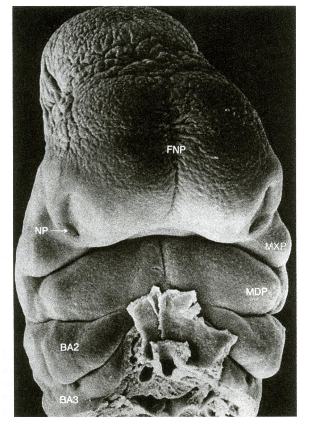
26 Migration of cranial neural crest cells and facial development
27
28
29 Normal lip formation
30

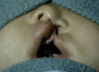
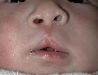
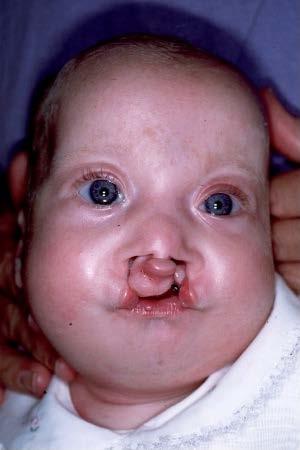
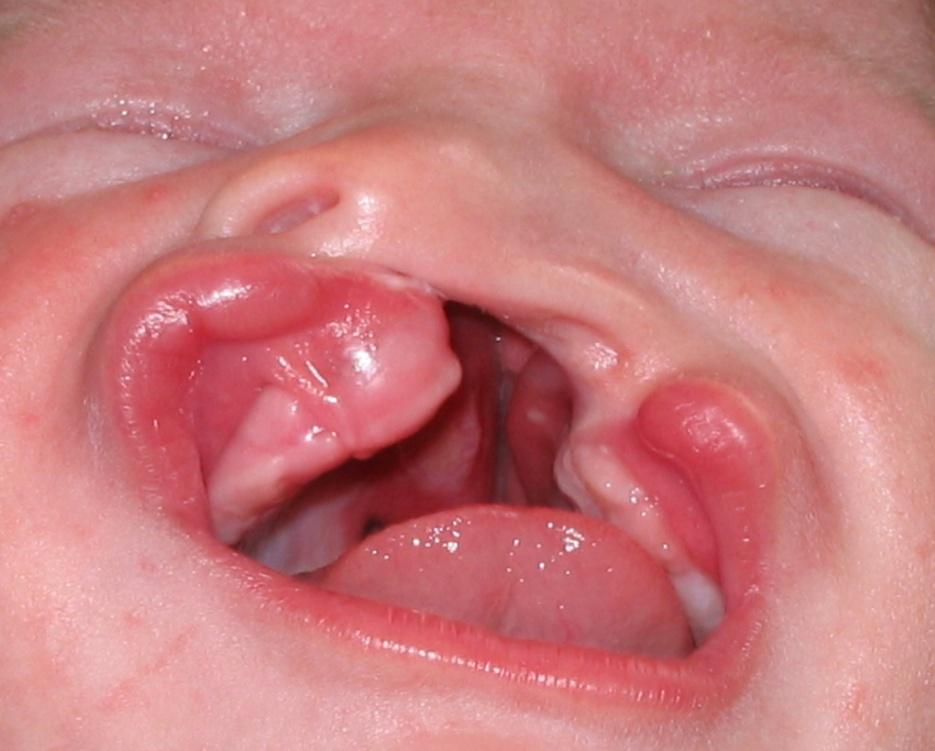
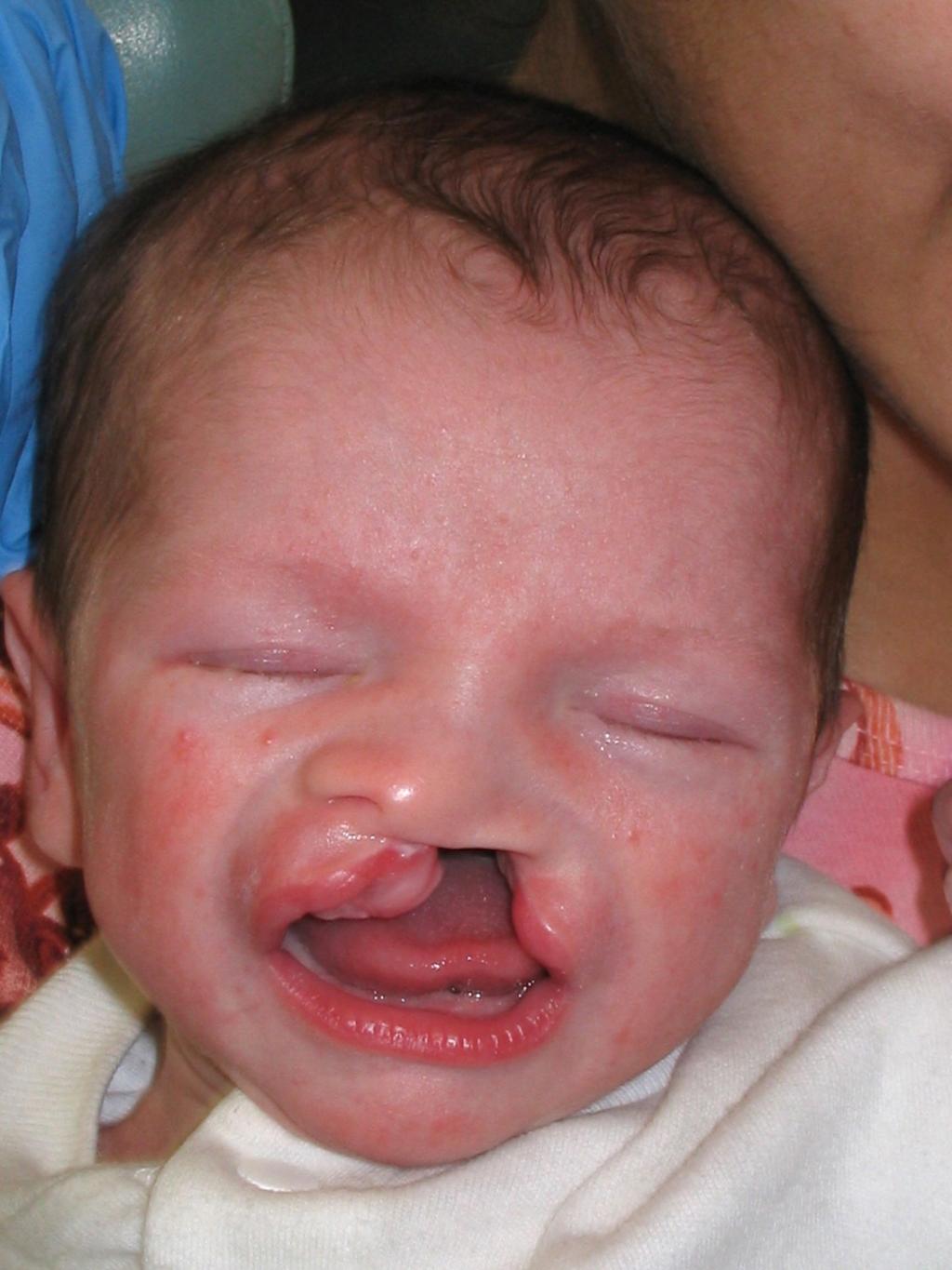
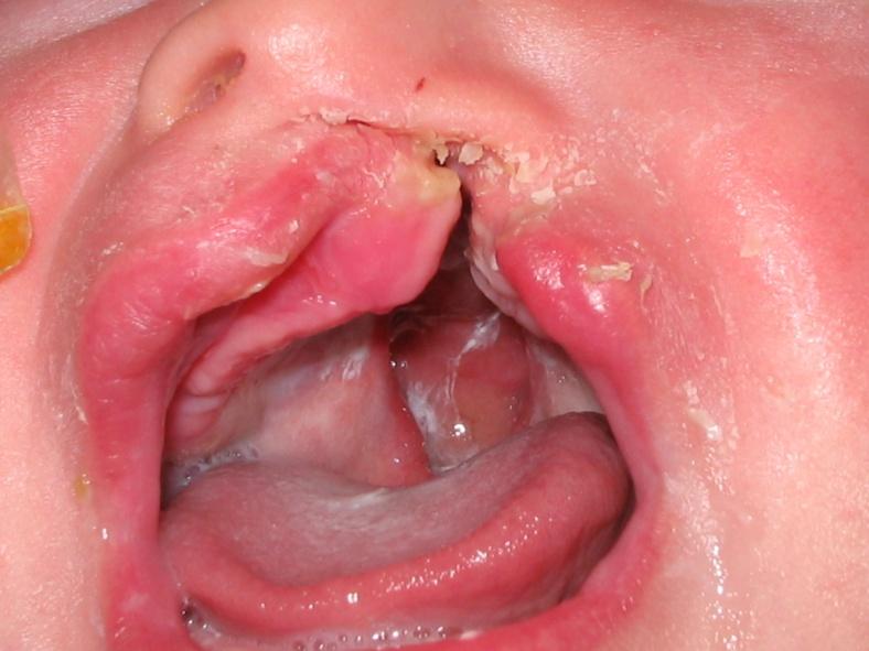
31 Unilateral and bilateral cleft lip with cleft palate


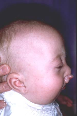
32 First arch syndrome Defects of external ear Mandibular hypoplasia Cleft palate (variable)

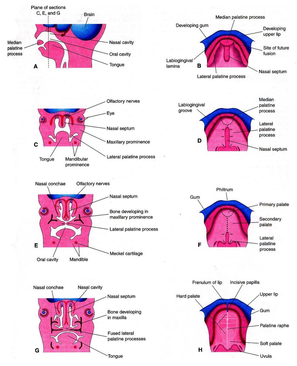
33 The anatomy of palatogenesis 6 wks 12 wks


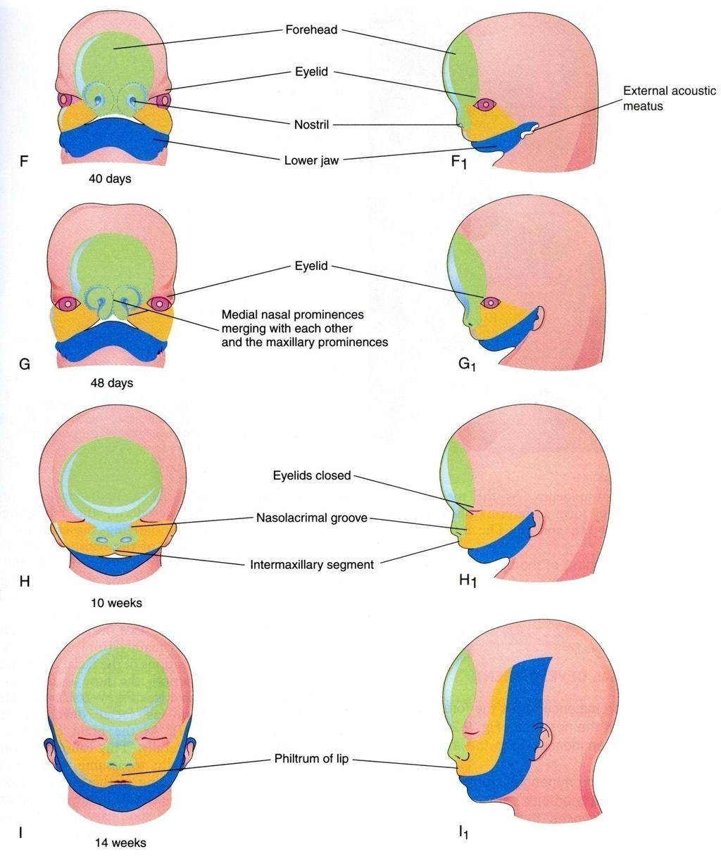
34 Formation of palate The oral cavity is delimited by the FNP, maxillary processes, and mandibular processes. The mandibular processes fuse in the midline, the two (bilateral) maxillary processes do not fuse at first and are separated from each other by the distal end of the FNP (the intermaxillary segment). Externally, the FNP is the origin of the philtrum of the upper lip.

35 Formation of palate

36 Cleft palate

37 Cleft palate (histology)

38 Cleft lip with cleft palate
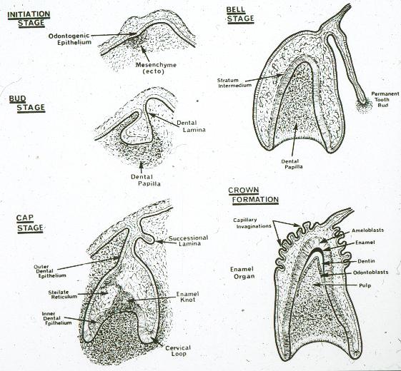
39 Tooth Development

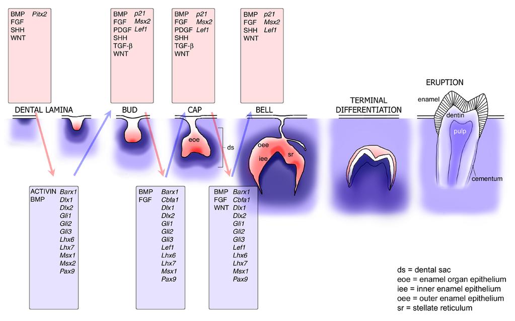
40 Molecular Regulation of Tooth Morphogenesis

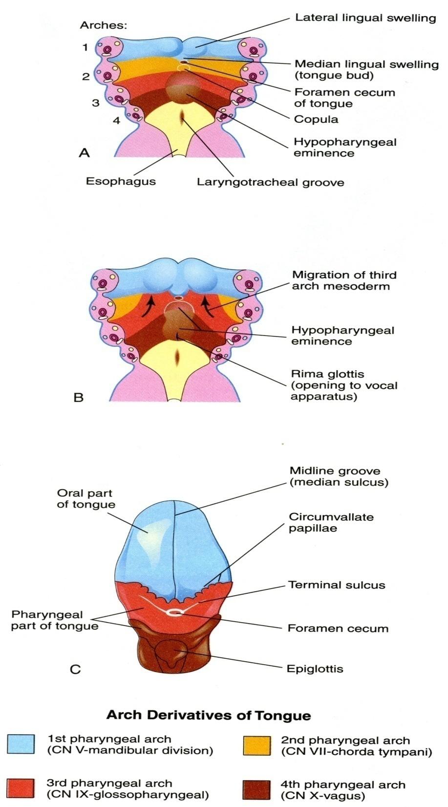
41 Formation of tongue (epithelium) The location of the oropharyngeal membrane represents the transition from ectoderm to endoderm in the oral cavity. This transition lies within the oral cavity, anterior to the 1st pharyngeal pouch. This is also the location of a transition in sensory innervation of the epithelium of the oral cavity from CN V (ectoderm) to CN IX (endoderm). The anterior 2/3 of the tongue forms from tongue buds derived from the 1st arch (= ectodermal, CN V innervation). Inside the oral cavity, the 3rd arch displaces the 2nd arch to the outside, such that the posterior 1/3 of tongue is derived from the 3rd arch (endodermal, CN IX), with the very posterior end of tongue derived from the 4th arch (endodermal, CN X).

42
43

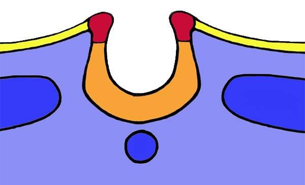
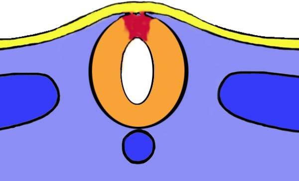
44 Thyroid
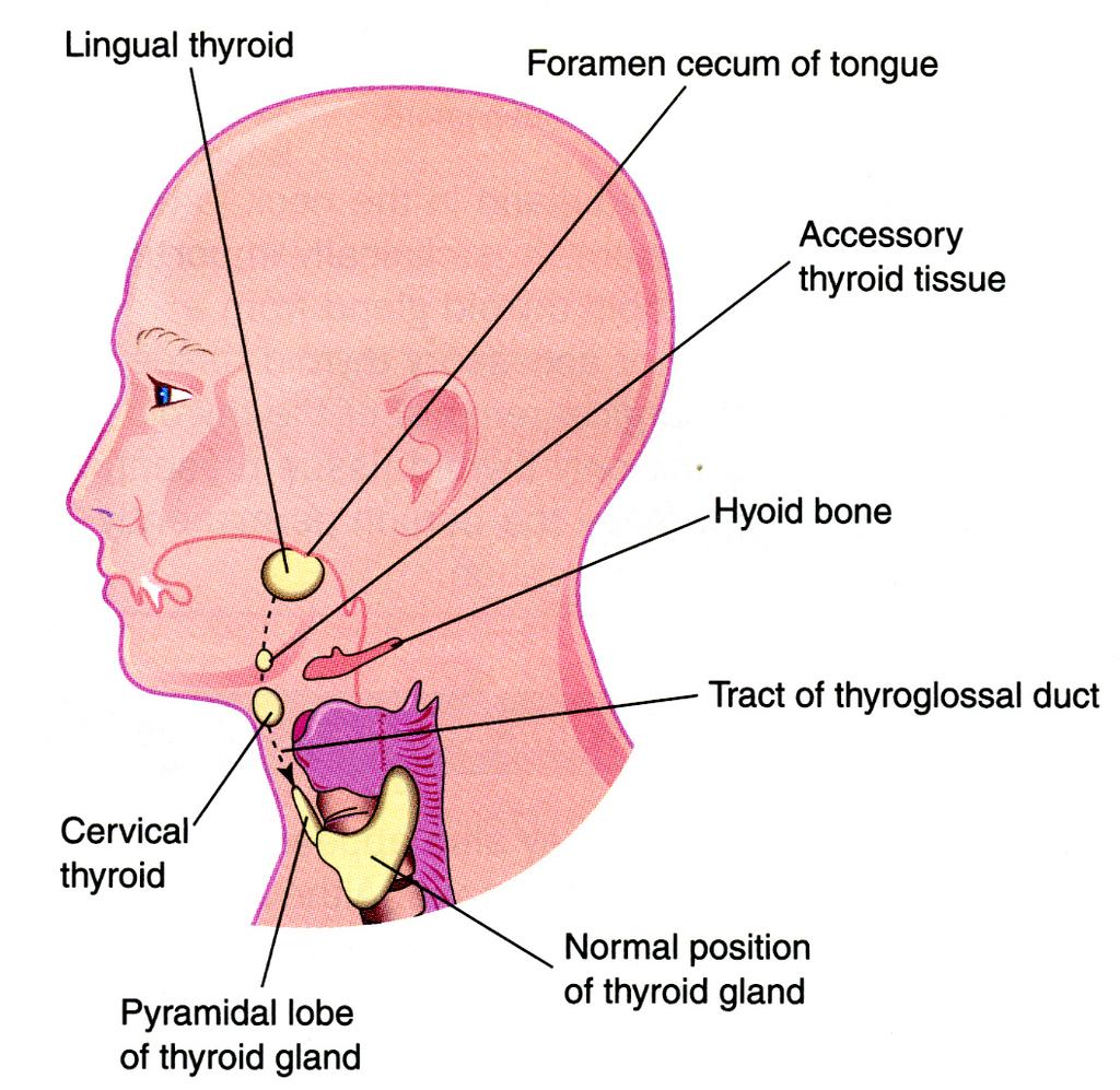
45
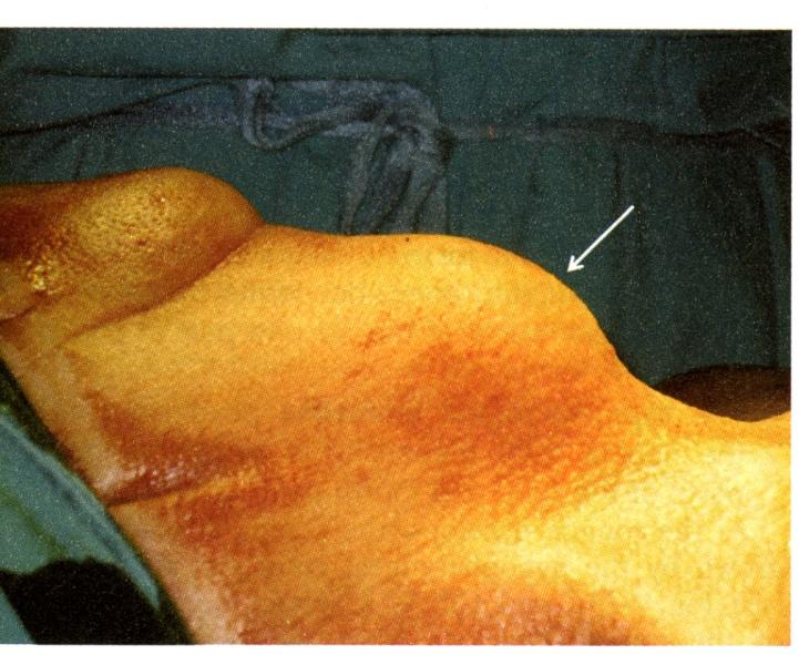
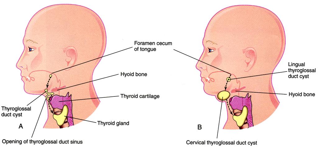
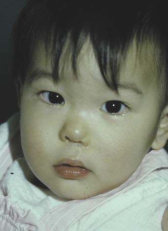
46 Cervical thyroglossal duct cyst

47 Imaging of a thyroglossal duct cyst
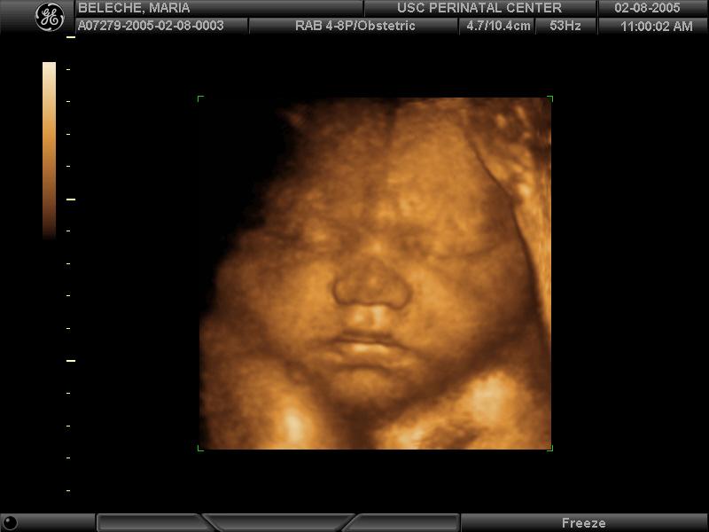
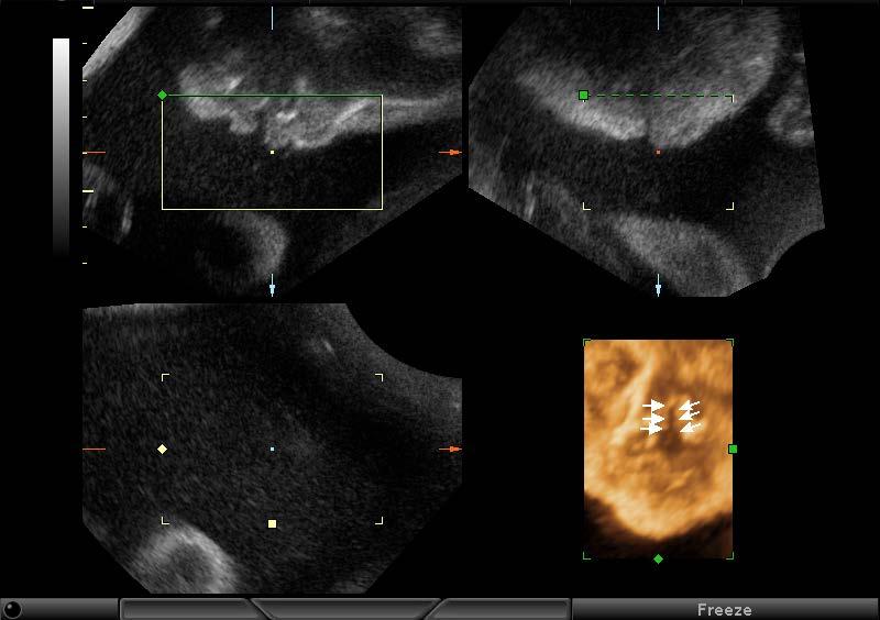
48 Pre-Natal Diagnosis Ultrasound Level II USN Operator dependent Isolated cleft palate difficult Amniocentesis Does not detect isolated CL/CP Chorionic Villus Sampling Does not detect isolated CL/CP
49
50
51 Craniofacial Team at CHLA Audiologist Pediatric Dentist Pediatric Pulmonologist Pediatrician Speech Pathologist Orthodontist Social Worker Pediatric psychologist Plastic Surgeon Oral Surgeon Otolaryngologist Craniofacial Nurse Geneticist Ophthalmologist
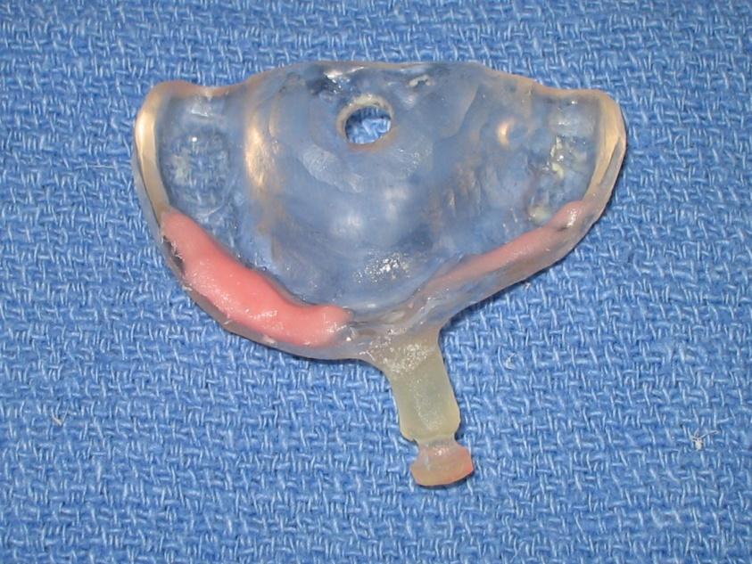
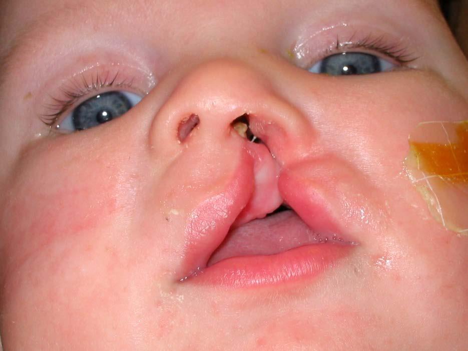
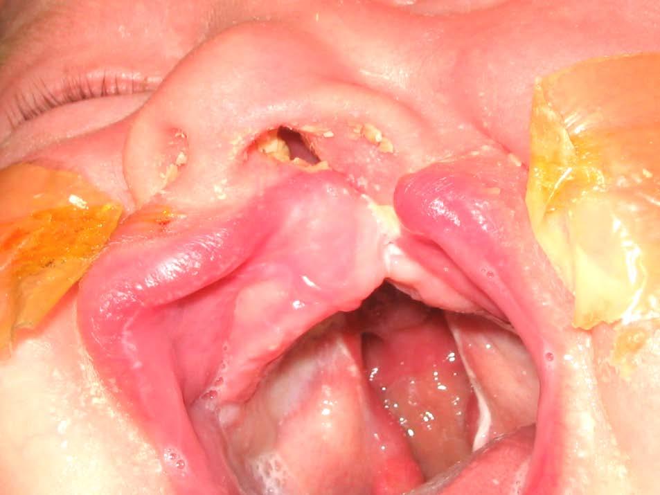
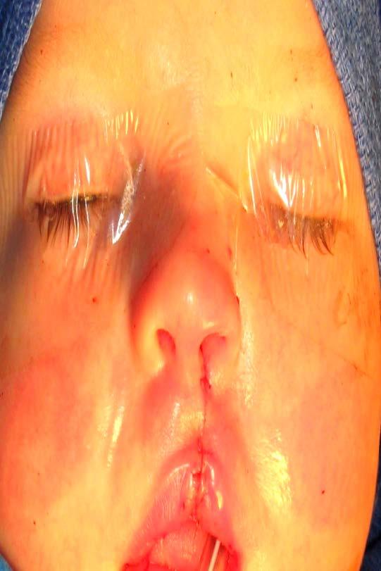
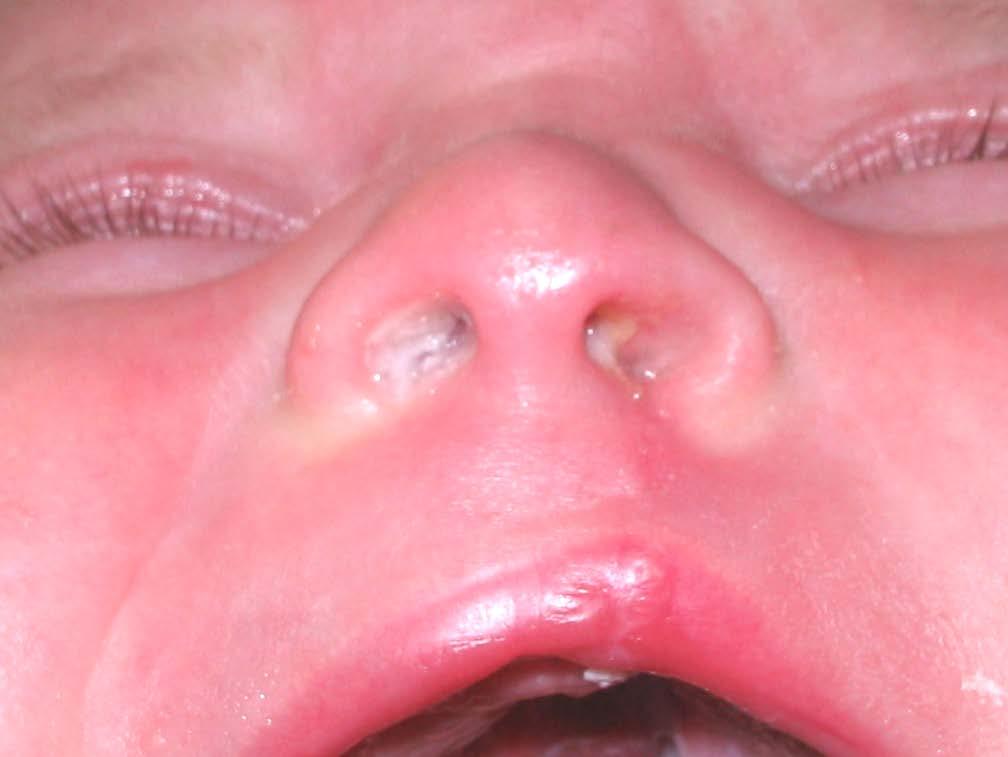
52 Nasoalveolar Molding (2-5 weeks begin) Cleft Lip Repair (3 months old) Cleft Palate Repair (1 year old) Cleft Lip and Palate Patient Alveolar bone graft (7 or 8 years old) Cleft Lip/Nose Revision Pharyngoplasty(speech dependant) Jaw Surgery (15 to 20 years old) Team Visits Cleft Lip Rhinoplasty (15 to 20 years old)
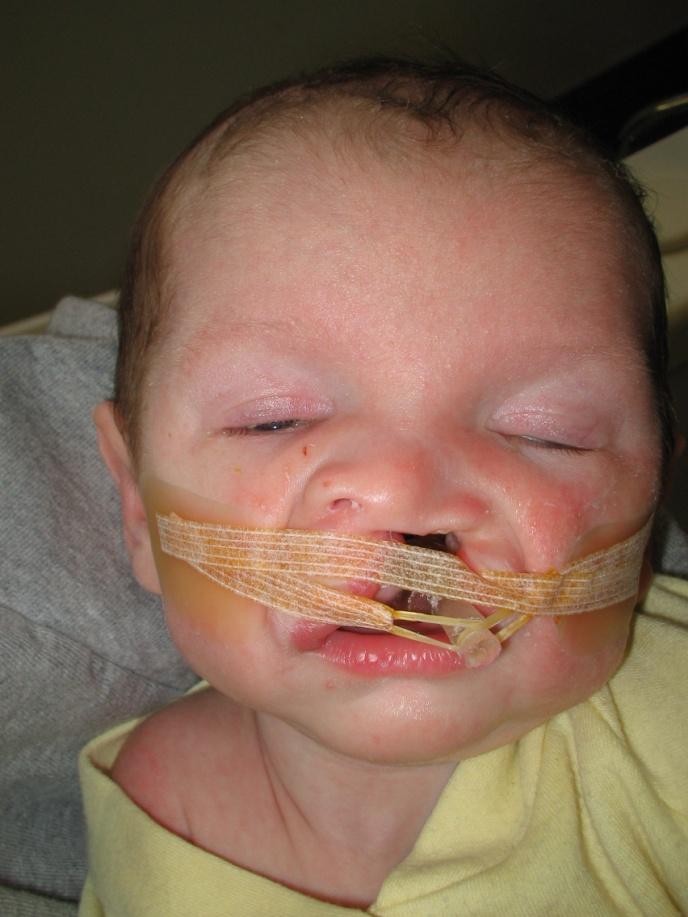
53 The Story of ashley

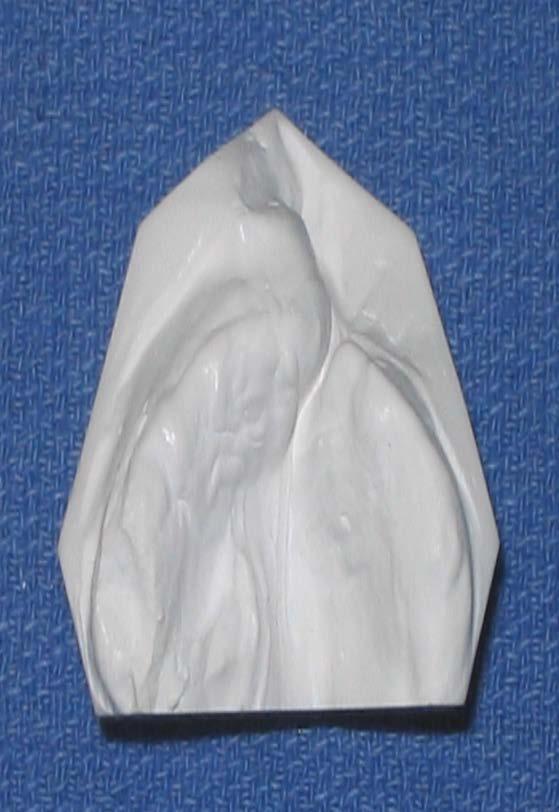
54

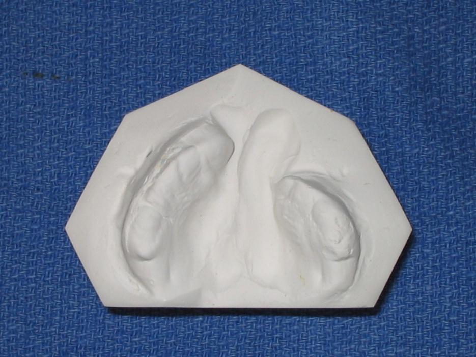
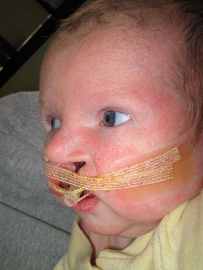
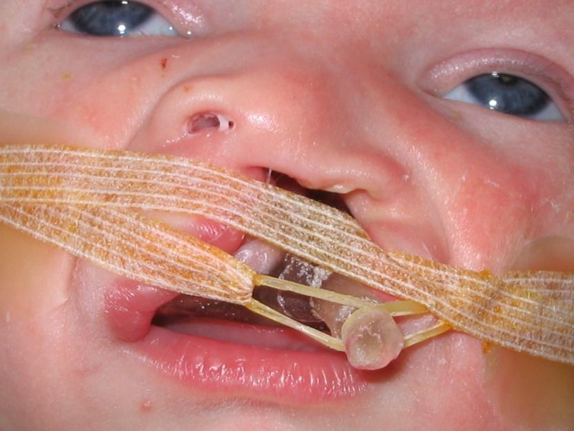
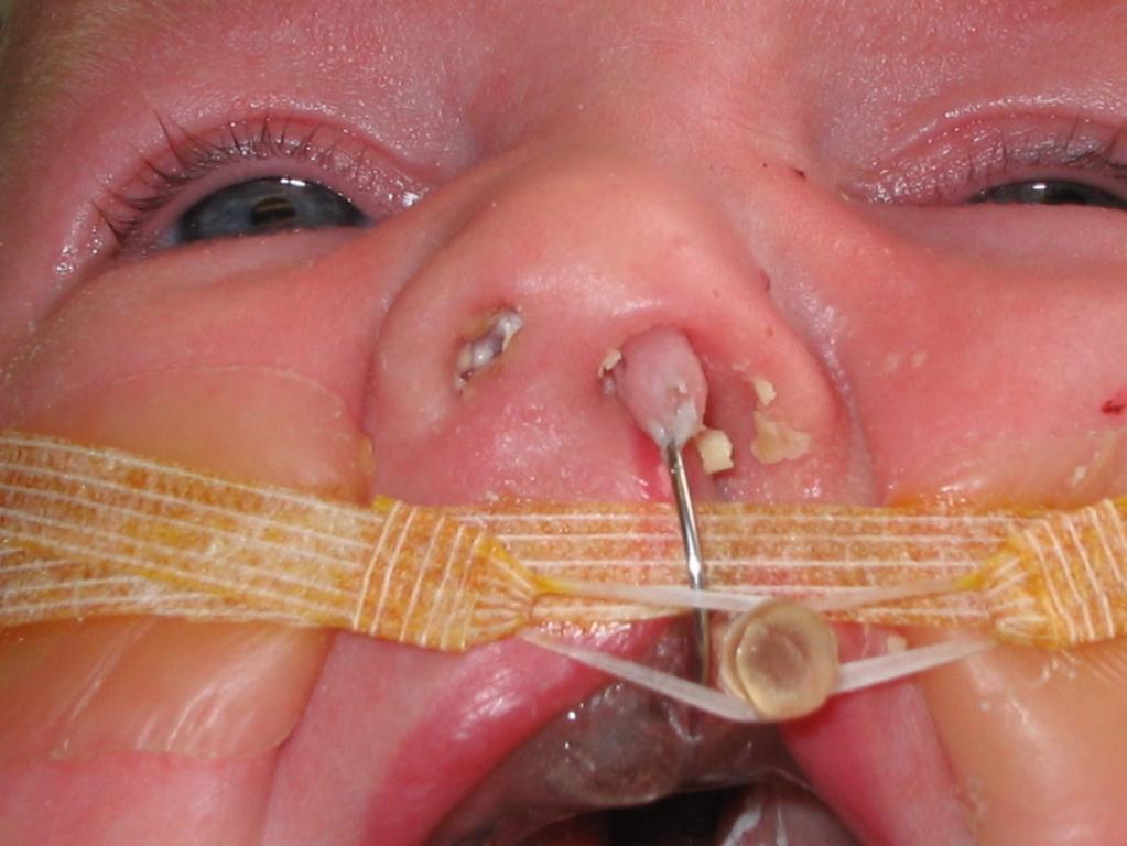
55 Nasoalveolar Molding
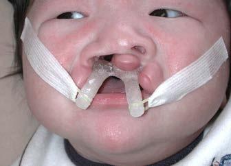
56 Protruding prolabium
57
58

59

60
61
62

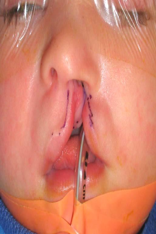
63 Pre-NAM Post-NAM Post-Surgery

64 Pre-op Post-op
Pharyngeal Apparatus. Pouches Endoderm Grooves Ectoderm Arch Neural Crest Somitomeres Aortic Arch - Vessel
 Pharyngeal Apparatus Pouches Endoderm Grooves Ectoderm Arch Neural Crest Somitomeres Aortic Arch - Vessel Segmental Organization Humans: Arch 1-4 prominent Arch 5 absent Arch 6 - transient First Arch Face
04 Development of the Face and Neck. Development of the Face Development of the neck
 04 Development of the Face and Neck Development of the Face Development of the neck Development of the face Overview of facial development The fourth week ~ the twelfth week of prenatal development Between
REVIEW OF CLINICAL EMBRYOLOGY OF HEAD AND NECK
 REVIEW OF CLINICAL EMBRYOLOGY OF HEAD AND NECK OUTLINE - EMBRYOLOGY UNDERLYING CLINICAL CONDITIONS I. EARLY DEVELOPMENT OF FACE: CLEFT LIP, CLEFT PALATE, OBSTRUCTED NASOLACRIMAL DUCT II. BRANCHIAL ARCHES
Remember from the first year embryology Trilaminar disc has 3 layers: ectoderm, mesoderm, and endoderm
 Development of face Remember from the first year embryology Trilaminar disc has 3 layers: ectoderm, mesoderm, and endoderm The ectoderm forms the neural groove, then tube The neural tube lies in the mesoderm
Development of the Pharyngeal Arches
 Development of the Pharyngeal Arches Thomas A. Marino, Ph.D. Temple University School of Medicine Competencies: Upon completion of this section of the course, the student must be able to: 1. Recall the
NEUROCRANIUM VISCEROCRANIUM VISCEROCRANIUM VISCEROCRANIUM
 LECTURE 4 SKULL NEUROCRANIUM VISCEROCRANIUM VISCEROCRANIUM VISCEROCRANIUM CRANIUM NEUROCRANIUM (protective case around brain) VISCEROCRANIUM (skeleton of face) NASOMAXILLARY COMPLEX MANDIBLE (DESMOCRANIUM)
Brain and spinal nerve. By: shirin Kashfi
 Brain and spinal nerve By: shirin Kashfi Nervous system: central nervous system (CNS) peripheral nervous system (PNS) Brain (cranial) nerves Spinal nerves Ganglions (dorsal root ganglions, sympathetic
Head and Neck I. PHARYNGEAL APPARATUS (FIGURE 12.1; TABLE 12.1)
") chapter 12 Head and Neck I. PHARYNGEAL APPARATUS (FIGURE 12.1; TABLE 12.1) The pharyngeal apparatus consists of the pharyngeal arches, pharyngeal pouches, pharyngeal grooves, and pharyngeal membranes,
chapter 12 Head and Neck I. PHARYNGEAL APPARATUS (FIGURE 12.1; TABLE 12.1) The pharyngeal apparatus consists of the pharyngeal arches, pharyngeal pouches, pharyngeal grooves, and pharyngeal membranes,
Cranial Nerves. Steven McLoon Department of Neuroscience University of Minnesota
 Cranial Nerves Steven McLoon Department of Neuroscience University of Minnesota 1 Course News Change in Lab Sequence Week of Oct 2 Lab 5 Week of Oct 9 Lab 4 2 Sensory and Motor Systems Sensory Systems:
Essentials in Head and Neck Embryology. Part 3 Development of the head, face, and oral cavity
 Essentials in Head and Neck Embryology Part 3 Development of the head, face, and oral cavity Outline General overview of prenatal development Embryonic period phase 1 Formation of bilaminar disk Formation
 Drawings illustrating the human pharyngeal apparatus. Drawings illustrating the human pharyngeal apparatus. Drawings illustrating the human pharyngeal apparatus. Drawings illustrating the human pharyngeal
Pharyngeal apparatus. - At the third week, it is a 3 layered structure: ectoderm, mesoderm and endoderm. This is called trilaminar disc
 Pharyngeal apparatus Remember from the first year embryology - The embryo was disc shaped in the second week of development (this is called embryonic disc) and it is a 2 layered disc (composed of two layers)---bilaminar
Review of Nervous System Anatomy
 For the real amazement, if you wish to be amazed, is this process. You start out as a single cell derived from the coupling of a sperm and an egg; this divides in two, then four, then eight, and so on,
DEVELOPMENTAL ANATOMY OF THE FACE, JAW AND NECK. O.M. Oluwatosin Department of Surgery
 DEVELOPMENTAL ANATOMY OF THE FACE, JAW AND NECK O.M. Oluwatosin Department of Surgery 1 2 By the end of this lecture, you should be able to: Discuss the embryology of the face Relate congenital anomalies
THYROID & PARATHYROID. By Prof. Saeed Abuel Makarem & Dr. Sanaa Al-Sharawy
 THYROID & PARATHYROID By Prof. Saeed Abuel Makarem & Dr. Sanaa Al-Sharawy 1 OBJECTIVES By the end of the lecture, the student should be able to: Describe the shape, position, relations and structure of
1/10/2013. What do neurons look like? Topic 14: Spinal Cord & Peripheral Nerves. How do neurons work? The nervous impulse. Specialized Neurons
 Topic 4: Spinal Cord & Peripheral Nerves What do neurons look like? Neurons What do they look like? How do they work? Neuronal and spinal organization What is the difference between neuron & nerve? How
Biology 323 Human Anatomy for Biology Majors Week 10; Lecture 1; Tuesday Dr. Stuart S. Sumida. Cranial Nerves and Soft Tissues of the Skull
 Biology 323 Human Anatomy for Biology Majors Week 10; Lecture 1; Tuesday Dr. Stuart S. Sumida Cranial Nerves and Soft Tissues of the Skull FOREBRAIN MIDBRAIN HINDBRAIN Forebrain: Cerebrum Perception,
-Ibrahim Al-Naser. -Dr Al- Muhtaseb. 1 P a g e
 -1 -Ibrahim Al-Naser - -Dr Al- Muhtaseb 1 P a g e The Digestive System The doctor started the lecture by talking about the class rules. The GI system is an organ system, it is divided into: The Alimentary
Congenital Neck Masses C. Stefan Kénel-Pierre, MD
 Congenital Neck Masses C. Stefan Kénel-Pierre, MD SUNY-LICH Medical Center Department of Surgery Case Presentation xx year old male presents with sudden onset left lower neck swelling x 1 week Denies pain,
Biology 340 Comparative Embryology Lecture 11 Dr. Stuart Sumida. Overview of Embryology of the Vertebrate Skull. Emphasis on Amniota
 Biology 340 Comparative Embryology Lecture 11 Dr. Stuart Sumida Overview of Embryology of the Vertebrate Skull Emphasis on Amniota Initial introduction to components parts of a vertebrate head. This lecture
CRANIAL NERVES. Dr. Amani A. Elfaki Associate Professor Department of Anatomy
 CRANIAL NERVES Dr. Amani A. Elfaki Associate Professor Department of Anatomy LEARNING OBJECTIVES Named the cranial nerves Identify the funcunal component of each cranial nerve Identify the effect of each
Trigeminal Nerve (V)
") Trigeminal Nerve (V) Lecture Objectives Discuss briefly how the face is developed. Follow up the course of trigeminal nerve from its point of central connections, exit and down to its target areas. Describe
Trigeminal Nerve (V) Lecture Objectives Discuss briefly how the face is developed. Follow up the course of trigeminal nerve from its point of central connections, exit and down to its target areas. Describe
Figure 1. Basic anatomy of the palate
 CHAPTER 10 CLEFT LIP AND PALATE Chen Yan, MD and Sanjay Naran, MD I. ANATOMY AND DEFINITIONS A. Cleft Lip (CL) alone, Cleft Lip with Cleft Palate (CLP), and Cleft Palate (CP) alone 1. CL alone and CLP
C h a p t e r PowerPoint Lecture Slides prepared by Jason LaPres North Harris College Houston, Texas
 C h a p t e r 15 The Nervous System: The Brain and Cranial Nerves PowerPoint Lecture Slides prepared by Jason LaPres North Harris College Houston, Texas Copyright 2009 Pearson Education, Inc., publishing
Peripheral Nervous System
 Peripheral Nervous System Sensory Receptors Motor Endings Cranial Nerves The Four Plexuses Extremities Review of Reflexes Fast, preprogrammed, inborn, automatic responses Occur in the CNS at the spinal
Subdivided into Vestibule & Oral cavity proper
 Extends from the lips to the oropharyngeal isthmus The oropharyngeal isthmus: Is the junction of mouth and pharynx. Is bounded: Above by the soft palate and the palatoglossal folds Below by the dorsum
SYLLABUS BDS I PROFESSIONAL GENERAL HUMAN ANATOMY INCLUDING EMBRYOLOGY AND HISTOLOGY
 GENERAL HUMAN ANATOMY INCLUDING EMBRYOLOGY AND HISTOLOGY I. General Anatomy 1. Anatomical terms 2. Skin, superficial fascia & deep fascia 3. Cardiovascular system, portal system, collateral circulation
Skeletal System. Prof. Dr. Malak A. Al-yawer Department of Anatomy/Embryology Section
 Skeletal System Prof. Dr. Malak A. Al-yawer Department of Anatomy/Embryology Section Learning objectives At the end of this lecture, the medical student will be able to: State the embryonic origin of skeletal
Lab 16: PNS: Nerves and Autonomic NS Hamilton Answers to Pre- Lab Assignments
 Lab 16: PNS: Nerves and Autonomic NS Hamilton Answers to Pre- Lab Assignments Pre-Lab Activity 1: 1. a. olfactory nerve b. optic nerve c. oculomotor nerve d. abducens nerve e. trochlear nerve f. trigeminal
Embryo#1. Mohammad Hisham Al-Mohtaseb باشق جهاد. 0 P a g e
 Embryo#1 Mohammad Hisham Al-Mohtaseb باشق جهاد 0 P a g e Before you start, it is important to link what you learn in gross anatomy with developmental stages discussed in embryology. Cells that form organs
Thyroid gland. importance. relations and connections. external laryngeal nerves. malformations.
 Thyroid gland 1. Recognize and understand the coverings of the thyroid gland and their clinical importance. 2. Recognize and understand the main parts of the thyroid gland and their locations, relations
Basic Anatomy and Physiology of the Lips and Oral Cavity. Dr. Faghih
 Basic Anatomy and Physiology of the Lips and Oral Cavity Dr. Faghih It is divided into seven specific subsites : 1. Lips 2. dentoalveolar ridges 3. oral tongue 4. retromolar trigone 5. floor of mouth 6.
Oral cavity : consist of two parts: the oral vestibule and the oral cavity proper. Oral vestibule : is slit like space between.
 Oral cavity Oral cavity : consist of two parts: the oral vestibule and the oral cavity proper Oral vestibule : is slit like space between the teeth, buccal gingiva, lips, and cheeks 1 Oral cavity Oral
Laboratory Exercise 3 Laboratory Examination of the Chicken Embryo Part II
 Biology 340, Comparative Embryology, Laboratory Exercise 3 Page 16 S. S. SUMIDA BIOLOGY 340 Comparative Embryology Laboratory Exercise 3 Laboratory Examination of the Chicken Embryo Part II Introduction
For the following questions, indicate the letter that corresponds to the SINGLE MOST APPROPRIATE ANSWER
 GROSS ANATOMY EXAMINATION May 15, 2000 For the following questions, indicate the letter that corresponds to the SINGLE MOST APPROPRIATE ANSWER 1. Pain associated with an infection limited to the middle
INTRODUCTION: ANATOMY UNDERLYING CLINICAL TESTS OF CRANIAL NERVES
 INTRODUCTION: ANATOMY UNDERLYING CLINICAL TESTS OF CRANIAL NERVES CRANIAL NERVE I - OLFACTORY I - OLFACTORY NERVE - SMELL TEST: SMELL ODORS (note: not ammonia; pain in nasal cavity CN5 DAMAGE: LOSS OF
Letty Moss-Salentijn DDS, PhD Dr. Edwin S.Robinson Professor of Dentistry (in Anatomy and Cell Biology)
") 10. Pharyngeal Arches Revisited and the PHARYNGEAL POUCHES Letty Moss-Salentijn DDS, PhD Dr. Edwin S.Robinson Professor of Dentistry (in Anatomy and Cell Biology) E-mail: lm23@columbia.edu READING ASSIGNMENT:
10. Pharyngeal Arches Revisited and the PHARYNGEAL POUCHES Letty Moss-Salentijn DDS, PhD Dr. Edwin S.Robinson Professor of Dentistry (in Anatomy and Cell Biology) E-mail: lm23@columbia.edu READING ASSIGNMENT:
Learning Outcomes. The Carotid 20/02/2013. Scalp, Face, Parotid. Layers of the Scalp. The Parotid Gland. The Scalp. The Carotid The Facial Artery
 Learning Outcomes The Scalp Layers of the Scalp Bleeding from the Scalp The Carotid The Facial Artery Major Muscles of the Face and Jaw(s) Muscles of Mastication Muscles of Facial Expression The Parotid
Human Nervous System:
 OLLI Brain: Making Sense of Our World: Lecture 3 Human Nervous System: The Motor & Sensory Divisions Copyright 2004 Pearson Education, Inc., publishing as Benjamin Cummings Organization of the Nervous
PERIPHERAL NERVOUS SYSTEM
 CHAPTER 13 PERIPHERAL NERVOUS SYSTEM Functional division of nervous system = afferent info to the CNS ascending spinal cord = efferent info from CNS descending spinal cord somatic skin, muscles visceral
Peripheral Nervous System Dr. Gary Mumaugh
 Peripheral Nervous System Dr. Gary Mumaugh Spinal Nerves Overview Thirty-one pairs of spinal nerves are connected to the spinal cord No special names; numbered by level of vertebral column at which they
What is Hemifacial Microsomia? By Pravin K. Patel, MD and Bruce S. Bauer, MD Children s Memorial Hospital, Chicago, IL
 What is Hemifacial Microsomia? By Pravin K. Patel, MD and Bruce S. Bauer, MD Children s Memorial Hospital, Chicago, IL 773-880-4094 Early in the child s embryonic development the structures destined to
Teeth, orofacial development and
 Teeth, orofacial development and cleft anomalies Miroslav Peterka Variability of jaws in vertebrates. (A) cartilaginous fish shark; (B) an example of a bone fish; (C ) amphibian frog; (D) reptile - turtle;
Introduction to Head and Neck Anatomy
 Introduction to Head and Neck Anatomy Nervous Tissue Controls and integrates all body activities within limits that maintain life Three basic functions 1. sensing changes with sensory receptors 2. interpreting
Course: Physical Assessment II Date: October 17, 2008 Doc: Practice Quiz 1
 Course: Physical Assessment II Date: October 17, 2008 Doc: Practice Quiz 1 This is the practice quiz we did in Class 4. The answers are at the end of the quiz should you wish to test yourself. Complete
Trigeminal nerve. Slide in bold and please go back to see the pictures, if I skipped any part of record that because it wasn t clear to me
 Trigeminal nerve Slide in bold and please go back to see the pictures, if I skipped any part of record that because it wasn t clear to me Hala nsour 2/26/2018 P a g e 1 this lecture contain two topics
Tracing the Cranial Nerves Osteologically
 CN I II III IV V 1 Supra-orbital ethmoidal nn. Ext. nasal V 2 Tracing the Cranial Nerves Osteologically Nucleus of Origin Olfactory tracts of frontal lobe of cerebrum Optic tracts from optic chiasma and
Embryology, anatomy and physiology of the oesophagus. Sarah Forsyth
 Embryology, anatomy and physiology of the oesophagus Sarah Forsyth Embryology Basics Endoderm forms scaffolding of GIT Endoderm forms the lining of the yolk sac Derivative of foregut Wk 4 - Foregut develops
Examination and Diseases of Cranial Nerves
 Cranial nerve evaluation is an important part of a neurologic exam. There are some differences in the assessment of cranial nerves with different species but the general principles are the same. Going
Anatomy of Oral Cavity DR. MAAN AL-ABBASI
 Anatomy of Oral Cavity DR. MAAN AL-ABBASI By the end of this lecture you should be able to: 1. Differentiate different parts of the oral cavity 2. Describe the blood and nerve supply of mucosa and muscles
b. The groove between the two crests is called 2. The neural folds move toward each other & the fuse to create a
 Chapter 13: Brain and Cranial Nerves I. Development of the CNS A. The CNS begins as a flat plate called the B. The process proceeds as: 1. The lateral sides of the become elevated as waves called a. The
APRIL
 APRIL - 2003 OCTOBER - 2003 February 2009 [KU 652] Sub. Code : 4131 FIRST B.D.S DEGREE EXAMINATION (Modified Regulations III) Paper I HUMAN ANATOMY, HISTOLOGY AND EMBRYOLOGY Time : Three hours
Unit VIII Problem 3 Neuroanatomy: Brain Stem, Cranial Nerves and Scalp
 Unit VIII Problem 3 Neuroanatomy: Brain Stem, Cranial Nerves and Scalp - Brain stem: It is connected to the cerebellum and cerebral hemispheres. Rostral end of brain stem: diencephalon is the area which
Review or skim Ch 12 on the vascular supply of the brain. Just look at pictures and legends for the clinical part at the end.
 Dental Neuroanatomy January 20 and 27, 10-12, 2011 Suzanne S. Stensaas, Ph.D. Dear Students: Please print these notes and bring them with you. My style is to use a Tablet PC and I draw on either a Word
Cranial nerves.
 Cranial nerves eaglezhyxzy@163.com Key Points of Learning Name Components Passing through Peripheral distribution Central connection Function Cranial nerves Ⅰ olfactory Ⅱ optic Ⅲ occulomotor Ⅳ trochlear
Brain and Cranial Nerves (Ch. 15) Human Anatomy lecture. caudal = toward the spinal cord)
 Human Anatomy lecture. caudal = toward the spinal cord)") Insight: Some cranial nerve disorders Brain and Cranial Nerves (Ch. 15) Human Anatomy lecture I. Overview (Directional terms: rostral = toward the forehead caudal = toward the spinal cord) A. 3 Major parts
Insight: Some cranial nerve disorders Brain and Cranial Nerves (Ch. 15) Human Anatomy lecture I. Overview (Directional terms: rostral = toward the forehead caudal = toward the spinal cord) A. 3 Major parts
Early Development of Neural Tube Development of Medulla Spinalis and Peripheral Nervous System. Assoc.Prof. E.Elif Güzel, M.D.
 Early Development of Neural Tube Development of Medulla Spinalis and Peripheral Nervous System Assoc.Prof. E.Elif Güzel, M.D. Third week of Embryogenesis Primitive streak/pit appears on the epiblast (day
MRI ANATOMY OF THE CRANIAL NERVES. Alexandra Borges Radiology Dpt. Instituto Português de Oncologia de Lisboa
 MRI ANATOMY OF THE CRANIAL NERVES Alexandra Borges Radiology Dpt. Instituto Português de Oncologia de Lisboa SENR 2014 CRANIAL NERVES Olfactory: I Optic: II Oculomotor nerves: III, IV, VI Trigeminal nerve:
Nervous System. The Peripheral Nervous System Agenda Review of CNS v. PNS PNS Basics Cranial Nerves Spinal Nerves Reflexes Pathways
 Nervous System Agenda Review of CNS v. PNS PNS Basics Cranial Nerves Spinal Nerves Sensory Motor Review of CNS v. PNS Central nervous system (CNS) Brain Spinal cord Peripheral nervous system (PNS) All
a) They are the most common cause of pediatric kidney failure. b) They are always symptomatic. c) They can be asymmetric.
 They are the most common cause of pediatric kidney failure. b) They are always symptomatic. c) They can be asymmetric.") Practice questions: 1. The paraxial mesoderm gives rise to somites. The structure of the somite a) is a loose mesenchymal sheet that will migrate toward the notochord. b) is an epithelial rosette with
Practice questions: 1. The paraxial mesoderm gives rise to somites. The structure of the somite a) is a loose mesenchymal sheet that will migrate toward the notochord. b) is an epithelial rosette with
Anatomy #9. Rashed AL-Jomared. The Cranial Nerves IX. Amneh Hazaimeh & Alanood Bostanji
 Anatomy #9 The Cranial Nerves IX Rashed AL-Jomared Amneh Hazaimeh & Alanood Bostanji السالم عليكم This lecture talks about the cranial nerves IX & X:: *Glossopharyngeal nerve : The nerve gets out of the
Neck of Condylar. Process. Anterior Border of Ramus. Mandibular. Foramen. Posterior Border of Ramus Incisive Fossa.
 Learning Outcomes The Mandible Surface Anatomy Muscle Attachments The (FOM) Muscles of the FOM The Tongue Muscles of the Tongue The Submandibular Region Submandibular Gland Sublingual Gland Lingual The
HBA THE BODY Head & Neck Written Examination October 23, 2014
 HBA 531 - THE BODY Head & Neck Written Examination October 23, 2014 Name: NOTE 2: When asked to trace nerve, artery, or vein pathways, do so by using arrows, e.g., structure a structure b structure c...
Preface... Contributors... 1 Embryology... 3
 Contents Preface... Contributors... vii xvii I. Pediatrics 1 Embryology... 3 Pearls... 3 Branchial Arch Derivatives... 3 Branchial Arch Anomalies: Cysts, Sinus, Fistulae... 4 Otologic Development... 4
Craniofacial Microsomia
 Patient and Family Education Craniofacial Microsomia Children with craniofacial microsomia (CFM) have a small or underdeveloped part of the face, usually the ear and jaw. The eye, cheek and neck may also
Oral Embryology and Histology
 Oral Embryology and Histology Chapter 8 Copyright 2018, Elsevier Inc. All Rights Reserved. 1 Learning Objectives Lesson 8.1: Oral Embryology 1. Pronounce, define, and spell the key terms. 2. Define embryology
UNIVERSITY OF NAIROBI
 UNIVERSITY OF NAIROBI UNIVERSITY EXAMINATIONS 2013/2014 LEVEL I MID-SEMESTER II EXAMINATION FOR THE DEGREE OF BACHELOR OF SCIENCE IN NURSING (BScN) AND BACHELOR OF PHARMACY (B.PHARM) MARKING SCHEME HNS101/UPC106:
ORAL CAVITY, ESOPHAGUS AND STOMACH
 ORAL CAVITY, ESOPHAGUS AND STOMACH 1 OBJECTIVES By the end of the lecture you should be able to: Describe the anatomy the oral cavity, (boundaries, parts, nerve supply). Describe the anatomy of the palate,
HBA THE BODY Head & Neck Written Examination - November 4, 2013
 HBA 531 - THE BODY Head & Neck Written Examination - November 4, 2013 NOTE 1: Whenever your answer refers to a cranial nerve, give both its number and its name. In the case of the trigeminal nerve, give
Anterior triangle of neck
 Anterior triangle of neck Dept. of Anatomy Zhou Hong Ying Outline boundary and subdivisions of ant. triangle contents of the triangle Muscles: suprahyoid muscles, infrahyoid muscles Nerves: CNⅩ, CNⅪ, CNⅫ,
DENTAL MANAGEMENT OF CLEFT LIP AND PALATE. J Harewood DDS MA MS
 DENTAL MANAGEMENT OF CLEFT LIP AND PALATE J Harewood DDS MA MS CLEFT LIP/PALATE: INCIDENCE Cleft lip and/or palate 1:1000 Varies with race Japan: 20: 10 000 Western Europe: 12: 10 000 USA: 10.2:10 000
SCHOOL OF ANATOMICAL SCIENCES Mock Run Questions. 4 May 2012
 SCHOOL OF ANATOMICAL SCIENCES Mock Run Questions 4 May 2012 1. With regard to the muscles of the neck: a. the platysma muscle is supplied by the accessory nerve. b. the stylohyoid muscle is supplied by
lecture #2 Done by : Tyma'a Al-zaben
 lecture #2 Done by : Tyma'a Al-zaben ** Hello SERTONIN! note:: the slide included within the sheet but make sure back to slide for pictures in the previous lecture we talk about ascending tract and its
ANTERIOR CERVICAL TRIANGLE (Fig. 2.1 )
") 2 Neck Anatomy ANTERIOR CERVICAL TRIANGLE (Fig. 2.1 ) The boundaries are: Lateral: sternocleidomastoid muscle Superior: inferior border of the mandible Medial: anterior midline of the neck This large triangle
2 Neck Anatomy ANTERIOR CERVICAL TRIANGLE (Fig. 2.1 ) The boundaries are: Lateral: sternocleidomastoid muscle Superior: inferior border of the mandible Medial: anterior midline of the neck This large triangle
The Pharynx. Dr. Nabil Khouri MD. MSc, Ph.D
 The Pharynx Dr. Nabil Khouri MD. MSc, Ph.D Introduction The pharynx is the Musculo-fascial halfcylinder that links the oral and nasal cavities in the head to the larynx and esophagus in the neck Common
Lec [8]: Mandibular nerve:
![Lec [8]: Mandibular nerve:](/thumbs/94/121295776.jpg "Lec [8]: Mandibular nerve:") Lec [8]: Mandibular nerve: The mandibular branch from the trigeminal ganglion lies in the middle cranial fossa lateral to the cavernous sinus. With the motor root of the trigeminal nerve [motor roots lies
Lec [8]: Mandibular nerve: The mandibular branch from the trigeminal ganglion lies in the middle cranial fossa lateral to the cavernous sinus. With the motor root of the trigeminal nerve [motor roots lies
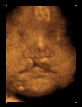
Normal fetal face and neck
 Normal fetal face and neck Maria A. Calvo-Garcia, MD. Associate Professor of Radiology Cincinnati Children s Hospital Medical Center Cincinnati, Ohio Disclosure I have no disclosures Goals & objectives
Cranial Nerves and Spinal Cord Flashcards
 1. Name the cranial nerves and their Roman numeral. 2. What is Cranial Nerve I called, and what does it 3. Scientists who are trying to find a way to make neurons divide to heal nerve injuries often study
Dr.Ban I.S. head & neck anatomy 2 nd y. جامعة تكريت كلية طب االسنان املرحلة الثانية
 جامعة تكريت كلية طب االسنان التشريح مادة املرحلة الثانية أ.م.د. بان امساعيل صديق 6102-6102 1 The Palate The palate forms the roof of the mouth and the floor of the nasal cavity. It is divided into two
Week 14. Development of the Musculoskeletal System
 Week 14 Development of the Musculoskeletal System Skeletal System Derived from: paraxial mesoderm somites and somitomeres sclerotome sclerotome differentiation induced by SHH from notochord and floor plate
SKELETAL MUSCLES. Objectives
 Objectives Body Wall Muscles Remember the pattern Hypaxial Muscle Overview Cervical body wall muscles Hypaxial Muscle Overview Thoracic body wall muscles Hypaxial Muscle Overview Abdominal body wall muscles
Tympanic Bulla Temporal Bone. Digastric Muscle. Masseter Muscle
 Superior view Hyoid Bone The hyoid bone does not articulate with any other bones. It is held in place by ligaments to the styloid process of the temporal bone and the thyroid cartilage of the larynx. It
Brainstem and Cranial Nerves II. Nerves covered in other lectures. A reminder about embryology. Prof. Stuart Bunt
 Brainstem and Cranial Nerves II Prof. Stuart Bunt Nerves covered in other lectures 1 Olfactory 2 Optic 3,4,6 Extraocular eye muscles 8 Vestibulo-cochlear 5 Motor and Sensory to the face and muscles of
Doctor Osama Asa ad Khader. Mohammad Alsalem
 6 Doctor 2015 Osama Asa ad Khader Mohammad Alsalem A quick revision for the spinal cord blood supply: Arterial Blood supply of spinal cord The spinal cord got its arterial supply by two ways: Longitudinal
Prevertebral Region, Pharynx and Soft Palate
 Unit 20: Prevertebral Region, Pharynx and Soft Palate Dissection Instructions: Step1 Step 2 Step 1: Insert your fingers posterior to the sternocleidomastoid muscle, vagus nerve, internal jugular vein,
Chapter 13: The Peripheral Nervous System
 Chapter 13: The Peripheral Nervous System Objectives: 1. Define peripheral nervous system and list its components. 2. Classify general sensory receptors by structure, stimulus detected, and body location.
Differentiation of the Placode Ectoderm Neural Crest Derivatives
 Dr. Andrea D. Székely Differentiation of the Placode Ectoderm Neural Crest Derivatives Acknowledgements to Dr Károly Altdorfer Dr Nándor Nagy EARLY DEVELOPMENT The neural crest and craniofacial placodes
Anatomy: head and Neck (6 questions) 1. Prevertebral Flexor Musculature (lying in front of the vertebrae) include all, EXCEPT: Longus Colli.
 1. Prevertebral Flexor Musculature (lying in front of the vertebrae) include all, EXCEPT: Longus Colli.") Anatomy: head and Neck (6 questions) 1. Prevertebral Flexor Musculature (lying in front of the vertebrae) include all, EXCEPT: Longus Colli. Rectus Capitis Anterior. Rectus Capitis Lateralis. Rectus Capitis
Anatomy: head and Neck (6 questions) 1. Prevertebral Flexor Musculature (lying in front of the vertebrae) include all, EXCEPT: Longus Colli. Rectus Capitis Anterior. Rectus Capitis Lateralis. Rectus Capitis
Cleft-Craniofacial Center
 Cleft-Craniofacial Center A Pioneering T eam 2 Welcome to the Cleft-Craniofacial Center at Children s Hospital of Pittsburgh The Cleft-Craniofacial Center at Children s Hospital of Pittsburgh has been
TRANSCRIPTIONS OF NARRATIONS FOR EMBRYOLOGY OF THE BRANCHIAL REGION
 TRANSCRIPTIONS OF NARRATIONS FOR EMBRYOLOGY OF THE BRANCHIAL REGION Branchial Arch Development, slide 2 This is a very familiar picture - a median sagittal section of a four week embryo. I have actually
Structure Location Function
 Frontal Bone Cranium forms the forehead and roof of the orbits Occipital Bone Cranium forms posterior and inferior portions of the cranium Temporal Bone Cranium inferior to the parietal bone forms the
The Brain and Cranial Nerves Pg. 129
 The Brain and Cranial Nerves Pg. 129 Three Main Regions of the Brain Forebrain Cerbral hemispheres Diencephalon Midbrain Brain stem Hindbrain Pons Cerebellum Medulla oblongata Forebrain Interprets sensory
Laboratory Exercise 4 Laboratory Examination of the Pig Embryo
 S. S. SUMIDA BIOLOGY 340 Comparative Embryology Laboratory Exercise 4 Laboratory Examination of the Pig Embryo Introduction With the use of serial cross-sections of chicken embryos, you recently completed
Cranial Nerves VII to XII
 Cranial Nerves VII to XII MSTN121 - Neurophysiology Session 13 Department of Myotherapy Cranial Nerve VIII: Vestibulocochlear Sensory nerve with two distinct branches. Vestibular branch transmits information
Functional components
 Facial Nerve VII cranial nerve Emerges from Pons Two roots Functional components: 1. GSA (general somatic afferent) 2. SA (Somatic afferent) 3. GVE (general visceral efferent) 4. BE (Special visceral/branchial
Omran Saeed. Luma Taweel. Mohammad Almohtaseb. 1 P a g e
 2 Omran Saeed Luma Taweel Mohammad Almohtaseb 1 P a g e I didn t include all the photos in this sheet in order to keep it as small as possible so if you need more clarification please refer to slides In
The Nervous System: Central Nervous System
 The Nervous System: Central Nervous System I. Anatomy of the nervous system A. The CNS & the body by: 1. monitoring of the body 2. & information between parts of the body 3. acting as a to gather, store,
COURS 8 : THE NERVOUS SYSTEM, PART II
 UE1 Anglais Pr Naguy Le 20/03/2018 à 16h00 Roneotypeur : Bouchamma Imane COURS 8 : THE NERVOUS SYSTEM, PART II Rappel : le CC du 4 mai à lieu sur les cours 7 et 8 «The Nervous System» et sera sous forme