04 Development of the Face and Neck. Development of the Face Development of the neck
|
|
|
- Nickolas Webb
- 5 years ago
- Views:
Transcription
1 04 Development of the Face and Neck Development of the Face Development of the neck
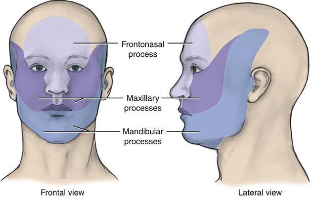
2 Development of the face Overview of facial development The fourth week ~ the twelfth week of prenatal development Between developing brain and heart All three embryonic layers are involved in facial development Five facial processes Single frontonasal process Paired maxillary process Paired mandibular process

3 Developing brain stomodeum
4
5 Table 4-1 BranchialArches and Derivative Structures First arches (mandibular) Second arches (hyoid) Third arches Fourth through Sixth arches Trigenimal nerve, muscles of mastication, mylohyoid and anterior belly of digastric, tensor tympani, tensor veli palatine muscles Facial nerve, stapedius muscle, muscles of facial expression, posterior belly of the digastric muscle, stylohyoid muscle Glossopharyngeal nerve, stylopharyngeal muscle Superior laryngeal branch and recurrent laryngeal branch of vagus nerve, levator veli palatine muscles, pharyngeal constrictors, intrinsic muscles of the laynx Malleus and incus of middle ear, including anterior ligament of the malleus, sphenomandibular ligament, and parts of sphenoid bone Stapes and parts of malleus and incus of middle ear, stylohyoid ligament, styloid process of the temporal bone, lesser cornu of hyoid bone, upper portion of body of hyoid bone Greater cornu of hyoid bone, lower portion of body of hyoid bone Laryngeal cartilage
6 Development of the face Overview of facial development Facial processes are centers of growth for the face Upper face : from frontonasal process Middle face : from maxillary process Lower face : from mandibular process Growth pattern of the face upper face rapid middle and lower face slow

 Most of the facial structures fusion of tissue on")
7 Development of the face Overview of facial development Fusion fusion of tissue on the same surface of the embryo e.g.) Most of the facial structures fusion of tissue on the different surface of the embryo e.g.) Palate
8 Development of the face Stomodeum and oral cavity formation Stomodeum (stomatodeum) a shallow depression in the embryonic surface ectoderm before fourth week Oropharyngeal membrane separates the stomodeum from the primitive pharynx Primitive pharynx (The cranial part of foregut, the beginning of the future digestive tract) FIRST event during fourth week Disintegration of the oropharyngeal membrane Stomodeum will form the oral cavity lined by oral epithelium

9 Embryo at third to fourth weeks of development.
10 Development of the face Mandibular arch and lower face formation During fourth week, two bulges of tissue appear inferior to stomodeum two mandibular processes These paired mandibular processes are formed in part by neural crest cells that migrated to the facial region, covered externally by ectoderm and internally by endoderm. Paired mandibular processes BOTH fuse at midline to form the mandibular arch mandibular arch, first branchial arch Meckel s cartilage forms within each side of mandibular arch, IMPORTANT in alveolar bone development Directly forms lower face, including lower lip. : mandible, mandibular teeth, associated tissues, tongue
11 Development of the face Frontonasal process and upper face formation During fourth week, frontonasal process is a bulge of tissue in upper facial area. Frontonasal process will form upper face, which includes forehead, bridge of nose, primary palate, nasal septum.
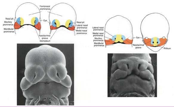
12 Development of the face Frontonasal process and upper face formation <Placode development> Rounded areas of specialized, thickened ectoderm Lens placodes : future eyes and associated tissues otic placodes : future internal ear and associated tissues Nasal placodes : form nasal (olfactory) pits, which develop into nasal cavities.
13 Development of the face Frontonasal process and upper face formation <Nose and paranasal sinus formation> During the fourth week, the tissue around the nasal placodes undergoes growth. Nasal pits : Placodes become submerged, forming depression in center of each Nasal pits develop into nasal cavities. medial nasal process : the middle portion of the nose from root to apex, center portion of upper lip, philtrum region lateral nasal processes : alae premaxillary segment : maxillary incisor teeth and associated tissues, primary palate, nasal septum
14
15 Development of the face Maxillary process and midface formation During fourth week, the maxillary processes be formed from increased growth of mandibular arch. These paired maxillary processes are formed in part by neural crest cells that migrated to the facial region, covered externally by ectoderm and internally by endoderm. Maxillary process will form the midface: Includes sides of upper lip, cheeks, secondary palate posterior portion of maxilla with maxillary canines and posteriors, associated tissues Zygomatic bones and temporal bones
+ each medial nasal process(middle) Lower lip: mandibular process intermaxillary")
16 Development of the face Maxillary process and midface formation < Upper and lower lip formation> Upper lip: each maxillary process (sides)+ each medial nasal process(middle) Lower lip: mandibular process intermaxillary segment

17 ** Developmental disturbances of the lips and associated tissue Cleft lip
18 Development of the neck The fourth week of prenatal development ~ fetal period Develop from the primitive pharynx and the branchial apparatus

19 Development of the Neck 19 Primitive pharynx formation Derived from the anterior part of the foregut and will form the primitive pharynx, the future oropharynx

20 Development of the Neck 20 Branchial apparatus formation Branchial apparatus branchial arches, branchial grooves and membranes, pharyngeal pouches ARCH GROOVE or CLEFT POUCH MEMBRANE
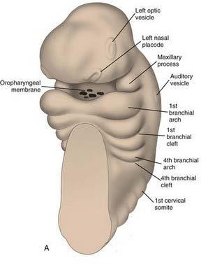
21 Branchial arch formation The fourth week of prenatal development Stacked bilateral swellings of tissue First branchial arch(mandibular arch ) ~ fourth mandibular arch Six pairs of U-shaped bars Mesenchyme formed by neural crest cells, ectoderm, and endoderm Fifth branchial arch is often so rudimentary Important structures of the face and neck Has its own developing cartilage, nerve, vascular, and muscular components FIRST branchial arch(mandibular arch) are involved in the formation of middle and lower face
 Reichert s cartilage : a middle ear bone, a process of the temporal bone, parts of the hyoid bone Mesoderm of the hyoid")
22 Lower four pairs arches are involved in the formation of the structure of the neck. Second branchial arch (=Hyoid arch) Reichert s cartilage : a middle ear bone, a process of the temporal bone, parts of the hyoid bone Mesoderm of the hyoid arch : facial muscle, middle ear muscle, suprahyoid muscle Facial nerve
23 Third branchial arch Parts of the hyoid bone Pharyngeal muscle Glossopharyngeal nerve Fourth and sixth branchial arch Most of the laryngeal cartilage Vagus nerve (+Glossopharyngeal nerve)
24 24 Table 4-2 BranchialArches and Derivative Structures First arches (mandibular) Second arches (hyoid) Third arches Fourth through Sixth arches Trigeminal nerve, muscles of mastication, mylohyoid and anterior belly of digastric, tensor tympani, tensor veli palatine muscles Facial nerve, stapedius muscle, muscles of facial expression, posterior belly of the digastric muscle, stylohyoid muscle Glossopharyngeal nerve, stylopharyngeal muscle Superior laryngeal branch and recurrent laryngeal branch of vagus nerve, levator veli palatine muscles, pharyngeal constrictors, intrinsic muscles of the laynx Malleus and incus of middle ear, including anterior ligament of the malleus, sphenomandibular ligament, and parts of sphenoid bone Stapes and parts of malleus and incus of middle ear, stylohyoid ligament, styloid process of the temporal bone, lesser cornu of hyoid bone, upper portion of body of hyoid bone Greater cornu of hyoid bone, lower portion of body of hyoid bone Laryngeal cartilage

25 Branchial groove and membrane formation External grooves, between neighboring branchial arches FIRST branchial groove Is located between the first and second branchial arches A mature structure of the head and neck First branchial membrane : tympanic membrane First branchial groove : external auditory meatus By the end of the seventh week, : Hyoid arch enlarges and overlaps the third, fourth, and sixth arches and cover them. : the last for branchial grooves are obliterated neck formation
26 Pharyngeal pouch formation Four well-defined pairs of pharyngeal pouches Endodermal evaginations form the lateral walls lining the pharynx Many structures of the face and neck. Table 4-3 Pharyngeal Pouches and Derivative Structures First pouches Tympanic membrane (with first branchial groove), tympanic cavity, mastoid antrum, auditory tube Second pouches Crypts and lymphatic nodules of the palatine tonsils Third and fourth pouches Parathyroid and thymus gland ** Developmental disturbances of the branchial apparatus Cervical cysts or Cervical sinuses

27
Pharyngeal Apparatus. Pouches Endoderm Grooves Ectoderm Arch Neural Crest Somitomeres Aortic Arch - Vessel
 Pharyngeal Apparatus Pouches Endoderm Grooves Ectoderm Arch Neural Crest Somitomeres Aortic Arch - Vessel Segmental Organization Humans: Arch 1-4 prominent Arch 5 absent Arch 6 - transient First Arch Face
Pharyngeal apparatus. - At the third week, it is a 3 layered structure: ectoderm, mesoderm and endoderm. This is called trilaminar disc
 Pharyngeal apparatus Remember from the first year embryology - The embryo was disc shaped in the second week of development (this is called embryonic disc) and it is a 2 layered disc (composed of two layers)---bilaminar
REVIEW OF CLINICAL EMBRYOLOGY OF HEAD AND NECK
 REVIEW OF CLINICAL EMBRYOLOGY OF HEAD AND NECK OUTLINE - EMBRYOLOGY UNDERLYING CLINICAL CONDITIONS I. EARLY DEVELOPMENT OF FACE: CLEFT LIP, CLEFT PALATE, OBSTRUCTED NASOLACRIMAL DUCT II. BRANCHIAL ARCHES
Remember from the first year embryology Trilaminar disc has 3 layers: ectoderm, mesoderm, and endoderm
 Development of face Remember from the first year embryology Trilaminar disc has 3 layers: ectoderm, mesoderm, and endoderm The ectoderm forms the neural groove, then tube The neural tube lies in the mesoderm
Essentials in Head and Neck Embryology. Part 3 Development of the head, face, and oral cavity
 Essentials in Head and Neck Embryology Part 3 Development of the head, face, and oral cavity Outline General overview of prenatal development Embryonic period phase 1 Formation of bilaminar disk Formation
Head and Neck I. PHARYNGEAL APPARATUS (FIGURE 12.1; TABLE 12.1)
") chapter 12 Head and Neck I. PHARYNGEAL APPARATUS (FIGURE 12.1; TABLE 12.1) The pharyngeal apparatus consists of the pharyngeal arches, pharyngeal pouches, pharyngeal grooves, and pharyngeal membranes,
chapter 12 Head and Neck I. PHARYNGEAL APPARATUS (FIGURE 12.1; TABLE 12.1) The pharyngeal apparatus consists of the pharyngeal arches, pharyngeal pouches, pharyngeal grooves, and pharyngeal membranes,
 Drawings illustrating the human pharyngeal apparatus. Drawings illustrating the human pharyngeal apparatus. Drawings illustrating the human pharyngeal apparatus. Drawings illustrating the human pharyngeal
NEUROCRANIUM VISCEROCRANIUM VISCEROCRANIUM VISCEROCRANIUM
 LECTURE 4 SKULL NEUROCRANIUM VISCEROCRANIUM VISCEROCRANIUM VISCEROCRANIUM CRANIUM NEUROCRANIUM (protective case around brain) VISCEROCRANIUM (skeleton of face) NASOMAXILLARY COMPLEX MANDIBLE (DESMOCRANIUM)
Development of the Pharyngeal Arches
 Development of the Pharyngeal Arches Thomas A. Marino, Ph.D. Temple University School of Medicine Competencies: Upon completion of this section of the course, the student must be able to: 1. Recall the
DEVELOPMENTAL ANATOMY OF THE FACE, JAW AND NECK. O.M. Oluwatosin Department of Surgery
 DEVELOPMENTAL ANATOMY OF THE FACE, JAW AND NECK O.M. Oluwatosin Department of Surgery 1 2 By the end of this lecture, you should be able to: Discuss the embryology of the face Relate congenital anomalies
Head and Neck Development and Malformations
 Head and Neck Development and Malformations Yang Chai, DDS, PhD Professor George and MaryLou Boone Chair Ostrow School of Dentistry of USC ychai@usc.edu C D E A. B Learning Objectives - Learn cranial neural
Embryo#1. Mohammad Hisham Al-Mohtaseb باشق جهاد. 0 P a g e
 Embryo#1 Mohammad Hisham Al-Mohtaseb باشق جهاد 0 P a g e Before you start, it is important to link what you learn in gross anatomy with developmental stages discussed in embryology. Cells that form organs
Congenital Neck Masses C. Stefan Kénel-Pierre, MD
 Congenital Neck Masses C. Stefan Kénel-Pierre, MD SUNY-LICH Medical Center Department of Surgery Case Presentation xx year old male presents with sudden onset left lower neck swelling x 1 week Denies pain,
SKELETAL MUSCLES. Objectives
 Objectives Body Wall Muscles Remember the pattern Hypaxial Muscle Overview Cervical body wall muscles Hypaxial Muscle Overview Thoracic body wall muscles Hypaxial Muscle Overview Abdominal body wall muscles
Structure Location Function
 Frontal Bone Cranium forms the forehead and roof of the orbits Occipital Bone Cranium forms posterior and inferior portions of the cranium Temporal Bone Cranium inferior to the parietal bone forms the
Cranial nerves.
 Cranial nerves eaglezhyxzy@163.com Key Points of Learning Name Components Passing through Peripheral distribution Central connection Function Cranial nerves Ⅰ olfactory Ⅱ optic Ⅲ occulomotor Ⅳ trochlear
Trigeminal Nerve (V)
") Trigeminal Nerve (V) Lecture Objectives Discuss briefly how the face is developed. Follow up the course of trigeminal nerve from its point of central connections, exit and down to its target areas. Describe
Trigeminal Nerve (V) Lecture Objectives Discuss briefly how the face is developed. Follow up the course of trigeminal nerve from its point of central connections, exit and down to its target areas. Describe
Infratemporal fossa: Tikrit University college of Dentistry Dr.Ban I.S. head & neck Anatomy 2 nd y.
 Infratemporal fossa: This is a space lying beneath the base of the skull between the lateral wall of the pharynx and the ramus of the mandible. It is also referred to as the parapharyngeal or lateral pharyngeal
Lec [8]: Mandibular nerve:
![Lec [8]: Mandibular nerve:](/thumbs/94/121295776.jpg "Lec [8]: Mandibular nerve:") Lec [8]: Mandibular nerve: The mandibular branch from the trigeminal ganglion lies in the middle cranial fossa lateral to the cavernous sinus. With the motor root of the trigeminal nerve [motor roots lies
Lec [8]: Mandibular nerve: The mandibular branch from the trigeminal ganglion lies in the middle cranial fossa lateral to the cavernous sinus. With the motor root of the trigeminal nerve [motor roots lies
Nose & Mouth OUTLINE. Nose. - Nasal Cavity & Its Walls. - Paranasal Sinuses. - Neurovascular Structures. Mouth. - Oral Cavity & Its Contents
 Dept. of Human Anatomy, Si Chuan University Zhou hongying eaglezhyxzy@163.com Nose & Mouth OUTLINE Nose - Nasal Cavity & Its Walls - Paranasal Sinuses - Neurovascular Structures Mouth - Oral Cavity & Its
AXIAL SKELETON SKULL
 AXIAL SKELETON SKULL CRANIAL BONES (8 total flat bones w/ 2 paired) 1. Frontal forms forehead & upper portion of eyesocket (orbital) 2. Parietal paired bones; form superior & lateral walls of cranium 3.
Oral cavity : consist of two parts: the oral vestibule and the oral cavity proper. Oral vestibule : is slit like space between.
 Oral cavity Oral cavity : consist of two parts: the oral vestibule and the oral cavity proper Oral vestibule : is slit like space between the teeth, buccal gingiva, lips, and cheeks 1 Oral cavity Oral
Anatomy and Physiology. Bones, Sutures, Teeth, Processes and Foramina of the Human Skull
 Anatomy and Physiology Chapter 6 DRO Bones, Sutures, Teeth, Processes and Foramina of the Human Skull Name: Period: Bones of the Human Skull Bones of the Cranium: Frontal bone: forms the forehead and the
Temporal region. temporal & infratemporal fossae. Zhou Hong Ying Dept. of Anatomy
 Temporal region temporal & infratemporal fossae Zhou Hong Ying Dept. of Anatomy Temporal region is divided by zygomatic arch into temporal & infratemporal fossae. Temporal Fossa Infratemporal fossa Temporal
SCHOOL OF ANATOMICAL SCIENCES Mock Run Questions. 4 May 2012
 SCHOOL OF ANATOMICAL SCIENCES Mock Run Questions 4 May 2012 1. With regard to the muscles of the neck: a. the platysma muscle is supplied by the accessory nerve. b. the stylohyoid muscle is supplied by
7. Development of digestive system I. Yolk sac. Primitive gut. Stomodeum. Teeth. Branchial clefts, pouches and arches. Tongue.
 7. Development of digestive system I. Yolk sac. Primitive gut. Stomodeum. Teeth. Branchial clefts, pouches and arches. Tongue. Gut tube the roof of the yolk sac is cephalocaudally and laterally folded
Subdivided into Vestibule & Oral cavity proper
 Extends from the lips to the oropharyngeal isthmus The oropharyngeal isthmus: Is the junction of mouth and pharynx. Is bounded: Above by the soft palate and the palatoglossal folds Below by the dorsum
Gross Anatomy of the. TEMPORAL BONE, EXTERNAL EAR, and MIDDLE EAR
 Gross Anatomy of the TEMPORAL BONE, EXTERNAL EAR, and MIDDLE EAR M1 Gross and Developmental Anatomy 9:00 AM, December 11, 2008 Dr. Milton M. Sholley Professor of Anatomy and Neurobiology Assignment: Head
The Pharynx. Dr. Nabil Khouri MD. MSc, Ph.D
 The Pharynx Dr. Nabil Khouri MD. MSc, Ph.D Introduction The pharynx is the Musculo-fascial halfcylinder that links the oral and nasal cavities in the head to the larynx and esophagus in the neck Common
THYROID & PARATHYROID. By Prof. Saeed Abuel Makarem & Dr. Sanaa Al-Sharawy
 THYROID & PARATHYROID By Prof. Saeed Abuel Makarem & Dr. Sanaa Al-Sharawy 1 OBJECTIVES By the end of the lecture, the student should be able to: Describe the shape, position, relations and structure of
By : Prof Saeed Abuel Makarem & Dr.Sanaa Alshaarawi
 By : Prof Saeed Abuel Makarem & Dr.Sanaa Alshaarawi OBJECTIVES By the end of the lecture, students shouldbe able to: List the nuclei of the deep origin of the trigeminal and facial nerves in the brain
Brain and spinal nerve. By: shirin Kashfi
 Brain and spinal nerve By: shirin Kashfi Nervous system: central nervous system (CNS) peripheral nervous system (PNS) Brain (cranial) nerves Spinal nerves Ganglions (dorsal root ganglions, sympathetic
Bones of the skull & face
 Bones of the skull & face Cranium= brain case or helmet Copyright The McGraw-Hill Companies, Inc. Permission required for reproduction or display. The cranium is composed of eight bones : frontal Occipital
Embryology: Development of digestive system
 Embryology: Development of digestive system Embryo folding incorporation of endoderm to form primitive gut. Outside of embryo yolk sac and allantois. Vitelline duct Stomodeum (primitive mouth) the oral
Gross Anatomy of the. TEMPORAL BONE, EXTERNAL EAR, and MIDDLE EAR. Assignment: Head to Toe Temporomandibular Joint (TMJ)
") Gross Anatomy the TEMPORAL BONE, EXTERNAL EAR, and MIDDLE EAR M1 Gross and Developmental Anatomy 9:00 AM, December 11, 2008 Dr. Milton M. Sholley Pressor Anatomy and Neurobiology Assignment: Head to Toe
Gross Anatomy the TEMPORAL BONE, EXTERNAL EAR, and MIDDLE EAR M1 Gross and Developmental Anatomy 9:00 AM, December 11, 2008 Dr. Milton M. Sholley Pressor Anatomy and Neurobiology Assignment: Head to Toe
Biology 218 Human Anatomy. Adapted from Martini Human Anatomy 7th ed. Chapter 6 The Skeletal System: Axial Division
 Adapted from Martini Human Anatomy 7th ed. Chapter 6 The Skeletal System: Axial Division Introduction The axial skeleton: Composed of bones along the central axis of the body Divided into three regions:
Biology 323 Human Anatomy for Biology Majors Week 10; Lecture 1; Tuesday Dr. Stuart S. Sumida. Cranial Nerves and Soft Tissues of the Skull
 Biology 323 Human Anatomy for Biology Majors Week 10; Lecture 1; Tuesday Dr. Stuart S. Sumida Cranial Nerves and Soft Tissues of the Skull FOREBRAIN MIDBRAIN HINDBRAIN Forebrain: Cerebrum Perception,
Letty Moss-Salentijn DDS, PhD Dr. Edwin S.Robinson Professor of Dentistry (in Anatomy and Cell Biology)
") 10. Pharyngeal Arches Revisited and the PHARYNGEAL POUCHES Letty Moss-Salentijn DDS, PhD Dr. Edwin S.Robinson Professor of Dentistry (in Anatomy and Cell Biology) E-mail: lm23@columbia.edu READING ASSIGNMENT:
10. Pharyngeal Arches Revisited and the PHARYNGEAL POUCHES Letty Moss-Salentijn DDS, PhD Dr. Edwin S.Robinson Professor of Dentistry (in Anatomy and Cell Biology) E-mail: lm23@columbia.edu READING ASSIGNMENT:
APRIL
 APRIL - 2003 OCTOBER - 2003 February 2009 [KU 652] Sub. Code : 4131 FIRST B.D.S DEGREE EXAMINATION (Modified Regulations III) Paper I HUMAN ANATOMY, HISTOLOGY AND EMBRYOLOGY Time : Three hours
Skeletal System -Axial System. Chapter 7 Part A
 Skeletal System -Axial System Chapter 7 Part A Skeleton Learn: Names of the s. Identify specific landmarks that allow: Bones to fit into each other, Organs to fit into the cavities, Muscles to attach,
Bones Ethmoid bone Inferior nasal concha Lacrimal bone Maxilla Nasal bone Palatine bone Vomer Zygomatic bone Mandible
 splanchnocranium - Consists of part of skull that is derived from branchial arches - The facial bones are the bones of the anterior and lower human skull Bones Ethmoid bone Inferior nasal concha Lacrimal
The Ear The ear consists of : 1-THE EXTERNAL EAR 2-THE MIDDLE EAR, OR TYMPANIC CAVITY 3-THE INTERNAL EAR, OR LABYRINTH 1-THE EXTERNAL EAR.
 The Ear The ear consists of : 1-THE EXTERNAL EAR 2-THE MIDDLE EAR, OR TYMPANIC CAVITY 3-THE INTERNAL EAR, OR LABYRINTH 1-THE EXTERNAL EAR Made of A-AURICLE B-EXTERNAL AUDITORY MEATUS A-AURICLE It consists
PTERYGOPALATINE FOSSA
 PTERYGOPALATINE FOSSA Outline Anatomical Structure and Boundaries Foramina and Communications with other spaces and cavities Contents Pterygopalatine Ganglion Especial emphasis on certain arteries and
Basic Anatomy and Physiology of the Lips and Oral Cavity. Dr. Faghih
 Basic Anatomy and Physiology of the Lips and Oral Cavity Dr. Faghih It is divided into seven specific subsites : 1. Lips 2. dentoalveolar ridges 3. oral tongue 4. retromolar trigone 5. floor of mouth 6.
Anatomy: head and Neck (6 questions) 1. Prevertebral Flexor Musculature (lying in front of the vertebrae) include all, EXCEPT: Longus Colli.
 1. Prevertebral Flexor Musculature (lying in front of the vertebrae) include all, EXCEPT: Longus Colli.") Anatomy: head and Neck (6 questions) 1. Prevertebral Flexor Musculature (lying in front of the vertebrae) include all, EXCEPT: Longus Colli. Rectus Capitis Anterior. Rectus Capitis Lateralis. Rectus Capitis
Anatomy: head and Neck (6 questions) 1. Prevertebral Flexor Musculature (lying in front of the vertebrae) include all, EXCEPT: Longus Colli. Rectus Capitis Anterior. Rectus Capitis Lateralis. Rectus Capitis
-Ibrahim Al-Naser. -Dr Al- Muhtaseb. 1 P a g e
 -1 -Ibrahim Al-Naser - -Dr Al- Muhtaseb 1 P a g e The Digestive System The doctor started the lecture by talking about the class rules. The GI system is an organ system, it is divided into: The Alimentary
Chapter 20: Branchial cleft anomalies, thyroglossal cysts and fistulae. P. D. M. Ellis. Branchial cleft anomalies. Embryology
 Chapter 20: Branchial cleft anomalies, thyroglossal cysts and fistulae P. D. M. Ellis Branchial cleft anomalies and thyroglossal cysts and fistulae are the end result of defects in development in the neck
Dr. Sami Zaqout Faculty of Medicine IUG
 Auricle External Ear External auditory meatus The Ear Middle Ear (Tympanic Cavity) Auditory ossicles Internal Ear (Labyrinth) Bony labyrinth Membranous labyrinth External Ear Auricle External auditory
Lecture #2. Respiratory system. Development
 Lecture #2 Respiratory system. Development Respiratory system - is a biological system consisting of specific organs and structures used for the process of respiration in an organism Breathing and Respiration
Trigeminal nerve. Slide in bold and please go back to see the pictures, if I skipped any part of record that because it wasn t clear to me
 Trigeminal nerve Slide in bold and please go back to see the pictures, if I skipped any part of record that because it wasn t clear to me Hala nsour 2/26/2018 P a g e 1 this lecture contain two topics
Parotid Gland, Temporomandibular Joint and Infratemporal Fossa
 M1 - Anatomy Parotid Gland, Temporomandibular Joint and Infratemporal Fossa Jeff Dupree Sanger 9-057 jldupree@vcu.edu Parotid gland: wraps around the mandible positioned between the mandible and the sphenoid
Skull basic structures. Neurocranium
 Assoc. Prof. Květuše Lovásová, M.V.D., PhD. Skull basic structures Skull consists of two groups of bones: neurocranium (bones forming the brain box) splanchnocranium (bones forming the facial skeleton)
Dr. Sami Zaqout, IUG Medical School
 The skull The skull is composed of several separate bones united at immobile joints called sutures. Exceptions? Frontal bone Occipital bone Vault Cranium Sphenoid bone Zygomatic bones Base Ethmoid bone
Tracing the Cranial Nerves Osteologically
 CN I II III IV V 1 Supra-orbital ethmoidal nn. Ext. nasal V 2 Tracing the Cranial Nerves Osteologically Nucleus of Origin Olfactory tracts of frontal lobe of cerebrum Optic tracts from optic chiasma and
Prevertebral Region, Pharynx and Soft Palate
 Unit 20: Prevertebral Region, Pharynx and Soft Palate Dissection Instructions: Step1 Step 2 Step 1: Insert your fingers posterior to the sternocleidomastoid muscle, vagus nerve, internal jugular vein,
Chapter 7: Head & Neck
 Chapter 7: Head & Neck Osteology I. Overview A. Skull The cranium is composed of irregularly shaped bones that are fused together at unique joints called sutures The skull provides durable protection from
Omran Saeed. Luma Taweel. Mohammad Almohtaseb. 1 P a g e
 2 Omran Saeed Luma Taweel Mohammad Almohtaseb 1 P a g e I didn t include all the photos in this sheet in order to keep it as small as possible so if you need more clarification please refer to slides In
Dr.Ban I.S. head & neck anatomy 2 nd y. جامعة تكريت كلية طب االسنان املرحلة الثانية
 جامعة تكريت كلية طب االسنان التشريح مادة املرحلة الثانية أ.م.د. بان امساعيل صديق 6102-6102 1 The Palate The palate forms the roof of the mouth and the floor of the nasal cavity. It is divided into two
Trigeminal Nerve Anatomy. Dr. Mohamed Rahil Ali
 Trigeminal Nerve Anatomy Dr. Mohamed Rahil Ali Trigeminal nerve Largest cranial nerve Mixed nerve Small motor root and large sensory root Motor root Nucleus of motor root present in the pons and medulla
Functional components
 Facial Nerve VII cranial nerve Emerges from Pons Two roots Functional components: 1. GSA (general somatic afferent) 2. SA (Somatic afferent) 3. GVE (general visceral efferent) 4. BE (Special visceral/branchial
Introduction to Head and Neck Anatomy
 Introduction to Head and Neck Anatomy Nervous Tissue Controls and integrates all body activities within limits that maintain life Three basic functions 1. sensing changes with sensory receptors 2. interpreting
Oral Embryology and Histology
 Oral Embryology and Histology Chapter 8 Copyright 2018, Elsevier Inc. All Rights Reserved. 1 Learning Objectives Lesson 8.1: Oral Embryology 1. Pronounce, define, and spell the key terms. 2. Define embryology
Cranium Facial bones. Sternum Rib
 Figure 7.1 The human skeleton. Skull Thoracic cage (ribs and sternum) Cranium Facial bones Sternum Rib Bones of pectoral girdle Vertebral column Sacrum Vertebra Bones of pelvic girdle (a) Anterior view
Structure and Nerve Supply of The Larynx
 Kingdom of Bahrain Arabian Gulf University College of Medicine and Medical sciences Structure and Nerve Supply of The Larynx This presentation was originally prepared by: Dr. Kumar Notes were added by:
TRANSCRIPTIONS OF NARRATIONS FOR EMBRYOLOGY OF THE BRANCHIAL REGION
 TRANSCRIPTIONS OF NARRATIONS FOR EMBRYOLOGY OF THE BRANCHIAL REGION Branchial Arch Development, slide 2 This is a very familiar picture - a median sagittal section of a four week embryo. I have actually
Anatomic Relations Summary. Done by: Sohayyla Yasin Dababseh
 Anatomic Relations Summary Done by: Sohayyla Yasin Dababseh Anatomic Relations Lecture 1 Part-1 - The medial wall of the nose is the septum. - The vestibule lies directly inside the nostrils (Nares). -
The Digestive System in the Head and Neck
 The Digestive System in the Head and Neck The Mouth The Lips The lips are two fleshy folds that surround the oral orifice They are covered on the outside by skin and are lined on the inside by mucous membrane
function - sensory & postganglionic sympathetic [communication from the internal carotid plexus in the cavernous sinus] innervation of the mucosa of
![function - sensory & postganglionic sympathetic [communication from the internal carotid plexus in the cavernous sinus] innervation of the mucosa of](/thumbs/74/71276096.jpg "function - sensory & postganglionic sympathetic [communication from the internal carotid plexus in the cavernous sinus] innervation of the mucosa of") Nerves I. Cranial nerves A. Olfactory (CN I) 1. Olfactory bulb 2. Olfactory tract B. Optic n. (CNII) function - carries visual sensory information from the neural retina to the diencephalon & midbrain
Nerves I. Cranial nerves A. Olfactory (CN I) 1. Olfactory bulb 2. Olfactory tract B. Optic n. (CNII) function - carries visual sensory information from the neural retina to the diencephalon & midbrain
Thyroid gland. importance. relations and connections. external laryngeal nerves. malformations.
 Thyroid gland 1. Recognize and understand the coverings of the thyroid gland and their clinical importance. 2. Recognize and understand the main parts of the thyroid gland and their locations, relations
Dr.Ban I.S. head & neck anatomy 2 nd y. جامعة تكريت كلية طب االسنان املرحلة الثانية أ.م.د. بان امساعيل صديق 6102/6102
 جامعة تكريت كلية طب االسنان التشريح مادة املرحلة الثانية أ.م.د. بان امساعيل صديق 6102/6102 Parotid region The part of the face in front of the ear and below the zygomatic arch is the parotid region. The
Bisection of Head & Nasal Cavity 頭部對切以及鼻腔. 解剖學科馮琮涵副教授 分機
 Bisection of Head & Nasal Cavity 頭部對切以及鼻腔 解剖學科馮琮涵副教授 分機 3250 E-mail: thfong@tmu.edu.tw Outline: The structure of nose The concha and meatus in nasal cavity The openings of paranasal sinuses Canals, foramens
For the following questions, indicate the letter that corresponds to the SINGLE MOST APPROPRIATE ANSWER
 GROSS ANATOMY EXAMINATION May 15, 2000 For the following questions, indicate the letter that corresponds to the SINGLE MOST APPROPRIATE ANSWER 1. Pain associated with an infection limited to the middle
EMBRYOLOGY NOTES. I. Summary of First Three Weeks a. Definitions b. Germ layers. Central Nervous System. III. Gastrointestinal System
 EMBRYOLOGY NOTES I. Summary of First Three Weeks a. Definitions b. Germ layers II. Central Nervous System III. Gastrointestinal System IV. Urogenital System and Homologues V. Cardiovascular System and
Parotid Gland. Parotid Gland. Largest of 3 paired salivary glands (submandibular; sublingual) Ramus of Mandible. Medial pterygoid.
 Ramus of Mandible. Medial pterygoid.") Parotid region Parotid Gland Largest of 3 paired salivary glands (submandibular; sublingual) Ramus of Mandible Medial pterygoid Cross section of mandible Masseter D S SCM Parotid Gland Mastoid Process
Parotid region Parotid Gland Largest of 3 paired salivary glands (submandibular; sublingual) Ramus of Mandible Medial pterygoid Cross section of mandible Masseter D S SCM Parotid Gland Mastoid Process
Chapter 7 Part A The Skeleton
 Chapter 7 Part A The Skeleton Why This Matters Understanding the anatomy of the skeleton enables you to anticipate problems such as pelvic dimensions that may affect labor and delivery The Skeleton The
Chapter 7. Skeletal System
 Chapter 7 Skeletal System 1 Skull A. The skull is made up of 22 bones: 8 cranial bones, 13 facial bones, and the mandible. B. The Cranium encloses and protects the brain, provides attachments for muscles,
Veins of the Face and the Neck
 Veins of the Face and the Neck Facial Vein The facial vein is formed at the medial angle of the eye by the union of the supraorbital and supratrochlear veins. connected through the ophthalmic veins with
Skeletal System: Skull.
 Skeletal System: Skull www.fisiokinesiterapia.biz Bones of the Skull SPLANCHNOCRANIUM Nasal (2) Maxilla (2) Lacrimal (2) Zygomatic (2) Palatine (2) Inferior concha (2) Vomer Mandible NEUROCRANIUM Frontal
Mohammad Hisham Al-Mohtaseb. Lina Mansour. Reyad Jabiri. 0 P a g e
 2 Mohammad Hisham Al-Mohtaseb Lina Mansour Reyad Jabiri 0 P a g e This is only correction for the last year sheet according to our record. If you already studied this sheet just read the yellow notes which
3. The Jaw and Related Structures
 Overview and objectives of this dissection 3. The Jaw and Related Structures The goal of this dissection is to observe the muscles of jaw raising. You will also have the opportunity to observe several
Dr. Sami Zaqout Faculty of Medicine IUG
 The Nose External Nose Nasal Cavity External Nose Blood and Nerve Supplies of the External Nose Blood Supply of the External Nose The skin of the external nose Branches of the ophthalmic and the maxillary
THE INTERIOR OF THE PHARYNX. By Dr. Muhammad Imran Qureshi
 THE INTERIOR OF THE PHARYNX By Dr. Muhammad Imran Qureshi The Cavity The cavity of the pharynx is divided into: 1. The Nasal part (called Nasopharynx) 2. The Oral part (called the Oropharynx), 3. And the
THE SKELETAL SYSTEM. Focus on the Skull
 THE SKELETAL SYSTEM Focus on the Skull Review Anatomical Terms Anterior/Posterior Dorsal/Ventral Medial/Lateral Superior/Inferior Bone Markings - Review Projections for attachment of muscles, ligaments
The Skull and Temporomandibular joint II Prof. Abdulameer Al-Nuaimi. E. mail:
 The Skull and Temporomandibular joint II Prof. Abdulameer Al-Nuaimi E-mail: a.al-nuaimi@sheffield.ac.uk E. mail: abdulameerh@yahoo.com Temporal fossa The temporal fossa is a depression on the temporal
human anatomy 2016 lecture fifteen Dr meethak ali ahmed neurosurgeon
 Cranial Nerves Organization of the Cranial Nerves The cranial nerves are named as follows: I. Olfactory II. Optic III. Oculomotor IV. Trochlear V. Trigeminal VI. Abducent VII. Facial VIII. Vestibulocochlear
A. The supraclavicular nerves supply sensory fibers to the skin of the clavicular area
 YR 1 GROSS ANATOMY WRITTEN EXAM 2 -- October 10, 1997. CHOOSE THE SINGLE BEST ANSWER FOR QUESTIONS 1-42. 1. Each of the following statements is CORRECT EXCEPT: A. The supraclavicular nerves supply sensory
Larynx - cartilaginous structure holding the vocal folds which protrude into airstream
 1! Larynx - cartilaginous structure holding the vocal folds which protrude into airstream 2! Flow increase - like thumb over garden hose Pressure drop - narrower space forces pressure drop due to speed
Development of Respiratory System. Dr. Sanaa Alshaarawy& Dr. Saeed Vohra
 Development of Respiratory System Dr. Sanaa Alshaarawy& Dr. Saeed Vohra OBJECTIVES At the end of the lecture the students should be able to: Identify the development of the laryngeotracheal (respiratory)
Maxilla, ORBIT and infratemporal fossa. Neophytos C Demetriades MD, DDS, MSc Associate professor European University of Cyprus School of Medicine
 Maxilla, ORBIT and infratemporal fossa Neophytos C Demetriades MD, DDS, MSc Associate professor European University of Cyprus School of Medicine MAXILLA Superior, middle, and inferior meatus Frontal sinus
Dr.Noor Hashem Mohammad Lecture (5)
") Dr.Noor Hashem Mohammad Lecture (5) 2016-2017 If the mandible is discarded, the anterior part of this aspect of the skull is seen to be formed by the hard palate. The palatal processes of the maxillae
Dr.Noor Hashem Mohammad Lecture (5) 2016-2017 If the mandible is discarded, the anterior part of this aspect of the skull is seen to be formed by the hard palate. The palatal processes of the maxillae
Branchial Cleft and Pouch Anomalies
 Branchial Cleft and Pouch Anomalies Prof.Mohamed Hesham Alexandria Faculty of Medicine Alexandria, Egypt Emberyological Basis Branchial Clefts 1st 2nd Pinna EAC 3rd 4th 4th 6th Cervical sinus Branchial
Evaluation and Management of Pediatric Neck masses
 Evaluation and Management of Pediatric Neck masses Steven T. Wright, M.D. Faculty Advisor: Ronald Deskin, M.D. The University of Texas Medical Branch Department of Otolaryngology Grand Rounds Presentation
SYLLABUS BDS I PROFESSIONAL GENERAL HUMAN ANATOMY INCLUDING EMBRYOLOGY AND HISTOLOGY
 GENERAL HUMAN ANATOMY INCLUDING EMBRYOLOGY AND HISTOLOGY I. General Anatomy 1. Anatomical terms 2. Skin, superficial fascia & deep fascia 3. Cardiovascular system, portal system, collateral circulation
University of Palestine. Midterm Exam 2013/2014 Total Grade:
 Course No: DNTS2208 Course Title: Head and Neck Anatomy Date: 09/11/2013 No. of Questions: (50) Time: 1hour Using Calculator (No) University of Palestine Midterm Exam 2013/2014 Total Grade: Instructor
Anatomy of the Trigeminal Nerve
 19 Anatomy of the Trigeminal Nerve.1 Introduction 0. The Central Part of the Trigeminal Nerve 1..1 Origin 1.. Trigeminal Nuclei.3 The Peripheral Part of the Trigeminal Nerve 4.3.1 Ophthalmic Nerve 4.3.
INTRODUCTION: ANATOMY UNDERLYING CLINICAL TESTS OF CRANIAL NERVES
 INTRODUCTION: ANATOMY UNDERLYING CLINICAL TESTS OF CRANIAL NERVES CRANIAL NERVE I - OLFACTORY I - OLFACTORY NERVE - SMELL TEST: SMELL ODORS (note: not ammonia; pain in nasal cavity CN5 DAMAGE: LOSS OF
بسم اهلل الرحمن الرحيم
 بسم اهلل الرحمن الرحيم Today we will talk about digestive system in the head & neck We have the mouth, teeth, tongue, palate & salivary glands all of these are included in this lecture *First we will start
Unit VIII Problem 9 Anatomy of The Ear
 Unit VIII Problem 9 Anatomy of The Ear - The ear is an organ with 2 functions: Hearing. Maintenance of equilibrium/balance. - The ear is divided into 3 parts: External ear. Middle ear (which is also known