The Quality Leader in 3D Cone Beam CT
|
|
|
- Ashley Davis
- 5 years ago
- Views:
Transcription

1


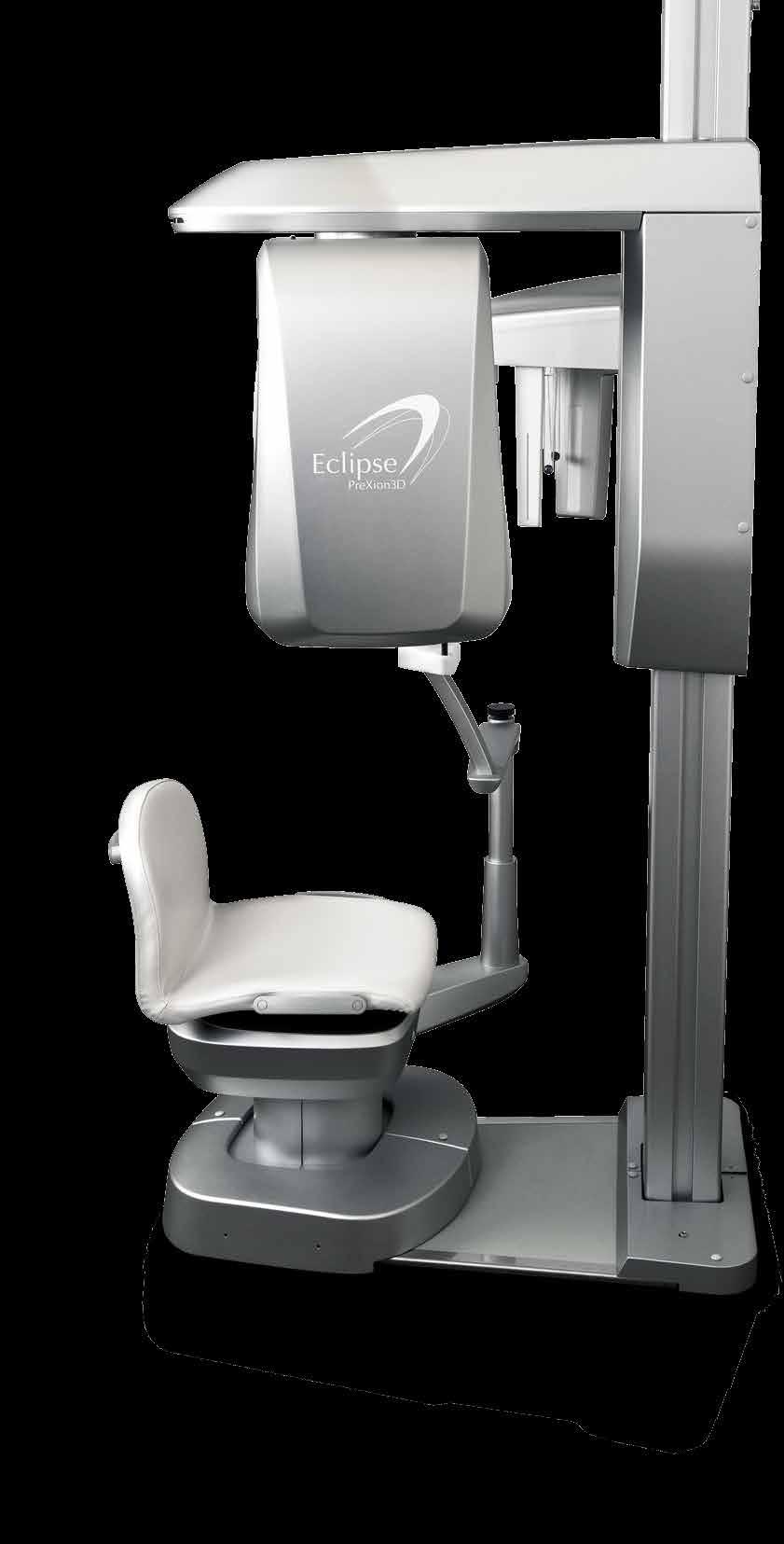
2 The Quality Leader in 3D Cone Beam CT The Complete 2-in-1 or 3-in-1 Multi-modality Solution PreXion, with over 15 years of innovation in the medical and dental fields, introduces the PreXion3D Eclipse. The PreXion 3D Eclipse a true 2-in-1 or 3-in-1 multi-modality solution combines the superb image quality that you have come to expect from the PreXion3D Elite with enhanced versatility all at an affordable cost that will benefit you and your patients. Ideal for dental offices that want to perform in-office surgical implant procedures, the PreXion3D Eclipse also excels at 3D imaging for diagnosis in general dentistry, periodontics, orthodontics, and endodontics, along with planning in oral-maxillofacial surgery. Included with your PreXion3D Eclipse is the PreXion3D Viewer Software a breakthrough program that contains easy-to-use modules for evaluating dedicated 2D Panoramic, 2D Cephalometric, and 3D images. Utilizing fully integrated software with state-of-the-art imaging technology, Prexion provides the clinician with the most accurate assessment of bone and surrounding anatomy. The PreXion3D Eclipse also comes with the PreXion data server solution, which allows you to seamlessly integrate our technology into your office no additional hardware required, no costly network upgrades, and no hidden charges.


3 A New Dimension in Imaging Highest Resolution A smaller focal spot increases resolution. The PreXion3D Eclipse uses a 0.2 mm focal spot smallest in its class. A Powerful Visual Aid for You and Your Patients The PreXion3D Eclipse boasts the smallest focal spot size in the industry providing clinicians with the highest quality image without distortion. 3D images help visualize dental anatomy via multiplanar and double oblique views, including the option for dynamic rotational slices creating excellent tools for diagnosing clinical criteria with higher levels of accuracy. In addition, the stunning images generated by the PreXion3D Eclipse provide a powerful visual aid for educating patients on available treatments. Sharp 0.2 mm PreXion3D Comparative Superiority Blurred 0.5 mm average Traditional panoramic images cannot evaluate the full buccal-lingual width. With 3D images generated by the PreXion3D Eclipse, clinicians can evaluate cases from different angles and planes, resulting in more accurate diagnoses. 3D Images PreXion3D Eclipse Images PreXion3D Eclipse s 3D image Traditional Panoramic Image Solely using 2D Panoramic radiography for diagnosis and treatment can increase risk and affect treatment outcomes.


4 Get the Right Picture S imultaneous Panoramic Image The PreXion3D Eclipse Offers Two Fields of View (FOV) and Four Scanning Modes for Precise Diagnoses Panoramic images are automatically generated with each scan. No separate panoramic scan is required. PreXion leads the market in the quantity and quality of projected views per scan, resulting in Flexible Capabilities with Dedicated Pan and Ceph clearer, higher quality images for your diagnosis. All Prexion3D Eclipse scan sizes are safe for Easily capture dedicated 2D panoramic for post-op or general exams. Optional Ceph is great for Orthodontics. general practitioners to read and do not require licensed radiology reports. Panoramic Modes Mode Standard Light Mode FOV 81 mm x 75 mm Scan Time 8.7 secs Scan Time 16 secs edicated 2D D Panoramic Image Projected Views 269 High Definition Mode FOV 81 mm x 75 mm Cephalometric Modes Scan Time 8.7 secs Mode PA Scan Time 12 secs, 15 secs Picture Size 7.99 in x in / 203 mm x 254 mm LA 10 secs, 12 secs in x in / 305 mm x 254 mm 8 secs 8.00 in x in / mm x 254 mm Projected Views 269 Carpus Ultra High Definition Mode Cephalometric Image FOV 81 mm x 75 mm Scan Time 17.4 secs Projected Views 538 Wide Mode FOV 113 mm x 72 mm Scan Time 9.1 secs x 2 times Projected Views 564 Prexion Diamond Warranty and Direct Sales & Support PreXion s direct sales and support offers a superior advantage over dealerships. Covers 100% parts and labor with no additional fees or deductibles Includes 1 full day on-site training Includes software updates and phone support Includes exclusive access to Prexion Online Help Center


5 Superior Clinical Results Implant PreXion3D can provide you with the highest quality images for both pre- and post-op assessment. Accurate evaluation of the implant position and the bone thickness can be quickly & accurately evaluated. DICOM data can be transferred for use in surgical guide fabrications and accuracy in the placements of dental implants. The PreXion3D Eclipse Provides Clinicians with the Highest Quality Images for Exceptional Clinical Results Endo Eclipse scans aid in both pre-endo treatments plus evaluation of post re-treatments of endodontic failures. Information gathered from the cone beam scans can accurately determine if a re-treatment will be successful or if the tooth requires an extraction. TMJ The clarity and crisp detail of PreXion s 3D imaging provides accurate assessment of the position and condition of the condyle. Measurement tools provide exact distances between the condyle and the bony fossa giving precise detail in aiding the design and/or positioning of corrective appliance. Perio The combination of 3D and multi-slice cone beam CT images provides a clear understanding of the bony defects within relation to surrounding anatomy. In addition to helping the dentist plan for treatment options, 3D CBCT imaging is an excellent tool for educating patients about their own perio issues and bone degeneration resulting in better treatment acceptance.
 Molar Both upper and lower molars are more accurately evaluated for position, number of root")


6 Superior Clinical Results (cont.) Molar Both upper and lower molars are more accurately evaluated for position, number of root canals, and the distance near the inferior alveolar canal if extractions are planned. Excellent for impacted third molar studies to distal third roots. Rotational and multi-slice features further enhance the accuracy of evaluations, especially with deep or curved roots. In addition, the Wide mode feature allows dentists to view the entire distal 3 rd molar root. Maxillary Sinus The sinus floor is included in a standard scan. Accurate diagnosis and evaluations of the sinus, including the nasal septum, is an added benefit. This information is also crucial when planning sinus lifts for visualizing depth, volume, landmarks or septae to consider during the planning phase. Conditions of sinusitis may reveal mucus or thick buildup in the sinus cavity and can be evaluated in both 3D and MPR images. Bone Augmentation Pre-surgical planning is essential for successful bone grafts. Accurate measurements for graft materials, membranes or surgical meshes can be accomplished prior to actual surgery. An additional benefit is the ability to accurately gauge the amount of graft material needed resulting in cost savings. Bone density can also be measured and determined prior to surgery. Root Abscess The cause of root abscess such as endodontic failure and implants can be accurately evaluated using the high quality images of the PreXion3D system, and assist in exact treatment planning options.


7 Breakthrough Software Fully Integrated PreXion3D Viewer Software Seamless Workflow Scan Once, View Many with the PreXion Data Server Solution The Prexion3D Viewer software, included with the PreXion3D Eclipse at no additional charge, contains easy-touse modules for evaluating dedicated 2D Panoramic, 2D Cephalometric, and 3D images. The Prexion3D Viewer software provides the clinician with the most accurate assessment of the bone and surrounding anatomy while making 1:1 exact measurements. This assures optimal implant placement without the superimposition of tissue or projection distortion compared to conventional panoramic systems. The Prexion3D Viewer images reconstruct to DICOM 3.0 format and are compatible with major 3 rd party surgical planning software systems. Prexion s scan once, view many solution allows immediate access to 3D scans from any computer or laptop on the office network without any hardware upgrades. The Prexion Data Server solution is unique because it won t slow down networks caused from downloading and loading scans from different rooms. PreXion3D Eclipse Scan Prexion3D Viewer Curved Planar Reconstruction (CPR) Mode Create custom troughs around arches and view cross-sections trans-axially. Within 30 seconds, patient scans are instantly accessible from several rooms at the same time. Prexion3D Movie Maker Easily create compelling 3D animated movies for patients and increase understanding and case acceptance. Consult Insurance Biller Doctor Office Laptop Implant Treatment Coordinator Operatory Multi-Data View Easily compare pre-operative scans with post operative scans, synchronize them and quantify measurable results on a single screen, slice-by-slice. PrexViewer CD Make patient discs with free 3D viewer. Implant Planning Easily locate and mark the Inferior Alveolar Nerve, measure 1:1 ratios, place and move implants in 3D.


8 Compact Styling Specifications Device Type Ergonomic design with a small footprint CBCT + Panorex + Ceph (Optional) FOV (Diameter by Height) The PreXion3D Eclipse offers a smart, stylish design that is both ergonomically-friendly to the patient and compact in size. The small footprint of the PreXion3D Eclipse allows it to fit in almost any location previously occupied by a panoramic x-ray system with no office expansion typically necessary. Light Mode 3.18 in x 2.95 in / 81 mm x 75 mm High Definition Mode 3.18 in x 2.95 in / 81 mm x 75 mm Ultra High Definition Mode 3.18 in x 2.95 in / 81 mm x 75 mm Wide Mode 4.45 in x 2.83 in / 113 mm x 72 mm Light Mode: 8.7 secs, High Definition mode: 8.7 secs CT Scan Mode Ultra High Definition mode: 17.4 secs, Wide mode: 9.1 secs x 2 Panorex Standard mode: 16 secs Ceph LA, PA, Carpus: 8, 10, 12, 15 secs Ergonomic Design X-ray Output 90 kv (50 to 90 kv in Ceph scan) 4 ma (1.0 to 4.0 ma in Ceph scan) *CT has 2.6 ma and 4 ma mode The PreXion3D Eclipse has an ergonomically CBCT Sensor FPD, 14 bits designed chair that results in a more relaxed Focal Spot 0.2 mm (200 µm) and stable patient. Power 120 V, Single phase, 1.5 kva Patient Position Seated Included Computers Two Computers: Console (1) and Viewer (1) (Network Client is available) Dimensions Unit : inches 5.71 vertical distance of the chinrest arm Live Video Camera with Scout View Easily scan a desired region of interest ~52.09 vertical distance of the chair : horizontal distance of the chair ~ horizontal distance of the chair ~ horizontal distance of the chair ~51.73 to fit in the same space as the panoramic x-ray system it is replacing horizontal distance of the chair vertical distance of the chair : The size of the PreXion3D Eclipse allows it 5.71 vertical distance of the chinrest arm Compact Size
9 USA PreXion, Inc. 411 Borel Avenue, Suite 550, San Mateo CA 94403, USA Tel: Fax: Japan XTrillion Kanda Suda-Cho, Chiyoda-ku Tokyo, Japan Tel: Fax: DC PU
CS 9300 Family. The power of flexibility
 CS 9300 Family The power of flexibility The new CS 9300 digital imaging system from Carestream Dental take the guesswork out of examinations The all-in-one CS 9300 is the most versatile multimodality imaging
Low Dose Excellent Image Quality Rapid Reconstruction
 Low Dose Excellent Image Quality Rapid Reconstruction Efficient 3 in 1 Dental X-ray System CBCT > Precise 3-D Anatomical structures - Accurate diagnosis for doctors - Safe implant for patients > Significant
Head to new heights with your imaging SCANORA 3D
 SCANORA 3D Head to new heights with your imaging Benefits at a glance The solution for dentomaxillofacial and ENT imaging Easy Patient seated for added stability during exposure. Clear, self-explinatory
True Low Dose. Exact time to display image on screen may vary upon computer and network configuration.
 RAYSCAN ALPHA PLUS True Low Dose Cone Beam CT Industry Leading Resolution High resolution images provide all the clinical information needed while keeping radiation exposure low. Endodontics - Smallest
X X X. GXS-700 Direct USB Digital Intraoral Sensors. Buy a Sensor Combo and a Digital Pan Unit, Receive $600 Off! GO.BENCO benco.
 8 0 0. G O. B E N C O b e n c o. c o m GS-700 Direct USB Digital Intraoral Sensors Designed to make migrating from film, or upgrading an existing digital system, easier than ever High quality image capture
ENGLISH. Cefla s.c. - Via Selice Provinciale 23/a, Imola - Italy Tel
 09-2017 NVGEGB161S00 According to the regulations in force, some products and/or features may have different availability and characteristics in areas outside of the European Union. Please contact your
3Shape X1 Scanning redefined
 3Shape X1 Scanning redefined Why choose the X1 Give your patients a great experience No head fixation and sleek design create a comfortable scanning experience for your patient High image quality low dose
PAN CEPH 3D CONE BEAM
 PAN CEPH 3D CONE BEAM 2D - 3D panoramic units PANORAMIC CEPHALOMETRIC 3D CONE BEAM IMAGING I-MAX TOUCH Tactile & naturally intuitive panoramic imaging Discover the simplicity and efficiency this unit can
3Shape X1 Scanning redefined
 3Shape X1 Scanning redefined Why choose the X1 Give your patients a great experience No head fixation and sleek design create a comfortable scanning experience for your patient High image quality low dose
DIAGNOSTIC IMAGING. OPTIMIZED.
 ABOUT LED DENTAL SEE THE DIFFERENCE Using our years of business insight and clinical experience as a foundation, LED Dental takes the uncertainty out of your imaging purchase decision. We offer our clients
STELLARIS 3D 4 IN 1 CBCT SOLUTION FOR ADVANCED DIAGNOSTICS
 STELLARIS 3D 4 IN CBCT SOLUTION FOR ADVANCED DIAGNOSTICS 3 STELLARIS 3D 4 IN CBCT SOLUTION FOR ADVANCED DIAGNOSTICS Stellaris 3D is a complete and compact, fully upgradeable 3D CBCT for a patient, Panoramic
THE WAIT IS OVER CS D. 3D imaging is now available for everyone
 THE WAIT IS OVER CS 8100 3D 3D imaging is now available for everyone COMPLEXITY IS NO LONGER THE STANDARD NOW THERE ARE MANY REASONS TO MOVE TO 2D/3D IMAGING Now it s possible to experience nothing but
CS 8100 FAMILY / CS D FAMILY / CS 9300 FAMILY EXTRAORAL SOLUTIONS EXTRAORDINARY POSSIBILITIES
 CS 8100 FAMILY / CS 8100 3D FAMILY / CS 9300 FAMILY EXTRAORAL SOLUTIONS EXTRAORDINARY POSSIBILITIES A SOLUTION FOR EVERY CLINIC AND ANY CONSULTATION The CS 8100 does more than my old machine and is half
XPan 3D Plus. FONA Every dental solution you need. Advanced dental technology. Headquarters THE ULTIMATE DIAGNOSTIC SOLUTION DIGITAL DENTISTRY
 FONA Every dental solution you need Through decades of experience and deep understanding of the dental profession, we deliver complete, reliable and accessible solutions.regardless of country or specialisation,
Digital Imaging from a new perspective
 TREATMENT CENTRES HANDPIECES HYGIENE SYSTEMS X-RAY SYSTEMS CEREC TREATMENT CENTRES HANDPIECES HYGIENE SYSTEMS X-RAY SYSTEMS CEREC SIRONA CREATING AND MAINTAINING VALUE. You are right to expect a great
Flexible Easy Competitive. SCANORA 3Dx - The in-office large field-of-view Cone Beam CT system for Head and Neck imaging
 Flexible Easy Competitive SCANORA 3Dx - The in-office large field-of-view Cone Beam CT system for Head and Neck imaging SCANORA 3Dx. The solution. SCANORA 3Dx makes advanced 3D imaging easy in the head
3D Accuitomo XYZ Slice View Tomograph. Super High-Resolution Images of Region of Interest
 3D Accuitomo XYZ Slice View Tomograph. Super High-Resolution Images of Region of Interest Thinking ahead. Focused on life. 2 Cone Beam X-Ray CT Imaging X-Ray Tube Imaging Intensifier Imaging Volume Voxel
Easy operation. Numerous diagnostic options. X-rays you can rely on: the ORTHOPHOS XG device family. All ORTHOPHOS XG 3Dready programs at a glance.
 CAD /CAM Systems Instruments Hygiene Systems Treatment Centers Imaging Systems Subject to technical changes and errors in the text, Order No. A91100-M47-B346-01-7600, Printed in Germany, Dispo-Nr. 04602,
Dental Line. 3D digital panoramic system. radiology ahead
 Dental Line 3D digital panoramic system radiology ahead new generation 3D digital panoramic unit 3D imaging s value available for anyone Following the incredible success of the innovative digital panoramic
Cone Beam 3D Imaging
 Cone Beam 3D Imaging NewTom Sets the Standard in 3D Maxillofacial Imaging Cone Beam 3D Imaging The Global Market Leader The Inventors n of Cone Beam 3D In 1996, QR srl developed the first generation of
3D CBCT Case Study Daniel McEowen, DDS Hagerstown, Maryland
 3D CBCT Report November 2011 Issue 3D CBCT Case Study Daniel McEowen, DDS Hagerstown, Maryland Failed endodontics is a common problem in dental practice today. More and more doctors and patients are exploring
2D AND 3D/2D WALL-MOUNTED PANORAMIC UNITS
 2D AND 3D/2D WALL-MOUNTED PANORAMIC UNITS KEEP YOUR CLINIC ONE STEP AHEAD! Wall-mounted concept: zero foot print 62kg - the lightest unit on the market Face to face positioning High Definition The fruit
ENGLISH. Distributed by: QR srl - Via Silvestrini, Verona Italy Tel
 GiANO - R15.1 - EN ENGLISH Distributed by: QR srl - Via Silvestrini, 20-37135 Verona Italy Tel. +39 045 8202727-045 583500 info@newtom.it www.newtom.it Manufacturer: CEFLA S.C. - CEFLA DENTAL GROUP Via
OP 3D Vision The upgradable 3D X-ray system for the strictest demands.
 OP 3D Vision The upgradable 3D X-ray system for the strictest demands. The solution for every task: KaVo OP 3D Vision. Regardless of which dental query you may have, the KaVo ORTHOPANTOMOGRAPH OP 3D Vision
VGi - R EN ENGLISH. QR srl - Via Silvestrini, Verona Italy Tel
 VGi - R15.1 - EN ENGLISH QR srl - Via Silvestrini, 20-37135 Verona Italy Tel. +39 045 8202727-045 583500 info@newtom.it www.newtom.it FIRST IN CONE BEAM, ACCURATE IN RESULTS. 360 degree imaging, reduced
ENGLISH. Cone Beam 3D Imaging
 ENGLISH Cone Beam 3D Imaging FIRST USER OF CONE BEAM IN DENTAL FIELD QR s.r.l. is the name that stands behind NewTom Cone Beam 3D imaging units and the creator of Cone Beam technology for the dental field.
3D Panoramic Cephalometric. Innovation, in reach. KODAK 9000 Extraoral Imaging System
 3D Panoramic Cephalometric Innovation, in reach 9000 KODAK 9000 Extraoral Imaging System Cephalometric Innovation made simple Innovation made simple We believe in innovation. We always have. In fact, our
I AM DEMANDING Type CMOS Flat Panel CMOS CMOS ø 40 x 40 mm, ø 60 x 60 mm, ø 80 x 80 mm, ø 110 x 80 mm
 TECHNICAL SPECIFICATIONS 1168 1501 1978 1237 1551-2351 Ø 1090 PANORAMIC CBCT CEPHALOMETRIC X-RAY SOURCE Tube type High frequency DC generator 2.8 mmal / 85 kv 7.0 mmal / 90 kv 2.8 mmal / 85 kv Operation
Profound understanding of anatomy
 ENGLISH Profound understanding of anatomy The unique Planmeca ProMax 3D product family offers equipment for all maxillofacial imaging. All volumes sizes from the smallest special cases to whole head images
Versatility And Expandability In One Panoramic.
 Orthoralix 9200 / 9200 DDE Versatility And Expandability In One Panoramic. Panoramic X-ray Systems Intraoral X-ray Systems Digital Intraoral Sensors Digital X-ray Phosphor Plates Intraoral Cameras Imaging
T h e D e n t a l C o m p a n y FROM DIAGNOSTIC SCAN TO SURGERY, WE SHAPE THE FUTURE OF DENTISTRY.
 T h e D e n t a l C o m p a n y FROM DIAGNOSTIC SCAN TO SURGERY, WE SHAPE THE FUTURE OF DENTISTRY. SIDEXIS SOFTWARE ORTHOPHOS SL D/D SEAMLESS THE NEW STANDARD IN CLINICAL DIAGNOSIS AND PATIENT COMMUNICATION
Profound understanding of anatomy
 ENGLISH Profound understanding of anatomy Planmeca ProMax 3D, the intelligent and multipurpose X-ray unit, is designed to obtain complete information on patient anatomy in the minutest detail. The unit
3D-MODEL CUSTOM-MADE MODELS SEGMENTATION AND PRODUCTION SERVICE OF BONE MODELS WITH HIGHEST 3D PRINTING RESOLUTION
 CUSTOM-MADE MODELS 3D-MODEL SEGMENTATION AND PRODUCTION SERVICE OF BONE MODELS WITH HIGHEST 3D PRINTING RESOLUTION FOLLOW US ON CUSTOM-MADE MODELS 3D-MODEL From a CT or CBCT scan, 3D-model service provides
CS Introducing the CS 1600 Intraoral Camera Revolutionary technology. Superior workflow.
 CS 1600 Introducing the CS 1600 Intraoral Camera Revolutionary technology. Superior workflow. Early caries identfication for healthier patients Earliest caries identification Early detection is the most
Profound understanding of anatomy
 ENGLISH Profound understanding of anatomy The unique Planmeca ProMax 3D product family offers equipment for all maxillofacial imaging. All volume sizes from the smallest special cases to whole head images
VistaVox S 3D from Dürr Dental
 VistaVox S 3D from Dürr Dental 3D and 2D X-ray images with exceptional image quality COMPRESSED AIR SUCTION IMAGING DENTAL CARE HYGIENE Taking diagnostics to the next level VistaVox S combines diagnostic
The Optimum Choice for Implantologist
 The Optimum Choice for Implantologist What is essential for your practice? What s the best way to choose a 3D X-ray machine for implant treatment planning? 02 Doctor says.. There are diagnostic limitations
- RCS Paris B
 Technical specifications PANORAMIC CBCT CEPHALOMETRIC X-ray source Tube type High frequency DC generator Total filtration >2.5 mm Al @ 90 kv Mode of operation Continuous Pulsed Continuous Tube voltage
clinical articles management advice practice profiles technology reviews Implant PRACTICE US Volume 3 No 2
 clinical articles management advice practice profiles technology reviews Implant PRACTICE US Volume 3 No 2 Sirona Sirona tailors solutions to the needs of the different markets within dentistry to ensure
Veraviewepocs 3D R100 & F40
 Veraviewepocs 3D R100 & F40 Innovative 3D Reuleaux Full Arch FOV Thinking ahead. Focused on life. Veraviewepocs 3D R100 A New Frontier in X-ray Diagnostics Veraviewepocs 3D R100 has changed the shape of
VATECH IMAGING SYSTEMS
 VATECH IMAGING SYSTEMS 2 About Vatech America 3 Vatech Assurance 4 PaX-i 6 PaX-i Insight 12 i3d Smart 16 PaX-i3D 20 Green CT 26 Green CT 2 32 i3d Premium 36 Ez3D-i 42 EzDent-i 45 EzRay Air Wall 46 HD Sensor
ENGLISH VGi - R ENG
 ENGLISH First in Cone Beam, Accurate in Results Cone Beam 3D Imaging First User of Cone Beam in Dental Field QR s.r.l. is the name that stands behind NewTom Cone Beam 3D imaging units and we were the creators
ADVANCED 3D IMAGING. CEFLA s.c. Via Selice Provinciale 23/a Imola Italy t newtom.
 CEFLA s.c. Via Selice Provinciale 23/a 40026 Imola Italy t. +39 045 8202727 045 583500 info@newtom.it newtom.it 05/2018 NVGEGB181S00 According to the standards in force, in extra-eu areas the availability
English. Perfect Vision
 English Perfect Vision Everything becomes clearer and simpler with a big F.O.V. The complete dentomaxillofacial volume ready for your diagnosis One scan provides you with an incredible amount of information
CASE REPORT. CBCT-Assisted Treatment of the Failing Long Span Bridge with Staged and Immediate Load Implant Restoration
 Computer Aided Implantology Academy Newsletter - Newsletter 20 - July 2009 CASE REPORT CBCT-Assisted Treatment of the Failing Long Span Bridge with Staged and Immediate Load Implant Restoration Case Report
ENGLISH. Cefla s.c. - Via Selice Provinciale 23/a, Imola - Italy Tel
 11-2016 N5GGB161S00 According to the regulations in force, some products and/or features may have different availability and characteristics in areas outside of the European Union. Please contact your
Xelis Dental - What New in
 Xelis Dental - What New in 1.0.6.1 Clinical Needs Why Xelis-Dental? Panorama Cephalography Intra-oral Digital Camera Traditional imaging systems - 2-Dimension view Incorrect anatomical information - Distortion
ORTHOPANTOMOGRAPH OP200 D ORTHOCEPH OC200 D. True dynamo leading through the decades.
 ORTHOPANTOMOGRAPH OP200 D ORTHOCEPH OC200 D True dynamo leading through the decades. 1 You can t dublicate the legacy. 1946 Professor Y.V. Paatero publishes his first paper on Panoramic Tomography. 1951
The Application of Cone Beam CT Image Analysis for the Mandibular Ramus Bone Harvesting
 44 The Application of Cone Beam CT Image Analysis for the Mandibular Ramus Bone Harvesting LivingWell Institute of Dental Research Lee, Jang-yeol, Youn, Pil-sang, Kim, Hyoun-chull, Lee Sang-chull Ⅰ. Introduction
User Guide for Dental and Maxillofacial Cone Beam Computed Tomography (CBCT)
") User Guide for Dental and Maxillofacial Cone Beam Computed Tomography (CBCT) Poster No.: C-0756 Congress: ECR 2014 Type: Educational Exhibit Authors: J. Ukkonen, J. Asp; Helsinki/FI Keywords: Education
User Guide for Dental and Maxillofacial Cone Beam Computed Tomography (CBCT) Poster No.: C-0756 Congress: ECR 2014 Type: Educational Exhibit Authors: J. Ukkonen, J. Asp; Helsinki/FI Keywords: Education
Dental Cone Beam CT. What is Dental Cone Beam CT?
 Scan for mobile link. Dental Cone Beam CT Dental cone beam computed tomography (CT) is a special type of x-ray equipment used when regular dental or facial x-rays are not sufficient. Your doctor may use
The Power Of Choice Pan. Ceph. 3D. Your Imaging Future Starts Today.
 NEW from Gendex! The Power Of Choice Pan. Ceph. 3D. Your Imaging Future Starts Today. Cone Beam 3D Imaging Systems Panoramic X-ray Systems Intraoral X-ray Systems Digital Intraoral Sensors Digital X-ray
Full mouth rehabilitation with digital workflow
 Jung-plant dental office Dr. Jae-min, Lee D.D.S. Full mouth rehabilitation with digital workflow Solutions featured: 3Shape TRIOS 3Shape Dental System 3Shape Implant Studio Case information On first visit,
fast accurate safe See the full picture: add a 3rd dimension to your patient evaluation to diagnose more effectively. BENEFITS OF NEWTOM 3G 6" 9" 12"
 See the full picture: add a 3rd dimension to your patient evaluation to diagnose more effectively. Examination Effective Dose Equivalent (ICRP tissue weights 2005) Panoramic Dose (ICRP tissue weights 2005)
Immediate loading and implant surgery with digital workflow
 Mirero Dental Clinic Dr. Jaemin Lee Immediate loading and implant surgery with digital workflow Solutions featured: 3Shape TRIOS 3Shape Dental System 3Shape Implant Studio Case information Patient, 32-year
CS 3600 / CS 3600 ACCESS A SMARTER WAY TO SCAN INTRAORAL SCANNING
 CS 3600 / CS 3600 ACCESS A SMARTER WAY TO SCAN INTRAORAL SCANNING R R HARNESS THE POWER OF INTRAORAL SCANNING AWARDS AND RECOGNITION CLINICIANS REPORT Excellent in Rapid Acquisition and Margin Detail Capture;
THE USE OF KEYSTONE EASYGUIDE CT SCANNING SOFTWARE FOR DIAGNOSIS, DIRECTION AND DEPTH DETERMINATION
 CT DIAGNOSTICS IN 3D IMPLANT TREATMENT PLANNING THE USE OF KEYSTONE EASYGUIDE CT SCANNING SOFTWARE FOR DIAGNOSIS, DIRECTION AND DEPTH DETERMINATION Timothy Kosinski, DDS, MAGD Assistant Clinical Professor
ENGLISH. Advanced tools for orthodontics
 ENGLISH Advanced tools for orthodontics A complete solution for orthodontics One unit one software Panoramic Cephalometric CBCT image 3D photo Planmeca ProMax unit 3D model scan Planmeca offers a complete
5G XL - R EN ENGLISH
 5G XL - R15.0 - EN ENGLISH Sede legale ed amministrativa - Headquarters QR srl - Via Selice Provinciale, 23/a - 40026 Imola - Bo (Italy) Stabilimento - Plant Via Fermi, 40-37136 Verona (Italy) Tel. +39
Keeping you focused on what you do best.
 Keeping you focused on what you do best. The right diagnosis on all patients with industry-leading depth and detail. Picture the perfect ultrasound system. Now take a closer look at Toshiba s Aplio 500
CS Introducing the CS 1600 Intraoral Camera Revolutionary caries detection aid. FIRE technology. Superior patient care.
 CS 1600 Introducing the CS 1600 Intraoral Camera Revolutionary caries detection aid. FIRE technology. Superior patient care. An aid in the process of early caries detection Caries discovered in early stages
Powered by. Dedicated MRI
 Powered by Dedicated MRI Provides the latest software and hardware upgrade configuration powered by exp technology: boosting productivity, increasing image quality, and adding new acquisition techniques.
AWARD-WINNING CONE BEAM 3D DENTAL IMAGING
 AWARD-WINNING CONE BEAM 3D DENTAL IMAGING Dedicated to Advancing Dental Treatment A COMPLETE 3D TREATMENT SOLUTION Your dental practice is unique that s why you need a flexible solution that works with
NewTom 5G XL EXTRA.VISION
 CEFLA s.c. Via Selice Provinciale 23/a 40026 Imola Italy t. +39 045 8202727 045 583500 info@newtom.it newtom.it 06/2018 N5GXGB181S00 According to the standards in force, in extra-eu areas the availability
Planmeca ProMax 3D s Planmeca ProMax 3D ENGLISH
 Planmeca ProMax 3D s Planmeca ProMax 3D ENGLISH Genuine all-in-one unit Planmeca ProMax 3D s and Planmeca ProMax 3D units are designed to obtain complete information on patient anatomy in the minutest
Monday Morning Pearls of Practice by Bobby Baig
 Dec 19, 2016 Monday Morning Pearls of Practice by Bobby Baig baig@buildyoursmile.com Prosthodontic Associates 2300 Yonge St, suite 905 Toronto, M4P1E4 www.buildyoursmile.com CBCT and Implant Dentistry:
3D Cone beam CT & Digital Radiography Dedicated to Otorhinolaryngology
 3D Cone beam CT & Digital Radiography Dedicated to Otorhinolaryngology Multi-functional imaging solution3 RAYSCAN m is an unique 2-in-1 imaging solution, combining Cone Beam CT and Digital Radiography,
Varian Acuity BrachyTherapy Suite One Room Integrated Image-Guided Brachytherapy
 Varian Acuity BrachyTherapy Suite One Room Integrated Image-Guided Brachytherapy The Acuity BrachyTherapy Suite Integrating Imaging, Planning, and Treatment in a Single Room Each component draws on the
CT SCAN PROTOCOL. Shoulder
 CT SCAN PROTOCOL Shoulder Purpose and Summary CT images made with this protocol are used to provide the orthopedic surgeon with a detailed 3D anatomical reconstruction of the patient s scapula and proximal
ORTHOPANTOMOGRAPH OP 3D Pro A platform for changing needs
 ORTHOPANTOMOGRAPH OP 3D Pro A platform for changing needs ORTHOPANTOMOGRAPH OP 3D Pro OP 3D Pro is the most comprehensive 3-in-1 platform designed for today and tomorrow, covering the entire maxillofacial
Solutions for Large Group Practices
 Solutions for Large Group Practices 2D imaging im aging 3D imaging 3D im aging Infection control ion c CAD/CAM solutions D/C AM solutions Information and monitoring l un ts Dental units One easy-to-use
Clinical details: Details of scan: CONE BEAM CT REPORT: Name: H. B. Gender: Reason for referral: Referred by:
 Name: H. B. Gender: Male DOB: 11/12/1950 Age: 64 Date taken: 16/11/2015 Date reported: 19/11/2015 Clinical details: Reason for referral: Referred by: Investigate symptoms related to left TMJ. Reconstructed
ORTHOPANTOMOGRAPH OP300. A platform for changing needs.
 ORTHOPANTOMOGRAPH OP300 A platform for changing needs. 1 Panoramic with cephalometric digital image Xray machine For me, peace of mind means a patient trusting in my care, time after time Leading the way
Dental Implants: A Predictable Solution for Tooth Loss. Reena Talwar, DDS PhD FRCD(C) Oral & Maxillofacial Surgeon Associate Clinical Professor
 Oral & Maxillofacial Surgeon Associate Clinical Professor") Dental Implants: A Predictable Solution for Tooth Loss Reena Talwar, DDS PhD FRCD(C) Oral & Maxillofacial Surgeon Associate Clinical Professor What are Dental Implants? Titanium posts used to replace missing
Dental Implants: A Predictable Solution for Tooth Loss Reena Talwar, DDS PhD FRCD(C) Oral & Maxillofacial Surgeon Associate Clinical Professor What are Dental Implants? Titanium posts used to replace missing
In-Office Cone Beam Computerized Tomography: Technology Review and Clinical Examples Michael Tischler, DDS
 Page 1 of 8 Issue Date: June 2008, Posted On: 6/26/2008 In-Office Cone Beam Computerized Tomography: Technology Review and Clinical Examples Michael Tischler, DDS Electronic Medical Record Research EMR
Using cone beam technology in orthodontics
 PRACTICE US Using cone beam technology in orthodontics Continuing education earn 2 ce credits! Edward Lin explains the benefits of adding cone beam technology to the orthodontic armamentaria Since cone
For true visualisation
 ENGLISH For true visualisation Planmeca ProModel is a patient-specific physical model for high-end maxillofacial operations and dental surgery. By reproducing the anatomy of the patient in real-size, Planmeca
SOREDEX. Pride.Passion.Performance.
 SOREDEX Pride.Passion.Performance. A full-service imaging company Soredex offers first-class imaging solutions by focusing on easy and fast workflow, contemporary design and innovative features. We are
D3D CBCT. See more Do More
 D3D CBCT See more Do More BIOLASE DaVinci Imaging D3D For capturing superior 3D image acquisitions with the lowest minimal dose for patient safety The BIOLASE DaVinci Imaging D3D has one of the lowest
Agenda: Dental Cone Beam Imaging
 Cone Beam Imaging Agenda: Dental Cone Beam Imaging *Definition and Functionality *Usage and diagnostics benefits *Comparative radiation information *Federal regulatory responsibilities: manufacturing *State
Extraoral Radiology October 10th, 2008
 Extraoral Radiology October 10th, 2008 Steven R. Singer, DDS srs2@columbia.edu 212.305.5674 November 8 th, 1895 Extraoral Projections Images can be produced in the dental office X-ray source can be Intraoral
The 2D x-ray family. dentsplysirona.com
 The 2D x-ray family dentsplysirona.com 02 I 03 As versatile as practice life The Orthophos 2D X-ray family offers the right solution for every practice. From entry into digital radiography to the perfect
IMAGE-GUIDED RADIATION THERAPY
 IMAGE-GUIDED RADIATION THERAPY Your Single Source Oncology Solutions Provider Plan. Target. Treat. At Best NOMOS, we design products and solutions that help medical professionals treat a variety of cancers.
Veraviewepocs 3D. F40 and R100 with innovative 3D Reuleaux Full Arch FOV. Thinking ahead. Focused on life.
 Veraviewepocs 3D F40 and R100 with innovative 3D Reuleaux Full Arch FOV Thinking ahead. Focused on life. Veraviewepocs 3D R100 A New Frontier in X-ray Diagnostics Veraviewepocs 3D R100 has changed the
Extraoral Imaging. Chapter 42. Copyright 2018, Elsevier Inc. All Rights Reserved. 1
 Extraoral Imaging Chapter 42 Copyright 2018, Elsevier Inc. All Rights Reserved. 1 Learning Objectives Lesson 42.1: Panoramic Imaging 1. Pronounce, define, and spell the key terms. 2. Discuss panoramic
ENDODONTICS ENDODONTICS
 158 159 & Heads Smarter and Safer Cordless Endodontic Handpiece with Torque Control and Auto Reverse Miniature Head For Ni-Ti files (ø2.35) Push Button Chuck MP-F20R MP-F16R MP-F10R 20:1 / 16:1 / 10:1
Intuitive. Intelligent. Innovative. General Imaging
 Intuitive. Intelligent. Innovative. General Imaging CLARITY CONFIDENCE EASE OF USE 2 Complete flexibility, outstanding quality For robust performance you can rely on for a wide range of clinical tasks,
ENGLISH. Advanced tools for orthodontics
 ENGLISH Advanced tools for orthodontics A complete solution for orthodontics One unit one software Panoramic Cephalometric CBCT image 3D photo Planmeca ProMax unit 3D model scan Planmeca offers a complete
Dental Implant Planning using Cone Beam CT imaging: a pictorial guide.
 Dental Implant Planning using Cone Beam CT imaging: a pictorial guide. Poster No.: C-1970 Congress: ECR 2015 Type: Educational Exhibit Authors: S. R. Rice, G. Price, S. Morley, T. Beale; London/UK Keywords:
Diagnosis and Treatment of a Large Central Ossifying Fibroma of the Mandible. Clinical Case
 Clinical Case Diagnosis and Treatment of a Large Central Ossifying Fibroma of the Mandible A 36 year old African American Female presented to the Department of Oral and Maxillofacial Surgery Clinic at
The agony and ecstasy of buying cone beam technology Part 1: The Ecstasy
 Clinical The agony and ecstasy of buying cone beam technology Part 1: The Ecstasy Dale A. Miles 1 Abstract Background: Since arriving in North America in 2001, cone beam computed tomography (CBCT) has
Advantage beyond imaging
 Advantage beyond imaging www.ondemand3d.com About OnDemand3D TM OnDemand3D TM provides sophisticated 3D image processing tools and enables easy sharing of images between practices a nd imaging specialists
Digital Treatment Planning and Surgical Guide Fabrication
 Digital Treatment Planning and Surgical Guide Fabrication Improve Patient Care Increase Profitability Maximize Productivity Introduction As patient demand for dental implant treatment continues to grow,
NewTom GiANO HR PERFECT.VISION
 CEFLA s.c. Via Selice Provinciale 23/a 40026 Imola Italy t. +39 045 8202727 045 583500 info@newtom.it newtom.it 06/2018 NHRGB181S00 According to the standards in force, in extra-eu areas the availability
3D CBCT Case Study CBCT Scanning in Sinus Bump Cases Robert E. Walinchus, DMD DICOI
 3D CBCT Report August 2011 Issue 3D CBCT Case Study CBCT Scanning in Sinus Bump Cases Robert E. Walinchus, DMD DICOI With the advances in technology it has been the goal of dentists today to perform advanced
Dolphin 3D Imaging 11.7 beta
 Dolphin 3D Imaging 11.7 beta Dolphin 3D Surgery Dolphin s 3D Surgery has expanded to include even more features! This revolutionary treatment planning and presentation software module now includes a fully
International Journal of Current Medical and Pharmaceutical Research
 ISSN: 2395-6429 International Journal of Current Medical and Pharmaceutical Research Available Online at http://www.journalcmpr.com DOI: http://dx.doi.org/10.24327/23956429.ijcmpr20170169 RESEARCH ARTICLE
Simplant. Guided Surgery. delivering restorative driven implant treatment
 Simplant Guided Surgery delivering restorative driven implant treatment Simplant the key to unlocking digital potential As part of the Denstsply Sirona Implants Digital Solutions offering, Simplant delivers
Planmeca CBCT units. ENT imaging. Ear Nose Throat ENGLISH
 CBCT units ENT imaging Ear Nose Throat ENGLISH Versatile units for ENT imaging ProMax 3D Max, ProMax 3D Mid and ProMax 3D Plus are our versatile CBCT units for dental and full maxillofacial imaging. With
Galileos: The Quintessence of Modern Dentistry
 Imagine the Possibilities Galileos: The Quintessence of Modern Dentistry Neal S. Patel, DDS The new CEREC Bluecam and the CERECdoctors.com faculty members continue to push the envelope with unrivaled digital