ENGLISH. Cefla s.c. - Via Selice Provinciale 23/a, Imola - Italy Tel
|
|
|
- Abraham Weaver
- 5 years ago
- Views:
Transcription


1 N5GGB161S00 According to the regulations in force, some products and/or features may have different availability and characteristics in areas outside of the European Union. Please contact your local distributor. ENGLISH Cefla s.c. - Via Selice Provinciale 23/a, Imola - Italy Tel info@newtom.it


 was the very first Cone Beam equipment in the world, which was installed in 1996.")


2 FIRST IN CONE BEAM, ACCURATE IN RESULTS. 360 degree imaging, reduced image scatter or artifacts. Smallest possible focal spot and single flat panel create the clearest images. First user of Cone Beam in dental field Dedicated digital sensor and specific image algorithms provide a full range of information. THE GLOBAL MARKET LEADER. QR s.r.l. is the name that stands behind NewTom Cone Beam 3D imaging units and we were the creators of Cone Beam technology for the dental field. NewTom 9000 (also known as Maxiscan ) was the very first Cone Beam equipment in the world, which was installed in It is the forefather of NewTom product line and, in general, of the entire X-Ray units based on Cone Beam technology. QR s 20 plus years of experience and success in research, development, manufacturing and distribution of NewTom products affirms our commitment to excellence and quality. QR s.r.l. is based in Italy and all NewTom products are designed and manufactured by our group. Our products represent the Italian tradition of specialized manufacture and NewTom is known all over the world for its reliability, high standards and state-of-the-art technology. QR s.r.l. is a comprehensive and independently working company consisting of a research and development department (hardware and software), production and technical assembling division, technical support staff, customer service, sales and marketing department and management offices. Our national and international sales network relies on strong and long-term partnerships with all our dealers and representatives spread all over the world. NewTom s team-oriented staff is committed to provide not only the best product available on the market, but also excellent before and after- sales support, as a happy customer is the best advertisement.


 CB3D Cone Beam X-ray The result is a more accurate image without missing information and a considerably lower radiation exposure.")


3 CONE BEAM TECHNOLOGY Cone Beam 3D vs. MSCT X-ray source HIRES SCAN NewTom 5G allows to irradiate small portion of body, in order to see small anatomical details. This can be useful for proper implant assessment, because it requires the visualization of all aspects of the mandibular canal and other small parts, such as tooth roots, periodontal ligaments and any present lesions. Only 3D High Resolution imaging produces both the quality and the quantity of details necessary to accurately view those small details. Accuracy is one of the most important factors, together with dose reduction, also for analyzing middle and inner ear pathologies. NewTom has been recognized as an important diagnostic tool for most of ear pathologies. Multiple Fields of View The scanner s FOV determines how much of the patient s Fan Beam X-ray MSCT MSCT uses a narrow fan beam that rotates around the patient acquiring thin axial slices with each revolution. In order to create a section of anatomy, many rotations must be done. During these repeated rotations, traditional CT emits a high radiation dose, but it leaves a gap of information between each rotation. Therefore software must stitch together the images and calculate what is missing. Cone Beam 3D imaging uses a cone-shaped beam to acquire the entire image in a scan using only one rotation. FP (Flat Panel Detector) CB3D Cone Beam X-ray The result is a more accurate image without missing information and a considerably lower radiation exposure. The American Academy of Oral and Maxillofacial Radiology (AAOMR) prescribes the use of Cone Beam 3D imaging when evaluating periodontal, implant, and oral/maxillofacial surgery patients. One NewTom scan obtains a complete dentomaxillofacial image in a single database of digital information. Various views of the information in 3D images can be created using NewTom NNT software. PRECISE 1:1 SCALE IMAGING With precise 1:1 scale imaging, NewTom technology eliminates the magnification errors of conventional cephalometric and opg imaging technology. 3D imaging allows the dental professional to identify potentially serious problems, such as airway passage obstructions and soft tissue abnormalities. CB3D imaging technology is the standard of care for implantologists, orthodontists, periodontists and oral/maxillofacial surgeons. Thanks to its design, more similar to a traditional CT, NewTom 5G helps the development of new fields of application. anatomy will be visualized. If using a flat panel detector (FP), the dimensions of their cylindrical FOV can be described as Diameter by Height (DxH). Nowadays, the need to scan different R.O.I. (Region Of Interest) with different dimensions is regulated by international standards in order to reduce the effective dose for the patient following the As Low As Reasonably Achievable (ALARA) dose principles. In particular, the use of a small FOV, in addition to reducing the dimension of the irradiated region, allows for a dramatic increase in the accuracy and resolution of images for all the pathologies and diagnosis where it is necessary to identify very small details at high definition. Small FOV are mostly used in dental field for endo, perio, implant surveys and for the localization of impacted teeth and in the medical field for finger fractures, ENT diagnoses and all the medical cases needed a very high definition. One single rotation of the biggest FOV permits to scan patients where the referring doctors need to see the major part of the anatomical part interested. In dental field, these FOV can irradiate big regions of the head, which includes the roof of the orbits and the Nasion down to the hyoid bone, and it is used in orthodontics, orthognatics and maxillofacial surgery. In medical field, big FOV permits to irradiate a big part of the body, allowing to visualize all the extremities (e.g. feet, ankles, wrists, hands and in some cases even hips prosthesis), airways for sleep apnea obstruction and cervical region. In all the cases NewTom has different dose protocols in order to further reduce the dose. Finally, NewTom Team has found the proper balance between FOV, dose and accuracy, using different dose protocols for each single FOV and diagnosis. Between these, medium FOV are also selectable. They are used in the dental field for TMJ, pano s and implant surveys, because they irradiate from the middle of the orbits down to the Menton (vertically) and condyle-to-condyle (horizontally). In the medical field they can be used for scanning in one acquisition both the TMJ and ear regions without useless and dangerous double exposures for most of the patients. 18x16 15x5 8x8 15x12 12x8 6x6


4 ECOSCAN The EcoScan is the novelty among the various scan protocols available on NewTom devices. This protocol reduces scan time and X-ray emission time as well as dosage, without affecting the high quality of the images. SafeBeam Technology for automatic dose exposure Only NewTom systems employ SafeBeam technology, the safest technology available for patient and staff. Featured in all NewTom units, SafeBeam automatically adjusts the radiation dosage according to the patient s age and size. This technology uses intermittent bursts of radiation, which last only milliseconds, during image acquisition. Other systems deliver a constant stream of radiation and the same amount of radiation, whether scanning a adult or a small child. SafeBeam technology automatically and continuously monitors system operations, thus eliminating the possibility of unnecessary exposures. In conjunction with our patented SafeBeam technology, when compared to other CB3D systems, NewTom 5G has a wider range of adjustments for the X-ray power and quantity (kv=110 and ma=1-20). As a result, patient exposure is tailored and image contrast remains consistent regardless of patient size or bone density. Greater patient comfort and treatment acceptance All NewTom units add a sense of comfort for the patients, allowing them to relax during the scan and limiting the patient movements, in order to improve the image quality. NewTom scans provide the practitioner and the patient with unprecedented visualization of anatomic information. This leads to a better diagnosis and better treatment planning, increasing the patient treatment knowledge. The result is a more cooperative and informed consent process along with understanding the need for treatment and improving the doctor-patient relationship.


, on paper, fi lm or")


5 NewTom NNT analysis software NewTom NNT analysis software is the perfect solution for 2D and 3D imaging. NNT allows the creation of different kinds of 2D and 3D images in a 16 bit grey-scale and it takes only few seconds to evaluate the data taken during the scan. The software is totally designed by NewTom engineers and, thanks to the various application modes specifically design for different fields of use, it fulfills all the requirements and needs of our clients. NNT, with a new integrated implant planning application, can easily identify and mark root inclination, position of impacted and supernumerary teeth, absorption, hyperplastic growth, tooth structure anomalies and the mandibular canal. The software delivers extremely high quality images which facilitate safer surgical planning. The images can be gathered and used in report templates which are defined by users and can be delivered digitally (burnt to a CD or DVD), on paper, fi lm or pdf. The software is available in different versions: the Expert version is used for taking scans, the Professional version permits data processing and the NNT Viewer gives other professionals the ability to view the images processed by NNT. The images can be exported in DICOM 3.0 format at any time, in order to allow easy sharing between imaging centers and referring doctors. The NNT DICOM Datasets are fully compatible with most third party software programs. SUPERIOR THIRD-PARTY COMPATIBILITY NewTom images are compatible with most major third-party software programs on the market as well as guided implant and maxillofacial surgery software. 3D imaging data is highly adaptable and can be imported and used in countless diagnostic and educational modes. Software segmentation adjusts the amount of soft tissue, underlines the hard tissue and accentuates the structure of the skull. Different intuitive software applications allow the creation of realistic models that can be positioned on images obtained from the scan. This creates infinite options that help in diagnosis, treatment planning, pre-surgical analysis, and patient education. NEWTOM 5G Main WS Newtork set-up PRINTING DICOM STORING PROCESSING DIAGNOSTIC VIEWING Only for remote support INTERNET Standard configuration at dealer s discretion


6 NewTom Implant Planning NewTom Implant Planning is a software package that allows the creation of 3D implant simulation on any PC. It can simulate the implant placement on 2D and 3D models, identify the mandibular canal, draw panoramics and cross sections of the bone model. It also shows the 3D bone model and calculates the bone density. NewTom Implant Planning is used to plan prosthesis implant surgery in a faster, safer and more efficient way. It also allows the ability to export in.stl format. CLINICAL CASES A useful communication & motivation tool All the images generated by NewTom Implant Planning can be used to communicate with the patient, in compliance with the compulsory rules about the informed consent. The most interesting cases can be saved on a CD-ROM through the image exporting functions. Thanks to the user-friendly interface, learning is a quick matter. 2D & 3D It generates panoramics, cross sections and 3D bone models reading axial slices. This helps identifying all the anatomic aspects of the patient, the mandibular canal, the bone structure and the exact implant positions, in order to facilitate the surgery. Measures and information NewTom Implant Planning can plan the prosthesis implant surgery by identifying both the implant and the mandibular canal position. It measures accurately the proportion and density of the bone and makes the surgery more effective and faster. Implants CB3D is one of the most effective tools available for analyzing implant sites. 3D images can accurately identify possible pathologies and structural abnormalities. Cross sectional and panoramic views facilitate various measurements such as: height and width of the implant sites, mandibular edentulous site, a potential implant site near the mental foramen, width of the buccal/lingual ridge and cortical bone density. 3D images highlight the cortical bone thickness, the cancellous bone density, the inferior alveolar nerve and mental foramen location. They also influence the choice of the appropriate implant to be used, its placement, its width and consideration of die back from dense cortical bone. SUPPORTED FORMATS NewTom Implant Planning reads axial slices saved in DICOM 3.0 or in NNT format, which is the same format used by NewTom 5G, NewTom VGi, NewTom GiANO and previously released systems (NewTom VG, NewTom 3G and NewTom 9000).


 radiology")


, above all in allowing the anatomical structures")

7 3D Endo-Perio In order to perform certain procedures, like treating a fractured tooth, mandibular canal therapy and caring for the surrounding tissue, endodontic and periodontic specialists require extremely high quality images that will allow them to identify every detail of the treatment area, make an accurate diagnosis, and establish an effective treatment plan. Upon carrying out a thorough examination of the area in question, the user will gain a full appreciation for the device s TMJ CB3D takes the examination of the Temporomandibular Joint to a new level. After a single scan, Sagittal and Coronal views can be sectioned to show joint space and pathologies. 3D image reconstruction can clearly provide detailed information of the TMJ and Cervical Spine anatomy. A wide panoramic view provides a quick screening tool, where differences in condylar and ramus height as well as other dental pathologies can be checked. less invasive nature and greater suitability. Oral and Maxillofacial Surgery Orthodontics This discipline deals with the correction of various sof t Thanks to its three-dimensional image acquisition and hard tissue diseases af flicting the maxillofacial capability, Cone Beam (CBCT) radiology generates area. Scans performed using NewTom devices precisely various image types including panoramic, illustrate specific characteristics, such as the presence cephalometric and 3D images, all of which are ideal of teeth or fractures, bone density and depth, and the for orthodontic and aesthetic procedures, as well as shape and the inclination of the root. Furthermore, in for the treatment of more serious diseases. 3D images the case of post-operative scans, the presence of any are capable of clearly illustrating specific details, such metallic elements will not af fect image quality. On the as the buccal bone and the roots of the teeth. For contrary, thanks to the low number of rays necessary, the purpose of determining the existence and the the scattering ef fect is almost non-existent, thus form of an impacted tooth (and its roots), above all in allowing the anatomical structures scanned to be clearly the maxilla, there is a significant difference between displayed. The detailed images obtained using the MIP the descriptive capacity of a two-dimensional and Volume options generate cooperative relationships radiographic plane and that which is offered by between doctors and their patients. three-dimensional imagery. 3D images provide a comprehensive representation of the scanned area, even allowing for the angle of view and the depth of the reconstructed images to be modified.


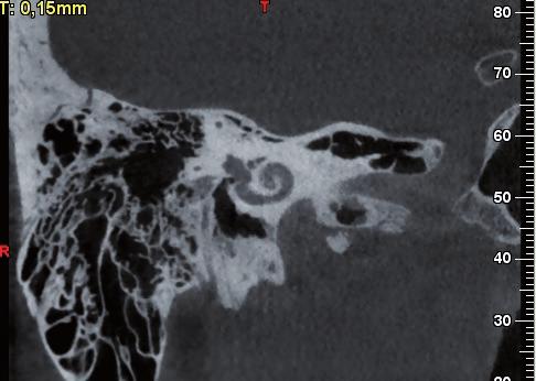

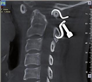
8 3D ENT Protocols The unit s exceptional precision and various FOV possibilities provide for a clear view of the airways, sinuses and ear structures. The scans are carried out using the most suitable radiological parameters in order to avoid any unnecessary ray emissions. During the pre-examination phase, the operator selects the most suitable protocol for the anatomical area to be analyzed. Most examinations carried out using conventional CAT/CT scans can also be performed with the scan devices, which, in contrast, provide for more detailed images while simultaneously decreasing patient exposure. Cervicals A cervical spine X-ray can help find the cause of symptoms such as neck, shoulder, upper back, or arm pain, as well as tingling, numbness, or weakness in the arm or hand. It can detect fractures in the cervical vertebrae, dislocation of the joints between the vertebrae, subluxations of the vertebral bodies and cervical abnormalities. CB3D is excellent for characterizing fractures and identifying osseous compromise of the vertebral canal because of the absence of superimposition from the transverse view. The higher contrast resolution of CB3D also provides improved visualization of subtle fractures. The scan can provide patient comfort by being able to reconstruct images in the axial, sagittal, coronal, and oblique planes from one patient positioning. Orthopedics Bone X-ray is used to detect fractures or dislocated joints, ensure that a fracture has been properly aligned, evaluate injury or damage from conditions such as infections, arthritis, abnormal bone growths, locate foreign objects, evaluate changes in bones and detect degenerative conditions of the bone. For all these pathologies, the multiple views due to the 3D dataset allow specialists to assess the degree of pathological displacement of any fractures or dislocations.


9 Veterinary NEWTOM BENEFITS NewTom 5G provides veterinary practices with an essential, non-invasive diagnostic tool. The research in this field is an important part for deepen our knowledge of what we know about diagnostic imaging and treatment in every medical fields. The value of 3D, however, is directly related to the quality of the images obtained. Poor images can result in missing a foreign body, bone cancer or a fracture. Besides, the risk of missing or misinterpreting a problem, due to poor radiographs, can cost the practice money. An exam performed with NewTom 5G can be useful for: nasal/sinus disease, middle ear/chronic external ear disease, thoracic imaging, pre-surgical planning, skull, spinal, pelvic evaluation, fracture evaluation, etc. NewTom 5G, from the company that was the first to use the Cone Beam technology in dental field, represents the newest in CB3D technology. NewTom 5G takes an image at every degree of rotation, 360 rotation = 360 images, increasing the range of possibilities for image manipulation. It couples a revolutionary flat panel X-ray detector technology with a very small focal spot (0.3mm), to produce the clearest, sharpest images possible. NewTom 5G features an adjustable field of view, which allows doctors to irradiate just the right volume, depending on the different clinical applications. The size of FOV can vary from the smallest 6x6 cm to the biggest 18x16 cm and they can be selected directly from the software, before the scan. NewTom 5G emits a lower radiation in comparison with conventional CT, by using a pulsed emission, that unlike other systems, activates the X-ray source only when required and, for a full scan, it takes no more than 5 seconds of total exposure. With a fascinating design, users can explore new clinical applications, thanks to the open, pass-through style gantry and a motorized patient table. The supine position of the patient during the scan and the reduced scan time add comfort and stability for an excellent result in terms of image quality and patient satisfaction. The small footprint and the variable positioning make NewTom 5G the best choice for locations, where space is at a premium. NewTom 5G does not need air-conditioned rooms, its weight does not require reinforced floor and it can function in rooms without complicated and expensive radiation protection structures. All the operations executed by NewTom, the patient s examination and the following calculations, are computer guided. The user, when performing the scan, is supported by user-friendly menus. Each step is associated to a mouse-activated icon. Following the same process, one can enter the integrated file of image-data. A greater comfort for patients leads to a better acceptance of the treatment. SafeBeam Technology adjusts the radiation dosage for patient safety. Multiple FOV and different scan modes are selectable from the software and adaptable to various fields of application. The margin of error is reduced thanks to the precise 1:1 scale and a 16-bit grey scale. NNT software makes the image sharing process easier.
10 SPECIFICATIONS X-ray source Focal spot Acquisition technique Scan time High frequency, rotating anode: 110 kv; 1-20 ma (pulsed mode) 0.3mm 3D Single scan and Cone Beam acquisition SafeBeam control reduces radiation based on patient size 18s to 36s NewTom Today s standard of care > Free Viewer and Sharing Application > Full DICOM 3.0 Compliant > Improved Software Integration > Small Footprint X-ray emission time 2,4s to 7,3s Image acquisition Image detector Signal grey scale 240/360/480 images 360 degree rotation Amorphous silicon flat panel, 20 cm x 25 cm Field of View (7.87 in x 9.84 in) 14-bit scanning, 16-bit reconstruction Multiples scan modes Centimeters FOV sizes D x H Inches Voxel size options (μm) Dimensions in centimeters [dimensions in inches] Standard Scan _ Boosted Scan 18 x x x x x x x x HiRes Scan 15 x x x x x x x x Patient positioning Supine Motorized patient table + laser Reconstruction time Less than 1 minute NewTom 5G with patient table Width 175 cm in Weights and dimensions Scan unit Depth (max) Height Gantry 360 cm in (with patient table w. stretcher) 178 cm in 58 cm in Total weight 650 kg lb (with patient table w. stretcher) Software Power Required NNT with free viewer and sharing application 220/230/240V~, 50/60 Hz Specifications subject to change without prior notice. NewTom 5G with patient table w. stretcher 0051
VGi - R EN ENGLISH. QR srl - Via Silvestrini, Verona Italy Tel
 VGi - R15.1 - EN ENGLISH QR srl - Via Silvestrini, 20-37135 Verona Italy Tel. +39 045 8202727-045 583500 info@newtom.it www.newtom.it FIRST IN CONE BEAM, ACCURATE IN RESULTS. 360 degree imaging, reduced
Cone Beam 3D Imaging
 Cone Beam 3D Imaging NewTom Sets the Standard in 3D Maxillofacial Imaging Cone Beam 3D Imaging The Global Market Leader The Inventors n of Cone Beam 3D In 1996, QR srl developed the first generation of
ENGLISH VGi - R ENG
 ENGLISH First in Cone Beam, Accurate in Results Cone Beam 3D Imaging First User of Cone Beam in Dental Field QR s.r.l. is the name that stands behind NewTom Cone Beam 3D imaging units and we were the creators
ENGLISH. Distributed by: QR srl - Via Silvestrini, Verona Italy Tel
 GiANO - R15.1 - EN ENGLISH Distributed by: QR srl - Via Silvestrini, 20-37135 Verona Italy Tel. +39 045 8202727-045 583500 info@newtom.it www.newtom.it Manufacturer: CEFLA S.C. - CEFLA DENTAL GROUP Via
ENGLISH. Cefla s.c. - Via Selice Provinciale 23/a, Imola - Italy Tel
 09-2017 NVGEGB161S00 According to the regulations in force, some products and/or features may have different availability and characteristics in areas outside of the European Union. Please contact your
ENGLISH. Cone Beam 3D Imaging
 ENGLISH Cone Beam 3D Imaging FIRST USER OF CONE BEAM IN DENTAL FIELD QR s.r.l. is the name that stands behind NewTom Cone Beam 3D imaging units and the creator of Cone Beam technology for the dental field.
5G XL - R EN ENGLISH
 5G XL - R15.0 - EN ENGLISH Sede legale ed amministrativa - Headquarters QR srl - Via Selice Provinciale, 23/a - 40026 Imola - Bo (Italy) Stabilimento - Plant Via Fermi, 40-37136 Verona (Italy) Tel. +39
ADVANCED 3D IMAGING. CEFLA s.c. Via Selice Provinciale 23/a Imola Italy t newtom.
 CEFLA s.c. Via Selice Provinciale 23/a 40026 Imola Italy t. +39 045 8202727 045 583500 info@newtom.it newtom.it 05/2018 NVGEGB181S00 According to the standards in force, in extra-eu areas the availability
Head to new heights with your imaging SCANORA 3D
 SCANORA 3D Head to new heights with your imaging Benefits at a glance The solution for dentomaxillofacial and ENT imaging Easy Patient seated for added stability during exposure. Clear, self-explinatory
CS 9300 Family. The power of flexibility
 CS 9300 Family The power of flexibility The new CS 9300 digital imaging system from Carestream Dental take the guesswork out of examinations The all-in-one CS 9300 is the most versatile multimodality imaging
NewTom 5G XL EXTRA.VISION
 CEFLA s.c. Via Selice Provinciale 23/a 40026 Imola Italy t. +39 045 8202727 045 583500 info@newtom.it newtom.it 06/2018 N5GXGB181S00 According to the standards in force, in extra-eu areas the availability
The Quality Leader in 3D Cone Beam CT
 The Quality Leader in 3D Cone Beam CT The Complete 2-in-1 or 3-in-1 Multi-modality Solution PreXion, with over 15 years of innovation in the medical and dental fields, introduces the PreXion3D Eclipse.
fast accurate safe See the full picture: add a 3rd dimension to your patient evaluation to diagnose more effectively. BENEFITS OF NEWTOM 3G 6" 9" 12"
 See the full picture: add a 3rd dimension to your patient evaluation to diagnose more effectively. Examination Effective Dose Equivalent (ICRP tissue weights 2005) Panoramic Dose (ICRP tissue weights 2005)
Low Dose Excellent Image Quality Rapid Reconstruction
 Low Dose Excellent Image Quality Rapid Reconstruction Efficient 3 in 1 Dental X-ray System CBCT > Precise 3-D Anatomical structures - Accurate diagnosis for doctors - Safe implant for patients > Significant
True Low Dose. Exact time to display image on screen may vary upon computer and network configuration.
 RAYSCAN ALPHA PLUS True Low Dose Cone Beam CT Industry Leading Resolution High resolution images provide all the clinical information needed while keeping radiation exposure low. Endodontics - Smallest
Flexible Easy Competitive. SCANORA 3Dx - The in-office large field-of-view Cone Beam CT system for Head and Neck imaging
 Flexible Easy Competitive SCANORA 3Dx - The in-office large field-of-view Cone Beam CT system for Head and Neck imaging SCANORA 3Dx. The solution. SCANORA 3Dx makes advanced 3D imaging easy in the head
English. Perfect Vision
 English Perfect Vision Everything becomes clearer and simpler with a big F.O.V. The complete dentomaxillofacial volume ready for your diagnosis One scan provides you with an incredible amount of information
PAN CEPH 3D CONE BEAM
 PAN CEPH 3D CONE BEAM 2D - 3D panoramic units PANORAMIC CEPHALOMETRIC 3D CONE BEAM IMAGING I-MAX TOUCH Tactile & naturally intuitive panoramic imaging Discover the simplicity and efficiency this unit can
Profound understanding of anatomy
 ENGLISH Profound understanding of anatomy The unique Planmeca ProMax 3D product family offers equipment for all maxillofacial imaging. All volume sizes from the smallest special cases to whole head images
Dental Line. 3D digital panoramic system. radiology ahead
 Dental Line 3D digital panoramic system radiology ahead new generation 3D digital panoramic unit 3D imaging s value available for anyone Following the incredible success of the innovative digital panoramic
I AM DEMANDING Type CMOS Flat Panel CMOS CMOS ø 40 x 40 mm, ø 60 x 60 mm, ø 80 x 80 mm, ø 110 x 80 mm
 TECHNICAL SPECIFICATIONS 1168 1501 1978 1237 1551-2351 Ø 1090 PANORAMIC CBCT CEPHALOMETRIC X-RAY SOURCE Tube type High frequency DC generator 2.8 mmal / 85 kv 7.0 mmal / 90 kv 2.8 mmal / 85 kv Operation
Easy operation. Numerous diagnostic options. X-rays you can rely on: the ORTHOPHOS XG device family. All ORTHOPHOS XG 3Dready programs at a glance.
 CAD /CAM Systems Instruments Hygiene Systems Treatment Centers Imaging Systems Subject to technical changes and errors in the text, Order No. A91100-M47-B346-01-7600, Printed in Germany, Dispo-Nr. 04602,
3D Cone beam CT & Digital Radiography Dedicated to Otorhinolaryngology
 3D Cone beam CT & Digital Radiography Dedicated to Otorhinolaryngology Multi-functional imaging solution3 RAYSCAN m is an unique 2-in-1 imaging solution, combining Cone Beam CT and Digital Radiography,
Profound understanding of anatomy
 ENGLISH Profound understanding of anatomy Planmeca ProMax 3D, the intelligent and multipurpose X-ray unit, is designed to obtain complete information on patient anatomy in the minutest detail. The unit
THE WAIT IS OVER CS D. 3D imaging is now available for everyone
 THE WAIT IS OVER CS 8100 3D 3D imaging is now available for everyone COMPLEXITY IS NO LONGER THE STANDARD NOW THERE ARE MANY REASONS TO MOVE TO 2D/3D IMAGING Now it s possible to experience nothing but
Xelis Dental - What New in
 Xelis Dental - What New in 1.0.6.1 Clinical Needs Why Xelis-Dental? Panorama Cephalography Intra-oral Digital Camera Traditional imaging systems - 2-Dimension view Incorrect anatomical information - Distortion
DIAGNOSTIC IMAGING. OPTIMIZED.
 ABOUT LED DENTAL SEE THE DIFFERENCE Using our years of business insight and clinical experience as a foundation, LED Dental takes the uncertainty out of your imaging purchase decision. We offer our clients
Digital Imaging from a new perspective
 TREATMENT CENTRES HANDPIECES HYGIENE SYSTEMS X-RAY SYSTEMS CEREC TREATMENT CENTRES HANDPIECES HYGIENE SYSTEMS X-RAY SYSTEMS CEREC SIRONA CREATING AND MAINTAINING VALUE. You are right to expect a great
Profound understanding of anatomy
 ENGLISH Profound understanding of anatomy The unique Planmeca ProMax 3D product family offers equipment for all maxillofacial imaging. All volumes sizes from the smallest special cases to whole head images
XPan 3D Plus. FONA Every dental solution you need. Advanced dental technology. Headquarters THE ULTIMATE DIAGNOSTIC SOLUTION DIGITAL DENTISTRY
 FONA Every dental solution you need Through decades of experience and deep understanding of the dental profession, we deliver complete, reliable and accessible solutions.regardless of country or specialisation,
2D AND 3D/2D WALL-MOUNTED PANORAMIC UNITS
 2D AND 3D/2D WALL-MOUNTED PANORAMIC UNITS KEEP YOUR CLINIC ONE STEP AHEAD! Wall-mounted concept: zero foot print 62kg - the lightest unit on the market Face to face positioning High Definition The fruit
T h e D e n t a l C o m p a n y FROM DIAGNOSTIC SCAN TO SURGERY, WE SHAPE THE FUTURE OF DENTISTRY.
 T h e D e n t a l C o m p a n y FROM DIAGNOSTIC SCAN TO SURGERY, WE SHAPE THE FUTURE OF DENTISTRY. SIDEXIS SOFTWARE ORTHOPHOS SL D/D SEAMLESS THE NEW STANDARD IN CLINICAL DIAGNOSIS AND PATIENT COMMUNICATION
NewTom GiANO HR PERFECT.VISION
 CEFLA s.c. Via Selice Provinciale 23/a 40026 Imola Italy t. +39 045 8202727 045 583500 info@newtom.it newtom.it 06/2018 NHRGB181S00 According to the standards in force, in extra-eu areas the availability
VistaVox S 3D from Dürr Dental
 VistaVox S 3D from Dürr Dental 3D and 2D X-ray images with exceptional image quality COMPRESSED AIR SUCTION IMAGING DENTAL CARE HYGIENE Taking diagnostics to the next level VistaVox S combines diagnostic
CT Imaging at the Point-of-Care
 ENGLISH True Dedication The new Planmed Verity Extremity CT Scanner revolutionizes extremity CT imaging. The compact unit brings 3D imaging at emergency departments, orthopedic clinics or trauma centers
D3D CBCT. See more Do More
 D3D CBCT See more Do More BIOLASE DaVinci Imaging D3D For capturing superior 3D image acquisitions with the lowest minimal dose for patient safety The BIOLASE DaVinci Imaging D3D has one of the lowest
The Optimum Choice for Implantologist
 The Optimum Choice for Implantologist What is essential for your practice? What s the best way to choose a 3D X-ray machine for implant treatment planning? 02 Doctor says.. There are diagnostic limitations
CT Scanning Protocol For V2R Guided Surgery Solutions
 CT Scanning Protocol For V2R Guided Surgery Solutions 2 V2R CT Scanning Protocol \\ Contents Contents General requirements... 3 V2R Dual Scan Protocol... 5 V2R Single Scan Protocol... 8 Overview... 10
User Guide for Dental and Maxillofacial Cone Beam Computed Tomography (CBCT)
") User Guide for Dental and Maxillofacial Cone Beam Computed Tomography (CBCT) Poster No.: C-0756 Congress: ECR 2014 Type: Educational Exhibit Authors: J. Ukkonen, J. Asp; Helsinki/FI Keywords: Education
User Guide for Dental and Maxillofacial Cone Beam Computed Tomography (CBCT) Poster No.: C-0756 Congress: ECR 2014 Type: Educational Exhibit Authors: J. Ukkonen, J. Asp; Helsinki/FI Keywords: Education
CT SCAN PROTOCOL. Shoulder
 CT SCAN PROTOCOL Shoulder Purpose and Summary CT images made with this protocol are used to provide the orthopedic surgeon with a detailed 3D anatomical reconstruction of the patient s scapula and proximal
Planmeca ProMax 3D s Planmeca ProMax 3D ENGLISH
 Planmeca ProMax 3D s Planmeca ProMax 3D ENGLISH Genuine all-in-one unit Planmeca ProMax 3D s and Planmeca ProMax 3D units are designed to obtain complete information on patient anatomy in the minutest
ORTHOPHOS XG 3 DS. X-ray systems. ORTHOPHOS XG 3 Digital panoramic X-ray for practical diagnostics.
 ORTHOPHOS XG 3 DS X-ray systems ORTHOPHOS XG 3 Digital panoramic X-ray for practical diagnostics. ORTHOPHOS XG 3 Standard panoramic X-ray with proven technology. competent successful Many years of competence
3D-MODEL CUSTOM-MADE MODELS SEGMENTATION AND PRODUCTION SERVICE OF BONE MODELS WITH HIGHEST 3D PRINTING RESOLUTION
 CUSTOM-MADE MODELS 3D-MODEL SEGMENTATION AND PRODUCTION SERVICE OF BONE MODELS WITH HIGHEST 3D PRINTING RESOLUTION FOLLOW US ON CUSTOM-MADE MODELS 3D-MODEL From a CT or CBCT scan, 3D-model service provides
Dental Cone Beam CT. What is Dental Cone Beam CT?
 Scan for mobile link. Dental Cone Beam CT Dental cone beam computed tomography (CT) is a special type of x-ray equipment used when regular dental or facial x-rays are not sufficient. Your doctor may use
THE USE OF KEYSTONE EASYGUIDE CT SCANNING SOFTWARE FOR DIAGNOSIS, DIRECTION AND DEPTH DETERMINATION
 CT DIAGNOSTICS IN 3D IMPLANT TREATMENT PLANNING THE USE OF KEYSTONE EASYGUIDE CT SCANNING SOFTWARE FOR DIAGNOSIS, DIRECTION AND DEPTH DETERMINATION Timothy Kosinski, DDS, MAGD Assistant Clinical Professor
OP 3D Vision The upgradable 3D X-ray system for the strictest demands.
 OP 3D Vision The upgradable 3D X-ray system for the strictest demands. The solution for every task: KaVo OP 3D Vision. Regardless of which dental query you may have, the KaVo ORTHOPANTOMOGRAPH OP 3D Vision
ORTHOPANTOMOGRAPH OP200 D ORTHOCEPH OC200 D. True dynamo leading through the decades.
 ORTHOPANTOMOGRAPH OP200 D ORTHOCEPH OC200 D True dynamo leading through the decades. 1 You can t dublicate the legacy. 1946 Professor Y.V. Paatero publishes his first paper on Panoramic Tomography. 1951
2
 1 2 3 4 5 6 7 8 9 10 11 12 13 Cine loop of tomosynthesis slice images through the chest. 14 Standard PA chest radiograph (left) and single slice from the tomosynthesis image dataset (right) of a patient
X X X. GXS-700 Direct USB Digital Intraoral Sensors. Buy a Sensor Combo and a Digital Pan Unit, Receive $600 Off! GO.BENCO benco.
 8 0 0. G O. B E N C O b e n c o. c o m GS-700 Direct USB Digital Intraoral Sensors Designed to make migrating from film, or upgrading an existing digital system, easier than ever High quality image capture
WITH. The Next Step in Office MRI
 WITH The Next Step in Office MRI Introducing S-scan the Next Step in Office MRI Based on extensive customer feedback and years of engineering, Esaote has designed the S-scan with exp Technology, an optimized
Versatility And Expandability In One Panoramic.
 Orthoralix 9200 / 9200 DDE Versatility And Expandability In One Panoramic. Panoramic X-ray Systems Intraoral X-ray Systems Digital Intraoral Sensors Digital X-ray Phosphor Plates Intraoral Cameras Imaging
Innovations 2017 & 2018
 Innovations 2017 & 2018 medicad 5.0 Hip 3D Spine 3D Knee 3D Shoulder 3D The Orthopedic Solution medicad Version 5.0 CHECK OUT WHAT'S NEW: Hip Automatic measuring of femoral or acetabular offset Automated
3D Panoramic Cephalometric. Innovation, in reach. KODAK 9000 Extraoral Imaging System
 3D Panoramic Cephalometric Innovation, in reach 9000 KODAK 9000 Extraoral Imaging System Cephalometric Innovation made simple Innovation made simple We believe in innovation. We always have. In fact, our
Powered by. Dedicated MRI
 Powered by Dedicated MRI Provides the latest software and hardware upgrade configuration powered by exp technology: boosting productivity, increasing image quality, and adding new acquisition techniques.
CS 8100 FAMILY / CS D FAMILY / CS 9300 FAMILY EXTRAORAL SOLUTIONS EXTRAORDINARY POSSIBILITIES
 CS 8100 FAMILY / CS 8100 3D FAMILY / CS 9300 FAMILY EXTRAORAL SOLUTIONS EXTRAORDINARY POSSIBILITIES A SOLUTION FOR EVERY CLINIC AND ANY CONSULTATION The CS 8100 does more than my old machine and is half
3D Accuitomo XYZ Slice View Tomograph. Super High-Resolution Images of Region of Interest
 3D Accuitomo XYZ Slice View Tomograph. Super High-Resolution Images of Region of Interest Thinking ahead. Focused on life. 2 Cone Beam X-Ray CT Imaging X-Ray Tube Imaging Intensifier Imaging Volume Voxel
Clinical details: Details of scan: CONE BEAM CT REPORT: Name: H. B. Gender: Reason for referral: Referred by:
 Name: H. B. Gender: Male DOB: 11/12/1950 Age: 64 Date taken: 16/11/2015 Date reported: 19/11/2015 Clinical details: Reason for referral: Referred by: Investigate symptoms related to left TMJ. Reconstructed
STELLARIS 3D 4 IN 1 CBCT SOLUTION FOR ADVANCED DIAGNOSTICS
 STELLARIS 3D 4 IN CBCT SOLUTION FOR ADVANCED DIAGNOSTICS 3 STELLARIS 3D 4 IN CBCT SOLUTION FOR ADVANCED DIAGNOSTICS Stellaris 3D is a complete and compact, fully upgradeable 3D CBCT for a patient, Panoramic
The Key to Confidence
 WITH The Key to Confidence The Key to Confidence More detail, better accuracy, greater confidence The G-scan Brio is a revolutionary MRI approach for all musculoskeletal applications, which allows you
Implant restoration in the aesthetic zone using guided surgery and immediate functional loading
 Prachatipat Hospital Prathumtani Province Dr. Nawakamon Suriyan Implant restoration in the aesthetic zone using guided surgery and immediate functional loading Digital Workflow: clinical patient information
GUIDED SURGERY TECHNIQUE
 GUIDED SURGERY TECHNIQUE INDEX WORKFLOW...4 OUR SOLUTIONS...5 PLANNING PROTOCOL - PARTIAL EDENTULISM...6 - TOTAL EDENTULISM...9 SOFTWARE FEATURES...12 SURGICAL COMPONENTS FOR GUIDED IMPLANTOLOGY...14
Extraoral Imaging. Chapter 42. Copyright 2018, Elsevier Inc. All Rights Reserved. 1
 Extraoral Imaging Chapter 42 Copyright 2018, Elsevier Inc. All Rights Reserved. 1 Learning Objectives Lesson 42.1: Panoramic Imaging 1. Pronounce, define, and spell the key terms. 2. Discuss panoramic
STANDARD OF PRACTICE. Dental CT Scanners CONTENTS. April 2011
 April 2011 STANDARD OF PRACTICE Approved by Council April 18, 2011 Dental CT Scanners This document is the standard of practice in relation to the use of dental computed tomography (CT) scanners with respect
Utilizing Digital Treatment Planning and Guided Surgery in Conjunction with Narrow Body Implants. by Timothy F. Kosinski, DDS, MAGD
 Utilizing Digital Treatment Planning and Guided Surgery in Conjunction with Narrow Body Implants by Timothy F. Kosinski, DDS, MAGD Implant dentistry is undergoing some amazing transformations. With the
3Shape X1 Scanning redefined
 3Shape X1 Scanning redefined Why choose the X1 Give your patients a great experience No head fixation and sleek design create a comfortable scanning experience for your patient High image quality low dose
3Shape X1 Scanning redefined
 3Shape X1 Scanning redefined Why choose the X1 Give your patients a great experience No head fixation and sleek design create a comfortable scanning experience for your patient High image quality low dose
Agenda: Dental Cone Beam Imaging
 Cone Beam Imaging Agenda: Dental Cone Beam Imaging *Definition and Functionality *Usage and diagnostics benefits *Comparative radiation information *Federal regulatory responsibilities: manufacturing *State
X-ray (Radiography) - Bone
 - Bone") Scan for mobile link. X-ray (Radiography) - Bone Bone x-ray uses a very small dose of ionizing radiation to produce pictures of any bone in the body. It is commonly used to diagnose fractured bones or
Scan for mobile link. X-ray (Radiography) - Bone Bone x-ray uses a very small dose of ionizing radiation to produce pictures of any bone in the body. It is commonly used to diagnose fractured bones or
Veraviewepocs 3D R100 & F40
 Veraviewepocs 3D R100 & F40 Innovative 3D Reuleaux Full Arch FOV Thinking ahead. Focused on life. Veraviewepocs 3D R100 A New Frontier in X-ray Diagnostics Veraviewepocs 3D R100 has changed the shape of
- RCS Paris B
 Technical specifications PANORAMIC CBCT CEPHALOMETRIC X-ray source Tube type High frequency DC generator Total filtration >2.5 mm Al @ 90 kv Mode of operation Continuous Pulsed Continuous Tube voltage
Simplant. Guided Surgery. delivering restorative driven implant treatment
 Simplant Guided Surgery delivering restorative driven implant treatment Simplant the key to unlocking digital potential As part of the Denstsply Sirona Implants Digital Solutions offering, Simplant delivers
The future of health is digital
 Dated: XX/XX/XXXX Name: XXXXXXXX XXXXXXXXXXX Birth Date: XX/XX/XXXX Date of scan: XX/XX/XXXX Examination of the anatomical volume: The following structures are reviewed and evaluated for bilateral symmetry,
DE UK. ultra low dose. your dental equipment sales, service and training partner. Planmeca
 DE UK your dental equipment sales, service and training partner Planmeca ultra low dose Pioneering low dose 3D imaging Planmeca ProMax 3D units offer a unique Planmeca Ultra Low Dose imaging protocol that
vertaplan the spine surgeon s software vertaplan System for successful reconstruction of the individual sagittal balance
 the spine surgeon s software System for successful reconstruction of the individual sagittal balance What do you think of patient-specific reconstruction of the spine geometry? Optimum surgical outcome
UNDERSTANDING DIGITAL DENTISTRY: CBCT AND INTRA-ORAL 30 SCANNING
 UNDERSTANDING DIGITAL DENTISTRY: CBCT AND INTRA-ORAL 30 SCANNING -=- & UNDERSTANDING DIGITAL DENTISTRY: CBCT AND INTRA-ORAL 30 SCANNING ----CBCTi-------iTERO------ NewTom VGi *Vertical Patient Positioning
3D/2D. Hyperion X5 Suspended imaging system. Data subject to change without notice. 03/2017 MX53DGB171S00
 www.my-ray.com Data subject to change without notice. 03/2017 MX53DGB171S00 According to the regulations in force, some products and/or features may have different availability and characteristics in areas
In-Office Cone Beam Computerized Tomography: Technology Review and Clinical Examples Michael Tischler, DDS
 Page 1 of 8 Issue Date: June 2008, Posted On: 6/26/2008 In-Office Cone Beam Computerized Tomography: Technology Review and Clinical Examples Michael Tischler, DDS Electronic Medical Record Research EMR
For true visualisation
 ENGLISH For true visualisation Planmeca ProModel is a patient-specific physical model for high-end maxillofacial operations and dental surgery. By reproducing the anatomy of the patient in real-size, Planmeca
SIMPLANT Guided Surgery delivering restorative-driven implant treatment
 SIMPLANT Guided Surgery delivering restorative-driven implant treatment SIMPLANT the key to unlocking digital potential As part of the DENTSPLY Implants Digital Solutions portfolio, SIMPLANT digital implant
CASE REPORT. CBCT-Assisted Treatment of the Failing Long Span Bridge with Staged and Immediate Load Implant Restoration
 Computer Aided Implantology Academy Newsletter - Newsletter 20 - July 2009 CASE REPORT CBCT-Assisted Treatment of the Failing Long Span Bridge with Staged and Immediate Load Implant Restoration Case Report
Straight to the point with your diagnosis.
 CAD/CAM SYSTEMS INSTRUMENTS HYGIENE SYSTEMS TREATMENT CENTERS IMAGING SYSTEMS HELIODENT DS INTRAORAL X-RAY WITH EXCELLENT IMAGE QUALITY Straight to the point with your diagnosis. T h e D e n t a l C o
NewTom 3G & VG Scanning Protocol
 NewTom 3G & VG Scanning Protocol Scanning Protocol for the NewTom 3G & VG Scanner For Use with ident General This protocol is written specifically for users of NewTom 3G & VG machines. Patient Scan with
 www.oralradiologists.com CONE BEAM CT REPORT CASE ---- Case Information Referring Doctor: - Patient Name: - Scan Date: December 1, 2015 Patient DOB: - Reason for Exam: - Study Details: icat Flex, 160x160x112
Devoted to the Advancement of Implant Dentistry
 Devoted to the Advancement of Implant Dentistry Devoted to the Advancement of Implant Dentistry Our ultimate goal is to provide you and your patients with the highest standards in implant case planning
Full ultrasound breast volumes. Faster scans. Streamlined workflow. ACUSON S2000 Automated Breast Volume Scanner. Answers for life.
 Full ultrasound breast volumes. Faster scans. Streamlined workflow. ACUSON S2000 Automated Breast Volume Scanner Answers for life. 1 ACQUIRE An automated whole breast solution. Reduced acquisition time.
3D/2D WALL MOUNTED UNIT
 Product launch document EN Page 1 sur 30 EN PRODUCT LAUNCH DOCUMENT REV03, September 2018 3D/2D WALL MOUNTED UNIT Product launch document EN Page 2 sur 30 INDEX 1. PRODUCT IDENTITY AND POSITIONNING...
AWARD-WINNING CONE BEAM 3D DENTAL IMAGING
 AWARD-WINNING CONE BEAM 3D DENTAL IMAGING Dedicated to Advancing Dental Treatment A COMPLETE 3D TREATMENT SOLUTION Your dental practice is unique that s why you need a flexible solution that works with
ORTHOPANTOMOGRAPH OP300. A platform for changing needs.
 ORTHOPANTOMOGRAPH OP300 A platform for changing needs. 1 Panoramic with cephalometric digital image Xray machine For me, peace of mind means a patient trusting in my care, time after time Leading the way
HDS PROCEDURE CODE GUIDELINES
 D0100 - D0999 Clinical Oral Evaluations D0120 - D0180 The codes in this section have been revised to recognize the cognitive skills necessary for patient evaluation. The collection and recording of some
Original Research THE USE OF REFORMATTED CONE BEAM CT IMAGES IN ASSESSING MID-FACE TRAUMA, WITH A FOCUS ON THE ORBITAL FLOOR FRACTURES
 DOI: 10.15386/cjmed-601 Original Research THE USE OF REFORMATTED CONE BEAM CT IMAGES IN ASSESSING MID-FACE TRAUMA, WITH A FOCUS ON THE ORBITAL FLOOR FRACTURES RALUCA ROMAN 1, MIHAELA HEDEȘIU 1, FLOAREA
The mission of our company is the development of an individual approach in functional dentistry.
 The Prosystom company started up in 2013. It was created by Russian doctors and IT specialists for carrying out scientific projects and practical work in functional dentistry. The mission of our company
INTRAORAL IMAGING ACCORDING TO YOUR NEEDS Intraoral X-rays
 INTRAORAL IMAGING ACCORDING TO YOUR NEEDS Intraoral X-rays FONA Intraoral X-rays INTRAORAL IMAGING ACCORDING TO YOUR NEEDS FONA intraoral X-ray devices are designed for ease of use, precision, reliability
Hyperion X9 3-in-1 Imaging System
 Hyperion X9 3-in-1 Imaging System 2 Hyperion X9, full imaging. 3 Hyperion X9, just right for me. The present and the future of my work. In three dimensions. Hyperion X9 offers me multiple possibilities
UNIVERSITY OF MEDICINE AND PHARMACY GR. T. POPA - IASI FACULTY OF DENTAL MEDICINE
 UNIVERSITY OF MEDICINE AND PHARMACY GR. T. POPA - IASI FACULTY OF DENTAL MEDICINE ABSTRACT CONTRIBUTIONS OF THREE-DIMENSIONAL IMAGING TO THE DIAGNOSIS AND MANAGEMENT OF CLEFT LIP AND PALATE PhD ADVISOR,
Varian Acuity BrachyTherapy Suite One Room Integrated Image-Guided Brachytherapy
 Varian Acuity BrachyTherapy Suite One Room Integrated Image-Guided Brachytherapy The Acuity BrachyTherapy Suite Integrating Imaging, Planning, and Treatment in a Single Room Each component draws on the
Introduction of FIREFLY Technology
 Introduction of FIREFLY Technology FIREFLY Technology is a unique, patent-pending, pre-surgical planning and intra - operative navigation technology that is focused on spinal applications and is derived
Cephalometric Analysis
 Cephalometric Analysis of Maxillary and Mandibular Growth and Dento-Alveolar Change Part III In two previous articles in the PCSO Bulletin s Faculty Files, we discussed the benefits and limitations of
Keywords: Mandible, Maxilla, Teeth, Cone Beam CT, Implants, Tumor and tumor-like conditions
 Dental Cone Beam CT: a primer for clinical radiologists e-poster: EP-1 Congress: ESHNR 2014 Type: Educational Poster Topic: ESHNR 2014 Authors: C. Vanhoenacker 1, F. Vanhoenacker 1, L. Vannitsen 1, A.
Planmeca ProMax 3D s Planmeca ProMax 3D ENGLISH
 Planmeca ProMax 3D s Planmeca ProMax 3D ENGLISH Learn more: Planmeca Imaging for ipad Genuine all-in-one unit Planmeca ProMax 3D s and Planmeca ProMax 3D units are designed to obtain complete information
Dolphin 3D Imaging 11.7 beta
 Dolphin 3D Imaging 11.7 beta Dolphin 3D Surgery Dolphin s 3D Surgery has expanded to include even more features! This revolutionary treatment planning and presentation software module now includes a fully