Veraviewepocs 3D R100 & F40
|
|
|
- Gregory Hart
- 5 years ago
- Views:
Transcription

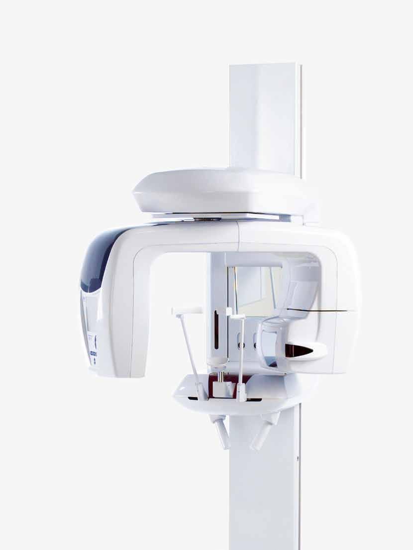
1 Veraviewepocs 3D R100 & F40 Innovative 3D Reuleaux Full Arch FOV Thinking ahead. Focused on life.
2 Veraviewepocs 3D R100 A New Frontier in X-ray Diagnostics Veraviewepocs 3D R100 has changed the shape of 3D. This unit's groundbreaking and patent pending 3D Reuleaux Full Arch FOV (field of view) provides a unique shape for full arch imaging. With 6 field of view options and Morita's world renowned image quality, Veraviewepocs 3D R100 is suitable for a wide variety of dental applications including implant planning. 1
3 2

4 3D Reuleaux Full Arch Field of View Blue line indicates new full arch FOV, equivalent to 100 mm. New Patent Pending Technology Morita's new and completely unique 3D Reuleaux Full Arch FOV abandons the typical cylinder with a new convex triangle shape. By more closely matching the natural dental arch form, this groundbreaking FOV reduces dosage by excluding areas outside the region of interest and allows a complete scan of the maxilla and/or the mandible. Not available on the Veraviewepocs 3D F40 model. 3


5 Various Fields of View Exposure Areas for Multiple Diagnostics The Veraviewepocs 3D R100 model offers a total of 6 exposure areas from 40 x 40 mm up to 100 x 80 mm for various diagnostic needs. The new full arch scan captures the maxilla and/or the mandible with the equivalent of 100 mm in diameter and two height options of 50 or 80 mm. Its full arch capability, reduced dosage, and exceptional clarity are ideal features for implant planning and oral surgery. This unit also offers small and medium field of view sizes suitable for endodontics, periodontics, as well as general dentistry. The Veraviewepocs 3D F40 model offers 40 x 80 mm and 40 x 40 mm fields of view, also suitable for a variety of applications. R100 Full Arch FOV Ø 80 FOV Ø 40 FOV Ø 100 mm Fields of View Ø 100 x H 80 mm* Ø 100 x H 50 mm* Ø 80 x H 80 mm Ø 80 x H 50 mm Ø 40 x H 80 mm Ø 40 x H 40 mm Veraviewepocs 3D R100 only Veraviewepocs 3D R100 and Veraviewepocs 3D F40 *3D Reuleaux Full Arch FOV 4

6 High Resolution Images With Dose Reduction Feature Dose Reduction Feature Through advanced engineering, a Dose Reduction Mode optimizes the intensity of the X-rays which lowers exposure for easily penetrated tissues. Dosage is reduced to a mere 60% of the standard mode.* By maximizing efficiency, soft tissue, such as the maxillary sinus membrane and skin, appear sharper than ever before with fewer artifacts.** Resolution & Clarity Veraviewepocs offers high resolution images of 125 µm voxel. It provides clear images of the periodontal pocket, the periodontal ligament, and the alveolar bone. It is extremely useful for implant therapy from planning to post-operative observation. Spatial Resolution MTF: Modulation Transfer Function MTF [%] Tube Voltage: 60 kv Tube Current: 1.0 ma MTF at 2 Lp/mm > 10 % Spatial Frequency [Lp/mm] Super-High Resolution for All Image Areas The resolution of Veraviewepocs is greater than 2 line pairs per mm (MTF 10%). The highly detailed images have a voxel size of mm per side, and the slice thickness and interval can be set between and mm. The expanded radiographic area of Ø 80 x H 80 mm maintains the same high resolution and voxel size as the smaller fields of view. 40 x 80 mm high resolution image taken in Dose Reduction Mode * For 40 X 80 mm exposures. ** Compared to standard exposure mode. 5


7 Easy 3D Positioning Flexibility Veraviewepocs offers flexibility in positioning methods. The region of interest can be positioned by the panoramic image, the bi-directional scout, or the 5 positioning laser beams. Panoramic Image with Scout Feature Before taking a 3D image, a high resolution panoramic exposure is taken to target the region of interest on the PC monitor. The C-arm will automatically move into the optimum patient position to get 3D images at the center of the region of interest. Bi-directional Scout After initial positioning is accomplished by the 3 positioning laser beams, bidirectional X-ray images can be taken to confirm that the position is accurate. If it is not, simply adjust the position of the image on the computer by placing the cursor at the center of the region of interest. Simply double click the cross to display the equivalent CT image. Clinical Case Example The panoramic image above suggests that there is an apical lesion on the distal root of tooth #26. Further inspection with a 3D image, however, shows that the lesion is on the buccal side of an extremely curved mesial root. Direct Positioning with 5 Laser Beams 5 positioning laser beams set the patient s position and align the region of interest. First, the patient s initial position is set using the 3 laser beams. Then, 2 additional laser beams are aligned to the region of interest. The C-arm will automatically move to the right position. 6


8 3D Images for Implant Planning Planning Process Successful placement of implants starts with the very critical and detailed planning process. Identification of structures such as the sinus cavity, inferior alveolar nerve, and clear views of the bone structure are needed. Veraviewepocs 3D R100 is ideal for implant planning with full arch imaging, industry leading clarity, and low dosage to the patient. Software i-dixel 2.0 software offers advanced implant planning features, plus compatibility with popular third party software. cmpr Image Processing Create cross sectional images of the dental arch. Mandibular Canal Tracing Highlight the mandibular canal for easier viewing, measuring the distance to the implant and determining its buccal and lingual position. 7

9 Advanced Software Features Confirm Implant Position with Volume Rendered Image A high resolution volume rendered image of the entire jaw can be created. This rendering makes it easy to explain each step of the implant planning and treatment process to the patient. Link to Implant Simulation Software By converting images to DICOM formats, implant simulation can be performed with other third party software. Presentation Preparation The data for implant devices including length and diameter can be used to superimpose an image of the device on a 3D image to show patients and others. Implant Library The implant library can be used to make realistic presentations for patients. 8


10 Clinical Cases Implantology The patient was seen for a routine follow up visit following implant placement of tooth #16. The implant had been placed 9 years earlier. The coronal, sagittal, and axial views all confirm good quality bone around the implant and an absence of any pathology related to the treatment. The fine detail of the bone, especially in the coronal view, allows the clinician and the patient to feel confident about the current health of the implant. Endodontics The patient presented with a radiolucency around tooth #26 that had previously been treated endodontically. Conventional 2D imaging was inconclusive so a cone beam CT image was taken with the 3D R100. The sagittal and coronal views both showed that the endodontic therapy was failing and there were apical lesions on both the buccal and palatal roots. The sagittal view clearly confirms a perforation of the Schneiderian membrane, while the coronal view identifies odontogenic maxillary sinusitis and mucosal thickening. The damage to the sinus membrane may have been overlooked if this case was diagnosed and treatment planned with an image that did not show the problem so clearly. 9


11 Oral Surgery The patient presented with pain in the maxillary left region. A cone beam CT image was taken with the 3D R100 and it was revealed that tooth #28 was in fact impacted and was causing problems for tooth #27. The axial view demonstrated extensive bone loss near the apical area of #27 due to the lack of arch space needed for #28 to erupt. The coronal view showed bone destruction all the way through the furcation of #27. The sagittal image not only shows the loss of osseous support around the entire apex of #27, but also shows damage to the sinus floor and mucosal thickening. A portion of the clinical images were provided by Kitasenju Radist Dental Clinic, i-view Imaging Center, Japan. 10

12 Panoramic Imaging After Image Layer Adjustment AF Automatic Positioning This function makes patient positioning nearly effortless. A light beam sensor automatically positions the unit without requiring the patient to move. The light beam sensor measures the distance to the patient s teeth, then the arm automatically moves into the optimal position. This process produces images with a high degree of reproducibility. Light Emitter DDAE (Digital Direct Auto Exposure) The DDAE function controls X-ray emission in real time depending on the area being examined and produces a wide dynamic range, as well as sharp and exceptionally clear images. CMOS Sensor AIE (Auto Image Enhancement) This software processing function uses a logarithmic conversion to adjust the overall density and to highlight shaded details, creating a better image. Computer X-ray Head Standard Panoramic The X-Y movement and arm rotation are coordinated by a computer control system to create a projection with the optimum image layer shape. Orthogonal Panoramic This projection controls the angle of X-ray penetration to reduce the overlapping of individual teeth. Shadow Reduction Panoramic This projection controls the angle of X-ray penetration to reduce the mandibular ramus shadow. PSD Sensor 11 AF Automatic positioning Technology DDAE Mechanism


13 Image Layer Adjustment After Exposure Panoramic Image Layer Adjustment The image layer for panoramic images can be adjusted after the exposure has been made to improve clarity and sharpness. The focus can be improved for points of varying depth as well as the surface. Select any point in the image for focus enhancement and then use the mouse wheel to make the adjustment. Before Image Layer Adjustment Preview images are shown in the green frame to support the manipulation of image layer adjustment After Image Layer Adjustment Image Layer Adjustment Options With various methods, the image layer can be adjusted to obtain optimum image results. Adjusted Image Layer Single point adjustment simply adjust the image layer alignment to the posterior and anterior direction. Adjusted Image Layer Two points adjustment the image layer position of the apical region can be adjusted separately at the mandibular and the maxilla. The layer position at the occlusal plane is fixed. Adjusted Image Layer Three point adjustment the image layer position of the apical region at the mandibular, maxilla, and occlusal plane can be adjusted independently. 12

14 Cephalometric Imaging High Speed The Veraviewepocs system offers high speed performance requiring only 4.9 seconds for a cephalometric scan. The speed helps ensure high quality images each and every time. For pediatric patients, the reduced scan time is especially helpful as repeat images due to patient movement are virtually eliminated. Low Dosage With only a tenth of the dosage compared to a conventional X-ray*, the exposure level is significantly reduced. High Quality Image with Wide Dynamic Range You obtain far more information about hard and soft tissue with just a single acquisition. Variable Imaging Processing The variable image processing technique generates optimum grayscale values by varying scanning speeds for hard and soft tissue. With this technique, the entire exposure time is only 4.1 seconds. Without this feature, the processing time is 4.9 seconds. Processing Time Imaging process can be completed within 20 seconds. Partial Cephalometric Images If not needed for examination, X-ray dosage can be reduced by eliminating the area behind the auditory canal. There are 3 partial image patterns. * Comparison made to Veraviewepocs film-based system 13
15 Specifications Trade name: Model: Type: Input voltage: Power consumption: Dimensions Main unit: With Cephalometric: Weight: X-ray generator Tube voltage: Tube current: Effective focal spot: Veraviewepocs 3D X550 Veraviewepocs 3D R100 Pan Veraviewepocs 3D R100 Pan/Ceph Veraviewepocs 3D F40 Pan Veraviewepocs 3D F40 Pan/Ceph EX-1: AC 120V 60 Hz, EX-2: 220/230/240 V 50/60 Hz 2.0 kva W 1,020 x D 1,330 x H 2,355 mm W 2,000 x D 1,330 x H 2,355 mm Approx. 190 kg Approx. 260 kg with Cephalometric 60-90kV (depending on exposure mode) 1-10mA (depending on exposure mode) 0.5 mm Panoramic image Exposure time: Imaging programs: Cephalometric image (option) Imaging area: Approx. 7.4 sec. for Fine High speed (Standard Mode) Approx. 15 sec. for Super Fine image (Standard Mode, 3D R100 only) Standard Panoramic (standard, orthogonal and shadow reduction projections) Magnification: 1.3 X throughout and 1.6 X throughout Pedodontic Panoramic (standard, orthogonal and shadow reduction projections) Magnification: 1.3 X throughout and 1.6 X throughout Maxillary Sinus Panoramic (front and back) Magnification: 1.5 X throughout TMJ Quadruple Image Magnification: 1.3 X throughout Posterior-anterior and Lateral 3D image Exposure time: 3D R100 imaging area: 3D Reuleaux Full Arch FOV: 3D F40 Imaging area: Approx. 9.4 seconds Ø 40 mm x H 40 mm, Ø 40 mm x H 80 mm Ø 80 mm x H 50 mm, Ø 80 mm x H 80 mm Ø 100 mm x H 50 mm, Ø 100 mm x H 80 mm Ø 40 mm x H 40 mm, Ø 40 mm x H 80 mm - Cephalometric is an optional feature. - The Veraviewepocs 3D must be fixed to the floor and the wall. - Always have patients wear X-ray protective equipment. Machine Dimensions & Suggested Operating Space Requirements Panoramic: 960 mm 40 mm EX-1: 96 mm EX-2: 70 mm 115 mm max. 1,775 mm min.1,055 mm 2,355 mm max. 1,020 mm max. 1,330 mm max. 1,380 mm 1,500 mm 730 mm 1,500 mm Panoramic/ Cephalometric: 1,490 mm 510 mm max. 1,775 mm min. 1,125 mm 2,355 mm 1,500 mm 845 mm max. 1,020 mm max. 1,330 mm max. 1,380 mm 2,300 mm 14
16 Diagnostic/Imaging Equipment Treatment Units Handpieces and Instruments Laser Equipment Laboratory Devices Educational and Training Systems Auxiliaries Developed and Manufactured by: J. Morita Mfg. Corporation 680 Higashihama Minami-cho, Fushimi-ku, Kyoto, Japan Tel: , Fax: Morita Global Website J. Morita Corporation 33-18, 3-Chome, Tarumi-cho Suita City, Osaka, Japan Tel: , Fax: J. Morita USA, Inc. 9 Mason lrvine, CA 92618, USA Tel: , Fax: J. Morita Europe GmbH Justus-von-Liebig-Strasse 27A, Dietzenbach, Germany Tel: , Fax: Siamdent Co., Ltd. 444 Olympia Thai Tower, 3rd Floor, Ratchadapisek Road, Samsennok, Huay Kwang, Bangkok 10310, Thailand Tel: , Fax: , J. Morita Corporation Australia & New Zealand Suite 2.05, 247 Coward Street, Mascot, NSW 2020, Australia Tel: , Fax: J. Morita Middle East 4 Tag Al Aoasaa, Saba Pacha 21311, Alexandria, Egypt Tel: , Fax:
Veraviewepocs 3D. F40 and R100 with innovative 3D Reuleaux Full Arch FOV. Thinking ahead. Focused on life.
 Veraviewepocs 3D F40 and R100 with innovative 3D Reuleaux Full Arch FOV Thinking ahead. Focused on life. Veraviewepocs 3D R100 A New Frontier in X-ray Diagnostics Veraviewepocs 3D R100 has changed the
3D Accuitomo XYZ Slice View Tomograph. Super High-Resolution Images of Region of Interest
 3D Accuitomo XYZ Slice View Tomograph. Super High-Resolution Images of Region of Interest Thinking ahead. Focused on life. 2 Cone Beam X-Ray CT Imaging X-Ray Tube Imaging Intensifier Imaging Volume Voxel
AdvErL Evo. Thinking ahead. Focused on life.
 AdvErL Evo Thinking ahead. Focused on life. Er:YAG Laser Technology The Ideal Periodontology Solution Periodontal Treatment (incision, excision, vaporization, ablation and coagulation) emoval of subgingival
Tri Auto ZX2. Thinking ahead. Focused on life.
 Tri Auto ZX2 Thinking ahead. Focused on life. Apical Control Observe the File Tip Position with the Apex Locator Function W.L. Automatic Stop Safety Function The Tri Auto ZX2 motor is linked with the canal
Root ZX mini Proven Accuracy in a New Compact Design. Thinking ahead. Focused on life.
 Root ZX mini Proven Accuracy in a New Compact Design Thinking ahead. Focused on life. Root ZX mini is compact, lightweight, and portable. It can be placed on the tray or any other convenient place. Convenient
Root ZX II Endodontic Predictability Morita Modular Innovation. Thinking ahead. Focused on life.
 Root ZX II Endodontic Predictability Morita Modular Innovation Thinking ahead. Focused on life. Thinking ahead. Focused on life. Root ZX II and Additional Modules The Possibilities Are Endless... With
Root ZX mini Design Meets Precision. Thinking ahead. Focused on life.
 Root ZX mini Design Meets Precision Thinking ahead. Focused on life. Color Options White Red Green Blue Compact and Lightweight Design Built on the industry standard Root ZX technology, Root ZX mini measures
Foundation Revolutionary Bone Augmentation Material. Thinking ahead. Focused on life. Regional Partner
 Foundation Revolutionary Bone Augmentation Material Thinking ahead. Focused on life. Regional Partner Easy Placement, Effective Results Tooth extraction. Socket is curetted and bone is exposed. Insertion
Digital Imaging from a new perspective
 TREATMENT CENTRES HANDPIECES HYGIENE SYSTEMS X-RAY SYSTEMS CEREC TREATMENT CENTRES HANDPIECES HYGIENE SYSTEMS X-RAY SYSTEMS CEREC SIRONA CREATING AND MAINTAINING VALUE. You are right to expect a great
Head to new heights with your imaging SCANORA 3D
 SCANORA 3D Head to new heights with your imaging Benefits at a glance The solution for dentomaxillofacial and ENT imaging Easy Patient seated for added stability during exposure. Clear, self-explinatory
The Quality Leader in 3D Cone Beam CT
 The Quality Leader in 3D Cone Beam CT The Complete 2-in-1 or 3-in-1 Multi-modality Solution PreXion, with over 15 years of innovation in the medical and dental fields, introduces the PreXion3D Eclipse.
Low Dose Excellent Image Quality Rapid Reconstruction
 Low Dose Excellent Image Quality Rapid Reconstruction Efficient 3 in 1 Dental X-ray System CBCT > Precise 3-D Anatomical structures - Accurate diagnosis for doctors - Safe implant for patients > Significant
VistaVox S 3D from Dürr Dental
 VistaVox S 3D from Dürr Dental 3D and 2D X-ray images with exceptional image quality COMPRESSED AIR SUCTION IMAGING DENTAL CARE HYGIENE Taking diagnostics to the next level VistaVox S combines diagnostic
AdvErL Evo. Thinking ahead. Focused on life.
 AdvErL Evo Thinking ahead. Focused on life. Er:YAG Laser Technology The Ideal Periodontic Solution Periodontal Treatment (incision, excision, vaporization, ablation and coagulation) Removal of subgingival
CS 9300 Family. The power of flexibility
 CS 9300 Family The power of flexibility The new CS 9300 digital imaging system from Carestream Dental take the guesswork out of examinations The all-in-one CS 9300 is the most versatile multimodality imaging
DIAGNOSTIC IMAGING. OPTIMIZED.
 ABOUT LED DENTAL SEE THE DIFFERENCE Using our years of business insight and clinical experience as a foundation, LED Dental takes the uncertainty out of your imaging purchase decision. We offer our clients
ENGLISH. Cefla s.c. - Via Selice Provinciale 23/a, Imola - Italy Tel
 09-2017 NVGEGB161S00 According to the regulations in force, some products and/or features may have different availability and characteristics in areas outside of the European Union. Please contact your
XPan 3D Plus. FONA Every dental solution you need. Advanced dental technology. Headquarters THE ULTIMATE DIAGNOSTIC SOLUTION DIGITAL DENTISTRY
 FONA Every dental solution you need Through decades of experience and deep understanding of the dental profession, we deliver complete, reliable and accessible solutions.regardless of country or specialisation,
Easy operation. Numerous diagnostic options. X-rays you can rely on: the ORTHOPHOS XG device family. All ORTHOPHOS XG 3Dready programs at a glance.
 CAD /CAM Systems Instruments Hygiene Systems Treatment Centers Imaging Systems Subject to technical changes and errors in the text, Order No. A91100-M47-B346-01-7600, Printed in Germany, Dispo-Nr. 04602,
Versatility And Expandability In One Panoramic.
 Orthoralix 9200 / 9200 DDE Versatility And Expandability In One Panoramic. Panoramic X-ray Systems Intraoral X-ray Systems Digital Intraoral Sensors Digital X-ray Phosphor Plates Intraoral Cameras Imaging
True Low Dose. Exact time to display image on screen may vary upon computer and network configuration.
 RAYSCAN ALPHA PLUS True Low Dose Cone Beam CT Industry Leading Resolution High resolution images provide all the clinical information needed while keeping radiation exposure low. Endodontics - Smallest
Cone Beam 3D Imaging
 Cone Beam 3D Imaging NewTom Sets the Standard in 3D Maxillofacial Imaging Cone Beam 3D Imaging The Global Market Leader The Inventors n of Cone Beam 3D In 1996, QR srl developed the first generation of
THE WAIT IS OVER CS D. 3D imaging is now available for everyone
 THE WAIT IS OVER CS 8100 3D 3D imaging is now available for everyone COMPLEXITY IS NO LONGER THE STANDARD NOW THERE ARE MANY REASONS TO MOVE TO 2D/3D IMAGING Now it s possible to experience nothing but
T h e D e n t a l C o m p a n y FROM DIAGNOSTIC SCAN TO SURGERY, WE SHAPE THE FUTURE OF DENTISTRY.
 T h e D e n t a l C o m p a n y FROM DIAGNOSTIC SCAN TO SURGERY, WE SHAPE THE FUTURE OF DENTISTRY. SIDEXIS SOFTWARE ORTHOPHOS SL D/D SEAMLESS THE NEW STANDARD IN CLINICAL DIAGNOSIS AND PATIENT COMMUNICATION
Flexible Easy Competitive. SCANORA 3Dx - The in-office large field-of-view Cone Beam CT system for Head and Neck imaging
 Flexible Easy Competitive SCANORA 3Dx - The in-office large field-of-view Cone Beam CT system for Head and Neck imaging SCANORA 3Dx. The solution. SCANORA 3Dx makes advanced 3D imaging easy in the head
I AM DEMANDING Type CMOS Flat Panel CMOS CMOS ø 40 x 40 mm, ø 60 x 60 mm, ø 80 x 80 mm, ø 110 x 80 mm
 TECHNICAL SPECIFICATIONS 1168 1501 1978 1237 1551-2351 Ø 1090 PANORAMIC CBCT CEPHALOMETRIC X-RAY SOURCE Tube type High frequency DC generator 2.8 mmal / 85 kv 7.0 mmal / 90 kv 2.8 mmal / 85 kv Operation
Dental Line. 3D digital panoramic system. radiology ahead
 Dental Line 3D digital panoramic system radiology ahead new generation 3D digital panoramic unit 3D imaging s value available for anyone Following the incredible success of the innovative digital panoramic
2D AND 3D/2D WALL-MOUNTED PANORAMIC UNITS
 2D AND 3D/2D WALL-MOUNTED PANORAMIC UNITS KEEP YOUR CLINIC ONE STEP AHEAD! Wall-mounted concept: zero foot print 62kg - the lightest unit on the market Face to face positioning High Definition The fruit
Straight to the point with your diagnosis.
 CAD/CAM SYSTEMS INSTRUMENTS HYGIENE SYSTEMS TREATMENT CENTERS IMAGING SYSTEMS HELIODENT DS INTRAORAL X-RAY WITH EXCELLENT IMAGE QUALITY Straight to the point with your diagnosis. T h e D e n t a l C o
X X X. GXS-700 Direct USB Digital Intraoral Sensors. Buy a Sensor Combo and a Digital Pan Unit, Receive $600 Off! GO.BENCO benco.
 8 0 0. G O. B E N C O b e n c o. c o m GS-700 Direct USB Digital Intraoral Sensors Designed to make migrating from film, or upgrading an existing digital system, easier than ever High quality image capture
Periapical Radiography
 Periapical Radiography BARBARA E. DIXON B.D.S., M.Sc., D.P.D.S. Main Indications Detection of Apical infection/inflammation Assessment of the periodontal status After trauma Assessment of Unerupted teeth
PAN CEPH 3D CONE BEAM
 PAN CEPH 3D CONE BEAM 2D - 3D panoramic units PANORAMIC CEPHALOMETRIC 3D CONE BEAM IMAGING I-MAX TOUCH Tactile & naturally intuitive panoramic imaging Discover the simplicity and efficiency this unit can
ORTHOPHOS XG 3 DS. X-ray systems. ORTHOPHOS XG 3 Digital panoramic X-ray for practical diagnostics.
 ORTHOPHOS XG 3 DS X-ray systems ORTHOPHOS XG 3 Digital panoramic X-ray for practical diagnostics. ORTHOPHOS XG 3 Standard panoramic X-ray with proven technology. competent successful Many years of competence
Profound understanding of anatomy
 ENGLISH Profound understanding of anatomy Planmeca ProMax 3D, the intelligent and multipurpose X-ray unit, is designed to obtain complete information on patient anatomy in the minutest detail. The unit
OP 3D Vision The upgradable 3D X-ray system for the strictest demands.
 OP 3D Vision The upgradable 3D X-ray system for the strictest demands. The solution for every task: KaVo OP 3D Vision. Regardless of which dental query you may have, the KaVo ORTHOPANTOMOGRAPH OP 3D Vision
Clinical details: Details of scan: CONE BEAM CT REPORT: Name: H. B. Gender: Reason for referral: Referred by:
 Name: H. B. Gender: Male DOB: 11/12/1950 Age: 64 Date taken: 16/11/2015 Date reported: 19/11/2015 Clinical details: Reason for referral: Referred by: Investigate symptoms related to left TMJ. Reconstructed
The Optimum Choice for Implantologist
 The Optimum Choice for Implantologist What is essential for your practice? What s the best way to choose a 3D X-ray machine for implant treatment planning? 02 Doctor says.. There are diagnostic limitations
EndoWave. Enhanced safety for root canal preparation. Thinking ahead. Focused on life.
 EndoWave Enhanced safety for root canal preparation Thinking ahead. Focused on life. Features that make a difference The triangular crosssection of the file minimizes the risk of fracture 3-point contact
Profound understanding of anatomy
 ENGLISH Profound understanding of anatomy The unique Planmeca ProMax 3D product family offers equipment for all maxillofacial imaging. All volumes sizes from the smallest special cases to whole head images
CASE REPORT. CBCT-Assisted Treatment of the Failing Long Span Bridge with Staged and Immediate Load Implant Restoration
 Computer Aided Implantology Academy Newsletter - Newsletter 20 - July 2009 CASE REPORT CBCT-Assisted Treatment of the Failing Long Span Bridge with Staged and Immediate Load Implant Restoration Case Report
- RCS Paris B
 Technical specifications PANORAMIC CBCT CEPHALOMETRIC X-ray source Tube type High frequency DC generator Total filtration >2.5 mm Al @ 90 kv Mode of operation Continuous Pulsed Continuous Tube voltage
The future of health is digital
 Dated: XX/XX/XXXX Name: XXXXXXXX XXXXXXXXXXX Birth Date: XX/XX/XXXX Date of scan: XX/XX/XXXX Examination of the anatomical volume: The following structures are reviewed and evaluated for bilateral symmetry,
Profound understanding of anatomy
 ENGLISH Profound understanding of anatomy The unique Planmeca ProMax 3D product family offers equipment for all maxillofacial imaging. All volume sizes from the smallest special cases to whole head images
English. Perfect Vision
 English Perfect Vision Everything becomes clearer and simpler with a big F.O.V. The complete dentomaxillofacial volume ready for your diagnosis One scan provides you with an incredible amount of information
The. Cone Beam. Conversation. A Townie endodontist shares 5 reasons she s sold on CBCT
 The Cone Beam Conversation A Townie endodontist shares 5 reasons she s sold on CBCT by Dr. Sonia Chopra Dr. Sonia Chopra is a practicing endodontist with 10 years of experience who currently practices
Extraoral Imaging. Chapter 42. Copyright 2018, Elsevier Inc. All Rights Reserved. 1
 Extraoral Imaging Chapter 42 Copyright 2018, Elsevier Inc. All Rights Reserved. 1 Learning Objectives Lesson 42.1: Panoramic Imaging 1. Pronounce, define, and spell the key terms. 2. Discuss panoramic
3Shape X1 Scanning redefined
 3Shape X1 Scanning redefined Why choose the X1 Give your patients a great experience No head fixation and sleek design create a comfortable scanning experience for your patient High image quality low dose
European Veterinary Dental College
 European Veterinary Dental College EVDC Training Support Document Preparation of Radiograph Sets (Cat and Dog) Document version : evdc-tsd-radiograph_positioning_(dog_and_cat)-20120121.docx page 1 of 13
The Application of Cone Beam CT Image Analysis for the Mandibular Ramus Bone Harvesting
 44 The Application of Cone Beam CT Image Analysis for the Mandibular Ramus Bone Harvesting LivingWell Institute of Dental Research Lee, Jang-yeol, Youn, Pil-sang, Kim, Hyoun-chull, Lee Sang-chull Ⅰ. Introduction
Extraoral Radiology October 10th, 2008
 Extraoral Radiology October 10th, 2008 Steven R. Singer, DDS srs2@columbia.edu 212.305.5674 November 8 th, 1895 Extraoral Projections Images can be produced in the dental office X-ray source can be Intraoral
3Shape X1 Scanning redefined
 3Shape X1 Scanning redefined Why choose the X1 Give your patients a great experience No head fixation and sleek design create a comfortable scanning experience for your patient High image quality low dose
STELLARIS 3D 4 IN 1 CBCT SOLUTION FOR ADVANCED DIAGNOSTICS
 STELLARIS 3D 4 IN CBCT SOLUTION FOR ADVANCED DIAGNOSTICS 3 STELLARIS 3D 4 IN CBCT SOLUTION FOR ADVANCED DIAGNOSTICS Stellaris 3D is a complete and compact, fully upgradeable 3D CBCT for a patient, Panoramic
Utilizing Digital Treatment Planning and Guided Surgery in Conjunction with Narrow Body Implants. by Timothy F. Kosinski, DDS, MAGD
 Utilizing Digital Treatment Planning and Guided Surgery in Conjunction with Narrow Body Implants by Timothy F. Kosinski, DDS, MAGD Implant dentistry is undergoing some amazing transformations. With the
Cephalometric Analysis
 Cephalometric Analysis of Maxillary and Mandibular Growth and Dento-Alveolar Change Part III In two previous articles in the PCSO Bulletin s Faculty Files, we discussed the benefits and limitations of
3D/2D WALL MOUNTED UNIT
 Product launch document EN Page 1 sur 30 EN PRODUCT LAUNCH DOCUMENT REV03, September 2018 3D/2D WALL MOUNTED UNIT Product launch document EN Page 2 sur 30 INDEX 1. PRODUCT IDENTITY AND POSITIONNING...
Planmeca ProMax 3D s Planmeca ProMax 3D ENGLISH
 Planmeca ProMax 3D s Planmeca ProMax 3D ENGLISH Genuine all-in-one unit Planmeca ProMax 3D s and Planmeca ProMax 3D units are designed to obtain complete information on patient anatomy in the minutest
User Guide for Dental and Maxillofacial Cone Beam Computed Tomography (CBCT)
") User Guide for Dental and Maxillofacial Cone Beam Computed Tomography (CBCT) Poster No.: C-0756 Congress: ECR 2014 Type: Educational Exhibit Authors: J. Ukkonen, J. Asp; Helsinki/FI Keywords: Education
User Guide for Dental and Maxillofacial Cone Beam Computed Tomography (CBCT) Poster No.: C-0756 Congress: ECR 2014 Type: Educational Exhibit Authors: J. Ukkonen, J. Asp; Helsinki/FI Keywords: Education
ADVANCED 3D IMAGING. CEFLA s.c. Via Selice Provinciale 23/a Imola Italy t newtom.
 CEFLA s.c. Via Selice Provinciale 23/a 40026 Imola Italy t. +39 045 8202727 045 583500 info@newtom.it newtom.it 05/2018 NVGEGB181S00 According to the standards in force, in extra-eu areas the availability
VGi - R EN ENGLISH. QR srl - Via Silvestrini, Verona Italy Tel
 VGi - R15.1 - EN ENGLISH QR srl - Via Silvestrini, 20-37135 Verona Italy Tel. +39 045 8202727-045 583500 info@newtom.it www.newtom.it FIRST IN CONE BEAM, ACCURATE IN RESULTS. 360 degree imaging, reduced
3D Panoramic Cephalometric. Innovation, in reach. KODAK 9000 Extraoral Imaging System
 3D Panoramic Cephalometric Innovation, in reach 9000 KODAK 9000 Extraoral Imaging System Cephalometric Innovation made simple Innovation made simple We believe in innovation. We always have. In fact, our
ORTHOPANTOMOGRAPH OP200 D ORTHOCEPH OC200 D. True dynamo leading through the decades.
 ORTHOPANTOMOGRAPH OP200 D ORTHOCEPH OC200 D True dynamo leading through the decades. 1 You can t dublicate the legacy. 1946 Professor Y.V. Paatero publishes his first paper on Panoramic Tomography. 1951
Spaceline EMCIA Ergonomically efficient. Thinking ahead. Focused on life.
 Spaceline EMCIA Ergonomically efficient Thinking ahead. Focused on life. The relaxed form of efficient treatment With its ergonomic-functional treatment unit Spaceline EMCIA, the company Morita provides
CS Introducing the CS 1600 Intraoral Camera Revolutionary technology. Superior workflow.
 CS 1600 Introducing the CS 1600 Intraoral Camera Revolutionary technology. Superior workflow. Early caries identfication for healthier patients Earliest caries identification Early detection is the most
Monday Morning Pearls of Practice by Bobby Baig
 Dec 19, 2016 Monday Morning Pearls of Practice by Bobby Baig baig@buildyoursmile.com Prosthodontic Associates 2300 Yonge St, suite 905 Toronto, M4P1E4 www.buildyoursmile.com CBCT and Implant Dentistry:
Intraoral radiographic techniques Introduction There are three main types of intraoral radiographs: Periapical radiograph Bitewing radiograph
 Intraoral radiographic techniques Introduction There are three main types of intraoral radiographs: Periapical radiograph Bitewing radiograph Occlusal radiograph The anatomic area of interest and type
In-Office Cone Beam Computerized Tomography: Technology Review and Clinical Examples Michael Tischler, DDS
 Page 1 of 8 Issue Date: June 2008, Posted On: 6/26/2008 In-Office Cone Beam Computerized Tomography: Technology Review and Clinical Examples Michael Tischler, DDS Electronic Medical Record Research EMR
International Journal of Current Medical and Pharmaceutical Research
 ISSN: 2395-6429 International Journal of Current Medical and Pharmaceutical Research Available Online at http://www.journalcmpr.com DOI: http://dx.doi.org/10.24327/23956429.ijcmpr20170169 RESEARCH ARTICLE
3D-MODEL CUSTOM-MADE MODELS SEGMENTATION AND PRODUCTION SERVICE OF BONE MODELS WITH HIGHEST 3D PRINTING RESOLUTION
 CUSTOM-MADE MODELS 3D-MODEL SEGMENTATION AND PRODUCTION SERVICE OF BONE MODELS WITH HIGHEST 3D PRINTING RESOLUTION FOLLOW US ON CUSTOM-MADE MODELS 3D-MODEL From a CT or CBCT scan, 3D-model service provides
高雄醫學大學 口腔醫學院 口腔病理影像科 牙科 X 光影像判讀 教學範例
 高雄醫學大學 口腔醫學院 口腔病理影像科 牙科 X 光影像判讀 教學範例 Content Image No. 001 Dentigerous cyst over left upper embedded canine--------------------- 頁 1 Image No. 002---------------------------------------------------------------
English. Perfectly in tune. Satelec Surgical Tips
 English Perfectly in tune Satelec Surgical Tips SATELEC, World Leader - Inventor of the piezoelectric technology applied to dentistry, - Constant R&D investments, in partnership with clinicians, universities
Dr.Sepideh Falah-kooshki
 Dr.Sepideh Falah-kooshki MAXILLA Premaxillary/median palatal suture (radiolucent). Incisive fossa and foramen (radiolucent). Nasal passages (radiolucent). Nasal septum (radiopaque). Anterior nasal spine
Limited To Endodontics Newsletter. Limited To Endodontics A Practice Of Endodontic Specialists June Volume 4
 Limited To Endodontics Newsletter LTE Limited To Endodontics A Practice Of Endodontic Specialists June 1 2011 Volume 4 Endodontic Treatment Utilizing 3D Imaging Recent advances in technology, such as rotary
GuidedService. The ultimate guide for precise implantations
 GuidedService The ultimate guide for precise implantations ABGuidedService The ultimate guide for precise implantations At A.B. Dental we've brought implantology into the future with a 3D digitally planned
DENTAL RADIOLOGY Identify basic facts and terms of radiology, to include fundamentals. with 70% accuracy.
 DENTAL RADIOLOGY Identify basic facts and terms of radiology, to include fundamentals of chemistry relating to radiology, with 70% accuracy. Radiation Physics Radiation Health and Safety Components of
Agenda: Dental Cone Beam Imaging
 Cone Beam Imaging Agenda: Dental Cone Beam Imaging *Definition and Functionality *Usage and diagnostics benefits *Comparative radiation information *Federal regulatory responsibilities: manufacturing *State
Immediate implant placement in the Title central incisor region: a case repo. Journal Journal of prosthodontic research,
 Immediate implant placement in the Title central incisor region: a case repo Author(s) Sekine, H; Taguchi, T; Yamagami, M; Alternative Takanashi, T; Furuya, K Journal Journal of prosthodontic research,
CT Scanning Protocol For V2R Guided Surgery Solutions
 CT Scanning Protocol For V2R Guided Surgery Solutions 2 V2R CT Scanning Protocol \\ Contents Contents General requirements... 3 V2R Dual Scan Protocol... 5 V2R Single Scan Protocol... 8 Overview... 10
5G XL - R EN ENGLISH
 5G XL - R15.0 - EN ENGLISH Sede legale ed amministrativa - Headquarters QR srl - Via Selice Provinciale, 23/a - 40026 Imola - Bo (Italy) Stabilimento - Plant Via Fermi, 40-37136 Verona (Italy) Tel. +39
Prosthetic Options in Implant Dentistry. Hakimeh Siadat, DDS, MSc Associate Professor
 Prosthetic Options in Dentistry Hakimeh Siadat, DDS, MSc Associate Professor Dental Research Center, Department of Prosthodontics & Dental s Faculty of Dentistry, Tehran University of Medical Sciences
Benefits of CBCT in Implant Planning
 10.5005/jp-journals-10012-1032 CLINICAL SCIENCE 1 Gregori M Kurtzman, 2 Douglas F Dompkowski 1 Private General Practice in Silver Spring, Maryland, USA 2 Private Periodontal Practice in Bethesda, Maryland,
Endodontics. Patented partial heat treatment Improved safety
 TM Endodontics Patented partial heat treatment Improved safety MANI Silk Files Novel NiTi rotary files which combine flexibility and stiffness for superior results. MANI Silk Files are an easy to learn,
3D Accuitomo Clinical Case Evidence
 3D Accuitomo Clinical Case Evidence The Advantages of DVT for Ear-, Nose- & Throat-Diagnostic Thinking ahead. Focused on life. Editorial Dear Colleagues, Index of Contents I am very happy to present you
THE USE OF KEYSTONE EASYGUIDE CT SCANNING SOFTWARE FOR DIAGNOSIS, DIRECTION AND DEPTH DETERMINATION
 CT DIAGNOSTICS IN 3D IMPLANT TREATMENT PLANNING THE USE OF KEYSTONE EASYGUIDE CT SCANNING SOFTWARE FOR DIAGNOSIS, DIRECTION AND DEPTH DETERMINATION Timothy Kosinski, DDS, MAGD Assistant Clinical Professor
fast accurate safe See the full picture: add a 3rd dimension to your patient evaluation to diagnose more effectively. BENEFITS OF NEWTOM 3G 6" 9" 12"
 See the full picture: add a 3rd dimension to your patient evaluation to diagnose more effectively. Examination Effective Dose Equivalent (ICRP tissue weights 2005) Panoramic Dose (ICRP tissue weights 2005)
Digital Dentistry Solution
 Digital Dentistry Solution Digital Dentistry Solution 02 Digital Dentistry Solution CAD/CAM System Mill - Clinic Optimized for glass-ceramic High speed and steady quality in milling Simple and elegant
VATECH IMAGING SYSTEMS
 VATECH IMAGING SYSTEMS 2 About Vatech America 3 Vatech Assurance 4 PaX-i 6 PaX-i Insight 12 i3d Smart 16 PaX-i3D 20 Green CT 26 Green CT 2 32 i3d Premium 36 Ez3D-i 42 EzDent-i 45 EzRay Air Wall 46 HD Sensor
Dental Cone Beam CT. What is Dental Cone Beam CT?
 Scan for mobile link. Dental Cone Beam CT Dental cone beam computed tomography (CT) is a special type of x-ray equipment used when regular dental or facial x-rays are not sufficient. Your doctor may use
Dental Morphology and Vocabulary
 Dental Morphology and Vocabulary Palate Palate Palate 1 2 Hard Palate Rugae Hard Palate Palate Palate Soft Palate Palate Palate Soft Palate 4 Palate Hard Palate Soft Palate Maxillary Arch (Maxilla) (Uppers)
3D Cone beam CT & Digital Radiography Dedicated to Otorhinolaryngology
 3D Cone beam CT & Digital Radiography Dedicated to Otorhinolaryngology Multi-functional imaging solution3 RAYSCAN m is an unique 2-in-1 imaging solution, combining Cone Beam CT and Digital Radiography,
3D CBCT Case Study Daniel McEowen, DDS Hagerstown, Maryland
 3D CBCT Report November 2011 Issue 3D CBCT Case Study Daniel McEowen, DDS Hagerstown, Maryland Failed endodontics is a common problem in dental practice today. More and more doctors and patients are exploring
Unitek Temporary Anchorage Device (TAD) System
 System") Planning and Placement Guide Treatment Planning Panoramic or P.A. X-ray - visualize root proximity Curved explorer tip - outline roots intraorally Bone sounding - measure tissue thickness using probe with
Planning and Placement Guide Treatment Planning Panoramic or P.A. X-ray - visualize root proximity Curved explorer tip - outline roots intraorally Bone sounding - measure tissue thickness using probe with
Contemporary Implant Dentistry
 Contemporary Implant Dentistry C H A P T ER 1 4 O F C O N T E M P OR A R Y O R A L A N D M A X I L L OFA C IA L S U R G E RY B Y : D R A R A S H K H O J A S T EH Dental implant is suitable for: completely
ENGLISH. Distributed by: QR srl - Via Silvestrini, Verona Italy Tel
 GiANO - R15.1 - EN ENGLISH Distributed by: QR srl - Via Silvestrini, 20-37135 Verona Italy Tel. +39 045 8202727-045 583500 info@newtom.it www.newtom.it Manufacturer: CEFLA S.C. - CEFLA DENTAL GROUP Via
Dental Implants: A Predictable Solution for Tooth Loss. Reena Talwar, DDS PhD FRCD(C) Oral & Maxillofacial Surgeon Associate Clinical Professor
 Oral & Maxillofacial Surgeon Associate Clinical Professor") Dental Implants: A Predictable Solution for Tooth Loss Reena Talwar, DDS PhD FRCD(C) Oral & Maxillofacial Surgeon Associate Clinical Professor What are Dental Implants? Titanium posts used to replace missing
Dental Implants: A Predictable Solution for Tooth Loss Reena Talwar, DDS PhD FRCD(C) Oral & Maxillofacial Surgeon Associate Clinical Professor What are Dental Implants? Titanium posts used to replace missing
EN PRODUCT LAUNCH DOCUMENT. I-MAX 3D 2016 Révision : V Product launch document EN Page 1 sur 29. Version 02, OCTOBER 2016
 Product launch document EN Page 1 sur 29 EN PRODUCT LAUNCH DOCUMENT Version 02, OCTOBER 2016 Product launch document EN Page 2 sur 29 INDEX 1. PRODUCT IDENTITY AND POSITIONNING... 3 2. TECHNICAL CHARACTERISTICS...
Dolphin 3D Imaging 11.7 beta
 Dolphin 3D Imaging 11.7 beta Dolphin 3D Surgery Dolphin s 3D Surgery has expanded to include even more features! This revolutionary treatment planning and presentation software module now includes a fully
DENTAL RADIOGRAPH INTERPRETATION
 DENTAL RADIOGRAPH INTERPRETATION Brook A. Niemiec, DVM Diplomate, American Veterinary Dental College Fellow, Academy of Veterinary Dentistry www.vetdentaltraning.com www.vetdentalrad.com Interpreting dental
APW: New Features, New Services, New Update
 APW Dental Services, PC DENTAL CT SCANS APW Dental Services, PC 34 East 62nd Street, New York, NY 10021 Vol. 2, October 2004 IN THIS ISSUE: BioDental Models Surgical Guides Craniofrontal Dysplasia 3D CONE
Senior Dental Insurance Scheduled Allowance
 Senior Dental Insurance Scheduled Allowance LIST OF COVERED DENTAL SERVICES The following is a complete list of those dental services which will be considered for payment by The American Progressive Life
The influence of sensor size and orientation on image quality in intra-oral periapical radiography
 Clinical The influence of sensor size and orientation on image quality in intra-oral periapical radiography Tony Druttman 1 The periapical view is one of the standard intra-oral radiographs by which diagnostic