NewTom 5G XL EXTRA.VISION
|
|
|
- Daisy Strickland
- 5 years ago
- Views:
Transcription


1 CEFLA s.c. Via Selice Provinciale 23/a Imola Italy t newtom.it 06/2018 N5GXGB181S00 According to the standards in force, in extra-eu areas the availability and specifications of some products and/or characteristics may vary. Please contact your local distributor for further information. Pictures are for illustration purpose only. NewTom THE ULTIMATE 3D

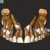
2 3 THE ONLY CBCT WITH LYING DOWN PATIENT POSITIONING. EXCELLENT IMAGE QUALITY WITH A DEVICE THAT FEATURES OUTSTANDING DIAGNOSTIC POTENTIAL. is the innovative device, featuring lying down patient positioning, able to offer high resolution volumetric images with extra low X-ray doses. Top quality CBCT for new medical applications.


3 4 CUTTING EDGE PERFORMANCE FOR ADVANCED DIAGNOSTICS. Quality and innovation with a device presenting exceptional features. Advanced diagnostics with, the only CBCT device with lying down patient positioning that offers excellent stabilisation and a broad range of FOVs for very high quality 3D and 2D images. CBCT technology allows to reduce the dose radiated to the patient up to 10-fold less than CT devices, with superior spatial resolution for bone tissue investigations. Special focus on patient health, enhanced by ECO Dose mode and the exclusive SafeBeam TM technology. is the first device with native FOV 21 x 19 cm for highly detailed investigations. The ideal device that produces clear, high definition images for orthopaedics, otorhinolaryngology, maxillofacial surgery and dentistry applications. SUPERIOR DIAGNOSTIC QUALITY Very high resolution 2D and 3D images with a broad range of FOV for an extensive selection of clinical applications. OPTIMAL LYING DOWN POSITION The only CBCT system with patient lying down positioning, a motordriven patient table and an open gantry. Perfect patient stabilisation considerably reduces any movement-induced artifacts. LOW X-RAY DOSE The ECO Scan mode and SafeBeam TM technology offered by further reduce the dose radiated to the patient, compared to examinations performed with CT technology. SPECIALIST SOFTWARE The adaptive user interface displays images and provides access to innovative 3D and 2D analysis functions for rapid and accurate diagnoses and optimal workflow.


4 6 7 UNCOMPROMISING QUALITY. Superior standard 3D examinations with a device designed for excellent performance. High definition volumetric images of bone tissue with a native isotopic voxel, non-overlapping sections and fewer artifacts. With CBCT technology, offers faster examinations and low doses of radiation with greater safety for the patient, better performance and an increasingly efficient workflow. The high quality images generated by are ideal for multiple medical fields, such as dentalmaxillofacial diseases, otorhinolaryngology applications, complete analysis of upper airways and precise examinations of bones and joints, limbs and the cervical spine. MSCT X-Ray Fan Beam CBCT X-Ray Cone Beam X-Ray Source THE CONVERGENCE OF TECHNOLOGY, PERFORMANCE AND SAFETY The powerful generator with rotating anode and smaller focal spot optimises performance, adapting the emission to the specific needs of the examination. A large flat panel sensor with a high signal-noise ratio improves image quality, expanding 3D and 2D diagnostic capacity. Innovative volumetric reconstruction algorithms control the image chain and enhance diagnostic potential, while minimising the presence of artifactsi. The exceptional user-friendliness makes the ideal choice for many image acquisition protocols, such as Ray2D examinations, the study of joint dynamics with CineX protocol, and very high resolution 3D diagnostics to examine bone tissues. 360 reconstruction The 360 scan acquires the entire volume with a single rotation. rapidly generates a complete dataset of axial, coronal and sagittal images as well as 3D renderings. extra FOV Vision The innovative extra FOV function allows to examine longitudinal anatomical parts. FOV 3D can be set from a minimum of Ø6 x h6 cm, up to a maximum native diameter of 21 cm or a height of 22 cm. HiRes analysis Clear and detailed very high resolution images to see bone micro-fractures or examine anatomical districts with micrometric details.


5 8 9 OPTIMAL LYING DOWN POSITIONING. User-friendliness, maximum stabilisation and quality for diagnoses using new medical applications. is the only CBCT device available on the market with patient in a lying down position. The motordriven patient table made of carbon fibre, which can be controlled from the on-board console or from the PC, allows to adapt the examination to any image acquisition need with patient lying down in a prone or supine, cranial-caudal or caudal-cranial position. The open gantry facilitates access to the scanning area and eliminates any sensation of claustrophobia and anxiety. Upper limb examinations are performed with the patient seated on the opposite side of the table. The lying down position is ideal for sedated, postsurgery and traumatised patients and to study sleep apnea. Reconstructed images are less subject to movement-induced artifacts and examination does not require the use of securing devices, thus ensuring better patient comfort. The on-board console offers a user-friendly interface to easily move the patient table along three axes, and to activate the alignment lasers by defining the exact reference points of the area of interest. ASSISTED ALIGNMENT The operator directly performs assisted alignment from the workstation via two scout images for automatic adjustment of the motor-driven patient table. The positioning and lock device has been specifically designed for the various dental and medical disciplines.

 8:8.")
6 10 11 LOW X-RAY DOSE. Wellbeing and safety are central to NewTom research. CBCT technology, compared to MSCT examinations, offers superior diagnostic quality for bone tissue with a much lower radiated dose. Exposure comparable to two 2D X-rays (AP and LL) generally needed for a preliminary examination*. *Koivisto et al. Effective radiation dose of a MSCT, two CBCT and one conventional radiography device in the ankle region, Journal of Foot and Ankle Research (2015) 8:8. MSCT (3D) (3D) 2D (AP + LL) offers high standard results with low radiation dose for the patient and exceptional performance as a result of unrivalled topnotch features: the high power generator improves filtration that protects against the more harmful low-energy radiations; X-ray emission occurs in pulsed mode during the scan for an extremely limited time, from a minimum of 0.9 s to a maximum of 5.4 s; finally, variable collimation limits exposure to regions of interest only. ECO Scan Low emission up to 0.9 seconds of emission for standard examinations. The ECO Scan protocol is ideal for post-surgery follow-ups and paediatric applications. SafeBeam TM The exclusive SafeBeam TM technology eliminates the risk of exposing the patient to an unnecessarily high dose by automatically adapting radiation levels to suit the patient s anatomical characteristics. Ray2D The Ray2D function allows to perform a preliminary low dose 2D X-ray examination, which can be followed, if necessary, by a high resolution 3D examination only of the area of interest, for in-depth diagnostics.


7 13 APPLICATION FIELDS. With, NewTom uses CBCT technology for new medical applications. Very high quality 2D and 3D images with a broad range of FOVs and dedicated software. An extraordinary potential for accurate diagnosis in all situations.


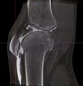

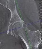
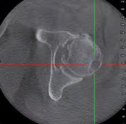
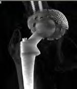
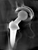
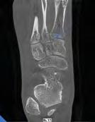
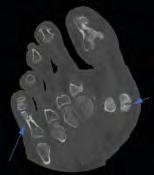
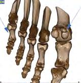
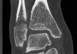
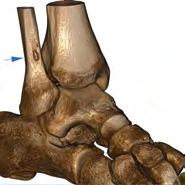
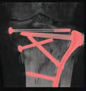
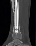
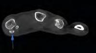
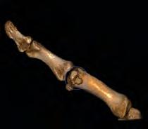

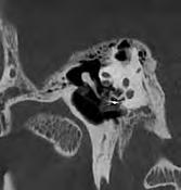
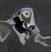
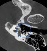
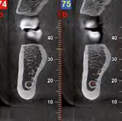
8 14 15 ORTHOPAEDIC APPLICATIONS. Images generated by, with their high resolution and quality, allow an in-depth study of the upper and lower limbs not only to diagnose fractures, dislocations, luxations or misalignment but also to define the bone and joint structure resulting from pathological alterations, to detect small bone fragments and to assess diseases in small joints, even when metal screws are present. Excellent acquisitions that exceed the limitations of CT examinations or those typical of 2D image acquisitions, in which a dedicated visual alignment cannot always prevent overlapping bone structures, thus generating a negative initial diagnosis even in cases presenting a high suspicion of fractures. offers perfect 3D images during post-surgery follow-up in the framework of osseointegration of prostheses, plates and bone implants, and in monitoring the healing status even with external immobilisation systems, such as, for example, plaster cast, splints or metal anchoring devices. LOWER LIMBS Planning and post-surgery assessment of implants, plates or prostheses. UPPER LIMBS Diagnosis of traumas with evidence of micro-fractures and follow-up treatment.


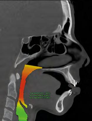
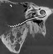

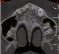
9 16 17 HEAD&NECK APPLICATIONS. INVESTIGATING NECK PAIN The better spatial resolution of CBCT, compared to MSCT, allows detailed analyses of trabecular and cortical structures to identify any dysplastic, inflammatory, traumatic or micro-traumatic elements. Relationships between vertebral bodies are also perfectly legible, thus highlighting any distortion or subluxation. 3D volumes generated with are ideal to examine the atlanto-occipital joint and in surgical programming for the application of osteosynthesis devices and prosthetics. UPPER AIRWAY EXAMINATION With dedicated FOVs, can generate the maxillary and frontal sinuses, nose and upper airways within a single scan, thus enabling the diagnosis of otorhinolaryngology diseases, such as sleep apnea (OSA). The analysis software offers dedicated tools for volume measurements, thus facilitating identification of the most critical shrinkage situations. is the only CBCT device that allows this examination to be performed with the patient in a lying down position. PLANNING AND VERIFYING MAXILLOFACIAL SURGERY generates the entire maxillofacial are within a single scan to verify the presence of fractures or other pathologies, characteristics of the bone and dental arches and the impact of dentition and its roots on both the mandibular canal and the maxillary sinuses. An essential tool for precise planning of surgical treatment and for post-surgery follow-up. Indeed, the presence of metal elements does not impact image quality as a result of innovative filters and the small quantity of radiation, which reduces the scattering effect to a minimum. INTERNAL EAR EXAMINATION Several studies show that CBCT images perfectly identify implant position both in the round window and the incus. This examination exposes the patient to fewer risks of exposure to ionising radiations; hence, it is preferable for middle ear implant follow-up examinations. NewTom s exclusive SafeBeam TM technology also allows to expose the patient only to the necessary dose.
")


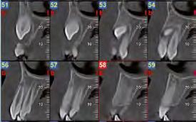
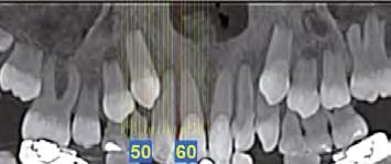
10 18 19 HEAD&NECK APPLICATIONS. TEMPOROMANDIBULAR JOINT (TMJ) EXAMINATION Diagnosis and anatomical assessment of the temporomandibular joint can be performed with high quality 3D images generated by. Sagittal and coronal slices provide optimal imaging of the joint zone to identify any pathologies and to assess the difference between condyle height and the mandibular branch. ENDODONTIC AND PERIODONTIC EXAMINATIONS is a particularly effective device to assess apical lesions, plan fractured tooth treatment and mandibular canal therapy, and treat tissue adjacent to the tooth. In fact, the detailed images generated by the device are very useful for endodontic therapy and periodontic examinations. To protect patient safety, FOV size can be adapted to the region of interest. ORTHODONTIC ANALYSIS The ideal application for tomographic, panoramic and cephalometric images acquired with is in examinations for aesthetic and orthodontic purposes and for the treatment of severe diseases. The realistic image provided by 3D examinations, compared to 2D diagnostics, allows to modify the visual angle and to adjust the thickness of reconstructed sections. Hence, the mutual position of dental elements and relations with the surrounding anatomical structures can be precisely assessed. These functions are essential to plan treatment, especially in the event of supernumerary and/or impacted teeth. DEFINITION AND CONTROL OF ORAL IMPLANT SURGERY Volumes obtained with are a highly effective tool to plan implant surgery, perform a realistic assessment of the site and choose the implant. Sizes in a 1:1 scale and detailed images of surrounding bone quality provide precise implant positioning indications. The simulation can be displayed on 3D rendering, and specialised 3D software can be used to precisely plan prosthetically guided implant surgery and customise the surgical template. Follow-up examinations will allow necessary assessments on the osseointegration process rate and on rejections, if any.


11 20 21 NNT, THE SOFTWARE FOR ALL SPECIALIST NEEDS. The versatile and powerful imaging software to perform the examination, process data and share the diagnosis. NNT is the essential tool to process and manage 2D and 3D images and X-ray videos (CineX). A software that adapts the user interface and offers dedicated analysis functions for the specific needs of radiologists and specialist physicians. Volume reconstruction algorithms and advanced filters created by NewTom s experience optimise final image quality and reduce the presence of artifacts and time required for image reconstruction. 3D volumes, 2D images and videos processed with the CineX function, which are compatible with DICOM 3.0 (IHE) standard, can be easily shared via the NNT Viewer version or printed on a 1:1 scale through customisable reports. DENTISTRY: CROSS SECTIONS IN PANORAMIC IMAGES Complete examination of the dental arches in cross sections to check shape, size and status of maxillary and mandibular bones and teeth. OTORHINOLARYNGOLOGY: FREE MULTIPLANAR SECTIONS Dynamic high resolution examination of the internal ear along non-orthogonal planes is essential to diagnose any diseases of the ossicular chain, stapes base, semicircular canals, cochlea and adjacent structures. GNATHOLOGY: DUAL TMJ VISION Simultaneous examination of both temporomandibular joints; symmetrical analysis and detection of problems or dysfunction deriving from joint diseases. RADIOLOGY: MULTI-SLICE EXAMINATION Examination of multiple image samples in Med-Like style with personalised orientation for the various assessments of anatomical areas scanned.

 that are")


12 22 23 SPECIALIST TOOLS. Ray2D With the innovative Ray2D function, generates 2D X-ray images (18 x 19 cm) that are perfect for both preliminary and post-surgery follow up examinations. It is possible to repeat the investigation from various angles to select the image with the best perspective. IMPLANT SITE ASSESSMENT Estimates bone density in a potential implant site, with Misch scale classification, to correctly plan treatment. CineX offers the exclusive CineX function to investigate moving anatomical structures, such as saliva ducts and joint mobility. This advanced technology uses a sequence of X-ray images to create an 18 x 19 cm format video that can also be exported to standard video format. AIRWAY VOLUME ANALISYS Estimating the actual upper airway space is essential to diagnose respiratory diseases and sleep apnea (OSA). 2D AND 3D EVALUATION The possibility to evaluate distances on 2D sections or with 3D rendering to verify any joint problems. ADVANCED REPORTS Advanced draft of medical reports to share on PACS, also available in automatic compiling mode.


13 SPECIALIST PLANNING SOFTWARE COMPLETE CONNECTIVITY. Excellent connectivity and integration with the modern systems adopted by NewTom. Workflow and clinical and diagnostic activities become much easier and highly performing. REMOTE ASSISTANCE By appropriately configuring the device to use the surgery s Internet connection, technical support can be provided from remote, and device status can be monitored. 3D/2D VIEWER Examinations can be shared with colleagues and patients by providing the Viewer directly on CD, DVD or a USB storage device. 1:1 PRINT Complete and flexible report for storing and sharing colour reports on photographic paper or grey scale reports on X-ray-equivalent transparencies. OTHER ACQUISITION DEVICES Compatibility with TWAIN standard and DICOM 3.0, guarantees the NNT software management of images from other 2D/3D image acquisition devices, such as video cameras, sensors, PSP and CBCT scanners. MULTI-STATION DISPLAY AND PROCESSING Image storage on a shared database in a local network that can be accessed from any workstation and ipad (only 2D). Management of multiple archives and access to passwordprotected data. 1:1 PRINT 3D/2D VIEWER REPORTS OTHER ACQUISITION DEVICE REMOTE ASSISTANCE 2D/3D IMAGE MANAGEMENT MULTI-STATION DISPLAY AND PROCESSING RIS/PACS INFORMATION SYSTEMS SOFTWARE SURGERY TREATMENT SYSTEMS 3D SCANNER SURGERY MANAGEMENT 3D MILLING PRINTERS RIS/PACS IHE compliant system that allows communication with RIS/PACS systems and DICOM printers. Complete services available: Print, Worklist, Storage Commitment, MPPS and Query/Retrieve. SURGERY MANAGEMENT SOFTWARE An open system designed for fast, efficient interfacing with the main dental surgery management software solutions via various standard VDDS, TWAIN and/or proprietary NNTBridge modes. SPECIALIST PLANNING SOFTWARE Exports in DICOM 3.0 format to specialist planning software to process orthodontic treatments, prostheses, implants, orthognatic and maxillofacial surgery. 3D MILLING PRINTERS Software modules are available to segment the reconstructed volume and export to STL format the surfaces required to create 3D models that can underpin planning and treatment. 3D SCANNER Prosthetically guided planning by integrating (via the dedicated software module) data in STL format from optical, intraoral or laboratory scanners, with volumetric data.
14 26 27 TECHNICAL SPECIFICATIONS. X-ray source Focal spot Exposure Control Sensor Grey scale 3D scan time 3D emission time 3D image acquisition High frequency generator, rotating-anode X-ray tube 0.3 mm SafeBeam to reduce exposure according to patient build Amorphous silicon flat panel 16-bit 18-36s 0.9s - 9.0s (single scan) Single scan with Cone Beam technology. 360 rotation Dimensions in centimetres (dimensions in inches) Available FOV Diameter x Height Resolution Selectable 3D scan modes Standard HiRes Eco Regular Boosted Enhanced 21 x 19 cm 18 x 16 cm 15 x 22 cm efov 15 x 12 cm 15 x 5 cm 12 x 8 cm 10 x 10 cm 10 x 5 cm 8 x 8 cm 8 x 5 cm 6 x 6 cm Selectable voxel size Standard Selectable voxel size HiRes Reconstruction time Ray2D image acquisition CineX image acquisition Patient positioning Weight Software DICOM nodes Power supply µm µm Less than 1 minute Digital Radiography (single shot, position selectable by user) 1-36s Serial Radiography, field of view 18x19 cm (WxH) Seated or lying down, prone or supine, in cranial-caudal or caudal-cranial position 660 Kg NewTom NNT with Viewer software, free IHE compliant (Print; Storage Commitment; WorkList MPPS; Query Retrieve) V~, V~, 10 V~, V~, 50/60 Hz Specifications subject to change without prior notice. 0051
ADVANCED 3D IMAGING. CEFLA s.c. Via Selice Provinciale 23/a Imola Italy t newtom.
 CEFLA s.c. Via Selice Provinciale 23/a 40026 Imola Italy t. +39 045 8202727 045 583500 info@newtom.it newtom.it 05/2018 NVGEGB181S00 According to the standards in force, in extra-eu areas the availability
5G XL - R EN ENGLISH
 5G XL - R15.0 - EN ENGLISH Sede legale ed amministrativa - Headquarters QR srl - Via Selice Provinciale, 23/a - 40026 Imola - Bo (Italy) Stabilimento - Plant Via Fermi, 40-37136 Verona (Italy) Tel. +39
ENGLISH. Cefla s.c. - Via Selice Provinciale 23/a, Imola - Italy Tel
 09-2017 NVGEGB161S00 According to the regulations in force, some products and/or features may have different availability and characteristics in areas outside of the European Union. Please contact your
NewTom GiANO HR PERFECT.VISION
 CEFLA s.c. Via Selice Provinciale 23/a 40026 Imola Italy t. +39 045 8202727 045 583500 info@newtom.it newtom.it 06/2018 NHRGB181S00 According to the standards in force, in extra-eu areas the availability
CS 9300 Family. The power of flexibility
 CS 9300 Family The power of flexibility The new CS 9300 digital imaging system from Carestream Dental take the guesswork out of examinations The all-in-one CS 9300 is the most versatile multimodality imaging
VGi - R EN ENGLISH. QR srl - Via Silvestrini, Verona Italy Tel
 VGi - R15.1 - EN ENGLISH QR srl - Via Silvestrini, 20-37135 Verona Italy Tel. +39 045 8202727-045 583500 info@newtom.it www.newtom.it FIRST IN CONE BEAM, ACCURATE IN RESULTS. 360 degree imaging, reduced
ENGLISH. Cefla s.c. - Via Selice Provinciale 23/a, Imola - Italy Tel
 11-2016 N5GGB161S00 According to the regulations in force, some products and/or features may have different availability and characteristics in areas outside of the European Union. Please contact your
Head to new heights with your imaging SCANORA 3D
 SCANORA 3D Head to new heights with your imaging Benefits at a glance The solution for dentomaxillofacial and ENT imaging Easy Patient seated for added stability during exposure. Clear, self-explinatory
Profound understanding of anatomy
 ENGLISH Profound understanding of anatomy The unique Planmeca ProMax 3D product family offers equipment for all maxillofacial imaging. All volume sizes from the smallest special cases to whole head images
I AM DEMANDING Type CMOS Flat Panel CMOS CMOS ø 40 x 40 mm, ø 60 x 60 mm, ø 80 x 80 mm, ø 110 x 80 mm
 TECHNICAL SPECIFICATIONS 1168 1501 1978 1237 1551-2351 Ø 1090 PANORAMIC CBCT CEPHALOMETRIC X-RAY SOURCE Tube type High frequency DC generator 2.8 mmal / 85 kv 7.0 mmal / 90 kv 2.8 mmal / 85 kv Operation
3D/2D. Hyperion X5 Suspended imaging system. Data subject to change without notice. 03/2017 MX53DGB171S00
 www.my-ray.com Data subject to change without notice. 03/2017 MX53DGB171S00 According to the regulations in force, some products and/or features may have different availability and characteristics in areas
English. Perfect Vision
 English Perfect Vision Everything becomes clearer and simpler with a big F.O.V. The complete dentomaxillofacial volume ready for your diagnosis One scan provides you with an incredible amount of information
PAN CEPH 3D CONE BEAM
 PAN CEPH 3D CONE BEAM 2D - 3D panoramic units PANORAMIC CEPHALOMETRIC 3D CONE BEAM IMAGING I-MAX TOUCH Tactile & naturally intuitive panoramic imaging Discover the simplicity and efficiency this unit can
Planmeca ProMax 3D s Planmeca ProMax 3D ENGLISH
 Planmeca ProMax 3D s Planmeca ProMax 3D ENGLISH Genuine all-in-one unit Planmeca ProMax 3D s and Planmeca ProMax 3D units are designed to obtain complete information on patient anatomy in the minutest
Profound understanding of anatomy
 ENGLISH Profound understanding of anatomy The unique Planmeca ProMax 3D product family offers equipment for all maxillofacial imaging. All volumes sizes from the smallest special cases to whole head images
CT Imaging at the Point-of-Care
 ENGLISH True Dedication The new Planmed Verity Extremity CT Scanner revolutionizes extremity CT imaging. The compact unit brings 3D imaging at emergency departments, orthopedic clinics or trauma centers
True Low Dose. Exact time to display image on screen may vary upon computer and network configuration.
 RAYSCAN ALPHA PLUS True Low Dose Cone Beam CT Industry Leading Resolution High resolution images provide all the clinical information needed while keeping radiation exposure low. Endodontics - Smallest
Cone Beam 3D Imaging
 Cone Beam 3D Imaging NewTom Sets the Standard in 3D Maxillofacial Imaging Cone Beam 3D Imaging The Global Market Leader The Inventors n of Cone Beam 3D In 1996, QR srl developed the first generation of
ENGLISH. Distributed by: QR srl - Via Silvestrini, Verona Italy Tel
 GiANO - R15.1 - EN ENGLISH Distributed by: QR srl - Via Silvestrini, 20-37135 Verona Italy Tel. +39 045 8202727-045 583500 info@newtom.it www.newtom.it Manufacturer: CEFLA S.C. - CEFLA DENTAL GROUP Via
DIAGNOSTIC IMAGING. OPTIMIZED.
 ABOUT LED DENTAL SEE THE DIFFERENCE Using our years of business insight and clinical experience as a foundation, LED Dental takes the uncertainty out of your imaging purchase decision. We offer our clients
XPan 3D Plus. FONA Every dental solution you need. Advanced dental technology. Headquarters THE ULTIMATE DIAGNOSTIC SOLUTION DIGITAL DENTISTRY
 FONA Every dental solution you need Through decades of experience and deep understanding of the dental profession, we deliver complete, reliable and accessible solutions.regardless of country or specialisation,
Dental Line. 3D digital panoramic system. radiology ahead
 Dental Line 3D digital panoramic system radiology ahead new generation 3D digital panoramic unit 3D imaging s value available for anyone Following the incredible success of the innovative digital panoramic
2D AND 3D/2D WALL-MOUNTED PANORAMIC UNITS
 2D AND 3D/2D WALL-MOUNTED PANORAMIC UNITS KEEP YOUR CLINIC ONE STEP AHEAD! Wall-mounted concept: zero foot print 62kg - the lightest unit on the market Face to face positioning High Definition The fruit
ENGLISH. Cone Beam 3D Imaging
 ENGLISH Cone Beam 3D Imaging FIRST USER OF CONE BEAM IN DENTAL FIELD QR s.r.l. is the name that stands behind NewTom Cone Beam 3D imaging units and the creator of Cone Beam technology for the dental field.
ENGLISH VGi - R ENG
 ENGLISH First in Cone Beam, Accurate in Results Cone Beam 3D Imaging First User of Cone Beam in Dental Field QR s.r.l. is the name that stands behind NewTom Cone Beam 3D imaging units and we were the creators
Flexible Easy Competitive. SCANORA 3Dx - The in-office large field-of-view Cone Beam CT system for Head and Neck imaging
 Flexible Easy Competitive SCANORA 3Dx - The in-office large field-of-view Cone Beam CT system for Head and Neck imaging SCANORA 3Dx. The solution. SCANORA 3Dx makes advanced 3D imaging easy in the head
OP 3D Vision The upgradable 3D X-ray system for the strictest demands.
 OP 3D Vision The upgradable 3D X-ray system for the strictest demands. The solution for every task: KaVo OP 3D Vision. Regardless of which dental query you may have, the KaVo ORTHOPANTOMOGRAPH OP 3D Vision
Profound understanding of anatomy
 ENGLISH Profound understanding of anatomy Planmeca ProMax 3D, the intelligent and multipurpose X-ray unit, is designed to obtain complete information on patient anatomy in the minutest detail. The unit
Low Dose Excellent Image Quality Rapid Reconstruction
 Low Dose Excellent Image Quality Rapid Reconstruction Efficient 3 in 1 Dental X-ray System CBCT > Precise 3-D Anatomical structures - Accurate diagnosis for doctors - Safe implant for patients > Significant
- RCS Paris B
 Technical specifications PANORAMIC CBCT CEPHALOMETRIC X-ray source Tube type High frequency DC generator Total filtration >2.5 mm Al @ 90 kv Mode of operation Continuous Pulsed Continuous Tube voltage
The Quality Leader in 3D Cone Beam CT
 The Quality Leader in 3D Cone Beam CT The Complete 2-in-1 or 3-in-1 Multi-modality Solution PreXion, with over 15 years of innovation in the medical and dental fields, introduces the PreXion3D Eclipse.
3D Cone beam CT & Digital Radiography Dedicated to Otorhinolaryngology
 3D Cone beam CT & Digital Radiography Dedicated to Otorhinolaryngology Multi-functional imaging solution3 RAYSCAN m is an unique 2-in-1 imaging solution, combining Cone Beam CT and Digital Radiography,
STELLARIS 3D 4 IN 1 CBCT SOLUTION FOR ADVANCED DIAGNOSTICS
 STELLARIS 3D 4 IN CBCT SOLUTION FOR ADVANCED DIAGNOSTICS 3 STELLARIS 3D 4 IN CBCT SOLUTION FOR ADVANCED DIAGNOSTICS Stellaris 3D is a complete and compact, fully upgradeable 3D CBCT for a patient, Panoramic
D3D CBCT. See more Do More
 D3D CBCT See more Do More BIOLASE DaVinci Imaging D3D For capturing superior 3D image acquisitions with the lowest minimal dose for patient safety The BIOLASE DaVinci Imaging D3D has one of the lowest
T h e D e n t a l C o m p a n y FROM DIAGNOSTIC SCAN TO SURGERY, WE SHAPE THE FUTURE OF DENTISTRY.
 T h e D e n t a l C o m p a n y FROM DIAGNOSTIC SCAN TO SURGERY, WE SHAPE THE FUTURE OF DENTISTRY. SIDEXIS SOFTWARE ORTHOPHOS SL D/D SEAMLESS THE NEW STANDARD IN CLINICAL DIAGNOSIS AND PATIENT COMMUNICATION
fast accurate safe See the full picture: add a 3rd dimension to your patient evaluation to diagnose more effectively. BENEFITS OF NEWTOM 3G 6" 9" 12"
 See the full picture: add a 3rd dimension to your patient evaluation to diagnose more effectively. Examination Effective Dose Equivalent (ICRP tissue weights 2005) Panoramic Dose (ICRP tissue weights 2005)
Hyperion X9 pro Professional 3-in-1 full-touch imaging system
 www.my-ray.com Data subject to changes without prior notice. 06/2018 M9PROGB181S00 According to the relevant regulations, in the extra-eu areas, some products and/or characteristics might have different
THE WAIT IS OVER CS D. 3D imaging is now available for everyone
 THE WAIT IS OVER CS 8100 3D 3D imaging is now available for everyone COMPLEXITY IS NO LONGER THE STANDARD NOW THERE ARE MANY REASONS TO MOVE TO 2D/3D IMAGING Now it s possible to experience nothing but
X X X. GXS-700 Direct USB Digital Intraoral Sensors. Buy a Sensor Combo and a Digital Pan Unit, Receive $600 Off! GO.BENCO benco.
 8 0 0. G O. B E N C O b e n c o. c o m GS-700 Direct USB Digital Intraoral Sensors Designed to make migrating from film, or upgrading an existing digital system, easier than ever High quality image capture
The Optimum Choice for Implantologist
 The Optimum Choice for Implantologist What is essential for your practice? What s the best way to choose a 3D X-ray machine for implant treatment planning? 02 Doctor says.. There are diagnostic limitations
CT Scanning Protocol For V2R Guided Surgery Solutions
 CT Scanning Protocol For V2R Guided Surgery Solutions 2 V2R CT Scanning Protocol \\ Contents Contents General requirements... 3 V2R Dual Scan Protocol... 5 V2R Single Scan Protocol... 8 Overview... 10
VistaVox S 3D from Dürr Dental
 VistaVox S 3D from Dürr Dental 3D and 2D X-ray images with exceptional image quality COMPRESSED AIR SUCTION IMAGING DENTAL CARE HYGIENE Taking diagnostics to the next level VistaVox S combines diagnostic
Planmeca ProMax 3D s Planmeca ProMax 3D ENGLISH
 Planmeca ProMax 3D s Planmeca ProMax 3D ENGLISH Learn more: Planmeca Imaging for ipad Genuine all-in-one unit Planmeca ProMax 3D s and Planmeca ProMax 3D units are designed to obtain complete information
3Shape X1 Scanning redefined
 3Shape X1 Scanning redefined Why choose the X1 Give your patients a great experience No head fixation and sleek design create a comfortable scanning experience for your patient High image quality low dose
Digital Imaging from a new perspective
 TREATMENT CENTRES HANDPIECES HYGIENE SYSTEMS X-RAY SYSTEMS CEREC TREATMENT CENTRES HANDPIECES HYGIENE SYSTEMS X-RAY SYSTEMS CEREC SIRONA CREATING AND MAINTAINING VALUE. You are right to expect a great
3Shape X1 Scanning redefined
 3Shape X1 Scanning redefined Why choose the X1 Give your patients a great experience No head fixation and sleek design create a comfortable scanning experience for your patient High image quality low dose
2
 1 2 3 4 5 6 7 8 9 10 11 12 13 Cine loop of tomosynthesis slice images through the chest. 14 Standard PA chest radiograph (left) and single slice from the tomosynthesis image dataset (right) of a patient
Versatility And Expandability In One Panoramic.
 Orthoralix 9200 / 9200 DDE Versatility And Expandability In One Panoramic. Panoramic X-ray Systems Intraoral X-ray Systems Digital Intraoral Sensors Digital X-ray Phosphor Plates Intraoral Cameras Imaging
Hyperion X9 3-in-1 Imaging System
 Hyperion X9 3-in-1 Imaging System 2 Hyperion X9, full imaging. 3 Hyperion X9, just right for me. The present and the future of my work. In three dimensions. Hyperion X9 offers me multiple possibilities
ENGLISH. Advanced tools for orthodontics
 ENGLISH Advanced tools for orthodontics A complete solution for orthodontics One unit one software Panoramic Cephalometric CBCT image 3D photo Planmeca ProMax unit 3D model scan Planmeca offers a complete
WITH. The Next Step in Office MRI
 WITH The Next Step in Office MRI Introducing S-scan the Next Step in Office MRI Based on extensive customer feedback and years of engineering, Esaote has designed the S-scan with exp Technology, an optimized
ENGLISH. Advanced tools for orthodontics
 ENGLISH Advanced tools for orthodontics A complete solution for orthodontics One unit one software Panoramic Cephalometric CBCT image 3D photo Planmeca ProMax unit 3D model scan Planmeca offers a complete
vertaplan the spine surgeon s software vertaplan System for successful reconstruction of the individual sagittal balance
 the spine surgeon s software System for successful reconstruction of the individual sagittal balance What do you think of patient-specific reconstruction of the spine geometry? Optimum surgical outcome
GUIDED SURGERY TECHNIQUE
 GUIDED SURGERY TECHNIQUE INDEX WORKFLOW...4 OUR SOLUTIONS...5 PLANNING PROTOCOL - PARTIAL EDENTULISM...6 - TOTAL EDENTULISM...9 SOFTWARE FEATURES...12 SURGICAL COMPONENTS FOR GUIDED IMPLANTOLOGY...14
3D Panoramic Cephalometric. Innovation, in reach. KODAK 9000 Extraoral Imaging System
 3D Panoramic Cephalometric Innovation, in reach 9000 KODAK 9000 Extraoral Imaging System Cephalometric Innovation made simple Innovation made simple We believe in innovation. We always have. In fact, our
CS 8100 FAMILY / CS D FAMILY / CS 9300 FAMILY EXTRAORAL SOLUTIONS EXTRAORDINARY POSSIBILITIES
 CS 8100 FAMILY / CS 8100 3D FAMILY / CS 9300 FAMILY EXTRAORAL SOLUTIONS EXTRAORDINARY POSSIBILITIES A SOLUTION FOR EVERY CLINIC AND ANY CONSULTATION The CS 8100 does more than my old machine and is half
THE USE OF KEYSTONE EASYGUIDE CT SCANNING SOFTWARE FOR DIAGNOSIS, DIRECTION AND DEPTH DETERMINATION
 CT DIAGNOSTICS IN 3D IMPLANT TREATMENT PLANNING THE USE OF KEYSTONE EASYGUIDE CT SCANNING SOFTWARE FOR DIAGNOSIS, DIRECTION AND DEPTH DETERMINATION Timothy Kosinski, DDS, MAGD Assistant Clinical Professor
For true visualisation
 ENGLISH For true visualisation Planmeca ProModel is a patient-specific physical model for high-end maxillofacial operations and dental surgery. By reproducing the anatomy of the patient in real-size, Planmeca
3D/2D WALL MOUNTED UNIT
 Product launch document EN Page 1 sur 30 EN PRODUCT LAUNCH DOCUMENT REV03, September 2018 3D/2D WALL MOUNTED UNIT Product launch document EN Page 2 sur 30 INDEX 1. PRODUCT IDENTITY AND POSITIONNING...
Xelis Dental - What New in
 Xelis Dental - What New in 1.0.6.1 Clinical Needs Why Xelis-Dental? Panorama Cephalography Intra-oral Digital Camera Traditional imaging systems - 2-Dimension view Incorrect anatomical information - Distortion
INNOVATING IN PERSONALISED SOLUTIONS
 INNOVATING IN PERSONALISED SOLUTIONS At the forefront of custom medical technologies 4 EXPERIENCE BUILT ON SUCCESS Innovation and research, AVINENT s twin foundations AVINENT is at the forefront of custom
3D Accuitomo XYZ Slice View Tomograph. Super High-Resolution Images of Region of Interest
 3D Accuitomo XYZ Slice View Tomograph. Super High-Resolution Images of Region of Interest Thinking ahead. Focused on life. 2 Cone Beam X-Ray CT Imaging X-Ray Tube Imaging Intensifier Imaging Volume Voxel
EN PRODUCT LAUNCH DOCUMENT. I-MAX 3D 2016 Révision : V Product launch document EN Page 1 sur 29. Version 02, OCTOBER 2016
 Product launch document EN Page 1 sur 29 EN PRODUCT LAUNCH DOCUMENT Version 02, OCTOBER 2016 Product launch document EN Page 2 sur 29 INDEX 1. PRODUCT IDENTITY AND POSITIONNING... 3 2. TECHNICAL CHARACTERISTICS...
The mission of our company is the development of an individual approach in functional dentistry.
 The Prosystom company started up in 2013. It was created by Russian doctors and IT specialists for carrying out scientific projects and practical work in functional dentistry. The mission of our company
clinical articles management advice practice profiles technology reviews Implant PRACTICE US Volume 3 No 2
 clinical articles management advice practice profiles technology reviews Implant PRACTICE US Volume 3 No 2 Sirona Sirona tailors solutions to the needs of the different markets within dentistry to ensure
AWARD-WINNING CONE BEAM 3D DENTAL IMAGING
 AWARD-WINNING CONE BEAM 3D DENTAL IMAGING Dedicated to Advancing Dental Treatment A COMPLETE 3D TREATMENT SOLUTION Your dental practice is unique that s why you need a flexible solution that works with
3D-MODEL CUSTOM-MADE MODELS SEGMENTATION AND PRODUCTION SERVICE OF BONE MODELS WITH HIGHEST 3D PRINTING RESOLUTION
 CUSTOM-MADE MODELS 3D-MODEL SEGMENTATION AND PRODUCTION SERVICE OF BONE MODELS WITH HIGHEST 3D PRINTING RESOLUTION FOLLOW US ON CUSTOM-MADE MODELS 3D-MODEL From a CT or CBCT scan, 3D-model service provides
Easy operation. Numerous diagnostic options. X-rays you can rely on: the ORTHOPHOS XG device family. All ORTHOPHOS XG 3Dready programs at a glance.
 CAD /CAM Systems Instruments Hygiene Systems Treatment Centers Imaging Systems Subject to technical changes and errors in the text, Order No. A91100-M47-B346-01-7600, Printed in Germany, Dispo-Nr. 04602,
ORTHOPANTOMOGRAPH OP200 D ORTHOCEPH OC200 D. True dynamo leading through the decades.
 ORTHOPANTOMOGRAPH OP200 D ORTHOCEPH OC200 D True dynamo leading through the decades. 1 You can t dublicate the legacy. 1946 Professor Y.V. Paatero publishes his first paper on Panoramic Tomography. 1951
B. CT protocols for the spine
 B. CT protocols for the spine Poster No.: A-003 Congress: ECR 2010 Type: Invited Speaker Topic: Neuro Authors: B. Tins; Oswestry/UK Keywords: CT, spine, diagnostic imaging protocol DOI: 10.1594/ecr2010/A-003
I AM EXCLUSIVE TECHNICAL SPECIFICATIONS. The first personal imaging plate scanner IMAGING PLATES. System. Windows recommended configuration
 TECHNICAL SPECIFICATIONS System Resolution... 20 lp/mm Scan Time (fast mode)...1,6s - 2,7s Scan Time (high definition mode)...2,1s - 3,6s Connection... Ethernet RJ-45 Dimensions... L. 154 x D. 204 x H.
Full ultrasound breast volumes. Faster scans. Streamlined workflow. ACUSON S2000 Automated Breast Volume Scanner. Answers for life.
 Full ultrasound breast volumes. Faster scans. Streamlined workflow. ACUSON S2000 Automated Breast Volume Scanner Answers for life. 1 ACQUIRE An automated whole breast solution. Reduced acquisition time.
Devoted to the Advancement of Implant Dentistry
 Devoted to the Advancement of Implant Dentistry Devoted to the Advancement of Implant Dentistry Our ultimate goal is to provide you and your patients with the highest standards in implant case planning
Extraoral Imaging. Chapter 42. Copyright 2018, Elsevier Inc. All Rights Reserved. 1
 Extraoral Imaging Chapter 42 Copyright 2018, Elsevier Inc. All Rights Reserved. 1 Learning Objectives Lesson 42.1: Panoramic Imaging 1. Pronounce, define, and spell the key terms. 2. Discuss panoramic
CBCT: A dentist perspective
 SEGi Review ISSN: 1985.5672 Vol.9, December 2015 CBCT: A dentist perspective Dr. Ranjana Garg, Faculty of Dentistry SEGi University. Dr. Vivek Vijay Gupta Faculty of Dentistry SEGi University Abstract
UNDERSTANDING DIGITAL DENTISTRY: CBCT AND INTRA-ORAL 30 SCANNING
 UNDERSTANDING DIGITAL DENTISTRY: CBCT AND INTRA-ORAL 30 SCANNING -=- & UNDERSTANDING DIGITAL DENTISTRY: CBCT AND INTRA-ORAL 30 SCANNING ----CBCTi-------iTERO------ NewTom VGi *Vertical Patient Positioning
Straight to the point with your diagnosis.
 CAD/CAM SYSTEMS INSTRUMENTS HYGIENE SYSTEMS TREATMENT CENTERS IMAGING SYSTEMS HELIODENT DS INTRAORAL X-RAY WITH EXCELLENT IMAGE QUALITY Straight to the point with your diagnosis. T h e D e n t a l C o
ORTHOPANTOMOGRAPH OP 3D Pro A platform for changing needs
 ORTHOPANTOMOGRAPH OP 3D Pro A platform for changing needs ORTHOPANTOMOGRAPH OP 3D Pro OP 3D Pro is the most comprehensive 3-in-1 platform designed for today and tomorrow, covering the entire maxillofacial
Digital mammography imaging from Carestream Health solutions for great workflow, productivity, and patient care.
 Digital Mammography Imaging on KODAK CR Systems Digital mammography imaging from Carestream Health solutions for great workflow, productivity, and patient care. Commercial distribution of the CR Mammography
SIMPLANT Guided Surgery delivering restorative-driven implant treatment
 SIMPLANT Guided Surgery delivering restorative-driven implant treatment SIMPLANT the key to unlocking digital potential As part of the DENTSPLY Implants Digital Solutions portfolio, SIMPLANT digital implant
GLOBAL INNOVATION BY DESIGN
 GLOBAL INNOVATION BY DESIGN For over 130 years Toshiba s research and development has improved the health and welfare of people around the world. Today, Toshiba Medical Systems offers a full range of diagnostic
CT SCAN PROTOCOL. Shoulder
 CT SCAN PROTOCOL Shoulder Purpose and Summary CT images made with this protocol are used to provide the orthopedic surgeon with a detailed 3D anatomical reconstruction of the patient s scapula and proximal
The Power Of Choice Pan. Ceph. 3D. Your Imaging Future Starts Today.
 NEW from Gendex! The Power Of Choice Pan. Ceph. 3D. Your Imaging Future Starts Today. Cone Beam 3D Imaging Systems Panoramic X-ray Systems Intraoral X-ray Systems Digital Intraoral Sensors Digital X-ray
User Guide for Dental and Maxillofacial Cone Beam Computed Tomography (CBCT)
") User Guide for Dental and Maxillofacial Cone Beam Computed Tomography (CBCT) Poster No.: C-0756 Congress: ECR 2014 Type: Educational Exhibit Authors: J. Ukkonen, J. Asp; Helsinki/FI Keywords: Education
User Guide for Dental and Maxillofacial Cone Beam Computed Tomography (CBCT) Poster No.: C-0756 Congress: ECR 2014 Type: Educational Exhibit Authors: J. Ukkonen, J. Asp; Helsinki/FI Keywords: Education
The Key to Confidence
 WITH The Key to Confidence The Key to Confidence More detail, better accuracy, greater confidence The G-scan Brio is a revolutionary MRI approach for all musculoskeletal applications, which allows you
INTRAORAL IMAGING ACCORDING TO YOUR NEEDS Intraoral X-rays
 INTRAORAL IMAGING ACCORDING TO YOUR NEEDS Intraoral X-rays FONA Intraoral X-rays INTRAORAL IMAGING ACCORDING TO YOUR NEEDS FONA intraoral X-ray devices are designed for ease of use, precision, reliability
 www.oralradiologists.com CONE BEAM CT REPORT CASE ---- Case Information Referring Doctor: - Patient Name: - Scan Date: December 1, 2015 Patient DOB: - Reason for Exam: - Study Details: icat Flex, 160x160x112
Utilizing Digital Treatment Planning and Guided Surgery in Conjunction with Narrow Body Implants. by Timothy F. Kosinski, DDS, MAGD
 Utilizing Digital Treatment Planning and Guided Surgery in Conjunction with Narrow Body Implants by Timothy F. Kosinski, DDS, MAGD Implant dentistry is undergoing some amazing transformations. With the
Lunar Prodigy Advance
 GE Medical Systems Lunar Prodigy Advance Direct-Digital Densitometry imagination at work Your practice needs to move fast, yet you want peace of mind. A partnership is a journey - expertise, support and
New era of dental imaging
 ENGLISH New era of dental imaging The revolutionary Planmeca ProMax X-ray unit provides a wide range of extraoral X-ray imaging modalities for the needs of modern dentistry; panoramic radiographs for imaging
Veraviewepocs 3D R100 & F40
 Veraviewepocs 3D R100 & F40 Innovative 3D Reuleaux Full Arch FOV Thinking ahead. Focused on life. Veraviewepocs 3D R100 A New Frontier in X-ray Diagnostics Veraviewepocs 3D R100 has changed the shape of
European Veterinary Dental College
 European Veterinary Dental College EVDC Training Support Document Preparation of Radiograph Sets (Cat and Dog) Document version : evdc-tsd-radiograph_positioning_(dog_and_cat)-20120121.docx page 1 of 13
I AM SAFER. Reliable X-ray technology that reduces radiation exposure
 I AM SAFER Reliable X-ray technology that reduces radiation exposure EN 40 cm / 80 cm / 110 cm (16 / 31 / 43 ) 70 cm (27 ) 9 cm (3,5 ) 36 cm (14 ) 79 cm / 119 cm / 149 cm (31 / 46 / 59 ) 87 cm (34 ) 81
CS 3600 / CS 3600 ACCESS A SMARTER WAY TO SCAN INTRAORAL SCANNING
 CS 3600 / CS 3600 ACCESS A SMARTER WAY TO SCAN INTRAORAL SCANNING R R HARNESS THE POWER OF INTRAORAL SCANNING AWARDS AND RECOGNITION CLINICIANS REPORT Excellent in Rapid Acquisition and Margin Detail Capture;
Diagnostics and treatment planning. Dr. Attila Szűcs DDS
 Diagnostics and treatment planning. Dr. Attila Szűcs DDS Considering both surgical Aim and prosthetic aspects in the planning of implant prosthetics Arrangements for implant therapy Preliminary examinations
Digital Dentistry Solution
 Digital Dentistry Solution Digital Dentistry Solution 02 Digital Dentistry Solution CAD/CAM System Mill - Clinic Optimized for glass-ceramic High speed and steady quality in milling Simple and elegant
Dental Cone Beam CT. What is Dental Cone Beam CT?
 Scan for mobile link. Dental Cone Beam CT Dental cone beam computed tomography (CT) is a special type of x-ray equipment used when regular dental or facial x-rays are not sufficient. Your doctor may use
DE UK. ultra low dose. your dental equipment sales, service and training partner. Planmeca
 DE UK your dental equipment sales, service and training partner Planmeca ultra low dose Pioneering low dose 3D imaging Planmeca ProMax 3D units offer a unique Planmeca Ultra Low Dose imaging protocol that
Dental Radiography Core Subject. Digital Radiography
 Dental Radiography Core Subject Digital Radiography Aims: To develop an understanding of the history of digital radiography, the different types of digital x-rays and the advantages and disadvantages of