RECONSTRUCTION OF RARE CRANIOFACIAL CLEFTS: AN EARLY EXPERIENCE
|
|
|
- Dominic Todd
- 5 years ago
- Views:
Transcription
1 ORIGINAL ARTICLE RECONSTRUCTION OF RARE CRANIOFACIAL CLEFTS: ABSTRACT Objective: To share our experience regarding the management of rare craniofacial clefts. Methodology: The study was and conducted at the department of plastic and reconstructive surgery, Hayatabad Medical Complex Peshawar and Al-Shifa Health Care Center Peshawar, from January 2008 to December A total of 08 patients with rare craniofacial clefts were treated. Diagnosis was based on clinical examination and radiographic findings on CT scan. Tessier's classification was used to describe these clefts. There were two case of unilateral cleft 7, two cases of bilateral cleft 7, two cases of cleft 30, one case of bilateral cleft 4 and a single case of proboscis lateralis. All the patients were treated surgically and the different clefts were repaired or reconstructed executing different procedures. Results: We achieved good results in all the cases regarding soft tissue defects. No surgical intervention was performed for bony defects. Conclusion: Proper assessment and referral of these rare craniofacial clefts to plastic surgeon in necessary for the optional surgical outcome. Key Words: Craniofacial cleft, reconstruction AN EARLY EXPERIENCE Tahmeedullah, Muhammad Bilal, Naseem ul Haq, Fabio Abenavoli Department of Plastic Surgery, Hayatabad Medical Complex, Peshawar - Pakistan INTRODUCTION likely to go unrecognized. However the reported incidence of rare craniofacial clefts is 1.5 to 6.0 Craniofacial clefts are major clefts per 100,000 live births. The incidence of rare affecting the face, the cranium, or both. These craniofacial clefts compared with common cleft lip congenital clefts cause distortion of the face and and palate malformations may range from 9.5 to cranium with deficiencies or excesses of tissue that per The etiological factors are genetic cleave anatomic planes in a linear fashion. They factors, radiation, infection, maternal metabolic are the most disfiguring facial anomalies. disorders, and use of drugs or chemicals by mother Craniofacial clefts exist in varying degrees of during pregnancy and dietary deficiencies (folic severity and almost all of them occur along the 1-3 acid). predictable embryologic lines. These clefts can be either complete or incomplete and can appear Varieties of separate classifications are alone or in association with other facial clefts. The 4 projected with regard to cleft lip and palate. The severity of craniofacial clefts varies considerably, clefts falling under the category of "Rare ranging from a barely perceptible notch on the lip Craniofacial Clefts" are also classified by many or on the nose or a scar-like structure on the a u t h o r s d i ff e r e n t l y. O n e o f t h e p o p u l a r cheek, to a dramatic separation of all layers of classifications is that proposed by Paul Tessier's as facial structures. In addition one cleft type can 4,5 "Rare Cranio Facial Clefts". The central point of manifest on one side of the face while a different 5 reference in this classification is the orbit. 1,2 type is present on other side. Paul Tessier's classification system based Craniofacial clefts are much rarer than on the basis of clinical findings in which the clefts simple cleft lip/palate. The exact incidence of are numbered from 0 to 14 depending upon the craniofacial cleft has not been identified because relationship to the orbit. The orbits divide the face of their rarity. Most often the milder forms are into upper and lower hemispheres and separate the 163
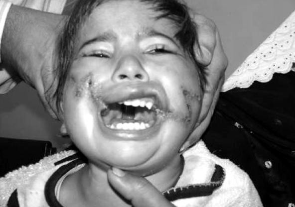
2 cranial clefts from the facial clefts. At times, facial craniofacial clefts because it gives the anatomical clefts extend through the orbit to become cranial description of the cleft and is internationally clefts. The clefts are numbered so that the facial recognized as standard reference (Figure 1). This component of the cleft and the cranial component classification has the merit of permitting all always add upto 14: 0 and 14, 1 and 13, 2 and 12, clinicians who deal with patients having these 3 and 11, 4 and 10, 5 and 9, 6 and 8. Tessier cleft malformations (pediatricians, geneticists, foetal number 7 is lateral most craniofacial cleft. The pathologists, surgeons, and obstetricians, soft tissue and skeletal components of a cleft are sonographers) to readily communicate with one seldom affected to the same extent. The skeletal another since it is based on anatomical and clinical 5 landmark tends to be more constant and reliable considerations.there were two cases of unilateral 5 than the soft tissue landmarks. cleft 7, two cases of bilateral cleft 7, two cases of cleft 30, one case of bilateral cleft 4 and a single Apart from the common cleft lip and case of proboscis lateralis. palate deformity the treatment of other types of craniofacial defects need much more complex CASE1: 4-6 diagnostic and treatment modalities. Due to their A female baby at the age of 3 months was complexity, the individual degree of cleft referred to our unit with bilateral Tessier cleft 4. formation and the different structures and organs The cleft had spared the philtral column and the i n v o l v e d, s u c c e s s f u l r e c o n s t r u c t i o n a n d entire nose. The cleft commenced medial to the rehabilitation in almost all the case demand a 6 oral commisures and passed lateral to the nasal multistep and multi-professional procedure. ala. The orbicularis muscle was located in the Besides careful examination, imaging lateral lip elements and there was no muscle in the techniques are necessary to assess the individual midline. The clefts passed through the cheeks into degree of skeletal involvement in the cleft the lower eyelids. The medial canthus and lacrimal formation. For correct diagnosis modern imaging systems were normal. Microphthalmia observed in techniques such as CT, MRI and 3-D CT allow the left eye. The skeletal component involved the better pre-op understanding of the problem and maxillary sinuses but the medial walls of the planning of the surgical procedures. sinuses were intact. The clefts were present medial to the infraorbital formina and terminated at the Considering these requirements we would medial aspect of orbital rim. like to present our experience in the surgical management of 08 patients with rare facial clefts. Operative Technique: METHODOLOGY AND RESULTS Over a period of two years i.e. from January 2008 to December 2009 a total of 08 patients with rare craniofacial clefts were treated. Diagnosis was base d on clinical examination and radiographic findings on CT scan. In our study we used the Tessier's classification to describe the Figure 1: Tessier s Calssification Only soft tissue reconstruction was performed with local flaps. The grooved or hypoplastic tissue extending in the path of the cleft between the upper lip and the lower eyelid was excised. The rotation advancement repair of the cleft lip was performed. The lateral cheeks advanced over the cleft region to close the soft tissue defect. 164


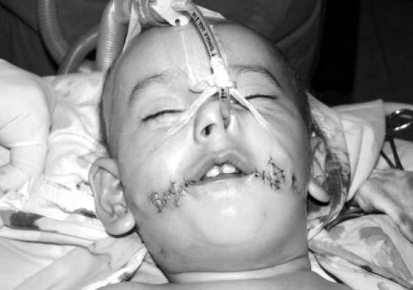
3 Figure 2 a: Pre-operative bilateral Tessier s # 4 Cleft Figure 2 b: Post-operative bilateral Tessier s # 4 Cleft The incision at the lower lid margin is continued 1-cm lateral to the lateral canthus. The nasal soft tissue is dissected to rotate the nasal ala caudally. The medial canthal ligament is dissected and medial canthopexy is performed with non- absorbable suture. The conjunctival fornix is reconstructed with mucous tissue within the cleft, and orbicularis oculi muscle is preserved and reconstructed. The cheek flap is advanced to the medial and cephalic direction and tightly sutured to the periosteum at the piriform aperture. A small rectangular flap, designed on the medial margin of the cheek flap, is inserted into the medial canthal area. Lip repair was performed by using the bilateral rotation- advancement technique, and the white skin roll and vermilion on the central portion of the lip were reconstructed with both lateral vermilion flaps, which were sutured in the midline (Figure 2a & b). CASE 2 & 3: Two patients aged 3 years and 4 years had unilateral cleft 7. Both were males & the cleft was on left side. On clinical examination there was no h e m i f a c i a l m i c r o s o m i a. T h e s o f t t i s s u e manifestation of cleft 7 in these patients was a variable degree of macrostomia. Malformation of the external ear was not found in any of the patient. Surgical Technique: The surgical technique consists of the creation of a straight-line closure of the mucosa, a red-lip commissural flap rotated from the superior part of the lip (which, as a rule, is an extended part of the natural red-lip component), a modiolus muscle reconstruction at the confluence of the orbicularis oris, zygomatic major, risorius and depressor anguli oris muscles performed and modified z-plasty flap for skin closure. The distance from the midpoint of the cupid's bow to the normal commisure is used for reference. The position of the new commissure on the cleft side is marked at the vermillioncutaneous junction on the upper lip and by sighting a perpendicular line, the corresponding point is noted at the vermillion- cutaneouis line on the lower lip. This proposed position of the commissure is confirmed by measuring the peak of cupid's bow on each side. Another mark is made on the upper lip vermillio-mucosa approximately 3-4 mm medial to the proposed commissural point as the inset for the inferiorly based vermillion mucosal flap. The cutaneous-mucosal margin of the cleft is marked and the critical points are tattooed with gention violet dye. After infiltration of local anesthetic with epinephrine, the cutaneous mucosal margins are incised to expose the underlying orbicularis oris musle. The splayed muscular elements are dissected in the subdermal plane. An inferiorly based rectangular vermillio- mucosal flap is raised off the underlying muscle ( pars marginalis) on the lower lip extending the base just lateral to the lower labial point of the new commissure. Next the orbiculris oris muscle is approximated side to side from lateral to medial. Near the position of the anticipated commissure, the pars marginalis is opposed end to end to complete construction of the sphincter. The cutaneous points of the new commissure are approximated by a dermal suture and the skin is closed in two layers. After the mucosal dog-ear is excised, the buccal mucosal cleft is closed, lateral to medial, toward the commissure and a Z plasty is performed. The vermillion mucosal flap is draped into the commissure and ecured to the pars marginalis (Figure 3,4 a & b). 165


4 Figure 3 a: Pre-operative left Tessier s # 7 Unilateral Cleft Figure 3 b: Post-operative left Tessier s # 7 Unilateral Cleft Figure 4 a: Pre-operative left Tessier s # 7 Unilateral Cleft Figure 4 b: Post-operative left Tessier s # 7 Unilateral Cleft CASE 4 & 5: CASE 6 & 7: Two patients aged 3 years each had Two patients presented with cleft 30. Both bilateral cleft 7. One of them was male and the were males. One patient was 18 years and the other female. Right sided hemifacial microsomia other was 28 years old. The cleft involved the was noted in female patient. In both the patients lower lip in the midline and there was no skeletal the eaxternal ears were normal and the clefts involvement. In one patient the lower lip was extended from the oral commissure toward the ear. notched only and in the second patient there was Tongue and soft palate were normal in both the midline involvenement of the more than two thirds patients but the muscles of mastication were of lower lip. Tongue and mandible were spared in underdeveloped (Masseter). Parotid glands and both the patients. ducts were present in both the patients. The posterior maxilla and mandibular rami were Surgical Technique: hypoplastic in the female patient. In both the patients a W shaped incision Surgical Technique: designed at the vermilion coetaneous junction of the cleft lip. The skin flaps dissected and elevated. Soft tissue repair performed for the The orbicularis oris muscle dissected separated correction of macrostomia. The same surgical from the mucosa and stitched in midline. A three technique applied as for the unilateral cleft 7 but layer wound closure done by stitching the mucosa, 8,9 on both sides (Figure 5,6 a & b). 10,11 muscle and skin separately (Figure 7,8 a & b). 166


5 Figure 5 a: Pre-operative bilateral Tessier s # 7 Cleft Figure 5 b: Post-operative bilateral Tessier s # 7 Cleft Figure 6 a: Pre-operative bilateral Tessier s # 7 Cleft Figure 6 b: Post-operative bilateral Tessier s # 7 Cleft Figure 7 a: Pre-operative Tessier s # 30 Cleft Figure 7 b: Post-operative Tessier s # 30 Cleft Figure 8 a: Pre-operative Tessier s # 30 Cleft Figure 8 b: Post-operative Tessier s # 30 Cleft 167


6 CASE 8: Surgical Technique A young male of 15 years presented to us Various methods have been suggested for with proboscis lateralis. It's a very rare congenital the treatment of proboscis lateralis, ranging from anomaly related to the median facial cleft and may the extirpation of the proboscis, to the tunneling be associated with other anomalies of the eye and method. We corrected the proboscis by making its adnexa and cleft lip or cleft palate. The incision around the proboscis. We lift up the incidence of proboscis lateralis is <1 per proboscis as a flap. Nasal mucosa of proboscis was 12,13 separated and excised. Defect in nasal mucosa was 100,000. The proboscis may be unilateral or stitched and the proboscis skin was replaced. Flap bilateral. There is a tube like rudimentary nasal debulking will be done in future (Figure 9 a & b). structure which is attached along the embryonic fusion lines between the anterior maxillary process A summary of salient features of the eight and the frontonasal process. The length of cases is given in Table proboscis may vary from 1 to 4 cm in length. It has a stratified columnar lining and sometime ends DISCUSSION as a blind tract or communicated to the nasal Craniofacial clefts are of particular cavity. relevance to plastic surgeons, Maxillofacial surgeons & ENT surgeons. While providing The patient which we received had group surgical treatment to patients with complex 2 proboscis on right side. Nasal airway was patent craniofacial deformities the plastic surgeons on right side and the patient complained of encounter certain inadequacies. Firstly the health discharge from proboscis. The patient had no other status of the patient varies greatly and most of the associated anomaly. times it is difficult to verify the fitness of the Figure 9 a: Pre-operative probosis lateralis Figure 9 b: Post-operative probosis lateralis Case No. Case 1 Case 2 & 3 Case 4 & 5 Case 6 & 7 Case 8 Table 1: Clinical features of Craniofacial Clefts in Our series Clinical Features Bilateral Tessier 4 cleft The cleft passing through the cheeks into the lower eyelid. Left microphthalmia Unilateral Tessier 7 cleft Both the cleft were on left side. No hemifial microsomia. No external ear deformities Bilateral Tessier 7 cleft Right sided hemifial microsoma observed in one patient Preauricular skin tags in one patient Tessier 30 cleft The obicularis Oris muslae was affected in the both cases. No bone involvement Probosis Lateralis Left side Probosis Lateralis Probosis connected to the nasal canting 168
7 patient for surgical treatment. Secondly the lack of proper equipment may restrict the anesthetist and the surgeon to achieve their task up to the mark. Also the quality of postoperative care requires high levels of expertise and trained staff. To the proper management of such patients can only be accomplished in designated craniofacial centers where they can be operated according to the standardized protocol. It must be realized that the optimal treatment for craniofacial clefts require multiple, staged reconstructive procedures at different ages of the children concerned. This series of 08 patients with rare craniofacial clefts represents our early experience in soft tissue reconstruction of these deformities. Apart from the surgical and anesthesiologic limitation we also find certain limitations in the proper diagnosis of such cases. The lack of 3D- CT scan confines us to diagnose the deformity on clinical examination and surgical exploration. Due to all these limitations we focused our surgical treatment on soft tissue reconstruction only. Tessier bilateral cleft 4 was reconstructed with local flaps by cheek advancement and lip repair by rotation advancement technique. No perioperative complication occurred in this patient. The unilateral and bilateral cleft 7 benefited from a single stage pure soft tissue repair. We did not find deformities of external ear and occurrence of preauricular tags in any unilateral cleft # 7 patients. This is in contrast to several reports where external ear deformities occur in approximately two thirds of patients cleft 7 7. However David et al described Tessier no 7 8,9 cleft never affecting the external ear. Median clefts of the lower lip are rare and just 68 cases have been reported till date. Soft tissue repair of cleft 30 as single stage procedure gave good result because there was no bony involvement. Proboscis lateralis is a rare anomaly among the nasal malformations. Many surgical options described for its correction in multiple 14,17,18 steps. In our case we also planned multistage procedure for its correction. In the first stage we have removed the nasal mucosa from the proboscis and close the nasal defect and in the second stage we will bulk the proboscis that was used for reconstruction of right nostril. Summarizing our results, we would like to mention that we achieved acceptable results in all the cases where single stage soft tissue reconstruction was the appropriate surgical treatment. Regular follow-up of these patient is also very important for the successful out come of surgical treatment. CONCLUSION While summarizing our early experience of the reconstruction of rare craniofacial clefts we would like to state that while operating with limited experience and equipments we have achieved acceptable results in all cases where one stage pure soft tissue reconstruction was the appropriate method of surgical treatment. We hope that in years to come we would be able to provide good care to patients with complex craniofacial deformities. REFERENCES 1. Eppley BL, van Aalst JA, Robey A, Havlik JR. The spectrum of orofacial clefting. Plast Reconst Surg 2005;115: Moore MH. Rare craniofacial clefts. J Craniofac Surg 1996;7: Prescott NJ, Winter RM, Malcolm S. Non syndromic cleft lip and palate: complex genetics and environmental effects. Ann Hum Genet 2001;65: Tolarova MM, Cervenka J. Classification and birth prevalence of orofacial clefts. Am J Med Genet 1998;75: Tessier P. Anatomical classification of facial, craniofacial, and latero-facial clefts. J Maxillofac Surg 1976;4: Johnston MC. Embryology of the head and neck. In: McCarthy JG, editor. Plastic Surgery. Philadelphia: WB Saunders; Sieg P, Hakim SG, Jacobson HS, Saka B, Hermes D. Rare facial clefts: treatment during charity missions in developing countries. Plast Reconstr Surg 2004;114: Aketa J, Nodai T, Kuga Y. A method for the repair of transverse facial clefts. Cleft Palate J 1980;17: Powell WJ, Jenkins HP. Transverse facial clefts: report of three cases. Plast Reconst Surg 1968;42: Yildirim I, Aydin Y, Cinar C, Ogur S. Management of total lower face cleft. Plast Reconst Surg 2002;109: Oostrom CAM, Vermeij-Keers C, Gilbert PM, van der Meulen JC. Median cleft of the lower lip and mandible: case reports, a new embryologic hypothesis and subdivision. Plast Reconstr Surg 1996;97: Bhatt YC. Proboscis lateralis: review of literature and a case report. Internet J Plast Surg 2008;5:
8 13. English GM. Congenital anomalies of the 16. Guerrero JM, Cogren MS, Kelly D. Proboscis nose, nasopharynx and paranasal sinuses. In: lateralis. Arch Ophthalmol 2001;119: E n g l i s h G M, e d i t o r. O t o l a r y n g o l o g y. Philadelphia: Lippincott; p Prescott NJ, Winter RM, Malcolm S. Nonsyndromic cleft lip and palate: complex 14. Metha L, Petrikovsky B, Tydings L. Lateral genetics and environmental effects. Ann Hum nasal proboscis: antenatal diagnosis and councelling. Obstet Gynecol 199;94: Genet 2001;65: Boo Chai K. The proboscis lateralis: 14 year 18. Acarturk S, Kivanc K. Proboscis lateralis: follow up. Plast Reconstr Surg 1985;75:569- evaluation of the anomaly and a review of two 77. cases. Plast Reconstr Surg 2006;117: Address for Correspondence: Dr. Tahmeedullah Assistant Professor Department of Plastic Surgery, Hayatabad Medical Complex, Peshawar - Pakistan. E mail: tahmeedullah101@yahoo.com JPMI 2011 Vol. 25 No. 02 :
Proboscis lateralis: report of two cases
 The British Association of Plastic Surgeons (2003) 56, 704 708 CASE REPORT Proboscis lateralis: report of two cases Lütfi Eroğlu a, *, Osman Ata Uysal b a Faculty of Medicine, Department of Plastic and
126 ISSN East Cent. Afr. J. surg. (Online)
") 126 Macrostomia Repair: Comparison of the Z- Plasty Repair with the Straight line Closure O.A. Olawoye 1, O.M. Fatungashe 2, B.A. Ayoade 3, A.O. Tade 3 Department of Plastic Surgery, University College
126 Macrostomia Repair: Comparison of the Z- Plasty Repair with the Straight line Closure O.A. Olawoye 1, O.M. Fatungashe 2, B.A. Ayoade 3, A.O. Tade 3 Department of Plastic Surgery, University College
Remember from the first year embryology Trilaminar disc has 3 layers: ectoderm, mesoderm, and endoderm
 Development of face Remember from the first year embryology Trilaminar disc has 3 layers: ectoderm, mesoderm, and endoderm The ectoderm forms the neural groove, then tube The neural tube lies in the mesoderm
Cleft Lip and Palate: The Effects on Speech and Resonance
 Ann W. Kummer, PhD, CCC-SLP Cincinnati Children s Cleft lip and/or palate can have a negative impact on both speech and resonance. The following is a summary of normal anatomy, the types and causes of
OPEN ACCESS ATLAS OF OTOLARYNGOLOGY, HEAD & NECK OPERATIVE SURGERY
 OPEN ACCESS ATLAS OF OTOLARYNGOLOGY, HEAD & NECK OPERATIVE SURGERY BUCCINATOR MYOMUCOSAL FLAP The Buccinator Myomucosal Flap is an axial flap, based on the facial and/or buccal arteries. It is a flexible
Combined tongue flap and V Y advancement flap for lower lip defects
 British Journal of Plastic Surgery (2005) 58, 258 262 CASE REPORTS Combined tongue flap and V Y advancement flap for lower lip defects Kenji Yano*, Ko Hosokawa, Tateki Kubo Department of Plastic and Reconstructive
Dr.ALI AL BAZZAZ PLASTIC SURGON CLEFT LIP AND PALATE
 Dr.ALI AL BAZZAZ PLASTIC SURGON CLEFT LIP AND PALATE Cleft lip (cheiloschisis) and cleft palate (palatoschisis), which can also occur together as cleft lip and palate, are variations of a type of clefting
UCL Repair: Emphasis on Muscle Dissection and Reconstruction
 UCL Repair: Emphasis on Muscle Dissection and Reconstruction Unilateral cleft lip repair is performed using rotation-advancement technique. Markings are made on columella base, redlines, Cupid s bow on
Face. Definition: The area between the two ears and from the chin to the eye brows. The muscles of the face
 Face Definition: The area between the two ears and from the chin to the eye brows. The muscles of the face The muscle of facial expression (include the muscle of the face and the scalp). All are derived
Upper Triangular Flap Method for Primary Repairs of Incomplete Unilateral Cleft Lip Patients. Minor to Two-Thirds Way Defects
 HEAD AND NECK SURGERY Upper Triangular Flap Method for Primary Repairs of Incomplete Unilateral Cleft Lip Patients Minor to Two-Thirds Way Defects Kyung S. Koh, MD, PhD,* Tae Suk Oh, MD,* and Jin Woo Song,
Management of Commonly Encountered Secondary Cleft Deformities of Face-A Case Series
 DOI: 10.7860/IJARS/2017/28759:2331 Surgery Section Case Series Management of Commonly Encountered Secondary Cleft Deformities of Face-A Case Series JACOB JOHN, ARJUN MADHU USHA, MUBARAK AZIZ, VINIT RENKAN
3-Deep fascia: is absent (except over the parotid gland & buccopharngeal fascia covering the buccinator muscle)
") The Face 1-Skin of the Face The skin of the face is: Elastic Vascular (bleed profusely however heal rapidly) Rich in sweat and sebaceous glands (can cause acne in adults) It is connected to the underlying
The Face 1-Skin of the Face The skin of the face is: Elastic Vascular (bleed profusely however heal rapidly) Rich in sweat and sebaceous glands (can cause acne in adults) It is connected to the underlying
What is Hemifacial Microsomia? By Pravin K. Patel, MD and Bruce S. Bauer, MD Children s Memorial Hospital, Chicago, IL
 What is Hemifacial Microsomia? By Pravin K. Patel, MD and Bruce S. Bauer, MD Children s Memorial Hospital, Chicago, IL 773-880-4094 Early in the child s embryonic development the structures destined to
New York Science Journal 2017;10(5) Primary Rhino-cheiloplasty in unilateral Cleft lip.
 Primary Rhino-cheiloplasty in unilateral Cleft lip.") Primary Rhino-cheiloplasty in unilateral Cleft lip Abd Elmoneim H. Hota 1, Magdy A. Abdelmoktader 1, Refaat I. El Badawy 2 and Ahmed F. Salem 1 1 Plastic Surgery Department Faculty of Medicine, Alazhar
Primary Rhino-cheiloplasty in unilateral Cleft lip Abd Elmoneim H. Hota 1, Magdy A. Abdelmoktader 1, Refaat I. El Badawy 2 and Ahmed F. Salem 1 1 Plastic Surgery Department Faculty of Medicine, Alazhar
ONE out of every eight hundred children in the United States is born with
 REPAIR OF THE CLEFT LIP ROBIN ANDERSON, M.D. Department of Plastic Surgery ONE out of every eight hundred children in the United States is born with a cleft lip, a cleft palate, or both. Within this group
Rotation-Advancement Principle. in Cleft Lip Closure. D. RALPH MILLARD, JR., M.D., F.A.C.S. Miami, Florida
 Rotation-Advancement Principle in Cleft Lip Closure D. RALPH MILLARD, JR., M.D., F.A.C.S. Miami, Florida Correction of prealveolar, alveolar, and postalveolar clefts poses a fivefold project: natural appearance,
Surgical Correction of Tessier Number 8 Cleft
 Surgical Correction of Tessier Number 8 Cleft Antonio Fuente-del-Campo, M.D., F.A.C.S. Mexico Cily, Mexiro The number 8 Tessier c!eft can be a discrete horizontal shadow at the level of the lateral canthus
Principles of Facial Reconstruction After Mohs Surgery
 Objectives Principles of Facial Reconstruction After Mohs Surgery Identify important functional anatomy and aesthetic units of the face. Describe techniques used in facial reconstruction. Discuss postoperative
Plastic Surgeon, Middlesbrough General Hospital, Stockton Children's Hospital, Newcastle Regional Hospital Board
 THE NASAL TIP IN BILATERAL HARE LIP By J. POTTER, F.R.C.S.Ed. Plastic Surgeon, Middlesbrough General Hospital, Stockton Children's Hospital, Newcastle Regional Hospital Board IN the problem of the bilateral
OPEN ACCESS ATLAS OF OTOLARYNGOLOGY, HEAD & NECK OPERATIVE SURGERY
 OPEN ACCESS ATLAS OF OTOLARYNGOLOGY, HEAD & NECK OPERATIVE SURGERY NASOLABIAL FLAP FOR ORAL CAVITY RECONSTRUCTION Harry Wright, Scott Stephan, James Netterville Designed as a true myocutaneous flap pedicled
Figure 1. Basic anatomy of the palate
 CHAPTER 10 CLEFT LIP AND PALATE Chen Yan, MD and Sanjay Naran, MD I. ANATOMY AND DEFINITIONS A. Cleft Lip (CL) alone, Cleft Lip with Cleft Palate (CLP), and Cleft Palate (CP) alone 1. CL alone and CLP
A TECHNIQUE FOR ONE STAGE REPAIR OF COMPLETE PALATAL CLEFT
 A TECHNIQUE FOR ONE STAGE REPAIR OF COMPLETE PALATAL CLEFT Pages with reference to book, From 105 To 107 Iftikhar Ahmad, M. Rafiq Khan, Abdullah Jan, Abdur Rasheed ( Department of E.N.T. and Head and Neck
Geometric triangular vermilion flap for correction of whistle deformity in unilateral cleft lip.
 ORIGINAL ARTICLE Geometric triangular vermilion flap for correction of whistle deformity in unilateral cleft lip. Abstract: Whistle deformity is one of the several deformities that a child with repaired
The Role of the Lip Adhesion Procedure. in Cleft Lip Repair*
 The Role of the Lip Adhesion Procedure in Cleft Lip Repair* RALPH HAMILTON, M.D. WILLIAM P. GRAHAM, III, M.D. PETER RANDALL, M.D. Philadelphia, Pa. 19104 Introduction A lip adhesion procedure utilizing
Head and Face Anatomy
 Head and Face Anatomy Epicranial region The Scalp The soft tissue that covers the vault of skull. Extends from supraorbital margin to superior nuchal line. Layers of the scalp S C A L P = skin = connective
Bones Ethmoid bone Inferior nasal concha Lacrimal bone Maxilla Nasal bone Palatine bone Vomer Zygomatic bone Mandible
 splanchnocranium - Consists of part of skull that is derived from branchial arches - The facial bones are the bones of the anterior and lower human skull Bones Ethmoid bone Inferior nasal concha Lacrimal
The Advantages of Two Stages in Repair. of Bilateral Cleft Lip. VICTOR SPINA, M.D. Sado Paulo, Brazil
 The Advantages of Two Stages in Repair of Bilateral Cleft Lip VICTOR SPINA, M.D. Sado Paulo, Brazil The suggestion of using two stages for the surgical correction of complete bilateral clefts of the lip
REVIEW OF CLINICAL EMBRYOLOGY OF HEAD AND NECK
 REVIEW OF CLINICAL EMBRYOLOGY OF HEAD AND NECK OUTLINE - EMBRYOLOGY UNDERLYING CLINICAL CONDITIONS I. EARLY DEVELOPMENT OF FACE: CLEFT LIP, CLEFT PALATE, OBSTRUCTED NASOLACRIMAL DUCT II. BRANCHIAL ARCHES
TRAUMA TO THE FACE AND MOUTH
 Dr.Yahya A. Ali 3/10/2012 F.I.C.M.S TRAUMA TO THE FACE AND MOUTH Bailey & Love s 25 th edition Injuries to the orofacial region are common, but the majority are relatively minor in nature. A few are major
Construction of the congenitally missing columella in midline clefts
 Construction of the congenitally missing columella in midline clefts Kurt-Wilhelm BÜTOW Department of Maxillo-Facial and Oral Surgery (Head: Prof. Kurt-W. Bütow, MChD(OMFSurg), DMD, PhD, DSc(Odont), FCMFOS),
NEUROCRANIUM VISCEROCRANIUM VISCEROCRANIUM VISCEROCRANIUM
 LECTURE 4 SKULL NEUROCRANIUM VISCEROCRANIUM VISCEROCRANIUM VISCEROCRANIUM CRANIUM NEUROCRANIUM (protective case around brain) VISCEROCRANIUM (skeleton of face) NASOMAXILLARY COMPLEX MANDIBLE (DESMOCRANIUM)
McGregor Flap Reconstruction of Extensive Lower Lip Defects Following Excision of Squamous Cell Carcinoma
 Kasr El Aini Journal of Surgery VOL., 12, NO 2 May 2011 27 McGregor Flap Reconstruction of Extensive Lower Lip Defects Following Excision of Squamous Cell Carcinoma Mohamed A. Albadawy, MD and Bassem M.
Anatomy and Physiology. Bones, Sutures, Teeth, Processes and Foramina of the Human Skull
 Anatomy and Physiology Chapter 6 DRO Bones, Sutures, Teeth, Processes and Foramina of the Human Skull Name: Period: Bones of the Human Skull Bones of the Cranium: Frontal bone: forms the forehead and the
Upper arch. 1Prosthodontics. Dr.Bassam Ali Al-Turaihi. Basic anatomy & & landmark of denture & mouth
 1Prosthodontics Lecture 2 Dr.Bassam Ali Al-Turaihi Basic anatomy & & landmark of denture & mouth Upper arch Palatine process of maxilla: it form the anterior three quarter of the hard palate. Horizontal
Cairo Dental Journal (24) Number (I), 77:84 January, Haitham Sayed Attia 3, Mohamed Saied Hamed 1 and Monteser El Koutobey 2
 Number (I), 77:84 January, Haitham Sayed Attia 3, Mohamed Saied Hamed 1 and Monteser El Koutobey 2") Cairo Dental Journal (24) Number (I), 77:84 January, 2008 Anthropometric Analysis of cases of Unilateral Cleft Lip Versus cases of Complete Unilateral Cleft Lip and Palate Haitham Sayed Attia 3, Mohamed
Cairo Dental Journal (24) Number (I), 77:84 January, 2008 Anthropometric Analysis of cases of Unilateral Cleft Lip Versus cases of Complete Unilateral Cleft Lip and Palate Haitham Sayed Attia 3, Mohamed
Nasolabial Flap Reconstruction of Oral Cavity Defects: A Report of 18 Cases
 J Oral Maxillofac Surg 58:1104-1108, 2000 Nasolabial Flap Reconstruction of Oral Cavity Defects: A Report of 18 Cases Yadranko Ducic, MD, FRCS (C),* and Mark Burye, DDS Purpose: This article describes
Oral cavity landmarks
 By: Dr. Ahmed Rabah Oral cavity landmarks The knowledge of oral anatomy and physiology will help the operator and provides enough landmarks to act as positive guide during denture construction. This subject
A REVIEW OF CLASSIFICATION SYSTEMS FOR CLEFT LIP AND PALATE PATIENTS- I. MORPHOLOGICAL CLASSIFICATIONS
 Review Article A REVIEW OF CLASSIFICATION SYSTEMS FOR CLEFT LIP AND PALATE PATIENTS- I. MORPHOLOGICAL CLASSIFICATIONS Syed Nasir Shah, Mariya Khalid, Muhammad Sartaj Khan Department of Prosthodontics,
Essentials in Head and Neck Embryology. Part 3 Development of the head, face, and oral cavity
 Essentials in Head and Neck Embryology Part 3 Development of the head, face, and oral cavity Outline General overview of prenatal development Embryonic period phase 1 Formation of bilaminar disk Formation
CHAPTER 8 SECTION 1.4 ORAL SURGERY TRICARE/CHAMPUS POLICY MANUAL M DEC 1998 SPECIAL BENEFIT INFORMATION
 TRICARE/CHAMPUS POLICY MANUAL 6010.47-M DEC 1998 SPECIAL BENEFIT INFORMATION CHAPTER 8 SECTION 1.4 Issue Date: October 8, 1986 Authority: 32 CFR 199.4(e)(10) I. DESCRIPTION There are certain oral surgical
Scientific Forum. Nostrilplasty: Raising, Lowering, Widening, and Symmetry Correction of the Alar Rim
 Nostrilplasty: Raising, Lowering, Widening, and Symmetry Correction of the lar Rim Richard Ellenbogen, MD; and Greg azell, MD ackground: lthough the alar rim has frequently been neglected in correction
04 Development of the Face and Neck. Development of the Face Development of the neck
 04 Development of the Face and Neck Development of the Face Development of the neck Development of the face Overview of facial development The fourth week ~ the twelfth week of prenatal development Between
Trigeminal Nerve (V)
") Trigeminal Nerve (V) Lecture Objectives Discuss briefly how the face is developed. Follow up the course of trigeminal nerve from its point of central connections, exit and down to its target areas. Describe
Trigeminal Nerve (V) Lecture Objectives Discuss briefly how the face is developed. Follow up the course of trigeminal nerve from its point of central connections, exit and down to its target areas. Describe
Anatomic Relations Summary. Done by: Sohayyla Yasin Dababseh
 Anatomic Relations Summary Done by: Sohayyla Yasin Dababseh Anatomic Relations Lecture 1 Part-1 - The medial wall of the nose is the septum. - The vestibule lies directly inside the nostrils (Nares). -
Kevin T. Kavanagh, MD
 Kevin T. Kavanagh, MD Axial Based upon a named artery. Survival length depends upon the artery not the width of the flap. Random Has random unnamed vessels supplying it. Survival length is directly proportional
-Ibrahim Al-Naser. -Dr Al- Muhtaseb. 1 P a g e
 -1 -Ibrahim Al-Naser - -Dr Al- Muhtaseb 1 P a g e The Digestive System The doctor started the lecture by talking about the class rules. The GI system is an organ system, it is divided into: The Alimentary
Vancouver, B.C., Canada
 THE "ALAR SHIFT" REVISITED By THEODORE F. WILKIE, B.A., M.D., F.R.C.S.(C), F.A.C.S. Vancouver, B.C., Canada IN the hands of many plastic surgeons certain procedures have an evanescent history. Usually
SCOPE OF PRACTICE PGY-6 PGY-7 PGY-8
 PGY-6 Round on all plastic surgery inpatients every day. Assess progress of patients and identify real or potential problems. Review patients progress with attending physicians daily and participate in
ofunusual and Occult Clefts of the Palate JOHN MARQUIS CONVERSE, MD. SIDNEY L. HOROWITZ, D.D.S. MELVIN H. BECKER M.D, New York New York-4:11
 TheUse oftomographyin: the Diagnosis ofunusual and Occult Clefts of the Palate JOHN MARQUIS CONVERSE, MD. SIDNEY L. HOROWITZ, D.D.S. MELVIN H. BECKER M.D, New York New York-4:11 The ma or1ty of patients
Lips and labial mucosa
 Lips and labial mucosa External portion of the lips: the vermilion border and the skin Vermilion border : the exposed red portion of the lip, covered by mucous membrane, no mucous glands Boundary: the
cally, a distinct superior crease of the forehead marks this spot. The hairline and
 4 Forehead The anatomical boundaries of the forehead unit are the natural hairline (in patients without alopecia), the zygomatic arch, the lower border of the eyebrows, and the nasal root (Fig. 4.1). The
The anatomical basis for a cleft lip defect is far
 PEDIATRIC/CRANIOFACIAL Comparison of Three Incisions to Repair Complete Unilateral Cleft Lip Srinivas Gosla Reddy, M.D.S., M.B.B.S. Rajgopal R. Reddy, B.D.S., M.B.B.S. Ewald M. Bronkhorst, Ph.D. Rajendra
RECONSTRUCTION OF SUBTOTAL DEFECTS OF THE NOSE BY ABDOMINAL TUBE FLAP. By MICHAL KRAUSS. Plastic Surgery Hospital, Polanica-Zdroj, Poland
 RECONSTRUCTION OF SUBTOTAL DEFECTS OF THE NOSE BY ABDOMINAL TUBE FLAP By MICHAL KRAUSS Plastic Surgery Hospital, Polanica-Zdroj, Poland RECONSTRUCTION of the nose is one of the composite procedures in
G l o s s a r y. The lack of closure of a normal body orifice or. passage
 A P P E N D I XE G l o s s a r y Allergic rhinitis Swelling of the membrane in the nasal chamber due to allergic reactions; the condition may obstruct breathing Alveolar ridge The bony arches of the maxilla
Prenatal Diagnosis of Cleft Lip
 Commentary Prenatal Diagnosis of Cleft Lip What the Sonologist Needs to Tell the Surgeon John. Mulliken, MD, eryl R. enacerraf, MD Division of Plastic Surgery, Children s Hospital (J..M.) Department of
Cleft-Craniofacial Center
 Cleft-Craniofacial Center A Pioneering T eam 2 Welcome to the Cleft-Craniofacial Center at Children s Hospital of Pittsburgh The Cleft-Craniofacial Center at Children s Hospital of Pittsburgh has been
Figure (2-6): Labial frenum and labial notch.
: Labial frenum and labial notch.") The anatomy of the edentulous ridge in the maxilla and mandible is very important for the design of a complete denture. The consistency of the mucosa and architecture of the underlying bone is different
The anatomy of the edentulous ridge in the maxilla and mandible is very important for the design of a complete denture. The consistency of the mucosa and architecture of the underlying bone is different
Head and neck cancer - patient information guide
 Head and neck cancer - patient information guide The development of reconstructive surgical techniques in the last 20 years has led to major advances in the treatment of patients with head and neck cancer.
Mc Gregor Flap for Lower Eyelid Defect
 IOSR Journal of Dental and Medical Sciences (IOSR-JDMS) e-issn: 2279-0853, p-issn: 2279-0861.Volume 16, Issue 4 Ver. V (April. 2017), PP 69-74 www.iosrjournals.org Mc Gregor Flap for Lower Eyelid Defect
Subciliary versus Subtarsal Approaches to Orbitozygomatic Fractures
 CME Subciliary versus Subtarsal Approaches to Orbitozygomatic Fractures Rod J. Rohrich, M.D., Jeffrey E. Janis, M.D., and William P. Adams, Jr., M.D. Dallas, Texas Learning Objectives: After studying this
Technique Guide. Titanium Wire with Barb and Needle. Surgical Technique Guide for Canthal Tendon Prodecures.
 Technique Guide Titanium Wire with Barb and Needle. Surgical Technique Guide for Canthal Tendon Prodecures. Indications/Features Indications The Synthes Titanium Wire with Barb and straight Needle is
Pharyngeal Apparatus. Pouches Endoderm Grooves Ectoderm Arch Neural Crest Somitomeres Aortic Arch - Vessel
 Pharyngeal Apparatus Pouches Endoderm Grooves Ectoderm Arch Neural Crest Somitomeres Aortic Arch - Vessel Segmental Organization Humans: Arch 1-4 prominent Arch 5 absent Arch 6 - transient First Arch Face
LATERAL CEPHALOMETRIC EVALUATION IN CLEFT PALATE PATIENTS
 POLSKI PRZEGLĄD CHIRURGICZNY 2009, 81, 1, 23 27 10.2478/v10035-009-0004-2 LATERAL CEPHALOMETRIC EVALUATION IN CLEFT PALATE PATIENTS PRADEEP JAIN, ANAND AGARWAL, ARVIND SRIVASTAVA Department of Plastic
be very thin and variable. Facial nerve branches that exit the parotid gland are deep to the SMAS.
 The Superficial musculoaponeurotic system (SMAS) fascia is a fanlike fascia that envelops the face and provides a suspensory sheet which distributes forces of facial expression.. The SMAS is continuous
Case Report Mid Facial Degloving Procedure: Managing A Case of Multiple Mid Face Fractures with Significant External Deformity
 55 Bangladesh J Otorhinolaryngol 2015; 21(1): 51-56 Case Report Mid Facial Degloving Procedure: Managing A Case of Multiple Mid Face Fractures with Significant External Deformity Akhil Chndra Biswas 1,
Maxilla, ORBIT and infratemporal fossa. Neophytos C Demetriades MD, DDS, MSc Associate professor European University of Cyprus School of Medicine
 Maxilla, ORBIT and infratemporal fossa Neophytos C Demetriades MD, DDS, MSc Associate professor European University of Cyprus School of Medicine MAXILLA Superior, middle, and inferior meatus Frontal sinus
Longitudinal outcome of pharyngoplasty
 Archives of Orofacial Sciences (2009), 4(1): 17-21 CASE REPORT Longitudinal outcome of pharyngoplasty Peter J. Anderson*, Roslynn K. Sells, David. J. David Australian Craniofacial Unit, Women s and Children
Closure of Palatal Fistula with Bucco-labial Myomucosal Pedicled Flap
 Annals of Pediatric Surgery, Vol 5, No 2, April 2009, PP 104-108 Original Article Closure of Palatal Fistula with Bucco-labial Myomucosal Pedicled Flap Mohamed M. EL-Leathy* and Mohamed F. Attia** Pediatric
Facial Trauma. Rural Emergency Services and Trauma Symposium 2008
 Rural Emergency Services and Trauma Symposium 2008 Facial Trauma Mitchell Stotland, MD Associate Professor of Surgery and Pediatrics Dartmouth-Hitchcock Medical Center Children s Hospital of Dartmouth
Titanium Wire with Barb and Needle. Surgical Technique Guide for Canthal Tendon Procedures.
 Titanium Wire with Barb and Needle. Surgical Technique Guide for Canthal Tendon Procedures. Technique Guide This publication is not intended for distribution in the USA. Instruments and implants approved
CT of Maxillofacial Fracture Patterns. CT of Maxillofacial Fracture Patterns
 CT of Maxillofacial Fracture Patterns CT of Maxillofacial Fracture Patterns Stuart E. Mirvis, M.D., FACR Department of Radiology University of Maryland School of Medicine Viking 1 1976 MGS 2001 Technology
Trigeminal Nerve Anatomy. Dr. Mohamed Rahil Ali
 Trigeminal Nerve Anatomy Dr. Mohamed Rahil Ali Trigeminal nerve Largest cranial nerve Mixed nerve Small motor root and large sensory root Motor root Nucleus of motor root present in the pons and medulla
Elements of Dysmorphology I. Krzysztof Szczałuba
 Elements of Dysmorphology I Krzysztof Szczałuba 9.05.2016 Common definitions (1) Dysmorphology: recognition and study of birth defects (congenital malformations) and syndromes [David Smith, 1960] Malformation:
Normal fetal face and neck
 Normal fetal face and neck Maria A. Calvo-Garcia, MD. Associate Professor of Radiology Cincinnati Children s Hospital Medical Center Cincinnati, Ohio Disclosure I have no disclosures Goals & objectives
SYLLABUS OF ORAL AND MAXILLOFACIAL SURGERY
 MEDICAL UNIVERSITY OF VARNA FACULTY OF DENTAL MEDICINE DEPARTMENT OF ORAL AND MAXILLOFACIAL SURGERY AND SID SYLLABUS OF ORAL AND MAXILLOFACIAL SURGERY (State examination) ACADEMIC YEAR 2015 2016 1. Asepsis
Nasolabial flap reconstruction in oral cancer
 Singh et al. World Journal of Surgical Oncology 2012, 10:227 WORLD JOURNAL OF SURGICAL ONCOLOGY RESEARCH Open Access Nasolabial flap reconstruction in oral cancer Seema Singh, Rajesh Kumar Singh and Manoj
PTERYGOPALATINE FOSSA
 PTERYGOPALATINE FOSSA Outline Anatomical Structure and Boundaries Foramina and Communications with other spaces and cavities Contents Pterygopalatine Ganglion Especial emphasis on certain arteries and
The America Association of Oral and Maxillofacial Surgeons classify occlusion/malocclusion in to the following three categories:
 Subject: Orthognathic Surgery Policy Effective Date: 04/2016 Revision Date: 07/2018 DESCRIPTION Orthognathic surgery is an open surgical procedure that corrects anomalies or malformations of the lower
Omran Saeed. Luma Taweel. Mohammad Almohtaseb. 1 P a g e
 2 Omran Saeed Luma Taweel Mohammad Almohtaseb 1 P a g e I didn t include all the photos in this sheet in order to keep it as small as possible so if you need more clarification please refer to slides In
CHAPTER 11 FACIAL PARALYSIS. Shailesh Agarwal, MD and Arash Momeni, MD
 CHAPTER 11 FACIAL PARALYSIS Shailesh Agarwal, MD and Arash Momeni, MD The facial nerve innervates a total of 23 paired muscles and the orbicularis oris muscle. The majority of muscles innervated by the
CHAPTER 17 FACIAL AESTHETIC SURGERY. Christopher C. Surek, DO and Mohammed S. Alghoul, MD. I. BROW LIFT (Figures 1 and 2)
") CHAPTER 17 FACIAL AESTHETIC SURGERY Christopher C. Surek, DO and Mohammed S. Alghoul, MD I. BROW LIFT (Figures 1 and 2) A. Open Coronal Brow Lift Technique 1. Coronal incision is made in the hair-bearing
CHAPTER 17 FACIAL AESTHETIC SURGERY Christopher C. Surek, DO and Mohammed S. Alghoul, MD I. BROW LIFT (Figures 1 and 2) A. Open Coronal Brow Lift Technique 1. Coronal incision is made in the hair-bearing
Surgical Treatment of Short Nose
 Surgical Treatment of Short Nose Dr. Otto YT Au MD (JEFFERSON, USA) 1957, MCPS (MANITOBA) 1963, FHKAM (SURGERY) 1995 Diplomate American Board Plastic Surgery Plastic Surgery Specialist Dr.OttoYTAu A nice
Trigeminal nerve. Slide in bold and please go back to see the pictures, if I skipped any part of record that because it wasn t clear to me
 Trigeminal nerve Slide in bold and please go back to see the pictures, if I skipped any part of record that because it wasn t clear to me Hala nsour 2/26/2018 P a g e 1 this lecture contain two topics
Craniofacial Microsomia
 Patient and Family Education Craniofacial Microsomia Children with craniofacial microsomia (CFM) have a small or underdeveloped part of the face, usually the ear and jaw. The eye, cheek and neck may also
Core Curriculum Syllabus Emergencies in Otolaryngology-Head and Neck Surgery FACIAL FRACTURES
 Core Curriculum Syllabus Emergencies in Otolaryngology-Head and Neck Surgery A. General Considerations FACIAL FRACTURES Look for other fractures like skull and/or cervical spine fractures Test function
Aesthetic reconstruction of the nasal tip using a folded composite graft from the ear
 The British Association of Plastic Surgeons (2004) 57, 238 244 Aesthetic reconstruction of the nasal tip using a folded composite graft from the ear Yong Oock Kim*, Beyoung Yun Park, Won Jae Lee Institute
OTOLARYNGOLOGY ONLINE. Preauricular sinus. Management. Dr. T. Balasubramanian 6/17/2010
 1 OTOLARYNGOLOGY ONLINE Preauricular sinus Management Dr. T. Balasubramanian 6/17/2010 This e book discusses the Etiopathogenesis of preauricular sinus. Even though it is commonly encountered surgical
5/20/2015. Mohs Surgery BCCA High risk anatomic locations. Mohs Surgery High risk anatomic locations. Mohs Surgery Histologically Aggressive BCCA
 Mohs Surgery BCCA High risk anatomic locations High risk areas H zone nasal ala, nasal septum, nasal ala groove, periorbital region, periauricual region, region around and in ear canal, ear pinna and scalp
Sensitivity of a Method for the Analysis of Facial Mobility. II. Interlandmark Separation
 Sensitivity of a Method for the Analysis of Facial Mobility. II. Interlandmark Separation CARROLL-ANN TROTMAN, B.D.S., M.A., M.S. JULIAN J. FARAWAY, PH.D. Objective: This study demonstrates a method of
Basic Anatomy and Physiology of the Lips and Oral Cavity. Dr. Faghih
 Basic Anatomy and Physiology of the Lips and Oral Cavity Dr. Faghih It is divided into seven specific subsites : 1. Lips 2. dentoalveolar ridges 3. oral tongue 4. retromolar trigone 5. floor of mouth 6.
A Study of Classification Systems for Maxillectomy Defects
 A Study of Classification Systems for Maxillectomy Defects Zubair Durrani FFDRCS, FRCS, FRCS (OMFS)* Syed Ghazanfar Hassan FFDRCS** Shomaila Ameer Alam BDS*** * Associate Professor & Consultant Oral and
Unilateral Cleft Lip Repair by using White-skin-roll Flap from Cleft Side of Lip
 Unilateral Cleft Lip Repair by using White-skin-roll Flap from Cleft Side of Lip Background: With all due consideration to the restoration of function, post-operative aesthetic appearance of the cleft
REDUPLICATION OF THE MOUTH AND MANDIBLE. Groote Schuur Hospital and the Red Cross Hospital, Cape Town, South Africa
 IJHtlsh Journnl of Plaslic NurgeO, (1973), 26, 84-89 REDUPLICATION OF THE MOUTH AND MANDIBLE By D. DaviEs, M.B., Ch.B., F.R.C.S., G. MoRgIso~r, M.B., Ch.B., F.C.S. and B. H. MILLER, B.D.S., F.D.S., Dip.Orth.
Tanta University. Faculty of Medicine. Plastic and Reconstructive Surgery Department. Doctorate Degree in Plastic Surgery
 Componenets : Tanta University Faculty of Medicine Plastic and Reconstructive Surgery Department Doctorate Degree in Plastic Surgery Students should fulfill the designated number of credit hours, including
Personal technique for definite repair of complete unilateral cleft lip: modified Millard technique
 Archives of Craniofacial Surgery Arch Craniofac Surg Vol.19 No.1, 3-12 https://doi.org/10.7181/acfs.2018.19.1.3 Personal technique for definite repair of complete unilateral cleft lip: modified Millard
OPEN ACCESS ATLAS OF OTOLARYNGOLOGY, HEAD & NECK OPERATIVE SURGERY
 OPEN ACCESS ATLAS OF OTOLARYNGOLOGY, HEAD & NECK OPERATIVE SURGERY PARAMEDIAN FOREHEAD FLAP NASAL RECONSTRUCTION SURGICAL TECHNIQUE Brian Cervenka, Travis Tollefson, Patrik Pipkorn The paramedian forehead
Original Research. Figure 1: (a) Unilateral complete cleft of the lip and palate, (b) unilateral complete skeletal cleft with a Simonart s band.
 Unilateral complete cleft of the lip and palate, (b) unilateral complete skeletal cleft with a Simonart s band.") Received: 14 th August 2015 Accepted: 19 th November 2015 Conflicts of Interest: None Source of Support: Nil Original Research Simonart s Bands and Facial Growth in Unilateral Cleft Lip and Palate Patients:
Received: 14 th August 2015 Accepted: 19 th November 2015 Conflicts of Interest: None Source of Support: Nil Original Research Simonart s Bands and Facial Growth in Unilateral Cleft Lip and Palate Patients: