Nail Biology: The Nail Apparatus. Nail plate Proximal nail fold Nail matrix Nail bed Hyponychium
|
|
|
- Irene Holmes
- 6 years ago
- Views:
Transcription

1
2 Nail Biology: The Nail Apparatus Nail plate Proximal nail fold Nail matrix Nail bed Hyponychium

3

4
5 Nail Biology: The Nail Apparatus Lies immediately above the periosteum of the distal phalanx The shape of the distal phalanx determines the shape and transverse curvature of the nail The intimate anatomic relationship between nail and bone accounts for the bone alterations in nail disorders and vice versa
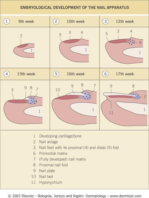
6 Nail Apparatus: Embryology Nail field develops during week 9 from the epidermis of the dorsal tip of the digit Proximal border of the nail field extends downward and proximally into the dermis to create the nail matrix primordium By week 15, the nail matrix is fully developed and starts to produce the nail plate


7 Nails develop from thickened areas of epidermis at the tips of each digit called nail fields. Later these nail fields migrate onto the dorsal surface surrounded laterally and proximally by folds of epidermis called nail folds.
8
9 Nail Func7on Protect the distal phalanx Enhance tactile discrimination Enhance ability to grasp small objects Scratching and grooming Natural weapon Aesthetic enhancement Pedal biomechanics
10 The Nail Plate Fully keratinized structure produced throughout life Results from maturation and keratinization of the nail matrix epithelium Attachments: Lateral: lateral nail folds Proximal: proximal nail fold (covers 1/3 of the plate) Inferior: nail bed Distal: separates from underlying tissue at the hyponychium
11 The Nail Plate Rectangular and curved in 2 axes Transverse and horizontal Smooth, although longitudinal ridging with age Ridge pattern used for forensic identification Homogeneously pink due to underling vessels Free edge is white
12 The Nail Plate Lunula: visible portion of the nail matrix white, half- moon shaped area plate loosely attached to underlying epithelium
13 The Nail Plate Onychocorneal Band Thin, distal transverse white band Marks distal portion of attachment of plate to bed Anatomic barrier against environmental hazards Disruption allows plate detachment (onycholysis) Onychodermal Band Thin, distal pink band separating onychocorneal band from the free edge of the plate
14
15 The Nail Plate: Transverse Anatomy Nail plate consists of three portions: Dorsal, intermediate, and ventral plates Dorsal and intermediate plates are produced by the nail matrix The ventral plate is produced by the nail bed Above the lunula, the plate consists only of the dorsal and intermediate portions
16 Nail Plate Thickness Plate progressively thickens from point of emergence to distal tip Mean thickness distal toenail: 1.65mm/1.38mm (m/f) Mean thickness distal fingernail: 0.6mm/0.5mm (m/f) Thickness with age, esp. in 1 st two decades Thickness depends on the length of the nail matrix and the nail bed
17 Nail Plate Thickness Thinning of the nails is usually a matrix disorder Thickening of the nails is usually a consequence of nail bed disorders
18 Proximal Nail Fold Consists of dorsal and ventral portions The dorsal portion is continuous with and anatomically similar to the skin of the dorsal digit but thinner and devoid of pilosebaceous units
19 Proximal Nail Fold The ventral portion is invisible from the exterior and is continuous proximally with the germinative nail matrix It adheres to and covers ¼ of the nail plate and keratinizes with a granular layer The limit between the proximal nail fold and the nail matrix can be histologically established at the site of disappearance of the granular layer
20 Proximal Nail Fold: Cu4cle Formed by the horny layer of the proximal nail fold Attached to the superficial nail plate Prevents separation of the plate from the fold Integrity of the cuticle is essential for nail homeostasis in this region
21 Proximal Nail Fold Dermis of the proximal fold contains capillaries that run parallel to the surface Arterial and venous limbs of the capillaries are arranged in parallel rows and appear as fine regular loops Proximal nail fold capillary morphology is altered in connective tissue diseases

22
23 Nail Matrix Specialized epithelial structure that lies above the midportion of the distal phalanx Consists of a proximal (dorsal) and a distal (ventral) portion Nail matrix keratinocytes keratinize in the absence of a granular layer to form the nail plate
24 Nail Matrix Kera7niza7on Maturation and differentiation of nail matrix keratinocytes occurs in a distally oriented diagonal axis (unlike the epidermis) Keratinization of the proximal (dorsal) nail matrix cells produces the dorsal nail plate Keratinization of the distal (ventral) nail matrix cells produces the intermediate nail plate
25 Nail Matrix Cornified onychocytes are composed mainly of keratin filaments, high sulfur matrix proteins, and the marginal band, which consists of precipitated cytoplasmic proteins During keratinization of onychocytes, DNases and RNases degrade nuclear fragments. Incomplete degradation of nuclear material results in transient leukonychial spots
26 Nail Matrix Melanocytes Usually quiescent but can become activated and synthesize melanin, which is transferred to surrounding keratinocytes Distal migration of melanin- containing keratinocytes gives rise to a diffuse or banded nail pigmentation (physiologic or pathologic) Nail matrix melanocytes of Caucasians do not contain mature melanosomes which are normally found in the nails of Asians and blacks
27 Nail Bed Extends from the distal margin of the lunula to the onychodermal band Nail bed epithelium is thin (2-5 cell layers) and firmly attached to the nail plate Nail bed keratinization produces a thin, horny layer that forms the ventral nail plate No granular layer and sparse melanocytes
28 Hyponychium Marks the anatomic area between the nail bed and the distal groove, where the nail plate detaches from the distal digit Anatomy is similar to plantar and volar skin (a granular layer is present) Normally covered by the distal nail plate
29 Basement Membrane Zone Antigenic structure is identical to that of the epidermis and is consistent throughout all portions of the nail apparatus Thus, the nails are commonly involved in diseases associated with attack on BMZ components
30 Blood and Nerve Supply Nail Apparatus: lateral digital arteries and nerves Nail Bed: encapsulated neurovascular structures called glomus bodies contain one to four AV anastomoses and nerve endings regulate blood supply to the digits in cold weather
31 Chemical Proper7es of the Nail Plate Low- sulfur keratins embedded in an amorphous matrix of high- sulfur proteins rich in cystine. Water (20%) <18% = brittle; >30% = opaque and soft Lipid (<5%): mainly cholesterol Trace inorganic elements: iron, zinc, calcium Do not contribute to nail hardness
32 Chemical Proper7es Nail keratins: 80% hard hair- type keratins Acidic Ha 1-4 and basic Hb 1-4 keratins 20% soft skin- type keratins Epithelial keratins 5, 6, 14, 16, 17
33 Nail Growth Proceeds from 15 weeks IUL until death Fingernails: 3mm per month 3-6 months for replacement Toenails: 1mm per month months for replacement
34 Nail Growth Decreased Growth Age > 50 Systemic illness Malnutrition Vascular disease Peripheral neuropathy Antimitotic drugs Onychomycosis Yellow nail syndrome Accelerated Growth Pregnancy Finger trauma Psoriasis Oral retinoids Itraconazole
35 Nail Clippings Can Be Evaluated For Drugs, chemicals and toxins DNA analysis Blood group typing Individual identification
36 Nail Signs 3 categories based on site of pathology: 1. Nail matrix 2. Nail bed 3. Nail plate (deposition of pigment)

37
38 Nail Signs due to Abnormal Matrix Func7on Beau s Lines Pitting Onychorrhexis Trachyonychia Onychomadesis Koilonychia
")
39 Beau s Lines Transverse depressions due to disruption of proximal matrix mitotic activity Depth: extent of damage Width: duration of insult Mechanical trauma Proximal nail fold dz Systemic insult (all nails)

40 Beau s Lines

41 Onychomadesis (nail shedding) Proximal detachment of the nail plate from the proximal nail fold Due to a severe insult that produces complete arrest of matrix activity Causes are the same as for Beau s Lines

42 Onychomadesis

43 PiPng Punctate depressions of the nail plate surface Foci of abnormal keratinization of the proximal matrix results in clusters of parakeratotic cells in the dorsal plate Clusters easily detach, leaving pits

44 Pitting
45 What diseases produce pipng? Psoriasis- deep and irregular Alopecia areata- superficial and geometric Eczema

46 Pitting

47 Onychorrhexis Longitudinal ridging and fissuring of the plate Diffuse thinning Indicates diffuse damage to the nail matrix Lichen planus Vasculopathy/ischemia Trauma, Tumors Normal aging

48 Trachyonychia (20 Nail Dystrophy) Nail roughness due to excessive longitudinal ridging Proximal nail matrix damage by: Alopecia areata Lichen planus Psoriasis Eczema

49 True Leukonychia
50 True Leukonychia Nail plate has a normal surface but loses its transparency and appears white because of parakeratotic cells within the ventral portion Caused by diseases that disturb distal nail matrix keratinization
51 True Leukonychia: 3 Morphologic Variants Punctate: opaque white spots, move distally with nail growth Due to trauma, common in kids Transverse: Multiple opaque white parallel lines, traumatic Women: matrix trauma from manicures Diffuse / Total Rare. Sometimes hereditary. May be assoc. w/ keratoderma and other congenital defects such as deafness

52 Par7al/Punctate Leukonychia

53 Transverse Leukonychia

54 Leukonychia Totalis

55 Koilonychia (Spoon Nails) Thinned, concave nail plate due to upward eversion of the lateral edges Physiologic in kids Iron deficiency anemia Plummer- Vinson Hemochromatosis


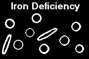
56 What is this disease?
57 Esophageal webs, iron deficiency anemia, and koilonychia.
58 Nail Signs due to Nail Bed Disorders Onycholysis Onychauxis Apparent Leukonychia Splinter hemorrhages

59 Onycholysis Distal nail plate detachment from bed Environmental exposure Psoriasis Infection UVR +/- TCN Hyperthyroidism Subungual tumor

60

61 Onycholysis

62
 Nail bed involvement by: Psoriasis Onychomycosis")
63 Onychauxis Nail plate appears thickened due to subungual scales (nail bed hyperkeratosis) Nail bed involvement by: Psoriasis Onychomycosis eczema


64 Onychauxis Ram s Horn Nails
65 Apparent Leukonychia Nails are white because of abnormalities in the color of the nail bed Nail plate transparency is maintained and the leukonychia does not move distally with nail growth White color fades with pressure
66 Apparent Leukonychia Terry s Nails: cirrhosis Whole nail is white except 2mm distal red band Muehrcke s Nails: hypoalbumin; chemotherapy Multiple transverse white bands parallel to lunula Half and Half Nails: chronic renal disease Leukonychia of the proximal half of the nail

67 Terry s Nails (cirrhosis)
68 Splinter Hemorrhages Dark- red, longitudinal, distal subungual lines Trauma Psoriasis Onychomycosis Proximal splinters Endocarditis Vasculitis Trichinosis APA Syndrome
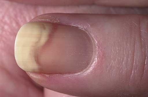
69 Nail Signs due to Deposi7on of Pigment Exogenous- convex proximal border Opposite of lunula Endogenous- concave proximal border Parallels lunula Subungual- onycholysis
Nail diseases This page outlines the terms used by dermatologists to describe diseases of the fingernails and toenails.
 Nail diseases This page outlines the terms used by dermatologists to describe diseases of the fingernails and toenails. Abnormalities of the nail plate surface Nail discolouration Abnormalities of the
Nails Examination and Disorders. Overview. Case 1 15/09/2016. Samantha Eisman. 25 year old woman Noticed at pedicure Single toe
 Nails Examination and Disorders Samantha Eisman Dermatologist MBChB/ MRCP/ FCDerm(SA)/ FACD Demystify nails Overview QUIZ Talk Examination nails and and site specific disease QUIZ answers and cover common
PERDUE NFPE: Tongue
 NFPE: Tongue NFPE: Nail Reference Guide Techniques: Inspection: Inspect the shape of the nails for - spooning - abnormal color or color patterns - splinter hemorrhages - white spots - presence of the lunula
Chapter 10 Nail Disorders & Diseases
 Chapter 10 Nail Disorders & Diseases MULTIPLE CHOICE 1. The surface of a healthy natural nail should be. a. smooth and spotted b. flexible and spotted c. smooth and unspotted d. pitted and rough ANS: C
Nail Matrix Disorders Nail Fold Disorders Nail Bed Disorders Nail Plate Disorders
 The most commonly reported nail conditions, seen by podiatrists, are: Onychocryptosis, 26% Onychauxis & onychogryphosis 22.8% Onychophosis 18.8% Onychomycosis 8.3% Nail Matrix Disorders Nail Fold Disorders
Poonkiat Suchonwanit, MD Hair and Scalp Disorders Unit Division of Dermatology Department of Medicine Ramathibodi Hospital
 Poonkiat Suchonwanit, MD Hair and Scalp Disorders Unit Division of Dermatology Department of Medicine Ramathibodi Hospital Hair loss Excessive hair growth Hair haft abnormalities Hair color Anagen Catagen
Diagnosis and Treatment of Infectious, Inflammatory and Neoplastic Nail Conditions
 OFFICE DERMATOLOGY, PART I 0025-7125/98 $8.00 +.OO NAIL DISORDERS Diagnosis and Treatment of Infectious, Inflammatory and Neoplastic Nail Conditions Phoebe Rich, MD Nail problems (Table 1) are common complaints
Chapter 4 Opener Pearson Education, Inc.
 Chapter 4 Opener Introduction The integumentary system is composed of: Skin Hair Nails Sweat glands Oil glands Mammary glands The skin is the most visible organ of the body Clinicians can tell a lot about
Integumentary System
 Chapter 5 Integumentary System 5-1 Skin: composed of dermis and epidermis Dermis. Gives structural strength. C.T. with many fibers, fibroblasts, macrophages. Some adipocytes and blood vessels. Contains
The Integumentary System. Mosby items and derived items 2010, 2006, 2002, 1997, 1992 by Mosby, Inc., an affiliate of Elsevier Inc.
 The Integumentary System The Skin Structure two primary layers called epidermis and dermis Epidermis Outermost and thinnest primary layer of skin Composed of several layers of stratified squamous epithelium
Skin. Kristine Krafts, M.D.
 Skin Kristine Krafts, M.D. Skin Lecture Objectives Describe the functions of skin. Describe the structure, location and function of the cell types found in epidermis: keratinocytes, melanocytes, Langerhans
B. Incorrect! The ectoderm does not produce the dermis. C. Incorrect! The dermis is derived from the mesoderm.
 Human Anatomy - Problem Drill 04: The Integumentary System Question No. 1 of 10 Instructions: (1) Read the problem and answer choices carefully, (2) Work the problems on paper as 1. From the inner cell
Chapter 5: Integumentary System
 Chapter 5: Integumentary System I. Overview of the Integumentary System A. List the five major functions of the integumentary system: 1. 2. 3. 4. 5. Il. Skin A. Epidermis 1. The epidermis consists of 2.
Skin and Body Membranes Body Membranes Function of body membranes Cover body surfaces Line body cavities Form protective sheets around organs
 Skin and Body Membranes Body Membranes Function of body membranes Cover body surfaces Line body cavities Form protective sheets around organs Classification of Body Membranes Epithelial membranes Cutaneous
Supplementary Online Content
 Supplementary Online Content Foley P, Gordon K, Griffiths, CEM, et al. Efficacy of guselkumab compared with adalimumab and placebo for psoriasis in specific body regions: a secondary analysis of 2 randomized
This section covers the basic knowledge of normal skin structure and function required to help understand how skin diseases occur.
 Background Knowledge Functions of normal skin Background Knowledge This section covers the basic knowledge of normal skin structure and function required to help understand how skin diseases occur. Learning
Classification. Distal & Lateral Subungual OM. White Superficial OM. Proximal Subungual OM. Candidal OM. Total dystrophic OM
 Onychomycosis Commonest dermatological condition Definition: Infection of the nail caused by fungi that include dermatophytes, non-dermatophyte moulds and yeasts (mainly Candida). 80% of all OM affects
Skin and Body Membranes
 4 Skin and Body Membranes PowerPoint Lecture Slide Presentation by Jerry L. Cook, Sam Houston University ESSENTIALS OF HUMAN ANATOMY & PHYSIOLOGY EIGHTH EDITION ELAINE N. MARIEB Skin and Body Membranes
Principles of Anatomy and Physiology
 Principles of Anatomy and Physiology 14 th Edition CHAPTER 5 The Integumentary System Introduction The organs of the integumentary system include the skin and its accessory structures including hair, nails,
Phoebe Rich MD Adjunct Professor OHSU Portland, Oregon
 Nail Tips for Diagnosis and Management of Nail Disorders Winter Clinical Dermatology Conference 2017 Hawaii Phoebe Rich MD Adjunct Professor OHSU Portland, Oregon Objectives diagnostic clues for benign
PowerPoint Lecture Slide Presentation by Patty Bostwick-Taylor, Florence-Darlington Technical College Skin and Body Membranes
 PowerPoint Lecture Slide Presentation by Patty Bostwick-Taylor, Florence-Darlington Technical College Skin and Body Membranes 4 Body Membranes Function of body membranes Cover body surfaces Line body cavities
Skin and Body Membranes
 Essentials of Human Anatomy & Physiology Elaine N. Marieb Seventh Edition Chapter 4 Skin and Body Membranes Slides 4.1 4.32 Lecture Slides in PowerPoint by Jerry L. Cook Skin and Body Membranes Function
Skin (Integumentary System) Wheater, Chap. 9
 Wheater, Chap. 9") Skin (Integumentary System) Wheater, Chap. 9 Skin (Integument) Consists of skin and associated derivatives Largest organ of body (21 ft 2 ; 9 lbs.; has 11 miles of blood vessels) Functions: Protection
Skin (Integumentary System) Wheater, Chap. 9 Skin (Integument) Consists of skin and associated derivatives Largest organ of body (21 ft 2 ; 9 lbs.; has 11 miles of blood vessels) Functions: Protection
Study of the Nail. Copyright 2013 SAP
 Study of the Nail Copyright 2013 SAP THE NAIL An appendage of the skin, this horny, translucent plate protects the tips of fingers and toes. Onyx is the technical term for the nail. The nail is composed
Unit 4 - The Skin and Body Membranes 1
 Unit 4 - The Skin and Body Membranes 1 I. Unit 4: Skin and Body Membranes A. Body Membranes 1. Function of body membranes a) Cover body surfaces b) Line body cavities c) Form protective sheets around organs
Examining the Fingernails When Evaluating Presenting Symptoms in Elderly
 Page 1 of 30 Examining the Fingernails When Evaluating Presenting Symptoms in Elderly Patients CME/CE Complete author affiliations and disclosures are at the end of this activity. Release Date: March 26,
The Integumentary System
 The Integumentary System Integument is skin Skin and its appendages make up the integumentary system A fatty layer (hypodermis) lies deep to it Two distinct regions Epidermis Dermis PHL 212 1 Function
Integumentary System
 Integumentary System Overview Functions 1. Protection 2. Excretion of wastes 3. Maintenance of T b 4. Synthesis of vitamin D 3 5. Storage of lipids 6. Detection of sensory stimuli Epidermis Tissue types
Chapter 5. Integumentary System 5-1
 Chapter 5 Integumentary System 5-1 Structures that are part of the integument Skin Hair Nails Glands Overview of Functions Protection Sensation Temperature regulation Vitamin D production Excretion Immunity
Dr Narmeen S. Ahmad. Lab 1
 Dr Narmeen S. Ahmad Lab 1 1 Tissues are groups of cells with a common structure (form) and function (job). There are (4) types of tissue: 1. Epithelial 2. Connective 3. Muscle 4. Nervous 2 Epithelial cells
Ch. 4: Skin and Body Membranes
 Ch. 4: Skin and Body Membranes I. Body Membranes A. Function of body membranes 1. Cover body surfaces 2. Line body cavities 3. Form protective sheets around organs II. Classification of Body Membranes
The Integumentary System: An Overview
 The Integumentary System: An Overview Functions: Protective covering Helps regulate body temperature Retards water loss from deeper tissues Houses sensory receptors Synthesizes biochemicals Excretes small
INTEGUMENTARY 1-Epidermis, 2-Dermis, Structure of thick and thin skin I- Epidermis . Stratum basale
 INTEGUMENTARY The skin (integument, cutis ) and its derivatives constitute the integumentary system. It form the external covering of the body and is the largest organ of the body. The skin consists of
The Integumentary System
 The Integumentary System The Integumentary System Integument is skin Skin and its appendages make up the integumentary system (See if you can name some appendages) A fatty layer (hypodermis) lies deep
Chapter 6 Skin and the Integumentary System. Skin Cells. Layers of Skin. Epidermis Dermis Subcutaneous layer beneath dermis not part of skin
 Chapter 6 Skin and the Integumentary System Composed of several tissues Maintains homeostasis Protective covering Retards water loss Regulates body temperature Houses sensory receptors Contains immune
Ch 4. Skin and Body Membranes
 Ch 4 Skin and Body Membranes TITLE HISTOLOGY SLIDES & NOTES ESSENTIAL QUESTION What tissues compose the integumentary system? Stratified Squamous Epithelium Stratified = several layers; Squamous = shape
7/10/18. Introduction. Integumentary System. Physiology. Anatomy. Structure of the Skin. Epidermis
 Introduction Integumentary System Chapter 22 Skin is largest and heaviest organ of body (7% of body weight) Houses receptors for touch, heat, cold, movement, and vibration No other body system is more
Anatomy and Physiology Homework: Chapters 3-4
 Anatomy and Physiology Homework: Chapters 3-4 CHAPTER 3: Cells and Tissues 1. The smallest unit of living tissue is called a. All living organisms are composed of these basic units where all life processes
2/5/2019. Organ System: Skin or Integumentary System. Hypodermis (or superficial fascia) Integumentary System - Learn and Understand
 Integumentary System - Learn and Understand") Integumentary System - Learn and Understand Skin is an organ comprised of all four tissues Each layer of the skin contributes to one or more of its numerous functions Skin is both strong and flexible Keratinization
Integumentary System - Learn and Understand Skin is an organ comprised of all four tissues Each layer of the skin contributes to one or more of its numerous functions Skin is both strong and flexible Keratinization
Name: Date: Class: Unit 1 Outline: Introduction, Cells, Tissues, and the Integumentary System
 Name: Date: Class: Unit 1 Outline: Introduction, Cells, Tissues, the Integumentary System Introduction to A&P The Human Body- An Orientation - study of the structure shape of the body its parts - study
Chapter 05. Lecture Outline. See separate PowerPoint slides for all figures and tables pre-inserted into PowerPoint without notes.
 Chapter 05 Lecture Outline See separate PowerPoint slides for all figures and tables pre-inserted into PowerPoint without notes. Copyright The McGraw-Hill Companies, Inc. Permission required for reproduction
Integumentary System (Script) Slide 1: Integumentary System. Slide 2: An overview of the integumentary system
 Slide 1: Integumentary System. Slide 2: An overview of the integumentary system") Integumentary System (Script) Slide 1: Integumentary System Slide 2: An overview of the integumentary system Skin is the body s largest and heaviest organ making up 15% of body weight. Most skin is 1 to
Integumentary System (Script) Slide 1: Integumentary System Slide 2: An overview of the integumentary system Skin is the body s largest and heaviest organ making up 15% of body weight. Most skin is 1 to
NAIL SURGERY TECHNIQUES
 NAIL SURGERY TECHNIQUES TRACEY C. VLAHOVIC, DPM FFPM RCPS (GLASG) ASSOCIATE PROFESSOR, J STANLEY AND PEARL LANDAU FACULTY FELLOW TEMPLE UNIVERSITY SCHOOL OF PODIATRIC MEDICINE, PHILA, PA THE AGENDA Pincer
LESSON ASSIGNMENT. The Human Integumentary and Fascial Systems. After completing this lesson, you should be able to:
 LESSON ASSIGNMENT LESSON 3 The Human Integumentary and Fascial Systems. TEXT ASSIGNMENT Paragraphs 3-1 through 3-14. LESSON OBJECTIVES After completing this lesson, you should be able to: 3-1. Define integumentary
Integumentary System. Integumentary System
 1. General aspects a. The integumentary system consists of several organs major organ of the system is the skin other organs are relatively small and they can be considered as specialized structures of
Hole s Human Anatomy and Physiology Eleventh Edition. Mrs. Hummer. Chapter 6
 Hole s Human Anatomy and Physiology Eleventh Edition Mrs. Hummer Chapter 6 1 Chapter 6 Skin and the Integumentary System Composed of several tissues Maintains homeostasis Protective covering Retards water
Figure 4.1. Using Figure 4.1, identify the following: 1) The region that contains adipose tissue is indicated by letter. Diff: 2 Page Ref: 115
 The region that contains adipose tissue is indicated by letter. Diff: 2 Page Ref: 115") Essentials of Anatomy and Physiology, 9e (Marieb) Chapter 4 Skin and Body Membranes Short Answer Figure 4.1 Using Figure 4.1, identify the following: 1) The region that contains adipose tissue is indicated
Essentials of Anatomy and Physiology, 9e (Marieb) Chapter 4 Skin and Body Membranes Short Answer Figure 4.1 Using Figure 4.1, identify the following: 1) The region that contains adipose tissue is indicated
Integumentary System and Body Membranes
 Integumentary System and Body Membranes The Skin and its appendages hair, nails, and skin glands Anatomy/Physiology NHS http://www.lab.anhb.uwa.edu.au/mb140/corepages/integumentary/integum.htm I. System
The Integumentary System
 The Integumentary System The Integumentary System Integument is skin Skin and its appendages make up the integumentary system A fatty layer (hypodermis) lies deep to it Two distinct regions Epidermis Dermis
BI 121 LAB. WEEK 2: Tissues (continued); Integumentary System
; Integumentary System") BI 121 LAB 2-1 WEEK 2: Tissues (continued); Integumentary System This week you will 1) Review the four major tissue types 2) Review the characteristics of epithelial tissues. 3) Learn the major characteristics
BI 121 LAB 2-1 WEEK 2: Tissues (continued); Integumentary System This week you will 1) Review the four major tissue types 2) Review the characteristics of epithelial tissues. 3) Learn the major characteristics
SKIN HISTOLOGY the microscopic anatomy of the Integument. Mikrogeo. com
 SKIN HISTOLOGY the microscopic anatomy of the Integument Mikrogeo. com Hair follicles, sweat glands, sebaceous glands (even teeth) are products of the epidermis,embryologically speaking ectododerm, that
****************************************************************************************************** INTEGUMENTARY SYSTEM
 BIOLOGY 211: HUMAN ANATOMY & PHYSIOLOGY ****************************************************************************************************** INTEGUMENTARY SYSTEM ******************************************************************************************************
Histology of the Eye
 Histology of the Eye Objectives By the end of this lecture, the student should be able to describe: The general structure of the eye. The microscopic structure of:»cornea.»retina. EYE BULB Three coats
Anatomy Ch 6: Integumentary System
 Anatomy Ch 6: Integumentary System Introduction: A. Organs are body structures composed of two or more different tissues. B. The skin and its accessory organs make up the integumentary system. Types of
Slide 1. Slide 2. Slide 3. Chapter 4: Body Membranes and the Integumentary System. Introduction. Membranes
 Slide 1 Chapter 4: Body Membranes and the Integumentary System Slide 2 Introduction Skin often reveals our inner workings and general health In most manual therapies, the skin is primary interface with
Lesson 3: The Human Integumentary and Fascial Systems
 Basic Human Anatomy Lesson 3: The Human Integumentary and Fascial Systems Welcome to Lesson 3 of the Basic Human Anatomy Course. Today, we ll be studying the Human Integumentary and Fascial Systems. I
CHAPTER 5 INTEGUMENTARY
 CHAPTER 5 INTEGUMENTARY skin under the skin other stuff cutaneous layer hypodermis (subcutaneous) accessory structures Cutaneous layer = skin epithelial layers = connective tissue layer = dermis Subcutaneous
Ex. 7: Integumentary
 Collin County Community College BIOL. 2401 Ex. 7: Integumentary. Skin or Integument Consists of three major regions Epidermis outermost superficial region Dermis middle region Hypodermis (superficial fascia)
Introduction. Skin and Body Membranes. Cutaneous Membranes Skin 9/14/2017. Classification of Body Membranes. Classification of Body Membranes
 Introduction Skin and Body Membranes Body membranes Cover surfaces Line body cavities Form protective and lubricating sheets around organs Classified in 5 categories Epithelial membranes 3 types- cutaneous,
Integumentary System. Packet #12
 Integumentary System Packet #12 Introduction Skin/Integument Skin, considered an organ, is the major component of the integumentary system. The integumentary system is also composed of other accessory
Internal medicine and medical investigation (ISSN: ) May 2017, Volume:2, Issue:2, Page: 57-62
 May 2017, Volume:2, Issue:2, Page: 57-62") Internal medicine and medical investigation (ISSN: 2474-7750) www.imminv.com May 2017, Volume:2, Issue:2, Page: 57-62 Determining the Prevalence and Types of Nail Involvement in Patients with Severe Acne
Internal medicine and medical investigation (ISSN: 2474-7750) www.imminv.com May 2017, Volume:2, Issue:2, Page: 57-62 Determining the Prevalence and Types of Nail Involvement in Patients with Severe Acne
Chapter 6: Integumentary System
 Chapter 6: Integumentary System 6.1 Introduction Why is skin considered to be an organ? What makes up the integumentary system? Integumentary System Skin (cutaneous membrane) Skin derivatives Sweat glands
Benign and malignant epithelial lesions: Seborrheic keratosis: A common benign pigmented epidermal tumor occur in middle-aged or older persons more
 Benign and malignant epithelial lesions: Seborrheic keratosis: A common benign pigmented epidermal tumor occur in middle-aged or older persons more common on the trunk; but extremities, head and neck are
Nail Disorders. Disclosure Statement: Objectives. Normal Nail Anatomy. Normal Nail Anatomy
 Nail Disorders Disclosure Statement: Co-Author, Dermatologic and Cosmetic Procedures in Office Practice. Elsevier, Inc., Philadelphia. 2012. Cutaneous Cryosurgery Fourth EditionCRC press 2014 Daniel Stulberg,
Unit 4 The Integumentary System
 Unit 4 The Integumentary System I. Classification of Body Membranes A. Epithelial Membranes (3) 1. Cutaneous Membrane > Stratified Squamous > Sits on Dense Connective Tissue > Skin: Epidermis & Dermis
The Nail Common terms & Anatomy Onychomycosis Causes and Background Onychomycosis Classification Treatment Basics
 FOX Podiatry Applications The Nail Common terms & Anatomy Onychomycosis Causes and Background Onychomycosis Classification Treatment Basics The Nail - Areas definitions Proximal nail fold Nail plate area
BIO 130 Anatomy and Physiology Spring, 2016 Exam 3 Name: Course ID Number. Section 1 Answer questions 1 40 on the scan sheet.
 BIO 130 Anatomy and Physiology Spring, 2016 Exam 3 Name: Course ID Number Section 1 Answer questions 1 40 on the scan sheet. 1. Which of the following is NOT a characteristic of epithelial tissue? a. It
Lab 7: Integumentary System Hamilton ANSWERS TO PRE- LAB ASSIGNMENTS
 Lab 7: Integumentary System Hamilton ANSWERS TO PRE- LAB ASSIGNMENTS Pre-Lab Activity 1: 1. a. epidermis b. dermis c. hypodermis d. adipose tissue e. hair f. sebaceous gland g. sweat gland 2. a Pre-Lab
Podiatric Perspectives on Nail Surgery
 Podiatric Perspectives on Nail Surgery Tracey C. Vlahovic, DPM Clinical Associate Professor, J. Stanley and Pearl Landau Faculty Fellow Temple University School of Podiatric Medicine Philadelphia, PA Common
Integument. Squamous Cell Carcinoma. Melanoma. Largest organ 30% of all clinical diagnoses 1/3 of all tumors
 Squamous Cell Carcinoma Integument Largest organ 30% of all clinical diagnoses 1/3 of all tumors Melanoma Epidermis Stratified, squamous keratinized epithelium Derived from ectoderm Appendages hair follicles
December 3, Name five bones in your body. Are bones living or dead? Explain. What is the function of bone marrow?
 December 3, 2013 Name five bones in your body. Are bones living or dead? Explain. What is the function of bone marrow? Skeletal, Muscular, and Integumentary Systems Chapter 36: Biology II The Skeleton
Integumentary System. Study of the Skin
 Integumentary System Study of the Skin Skin is used to: Maintain homeostasis Provide a protective covering Slow down water loss from deeper tissues House sensory receptors Synthesize various biochemicals
ABCD rule. apocrine glands. arrector pili. ceruminous glands. contact dermatitis
 ABCD rule assessing moles: asymmetric, broder irregularity, color, diameter (larger than 6mm) apocrine glands arrector pili sweat glands in the pubic and underarm areas that secrete thicker sweat, that
The Integumentary System: ANATOMY Includes: - Skin (integument) MEMBRANES. PHYSIOLOGY (functions) Protection. EPITHELIAL (cont.
 MEMBRANES. PHYSIOLOGY (functions) Protection. EPITHELIAL (cont.") Did you know. Membranes & The Integumentary System The skin is the largest organ of the human body. It has a surface area of about 25 square-feet! You shed about 1.5 pounds of skin particles each year.
Did you know. Membranes & The Integumentary System The skin is the largest organ of the human body. It has a surface area of about 25 square-feet! You shed about 1.5 pounds of skin particles each year.
Hole s Essentials of Human Anatomy & Physiology
 Hole s Essentials of Human Anatomy & Physiology David Shier Jackie Butler Ricki Lewis Created by Dr. Melissa Eisenhauer Head Athletic Trainer/Assistant Professor Trevecca Nazarene University Chapter 6
INTEGUMENTARY SYSTEM CHAPTER 4
 INTEGUMENTARY SYSTEM CHAPTER 4 FUNCTIONS Waterproofs Protein called keratin Protection 1 st line of defense against pathogens, chemicals & abrasions Insulation Regulates heat loss by controlling blood
POST-INJURY INTERVALS 1
 POST-INJURY INTERVALS 1 Introduction 1 Contusion dating 2 Skin 2 Brain 5 Hypoxic/ischemic injury and increased intracranial pressure 18 Brain incidentals (non-injurious) 21 Sexual violence 27 INTRODUCTION
Mohs surgery for the nail unit
 Mohs surgery for the nail unit olivier.cogrel@chu-bordeaux.fr Dermatologic surgery, Mohs surgery and lasers unit CHU Bordeaux, France Squamous cell carcinoma +++ Acral lentiginous melanoma Lichte et al.
2 Anonychia/Micronychia
 2 Anonychia/Micronychia Total or partial absence of the nail May be congenital or acquired Table 2.1. Causes of anonychia/micronychia Congenital Acquired Trauma Amniotic bands Bullous Teratogens (drugs,
Integumentary System
 Integumentary System The integumentary system is commonly known as the Skin Largest organ of human body 10% total body weight and would cover over 20 square feet Functions of Skin 1. Protection Barrier
Presentation Notes Be Visually Aware Nail Diseases and Disorders. Be Visually Aware. Nail Diseases and Disorders
 Slide 1 Be Visually Aware Nail Diseases and Disorders Page1 Slide 2 Copyright Copyright Texas Education Agency, 2013. These Materials are copyrighted and trademarked as the property of the Texas Education
Dentin Formation(Dentinogenesis)
") Lecture four Dr. Wajnaa Oral Histology Dentin Formation(Dentinogenesis) Dentinogenesis begins at the cusp tips after the odontoblasts have differentiated and begin collagen production. Dentinogenesis growth
Lecture four Dr. Wajnaa Oral Histology Dentin Formation(Dentinogenesis) Dentinogenesis begins at the cusp tips after the odontoblasts have differentiated and begin collagen production. Dentinogenesis growth
Warm Up. You have 10 minutes to complete your poster and prepare what you would like to share with the class.
 Warm Up You have 10 minutes to complete your poster and prepare what you would like to share with the class. Reflection 1. What were 2 similarities between your classification scheme and others in the
Anatomy Revision Papers. Part 4. Internal Structure of the Foot
 Anatomy Revision Papers Part 4 Internal Structure of the Foot Page 2 Page 3 Page 4 Page 5 Page 5 Page 6 Page 6 Page 6 Sagittal Section of the Foot Frontal Section of Foot Ligaments within the foot Corium
Chapter 5 The Integumentary System. Copyright 2009, John Wiley & Sons, Inc. 1
 Chapter 5 The Integumentary System Copyright 2009, John Wiley & Sons, Inc. 1 Introduction The organs of the integumentary system include the skin and its accessory structures including hair, nails, and
Lymphoid Organs. Dr. Sami Zaqout. Dr. Sami Zaqout IUG Faculty of Medicine
 Lymphoid Organs Dr. Sami Zaqout Cells of the Immune System Lymphocytes Plasma cells Mast cells Neutrophils Eosinophils Cells of the mononuclear phagocyte system Distribution of cells of the immune system
4 Skin and Body Membranes Study Guide
 Name: SKIN AND BODY MEMBRANES: 4 Skin and Body Membranes Study Guide Period: Body membranes, which cover body surfaces, line its cavities, and form protective sheets around organs, fall into two major
11/8/2012. Chapter 6 Part 1 Objectives: Skin = Integument = Cutaneous Membrane. The Structure of Skin. Epidermis
 Chapter 6 Part 1 Objectives: Define organ, and associate the skin as an organ of the integumentary system. List the general functions of the skin. Describe the structure of the layers of the skin. Summarize
Anatomy and Physiology I Student Outline The Integumentary System. Integumentary System. Page 1
 Anatomy and Physiology I Student Outline The Integumentary System Integumentary System Page 1 Have a very clear understanding of the each particular tissue and their unique functions in each layer of the
PowerPoint Lecture Slide Presentation by Patty Bostwick-Taylor, Florence-Darlington Technical College Skin and Body Membranes
 PowerPoint Lecture Slide Presentation by Patty Bostwick-Taylor, Florence-Darlington Technical College Skin and Body Membranes 4 Body Membranes Function of body membranes Cover body surfaces Line body cavities
CH 05 THE INTEGUMENTARY SYSTEM
 CH 05 THE INTEGUMENTARY SYSTEM This system consists of skin and its derivatives. The skin is one of the largest organs of the body in terms of surface area. The functions of the integumentary system include:
Describe the functions of the vertebrate integumentary system. Discuss the structure of the skin and how it relates to function.
 Chapter 5 Describe the functions of the vertebrate integumentary system. Discuss the structure of the skin and how it relates to function. Explain the basis for different skin colors. Describe the structure
THE INTEGUMENTARY SYSTEM. Body Membranes & Skin
 THE INTEGUMENTARY SYSTEM Body Membranes & Skin TYPES OF MEMBRANES Epithelial Membranes includes layer of epithelial cells and connective tissue Serous Cutaneous Mucous Connective Tissue Membranes solely
Treatment of Chronic Idiopathic Onychodystrophy with Intake of Carotene-rich Food
 6 Treatment of Chronic Idiopathic Onychodystrophy with Intake of Carotene-rich Food Jin Young Jung, M.D., Mi Ryung Roh, M.D., Kee Yang Chung, M.D., Ph.D. Department of Dermatology and Cutaneous Biology
The Language of Anatomy. (Anatomical Terminology)
") The Language of Anatomy (Anatomical Terminology) Terms of Position The anatomical position is a fixed position of the body (cadaver) taken as if the body is standing (erect) looking forward with the upper
The Language of Anatomy (Anatomical Terminology) Terms of Position The anatomical position is a fixed position of the body (cadaver) taken as if the body is standing (erect) looking forward with the upper
Chapter 5 The Integumentary System. Copyright 2009, John Wiley & Sons, Inc. 1
 Chapter 5 The Integumentary System Copyright 2009, John Wiley & Sons, Inc. 1 Introduction The organs of the integumentary system include the skin and its accessory structures including hair, nails, and
Oral histology. How is the oral mucosa different from skin?
 Oral histology Lec.13 Oral mucosa: Dr.Nada AL.Ghaban The oral mucosa is the mucous membrane lining the inside of the mouth and consists of stratified squamous epithelium termed oral epithelium and an underlying
Epidermis. Integumentary system
 Epidermis the doctor mentioned at the begging of the lecture that the slides is from different sources and has information and details that is enough for us so we don t have to go back and read from the
Skin Basics. About skin. Reviewed By: Kimberly Bazar, M.D., AAD Mary Ellen Luchetti, M.D., AAD
 Page 1 of 5 Skin Basics Reviewed By: Kimberly Bazar, M.D., AAD Mary Ellen Luchetti, M.D., AAD Summary The skin is the largest organ in the body. It consists of three layers the epidermis, the dermis and
MECHANISMS OF HUMAN DISEASE: LABORATORY SESSION PATHOLOGY OF THE SKIN LAB. Friday, February 13, :30 am 11:00 am
 MECHANISMS OF HUMAN DISEASE: LABORATORY SESSION PATHOLOGY OF THE SKIN LAB Friday, February 13, 2009 9:30 am 11:00 am FACULTY COPY GOALS: Describe the basic clinical and morphologic features of various