Clinicopathologic Self- Assessment S003 AAD 2017
|
|
|
- Bonnie Wilkinson
- 5 years ago
- Views:
Transcription
1 Clinicopathologic Self- Assessment S003 AAD 2017 Clay J. Cockerell, M.D. Director, Cockerell Dermatopathology Director, Division of Dermatopathology UT Southwestern Medical Center July 2017
2 No relevant conflicts
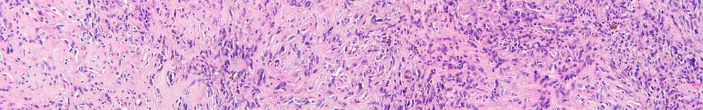
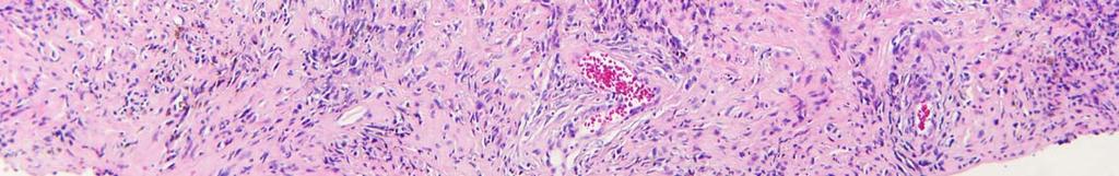
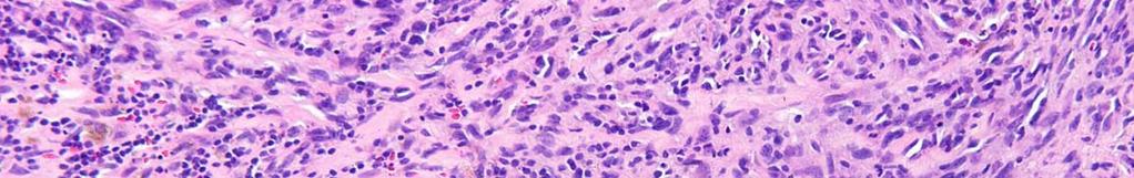
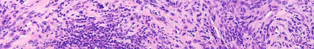
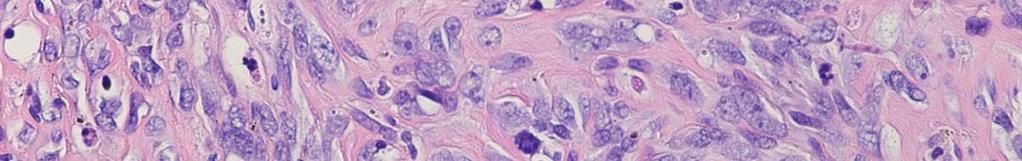
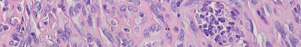
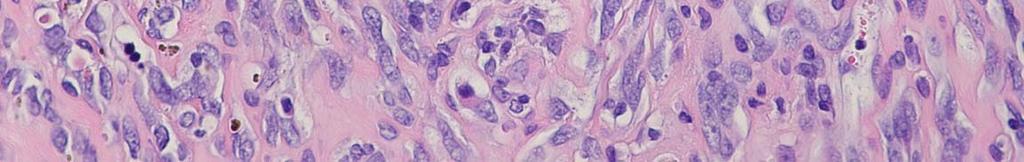
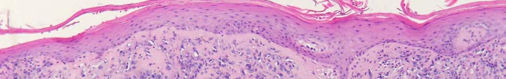
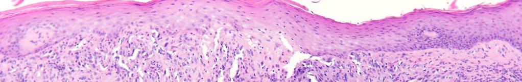
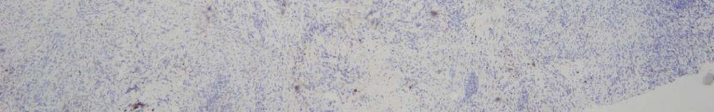
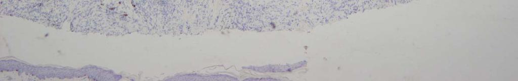
3 CASE 1
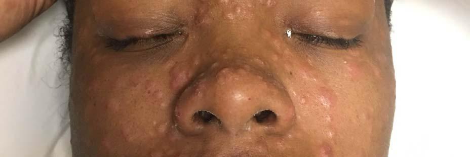
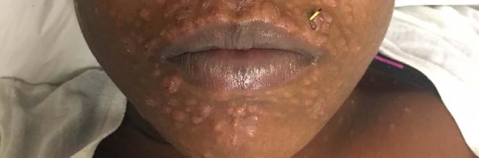
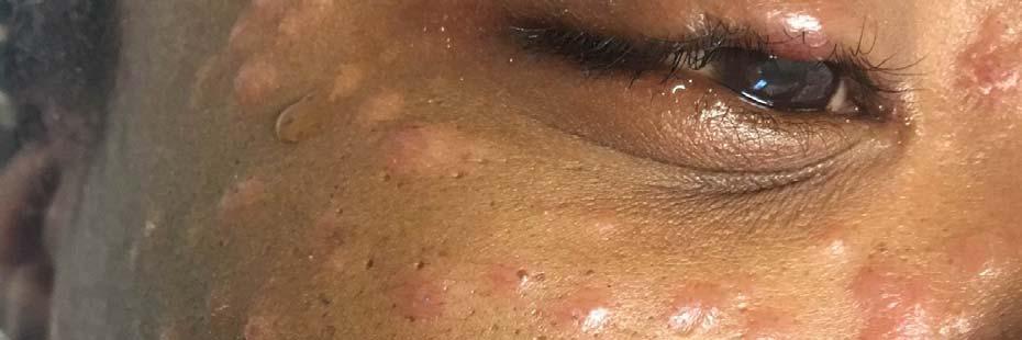
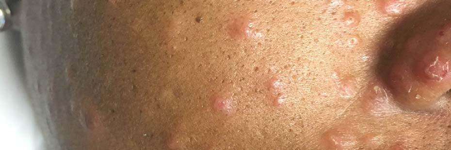
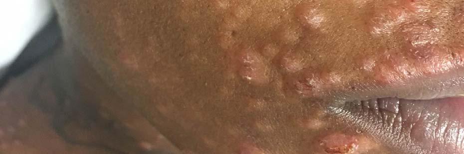
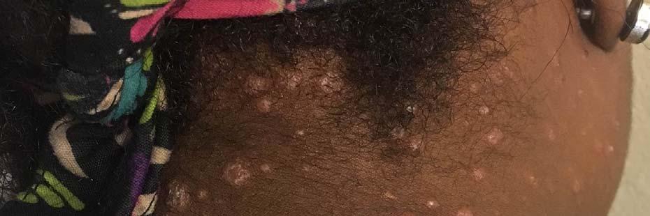
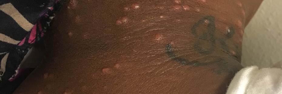
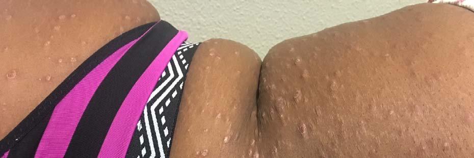
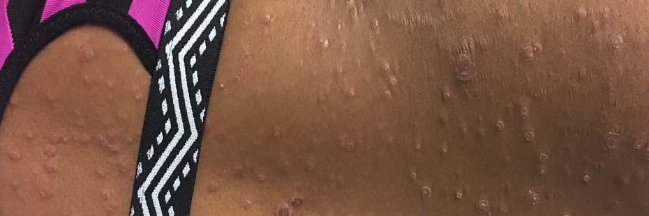
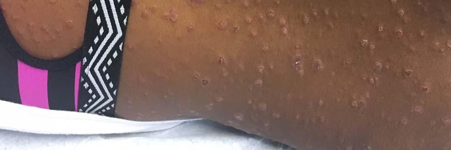
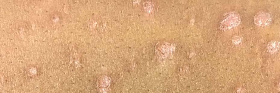
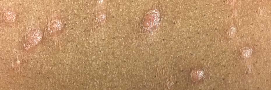
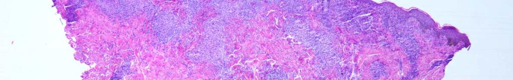
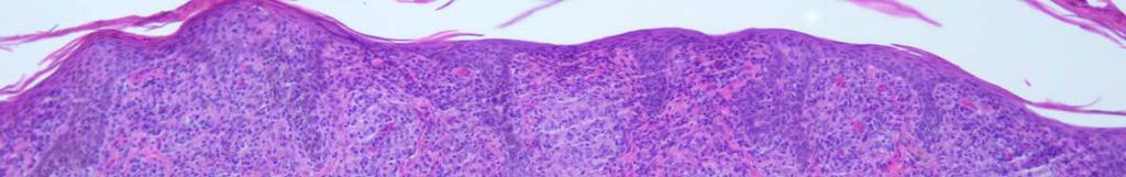
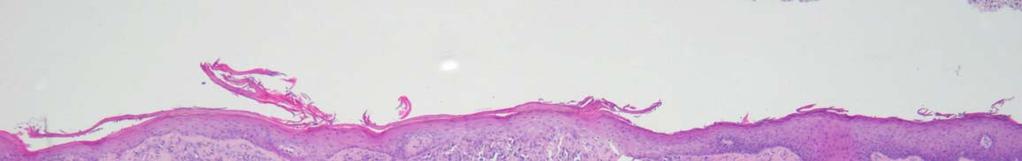
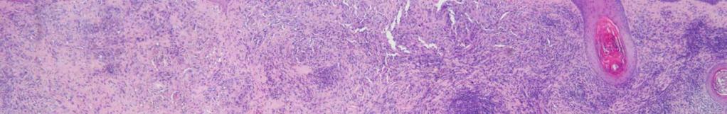
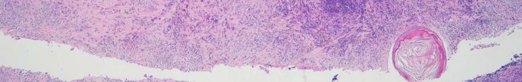
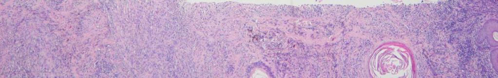
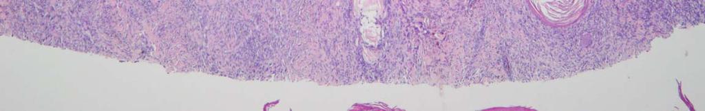
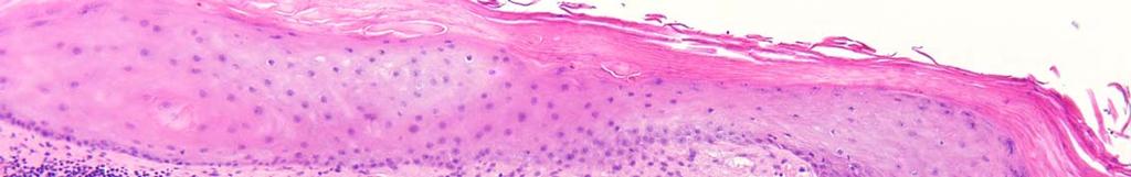
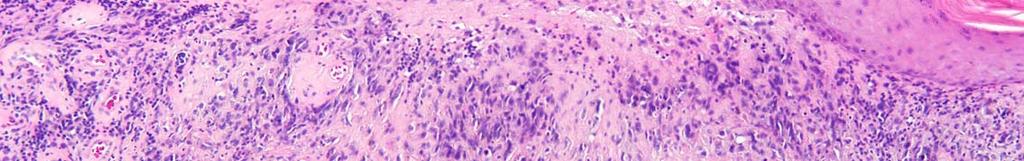
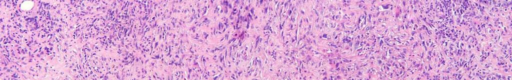
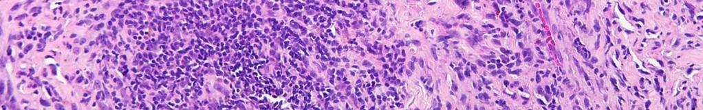
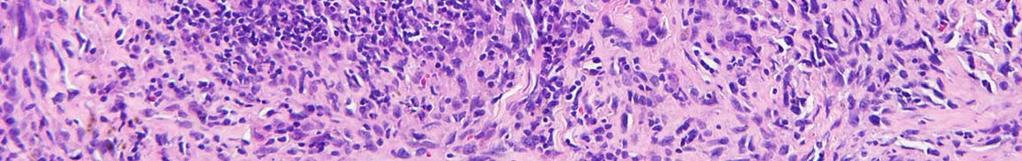
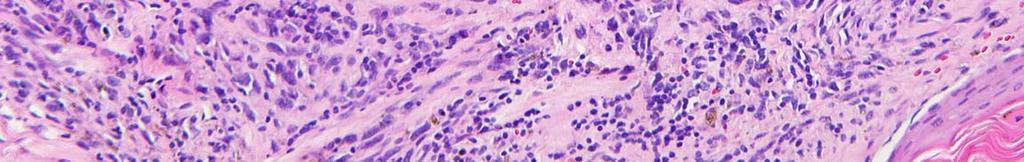
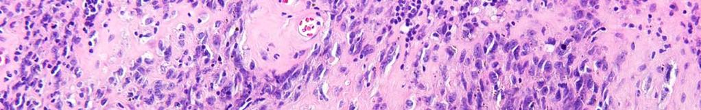
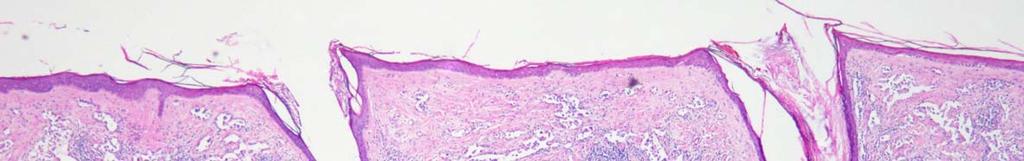
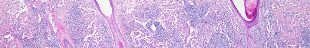
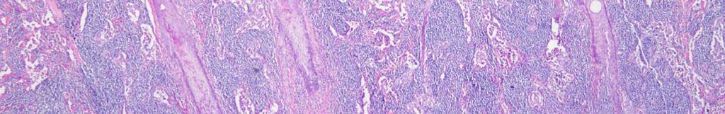
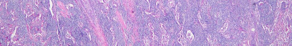
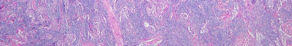
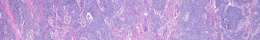
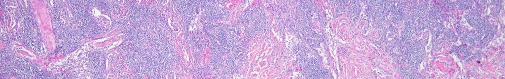
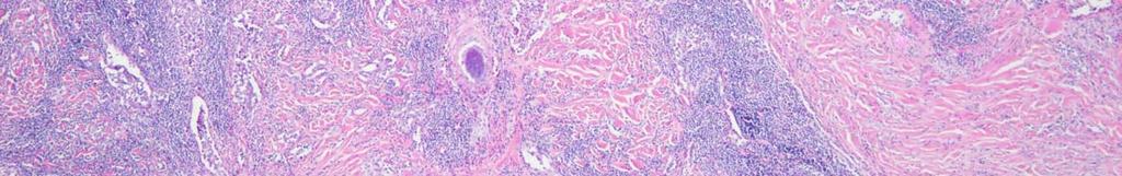
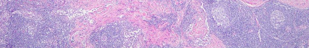
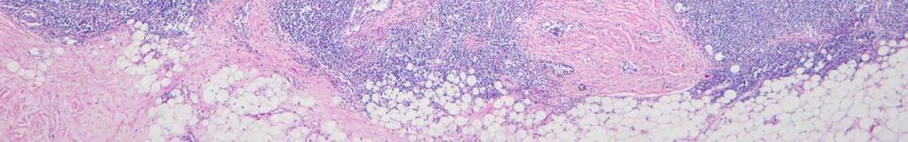
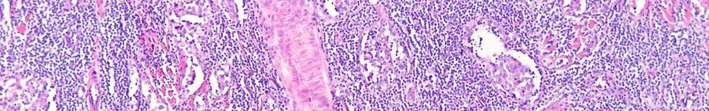
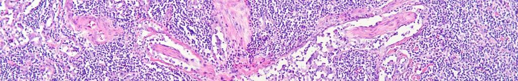
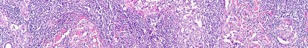
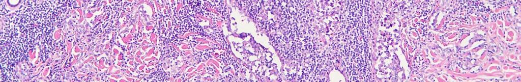
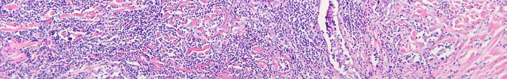
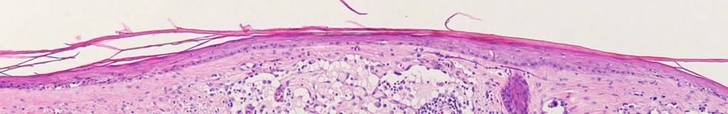
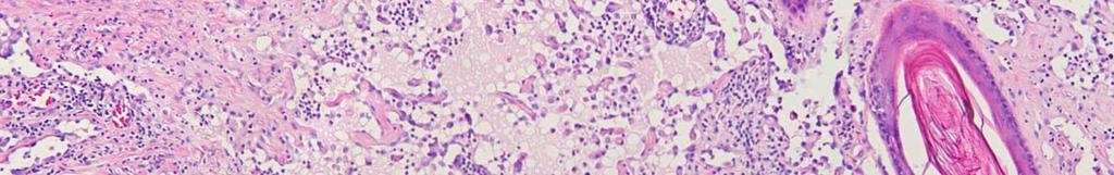
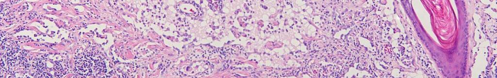
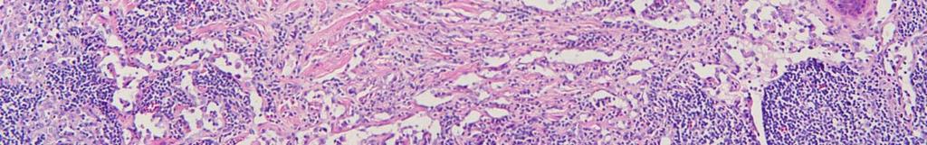
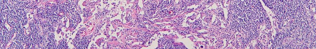
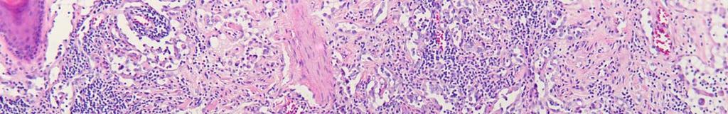
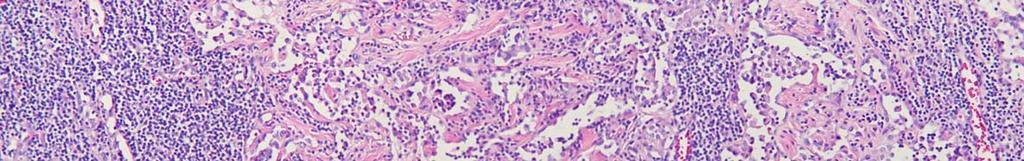
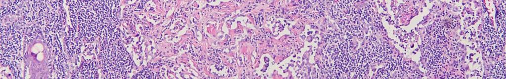
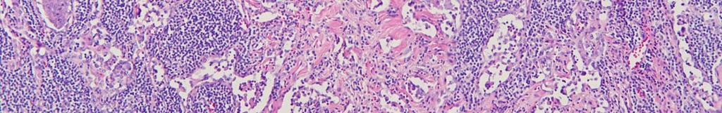
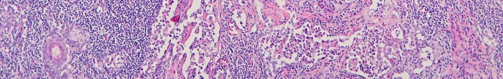
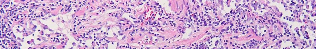
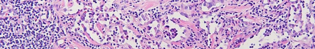
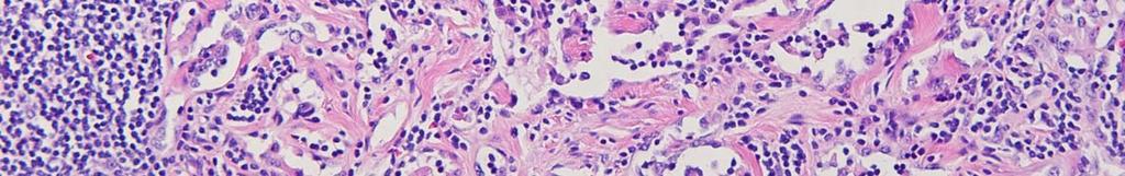
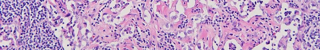
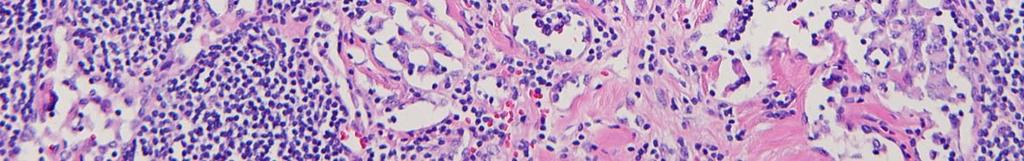
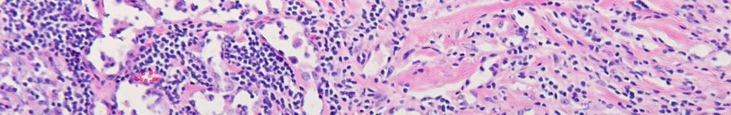
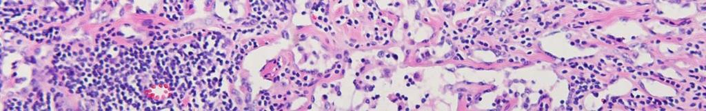
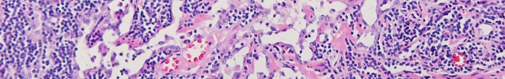
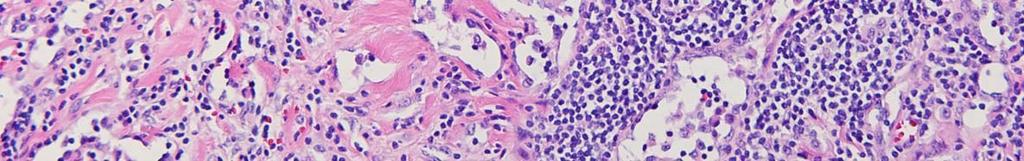
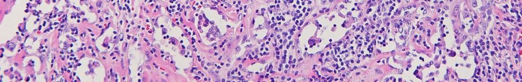
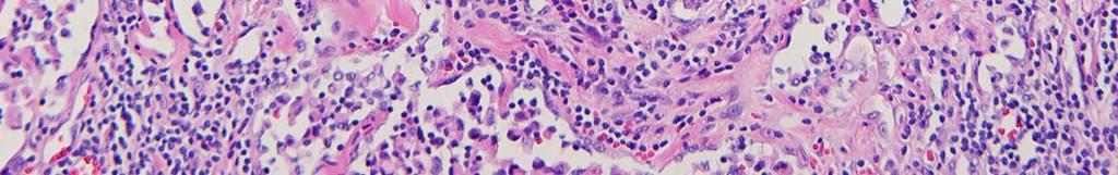
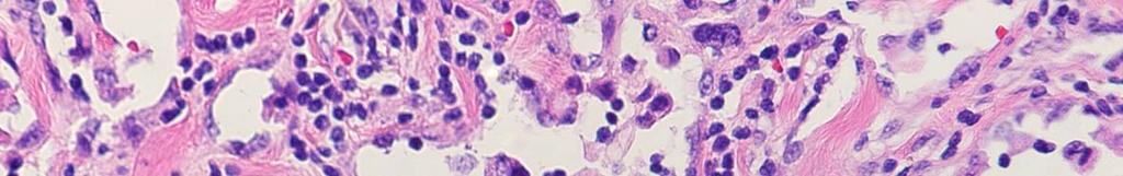
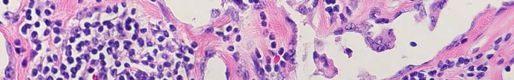
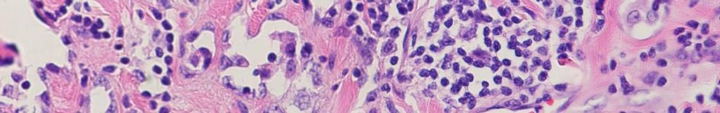
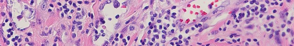
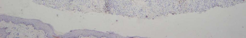
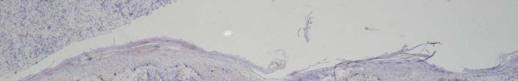
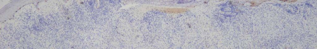
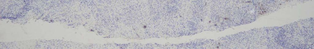
4 CASE 1 25 year-old African-American female Itchy, painful eruption over entire body for past 8 weeks Flu-like symptoms preceded eruption Fever, arthralgia, myalgia, sore throat

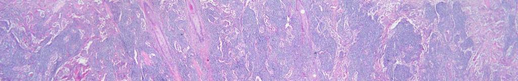
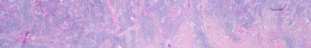
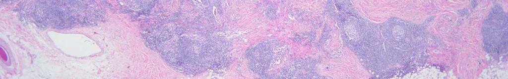
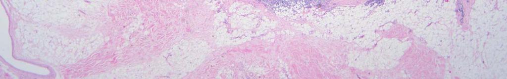
5
6

7
8

9
10 Pathology


11

12
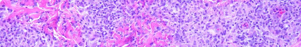
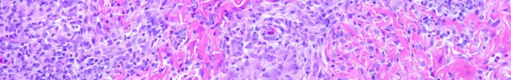
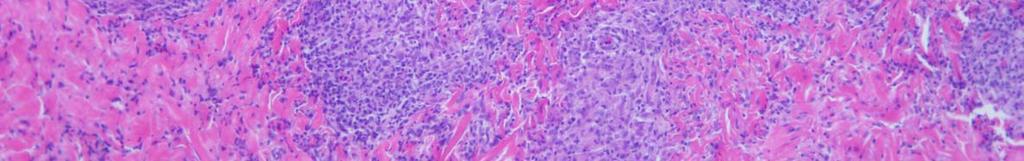
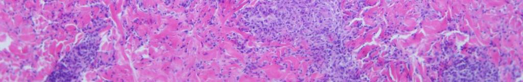
13
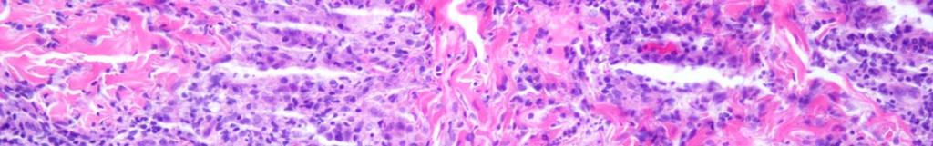
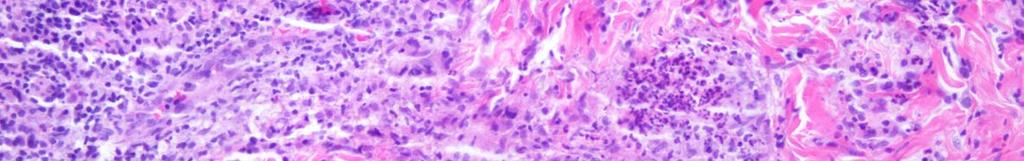
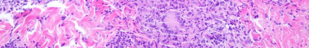
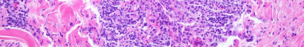
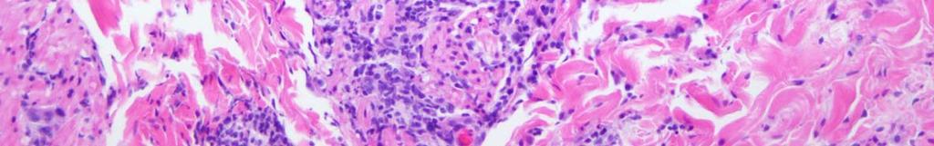
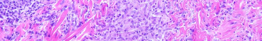
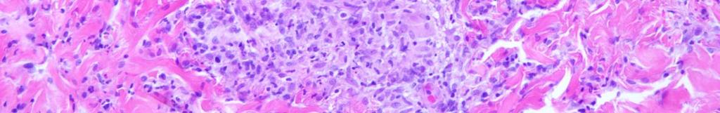
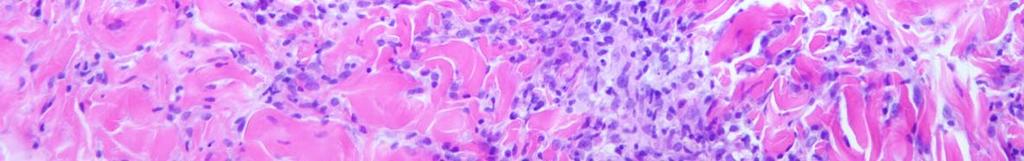
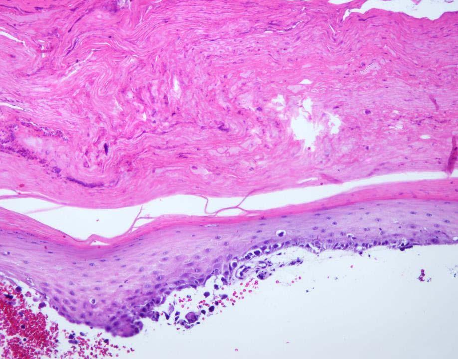
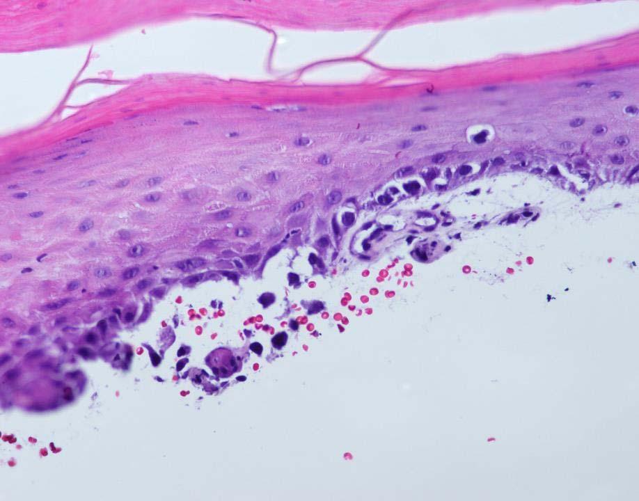
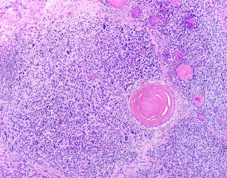
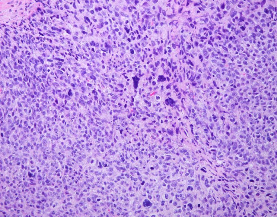
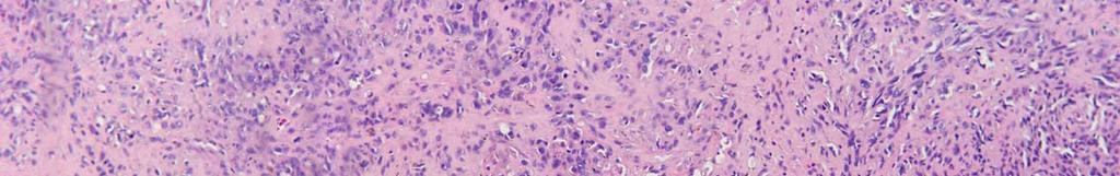
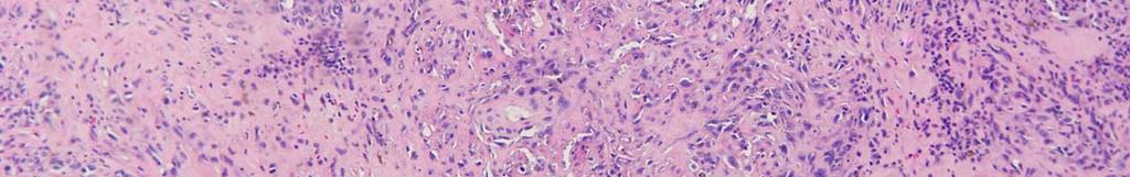
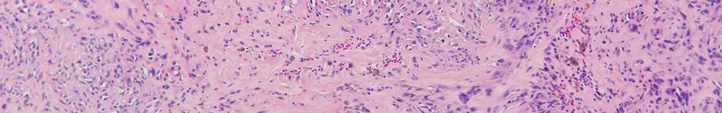
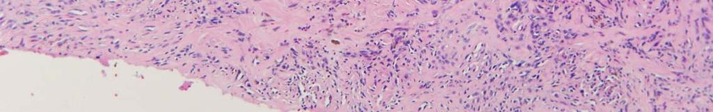
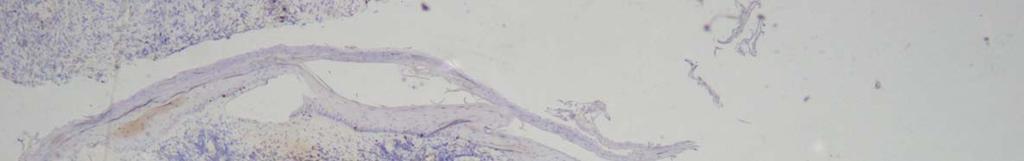
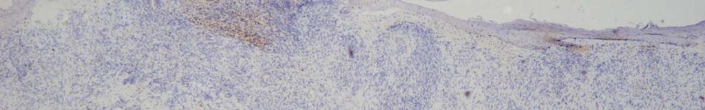
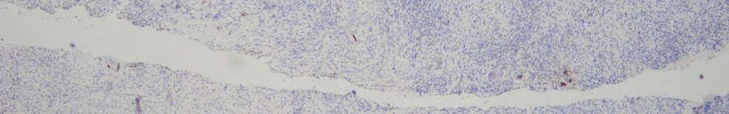
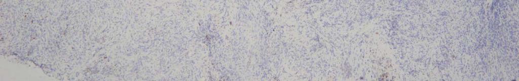
14
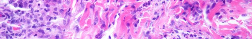
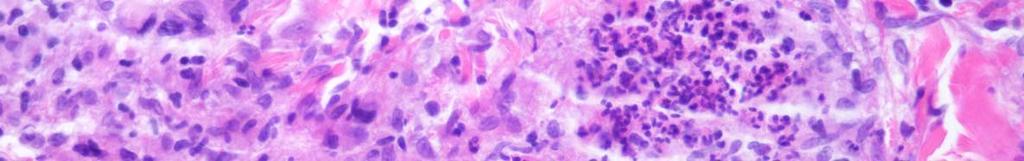
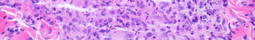
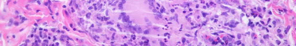
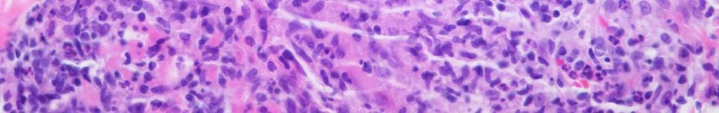
15
16 Question 1 What is the most likely diagnosis? A) Sarcoidosis B) Disseminated cryptococosis C) Lepromatous leprosy D) Granulomatous mycosis fungoides E) Granulomatous secondary syphilis
17 Question 1 What is the most likely diagnosis? A) Sarcoidosis B) Disseminated cryptococosis C) Lepromatous leprosy D) Granulomatous mycosis fungoides E) Granulomatous secondary syphilis
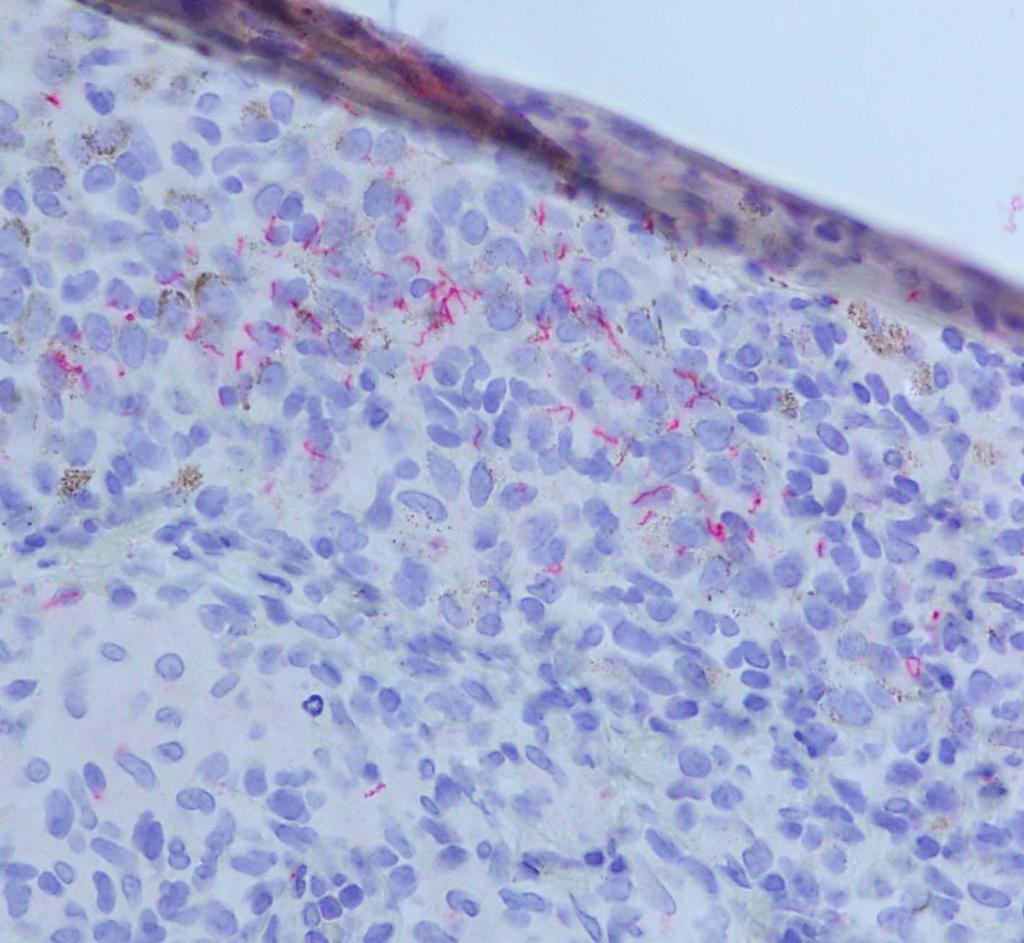
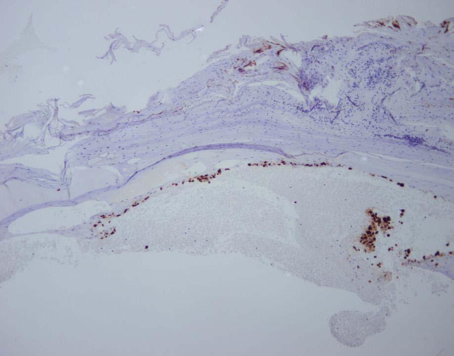
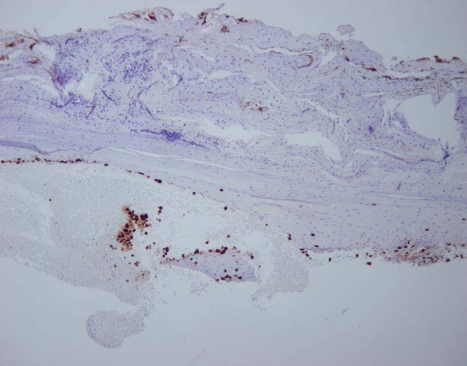
18 Question 2 Which special stain would be useful to confirm the diagnosis? A) Ziehl-Neelsen stain B) Fite stain C) PAS stain D) GMS stain E) IHC using Anti Treponema antibody
19 Question 2 Which special stain would be useful to confirm the diagnosis? A) Ziehl-Neelsen stain B) Fite stain C) PAS stain D) GMS stain E) IHC using Anti Treponema antibody
20
21 Granulomatous Syphilis Prominent granulomatous inflammation relatively uncommon in secondary syphilis Most common with tertiary syphilis When present, usually late stage secondary syphilis Majority cases 16 weeks or longer in duration Reported in acute cases also (10 days) Occasionally in HIV/AIDS patients: altered immunity results in unusual histology J Cutan Pathol 2014; 41:

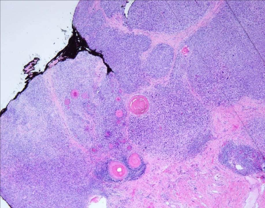
22 Granulomatous Syphilis Histiocytes, lymphocytes and variable number of plasma cells (sparse to numerous) Granulomas epithelioid, palisading, tuberculoid, sarcoidal or interstitial granuloma annulare-like +/- multinucleated giant cells Usually not as tightly sarcoidal as sarcoidosis Arranged in a T-shape or candelabra-like pattern Extension along nerves, follicular and eccrine structures IHC with Anti-Treponema pallidum antibody stain of choice Higher sensitivity (90%) and specificity than traditional silver impregnation techniques J Cutan Pathol 2014; 41:

23 Think syphilis! Still the great imitator! Sometimes in order to be a good dermatopathologist, you have to Moral of the Story
24 CASE 2
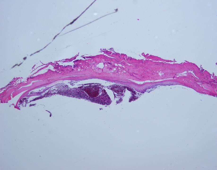
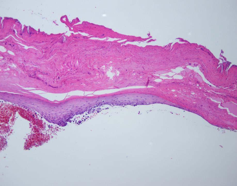
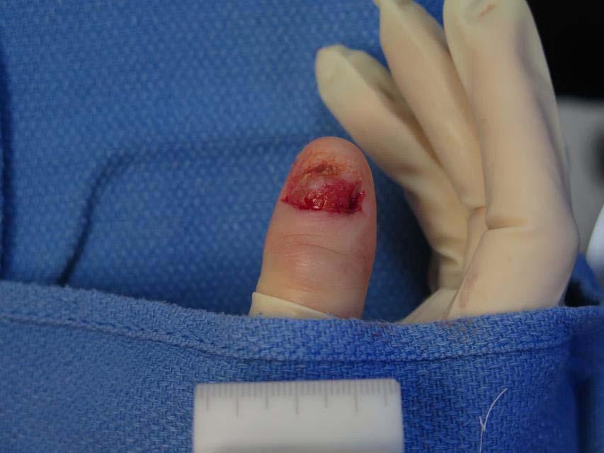
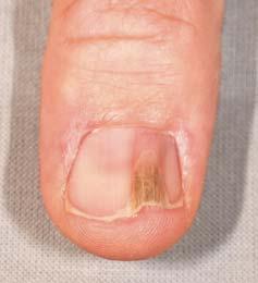
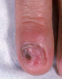
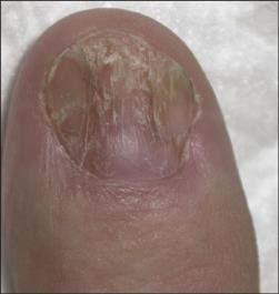
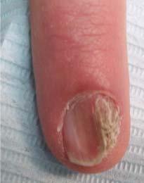
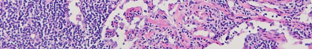
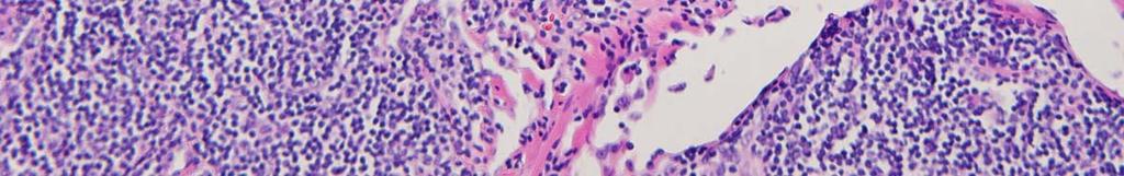
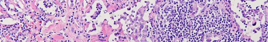
25 Case 2 42 year old woman female with 6 year history of right thumbnail dystrophy Had been treated for onychomycosis for over 1 year Nail clipping 1 year previously was negative for hyphae
26


27 Nail clipping and culture H&E and PAS negative No visible growth on agar slant H&E PAS

28 Kerydin (tavaborole) topical solution 5% White vinegar soaks
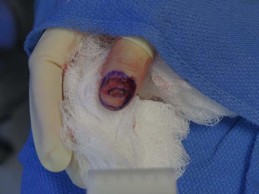
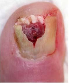
29 Case History, cont. Follow up April 2016: some improvement but no good regrowth October 2016: patient reported that nail fell off Small, skin-colored subungual papule noted Shave biopsy performed
30
31
32
33
34 Question 1 What is the most likely diagnosis? A) Melanoma B) Squamous cell carcinoma C) Onychomycosis D) Lichen planus E) Pemphigus vulgaris
35 Question 1 What is the most likely diagnosis? A) Melanoma B) Squamous cell carcinoma C) Onychomycosis D) Lichen planus E) Pemphigus vulgaris
36 Sox10
37

38 October 2016
39
40
41
42
43
44
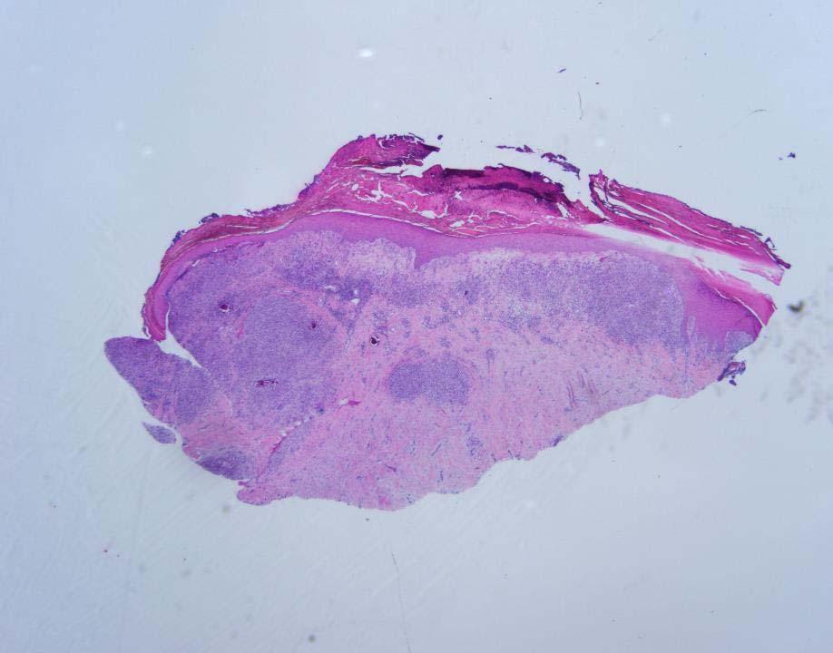
45 Final diagnosis MALIGNANT MELANOMA, RIGHT THUMB NAIL BED Clarks level IV, 3.5mm thickness, ulcerated All margins involved Mitotic index: 2/mm 2 Perineural involvement noted
46 Follow up PET scan negative for metastatic disease Amputation performed distal to PIP joint Lymph node dissection; no nodal metastasis Continued monitoring by dermatology, surgical oncology, and medical oncology


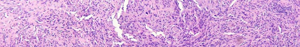
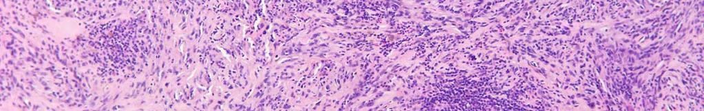
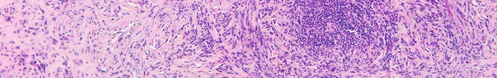
47 Amelanotic Subungual Melanoma: May Simulate Other Conditions
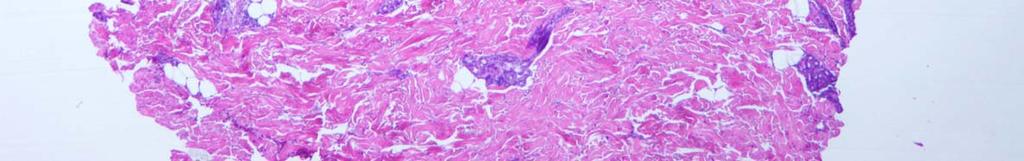
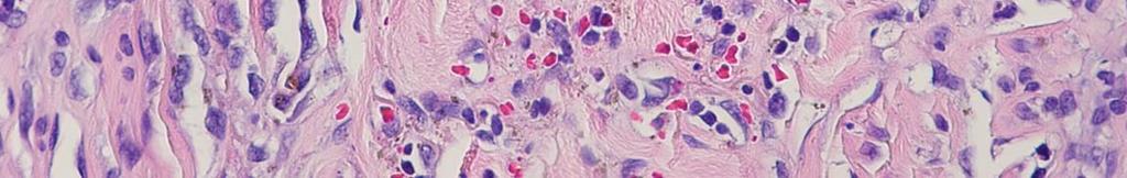
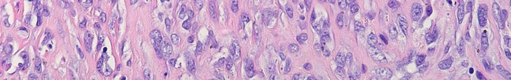
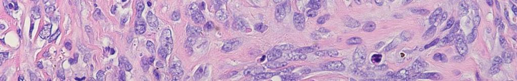
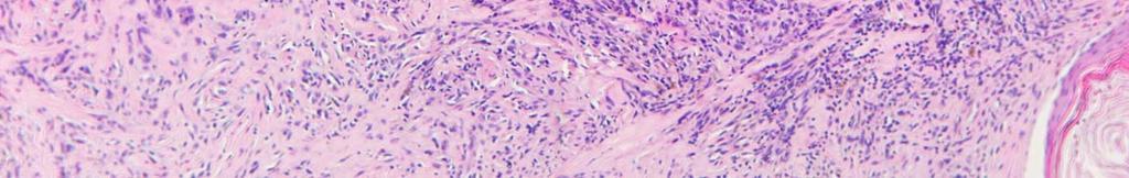
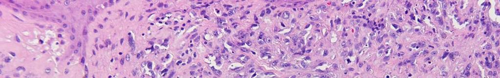
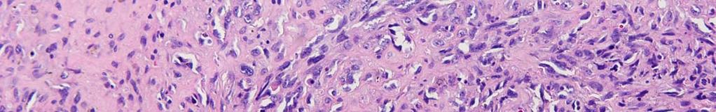
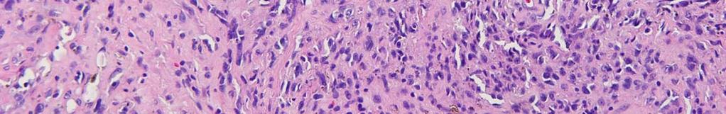
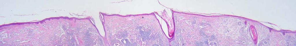
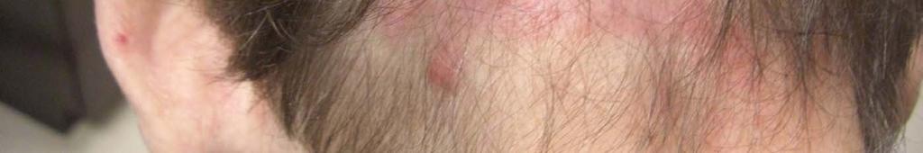
48 Question 2 What percentage of nail unit melanomas are amelanotic? A) 2% B) 5% C) 10% D) 15% E) 25%
49 Question 2 What percentage of nail unit melanomas are amelanotic? A) 2% B) 5% C) 10% D) 15% E) 25%
50 Nail Unit Melanoma Only 1-2% of all melanomas in Caucasians; 15-25% in Asians and Blacks Fingers>>Toes; most commonly on thumb and hallux 25% amelanotic <2.5mm 88% 5 year survival >2.5mm 44% 5 year survival
51 Nail unit melanoma No clear-cut relationship with UV exposure Genetically more akin to MM arising in non-sunexposed skin BRAF less prevalent KIT is more common NRAS in 4/16 nail unit melanomas
52 Challenges in diagnosing nail unit melanoma Clinically mimics other nail diseases May be present for many months prior to diagnosis Often asymptomatic; low motivation to seek medical advice Physicians reluctant to perform nail biopsies Time consuming Lack of familiarity with procedure Lack of proper instrumentation Insufficient sampling, sampling error
53 What can we do better? Maintain high clinical suspicion Lower threshold to biopsy nail abnormalities involving JUST ONE NAIL Obtain sufficient tissue given high risk of sampling error Educate patients, residents, fellows Only submit specimen to dermatopathologist with expertise and SEND PHOTO along with biopsy!
54 CASE 3
55 Case History 73 year old male with long standing lesion on the occipital scalp Clinical impression: rule out MALIGNANT MELANOMA
56
57
58
59
60
61
62
63
64
65
66
67 Diagnosis Malignant melanoma, > 2.1 mm in thickness, Clark s level 4 3 mitoses/mm 2 Non-ulcerated Patient referred to general surgery for wide excision
68
69
70
71
72
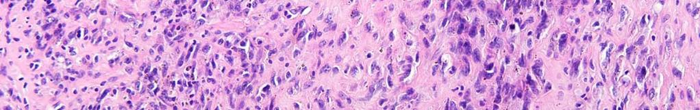
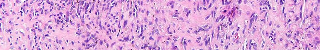
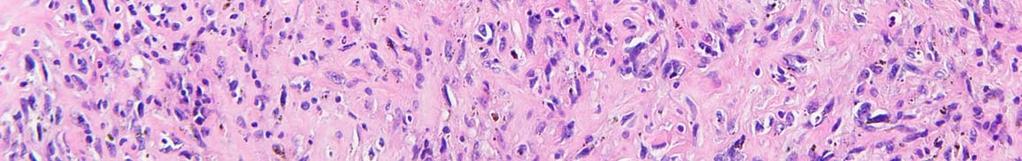
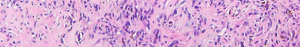
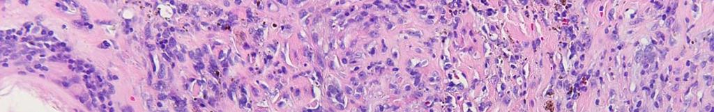
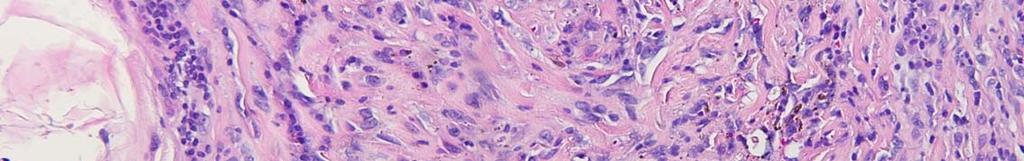
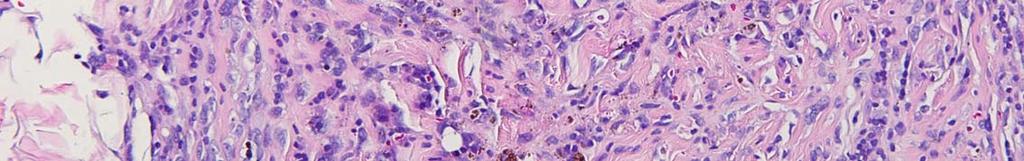
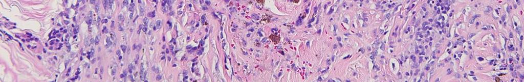
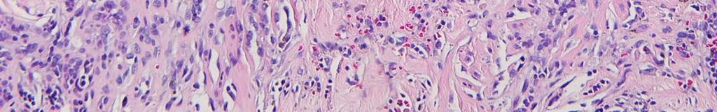
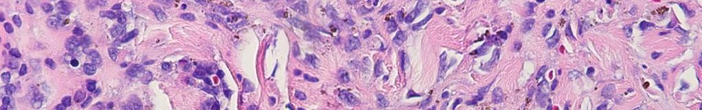
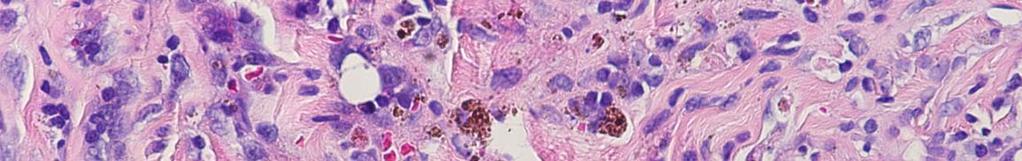
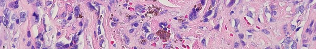
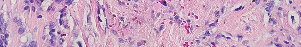
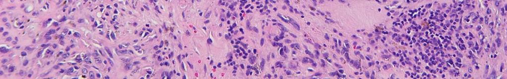
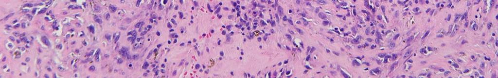
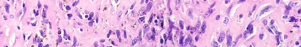
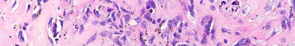
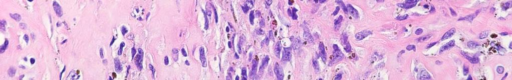
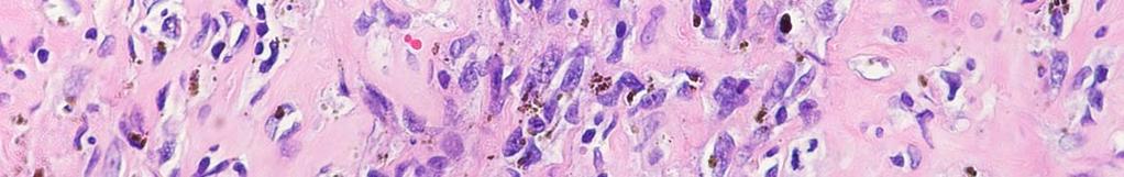
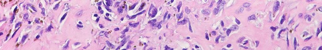
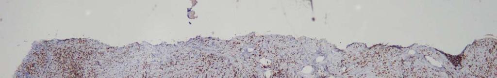
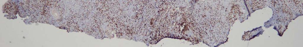
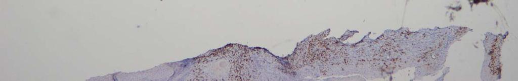
73
74
75

76

77
78 Which is the Most Likely Diagnosis? A. Angiomatoid melanoma B. Angiosarcoma C. Nodular fasciitis D. Malignant fibrous histiocytoma E. None of the above
79 Which is the Most Likely Diagnosis? A. Angiomatoid melanoma B. Angiosarcoma C. Nodular fasciitis D. Malignant fibrous histiocytoma E. None of the above
80 Clinical Course Re-excision revealed features characteristic of angiosarcoma Clinical pictures were reviewed and special stains performed - ERG & CD Melan-A & SOX S-100 protein highlighted many cells, including a discontiguous nodule Final diagnosis: pigmented epithelioid angiosarcoma simulating melanoma


81


82

83 ERG

84 CD31
85 SOX10
86 S100
87 Question 1 Angiosarcoma most commonly affects all of the following sites except: A) Face B) Scalp C) Areas of chronic lymphedema D) Previously irradiated area E) Palms and soles
88 Question 1 Angiosarcoma most commonly affects all of the following sites except: A) Face B) Scalp C) Areas of chronic lymphedema D) Previously irradiated area E) Palms and soles
89 Question 2 Angiosarcoma is associated with amplification and overexpression of: A) MYC B) FLT4 C) erbb-2 D) A&B E) None of the above
90 Question 2 Angiosarcoma is associated with amplification and overexpression of: A) MYC B) FLT4 C) erbb-2 D) A&B E) None of the above
91 Teaching Points Soft tissue neoplasms are often multi-phasic histologically Samples taken from one area may differ significantly from other areas Superficial shaves and punches VERY prone to sampling error Incision biopsy ideal Multiple variants of angiosarcoma described Epithelioid may simulate epithelial neoplasms and melanoma Special stains may have aberrant patterns and can lead to erroneous diagnoses Take and send clinical picture of any unusual neoplasm to increase accuracy of diagnosis!

92 Beware of Imitators!!
Dermatopathology: The tumor is composed of keratinocytes which show atypia, increase mitoses and abnormal mitoses.
 Squamous cell carcinoma (SCC): A common malignant tumor of keratinocytes arising in the epidermis, usually from a precancerous condition: 1- UV induced actinic keratosis, usually of low grade malignancy.
A PRACTICAL APPROACH TO ATYPICAL MELANOCYTIC LESIONS BIJAN HAGHIGHI M.D, DIRECTOR OF DERMATOPATHOLOGY, ST. JOSEPH HOSPITAL
 A PRACTICAL APPROACH TO ATYPICAL MELANOCYTIC LESIONS BIJAN HAGHIGHI M.D, DIRECTOR OF DERMATOPATHOLOGY, ST. JOSEPH HOSPITAL OBJECTIVES Discuss current trends and changing concepts in our understanding of
Living Beyond Cancer Skin Cancer Detection and Prevention
 Living Beyond Cancer Skin Cancer Detection and Prevention Cutaneous Skin Cancers Identification Diagnosis Treatment options Prevention What is the most common cancer in people? What is the most common
Desmoplastic Melanoma R/O BCC. Clinical Information. 74 y.o. man with lesion on left side of neck r/o BCC
 R/O BCC Sabine Kohler, M.D. Professor of Pathology and Dermatology Dermatopathology Service Stanford University School of Medicine Clinical Information 74 y.o. man with lesion on left side of neck r/o
Case Scenario 1 Worksheet. Primary Site C44.4 Morphology 8743/3 Laterality 0 Stage/ Prognostic Factors
 CASE SCENARIO 1 9/10/13 HISTORY: Patient is a 67-year-old white male and presents with lesion located 4-5cm above his right ear. The lesion has been present for years. No lymphadenopathy. 9/10/13 anterior
Springer Healthcare. Staging and Diagnosing Cutaneous Melanoma. Concise Reference. Dirk Schadendorf, Corinna Kochs, Elisabeth Livingstone
 Concise Reference Staging and Diagnosing Cutaneous Melanoma Dirk Schadendorf, Corinna Kochs, Elisabeth Livingstone Extracted from Handbook of Cutaneous Melanoma: A Guide to Diagnosis and Treatment Published
Update on Cutaneous Mesenchymal Tumors. Thomas Brenn
 Update on Cutaneous Mesenchymal Tumors Thomas Brenn Cutaneous Mesenchymal Tumours Wide morphological and biological spectrum Myofibroblastic, smooth muscle, neural, vascular, apidocytic, undifferentiated;
Basal cell carcinoma 5/28/2011
 Goal of this Presentation A practical approach to the diagnosis of cutaneous carcinomas and their mimics Thaddeus Mully, MD University of California San Francisco To review common non-melanoma skin cancers
Cutaneous Adnexal Tumors
 Cutaneous Adnexal Tumors Lesions with Predominant Follicular Differentiation Special Emphasis on Basal Cell Carcinoma 2014-04-01 Prof. Dr. med. Katharina Glatz Pathologie Cutaneous Adnexal Tumors Hair
المركب النموذج--- سبيتز وحمة = Type Spitz's Nevus, Compound SPITZ NEVUS 1 / 7
 SPITZ NEVUS 1 / 7 Epidemiology An annual incidence rate of 1.4 cases of Spitz nevus per 100,000 individuals has been estimated in Australia, compared with 25.4 per 100,000 individuals for cutaneous melanoma
Vernon K. Sondak. Department of Cutaneous Oncology Moffitt Cancer Center Tampa, Florida
 Vernon K. Sondak Department of Cutaneous Oncology Moffitt Cancer Center Tampa, Florida Australasian Melanoma Conference 2016 Sydney, NSW, Australia October 29, 2016 Disclosures Dr. Sondak is a compensated
An Overview of Melanoma. Harriet Kluger, M.D. Associate Professor Section of Medical Oncology Yale Cancer Center
 An Overview of Melanoma Harriet Kluger, M.D. Associate Professor Section of Medical Oncology Yale Cancer Center Melanoma Statistics Median age at presentation 45-55 55 years Incidence: 2003 54,200 cases
Diploma Examination. Dermatopathology: First paper. Tuesday 20 March Candidates must answer FOUR questions. Time allowed: 3 hours
 Dermatopathology: First paper Tuesday 20 March 2018 Candidates must answer FOUR questions Time allowed: 3 hours 1. Give an account of the genetic aberrations encountered in Spitzoid neoplasms and how these
David B. Troxel, MD. Common Medicolegal Situations: Misdiagnosis of Melanoma
 Common Medicolegal Situations: Misdiagnosis of Melanoma David B. Troxel, MD Medical Director, The Doctors Company, Napa, California Clinical Professor Emeritus, University of California at Berkeley Past
Benign versus Cancerous Lesions How to tell the difference FMF 2014 Christie Freeman MD, CCFP, DipPDerm, MSc
 1 Benign versus Cancerous Lesions How to tell the difference FMF 2014 Christie Freeman MD, CCFP, DipPDerm, MSc Benign lesions Seborrheic Keratoses: Warty, stuck-on Genetics and birthdays Can start in late
You Are Going to Cut How Much Skin? Locoregional Surgical Treatment. Justin Rivard MD, MSc, FRCSC September 21, 2018
 You Are Going to Cut How Much Skin? Locoregional Surgical Treatment Justin Rivard MD, MSc, FRCSC September 21, 2018 Presenter Disclosure Faculty/Speaker: Justin Rivard Relationships with financial sponsors:
Dr Rosalie Stephens. Mr Richard Martin. Medical Oncologist Auckland City Hospital Auckland
 Dr Rosalie Stephens Medical Oncologist Auckland City Hospital Auckland Mr Richard Martin General Surgeon Melanoma Unit Team Waitemata District Health Board Auckland 8:30-9:25 WS #99: Interactive Case Studies
Selected Pseudomalignant Soft Tissue Tumors of the Skin and Subcutis
 Selected Pseudomalignant Soft Tissue Tumors of the Skin and Subcutis Andrew L. Folpe, M.D. Professor of Laboratory Medicine and Pathology Mayo Clinic, Rochester, MN folpe.andrew@mayo.edu 2016 MFMER slide-1
Diploma examination. Dermatopathology: First paper. Tuesday 21 March Candidates must answer FOUR questions ONLY. Time allowed: Three hours
 Dermatopathology: First paper Tuesday 21 March 2017 1. Discuss the role of fluorescent in-situ hybridization (FISH) and emerging molecular techniques in the diagnosis of cutaneous melanocytic lesions,
IT S FUNDAMENTAL MY DEAR WATSON! A SHERLOCKIAN APPROACH TO DERMATOLOGY
 IT S FUNDAMENTAL MY DEAR WATSON! A SHERLOCKIAN APPROACH TO DERMATOLOGY Skin, Bones, and other Private Parts Symposium Dermatology Lectures by Debra Shelby, PhD, DNP, FNP-BC, FADNP, FAANP Debra Shelby,
Clinicopathologic Self-Assessment
 Clinicopathologic Self-Assessment Handout Symposium (S003), July 27 th 2017 Maija Kiuru MD PhD Assistant Professor, Departments of Dermatology & Pathology University of California Davis CASE 1: History
Dermatopathology. Dr. Rafael Botella Estrada. Hospital La Fe de Valencia
 Dermatopathology Dr. Rafael Botella Estrada. Hospital La Fe de Valencia Melanoma and mimics Dr. Martin Mihm Malignant lesions result from the accumulation of mutations Class I lesions (benign) Class II
Financial disclosures
 Mesenchymal Neoplasms with Melanocytic Differentiation By Konstantinos Linos MD, FCAP, FASDP Bone, Soft Tissue and Dermatopathology Assistant Professor of Pathology Dartmouth-Hitchcock Medical Center Geisel
Malignant Peripheral Nerve Sheath Tumor
 C H A P T E R 120 Malignant Peripheral Nerve Sheath Tumor Currently, malignant peripheral nerve sheath tumor (MPNST) is the most commonly used generic name for the neoplasms known in the past as neurosarcoma,
History A 89 year old gentleman presenting with a scalp/forehead nodule. Patient had squamous cell carcinoma 18 m at same site, excised. Outside diagn
 Case III History A 89 year old gentleman presenting with a scalp/forehead nodule. Patient had squamous cell carcinoma 18 m at same site, excised. Outside diagnoses: Squamous cell carcinoma. R/O: SCC, Melanoma,
Cutaneous Mesenchymal Neoplasms with EWSR1 Rearrangement
 Cutaneous Mesenchymal Neoplasms with EWSR1 Rearrangement By Konstantinos Linos MD, FCAP, FASDP Bone, Soft Tissue and Dermatopathology Assistant Professor of Pathology Dartmouth-Hitchcock Medical Center
Enterprise Interest Nothing to declare
 Enterprise Interest Nothing to declare Diagnoses one would not like to miss in soft tissue pathology early in your career Marta Sbaraglia, MD Department of Pathology Hospital of Treviso University of Padua
Contrast with Australian Guidelines A/Pr Pascale Guitera,
 Contrast with Australian Guidelines A/Pr Pascale Guitera, Dermatologist, Sydney University NO CONFLICT OF INTEREST Sydney Melanoma Diagnostic Centre, RPAH 2011 2008 225 pages 16 pages http://www.cancer.org.au/file/healthprofessionals/clinica
TEST MENU BY SPECIALTY
 1 TEST MENU BY SPECIALTY Breast Pathology Surgical excisions, needle core biopsies, and plastic surgery accepted from all sites Assessment of margins Axillary lymph node dissections Sentinel lymph nodes
Cutaneous Melanoma: Epidemiology (USA) The Sentinel Node in Head and Neck Melanoma. Cutaneous Melanoma: Epidemiology (USA)
 The Sentinel Node in Head and Neck Melanoma. Cutaneous Melanoma: Epidemiology (USA)") The Sentinel Node in Head and Neck Melanoma Cutaneous Melanoma: Epidemiology (USA) 6 th leading cause of cancer among men and women 68,720 new cases of invasive melanoma in 2009 8,650 deaths from melanoma
The Sentinel Node in Head and Neck Melanoma Cutaneous Melanoma: Epidemiology (USA) 6 th leading cause of cancer among men and women 68,720 new cases of invasive melanoma in 2009 8,650 deaths from melanoma
CUTANEOUS PSEUDONEOPLASTIC MESENCHYMAL LESIONS ( PSEUDOSARCOMAS )
") CUTANEOUS PSEUDONEOPLASTIC MESENCHYMAL LESIONS ( PSEUDOSARCOMAS ) Mark R. Wick, M.D. University of Virginia Health System Charlottesville, VA www.markwickmd.com; mrw9c@virginia.edu PSEUDONEOPLASMS & PSEUDOTUMORS
CUTANEOUS PSEUDONEOPLASTIC MESENCHYMAL LESIONS ( PSEUDOSARCOMAS ) Mark R. Wick, M.D. University of Virginia Health System Charlottesville, VA www.markwickmd.com; mrw9c@virginia.edu PSEUDONEOPLASMS & PSEUDOTUMORS
أملس عضلي غرن = Leiomyosarcoma. Leiomyosarcoma 1 / 5
 Leiomyosarcoma 1 / 5 EPIDEMIOLOGY Exact incidence is unknown, but older studies suggest that leiomyosarcomas comprise approximately 3 percent of soft-tissue sarcomas. Superficial leiomyosarcoma occurs
Melanoma and Dermoscopy. Disclosure Statement: ABCDE's of melanoma. Co-President, Usatine Media
 Melanoma and Dermoscopy Richard P. Usatine, MD, FAAFP Professor, Family and Community Medicine Professor, Dermatology and Cutaneous Surgery Medical Director, University Skin Clinic University of Texas
Protocol applies to melanoma of cutaneous surfaces only.
 Melanoma of the Skin Protocol applies to melanoma of cutaneous surfaces only. Procedures Biopsy (No Accompanying Checklist) Excision Re-excision Protocol revision date: January 2005 Based on AJCC/UICC
Update on 8 th Edition Cutaneous AJCC Staging of Primary Cutaneous Melanoma. Michael T. Tetzlaff MD, PhD
 Update on 8 th Edition Cutaneous AJCC Staging of Primary Cutaneous Melanoma Michael T. Tetzlaff MD, PhD Associate Professor Departments of Pathology (Dermatopathology) and Translational and Molecular Pathology
Pathology Mystery and Surprise
 Pathology Mystery and Surprise Tim Smith, MD Director Anatomic Pathology Medical University of South Carolina Disclosures No conflicts to declare Some problem cases Kidney tumor Scalp tumor Bladder tumor
Squamous cell carcinoma of the nail bed: Three case reports
 Journal of Orthopaedic Surgery 2004:12(2):248 252 Squamous cell carcinoma of the nail bed: Three case reports TC Wong, FK Ip, WC Wu Department of Orthopaedics and Traumatology, Pamela Youde Nethersole
SKIN CANCER. Most common cancer diagnosis 40% of all cancers
 SKIN CANCER Most common cancer diagnosis 40% of all cancers OBJECTIVES Review common and uncommon cancers of the skin. Special emphasis on melanoma and dysplastic nevus Review pathology/tnm/staging, which
ORAL MELANOMA Definition Epidemiology Clinical Presentation
 ORAL MELANOMA Definition Melanoma is a highly malignant neoplasia, arising from melanocytes, the cells that produce the brownish pigment melanin. Melanin is the determinant in skin colour and protects
Clinical Pathological Conference. Malignant Melanoma of the Vulva
 Clinical Pathological Conference Malignant Melanoma of the Vulva History F/48 Chinese Married Para 1 Presented in September 2004 Vulval mass for 2 months Associated with watery and blood stained discharge
Evening specialty conference: Liver
 Evening specialty conference: Liver Joseph Misdraji, M.D. Disclosure of Relevant Financial Relationships Disclosure of Relevant Financial Relationships USCAP requires that all planners (Education Committee)
Tips on getting the most from your alopecia pathology reports. D irector, H a ir C linic, Boston Medical C e n ter
 Tips on getting the most from your alopecia pathology reports Lynne J. Goldberg, MD J a g Bhawan Professor o f Dermatology a n d Pathology & Laboratory Medicine Boston U n iversity School of Medicine D
21/07/2017. Hobnail endothelial cells are not the same as epithelioid endothelial cells
 UPDATE IN CUTANEOUS VASCULAR S DERMATOPATHOLOGY SESSION BELFAST PATHOLOGY JUNE 21/2017 Dr E Calonje St John s Institute of Dermatology, London, United Kingdom THE FAMILY OF VASCULAR S WITH EPITHELIOID
57th Annual HSCP Spring Symposium 4/16/2016
 An Unusual Malignant Spindle Cell Lesion to Involve the Breast Erinn Downs-Kelly, D.O. Associate Professor of Pathology University of Utah & ARUP Laboratories No disclosures Case 39 y/o female with no
BACK TO TABLE OF CONTENTS FOCUS ON MELANOMA Oncology Annual Report BAPTIST HEALTH LEXINGTON ONCOLOGY ANNUAL REPORT
 FOCUS ON MELANOMA 2014 Oncology Annual Report BAPTIST HEALTH LEXINGTON 1 2014 ONCOLOGY ANNUAL REPORT TABLE OF CONTENTS What is melanoma?...3 Who is at risk for melanoma?...3 What causes melanoma?...4 What
Spectrum of clinical presentations
 Spectrum of clinical presentations Case History A 7-day-old male patient born full-term via uncomplicated vaginal delivery was seen for multiple erythematous red-brown purpuric lesions that were present
Whitney A. High, MD, JD, MEng
 ADS Dermatopathology Meeting 2014 Selected Adnexal Tumors Whitney A. High, MD, JD, MEng Associate Professor, Dermatology & Pathology Director of Dermatopathology (Dermatology) University of Colorado School
For additional information on meeting the criteria for Mohs, see Appendix 2.
 Position Statement on Appropriate Uses of Paraffin Sections in Association (Approved by the Board of Directors: August 1, 2011; Revised November 5, 2011; Revised August 9, 2014) According to AMA/CPT, Mohs
Subject Index. Dry desquamation, see Skin reactions, radiotherapy
 Subject Index Actinic keratosis disseminated disease 42 surgical excision 42 AIDS, see Kaposi s sarcoma Amifostine, skin reaction prophylaxis 111 Basal cell carcinoma, superficial X-ray therapy Bowen s
Skin lesions The Good and the Bad. Dr Virginia Hubbard Ipswich Hospital NHS Trust Barts and the London School of Medicine and Dentistry
 Skin lesions The Good and the Bad Dr Virginia Hubbard Ipswich Hospital NHS Trust Barts and the London School of Medicine and Dentistry Case 1 32 year old woman Australian Lesion on back New hair growing
Merkel Cell Carcinoma Case # 2
 DISCHARGE SUMMARY Admitted: 10/11/2010 Discharged: 10/13/2010 Merkel Cell Carcinoma Case # 2 Chief Compliant: A 79 year old lady status post tumor on the scalp excision and left neck likely dissection
Case Report Clear Cell Basal Cell Carcinoma
 SAGE-Hindawi Access to Research Volume 2011, Article ID 386921, 4 pages doi:10.4061/2011/386921 Case Report Clear Cell Basal Cell Carcinoma Deba P. Sarma, 1 Daniel Olson, 1 Jennifer Olivella, 1 Tracey
Malignant Melanoma Early Stage. A guide for patients
 This melanoma patient brochure is designed to help educate melanoma patients and their caregivers. It was developed under the guidance of Dr. Michael Smylie, Professor, Department of Oncology, University
Melanoma Update: 8th Edition of AJCC Staging System
 Melanoma Update: 8th Edition of AJCC Staging System Rosalie Elenitsas, M.D. Professor of Dermatology Director, Dermatopathology University of Pennsylvania DISCLOSURE OF RELATIONSHIPS WITH INDUSTRY None
Histopathology: granulomatous inflammation, including tuberculosis
 Histopathology: granulomatous inflammation, including tuberculosis These presentations are to help you identify basic histopathological features. They do not contain the additional factual information
Melanoma Case Scenario 1
 Melanoma Case Scenario 1 History and physical 11/5/16 Patient is a single, 48-year-old male in good health who presented to his primary physician for a yearly physical exam during which a 3.4 x 2.8 x 1.5
CHRONIC INFLAMMATION
 CHRONIC INFLAMMATION Chronic inflammation is an inflammatory response of prolonged duration often for months, years or even indefinitely. Its prolonged course is proved by persistence of the causative
PATHOLOGY OF THE SKIN 2. Tumours of the skin
 PATHOLOGY OF THE SKIN 2. Tumours of the skin Máirín E. McMenamin MB MRCPI FRCPath Dip (Dermatopathol) RCPath St. James s Hospital and University of Dublin, Trinity College Tumour (Neoplasia) Benign or
Radiology Pathology Conference
 Radiology Pathology Conference Sharlin Johnykutty,, MD, Cytopathology Fellow Sara Majewski, MD, Radiology Resident Friday, August 28, 2009 Presentation material is for education purposes only. All rights
A 40-year old male with follicular papule and pustule at central face area for 3 months
 A 40-year old male with follicular papule and pustule at central face area for 3 months GMS- Neg AFB-Neg Fite stain - neg HISTOPATHOLOGICAL DIFFERENTIAL DIAGNOSIS CASEOUS GRANULOMA INFECTION -MYCOBACTERIUM
Use of Histopathology and Microbiology in Veterinary Dermatology
 Use of Histopathology and Microbiology in Veterinary Dermatology Dr R D Last BVSc; M.Med.Vet (Pathology) Specialist Veterinary Pathologist Introduction The skin is the largest of the organ systems and
Skin Cancer. 5 Warning Signs. American Osteopathic College of Occupational and Preventive Medicine OMED 2012, San Diego, Monday, October 8, 2012 C-1
 Skin Cancer AMERICAN OSTEOPATHIC COLLEGE OF OCCUPATIONAL & PREVENTIVE MEDICINE OMED 2012 October 8, 2012 E. Robert Wanat II, D.O., M.P.H. Learning Objectives: Identify the 3 Basic Types of Skin Cancer
Modern therapy in oncology Metastatic melanoma
 Modern therapy in oncology Metastatic melanoma Anna Buda-Nowak Oncology Department; University Hospital in Cracow Melanoma Malignant skin neoplasm derived from neuroectodermal melanomatous cells. The incidence:
Financial disclosures
 Cutaneous Mesenchymal Neoplasms with EWSR1 Rearrangement By Konstantinos Linos MD, FCAP, FASDP Bone, Soft Tissue and Dermatopathology Assistant Professor of Pathology Dartmouth-Hitchc Geisel School of
Non-Hodgkin lymphomas (NHLs) Hodgkin lymphoma )HL)
 Hodgkin lymphoma )HL)") Non-Hodgkin lymphomas (NHLs) Hodgkin lymphoma )HL) Lymphoid Neoplasms: 1- non-hodgkin lymphomas (NHLs) 2- Hodgkin lymphoma 3- plasma cell neoplasms Non-Hodgkin lymphomas (NHLs) Acute Lymphoblastic Leukemia/Lymphoma
Non-Hodgkin lymphomas (NHLs) Hodgkin lymphoma )HL) Lymphoid Neoplasms: 1- non-hodgkin lymphomas (NHLs) 2- Hodgkin lymphoma 3- plasma cell neoplasms Non-Hodgkin lymphomas (NHLs) Acute Lymphoblastic Leukemia/Lymphoma
Skin Malignancies Non - Melanoma & Melanoma Marilyn Ng, MD Dept. of Surgery M&M Conference Downstate Medical Center July 19, 2012
 Skin Malignancies Non - Melanoma & Melanoma Marilyn Ng, MD Dept. of Surgery M&M Conference Downstate Medical Center July 19, 2012 Case Presentation 57 yo man with 3 month hx of a nonhealing < 1 cm right
Mohs surgery for the nail unit
 Mohs surgery for the nail unit olivier.cogrel@chu-bordeaux.fr Dermatologic surgery, Mohs surgery and lasers unit CHU Bordeaux, France Squamous cell carcinoma +++ Acral lentiginous melanoma Lichte et al.
Differential Diagnosis of Oral Masses. Palatal Lesions
 Differential Diagnosis of Oral Masses Palatal Lesions Palatal Masses Periapical Abscess Torus Palatinus Mucocele Lymphoid Hyperplasia Adenomatous Hyperplasia Benign Salivary Neoplasms Malignant Salivary
Melanoma Case Scenario 1
 Melanoma Case Scenario 1 History and physical 11/5/16 Patient is a single, 48-year-old male in good health who presented to his primary physician for a yearly physical exam during which a 3.4 x 2.8 x 1.5
The Relevance of Cytologic Atypia in Cutaneous Neural Tumors
 The Relevance of Cytologic Atypia in Cutaneous Neural Tumors Recent Findings - New Developments New Problems Zsolt B. Argenyi, M.D. Professor of Pathology & Dermatology Director of Dermatopathology Department
Elsevier B.V.; この論文は出版社版でありま Right 引用の際には出版社版をご確認ご利用ください This is
 Title Refractory cutaneous lichenoid sarc tranilast. Author(s) Nakahigashi, Kyoko; Kabashima, Kenj Utani, Atsushi; Miyachi, Yoshiki Citation Journal of the American Academy of 63(1): 171-172 Issue Date
A superficial radiotherapy B single pass curettage C excision with 2 mm margins D excision with 5 mm margins E Mohs micrographic surgery.
 1- A 63-year-old woman presents with a non-healing lesion on her right temple that has been present for over two years. On examination there is a 6 mm well defined lesion with central ulceration, telangiectasia
Oral Tumors in Dogs Gingival Enlargement
 Oral Tumors in Dogs Is that lump you re seeing in your dog s mouth normal? Or is it something to be concerned about? The easiest way to know for sure is to have it evaluated by a veterinarian. When you
World Articles of Ear, Nose and Throat Page 1
 World Articles of Ear, Nose and Throat ---------------------Page 1 Primary Malignant Melanoma of the Tongue: A Case Report Authors: Nanayakkara PR*, Arudchelvam JD** Ariyaratne JC*, Mendis K*, Jayasekera
WHAT DOES THE PATHOLOGY REPORT MEAN?
 Melanoma WHAT IS MELANOMA? Melanoma is a type of cancer that affects cells called melanocytes. These cells are found mainly in skin but also in the lining of other areas such as nose and rectum, and also
Dr. Brent Doolan, BSc MBBS MPH
 Impact of partial biopsies on the need for complete excisional surgery in the management of cutaneous melanomas: A multi-centre review Dr. Brent Doolan, BSc MBBS MPH Peter MacCallum Cancer Centre, Melbourne
Disseminated cutaneous BCG infection following BCG immunotherapy in patients with lepromatous leprosy
 Lepr Rev (2015) 86, 180 185 CASE REPORT Disseminated cutaneous BCG infection following BCG immunotherapy in patients with lepromatous leprosy GEETI KHULLAR*, TARUN NARANG*, KUSUM SHARMA**, UMA NAHAR SAIKIA***
Introduction. Results. Discussion. Histopathologic and immunohistochemical findings. Results. conclusions,
 1/5 2/5 Carcinoma distinctive carcinoma. form erysipeloides (CE), metastasis. which clinically Itfrom has resembles been termed erysipelas, is an uncommon, but may extend It164 toclassically back, presents
Case Report A Rare Cutaneous Adnexal Tumor: Malignant Proliferating Trichilemmal Tumor
 Case Reports in Medicine Volume 2015, Article ID 742920, 4 pages http://dx.doi.org/10.1155/2015/742920 Case Report A Rare Cutaneous Adnexal Tumor: Malignant Proliferating Trichilemmal Tumor Omer Alici,
Maligna Melanoma and Atypical Fibroxanthoma: An Unusual Collision Tumour G Türkcü 1, A Keleş 1, U Alabalık 1, D Uçmak 2, H Büyükbayram 1 ABSTRACT
 Maligna Melanoma and Atypical Fibroxanthoma: An Unusual Collision Tumour G Türkcü 1, A Keleş 1, U Alabalık 1, D Uçmak 2, H Büyükbayram 1 ABSTRACT Two different neoplasia in the same biopsy material called
Cutaneous Sarcoidosis Misdiagnosed as Leprosy. Report of Two Cases and Review of Literature
 Indian J Lepr 2016, 88 : 177-183 Hind Kusht Nivaran Sangh, New Delhi http://www.ijl.org.in Case Report Cutaneous Sarcoidosis Misdiagnosed as Leprosy. Report of Two Cases and Review of Literature 1 2 3
Pathology of the Thyroid
 Pathology of the Thyroid Thyroid Carcinoma Arising from Follicular Cells 2015-01-19 Prof. Dr. med. Katharina Glatz Pathologie Carcinomas Arising from Follicular Cells Differentiated Carcinoma Papillary
USCAP 2011: ASDP companion meeting. Steven D. Billings 1
 USCAP 2011: ASDP companion meeting. Steven D. Billings (billins@ccf.org) 1 Spindle cell tumors that make you say, Oh $*&%! This lecture will focus on examples of cutaneous tumors that present particular
Cellular Neurothekeoma
 Cellular Neurothekeoma Scott W Binder, MD Pritzker Professor of Pathology & Dermatology Sr. Vice Chair Director, Pathology Clinical Services Chief, Dermatopathology Geffen/UCLA School of Medicine Clinical
1/10/2018. Soft Tissue Tumors Showing Melanocytic Differentiation. Overview. Desmoplastic/ Spindle Cell Melanoma
 2016 MFMER slide-1 2016 MFMER slide-2 2016 MFMER slide-3 Soft Tissue Tumors Showing Melanocytic Differentiation Andrew L. Folpe, M.D. Professor of Laboratory Medicine and Pathology Mayo Clinic, Rochester,
Identifying Skin Cancer. Mary S. Stone MD Professor of Dermatology and Pathology University of Iowa Carver College of Medicine March, 2018
 Identifying Skin Cancer Mary S. Stone MD Professor of Dermatology and Pathology University of Iowa Carver College of Medicine March, 2018 American Cancer Society web site Skin Cancer Melanoma Non-Melanoma
Objectives. Salivary Gland FNA: The Milan System. Role of Salivary Gland FNA 04/26/2018
 Salivary Gland FNA: The Milan System Dr. Jennifer Brainard Section Head Cytopathology Cleveland Clinic Objectives Introduce the Milan System for reporting salivary gland cytopathology Define cytologic
Overview of Cutaneous Lymphomas: Diagnosis and Staging. Lauren C. Pinter-Brown MD, FACP Health Sciences Professor of Medicine and Dermatology
 Overview of Cutaneous Lymphomas: Diagnosis and Staging Lauren C. Pinter-Brown MD, FACP Health Sciences Professor of Medicine and Dermatology Definition of Lymphoma A cancer or malignancy that comes from
World Journal of Colorectal Surgery
 World Journal of Colorectal Surgery Volume 6, Issue 5 2016 Article 9 Basal Cell Cancer Carcinoma of the anus: Case Reports and Review of the Literature Christopher Dwyer MD Marc Brozovich MD, FACS, FASCRS
Important Decisions in Dermatopathology: The Clinico- Pathologic Correlation. Dermatopathology Specialists Needed. Changing Trends
 Important Decisions in Dermatopathology: The Clinico- Pathologic Correlation Uma Sundram, MD, PhD Departments of Pathology and Dermatology Stanford University May 29, 2008 Dermatopathology Specialists
Malignant tumors of melanocytes : Part 3. Deba P Sarma, MD., Omaha
 Malignant tumors of melanocytes : Part 3 Deba P Sarma, MD., Omaha Let s go over one case of melanoma using the following worksheet. Of the various essential information that needs to be included in the
Periocular Malignancies
 Periocular Malignancies Andrew Gurwood, O.D., F.A.A.O., Dipl. Marc Myers, O.D., F.A.A.O. Drs. Myers and Gurwood have no financial interests to disclose. Course Description Discussion of the most common
Challenges in Melanoma Diagnosis and Management
 Challenges in Melanoma Diagnosis and Management Winter Clinical Dermatology Conference - Hawaii Darrell S. Rigel, MD MS Clinical Professor of Dermatology New York University Medical Center DISCLOSURE OF
Melanoma. Kaushik Mukherjee MD A. Scott Pearson MD
 Melanoma Kaushik Mukherjee MD A. Scott Pearson MD Disclosures You still have to study Not all inclusive No Western blots Extensive use of Google Image Search and Sabiston Melanoma Basics 8 th most common
Metastatic Melanoma. Cynthia Kwong February 16, 2017 SUNY Downstate Medical Center Department of Surgery Grand Rounds
 Metastatic Melanoma Cynthia Kwong February 16, 2017 SUNY Downstate Medical Center Department of Surgery Grand Rounds Case Presentation 77 year old male with previous history of scalp melanoma and thyroid
DESCRIPTIONS FOR MED 3 ROTATIONS Dermatology A3S
 Regardless of your future field of practice, you will be exposed to a considerable amount of dermatology and this rotation provides you the chance to see a range of skin diseases. You will have the opportunity
Interesting Case Series. Desmoplastic Melanoma
 Interesting Case Series Desmoplastic Melanoma Anthony Maurice Kordahi, MD, Joshua B. Elston, MD, Ellen M. Robertson, MD, and C. Wayne Cruse, MD Division of Plastic Surgery, Department of Surgery, University
Melanoma: The Basics. What is a melanocyte?
 Melanoma: The Basics What is a melanocyte? A melanocyte is a normal cell, found in the skin, which produces melanin. Melanin is a black or dark brown pigment that is seen in the skin, hair, and parts of
Index. Angiosarcoma diagnosis, 47 lymphedema-related vs. non-lymphedemarelated, 48
 A Acneiform rash biopsy, 134 cetuximab, EGFR, 132 133 diagnosis, 131 patient history, 131 134 treatment, 134 135 Acne vulgaris, 109 AGA. See Androgenetic alopecia Alopecia areata, 148 American Joint Committee