Pathological Pigmentation
|
|
|
- Brett Powers
- 5 years ago
- Views:
Transcription

1 Pathological Pigmentation By Dr. Hemn Hassan Othman PhD, Pathology, Fall /20/2018 1
2 Pathological Pigmentation: Pigments: Pigments are colored substances accumulate abnormally within the tissue and organs, it can be: 1) Endogenous 1.Hemosiderin 2.Bilirubin 3.Lipofuscin 4.Melanin 2) Exogenous
3 1. Hemosiderin Local breakdown of red blood cells in tissues, e. g. in internal hemorrhage. The hemosiderin is a term refers to hemoglobin-derived, golden-yellow or golden-brown granules seen intracellularly.
4
5 Localized hemosiderosis results from local hemorrhage e.g.: a. The common bruise b. Pulmonary hemorrhage c. Cerebral hemorrhage Systemic hemosiderosis occurs in cases of: a. Increased absorption of iron. b. Impaired utilization of iron. c. Hemolytic anemia. d. Excessive blood transfusion.

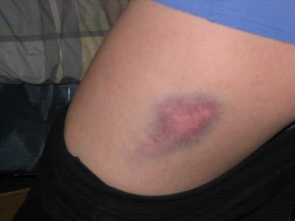
6 common bruise
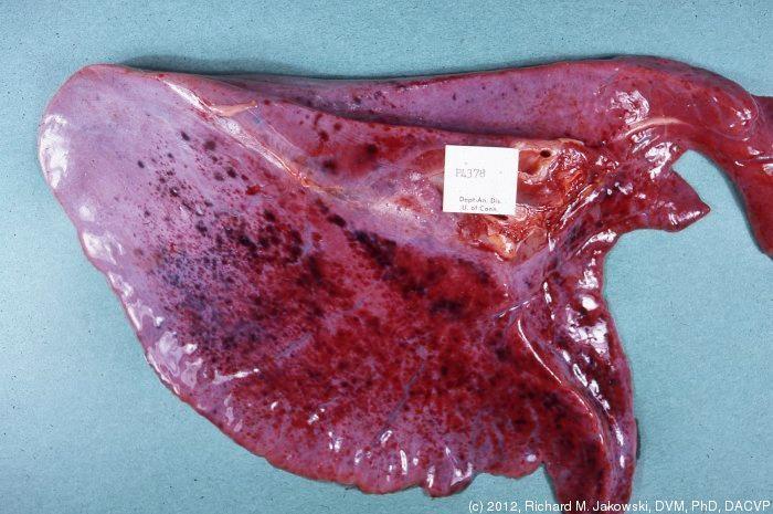
7 Pulmonary hemorrhage
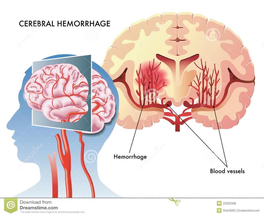
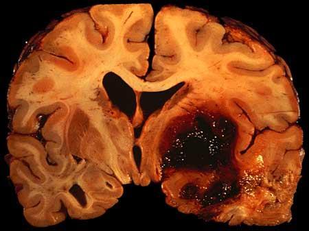
8 Cerebral hemorrhage

9 Hemosiderin-laden macrophages in the lung

 within the cytoplasm of")


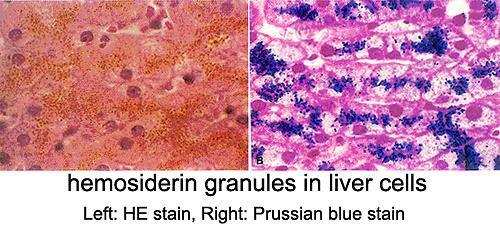
10 Organ: Liver Lesion: 1. Deposition of fine, golden-brown granules (black arrow) within the cytoplasm of hepatocytes (A). 2. The Prussian blue stain (specific for iron) shows bluish coloration of the deposited granules (B). Diagnosis: Hemosiderosis A B
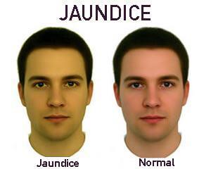
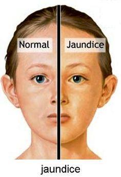
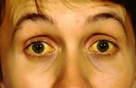
11 2. Bilirubin The bilirubin is the major pigment of bile. It is derived from the hemoglobin but it contains no iron. Pathological accumulation of the bilirubin (bilirubinosis) within cells of all tissues and within body fluids is referred to as jaundice or icterus which is characterized by yellowing of the skin, mucous membranes and the sclera of the eyes.
12

13
14 Pathological accumulation of bilirubin occur in the liver as a result of obstruction to the bile flow (e.g., obstruction of the common bile duct by a stone), in this case liver will possess yellowish-green color grossly.

15 Yellow swollen liver with swollen gall bladder
")
16 Greenish swollen liver (Jaundice)


17 Organ: Liver Lesion: Yellowish deposits are apparent within the bile canaliculi, kupffer cells and hepatocytes. Lesion diagnosis: Bilirubinosis or cholestasis Etiology diagnosis: Bile duct obstruction due to gall stone


18 Organ: Liver Lesion: Yellowish deposits are apparent within the bile ducts, bile canaliculi, kupffer cells and hepatocytes. Lesion diagnosis: Bilirubinosis or cholestasis Etiology diagnosis: Bile duct obstruction due to gall stone
19 3. Lipofuscin This is a yellowish brown pigment having high lipid content, often found in the atrophied cells or in old age individual. It is particularly common in the heart muscle, and the term brown atrophy is often applied. It is also found in liver cells, testes and nerve cells as an ageing marks.
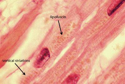
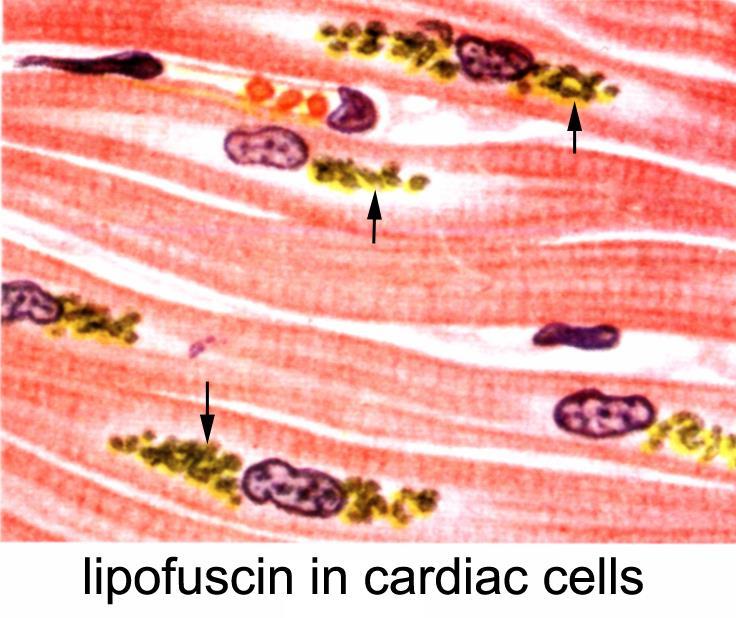
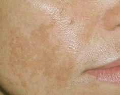
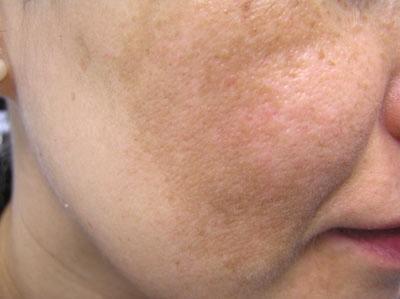
20
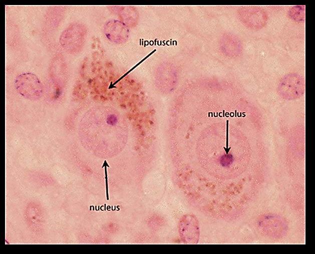
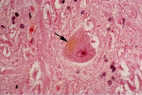
21 Lipofuscin in nerve cell

22 Lipofuscinosis due to ageing
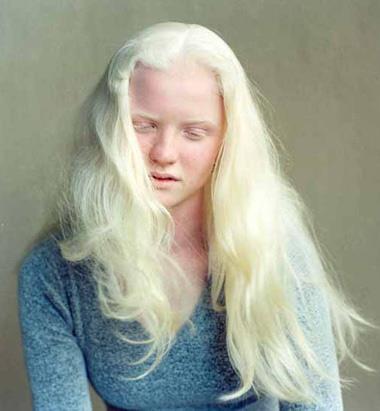
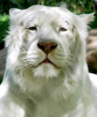
23 Lipofuscinosis in the face
24 4. Melanin Melanin is a normal pigment found in the form of fine brown to dark granules in the skin, choroid and iris of the eye, hair follicles, meninges and adrenal medulla. An abnormal accumulation of melanin pigment in the skin or other tissues is referred to as melanosis or melanism, e.g.: a. Benin nevus (mole) of the skin. b. melanosis of the colon (melanosis coli). c. melanosis bulbi (melanosis oculi) of the eye.

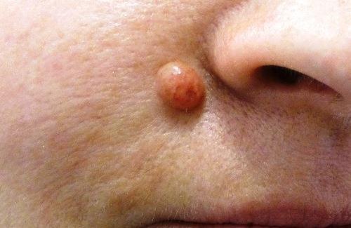
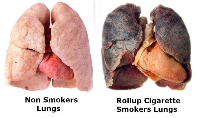
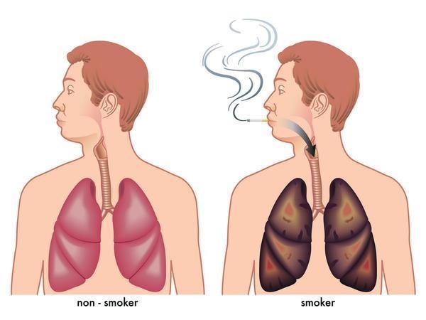
25 Benin nevus

26 Benin nevus

27 Benign nevus should be differentiated from Melanoma which is a malignant tumor (cancer) originated from the melanocytes, it s one of the aggressive tumors which grow rapidly with ability of early metastasis.

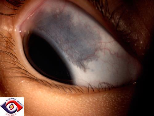
28 Melanosis bulbi

29 Melanosis in face

30 Melanosis due to sun burn


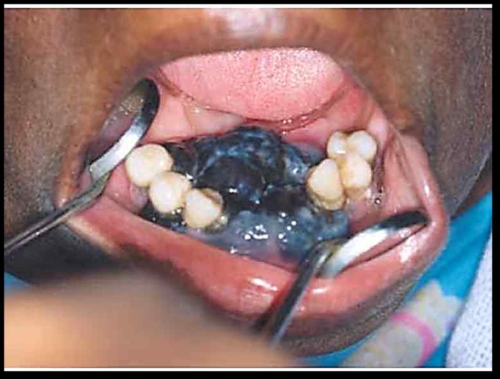
31 Melanosis in the oral cavity
32 Complete absence of melanin pigment is called (albinism) which is a congenital defect in which the affected individual is called an albino, occur in human and animals.
33
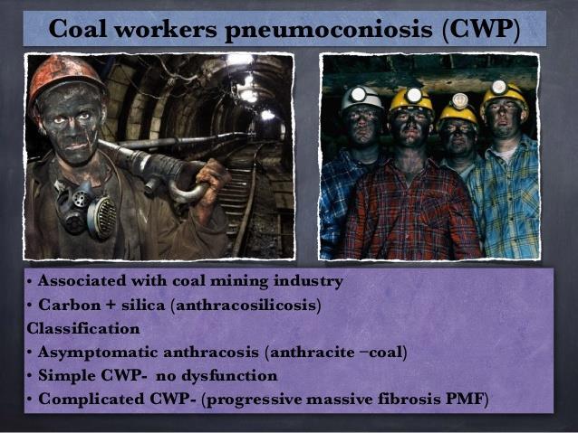
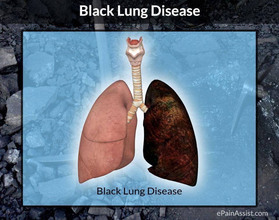
34 Exogenous Pigments Coal dust particles It is the most common exogenous pigment. Pathological accumulation of carbon particles in the lungs is referred to as anthracosis miners diseases Pneumoconiosis which is characterized by blackish discoloration of lung tissue.
35
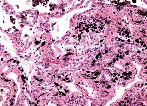

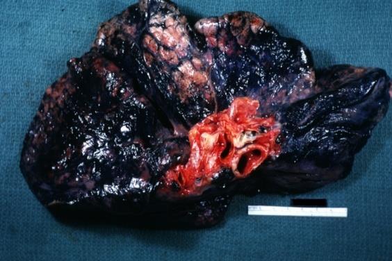
36 Anthracosis
37
38

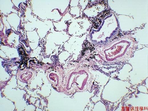
39 Anthracosis in habitual smokers
Morphological changes (accumulations) occur inside and outside cells
 occur inside and outside cells") MIXED ACCUMULATIONS (DEGENERATIONS) Morphological changes (accumulations) occur inside and outside cells The group includes: - chromoproteins metabolism disturbances; - lipoproteins metabolism disturbances;
MIXED ACCUMULATIONS (DEGENERATIONS) Morphological changes (accumulations) occur inside and outside cells The group includes: - chromoproteins metabolism disturbances; - lipoproteins metabolism disturbances;
Hemosiderin. Livia Vida 2018
 Hemosiderin Livia Vida 2018 Questions Histochemical caracteristics of the different pigments. Exogenous pigments. Hemoglobinogenic pigments. Causes and forms of jaundice. Hemoglobinogenic pigments. Pathological
Non-hematogenous endogenous pigments
 Non-hematogenous endogenous pigments 0 This group contains the following : 1. Melanins. 2. Lipofuscins. 3. Chromaffin. 4. Pseudomelanosis. 5. Dubin-Johnson pigments. 6. Ceroid-type lipofuscins. 7. Hamazaki-Weisenberg
Cell injury, adaptation and death. Unite one Second Lab.
 Cell injury, adaptation and death Unite one Second Lab. The two lung abscesses seen here are examples of liquefactive necrosis in which there is a liquid center in an area of tissue injury. One abscess
Pigments and accumulations
 Pigments and accumulations Intracellular Accumulations Normal cellular constituent vs. abnormal substance Transient vs. permanent Harmless vs. toxic Cytoplasm vs. nucleus Cell produced vs. produced other
PATHOLOGY Intracellular Degeneration LAB 1
 PATHOLOGY Intracellular Degeneration LAB 1 Cellular swelling Liver Organ :- Liver Lesion :- 1. Narrowing of hepatic sinusoids due to the swelling of hepatocyte. 2. The cytoplasm of affected hepatocyte
This is Learning Component 6 in Learning Module 1. We will show examples of features ( things ) including mineral deposits, urates, pigments, dust,
 including mineral deposits, urates, pigments, dust,") This is Learning Component 6 in Learning Module 1. We will show examples of features ( things ) including mineral deposits, urates, pigments, dust, plant material, and amyloid. 1 Calcium salts are the
This is Learning Component 6 in Learning Module 1. We will show examples of features ( things ) including mineral deposits, urates, pigments, dust, plant material, and amyloid. 1 Calcium salts are the
Lysosomes. Gr: lysis solution, soma body. Membrane bounded vesicles. Usually round ovoid or irregular electron dense bodies m.
 Lysosomes Gr: lysis solution, soma body Membrane bounded vesicles Usually round ovoid or irregular electron dense bodies 0.05 0.5 m. Lysosomes No. varies from a few to several hundred per cell, in different
Pigmented lesions of the Oral cavity
 Oral medicine أ.م.د احسان عبد هللا كميل Pigmented lesions of the Oral cavity Pigmented oral lesions are a large group of disorders in which the dark or brown color is the essential clinical characteristic.
Place and role of the pathology in the medicine. Structure of pathology and methods of investigation
 Place and role of the pathology in the medicine. Structure of pathology and methods of investigation Dr. Attila Zalatnai (Just for educational purposes) Without pathology there is no modern diagnostics!
BCM 317 LECTURE OJEMEKELE O.
 BCM 317 LECTURE BY OJEMEKELE O. JAUNDICE Jaundice is yellowish discoloration of the skin, sclera and mucous membrane, resulting from an increased bilirubin concentration in the body fluid. It is usually
Histopathology: Cell necrosis and cytoplasmic accumulations
 Histopathology: Cell necrosis and cytoplasmic accumulations These presentations are to help you identify basic histopathological features. They do not contain the additional factual information that you
11/8/2012. Chapter 6 Part 1 Objectives: Skin = Integument = Cutaneous Membrane. The Structure of Skin. Epidermis
 Chapter 6 Part 1 Objectives: Define organ, and associate the skin as an organ of the integumentary system. List the general functions of the skin. Describe the structure of the layers of the skin. Summarize
Disturbances of Circulation, Lab 1: Edema and Congestion/Hyperemia. Shannon Martinson, Feb
 Disturbances of Circulation, Lab 1: Edema and Congestion/Hyperemia Shannon Martinson, Feb 2017 http://people.upei.ca/smartinson/ Case #1 Signalment and History: 6-month old feeder lamb found dead on pasture
Skin and Body Membranes
 Essentials of Human Anatomy & Physiology Elaine N. Marieb Seventh Edition Chapter 4 Skin and Body Membranes Slides 4.1 4.32 Lecture Slides in PowerPoint by Jerry L. Cook Skin and Body Membranes Function
Skin and Body Membranes
 4 Skin and Body Membranes PowerPoint Lecture Slide Presentation by Jerry L. Cook, Sam Houston University ESSENTIALS OF HUMAN ANATOMY & PHYSIOLOGY EIGHTH EDITION ELAINE N. MARIEB Skin and Body Membranes
Definition of bilirubin Bilirubin metabolism
 Definition of bilirubin Bilirubin metabolism obilirubin formation otransport of bilirubin in plasma ohepatic bilirubin transport oexcretion through intestine Other substances conjugated by glucuronyl transferase.
HEMOLYSIS AND JAUNDICE:
 1 University of Papua New Guinea School of Medicine and Health Sciences Division of Basic Medical Sciences Discipline of Biochemistry and Molecular Biology PBL SEMINAR HEMOLYSIS AND JAUNDICE: An overview
VPM Pigment and other tissue deposits. Shannon Martinson
 VPM 152 - Pigment and other tissue deposits Shannon Martinson http://people.upei.ca/smartinson/ Case 1 Signalment: 2 month old heifer beef calf Clinical History: Lateral recumbency for 4 days Tachycardia,
DIFFERENTIAL DIAGNOSIS OF JAUNDICE
 CHARLES L. HARTSOCK, M.D. The yellow or greenish yellow staining of the blood plasma and body tissues, to which the clinical term jaundice has been applied, is due to an excessive amount of one of the
HEMOLYSIS & JAUNDICE: An Overview
 HEMOLYSIS & JAUNDICE: An Overview University of Papua New Guinea School of Medicine and Health Sciences Division of Basic Medical Sciences Discipline of Biochemistry and Molecular Biology PBL MBBS III
JAUNDICE. Zdeněk Fryšák 3rd Clinic of Internal Medicine Nephrology-Rheumatology-Endocrinology Faculty Hospital Olomouc
 JAUNDICE Zdeněk Fryšák 3rd Clinic of Internal Medicine Nephrology-Rheumatology-Endocrinology Faculty Hospital Olomouc Definition of Jaundice Icterus A yellowish staining of the skin, sclerae and deeper
-Liver function tests -
 -Liver function tests - Biochimestry teamwork Osamah Al-Jarallah Abdulaziz Al-Shamlan Abdullah Al-Mazyad Turki Al-Otaibi Khalid Al-Khamis Saud Al-awad KhaledAlmohaimede Meshal Al-Otaibi Al-Anood Asiri
General Pathology Theory Syllabus for II B.D.S.
 General Pathology Theory Syllabus for II B.D.S. Sr. No. Topic (Must Know) (Desirable to know) 1.Introduction to Pathology - Different sections in pathology - The Cell in health - Normal cell structure
IIPA Ready Iridology IIPA Ready Iridology
 IIPA Ready Iridology Class 1 ~ Introduction, Terms and Basics Class 2 ~ Anatomy of the Eye Class 3 ~ Collarette Class 4 ~ Zones and Constitutional Types Class 5 ~ Pigmentation Class 6 ~ Lacunea Class 7
7/10/18. Introduction. Integumentary System. Physiology. Anatomy. Structure of the Skin. Epidermis
 Introduction Integumentary System Chapter 22 Skin is largest and heaviest organ of body (7% of body weight) Houses receptors for touch, heat, cold, movement, and vibration No other body system is more
The basis of Disease
 General Curriculum The basis of Disease ZHOU REN 周韧 Prof., M.D., Ph.D. Institute of Pathology & Forensic Medicine Department of Pathology & Patho-physiology Zhenjiang University Judicial Evidence & Evaluation
SECTION 2 CELL INJURY
 Adapted myocyte Normal myocyte Reversibly-injured myocyte SECTION 2 CELL INJURY Cell death 5/4/2014 1 5/4/2014 2 Reversible Degeneration Irreversible Cellular Swelling Fatty Change Hyaline Change Amyloid
Skin and Body Membranes Body Membranes Function of body membranes Cover body surfaces Line body cavities Form protective sheets around organs
 Skin and Body Membranes Body Membranes Function of body membranes Cover body surfaces Line body cavities Form protective sheets around organs Classification of Body Membranes Epithelial membranes Cutaneous
Describe the functions of the vertebrate integumentary system. Discuss the structure of the skin and how it relates to function.
 Chapter 5 Describe the functions of the vertebrate integumentary system. Discuss the structure of the skin and how it relates to function. Explain the basis for different skin colors. Describe the structure
Glucose-6-Phosphate Dehydrogenase
 Glucose-6-Phosphate Dehydrogenase Is the major enzyme in the pentose phosphate pathway (also called the phosphogluconate pathway or the hexose monophosphate shunt) which is a metabolic pathway parallel
BIOL 2458 CHAPTER 19 Part 1 SI 1. List the types of extracellular fluids. 2. Intracellular fluid makes up of the body fluids. Where is it found?
 BIOL 2458 CHAPTER 19 Part 1 SI 1 1. Extracellular fluid makes up of the body fluids. List the types of extracellular fluids. 2. Intracellular fluid makes up of the body fluids. Where is it found? 3. In
The Integumentary System. Mosby items and derived items 2010, 2006, 2002, 1997, 1992 by Mosby, Inc., an affiliate of Elsevier Inc.
 The Integumentary System The Skin Structure two primary layers called epidermis and dermis Epidermis Outermost and thinnest primary layer of skin Composed of several layers of stratified squamous epithelium
THE UNIVERSITY OF JORDAN FACULTY OF MEDICINE DEPARTMENT OF PATHOLOGY
 THE UNIVERSITY OF JORDAN FACULTY OF MEDICINE DEPARTMENT OF PATHOLOGY INTRODUCTION TO ANEMIA Third year medical students First semester 2018/2019 Dr. RBC DISORDERS Lecturer: Dr. Tariq Al-Adaily Email: TNALADILY@ju.edu.jo
PowerPoint Lecture Slide Presentation by Patty Bostwick-Taylor, Florence-Darlington Technical College Skin and Body Membranes
 PowerPoint Lecture Slide Presentation by Patty Bostwick-Taylor, Florence-Darlington Technical College Skin and Body Membranes 4 Body Membranes Function of body membranes Cover body surfaces Line body cavities
Understanding Skin Colour
 Understanding Skin Colour SKIN COLOUR The natural colour of skin without any pigments is yellowish. However, we are all aware of the different colours of skin and these differences are determined by the
-sheet 3. -Waseem Alhaj. Maha Shomaf
 -sheet 3 -Basheer egbaria -Waseem Alhaj Maha Shomaf 1 P a g e Viral hepatitis have many types each type is associated with different outcomes complication, some can result in acute one,others result in
THE CLASSIFICATION OF ANEMIA*
 THE CLASSIFICATION OF ANEMIA* RUSSELL L. HADEN, M.D. SUMMARY A laboratory and clinical classification of anemia has been outlined. The results of the blood study have been correlated with the clinical
Unit 4 - The Skin and Body Membranes 1
 Unit 4 - The Skin and Body Membranes 1 I. Unit 4: Skin and Body Membranes A. Body Membranes 1. Function of body membranes a) Cover body surfaces b) Line body cavities c) Form protective sheets around organs
Liver Function Tests
 Liver Function Tests The liver is of vital importance in intermediary metabolism and in the detoxification and elimination of toxic substances. Damage to the organ may not obviously affects its activity
Ch. 4: Skin and Body Membranes
 Ch. 4: Skin and Body Membranes I. Body Membranes A. Function of body membranes 1. Cover body surfaces 2. Line body cavities 3. Form protective sheets around organs II. Classification of Body Membranes
Integumentary System
 Integumentary System The integumentary system is commonly known as the Skin Largest organ of human body 10% total body weight and would cover over 20 square feet Functions of Skin 1. Protection Barrier
Yellowish Discoloration to the Tissues of the Body
 Yellowish Discoloration to the Tissues of the Body (Jaundice or Icterus) Basics OVERVIEW Yellowish discoloration to the gums and other tissues of the body (known as jaundice or icterus ) Serum total bilirubin
Cholangitis/ Cholangiohepatitis Syndrome (Inflammation of the Bile Duct System and Liver) Basics
 Basics") Glendale Animal Hospital 623-934-7243 www.familyvet.com Cholangitis/ Cholangiohepatitis Syndrome (Inflammation of the Bile Duct System and Liver) Basics OVERVIEW The liver is the largest gland in the body;
Glendale Animal Hospital 623-934-7243 www.familyvet.com Cholangitis/ Cholangiohepatitis Syndrome (Inflammation of the Bile Duct System and Liver) Basics OVERVIEW The liver is the largest gland in the body;
Mycotoxin Lesions in the Slaughter House-Broilers
 Mycotoxin Lesions in the Slaughter House-Broilers SPECIAL NUTRIENTS, INC. THE MYCOTOXINS SPECIALIST www.mycotoxin.com INTRODUCTION Traditionally, the presence of mycotoxins capable of causing damage in
Extracellular degeneration
 Extracellular degeneration By Dr. Hemn Hassan Othman PhD, Pathology Fall 2016 1/17/2017 1 Extracellular Degenerations I / Hyaline Degeneration (Hyalinization): The ward hyaline is derived from the Latin
Gastrointestinal System: Accessory Organ Disorders
 Gastrointestinal System: Accessory Organ Disorders Mary DeLetter, PhD, RN Associate Professor Dept. of Baccalaureate and Graduate Nursing Eastern Kentucky University Disorders of Accessory Organs Portal
Etiology Bacteria Sand particles Particles of ingesta / intestinal contents Desquamated epithelium
 10 CONCRETIONS Concretions Calculi o Urinary Calculi o Biliary Calculi o Salivary Calculi o Pancreatic Calculi o Enteric Calculi Piliconcretions Phytoconcretions Polyconcretions Model Questions CONCRETIONS
Cornell Notes Name: Date: Topic: CH 4
 *We are revisiting Ch 3B on body tissues (Connective) prior to our study of Ch 4 Integumentary. Start on p.90 I. Connective Tissue A. Functions of Connective 1. Protection 2. Support 3. Binding Together
Integumentary System (Skin) Unit 6.3 (6 th Edition) Chapter 7.3 (7 th Edition)
 Unit 6.3 (6 th Edition) Chapter 7.3 (7 th Edition)") Integumentary System (Skin) Unit 6.3 (6 th Edition) Chapter 7.3 (7 th Edition) 1 Learning Objectives Identify the major components (anatomy) of skin Differentiate between the two types of skin glands Explain
Integumentary System (Skin) Unit 6.3 (6 th Edition) Chapter 7.3 (7 th Edition) 1 Learning Objectives Identify the major components (anatomy) of skin Differentiate between the two types of skin glands Explain
Anatomy and Physiology
 Anatomy and Physiology For The First Class 2 nd Semester Erythrocytes = Red Blood Cells (RBC) Erythrocytes = Red Blood Cells Red blood cells are biconcave discs, they have no nucleus and cytoplasmic organelles.
Chapter 05. Lecture Outline. See separate PowerPoint slides for all figures and tables pre-inserted into PowerPoint without notes.
 Chapter 05 Lecture Outline See separate PowerPoint slides for all figures and tables pre-inserted into PowerPoint without notes. Copyright The McGraw-Hill Companies, Inc. Permission required for reproduction
Chapter 18 LIVER BILIARY TRACT
 Chapter 18 LIVER & BILIARY TRACT DUCT SYSTEM N O FIBROUS TISSUE PORTAL TRIAD CENTRAL VEIN PATTERNS OF HEPATIC INJURY Degeneration: Balooning, feathery degeneration, fat, pigment Inflammation:
Biochemistry Liver Function Tests (LFTs)
") HbA NH 2 H 2 O 2 KClO3 Cl 2 O 7 PO 4 CH2O NAOH KMnO 4 M E D I C I N E KING SAUD UNIVERSITY Co 2 COOH MgCl 2 H 2 O Important Extra Information Doctors slides Doctors notes SO 2 HCN CCl 4 CuCl 2 SiCl 4 Biochemistry
HbA NH 2 H 2 O 2 KClO3 Cl 2 O 7 PO 4 CH2O NAOH KMnO 4 M E D I C I N E KING SAUD UNIVERSITY Co 2 COOH MgCl 2 H 2 O Important Extra Information Doctors slides Doctors notes SO 2 HCN CCl 4 CuCl 2 SiCl 4 Biochemistry
Section 8 Liver and Gallbladder
 General and Systemic Histopathology C601 and C602 Section 8 As we will see in this unit, the liver is subject to many types of injury. Additionally, many systemic diseases have a liver component and sometimes
Light yellow to dark golden yellow Clear ph range Specific gravity Sediments
 #11 Objectives: Understand specific gravity and identify normal specific gravity values for urine Learn to use a urine hydrometer to measure specific gravity Define specific gravity and identify normal
The Urinary System. Lab Exercise 38. Objectives. Introduction
 Lab Exercise The Urinary System Objectives - Be able to identify the structures of the urinary system and give their function - Be able to recognize the gross anatomy of the kidney - Identify the components
Surgical Treatment of special Tumours. Winnie Achilles Tierklinik Hollabrunn Lastenstrasse Hollabrunn
 Surgical Treatment of special Tumours Winnie Achilles Tierklinik Hollabrunn Lastenstrasse 2 2020 Hollabrunn boexi@gmx.de Hepatocellular Tumours Hepatocellular Carcinoma, hepatocellular adenoma, and hepatoblastoma
PowerPoint Lecture Slide Presentation by Patty Bostwick-Taylor, Florence-Darlington Technical College Skin and Body Membranes
 PowerPoint Lecture Slide Presentation by Patty Bostwick-Taylor, Florence-Darlington Technical College Skin and Body Membranes 4 Body Membranes Function of body membranes Cover body surfaces Line body cavities
Approach to the Patient with Liver Disease
 Approach to the Patient with Liver Disease Diagnosis of liver disease Careful history taking Physical examination Laboratory tests Radiologic examination and imaging studies Liver biopsy Liver diseases
Cellular Injury. Intracellular degeneration. By Dr. Hemn Hassan Othman PhD, Pathology Fall /20/2018 1
 Cellular Injury Intracellular degeneration By Dr. Hemn Hassan Othman PhD, Pathology Fall 2018 10/20/2018 1 Types of cell injury Cell injury is divided into: 1. Reversible cell injury 2. Irreversible cell
Your Skin. Section 14.2 Your Skin, Hair, and Nails
 Your Skin The skin covers and protects the body from injury, infection, and water loss. The skin also helps to regulate body temperature and gathers information from the environment. Protection The skin
EDUCATIONAL COMMENTARY BLOOD CELL IDENTIFICATION
 EDUCATIONAL COMMENTARY BLOOD CELL IDENTIFICATION Educational commentary is provided through our affiliation with the American Society for Clinical Pathology (ASCP). To obtain FREE CME/CMLE credits click
Describing and interpreting gross lesions. Prepared for VPM 4600, May 2018; Shannon Martinson
 Describing and interpreting gross lesions Prepared for VPM 4600, May 2018; Shannon Martinson How to Describe (and Interpret) Lesions Step 1 Step 2 Step 3 Step 4 Look at the specimen: Is it normal or abnormal
Pathophysiology I Liver and Biliary Disease
 Pathophysiology I Liver and Biliary Disease The Liver The liver is located in the right upper portion of the abdominal cavity just beneath the right side of the rib cage. The liver has many functions that
Introduction. Skin and Body Membranes. Cutaneous Membranes Skin 9/14/2017. Classification of Body Membranes. Classification of Body Membranes
 Introduction Skin and Body Membranes Body membranes Cover surfaces Line body cavities Form protective and lubricating sheets around organs Classified in 5 categories Epithelial membranes 3 types- cutaneous,
Radiology of hepatobiliary diseases
 GI cycle - Lecture 14 436 Teams Radiology of hepatobiliary diseases Objectives 1. To Interpret plan x-ray radiograph of abdomen with common pathologies. 2. To know the common pathologies presentation.
Chapter 4 The Integumentary System and Body Membranes. HAP Susan Chabot Lemon Bay High School
 Chapter 4 The Integumentary System and Body Membranes HAP Susan Chabot Lemon Bay High School Classification of Body Membranes Epithelial Membranes Cutaneous Membranes = The Skin Mucous Membranes Serous
Page 1. Chapter 37: Chemical Control of the Animal Body - The Endocrine System
 Chapter 37: Chemical Control of the Animal Body - The Endocrine System Endocrine System: Hormones and the various cells that secrete and receive them Types of Glands: 1) Endocrine Glands: Release substances
Page 1. Chapter 37: Chemical Control of the Animal Body - The Endocrine System. Target Cells: Cells specialized to respond to hormones
 Chapter 37: Chemical Control of the Animal Body - The Endocrine System Endocrine System: Hormones and the various cells that secrete and receive them Types of Glands: 1) Endocrine Glands: Release substances
Lec.2 Medical Physiology Blood Physiology Z.H.Kamil
 Destruction of Red Blood Cells When red blood cells are delivered from the bone marrow into the circulatory system, they normally circulate an average of 120 days before being destroyed. Even though mature
CHAPTER 7:3 INTEGUMENTARY SYSTEM
 CHAPTER 7:3 INTEGUMENTARY SYSTEM I. OBJECTIVES A. Label a diagram of a cross section of the skin B. Differentiate between the two types of skin glands C. Identify six functions of the skin D. Provide the
BCH472 [Practical] 1
![BCH472 [Practical] 1](/thumbs/89/98494556.jpg "BCH472 [Practical] 1") BCH472 [Practical] 1 Physical Examination Chemical Examination 2 ph Color Specific Gravity Volume Odor Appearance Acidic: -Diabetic Ketoacidosis. -Starvation. -UTIs (E. coli). Alkaline: -UTIs (ureasplitting
BCH472 [Practical] 1 Physical Examination Chemical Examination 2 ph Color Specific Gravity Volume Odor Appearance Acidic: -Diabetic Ketoacidosis. -Starvation. -UTIs (E. coli). Alkaline: -UTIs (ureasplitting
Malignant tumors of melanocytes: Part 1. Deba P Sarma, MD., Omaha
 Malignant tumors of melanocytes: Part 1 Deba P Sarma, MD., Omaha The melanocytic tumor is one of the most difficult and confusing areas in Dematopathology. It is true that most (95%) of such lesions are
Appendix : Dermoscopy
 Go Back to the Top To Order, Visit the Purchasing Page for Details APP Appendix : Dermoscopy Dermoscopy, also known as dermatoscopy, epiluminoscopy and epiluminescent microscopy, is an effective non-invasive
Blood. Plasma. The liquid part of blood is called plasma. 1. Pale yellow fluid; forms more than half the blood volume.
 11 Blood FOCUS: Blood consists of plasma and formed elements. The plasma is 91% water with dissolved or suspended molecules, including albumin, globulins, and fibrinogen. The formed elements include erythrocytes,
VPM Pigment and other tissue deposits. Shannon Martinson
 VPM 152 - Pigment and other tissue deposits Shannon Martinson http://people.upei.ca/smartinson/ Case 1: Signalment: 2 month old heifer beef calf Clinical History: Lateral recumbency for 4 days. Tachycardia,
monotonous, stippled, round, smoothcontoured nuclei and scanty acidophilic or
 monotonous, stippled, round, smoothcontoured nuclei and scanty acidophilic or vacuolated cytoplasm. The cells are surrounded by a loose fibrillary stroma that is traversed by delicate capillaries. Ill
The Integumentary System
 The Integumentary System The Integumentary System Integument is skin Skin and its appendages make up the integumentary system (See if you can name some appendages) A fatty layer (hypodermis) lies deep
06/11/1431. Chapter 5. Ra'eda Almashaqba
 Chapter 5 1 Skin The skin is composed of three layers, the epidermis, dermis, and subcutaneous tissue. The skin is thicker on the palms of the hands and soles of the feet and is continuous with the mucous
Pathology of the Liver and Biliary Tract 5 Diseases of the Biliary Tract. Shannon Martinson, April 2016
 Pathology of the Liver and Biliary Tract 5 Diseases of the Biliary Tract Shannon Martinson, April 2016 http://people.upei.ca/smartinson/ OUTLINE Normal anatomy & function Hepatobiliary Injury and responses
ANATOMY & PHYSIOLOGY ONLINE COURSE - SESSION 13 THE DIGESTIVE SYSTEM
 ANATOMY & PHYSIOLOGY ONLINE COURSE - SESSION 13 THE DIGESTIVE SYSTEM The digestive system also known as the alimentary canal or gastrointestinal tract consists of a series of hollow organs joined in a
4 Skin and Body Membranes Study Guide
 Name: SKIN AND BODY MEMBRANES: 4 Skin and Body Membranes Study Guide Period: Body membranes, which cover body surfaces, line its cavities, and form protective sheets around organs, fall into two major
X-Plain Pancreatic Cancer Reference Summary
 X-Plain Pancreatic Cancer Reference Summary Introduction Pancreatic cancer is the 4th leading cause of cancer deaths in the U.S. About 37,000 new cases of pancreatic cancer are diagnosed each year in the
NAEVUS OF OTA* naevus" occurring in the skin areas supplied by the ophthalmic and maxillary
 Brit. J. Ophthal. (1965) 49, 364 NAEVUS OF OTA* BY G. P. GUPTA AND D. N. GANGWAR From the Muslim University Institute of Ophthalmology and Gandhi Fye Hospital, Aligarh, India THE naevus of Ota is characterized
LENTIGO SIMPLEX. Epidemiology
 LENTIGO SIMPLEX Epidemiology The frequency of lentigo simplex in children and adults has not been determined. There does not appear to be a racial or gender predilection. Lentigo simplex is the most common
CYTOLOGY OF THE LIVER
 CYTOLOGY OF THE LIVER Maxey L. Wellman, DVM, PhD, DACVP (Clinical Pathology) Professor, Department of Veterinary Biosciences, College of Veterinary Medicine, The Ohio State University, Columbus, OH, USA
Red cell disorder. Dr. Ahmed Hasan
 Red cell disorder Dr. Ahmed Hasan Things to be learned in this lecture Definition and clinical feature of anemia. Classification of anemia. Know some details of microcytic anemia Question of the lecture:
Skin. the largest organ of the body 1 mm to 2 mm thick almost 2 square meters 6% of a person s body weight
 Skin the largest organ of the body 1 mm to 2 mm thick almost 2 square meters 6% of a person s body weight Functions of the Skin protection disease-causing organisms dangerous chemicals blood loss fluid
Chapter 2 Normal Components
 Chapter 2 Normal Components Epithelial Elements Tracheal and Bronchial Respiratory Epithelium Normal bronchial respiratory epithelium usually appears as monolayer tissue fragments and strips in bronchoscopic
Participants Identification No. % Evaluation. Mitotic figure Educational Erythrocyte precursor, abnormal/
 Cell Identification BMD-09 Participants Identification No. % Evaluation Mitotic figure 233 96.7 Educational Erythrocyte precursor, abnormal/ 4 1.7 Educational dysplastic nuclear features Erythrocyte precursor
Integumentary System. Packet #12
 Integumentary System Packet #12 Introduction Skin/Integument Skin, considered an organ, is the major component of the integumentary system. The integumentary system is also composed of other accessory
Physiological functions of the liver. Describe the major functions of the liver with respect to metabolism,detoxification & excretion of hydrophobic
 Physiological functions of the liver. Describe the major functions of the liver with respect to metabolism,detoxification & excretion of hydrophobic substances. Describe the formation of bile,its constitents
Histopathology: skin pathology
 Histopathology: skin pathology These presentations are to help you identify, and to test yourself on identifying, basic histopathological features. They do not contain the additional factual information
Ex. 7: Integumentary
 Collin County Community College BIOL. 2401 Ex. 7: Integumentary. Skin or Integument Consists of three major regions Epidermis outermost superficial region Dermis middle region Hypodermis (superficial fascia)
Comparative Study of the Serum Bilirubin and Various Other Liver Related Enzymes in Different Types of Jaundice
 DOI: 10.21276/aimdr.2018.4.4.BC12 Original Article ISSN (O):2395-2822; ISSN (P):2395-2814 Comparative Study of the Serum Bilirubin and Various Other Liver Related Enzymes in Different Types of Kedar Prasad
Name Score. The Neck Bone s Connected to the Head Bone
 Name Score The Neck Bone s Connected to the Head Bone You have pictures of either organs or organ system. Each part has been identified. Color each part a different color. Digestive System - 1 - Nerve
The Integumentary System
 The Integumentary System The Integumentary System Integument is skin Skin and its appendages make up the integumentary system A fatty layer (hypodermis) lies deep to it Two distinct regions Epidermis Dermis
DISEASES OF THE RESPIRATORY SYSTEM LECTURE 5 DR HEYAM AWAD FRCPATH
 DISEASES OF THE RESPIRATORY SYSTEM LECTURE 5 DR HEYAM AWAD FRCPATH RESTRICTIVE, INTERSTITIAL LUNG DISESAES. FIROSING DISESES. GRANULOMATOUS DISEASES. EOSINOPHILIC. SMOKING RELATED. FIBROSING DISEASES