Characteristics of the ocular fundus in primates
|
|
|
- Esther Bruce
- 5 years ago
- Views:
Transcription

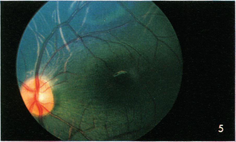
1 J. Anat. (1967), 101, 4, pp With 16 figures Printed in Great Britain Characteristics of the ocular fundus in primates LEE R. WOLIN AND L. C. MASSOPUST, JR. Laboratory of Neurophysiology, Cleveland Psychiatric Institute, Cleveland, Ohio In recent years we have seen a marked increase in research on the visual system. The last few years in particular have been characterized by an increasing use of primates for vision research. As an aspect of comparative research on the visual system, we have been interested in variations of the gross morphology of the retina (Ordy, Massopust & Wolin, 1962). We take routine ocular fundus photographs of each species of animal used. Since we have been able to assemble a collection of fundus photographs of a reasonably representative selection of primates (based on Simpson's Classification, 1945) we felt this would be of interest, as only a limited selection of primate retinal fundus photographs is readily accessible in the literature (Ordy, et al., 1962; Samorajski, Ordy & Keefe, 1966; Smith, Reynolds, Rane & Justice, 1964). The fundus photography is a relatively simple procedure. The pupil is dilated with cyclopentolate hydrochloride (Cyclogyl), following which the animal is anaesthetized, usually with thiamylal sodium (Suritol). The photographs are taken with a Zeiss fundus camera on Kodachrome II film. Species having a highly reflective retina and possibly a tapetum lucidum require insertion of a neutral density filter in the optical path of the camera. Two persons are required for the procedure, one to hold the subject's head and maintain proper orientation of the eye, while the second person manipulates the camera. The fundi of primates follow two basic patterns, one showing a clearly defined macular area and fovea and the other a less well-defined central area with no clear macula or fovea (see figures). The tree shrew (Tupaia) which has a controversial taxonomic status does not fit either pattern. Species including New and Old World, monkeys, gibbon and man, and in addition the tarsier, all show the first patterns (Figs. 1-9 and 1 1). Nerve fibres and blood vessels originating in a circular or oval optic disc (nerve head or papilla) converge on the central area of a retina containing a well-defined macula. The macula is largely free of vessels, differs slightly in pigmentation from the surrounding retina, and is outlined by a ring light reflex. The foveal depression is readily visualized and often shows a spot reflex with bright light and a deep focus. The choroidal vessels can often be seen. There is considerable variation in the vascular patterns within species and a difference between the representatives of the various species shown here should not be regarded as species specific. The general pattern of arcuate vessels with branches terminating near the macula or central retinal area is characteristic of all primates. According to Johnson (1901) this vascular pattern is also found in many carnivores. Certainly the cat fundus is quite similar in appearance to that of the loris (Fig. 12). The predominantly horizontal orientation of the arterioles in Fig. 1 and the pre-


2 694 LEE R. WOLIN AND L. C. MASSOPUST


3 .-.M.-.-.X- Ocular fundus in primates japmo T A qs s- At -a.. Ho i.s _..S, j.-- -; / A:..e _rst 695
 in the higher species is from green to greenish blue with the pigment being relatively uniformly")
4 696 LEE R. WOLIN AND L. C. MASSOPUST dominantly vertical orientation of the venules in Fig. 3 are strictly individual patterns. Individual variations in pigmentation, within species, are also found. Except in man, coloration of the retina (as viewed and photographed with the Zeiss light sources) in the higher species is from green to greenish blue with the pigment being relatively uniformly distributed over the retina (Figs. 2-9). The area near the macula is close to natural colour in each figure, while the bright blue appearing in Fig. 5, and the purple in Figs. 7, 8 and 15 is artifactual. The major differences to be noted among these species are: size and regularity of the border of macular area, size and shape of the optic disc, apparent 'texturing' of retina, and pigmentation of retina. Species including the aotes, lemurs, lorises and tree shrews (the tarsier being the exception) show a somewhat different appearance (Figs. 10, 12-16). The optic disc and vascular distribution are similar to those in the higher species and the distribution and termination of the arteries and veins define the central retinal area. There is, however, no defined macula or fovea. In some instances (see Fig. 10) there is a shadowing indicative of a slight depression over a broad central area; however, no pigment difference is noticeable nor can any ring reflex be obtained. In those animals apparently having a tapetum, the retina has a spotted and sometimes mosaic appearance (see Figs ). Such a highly reflecting retina sometimes makes it difficult to photograph the retina, vasculature and nerve head simultaneously, whilst the choroidal vessels are completely obscured (Fig. 12). The retina in these species varies in colour from orange through yellow to greenish yellow, with little of Fig. 1. Man (Homo sapiens). Lightly pigmented (blond) subject. Note light pigmentation of retina, large round nerve head, large slightly oval macula, smooth appearance of retinal surface and termination of vessels outside fovea centralis. Fig. 2. Gibbon (Hylobates lar). Quite similar in appearance to human fundus, nerve head is slightly obscured to higher reflectivity, macula rounder in shape, and generally darker pigmentation of retina. Fig. 3. Baboon (Papio hamadryas). Although the general characteristics are again close to those of human, note deeper (green) pigmentation and slightly textured appearance of retinal surface. Fig. 4. Rhesus monkey (Macaca mulatta). The oval shape of nerve head distinguishes this retina from those preceding. Fig. 5. Patas monkey (Erythrocebus patas). Very much like rhesus fundus. Deep pigmentation made macular ring reflex difficult to obtain although part of it is seen and outline of macular area is easy to determine. Horizontal striations are due to separation of nerve fibres coursing toward optic nerve. This is seen in individuals of many species and has on at least one occasion been misinterpreted as a speciescharacteristic and described as outlining the shape and extent of the macular area. Light coloured (pink) lines are choroidal vessels. Bright blue area at top is photographic artifact. Fig. 6. Colobus monkey (Colobus polykomos). Characteristic higher primate retina. Note relatively lighter pigmentation which in part permitted photography of extremely well defined macular ring reflex. Macular area is quite regular and nerve head is round as in human. Fig. 7. Marmoset (Oedipomidas oedipus). This eye was the smallest (and most difficult to photograph) of all the primate eyes. Macula is determinable by deeper pigmentation although we were unable to photograph, even in part, a ring reflex. The macular and foveal reflexes can however be obtained with an ophthalmoscope. Fig. 8. Cebus monkey (Cebus albifrons). Note relatively large well-defined macular area. Retina shows some texturing which in this photograph is shown by the irregular character of the ring reflex. The purplish haze is an external reflection artefact.

5 Ocular fundus in primates 697 the darker pigments appearing in the more central portion of the retina. The periphery of the retina near the ora serrata or ora terminalis has a dense accumulation of melanin sometimes giving an appearance similar to that of retinitis pigmentosa in man. One of the authors (L. R. W.) had the opportunity to examine the fundi of a number of prosimian species not represented here. Some differences in appearance of vasculature, pigmentation and reflectivity of the retina were observed, but all appeared to fall within the range of variation represented in Figs The tarsier is of particular interest as it is the only prosimian in which we have been able to see and photograph a 'macula'. Johnson (1901) states that, "...in Simiae the macula is invariably present, whilst in the Prosimiae it is entirely wanting...' Polyak (1957), on the other hand, refers to the '.. central fovea and area of the Night Monkey...', a simian, while describing a '...well-developed yellow spot or "macula" temporal to the disk...' in tarsier, a prosimian. This species has been described as nocturnal in behaviour and certainly has the gross characteristics of a nocturnal animal. The eyes are quite large in proportion to the head. The pupils when fully dilated are almost as large as the visible portion of the eye. The pupil when contracted is elongated rather than round but, in contrast to other nocturnal species observed, the elongation of the tarsier pupil is horizontal rather than vertical. Fig. 9. Squirrel monkey (Saimiri sciureus). This fundus also shows a large macula. 'Texturing' also is apparent not only at the ring reflex but over a much larger area. Blue pigmentation of retina is 'true' colour in this species. Note also extensive appearance of choroidal vessels. Fig. 10. Owl monkey (Aotus trivirgatus). This is the first nocturnal species represented, but is more closely related to Cebus and Saimiri than to other nocturnal species. No macula or fovea can be grossly visualized though a 'foveal' area is found in histological section. Note 'spotted' appearance of retina, which is found in most nocturnal species. Fig. 11. Tarsier (Tarsius syrichta). Highest of the prosiminians here represented. This eye is unique among nocturnal species in revealing a macular ring reflex and foveal spot (the photograph barely does justice to these features which are most distinctly seen with an ophthalmoscope). Note the relatively small nerve head and complex intertwining of vessels as they emerge. The beginning of dense peripheral deposits of pigment can be seen in the temporal and inferior portions of the photograph. Fig. 12. Slow loris (Nycticebus). This is the most highly reflective of the primate retinae shown. Use of neutral density filters in order to photograph the major portion of the retina produced blacking out of the optic nerve head. Note small size of nerve head and relatively large distance between nerve head and central retinal area (as defined by terminal pattern of blood vessels). Fig. 13. Lemur (Lemur fulvis). One of the more darkly pigmented retinae among the nocturnal species, this retina has an extremely 'mottled' appearance. Dense accumulation of pigment may be noted just below the nerve head. The periphery of this retina (not shown in plate is) almost black due to the density of the pigment. Fig Tree shrew (Tupaia glis). These three photographs survey the retina from nasal to temporal extremes. Unlike any other eye which we have observed, the vessels radiate in spokelike fashion from the nerve head. Veins and arteries alternate except in the temporal direction where it appears that three arteries extend toward the central visual portion of the retina. Fig. 15 was photographed directly through the centre of the pupil along the apparent optical axis of the eye. Fig. 14 was taken with the camera directed approximately thirty degrees in a nasal direction, while Fig. 16 required an orientation of more than forty-five degrees toward the posterior (temporal) portion of the eye. It is in the posterior part of the retina that an area having the appearance of a central visual area is observed.

6 698 LEE R. WOLIN AND L. C. MASSOPUST The retina of the tarsier has been described by Polyak (1957) as a pure rod retina but showing a clearly defined macula. In this latter respect it differs from all other nocturnal species examined. We have not yet obtained a tarsier eye and so have not determined whether the tarsier has a tapetum lucidum (Polyak does not mention this) but it was apparent while doing the photography that the retina was highly reflective, more reflective than that of any of the diurnal species examined, but less reflective than many of the nocturnal species. Aotes also have a 'fovea' in histological section (Polyak, 1957) but this cannot be seen ophthalmoscopically. Aotes, like the tarsier, have an all rod retina. The aotus, loris, lemur, bushbaby, and galago all show a slightly shadowed central area when viewed under dim illumination, but no macular ring reflex or foveal spot reflex can be elicited. The tree shrew which is reported to have a pure cone retina rather than a pure rod or duplex retina, deviates considerably from the two patterns described above (Figs ). It has been described whether this species should or should not be classified as a primate (Campbell, 1966; Goodman, 1966; Martin, 1966; McKenna, 1966; Van Valen, 1965). It has the appearance of an insectivore with a long narrow snout and with the eyes set in a lateral orientation. The optic disc is located such that if one looks directly into the eye along the optical axis the disc is in the anterior portion of the field (anterior and posterior here refer to the nasal and occipital directions along the median sagittal plane of the head) (Fig. 15). The vessels radiate in a spoke-like fashion (Fig. 14) rather than predominantly vertically as in the other species. The several arteries (three in Figs. 14 and 15) projecting in the posterior direction are somewhat smaller than the others and seem to terminate just anterior to what is probably the 'central retinal area' (Fig. 16). This area is located far in the posterior portion of the eye and, while it appears largely free of blood vessels, shows no evidence of a macula or fovea. The tree shrew looks 'straight ahead' at an object (Sanderson, 1963; author's observation) rather than cocking the head to one side, thus indicating that the central retinal area is in fact in the posterior portion of the eye and that the tree shrew probably employs binocular visual fixation. The controversy concerning the classification of the tree shrew must undoubtedly be based upon a number of considerations. The morphology of the eye of the animal is certainly unlike that of any other primate we have observed. This would be another factor in support of the classification of tree shrews as a separate order of mammals (Martin, 1966; McKenna, 1966; Van Valen, 1965). SUMMARY This report presents colour illustrations of the ocular fundus of fourteen primate species spanning the Order Primates. Both nocturnal and diurnal species are represented. Similarities and differences in the appearance of the fundi, particularly pigment variations, vascular patterns, and the appearance of a macula and fovea are noted. Supported in part by grant FR from the National Institutes of Health. We express our appreciation to Wallace Wendt, D.V.M. for his assistance with

7 Ocular jundus in primates 699 anaesthesia and photography; to L. J. Goss, D.V.M., Ph.D. and Mr R. T. Reuther of the Cleveland Zoological Society for making available to us the colobus monkey; to Dr William Montagna, Director of the Oregon Regional Primate Research centre, and to the many staff members who were most helpful in assisting with the examination of a variety of prosimian species and with the photography of the fundus of the tarsier and lemur; and to Dr Kenneth Swan of the University of Oregon Medical School for making available to us the facilities of the Eye Clinic. REFERENCES CAMPBELL, C. B. G. (1966). Taxonomic status of tree shrews. Science N.Y. 153, 436. GOODMAN, M. (1966). Phyletic position of tree shrews. Science N.Y. 153, JOHNSON, G. L. (1901). Contributions to the comparative anatomy of the mammalian eye, chiefly based on opthalmoscopic examination. Phil. Trans. R. Soc. B, 194, MARTIN, R. D. (1966). Tree shrews: unique reproductive mechanism of systematic importance. Science N. Y. 152, MCKENNA, M. C. (1966). Paleontology and the origin of the primates. Folia primatol. 4, ORDY, J. M., MAssOPUST, L. C., JR. & WOLIN, L. R. (1962). Postnatal development of the retina, electroretinogram, and acuity in the rhesus monkey. Expl Neurol. 5, POLYAK, S. (1957). The Vertebrate Visual System. Ed. H. Kluver. Chicago: University of Chicago Press. SAMORAJSKI, T., ORDY, J. M. & KEEFE, J. R. (1966). Structural organization of the retina in the tree shrew (Tupaia glis). J. Cell Biol. 28, SANDERSON, 1. T. (1963). The Monkey Kingdom. Philadelphia: Chilton Books. SIMPSON, G. G. (1945). The principles of classification and a classification of mammals. Bull. Am. Mus. nat. hist. 85, New York. SMITH, J. L., REYNOLDS, D. H., RANE, L. & JUSTICE, J., JR. (1964). The fundus oculi in the squirrel, owl and marmoset monkey. Am. J. Ophthal. 57, VAN VALEN, L. (1965). Tree shrews, primates, and fossils. Evolution 19,
4/22/16. Eye. External Anatomy of Eye. Accessory Structures. Bio 40B Dr. Kandula
 Eye Bio 40B Dr. Kandula External Anatomy of Eye Accessory Structures l Eyebrows l Levator Palpebrae Superioris - opens eye l Eyelashes l Ciliary glands modified sweat glands l Small sebaceous glands l
Fundus Autofluorescence. Jonathan A. Micieli, MD Valérie Biousse, MD
 Fundus Autofluorescence Jonathan A. Micieli, MD Valérie Biousse, MD The retinal pigment epithelium (RPE) has many important functions including phagocytosis of the photoreceptor outer segments Cone Rod
Image Formation and Phototransduction. By Dr. Abdelaziz Hussein Lecturer of Physiology
 Image Formation and Phototransduction By Dr. Abdelaziz Hussein Lecturer of Physiology Vision Vision is a complex process through which an image of the external environment is formed on the photosensitive
RETINAL DETACHMENT AT THE POSTERIOR POLE*
 Brit. J. Ophthal. (1958) 42, 749. RETINAL DETACHMENT AT THE POSTERIOR POLE* BY CALBERT I. PHILLIPSt Institute of Ophthalmology, University oflondon THE common feature of the cases to be described in this
September 9, 2013: The layout of the visual system, the retina and the lateral geniculate nucleus
 September 9, 2013: The layout of the visual system, the retina and the lateral geniculate nucleus 1 Basic Wiring of the Visual System 2 The world seen by the two eyes Seen by both eyes Seen by both eyes
(Received 8 March 1965)
") J. Physiol. (1965), 180, pp. 837-845 837 With 1 plate and 4 text-figures Printed in Great Britain THE EFFECT OF OCCLUDING THE RETINAL AND CHOROIDAL CIRCULATIONS ON THE ELECTRO- RETINOGRAM OF MONKEYS BY
J. Physiol. (1965), 180, pp. 837-845 837 With 1 plate and 4 text-figures Printed in Great Britain THE EFFECT OF OCCLUDING THE RETINAL AND CHOROIDAL CIRCULATIONS ON THE ELECTRO- RETINOGRAM OF MONKEYS BY
ID# Exam 1 PS 325, Fall 2001
 ID# Exam 1 PS 325, Fall 2001 As always, the Skidmore Honor Code is in effect, so keep your eyes foveated on your own exam. I tend to think of a point as a minute, so be sure to spend the appropriate amount
Vision I. Steven McLoon Department of Neuroscience University of Minnesota
 Vision I Steven McLoon Department of Neuroscience University of Minnesota 1 Eye Cornea Sclera Conjunctiva 2 Eye The conjunctiva lines the inner surface of the eyelids and outer surface of the sclera. 3
CHAPTER 8 EVALUATION OF FUNDUS IMAGE ANALYSIS SYSTEM
 CHAPTER 8 EVALUATION OF FUNDUS IMAGE ANALYSIS SYSTEM Diabetic retinopathy is very common retinal disease associated with diabetes. Efforts to prevent diabetic retinopathy though have yielded some results;
INTRODUCTION: ****************************************************************************************************
 BIOLOGY 211: HUMAN ANATOMY & PHYSIOLOGY **************************************************************************************************** EYES AND VISION ****************************************************************************************************
Retinal Photography in the Newborn
 Arch. Dis. Childh., 1969, 44, 499. Retinal Photography in the Newborn C. J. BULPITT and J. D. BAUM From M.R.C. Clinical Pharmacology Research Group, Department of Medicine, Royal Postgraduate Medical School,
Medical School Histology Basics. VIBS 289 lab. Eye
 Medical School Histology Basics VIBS 289 lab Eye Larry Johnson Texas A&M University Aqueous humor OUTLINE OVERVIEW CELLULAR STRUCTURES THROUGH WHICH LIGHT PASSES A. CORNEA B. LENS C. RETINA STRUCTURES
Five Things You re Missing with Your Fundus Camera
 ebook Five Things You re Missing with Your Fundus Camera By Donald J. Siegel, OD, Sun City West Eye Care Sponsored by: Before I began incorporating EIDON true-color imaging into my practice, my retinal
Central venous occlusion
 Central venous occlusion Central venous occlusion (right eye) There are dark haemorrhages at the macula and all over the retina. Choroidal haemangioma A choroidal haemangioma has salmon pink colour. There
OCT Angiography in Primary Eye Care
 OCT Angiography in Primary Eye Care An Image Interpretation Primer Julie Rodman, OD, MS, FAAO and Nadia Waheed, MD, MPH Table of Contents Diabetic Retinopathy 3-6 Choroidal Neovascularization 7-9 Central
OCULAR EFFECTS OF ORAL CONTRACEPTIVES. II. STUDIES IN THE RHESUS MONKEY
 FERTILITY AND STERILITY Copyright 1975 The American Fertility Society Vol. 26. No.9. September 1975 Printed in U.S A. OCULAR EFFECTS OF ORAL CONTRACEPTIVES. II. STUDIES IN THE RHESUS MONKEY VICTOR A. DRILL,
The Visual System. Retinal Anatomy Dr. Casagrande February 2, Phone: Office: T2302 MCN
 The Visual System Retinal Anatomy Dr. Casagrande February 2, 2004 Phone: 343-4538 Email: vivien.casagrande@mcmail.vanderbilt.edu Office: T2302 MCN Reading assignments and Good Web Sites Chapter 2 in Tovée,
Step 4: Ask permission to turn off lights or draw the curtains
 STEPS OF EYE EXAMINATION - FUNDUS Step 1: Approach the patient Read the instructions carefully for clues Shake hands, introduce yourself Ask permission to examine him I would like to examine your eyes,
Year 1 MBChB Clinical Skills Session Ophthalmoscopy
 Year 1 MBChB Clinical Skills Session Ophthalmoscopy Reviewed & ratified by: Dr V Taylor-Jones, Mr M Batterbury Consultant Ophthalmologist Learning objectives o To understand the anatomy and physiology
UNIOCULAR APLASIA OF THE OPTIC NERVE*
 Brit. J. Ophthal. (1962) 46, 51. UNIOCULAR APLASIA OF THE OPTIC NERVE* BY FROMA SOMERVILLE London APLASIA of the optic nerve occurring as an isolated abnormality in an eye of normal external appearance
F/G 6/18 MECHANISMS OCT 75 T LAWHILL, F SHARP, N SPEED DAMD17-74-C-4026 IHEF. NL UNCLASSIFIlED
 F/G 6/18 MECHANISMS 7 A-AI03 183 LOUISVILLE UNIV OF RETINAL KY SCHOOL DAMAGE OF FROM MEDICINE CHRONIC LASER RAOIATION.(U) OCT 75 T LAWHILL, F SHARP, N SPEED DAMD17-74-C-4026 IHEF NL UNCLASSIFIlED )J yreport
Year 2 MBChB Clinical Skills Session Ophthalmoscopy. Reviewed & ratified by: Mr M Batterbury Consultant Ophthalmologist
 Year 2 MBChB Clinical Skills Session Ophthalmoscopy Reviewed & ratified by: o Mr M Batterbury Consultant Ophthalmologist Learning objectives o To understand the anatomy and physiology of the external and
Sense of Vision. Chapter 8. The Eye and Vision. The Eye Orbit. Eyebrows, Eyelids, Eyelashes. Accessory Organs 5/3/2016.
 Sense of Vision Chapter 8 Special Senses The Eye and Vision 70 percent of all sensory receptors are in the eyes Each eye has over 1 million nerve fibers Protection for the eye Most of the eye is enclosed
Automated Detection of Vascular Abnormalities in Diabetic Retinopathy using Morphological Entropic Thresholding with Preprocessing Median Fitter
 IJSTE - International Journal of Science Technology & Engineering Volume 1 Issue 3 September 2014 ISSN(online) : 2349-784X Automated Detection of Vascular Abnormalities in Diabetic Retinopathy using Morphological
SPECIAL SENSES PART I: OLFACTION & GUSTATION
 SPECIAL SENSES PART I: OLFACTION & GUSTATION 5 Special Senses Olfaction Gustation Vision Equilibrium Hearing Olfactory Nerves Extend through cribriform plate into nasal cavity on both sides of nasal septum
Construction of the Visual Image
 Construction of the Visual Image Anne L. van de Ven 8 Sept 2003 BioE 492/592 Sensory Neuroengineering Lecture 3 Visual Perception Light Photoreceptors Interneurons Visual Processing Ganglion Neurons Optic
Special Senses: The Eye
 Unit 4 Special Senses: The Eye ESSENTIALS OF HUMAN ANATOMY & PHYSIOLOGY The Senses General senses of touch Temperature Pressure Pain Special senses Smell Taste Sight Hearing Equilibrium The Eye and Vision
p f .'''a.".: ;i.l i PAPILLOEDEMA brain. The ophthalmoscope may be looked upon as the Plate i is that of papilloedema of the optic nerve.
 382 * s. ii d:.'. ; z. j:.'''a.".: : ;i.l i 4 PAPILLOEDEMA FIG. I.-Papilloedema or 'choked disc' due to raised intracranial tension. In this instance the papilloedema was due to a frontal lobe tumour.
Reports 981. Reflection of light by small areas of the ocular fundus. ROBERT W. FLOWER, D.
 Reports 981 From the Department of Ophthalmology, School of Medicine, and the Department of Pathobiology, School of Veterinary Medicine, University of Pennsylvania, Philadelphia. This research was supported
COMMUNICATIONS PHOTOCOAGULATION OF THE RETINA* OPHTHALMOSCOPIC AND HISTOLOGICAL FINDINGS. photocoagulation of the rabbit's retina.
 Brit. J. Ophthal. (1963) 47, 577. COMMUNICATIONS PHOTOCOAGULATION OF THE RETINA* OPHTHALMOSCOPIC AND HISTOLOGICAL FINDINGS BY A. LAVYEL Haifa, Israel SINCE the introduction of the photocoagulator by Meyer-Schwickerath
Visual fields in diabetic retinopathy
 Brit. J. Ophthal. (I97I) 55, I83 Visual fields in diabetic retinopathy K. I. WISZNIA, T. W. LIEBERMAN, AND I. H. LEOPOLD From the Department of Ophthalmology, Mount Sinai School of Medicine, City University
Histology of the Eye
 Histology of the Eye Objectives By the end of this lecture, the student should be able to describe: The general structure of the eye. The microscopic structure of:»cornea.»retina. EYE BULB Three coats
VISUAL FIELDS. Visual Fields. Getting the Terminology Sorted Out 7/27/2018. Speaker: Michael Patrick Coleman, COT & ABOC
 VISUAL FIELDS Speaker: Michael Patrick Coleman, COT & ABOC Visual Fields OBJECTIVES: 1. Explain what is meant by 30-2 in regards to the Humphrey Visual Field test 2. Identify the difference between a kinetic
Psy393: Cognitive Neuroscience. Prof. Anderson Department of Psychology Week 3
 Psy393: Cognitive Neuroscience Prof. Anderson Department of Psychology Week 3 The Eye: Proof for the existence of God? And then there was light Optics Perception Absorption Eye is receiver not sender Plato
ASSESSING THE EYES. Structures. Eyelids Extraocularmuscles Eyelashes Lacrimal glands: Lacrimal ducts Cornea Conjunctiva Sclera Pupils Iris.
 ASSESSING THE EYES Structures External Eyelids Extraocularmuscles Eyelashes Lacrimal glands: Lacrimal ducts Cornea Conjunctiva Sclera Pupils Iris 1 2 Structures Internal Optic disc Physiological cup Retinal
Neural circuits PSY 310 Greg Francis. Lecture 05. Rods and cones
 Neural circuits PSY 310 Greg Francis Lecture 05 Why do you need bright light to read? Rods and cones Photoreceptors are not evenly distributed across the retina 1 Rods and cones Cones are most dense in
Carlson (7e) PowerPoint Lecture Outline Chapter 6: Vision
 PowerPoint Lecture Outline Chapter 6: Vision") Carlson (7e) PowerPoint Lecture Outline Chapter 6: Vision This multimedia product and its contents are protected under copyright law. The following are prohibited by law: any public performance or display,
Carlson (7e) PowerPoint Lecture Outline Chapter 6: Vision This multimedia product and its contents are protected under copyright law. The following are prohibited by law: any public performance or display,
Supplemental Table 1. List of cell lines used
 Supplemental Table 1. List of cell lines used Common species name Latin species name Source Common Marmoset Southern Lesser Bushbaby Callithrix jacchus In-house (1 line) Coriell number (if applicable)
Vision Seeing is in the mind
 1 Vision Seeing is in the mind Stimulus: Light 2 Light Characteristics 1. Wavelength (hue) 2. Intensity (brightness) 3. Saturation (purity) 3 4 Hue (color): dimension of color determined by wavelength
Biological Bases of Behavior. 6: Vision
 Biological Bases of Behavior 6: Vision Sensory Systems The brain detects events in the external environment and directs the contractions of the muscles Afferent neurons carry sensory messages to brain
ZEISS AngioPlex OCT Angiography. Clinical Case Reports
 Clinical Case Reports Proliferative Diabetic Retinopathy (PDR) Case Report 969 PROLIFERATIVE DIABETIC RETINOPATHY 1 1-year-old diabetic female presents for follow-up of proliferative diabetic retinopathy
PRISCOL AND RETINAL ARTERY OCCLUSIONS*
 Brit. J. Ophthal. (1955) 39, 98. PRISCOL AND RETINAL ARTERY OCCLUSIONS* PRELIMINARY REPORT BY LLOYD M. WEEREKOON Western Ophthalmic Hospital, London OCCLUSION of the retinal arteries often ends tragically
RETINAL CONDITIONS RETINAL CONDITIONS
 GENERAL INFORMATION RETINAL CONDITIONS RETINAL CONDITIONS WHAT ARE RETINAL CONDITIONS? Retinal conditions affect the light-sensitive tissue at the back of eye known as the retina. They include diseases
Dr/ Marwa Abdellah EOS /16/2018. Dr/ Marwa Abdellah EOS When do you ask Fluorescein angiography for optic disc diseases???
 When do you ask Fluorescein angiography for optic disc diseases??? 1 NORMAL OPTIC DISC The normal optic disc on fluorescein angiography is fluorescent due to filling of vessels arising from the posterior
Neuroscience - Problem Drill 13: The Eye and Visual Processing
 Neuroscience - Problem Drill 13: The Eye and Visual Processing Question No. 1 of 10 needed, (3) Pick the answer, and (4) Review the core concept tutorial as needed. 1. Which of the following statements
Ross. *March, 1927, an attempt was made to classify congenital defects
 608 'HE BRITISH JOURNAL OF OPHTHALMOLOGY A CASE OF ATYPICAL COLOBOMA ASSOCIATED WITH ABNORMAL RETINAL VESSELS BY IDA MANN AND JAMES A. LONDON CARLISLE Ross IN a paper on" Certain abnormal conditions of
Deep learning on biomedical images. Ruben Hemelings Graduate VITO KU Leuven. Data Innova)on Summit March, #DIS2017
on Summit March, #DIS2017") Deep learning on biomedical images Ruben Hemelings Graduate Researcher @ VITO KU Leuven Data Innova)on Summit March, 30 2017 #DIS2017 Research Automated analysis of blood vessels with deep learning 30th
Deep learning on biomedical images Ruben Hemelings Graduate Researcher @ VITO KU Leuven Data Innova)on Summit March, 30 2017 #DIS2017 Research Automated analysis of blood vessels with deep learning 30th
eye as a camera Kandel, Schwartz & Jessel (KSJ), Fig 27-3
, Fig 27-3") eye as a camera Kandel, Schwartz & Jessel (KSJ), Fig 27-3 retinal specialization fovea: highest density of photoreceptors, aimed at where you are looking -> highest acuity optic disk: cell-free area, where
eye as a camera Kandel, Schwartz & Jessel (KSJ), Fig 27-3 retinal specialization fovea: highest density of photoreceptors, aimed at where you are looking -> highest acuity optic disk: cell-free area, where
Annette Sims, MD, Ophthalmologist next Tuesday! Hooray!!
 BI 358 Lecture 18 Annette Sims, MD, Ophthalmologist next Tuesday! Hooray!! I. Announcements Quiz 5 returned at end of lecture. Eye Dissection & Vision lab next Tuesday > Lecture by Dr. Sims! Final Quiz
UNIQUE CHANGES IN THE RETINAL VEINS OF A DIABETIC PATIENT*
 Brit. J. Ophthal. (1962) 46, 737. UNIQUE CHANGES IN THE RETINAL VEINS OF A DIABETIC PATIENT* BY Nuffield Laboratory of Ophthalmology, Oxford, and Cape Town, Union of South Africa THis paper illustrates
Annette Sims, MD, Ophthalmologist next Tuesday! Hooray!!
 BI 358 Lecture 18 Annette Sims, MD, Ophthalmologist next Tuesday! Hooray!! I. Announcements Quiz 5 returned at end of lecture. Eye Dissection & Vision lab next Tuesday > Lecture by Dr. Sims! Final Quiz
THE VISUAL WORLD! Visual (Electromagnetic) Stimulus
 Stimulus") THE VISUAL WORLD! Visual (Electromagnetic) Stimulus Perceived color of light is determined by 3 characteristics (properties of electromagnetic energy): 1. Hue: the spectrum (wavelength) of light (color)
THE VISUAL WORLD! Visual (Electromagnetic) Stimulus Perceived color of light is determined by 3 characteristics (properties of electromagnetic energy): 1. Hue: the spectrum (wavelength) of light (color)
ADVANCED DIAGNOSTIC TECHNIQUES
 DIVISION OF VISION SCIENCES SESSION: 2008/2009 DIET: 1ST ADVANCED DIAGNOSTIC TECHNIQUES VISP216 LEVEL:2 MODULE LEADER: DR GUNTER LOFFLER B.Sc/B.Sc. (HONS) OPTOMETRY MAY 2009 DURATION: 2 HOURS CANDIDATES
Cirrus TM HD-OCT. Details defi ne your decisions
 Cirrus TM HD-OCT Details defi ne your decisions 2 With high-defi nition OCT Carl Zeiss Meditec takes you beyond standard spectral domain Built on 10 years experience at the vanguard of innovation, Carl
The Special Senses: Part A
 PowerPoint Lecture Slides prepared by Janice Meeking, Mount Royal College CHAPTER 15 The Special Senses: Part A Warm Up What is the function of the eyeball? List any structures of the eyeball that you
Visual Physiology. Perception and Attention. Graham Hole. Problems confronting the visual system: Solutions: The primary visual pathways: The eye:
 Problems confronting the visual system: Visual Physiology image contains a huge amount of information which must be processed quickly. image is dim, blurry and distorted. Light levels vary enormously.
Ganglion cell analysis by optical coherence tomography (OCT) Jonathan A. Micieli, MD Valérie Biousse, MD
 Jonathan A. Micieli, MD Valérie Biousse, MD") Ganglion cell analysis by optical coherence tomography (OCT) Jonathan A. Micieli, MD Valérie Biousse, MD Figure 1. Normal OCT of the macula (cross section through the line indicated on the fundus photo)
Ganglion cell analysis by optical coherence tomography (OCT) Jonathan A. Micieli, MD Valérie Biousse, MD Figure 1. Normal OCT of the macula (cross section through the line indicated on the fundus photo)
ASSUMPTION OF COGNITIVE UNIFORMITY
 The Human Brain cerebral hemispheres: two most important divisions of the brain, separated by the longitudinal fissure corpus callosum: a large bundle of axons that constitutes the major connection between
QUANTIFICATION OF PROGRESSION OF RETINAL NERVE FIBER LAYER ATROPHY IN FUNDUS PHOTOGRAPH
 QUANTIFICATION OF PROGRESSION OF RETINAL NERVE FIBER LAYER ATROPHY IN FUNDUS PHOTOGRAPH Hyoun-Joong Kong *, Jong-Mo Seo **, Seung-Yeop Lee *, Hum Chung **, Dong Myung Kim **, Jeong Min Hwang **, Kwang
LEUKAEMIA*t INFILTRATION OF THE IRIS IN CHRONIC LYMPHATIC. pattemn * Received for pubiication November io, i967.
 Brit. J. Ophthal. (1968) 52, 781 INFILTRATION OF THE IRIS IN CHRONIC LYMPHATIC LEUKAEMIA*t BY BRIAN MARTIN The General Infirmary, Leeds OCULAR involvement is common in the leukaemias though the anterior
A dynamic approach for optic disc localization in retinal images
 ISSN 2395-1621 A dynamic approach for optic disc localization in retinal images #1 Rutuja Deshmukh, #2 Karuna Jadhav, #3 Nikita Patwa 1 deshmukhrs777@gmail.com #123 UG Student, Electronics and Telecommunication
Flashers and Floaters
 Flashers and Floaters Introduction Sometimes people see small, moving spots or specks in their field of vision. These sensations are called floaters. About 7 out of 10 people experience floaters at some
On Different Wavelengths: The Spectrum of Retinal Imaging. On Different Wavelengths: The Spectrum of Retinal Imaging. Wavelength Specific Imaging
 On Different Wavelengths: The Spectrum of Retinal Imaging Timothy J. Bennett, CRA, FOPS, OCT-C Penn State Hershey Eye Center Hershey, PA On Different Wavelengths: The Spectrum of Retinal Imaging Wavelengths
RETINITIS PIGMENTOSA* A NEW THERAPEUTIC APPROACH
 Brit. J. Ophthal. (1963) 47, 144. RETINITIS PIGMENTOSA* A NEW THERAPEUTIC APPROACH BY LALIT P. AGARWAL, S. R. K. MALIK, MADAN MOHAN, AND P. R. KARWAL From the Department of Ophthalmology, All-India Institute
Special Senses PART A
 8 Special Senses PART A PowerPoint Lecture Slide Presentation by Jerry L. Cook, Sam Houston University ESSENTIALS OF HUMAN ANATOMY & PHYSIOLOGY EIGHTH EDITION ELAINE N. MARIEB The Senses General senses
let's continue talking about the eye,
 Eye is mainly composed of 3 layers: External layer, which called The Sclera which is a hard connective tissue that gives the eye its round shape. Extension of the sclera into the front is the cornea, which
Surgical Anatomy Ear and Eye. Presenters: Dr. Jim Hurrell and Dr. Dennis McCurnin
 Surgical Anatomy Ear and Eye Presenters: Dr. Jim Hurrell and Dr. Dennis McCurnin A Warm Welcome from My Faculty TEAM and Me!!! 2 The Pledge of Allegiance 3 The Senses 4 Hearing 3 Layers of Ear EXTERNAL
IMAGE OF THE MOMENT PRACTICAL NEUROLOGY
 178 PRACTICAL NEUROLOGY IMAGE OF THE MOMENT Gawn G. McIlwaine*, James H. Vallance* and Christian J. Lueck *Princess Alexandra Eye Pavilion, Chalmers Street, Edinburgh UK; The Canberra Hospital, P.O. Box
Benign melanoma of the choroid
 Brit. J. Ophthal. (I969) 53, 621 Benign melanoma of the choroid Recognition of malignant change using clinical photographic techniques E. S. ROSEN* AND A. GARNERt Manchester Roval Eye Hospital* and the
4/19/2018 FUNDUS AUTOFLUORESCENCE. Fluorescence Imaging. Fundus Autofluorescence (FAF) Fluorescence. Fluorescence
 Fluorescence. Fluorescence") I have no financial or proprietary interest in the subject matter of this presentation. FUNDUS AUTOFLUORESCENCE Timothy J. Bennett, CRA, OCT-C, FOPS Penn State Eye Center Hershey, PA Fluorescence Imaging
I have no financial or proprietary interest in the subject matter of this presentation. FUNDUS AUTOFLUORESCENCE Timothy J. Bennett, CRA, OCT-C, FOPS Penn State Eye Center Hershey, PA Fluorescence Imaging
SPECIAL SENSES. Anatomy & Physiology
 SPECIAL SENSES Anatomy & Physiology BELL WORK: DEFINE LACRIMAL ACHROMATIC OTOSCOPE TENNITIS VERTIGO STANDARD 25) Define key terms associated with vision disorders, ear disorders, nose disorders, and mouth
THE EYE: RETINA AND GLOBE
 Neuroanatomy Suzanne Stensaas February 24, 2011, 10:00-12:00 p.m. Reading: Waxman Ch. 15. Your histology and gross anatomy books should be useful. Reading: Histology of the Eye from any histology book
XUE HUI Department of Histology& Embryology, Basic Medicine College of Jilin University
 SENSE ORGAN XUE HUI Department of Histology& Embryology, Basic Medicine College of Jilin University EYE fibrous globe lens photosensitive cells a system of cells and nerves concentric layers the sclera
Arterial Branching in Man and Monkey
 Published Online: 1 March, 1982 Supp Info: http://doi.org/10.1085/jgp.79.3.353 Downloaded from jgp.rupress.org on December 13, 2018 Arterial Branching in Man and Monkey M. ZAMIR and J. A. MEDEIROS From
Classifying receptors
 Sense organs Specialized nerves that detect changes in external environment Translate via nerve impulses to CNS Classifying receptors Chemoreceptors Electroreceptors Mechanoreceptors Photo (radiation)
4. Which letter in figure 9.1 points to the fovea centralis? Ans: b
 Chapter 9: The Sensory System 1. Proprioceptors are involved in the sense of A) pain. B) temperature. C) pressure. D) movement of limbs. 2. Which are chemoreceptors? A) taste B) olfactory C) proprioceptors
Cirrus TM HD-OCT. Details define your decisions
 Cirrus TM HD-OCT Details define your decisions 2 With high-definition OCT Carl Zeiss Meditec takes you beyond standard spectral domain Built on 10 years experience at the vanguard of innovation, Carl Zeiss
Eye Movements. Geometry of the Orbit. Extraocular Muscles
 Eye Movements Geometry of the Orbit The eye (oculus) is located in the anterior aspect of the orbit: the equator of the eye (defined by a coronal plane passing through its middle) lies at the margin of
Yasser R. Serag, MD Tamer Wasfi, MD El- Saied El-Dessoukey, MD Magdi S. Moussa, MD Anselm Kampik, MD
 Microperimetric Evaluation of Brilliant Blue G- assisted Internal Limiting Membrane Peeling By Yasser R. Serag, MD Tamer Wasfi, MD El- Saied El-Dessoukey, MD Magdi S. Moussa, MD Anselm Kampik, MD The internal
Funduscopic Interpretation Understanding the Fundus: is that normal?
 Funduscopic Interpretation Understanding the Fundus: is that normal? Gillian McLellan BVMS PhD DVOphthal DECVO DACVO MRCVS With thanks to Christine Heinrich and all who contributed images Fundus Retina
The World of Primates
 The World of Primates From mouse lemurs to gorillas, the Primates are an extremely diverse and successful Order of mammals. There is no single feature that makes an animal a primate, but rather a suite
tracking progression we can better manage our patients. Like any tool, any instrument you ve got to
 EIYESS ALBEINUTI, MD 1 As we know OCT has become very instrumental in taking care of glaucoma patients whether we have the ability to objectively image the RNFL and therefore pickup earlier signs of damage
The nerve-fiber layer of the primate retina: an autoradiographic study
 The nerve-fiber layer of the primate retina: an autoradiographic study Thomas E. Ogden The intraretinal path taken by fibers of the arcuate bundle was studied in four Rhesus monkeys. The fibers incorporated
FACING YOUR FUNDIC FEARS: EXAMINATION OF THE OCULAR FUNDUS J. Seth Eaton, VMD, DACVO Cornell University Veterinary Specialists
 FACING YOUR FUNDIC FEARS: EXAMINATION OF THE OCULAR FUNDUS J. Seth Eaton, VMD, DACVO Cornell University Veterinary Specialists The goal of a thorough fundus examination is to clinically evaluate the structures
Copyright 2009 Pearson Education, Inc.
 Outline Nervous System Sensory Systems I. II. III. IV. V. VI. Biol 105 Lecture 11 Chapter 9 Senses Sensory receptors Touch Vision Hearing and balance Smell Senses Sensory receptor cells Sensory receptors
Taste buds Gustatory cells extend taste hairs through a narrow taste pore
 The Special Senses Objectives Describe the sensory organs of smell, and olfaction. Identify the accessory and internal structures of the eye, and explain their function. Explain how light stimulates the
Efferent innervation of the retina
 Efferent innervation of the retina II. Morphologic study of the monkey retina' Francisco M. Honrubia and James H. Elliott Silver impregnation of the monkey's retina has been employed to study the presence
THE SPECIAL SENSES. Introduction Vision
 THE SPECIAL SENSES Introduction Vision RECEPTORS Structures designed to respond to stimuli Variable complexity RECEPTORS: GENERAL PROPERTIES Transducers Receptor Potential Generator Potential RECEPTORS
Special Senses: Vision
 ighapmlre24pg223_230 5/12/04 2:27 PM Page 223 impos03 302:bjighapmL:ighapmLrevshts:layouts: NAME LAB TIME/DATE Special Senses: Vision REVIEW SHEET exercise 24 Anatomy of the Eye 1. Name five accessory
Coagulative necrosis in a malignant melanoma of the choroid at the macula with extensive subretinal hemorrhage
 Coagulative necrosis in a malignant melanoma of the choroid at the macula with extensive subretinal hemorrhage Robert D. Yee, Robert Y. Foos, and Bradley R. Straatsma The authors present a case report
Advances in assessing and managing vision impairment
 Advances in assessing and managing vision impairment John Grigg Associate Professor and Head Discipline of Ophthalmology Consultant Ophthalmologist Sydney Eye Hospital and The Children s Hospital at Westmead
again about ten days before enlisting, and left work to join. following case at some length: noticed that his sight was defective some eighteen months
 MULTIPLE ANEURISMS OF RETINAL ARTERIES. speaking of broad tendencies rather than of individuals, and again it is possible that not a few of those who reject Western advice on the ground of "female opposition"
Visual System. By: Jordan Koehling
 Visual System By: Jordan Koehling What is the Visual System Photoreceptors are the type of sensory neuron involved in sight. It is part of the CNS and allows us to see our surroundings using visual light
Improved use of tapetal reflection for eye-position monitoring
 Improved use of tapetal reflection for eye-position monitoring John D. Pettigrew, Michael L. Cooper,* and Gary G. Blasdel** A new technique is described for eye-position monitoring in species with strong
PSY 214 Lecture 5 (09/19/2010) (Vision) Dr. Achtman PSY 214. Lecture 5 Topic: Introduction to Vision Chapter 3, pages 55-71
 (Vision) Dr. Achtman PSY 214. Lecture 5 Topic: Introduction to Vision Chapter 3, pages 55-71") Corrections: No corrections needed Announcements: After the completion of chapter 4 a movie will be shown First test is October 3, 2011 Dr. Achtman is available during her office hours The test will include
Corrections: No corrections needed Announcements: After the completion of chapter 4 a movie will be shown First test is October 3, 2011 Dr. Achtman is available during her office hours The test will include
Ocular Anatomy for the Paraoptometric
 Ocular Anatomy for the Paraoptometric Minnesota Optometric Association Paraoptometric CE Friday September 30, 2016 Lindsay A. Sicks, OD, FAAO Assistant Professor, Illinois College of Optometry lsicks@ico.edu
The Eye. Cognitive Neuroscience of Language. Today s goals. 5 From eye to brain. Today s reading
 Cognitive Neuroscience of Language 5 From eye to brain Today s goals Look at the pathways that conduct the visual information from the eye to the visual cortex Marielle Lange http://homepages.inf.ed.ac.uk/mlange/teaching/cnl/
The Orbit. The Orbit OCULAR ANATOMY AND DISSECTION 9/25/2014. The eye is a 23 mm organ...how difficult can this be? Openings in the orbit
 The eye is a 23 mm organ...how difficult can this be? OCULAR ANATOMY AND DISSECTION JEFFREY M. GAMBLE, OD COLUMBIA EYE CONSULTANTS OPTOMETRY & UNIVERSITY OF MISSOURI DEPARTMENT OF OPHTHALMOLOGY CLINICAL