(Received 8 March 1965)
|
|
|
- Wilfred Blair
- 5 years ago
- Views:
Transcription

1 J. Physiol. (1965), 180, pp With 1 plate and 4 text-figures Printed in Great Britain THE EFFECT OF OCCLUDING THE RETINAL AND CHOROIDAL CIRCULATIONS ON THE ELECTRO- RETINOGRAM OF MONKEYS BY T. FUJINO AND D. I. HAMASAKI From the Department of Ophthalmology, Bascom Palmer Eye Institute, University of Miami School of Medicine, Miami, Florida, U.S.A. (Received 8 March 1965) Several methods have been tried to isolate the photoreceptors of the retina in normal physiological condition in order to learn more about their contribution to the e.r.g. One such approach is that of Brown & Watanabe (1962) who blocked the retinal circulation in cats and monkeys by inserting a stainless-steel rod through the vitreous humour and pressed it against the retinal vessels at the optic disk. Since the retinal vessels extend only to the outer plexiform layer, it is generally assumed that the inner layers of the retina are nourished by the retinal circulation while the photoreceptors are maintained by the choroidal circulation. When the retinal circulation was occluded as described, Brown & Watanabe reported a selective loss of the b-wave, leaving a response which appeared identical to the PIII component of Granit (1933). The response had its maximum amplitude when the micro-electrode was at the distal tip of the receptors and in the fovea, and Brown & Watanabe have designated it as a receptor potential. That asphyxia of the eye can abolish the b-wave and leave the PI and PIII components was shown by Granit (1933). Arden & Greaves (1956) produced retinal ischaemia by raising the intraocular pressure above systolic pressure and reported that the b-wave was the most resistant wave in the rabbit. The effects of choroidal occlusion on the e.r.g. has not been reported. Because the b-wave is more sensitive than PIII to most agents, the retention of the P III component and loss of the b-wave may indicate little about the site of origin of these two components. Thus it was felt that a comparison of the changes produced in the e.r.g. by independent retinal and choroidal occlusion would provide information on the role played by them in maintaining normal electrical activity of the retina. Surgical methods were used to occlude each circulation independently and reversibly, and it will be shown that during the early stages

2 838 T. FUJINO AND D. I. HAMASAKI the b-wave was the first to be lost with either retinal or choroidal occlusion. The PI and PIII components which remained, however, could be maintained for at least 60 min only when the choroidal circulation was intact. METHODS The experiments were carried out on 31 animals-26 Saimiri 8ciurea, two Aotes trivirgatus, two Macaca irus and one Rhesu8 macaque. All were performed under Nembutal (pentobarbitone sodium) anaesthesia with local infusion of Xylocaine (lidocaine hydrochloride) around the surgical wounds. The operative procedures used for isolating and occluding each circulation will be presented with the results. E.r.g.s were recorded with a cotton-wick electrode placed on the anaesthetized cornea. The pupils were dilated by 1 % atropine sulphate and 10% neo-synephrine (phenylephrine hydrochloride). The over-all time constant of the recording system was 1 sec, and permanent records were made on a Grass Polygraph. Stimuli were provided by a car headlamp bulb focused in the plane of the pupil. The last lens was 115 cm from the pupil. Its diameter was 6-5 cm, and its luminance as seen in Maxwellian view was 7 x 105 ft.l. The duration of the stimulus was approxiimately 1 sec. At the completion of each experiment, the integrity of the circulation was checked by the injection of Indian ink into the circulatory system. In the early experiments, the ink was injected into the carotid artery but the results were difficult to interpret because the exact pressure of the injection was not known. Thereafter, the ink was injected slowly into the left side of the heart and the ink was carried to the eye under the action of the heart alone. The use of Indian ink was reliable and proved essential for the interpretation of the results. The eyes were placed in Kolmer's fixative and routine paraffin sections 7,u thick were made. Sections from representative areas of the retina were examined for ink in the blood vessels. RESULTS Effect of occluding both retinal and choroidal circulations The anaesthetized animal was placed in a head-holder and dark-adapted for min. After the control e.r.g.s were recorded, the eye was enucleated under dim red illumination and placed back into the socket. The changes in the e.r.g. were then followed and the results of one experiment are shown in Text-fig. 1. The other eye served as the control and remained normal throughout the experiment. Immediately after removing all blood flow to the eye, the b-wave rapidly decreased in amplitude and was completely abolished after 60 sec. The remaining PI and PIII components gradually decreased, and PI was not present in records after 5 min. The loss of PIII varied from 20 to 40 min after the occlusion of all blood flow. Note the increase in PI after the enucleation. This enhancement was usually seen when there was an interference with the choroidal circulation. It is interesting to note that the off-response disappeared significantly earlier than the on-response. This was not due to dark-adaptation for a stimulus of long duration, sec, still did not evoke an off-response.
3 OCULAR CIRCULATION AND E.R.C sec 30 sec 60 sec 5 min 10 min 20 min 30 min 40 min Text-fig. 1. Effect of occluding all blood flow to the eye (enucleation) on the e.r.g. of one Saimiri monkey. Numbers at left represent times after the enucleation. Calibration, 100 1tV; time, 1 sec. Effect of occluding the retinal circulation In order to isolate the retinal circulation, the intraorbital portion of the optic nerve was dissected free from the surrounding tissues and blood vessels. All the extraocular muscles were cut 2-3 mm from their insertions. With the aid of an operating microscope, a fine thread was passed around the optic nerve close to the globe, special care being taken not to injure or interfere with the ciliary vessels. The ends of the thread were passed through narrow polyethylene tubing which was then pushed up to the optic nerve.
4 840 T. FUJINO AND D. I. HAMASAKI The animal was placed in the head-holder and the control e.r.g.s obtained. To occlude the retinal circulation, the tubing was pushed gently against the optic nerve to compress the nerve and ophthalmic vessels. This resulted in an immediate reduction in the amplitude of the b-wave similar to that seen following enucleation. After sec, the b-wave was completely abolished and only the PI and PIII components remained. Releasing the pressure on the optic nerve allowed the blood to return to the retina and within 5-10 sec a small b-wave appeared. The e.r.g. returned to the control level after about 1 min. Right Left 0 10 sec 30 sec 45 sec 60 sec 5 min 20 min Text-fig. 2. Comparison of the effect of occluding both the retinal and choroidal circulation (right eye) with that following occlusion of the retinal circulation (left eye) in a Rhe monkey. Numbers at left represent times after the occlusion. Calibration, 100,uV; time, 1 sec. To study the long-term effects of retinal circulation occlusion, stronger pressure was exerted on the tubing and the optic nerve and vessels cut through. In Text-fig. 2 the effects of occluding the retinal circulation (left eye) are compared with those following occlusion of both the retinal and choroidal circulation by the polyethylene tubing method (right eye). In this animal the anterior segment of the eye with the crystalline lens was removed to eliminate any possibility that the procedures were raising the intraocular pressures (see choroidal occlusion). It can be seen that the b-wave was the first wave to be affected by both procedures. The b-wave was almost completely abolished after 60 sec

5 OCULAR CIRCULATION AND E.R.G. 841 leaving small PI and large PIII components. With further time, PI and PIII were depressed in the eye without any circulation while the eye with only choroidal blood flow maintained PI and PIII responses for at least 20 min. Other experiments showed that the PI and PIII components could be maintained for at least 60 min by the choroidal circulation. Histological examination of the left eye showed Indian ink in the choroidal blood vessels and complete absence from the retinal vessels. As expected, no ink was present in either the retinal or the choroidal circulations of the right eye. From these two types of experiments it can be seen that while the b-wave requires the retinal circulation to be intact, the PI and PIII components can be maintained by the choroidal circulation alone. Occlusion of the choroidal circulation The surgical procedures used for isolating the ciliary vessels were similar to those for isolating the retinal circulation. A fine thread was passed around the optic nerve with the aid of the operating microscope, but this time the ends were pulled away from the optic nerve to surround the ciliary vessels and surrounding tissues. Again, all the extraocular muscles were cut close to their insertions. In the early experiments when the polyethylene tubing method was used to compress the ciliary vessels, it was found that the Indian ink entered neither the choroidal nor the retinal vessels. Subsequently it was found that the tubing was either pressing against the globe to raise the intraocular pressure above systolic pressure or was pressing against the optic nerve to block retinal blood flow. In either case, the flow to the retina and choroid was blocked and the results were unsatisfactory. Another method attempted for choroidal occlusion should be mentioned here although the results were not satisfactory. The four vortex veins were dissected free and threads passed around them. When these vessels were tied off there was a marked reduction in the b-wave and an increase in the a- and c-waves. However, even after 45 min a small e.r.g. with all components depressed could be evoked from the eye. Histological examination of the retinas showed all the retinal vessels filled with Indian ink but a substantial amount also present in the choroid. The method which was found to be most satisfactory for occluding the choroidal circulation without affecting the retinal blood supply was to cut the ciliary vessels. The thread was passed around the ciliary vessels as described and was used to pull all the ciliary vessels away from the optic nerve. All the vessels were cut with care so as not to injure the optic nerve. Immediately after cutting the ciliary vessels of the left eye (Text-fig. 3),
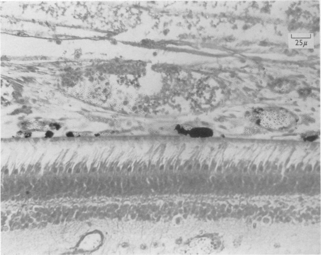
6 842 T. FUJINO AND D. I. HAMASAKI there was a rapid loss of the b-wave which left the PI and PIII components. The retinal circulation was occluded in the right eye as described above, and it can be seen that as before the b-wave was the first to be lost. With time, however, the PI and PIII components gradually decreased in the left eye until only a small response could be elicited at 20 min. The right eye at this time responded with large PI and PIII components. Right Left 0 30 sec 45 sec 60 sec 5 min 10 min 20 min Text-fig. 3. Comparison of the effect of occluding the retinal circulation (right eye) and choroidal circulation (left eye) in a Saimiri monkey. Numbers at left represent times after the occlusion. Calibration, 100 /JpV; time, 1 sec. In both eyes, the off-response disappeared much sooner than the onresponse. Photomicrographs from the eyes of this animal are shown in P1. 1. In order to demonstrate the Indian ink better the melanin pigment of the choroid was bleached by 0-5 % potassium permanganate solution. Note the presence of Indian ink in the small capillaries of the retina of the left eye (P1. la), and the large amounts of Indian ink in the choroidal vessels of the right eye (P1. 1 b). The corresponding circulations, the choroidal of the left eye and the retinal of the right eye, were empty. Choroidal occlusion was attempted in 18 eyes of which only three showed complete occlusion with complete absence of Indian ink in the choroidal circulation. The more usual observations are shown in Text-fig. 4. The ciliary vessels were cut in both eyes and again the b-wave was the wave first affected. In the right eye the small b-wave which remained after 2 min increased with further time and at the termination of the experiment, at 30 min, all components of the e.r.g. were present although much
7 OCULAR CIRCULATION AND E.R.G. 843 reduced. The same changes were seen in the left eye with the b-wave remaining very small throughout the experiment. In both eyes, there was a great increase in the amplitude of the c-wave following the reduction of the choroidal blood flow. Examination of the retinas showed Indian ink in the retinal circulation of both eyes. The choroidal circulation in both eyes showed small amounts of Indian ink in capillaries close to the head of the optic nerve with the left eye showing less Indian ink than the right. Right Left sec 30 sec 60 sec 2 min 5 min 15 min 30 min Text-fig. 4. Changes in the e.r.g. following the cutting of all ciliary vessels in both the right and left eyes of a Saimiri. Note that all components of the e.r.g. were present at the termination of the experiment. Numbers at left represent times after the operative procedures. Calibration, 100,uV; time, 1 sec. Careful histological study of the sections through the optic nerve showed a branch of the central retinal artery which passed to the choroid and ink could be followed in this collateral to the choroid. Examination of neoprene-injected eyes showed this retino-choroidal collateral very clearly.

8 844 T. FUJINO AND D. I. HAMASAKI DISCUSSION The results of this study demonstrate the role of the retinal and choroidal circulations in maintaining normal electrical activity of the retina. It was shown that while the b-wave required both the retinal and choroidal circulations to be intact, the PI and PIII components could be maintained adequately for at least 1 hr by the choroidal circulation. The changes in the e.r.g. produced by the occlusions of all blood flow to the eye were identical to those reported by Granit (1933) in the cat. The enhancement of the c-wave following interference with the ocular circulation was also noted by Granit. The time of abolition of all components of the e.r.g. varied from 20 to 40 min, and probably depended on the condition of the eye and the animal at the time of occlusion of blood supply. Assuming that the anatomical distribution of the blood vessels does separate physiologically the elements of the retina then it might be expected that occlusion of the retinal blood supply would abolish all electrical responses from elements in the inner layers of the retina. Thus if the b-wave arises from the bipolar cells as the evidence at the moment indicates (see Tomita, 1963), then occlusion of retinal blood flow would be expected to abolish the b-wave. This was indeed what was found here and previously by Brown & Watanabe (1962). The remaining PI and P III components should then arise from elements in the outer layers of the retina and the choroid. And it might be expected that the primary effect of occluding the choroidal blood supply would be on the PI and PIII components with the concurrent loss of the b-wave secondary to this. But the results showed that choroidal occlusion also affected the b-wave significantly earlier than the other two components. Hence, the early loss of the b-wave merely indicates that the elements which give rise to it are more sensitive to changes than those giving rise to the PI and PIII components. The PIII component, on the other hand, reacted differently depending on the particular circulation occluded. The loss of PIII when the choroidal circulation was occluded demonstrated that it was sensitive to alterations in the blood-flow pattern, and thus its maintenance by the choroidal circulation alone indicated that it probably arises from the outer layers of the retina or the choroid. The choroid and pigment epithelium can be ruled out inasmuch as the PIII component can be obtained from the isolated retina without these structures. Therefore, the receptors with their nuclei appear to be the most likely elements involved, and the observations support Brown & Watanabe's conclusion that the PIII component arises from the receptors.

9 The Journal of Physiology. Vol. 180, No. 4 Plate 1 :.._..e.. il4i ttl 7~~~~~~~~~~~~~~~~~~~~~~~~~~~~~~~~~~~~~~~~~~~~~~~~~~~~~~~~~~~~~~~~~~~~... -_* 25,u #^bt~~~~- a A b T. FUJINO AND D. I. HIAMASAKI (Facing p. 845)

10 OCULAR CIRCULATION AND E.R.G. 845 SUMMARY 1. When all blood flow to the eye was blocked, there was an immediate loss of the b-wave leaving the PI and PIII components. PI was lost after 5 min, and PIII min after the occlusion. 2. Retinal circulation occlusion resulted in an immediate depression of the b-wave followed by its complete abolition in 60 sec. The remaining PI and PIII components could be maintained for at least 60 min by the choroidal circulation. 3. Choroidal occlusion also resulted in a loss of the b-wave at times comparable to that seen with retinal occlusion. The remaining PI and PIII could not be maintained by the retinal circulation and were lost after min. 4. The observation that the b-wave was the first wave to be lost under all experimental conditions demonstrated the high sensitivity of the elements which give rise to it to alterations in blood-flow patterns. Little can be said about the identity of these structures. a. The results support Brown & Watanabe's (1962) conclusion that PIII arises from the receptors. WAe wish to thank Mr 0. Navarro for his valuable technical assistance with the experiments and with the histological preparations. This investigation was supported in part by a Public Health Service Research Grant, NB from the National Institute of Neurological Diseases and Blindness and in part by a Fight for Sight Post-Doctoral Research Fellowship (G) F-185, of the National Council to Combat Blindness, Inc., New York, New York held by Dr T. Fujino. REFERENCES ARDEN, G. B. & GREAVES, D. P. (1956). The reversible alterations of the electroretinogram of the rabbit after occlusion of the retinal circulation. J. Physiol. 133, BRowN, K. T. & WATANABE, K. (1962). Isolation and identification of a receptor potential from the pure cone fovea of the monkey retina. Nature, Lond., 193, GRANr, R. (1933). The components of the retinal action potential and their relation to the discharge in the optic nerve. J. Phy8iol. 77, TOmITA, T. (1963). Electrical activity of the vertebrate retina. J. opt. Soc. Amer. 53, EXPLANATION OF PLATE (a) Right retina and choroid of squirrel monkey. Note the presence of Indian ink in the choriocapillaries and the complete absence from the two retinal vessels shown. The choroidal melanin pigment was bleached with 0-5% potassium permanganate. E.r.g.s from this eye shown in Text-fig. 3. (b) Left retina and choroid of squirrel monkey. Note the presence of Indian ink in the retinal vessels and the complete absence from the choroidal vessels. The choroidal melanin pigment was bleached with 0.5% potassium permanganate. E.r.g.s from this eye shown in Text-fig. 3.
Introduction to Full Field ERGs
 Introduction to Full Field ERGs ISCEV Full Field ERG Standard (Recording protocols and their physiological basis) Laura J. Frishman, PhD University of Houston October 17, 2016 Cellular origins and mechanisms
Basic Electrophysiology, the Electroretinogram (ERG) and the Electrooculogram (EOG) - Signal origins, recording methods and clinical applications
 and the Electrooculogram (EOG) - Signal origins, recording methods and clinical applications") Basic Electrophysiology, the Electroretinogram (ERG) and the Electrooculogram (EOG) - Signal origins, recording methods and clinical applications The body is a complex machine consisting of the central
Basic Electrophysiology, the Electroretinogram (ERG) and the Electrooculogram (EOG) - Signal origins, recording methods and clinical applications The body is a complex machine consisting of the central
Vision I. Steven McLoon Department of Neuroscience University of Minnesota
 Vision I Steven McLoon Department of Neuroscience University of Minnesota 1 Eye Cornea Sclera Conjunctiva 2 Eye The conjunctiva lines the inner surface of the eyelids and outer surface of the sclera. 3
Evidence that a-wave Latency of the Electroretinogram Is Determined Solely by Photoreceptors
 Evidence that a-wave Latency of the Electroretinogram Is Determined Solely by Photoreceptors Hui Qiu*, Eriko Fujiwara, Mu Liu, Byron L. Lam and D. I. Hamasaki *Department of Ophthalmology, Hamamatsu University
The Orbit. The Orbit OCULAR ANATOMY AND DISSECTION 9/25/2014. The eye is a 23 mm organ...how difficult can this be? Openings in the orbit
 The eye is a 23 mm organ...how difficult can this be? OCULAR ANATOMY AND DISSECTION JEFFREY M. GAMBLE, OD COLUMBIA EYE CONSULTANTS OPTOMETRY & UNIVERSITY OF MISSOURI DEPARTMENT OF OPHTHALMOLOGY CLINICAL
Copyright 2009 Pearson Education, Inc.
 Outline Nervous System Sensory Systems I. II. III. IV. V. VI. Biol 105 Lecture 11 Chapter 9 Senses Sensory receptors Touch Vision Hearing and balance Smell Senses Sensory receptor cells Sensory receptors
The Visual System. Retinal Anatomy Dr. Casagrande February 2, Phone: Office: T2302 MCN
 The Visual System Retinal Anatomy Dr. Casagrande February 2, 2004 Phone: 343-4538 Email: vivien.casagrande@mcmail.vanderbilt.edu Office: T2302 MCN Reading assignments and Good Web Sites Chapter 2 in Tovée,
XUE HUI Department of Histology& Embryology, Basic Medicine College of Jilin University
 SENSE ORGAN XUE HUI Department of Histology& Embryology, Basic Medicine College of Jilin University EYE fibrous globe lens photosensitive cells a system of cells and nerves concentric layers the sclera
Eye Fluids. Dr. Mohamed Saad Daoud
 Eye Fluids 1 Reference Books: Text Book of Medical physiology (Guyton and Hall) Eleventh edition 2 Fluid System of the Eye (Intraocular Fluid) The eye is filled with intraocular fluid, which maintains
Histology of the Eye
 Histology of the Eye Objectives By the end of this lecture, the student should be able to describe: The general structure of the eye. The microscopic structure of:»cornea.»retina. EYE BULB Three coats
Reliability and Significance of Measurements of a-wave Latency in Rats
 Reliability and Significance of Measurements of a-wave Latency in Rats Eriko Fujiwara*, Hui Qiu, Mu Liu, Byron L. Lam, J.-M. Parel, G. Inana and D. I. Hamasaki *Department of Ophthalmology, Fukuoka University
Electroretinographic abnormalities and advanced multiple sclerosis
 Electroretinographic abnormalities and advanced multiple sclerosis James Pitzer Gills, Jr. Reduced electroretinographic responses were present in patients with advanced multiple sclerosis. The observed
The Cellular Basis of Electroretinogram (ERG) Signals
 Signals") The Cellular Basis of Electroretinogram (ERG) Signals Laura J. Frishman, PhD University of Houston October 19, 2015 Cellular origins and mechanisms of generation of the various waves of the ERG Sherry,
The Cellular Basis of Electroretinogram (ERG) Signals Laura J. Frishman, PhD University of Houston October 19, 2015 Cellular origins and mechanisms of generation of the various waves of the ERG Sherry,
Understanding how the ERG is generated helps immensely to interpret what it means.
 LECTURE 7 THE ELECTRORETINOGRAM (ERG) The electroretinogram is a field potential recorded from the cornea of the intact eye in response to light. It represents the total electrical activity of all the
of the retina is so small, in relation to that of the R-membrane, that no
 J. Physiol. (1965), 176, pp. 429-461 429 With 1 plate and 12 text-figures Printed in Great Britain SOME PROPERTES OF COMPONENTS OF THE CAT ELECTRORETNOGRAM REVEALED BY LOCAL RECORDNG UNDER OL BY G. B.
THE EYE: RETINA AND GLOBE
 Neuroanatomy Suzanne Stensaas February 24, 2011, 10:00-12:00 p.m. Reading: Waxman Ch. 15. Your histology and gross anatomy books should be useful. Reading: Histology of the Eye from any histology book
Destruction of the Indoleomine-Accumuloting Amocrine Cells Alters the ERG of Rabbits
 Destruction of the Indoleomine-Accumuloting Amocrine Cells Alters the ERG of Rabbits Kazuo Nakarsuka* and D. I. Hamasaki The indoleamine-accumulating amacrine cells in the rabbit's retina were destroyed
Special Senses: The Eye
 Unit 4 Special Senses: The Eye ESSENTIALS OF HUMAN ANATOMY & PHYSIOLOGY The Senses General senses of touch Temperature Pressure Pain Special senses Smell Taste Sight Hearing Equilibrium The Eye and Vision
From last week: The body is a complex electrical machine. Basic Electrophysiology, the Electroretinogram ( ERG ) and the Electrooculogram ( EOG )
 and the Electrooculogram ( EOG )") From last week: Differential Amplification This diagram shows a low frequency signal from the patient that differs between the two inputs and is therefore amplified, with an interfering high frequency
From last week: Differential Amplification This diagram shows a low frequency signal from the patient that differs between the two inputs and is therefore amplified, with an interfering high frequency
The electrically evoked response of the visual system (EER)
") The electrically evoked response of the visual system (EER) III. Further contribution to the origin of the EER Albert M. Potts and Jiro Inoue We have shown recently that an occipital correlate of the electric
The electrically evoked response of the visual system (EER) III. Further contribution to the origin of the EER Albert M. Potts and Jiro Inoue We have shown recently that an occipital correlate of the electric
AQUEOUS VEINS IN RABBITS*
 Brit. J. Ophthal., 35, 119. AQUEOUS VEINS IN RABBITS* BY D. P. GREAVES AND E. S. PERKINS Institute of Ophthalmology, London Director of Research, Sir Stewart Duke-Elder IN the course of investigations
Frequency & Amplitude Ranges for Bioelectric Signals
 Frequency & Amplitude Ranges for Bioelectric Signals Signal Frequency range (Hz) Amplitude range(mv) ECG 0.01 300 0.05 3 EEG 0.1 100 0.001 1 EOG 0.1 10 0.001 0.3 EMG 50 3000 0.001 100 Electro-oculogram
Medical School Histology Basics. VIBS 289 lab. Eye
 Medical School Histology Basics VIBS 289 lab Eye Larry Johnson Texas A&M University Aqueous humor OUTLINE OVERVIEW CELLULAR STRUCTURES THROUGH WHICH LIGHT PASSES A. CORNEA B. LENS C. RETINA STRUCTURES
Presentation On SENSATION. Prof- Mrs.Kuldeep Kaur
 Presentation On SENSATION Prof- Mrs.Kuldeep Kaur INTRODUCTION:- Sensation is a specialty area within Psychology that works at understanding how are senses work and how we perceive stimuli in the environment.
Senses and Sense Organs
 Senses and Sense Organs SENSORY SYSTEMS Human experience is effected by both internal and external stimuli. Humans are able to distinguish among many different types of stimuli by means of a highly developed
Taste buds Gustatory cells extend taste hairs through a narrow taste pore
 The Special Senses Objectives Describe the sensory organs of smell, and olfaction. Identify the accessory and internal structures of the eye, and explain their function. Explain how light stimulates the
INTRODUCTION: ****************************************************************************************************
 BIOLOGY 211: HUMAN ANATOMY & PHYSIOLOGY **************************************************************************************************** EYES AND VISION ****************************************************************************************************
COMMUNICATIONS PHOTOCOAGULATION OF THE RETINA* OPHTHALMOSCOPIC AND HISTOLOGICAL FINDINGS. photocoagulation of the rabbit's retina.
 Brit. J. Ophthal. (1963) 47, 577. COMMUNICATIONS PHOTOCOAGULATION OF THE RETINA* OPHTHALMOSCOPIC AND HISTOLOGICAL FINDINGS BY A. LAVYEL Haifa, Israel SINCE the introduction of the photocoagulator by Meyer-Schwickerath
OCCLUSIVE VASCULAR DISORDERS OF THE RETINA
 OCCLUSIVE VASCULAR DISORDERS OF THE RETINA Learning outcomes By the end of this lecture the students would be able to Classify occlusive vascular disorders (OVD) of the retina. Correlate the clinical features
Test Bank Chapter 2: The Beginnings of Perception
 Test Bank Chapter 2: The Beginnings of Perception MULTIPLE CHOICE 1. Our perception of the environment depends on a. the properties of the objects in the environment. b. the properties of the electrical
The Sense Organs 10/13/2016. The Human Eye. 1. Sclera 2. Choroid 3. Retina. The eye is made up of three layers:
 The human body gathers information from the outside world by using the five senses of: The Sense Organs 12.3 Sight Hearing Taste Smell Touch This information is essential in helping the body maintain homeostasis.
UNDERSTAND MORE ABOUT UVEITIS UVEITIS
 UNDERSTAND MORE ABOUT UVEITIS UVEITIS Uveitis What is uveitis? Uveitis is inflammation of the uvea, the middle layer of your eye. The eye is shaped much like a tennis ball, with three different layers
Special Senses PART A
 8 Special Senses PART A PowerPoint Lecture Slide Presentation by Jerry L. Cook, Sam Houston University ESSENTIALS OF HUMAN ANATOMY & PHYSIOLOGY EIGHTH EDITION ELAINE N. MARIEB The Senses General senses
Biology. Slide 1 of 49. End Show. Copyright Pearson Prentice Hall
 Biology 1 of 49 2 of 49 Sensory Receptors Neurons that react directly to stimuli from the environment are called sensory receptors. Sensory receptors react to stimuli by sending impulses to other neurons
o A cushion of fat surrounds most of the eye
 Name Period SPECIAL SENSES The Senses of touch o Temperature o Pressure o Pain o Smell o Taste o Sight o Hearing o Equilibrium The Eye and Vision are in the eyes has over a o Most of the eye is enclosed
Unit VIII Problem 8 Anatomy: Orbit and Eyeball
 Unit VIII Problem 8 Anatomy: Orbit and Eyeball - The bony orbit: it is protecting our eyeball and resembling a pyramid: With a base directed: anterolaterally. And an apex directed: posteromedially. Notes:
Neuroscience - Problem Drill 13: The Eye and Visual Processing
 Neuroscience - Problem Drill 13: The Eye and Visual Processing Question No. 1 of 10 needed, (3) Pick the answer, and (4) Review the core concept tutorial as needed. 1. Which of the following statements
Fundus Autofluorescence. Jonathan A. Micieli, MD Valérie Biousse, MD
 Fundus Autofluorescence Jonathan A. Micieli, MD Valérie Biousse, MD The retinal pigment epithelium (RPE) has many important functions including phagocytosis of the photoreceptor outer segments Cone Rod
Recovery of Retinal Pigment Epithelial Function After Ischemia in the Rabbit
 Investigative Ophthalmology & Visual Science, Vol. 32, No. 1, January 1991 Copyright Association for Research in Vision and Ophthalmology Recovery of Retinal Pigment Epithelial Function After Ischemia
Visual Physiology. Perception and Attention. Graham Hole. Problems confronting the visual system: Solutions: The primary visual pathways: The eye:
 Problems confronting the visual system: Visual Physiology image contains a huge amount of information which must be processed quickly. image is dim, blurry and distorted. Light levels vary enormously.
Annette Sims, MD, Ophthalmologist next Tuesday! Hooray!!
 BI 358 Lecture 18 Annette Sims, MD, Ophthalmologist next Tuesday! Hooray!! I. Announcements Quiz 5 returned at end of lecture. Eye Dissection & Vision lab next Tuesday > Lecture by Dr. Sims! Final Quiz
Annette Sims, MD, Ophthalmologist next Tuesday! Hooray!!
 BI 358 Lecture 18 Annette Sims, MD, Ophthalmologist next Tuesday! Hooray!! I. Announcements Quiz 5 returned at end of lecture. Eye Dissection & Vision lab next Tuesday > Lecture by Dr. Sims! Final Quiz
CIRCULATION IN THE IRIS AND CILIARY PROCESSES
 Brit. J. Ophthal. (1965) 49, 6 CIRCULATION IN THE IRIS AND CILIARY PROCESSES POSSIBLE RECIPROCAL RELATIONSHIP*t BY PAUL HENKIND Department ofpathology, Institute of Ophthalmology, University oflondon A
4/22/16. Eye. External Anatomy of Eye. Accessory Structures. Bio 40B Dr. Kandula
 Eye Bio 40B Dr. Kandula External Anatomy of Eye Accessory Structures l Eyebrows l Levator Palpebrae Superioris - opens eye l Eyelashes l Ciliary glands modified sweat glands l Small sebaceous glands l
Australian and New Zealand College of Veterinary Scientists. Fellowship Examination. Veterinary Ophthalmology Paper 1
 Australian and New Zealand College of Veterinary Scientists Fellowship Examination June 2014 Veterinary Ophthalmology Paper 1 Perusal time: Twenty (20) minutes Time allowed: Three (3) hours after perusal
Oxygen Distribution in the Macaque Retina
 Oxygen Distribution in the Macaque Retina Jameel Ahmed* Rod D. Braun,^ Robert Dunn, Jr. * and Robert A. Linsenmeier*X Purpose. Oxygen distribution was characterized in the macaque retina, which is more
Ocular Anatomy for the Paraoptometric
 Ocular Anatomy for the Paraoptometric Minnesota Optometric Association Paraoptometric CE Friday September 30, 2016 Lindsay A. Sicks, OD, FAAO Assistant Professor, Illinois College of Optometry lsicks@ico.edu
02/03/2014. Average Length: 23mm (Infant ~16mm) Approximately the size of a quarter Volume: ~5mL
 Approximately the size of a quarter Volume: ~5mL") Identify the anatomy of the eye. Explain the basic physiology of the parts of the eye. Briefly discuss various surgeries related to different parts of the anatomy. Average Length: 23mm (Infant ~16mm) Approximately
Identify the anatomy of the eye. Explain the basic physiology of the parts of the eye. Briefly discuss various surgeries related to different parts of the anatomy. Average Length: 23mm (Infant ~16mm) Approximately
RETINITIS PIGMENTOSA* A NEW THERAPEUTIC APPROACH
 Brit. J. Ophthal. (1963) 47, 144. RETINITIS PIGMENTOSA* A NEW THERAPEUTIC APPROACH BY LALIT P. AGARWAL, S. R. K. MALIK, MADAN MOHAN, AND P. R. KARWAL From the Department of Ophthalmology, All-India Institute
Answer three questions out of four questions.
 Ancillary Material: Nil ACADEMIC UNIT OF OPHTHALMOLOGY & ORTHOPTICS Summer Semester 2016 ELECTRODIAGNOSIS 1 Hour 30 Minutes You are advised to use the 4 leaf answer book. There are four questions. Attempt
Vision Seeing is in the mind
 1 Vision Seeing is in the mind Stimulus: Light 2 Light Characteristics 1. Wavelength (hue) 2. Intensity (brightness) 3. Saturation (purity) 3 4 Hue (color): dimension of color determined by wavelength
Image Formation and Phototransduction. By Dr. Abdelaziz Hussein Lecturer of Physiology
 Image Formation and Phototransduction By Dr. Abdelaziz Hussein Lecturer of Physiology Vision Vision is a complex process through which an image of the external environment is formed on the photosensitive
Sense of Vision. Chapter 8. The Eye and Vision. The Eye Orbit. Eyebrows, Eyelids, Eyelashes. Accessory Organs 5/3/2016.
 Sense of Vision Chapter 8 Special Senses The Eye and Vision 70 percent of all sensory receptors are in the eyes Each eye has over 1 million nerve fibers Protection for the eye Most of the eye is enclosed
Retinal Tear and Detachment
 Retinal Tear and Detachment Introduction The retina is the layer of tissue in the back of the eye that is responsible for vision. It is attached to the choroid tissue, which supplies the retina with blood.
Acquired unilateral loss adaptation
 Brit. J. Ophthal. (I 97 I) 55, 38 Acquired unilateral loss adaptation J. H. KELSEY AND G. B. ARDEN Moorfields Eye Hospital, City Road, London of dark Poor dark adaptation is a not uncommon symptom and
let's continue talking about the eye,
 Eye is mainly composed of 3 layers: External layer, which called The Sclera which is a hard connective tissue that gives the eye its round shape. Extension of the sclera into the front is the cornea, which
Franklin, 1933; Waterman, 1933]; indeed, the only negative findings, [Waterman, 1933]. Inasmuch, then, as Donegan was misled with
![Franklin, 1933; Waterman, 1933]; indeed, the only negative findings, [Waterman, 1933]. Inasmuch, then, as Donegan was misled with](/thumbs/91/106294335.jpg "Franklin, 1933; Waterman, 1933]; indeed, the only negative findings, [Waterman, 1933]. Inasmuch, then, as Donegan was misled with") 381 6I2.I34:6I2.893 THE CONSTRICTOR RESPONSE OF THE INFERIOR VENA CAVA TO STIMULATION OF THE SPLANCHNIC NERVE BY K. J. FRANKLIN AND A. D. McLACHLIN (From the University Department of Pharmacology, Oxford)
381 6I2.I34:6I2.893 THE CONSTRICTOR RESPONSE OF THE INFERIOR VENA CAVA TO STIMULATION OF THE SPLANCHNIC NERVE BY K. J. FRANKLIN AND A. D. McLACHLIN (From the University Department of Pharmacology, Oxford)
Around The Globe in 60 Minutes
 Around The Globe in 60 Minutes Around the GLOBE in Sixty Minutes Basic Ocular Anatomy, Examination, and Diagnostic Techniques Introduction Focusing on canine and feline ocular anatomy and basic examination
SPECIAL SENSES. Anatomy & Physiology
 SPECIAL SENSES Anatomy & Physiology BELL WORK: DEFINE LACRIMAL ACHROMATIC OTOSCOPE TENNITIS VERTIGO STANDARD 25) Define key terms associated with vision disorders, ear disorders, nose disorders, and mouth
Uveal Melanoma. Protocol applies to malignant melanoma of the uvea.
 Uveal Melanoma Protocol applies to malignant melanoma of the uvea. Protocol revision date: January 2005 Based on AJCC/UICC TNM, 6 th edition Procedures Cytology (No Accompanying Checklist) Biopsy (No Accompanying
Scrub In. What is the function of vitreous humor? What does the pupil do when exposed to bright light? a. Maintain eye shape and provide color vision
 Scrub In What is the function of vitreous humor? a. Maintain eye shape and provide color vision b. Maintain eye shape and refract light rays c. Provide night vision and color vision d. Provide night vision
2/3/17. Visual System I. I. Eye, color space, adaptation II. Receptive fields and lateral inhibition III. Thalamus and primary visual cortex
 1 Visual System I I. Eye, color space, adaptation II. Receptive fields and lateral inhibition III. Thalamus and primary visual cortex 2 1 2/3/17 Window of the Soul 3 Information Flow: From Photoreceptors
chorioretinal atrophy
 British Journal of Ophthalmology, 1987, 71, 757-761 Retinal microangiopathy in pigmented paravenous chorioretinal atrophy SURESH R LIMAYE AND MUNEERA A MAHMOOD From the Ophthalmology Service, DC General
Surgical Anatomy Ear and Eye. Presenters: Dr. Jim Hurrell and Dr. Dennis McCurnin
 Surgical Anatomy Ear and Eye Presenters: Dr. Jim Hurrell and Dr. Dennis McCurnin A Warm Welcome from My Faculty TEAM and Me!!! 2 The Pledge of Allegiance 3 The Senses 4 Hearing 3 Layers of Ear EXTERNAL
Flashers and Floaters
 Flashers and Floaters Introduction Sometimes people see small, moving spots or specks in their field of vision. These sensations are called floaters. About 7 out of 10 people experience floaters at some
Electro Retinogram Basics and Major Clinical Applications
 Ophthalmic Instrumentation Electro Retinogram Basics and Major Clinical Applications Meena C K. DNB, Thomas Cherian MS, Elizabeth Chacko BSc Optometry Electroretinogram is the electrical potential generated
Adenocarcinorna of the Ciliary Body A Report of 2 Cases in Dogs
 Path. vet. 5: 122-126 (1968) From the Ophthalmic Pathology Laboratory, Department of Ophthalmology, New York University School of Medicine, New York Adenocarcinorna of the Ciliary Body A Report of 2 Cases
Assisting in Ophthalmology. Copyright 2011, 2007, 2003, 1999 by Saunders, an imprint of Elsevier Inc. All rights reserved.
 Assisting in Ophthalmology Learning Objectives Define, spell, and pronounce the terms listed in the vocabulary. Apply critical thinking skills in performing patient assessment and care. Explain the differences
Chapter 18. The Senses SENSORY RECEPTION. Introduction: Superhuman Senses. Introduction: Superhuman Senses
 Introduction: Superhuman Senses Chapter 18 The Senses! Three senses found in some animals but not humans Echolocation locating objects by detecting echoes of emitted sound waves Electroreception ability
deemed necessary to repeat the work using a further small number of animals, and performing suitable control experiments.
 J. Physiol. (I938) 93, 75-80 6I2.II9:6II.I33 75 THE CAROTID SINUS AND BLOOD REGENERATION BY A. L. LATNER From the Department of Physiology, University of Liverpool (Received 18 October 1937) IN a previous
Lecture 3 Vision 2 The Retina
 Lecture 3 Vision 2 The Retina All lecture material from the following two links: 1) http://hubel.med.harvard.edu/book/bcontex.htm 2) http://www.ib.cnea.gov.ar/~redneu/2013/books/principles%20of%20neural%20science%20%20kandel/gateway.ut.ovid.com/gw2/ovidweb.cgisidnjhkoalgmeho00dbookimagebookdb_7c_2fc~32.htm
Work Sheet And Course Hand Out
 Work Sheet And Course Hand Out This course provides the primary care health professional with a basic understanding of the eye, its function and the assessment of common sight- and non-sight threatening
Anitschkov (1936) investigated the effect of chemoreceptor denervation. of ammonium chloride. He maintained, however, that the hyperpnoea was
 investigated the effect of chemoreceptor denervation. of ammonium chloride. He maintained, however, that the hyperpnoea was") J. Phy8iol. (1962), 161, pp. 351-356 351 With 4 text-figure8 Printed in Great Britain THE ROLE OF THE CHEMORECEPTORS IN THE HYPERPNOEA CAUSED BY INJECTION OF AMMONIUM CHLORIDE BY N. JOELS AND E. NEIL From
J. Phy8iol. (1962), 161, pp. 351-356 351 With 4 text-figure8 Printed in Great Britain THE ROLE OF THE CHEMORECEPTORS IN THE HYPERPNOEA CAUSED BY INJECTION OF AMMONIUM CHLORIDE BY N. JOELS AND E. NEIL From
-Detect heat or cold and help maintain body temperature
 Sensory Receptors -Transduce stimulus energy and transmit signals to the central nervous system -Reception occurs when a receptor detectd a stimulus -Perception occurs in the brain as this information
Sensation and Perception. A. Sensation: awareness of simple characteristics B. Perception: making complex interpretations
 I. Overview Sensation and Perception A. Sensation: awareness of simple characteristics B. Perception: making complex interpretations C. Top-Down vs Bottom-up Processing D. Psychophysics -- thresholds 1.
Introduction How the eye works
 1 Introduction Diabetic retinopathy is a condition that can cause permanent loss of eyesight and even blindness. It is a major cause of loss of vision. But if a person with diabetes receives proper eye
Frequently Asked Questions about General Ophthalmology:
 1. Normal Eye Structure The eye is a slightly asymmetrical globe, about an inch in diameter. The parts of the eye include: Cornea (a clear dome over the iris), Iris (the pigmented part); Pupil (the black
(Received 13 October 1949)
") 160 J. Physiol. (I950) III, I6o-I73 6I2.843. I6 AN ANALYSIS OF THE RESPONSE FROM SINGLE VISUAL-PURPLE-DEPE NDENT ELEMENTS, IN THE RETINA OF THE CAT BY K. 0. DONNER AND E. N. WILLMER From the Nobel Institute
160 J. Physiol. (I950) III, I6o-I73 6I2.843. I6 AN ANALYSIS OF THE RESPONSE FROM SINGLE VISUAL-PURPLE-DEPE NDENT ELEMENTS, IN THE RETINA OF THE CAT BY K. 0. DONNER AND E. N. WILLMER From the Nobel Institute
RETINAL CONDITIONS RETINAL CONDITIONS
 GENERAL INFORMATION RETINAL CONDITIONS RETINAL CONDITIONS WHAT ARE RETINAL CONDITIONS? Retinal conditions affect the light-sensitive tissue at the back of eye known as the retina. They include diseases
SPECIAL SENSES PART I: OLFACTION & GUSTATION
 SPECIAL SENSES PART I: OLFACTION & GUSTATION 5 Special Senses Olfaction Gustation Vision Equilibrium Hearing Olfactory Nerves Extend through cribriform plate into nasal cavity on both sides of nasal septum
Guess: Correct or Incorrect. Trial (perform in random order)
") AP Biology Senses Lab Names Per. Our senses are constantly bombarded with various stimuli from the environment, which are relayed to the central nervous system where the information is interpreted. In
AP Biology Senses Lab Names Per. Our senses are constantly bombarded with various stimuli from the environment, which are relayed to the central nervous system where the information is interpreted. In
Written by Administrator Wednesday, 13 January :27 - Last Updated Thursday, 21 January :34
 angle closure glaucoma A type of glaucoma caused by a sudden and severe rise in eye pressure. Occurs when the pupil enlarges too much or too quickly, and the outer edge of the iris blocks the eye s drainage
The Eye. The Orbit. The EYE What a Trip!!! - The Anterior Segment 5/12/2015. Jill J Luebbert, CPOT, ABOC
 The EYE What a Trip!!! - The Anterior Segment Jill J Luebbert, CPOT, ABOC The Eye The Orbit Bony socket containing the eye and most of its accessory organs consisting of 7 bones 1 The Seven Bones of the
Macular hole. Information for patients Ophthalmology (Vitreal Retina) Large Print
 Large Print") Macular hole Information for patients Ophthalmology (Vitreal Retina) Large Print page 2 of 16 What is the macula? The back of the eye has a light-sensitive lining called the retina, similar to the film
Macular hole Information for patients Ophthalmology (Vitreal Retina) Large Print page 2 of 16 What is the macula? The back of the eye has a light-sensitive lining called the retina, similar to the film
6I :6I2.I83 BY ALISON S. DALE. concluded that the apparent vaso-constriction obtained by F r6 hli c h and
 6I2.313.87:6I2.I83 A REVERSED ACTION OF THE CHORDA TYMPANI ON THE VENOUS OUTFLOW FROM THE SUBMAXILLARY GLAND. BY ALISON S. DALE. (From the Physiological Laboratory, Cambridcgel.) INTRODUCTORY. FROiHLICH
F/G 6/18 MECHANISMS OCT 75 T LAWHILL, F SHARP, N SPEED DAMD17-74-C-4026 IHEF. NL UNCLASSIFIlED
 F/G 6/18 MECHANISMS 7 A-AI03 183 LOUISVILLE UNIV OF RETINAL KY SCHOOL DAMAGE OF FROM MEDICINE CHRONIC LASER RAOIATION.(U) OCT 75 T LAWHILL, F SHARP, N SPEED DAMD17-74-C-4026 IHEF NL UNCLASSIFIlED )J yreport
Sense Organs. Chapter 38
 Sense Organs Chapter 38 Chemical Senses Chemoreceptors are the receptors responsible for smell and taste. Because all members of the animal kingdom have developed a sense of taste and/or smell, chemoreceptors
Test Bank for Medical Surgical Nursing An Integrated Approach 3rd Edition by White
 Test Bank for Medical Surgical Nursing An Integrated Approach 3rd Edition by White Link full download : http://testbankair.com/download/test-bank-for-medical-surgical-nursing-anintegrated-approach-3rd-edition-by-white/
Year 2 MBChB Clinical Skills Session Ophthalmoscopy. Reviewed & ratified by: Mr M Batterbury Consultant Ophthalmologist
 Year 2 MBChB Clinical Skills Session Ophthalmoscopy Reviewed & ratified by: o Mr M Batterbury Consultant Ophthalmologist Learning objectives o To understand the anatomy and physiology of the external and
Modifiers and Retransmitters (Secondary Light Sources)
") Vision and Light Vision Generators Transmitters (Light Sources) Modifiers and Retransmitters (Secondary Light Sources) Receivers Decoder Encoders Interpreter (Eyes) (Brain) Sun, Discharge lamps, fluorescent
Vision and Light Vision Generators Transmitters (Light Sources) Modifiers and Retransmitters (Secondary Light Sources) Receivers Decoder Encoders Interpreter (Eyes) (Brain) Sun, Discharge lamps, fluorescent
THE CHRONIC GLAUCOMAS
 THE CHRONIC GLAUCOMAS WHAT IS GLAUCOMA? People with glaucoma have lost some of their field of all round vision. It is often the edge or periphery that is lost. That is why the condition can be missed until
Introduction to Physiological Psychology
 Introduction to Physiological Psychology Vision ksweeney@cogsci.ucsd.edu cogsci.ucsd.edu/~ksweeney/psy260.html This class n Sensation vs. Perception n How light is translated into what we see n Structure
Primary Angle Closure Glaucoma
 www.eyesurgeonlondon.co.uk Primary Angle Closure Glaucoma What is Glaucoma? Glaucoma is a condition in which there is damage to the optic nerve. This nerve carries visual signals from the eye to the brain.
2. WINDOWS OF KNOWLEDGE
 CONTENT 2. WINDOWS OF KNOWLEDGE Vision - The protective measures of eyes. - Structure of human eye, Working of eye lens, - Photo receptors in the retina, Sense of vision. - Disorders & diseases of eyes,
Volume 23 Number 5. Reports 683
 Volume 2 Number 5 Reports 68 in the vitreous body of the cat. Acta Physiol Scand 84:261, 1972. 6. Niemeyer G: The function of the retina in the perfused eye. Doc Ophthalmol 9(1):5, 1975. 7. Enroth-Cugell
is the clear, transparent part at the front of the eye. It allows light to enter the eye and it also refracts (focuses) the light onto the retina.
 the light onto the retina.") Senses- Vision Light is a small part (1/70th) of the total electromagnetic (EM) spectrum. The EM band extends from radio waves at one extreme to x-rays at the other. The eye detects light and converts
Senses- Vision Light is a small part (1/70th) of the total electromagnetic (EM) spectrum. The EM band extends from radio waves at one extreme to x-rays at the other. The eye detects light and converts
Specialist Referral Service Willows Information Sheets. Cataract surgery
 Specialist Referral Service Willows Information Sheets Cataract surgery An operating microscope in use A total cataract - the normally black pupil is bluish white Cataract surgery These notes do not cover
ID# Exam 1 PS 325, Fall 2001
 ID# Exam 1 PS 325, Fall 2001 As always, the Skidmore Honor Code is in effect, so keep your eyes foveated on your own exam. I tend to think of a point as a minute, so be sure to spend the appropriate amount