Title. Author(s)Saito, Wataru; Kase, Satoru; Ohgami, Kazuhiro; Mori, CitationActa Ophthalmologica, 88(3): Issue Date Doc URL.
|
|
|
- Sheryl Sybil Quinn
- 5 years ago
- Views:
Transcription
1 Title Intravitreal anti-vascular endothelial growth factor oedema Author(s)Saito, Wataru; Kase, Satoru; Ohgami, Kazuhiro; Mori, CitationActa Ophthalmologica, 88(3): Issue Date Doc URL Rights The definitive version is available at Type article (author version) File Information AO88-3_ pdf Instructions for use Hokkaido University Collection of Scholarly and Aca
2 1 Intravitreal anti-vascular endothelial growth factor therapy with bevacizumab for tuberous sclerosis with macular edema Wataru Saito, MD, 1 Satoru Kase, MD, 1 Kazuhiro Ohgami, MD 1, Shohei Mori, MD 1, Shigeaki Ohno, MD 1 1 Department of Ophthalmology and Visual Sciences, Hokkaido University Graduate School of Medicine, Sapporo, Japan Correspondence to: Wataru Saito, MD Department of Ophthalmology and Visual Sciences, Hokkaido University Graduate School of Medicine, Nishi 7 chome, Kita 15 jou, Kita-ku, Sapporo , Japan, Phone: ; Fax: ; wsaito@med.hokudai.ac.jp Financial disclosure: None. 1
3 2 ABSTRACT. Purpose: To describe two patients with macular edema secondary to tuberous sclerosis complex (TSC), who were treated with intravitreal bevacizumab injection. Methods: Interventional case reports. Bevacizumab 1.25 mg was injected into the vitreous of two patients with TSC-associated macular edema/ exudative retinal detachment. VEGF concentration in the vitreous fluid was measured by ELISA in one of these patients. Results: Case year-old woman with TSC was diagnosed as having multiple retinal hamartomas in both eyes. Eleven years later, the patient developed macular edema with epiretinal membrane formation in the right eye. The patient underwent pars plana vitrectomy with retinal photocoagulation for retinal tumors. VEGF concentration in the vitreous fluid was high compared to that in patients without retinal vascular diseases. Recurrent macular edema was resolved by intravitreal injection of bevacizumab. Case year-old woman with TSC-associated retinal hamartoma, temporally showing macular exudative retinal detachment, developed the neovascularization originated from the tumor. By intravitreal bevacizumab injection, the 2
4 3 tumor size markedly reduced with regression of the neovascularization. Conclusions: These results suggest that VEGF derived from retinal hamartomas causes macular edema associated with TSC. Intravitreal injections of bevacizumab may be a useful therapeutic option for macular edema secondary to TSC. 3
5 4 Keywords: bevacizumab - exudative retinal detachment - macular edema - retinal hamartomas - tuberous sclerosis - vascular endothelial growth factor 4
6 5 Introduction Tuberous sclerosis complex (TSC) is a syndrome characterized by hamartomatous growths in multiple organs (Roach et al. 1998). Patients with TSC generally develop retinal astrocytic hamartomas; however clinical symptoms of patients with the retinal hamartomas are generally asymptomatic and most astrocytic hamartomas remains unchanged in size or regress during life (Zimmer-Galler & Robertson 1995). Symptomatic ocular complications rarely represent and include macular edema, exudative retinal detachment (RD), vitreous hemorrhage (VH), and neovascular glaucoma (Mennel et al. 2007). Symptomatic macular edema or exudative RD has previously been reported in only 7 eyes (Mennel et al. 2007). Argon laser photocoagulation or photodynamic therapy against retinal hamartoma may be effective for these complications (Bloom & Mahl 1991; Mennel et al. 2007). However, multiple laser treatment procedures may cause laser-induced choroidal neovascularization (Bloom & Mahl 1991). Thus, the therapeutic strategy for macular edema secondary to TSC, and the pathogenic mechanism are controversial. We report two patients with macular edema/ exudative RD secondary to TSC who underwent intravitreal 5
7 6 bevacizumab injection, from the result of high intravitreal vascular endothelial growth factor (VEGF) levels in one patient. Case reports Case 1 In 1995, a 22-year-old woman with TSC was referred for ophthalmologic examination. At 8 years of age, the patient was diagnosed as having TSC based on clinical findings including facial angiofibromas, renal angiomyolipoma, and subependymal nodules. The patient s visual acuities were 20/15 OU. Funduscopic examination demonstrated scattered slightly elevated gray-white tumors with partially calcification located in the retinal surface OU. Fluorescein angiography (FA) demonstrated early hyperfluorescence with late leakage corresponding to retinal tumors OU. The patient was diagnosed as having retinal hamartoma type 3 associated with TSC. In 1998, vitreous hemorrhage (VH) occurred OD, but spontaneously resolved within six months. In 2002, visual acuity decreased to 20/ 400 OD. Ophthalmoscopic examination showed posterior subcapsular cataract, cystoid macular edema, fibrovascular membrane formation, and retinochoroidal atrophic lesion located on the 6
8 7 superior side of the macula OD, although left eye remained no changed. Thereafter, the patient discontinued follow-up. In 2004, the patient complained of blurred vision OD, whereas visual acuity remained unchanged. The patient underwent cataract surgery of the right eye, because cataract developed despite disappearance of macular edema. Eight weeks after cataract surgery, the optics of the intraocular lens was completely closed by contraction of the anterior capsule OD. Visual acuity improved to 20/200 after YAG laser capsulotomy for the anterior capsule. In May 2006, visual acuity decreased to 20/500 OD. Macular edema recurred OD (Fig. 1A) and late-phase FA demonstrated cystoid macular edema and marked leakage from retinal tumors and retinal capillary vessels (Fig. 1B). Optical coherence tomography (OCT) revealed macular edema with epiretinal membrane OD (Fig. 1C). In November 2006, the patient underwent pars plana vitrectomy (PPV) with epiretinal membrane removal and retinal photocoagulation to the retinal tumors. After obtaining informed consent, VEGF concentration in the vitreous fluid collected was measured by ELISA (BioSource International, Camarillo, CA). The concentration was 68.5 pg/ml in this patient, while vitreal VEGF levels in ten patients with macular hole were under 7
9 8 detectable levels. One month after second surgery, visual acuity improved to 20/ 300. Funduscopy revealed photocoagulation scars corresponding to the tumors and disappearance of macular edema. The dye leakage from retinal capillary vessels in FA decreased (Fig. 2A~C). Five months after PPV, visual acuity decreased to 20/ 500 OD. Macular edema recurred (Fig. 3A), and FA showed increased retinal capillary leakage. Based on the patient s high intravitreal VEGF level, after informed consent following IRB approval, the patient underwent intravitreal injection of bevacizumab 1.25 mg. One week after the injection, macular edema rapidly improved (Fig. 3B) and visual acuity increased to 20/400. Thereafter, macular oedema recurred again; recurrent macular oedema disappeared by performing additional laser photocoagulation for tumours causative of exudation. Case 2 32-year-old woman presented with central visual loss of the right eye for the last two weeks. The patient was diagnosed as having tuberous sclerosis because she had a 8
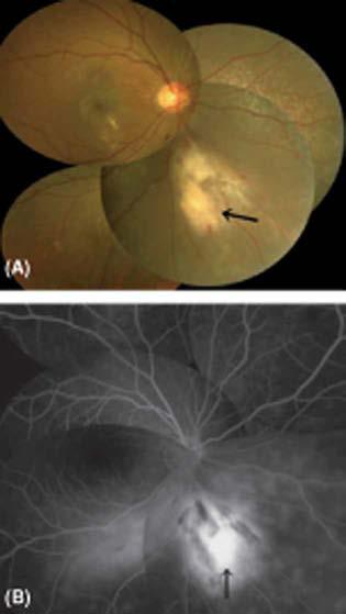
10 9 medical history of seizure and pathological diagnosis of subependymal giant cell astrocytoma and facial angiofibromas. Her visual acuities was 20/30 OD, and 20/15 OS. Anterior segment and lens were clear, OU. Funduscopy revealed macular exudative RD with lipoid exudates and an elevated, oval-shaped whitish retinal tumor without calcification, located in parapapillary area (Fig. 4A). The tumor located on the retinal surface and involved neovascularization on the surface (Fig. 4A, arrow). Late-phase FA revealed marked leakage from the tumor, the neovascularization, and retinal capillary vessels (Fig. 4B). A diagnosis of retinal hamartoma type 1 in association with TSC was made. One month later, exudative RD spontaneously regressed and visual acuity improved to 20/20, OD. However, the neovascularization originated from the tumor remained unchanged. Since we considered that the neovascularization caused VH like case 1, after informed consent, the patient received intravitreal bevacizumab 1.25 mg injection. One week after the injection, the tumor size reduced with regression of the neovascularization (Fig. 5A). The late leakage from the tumor and retinal capillary vessels in FA decreased (Fig. 5B). Three months after the injection, the tumor almost regressed (Fig. 5C) and visual acuity increased of 9
11 10 20/ 16, OD. Discussion TSC has been determined to result from mutations in TSC1 (hamartin) and TSC 2 (tuberin) tumor suppressor gene (Cheadle et al 2000). TSC1 and TSC2 proteins are responsible for the regulation of cell growth and tumourigenesis: TSC1/ TSC2 complex controls Ras homologue enriched in brain (Rheb) and subsequently inhibits the mammalian target of rapamycin (mtor), a key regulator in signaling pathway of cell proliferation and organ size (Schwartz et al 2007). Therefore, mutated TSC1/TSC2 complex causes uncontrolled cell growth and proliferation. Moreover, it is elucidated that hamartomas associated with TSC, involving the other organs (kidney, brain, and skin) except the eyes, are highly angiogenic (Arbiser et al. 2002) and can express angiogenic factors including VEGF (Nguyen-Vu et al. 2001). However, as far as we know, there are no reports which detected expression of VEGF in retinal hamarotmas. Therefore, we hypothesized that retinal hamartomas are also highly angiogenic and express VEGF, which lead to ocular complication including macular edema. In this study, VEGF concentration in the vitreous fluid taken intraoperatively in case 10
12 11 1 was apparently high, compared to the levels in patients without retinal vascular diseases. Retinal hamartomas in two patients involved VH (case 1) and the neovascularization originated from retinal tumors (case 2), suggesting that retinal hamartomas associated with TSC are well-vascularized tumors and possibly express VEGF. Based on the result of high intravitreal VEGF concentration in case 1, intravitreal bevacizumab injection was tried in two patients with TSC-associated macular edema/ exudative RD. After injection, not only macular edema, VA, OCT, and FA findings rapidly improved (Case 1) but the tumor size and tumor-associated neovascularization were rapidly attenuated (Case 2), although these two patients possibly may have recurrence of macular edema after a long-term follow-up. Therefore, the results in this study strongly suggest that VEGF derived from retinal astrocytic hamartomas plays a potential role in the pathogenesis of macular edema/ exudative RD secondary to TSC. As far as we know, there are no reports demonstrating effectiveness of intravitreal bevacizumab injection in patients with TSC-associated macular edema. In both patients 1 and 2, macular oedema disappeared rapidly by a single intravitreal bevacizumab injection. However, macular oedema secondary to TSC may be repeated, 11
13 12 such as in patient 1, in a long-term follow-up.when macular oedema recurred, performing intravitreal bevacizumab injection 3 6 times continuously every 4 6 weeks may be considered (Soliman et al. 2008). Moreover, additional retinal photocoagulation or photodynamic therapy for tumours causative of exudation may also be considered after intravitreal bevacizumab injection. When we follow up patients with TSC, to detect initial or recurrent macular oedema in early stage by performing OCT regularly is mandatory for preventing poor visual prognosis (Massin et al. 2006). In conclusion, intravitreal anti-vegf antibody injection including bevacizumab might be a therapeutic option for macular edema/ exudative RD secondary to TSC. 12
14 13 Acknowledgements This study was supported by a grant for Research on Sensory and Communicative Disorders from the Ministry of Health, Labor and Welfare, and by Grants-in-Aid for Scientific Research from the Ministry of Education, Culture, Sports, Science, and Technology (MEXT), Japan. 13
15 14 References Arbiser JL, Brat D, Hunter S, et al. (2002): Tuberous sclerosis-associated lesions of the kidney, brain, and skin are angiogenic neoplasms. J Am Acd Dermatol 46: Bloom SM & Mahl CF (1991): Photocoagulation for serous detachment of the macula secondary to retinal astrocytoma. Retina 11: Cheadle JP, Reeve MP, Sampson JR & Kwiatkowski DJ (2000): Molecular genetics advances in tuberous sclerosis. Hum Genet 107: Massin A, Gaudric A, Erginay A & Gaudric A (2006): Optical coherence tomography: a key to the future management of patients with diabetic macular oedema. Acta Ophthalmol Scand 84: Mennel S, Meyer CH, Peter S, Schmidt JC et al. (2007): Current treatment modalities for exudative retinal hamartomas secondary to tuberous sclerosis: review of the literature. Acta Ophthalmol Scand 85: Nguyen-Yu PA, Fackler I, Rust A, et al (2001): Loss of tuberin, the tuberous-sclerosis-complex-2 gene product is associated with angiogenesis. J Cutan Pathol 28:
16 15 Roach ES, Gomez MR & Northrup H (1998): Tuberous sclerosis complex consensus conference: revised clinical diagnostic criteria. J Child Neurol 13: Schwartz RA, Fernandez G, Kotulska K & Jozwiak S (2007): Tuberous sclerosis complex: advances in diagnosis, genetics, and management. J Am Acad Dermatol 57: Soliman W, Vinten M, Sander B, Soliman KA, Yehya S, Rahman MS & Larsen M (2008): Optical coherence tomography and vessel diameter changes after intravitreal bevacizumab in diabetic macular oedema. Acta Ophthalmol 86: Zimmer-Galler IE & Robertson DM (1995): Long-term observation of retinal lesions in tuberous sclerosis. Am J Ophthalmol 119:
17 16 Figure legends Fig. 1. Fundus photographs of the right eye before pars plana vitrectomy in case 1. A, Fundus photograph demonstrates scattered slight elevated whitish retinal tumors located on the surface of the retina, with partially calcification (arrow). Macular edema with epiretinal membrane and fibrovascular membrane are also seen. B, Late-phase fluorescein angiography reveals cystoid macular edema and marked leakage from retinal tumors and retinal capillaries. C, Horizontal image of optical coherence tomography through the fovea shows macular edema with epiretinal membrane. Fig. 2. Fundus photographs of the right eye one month after pars plana vitrectomy in case 1. A, Fundus photograph demonstrates disappearance of macular edema and retinal photocoagulation scars corresponding to retinal hamartomas. B, Late-phase fluorescein angiography reveals decreased leakage from retinal tumors and retinal capillaries, compared to that of before vitrectomy. C, Optical coherence tomography image shows the disappearance of macular edema. 16
18 17 Fig. 3. Optical cohelence tomography (OCT) findings of the right eye before (A) and one week after (B) intravitreal bevacizumab injection in case 1. A, OCT image shows the recurrence of macular edema. B, Macular edema markedly reduces. Fig. 4. Fundus photographs of the right eye at the initial onset in case 2. A, Fundus photograph shows macular serous retinal detachment with lipoid exudates and an elevated whitish retinal tumor located on infero-nasal side of the optic disc. The tumor complicates neovascularization on the surface (arrow). B, Late-phase fluorescein angiography reveals marked leakage from the tumor, neovascularization (arrow), and retinal capillary vessels. Fig. 5. Fundus photographs of the right eye one week after (A,B) and three months after (C) intravitreal bevacizumab injection in case 2. A, The tumor size reduces to less than one-half, with regression of the neovascularization. B, The late leakage from the tumor and retinal capillary vessels in fluorescein angiography decreases. C, The tumor almost regresses. 17
19 18 Figure1 Figure2 18

20 19 Figure3 Figure4 19

21 20 Figure5 20
Tuberous sclerosis presenting as atypical aggressive retinal astrocytoma with proliferative retinopathy and vitreous haemorrhage
 Case Report Brunei Int Med J. 2015; 11 (1): 49-53 Tuberous sclerosis presenting as atypical aggressive retinal astrocytoma with proliferative retinopathy and vitreous haemorrhage Pui Ling TANG and Mae-Lynn
Autoimmune retinopathy associated with colonic adeno. The original publication is available at Instructions for use
 Title Autoimmune retinopathy associated with colonic adeno Author(s)Saito, Wataru; Kase, Satoru; Ohguro, Hiroshi; Ishida CitationGraefe's Archive for Clinical and Experimental Ophth Issue Date 2013-05
Title. CitationJapanese Journal of Ophthalmology, 50(6): Issue Date Doc URL. Rights. Type. File Information
: Issue Date Doc URL. Rights. Type. File Information") Title Chronic panuveitis and scleritis in a patient with c Author(s)Saito, Wataru; Saito, Akari; Namba, Kenichi; Kase, S CitationJapanese Journal of Ophthalmology, 50(6): 558-561 Issue Date 2006 Doc URL
Title Chronic panuveitis and scleritis in a patient with c Author(s)Saito, Wataru; Saito, Akari; Namba, Kenichi; Kase, S CitationJapanese Journal of Ophthalmology, 50(6): 558-561 Issue Date 2006 Doc URL
Diagnosis and treatment of diabetic retinopathy. Blake Cooper MD Ophthalmologist Vitreoretinal Surgeon Retina Associates Kansas City
 Diagnosis and treatment of diabetic retinopathy Blake Cooper MD Ophthalmologist Vitreoretinal Surgeon Retina Associates Kansas City Disclosures Consulted for Novo Nordisk 2017,2018. Will be discussing
Diabetic Retinopathy
 Diabetic Retinopathy Diabetes can be classified into type 1 diabetes mellitus and type 2 diabetes mellitus, formerly known as insulin-dependent diabetes mellitus, and non-insulin diabetes mellitus, respectively.
Combined treatment for Coats disease: retinal laser photocoagulation combined with intravitreal bevacizumab injection was effective in two cases
 Kodama et al. BMC Ophthalmology 2014, 14:36 CASE REPORT Open Access Combined treatment for Coats disease: retinal laser photocoagulation combined with intravitreal bevacizumab injection was effective in
Moncef Khairallah, MD
 Moncef Khairallah, MD Department of Ophthalmology, Fattouma Bourguiba University Hospital Faculty of Medicine, University of Monastir Monastir, Tunisia INTRODUCTION IU: anatomic form of uveitis involving
Retina Conference. Janelle Fassbender, MD, PhD University of Louisville Department of Ophthalmology and Visual Sciences 09/04/2014
 Retina Conference Janelle Fassbender, MD, PhD University of Louisville Department of Ophthalmology and Visual Sciences 09/04/2014 Subjective CC/HPI: 64 year old Caucasian female referred by outside ophthalmologist
ZEISS AngioPlex OCT Angiography. Clinical Case Reports
 Clinical Case Reports Proliferative Diabetic Retinopathy (PDR) Case Report 969 PROLIFERATIVE DIABETIC RETINOPATHY 1 1-year-old diabetic female presents for follow-up of proliferative diabetic retinopathy
Michael P. Blair, MD Retina Consultants, Ltd Libertyville/Des Plaines, Illinois Clinical Associate University of Chicago 17 October 2015
 Michael P. Blair, MD Retina Consultants, Ltd Libertyville/Des Plaines, Illinois Clinical Associate University of Chicago 17 October 2015 So What Parts of the Eye Retina are Affected by VHL Neural tissue
A retrospective nonrandomized study was conducted at 3
 Department of Ophthalmology, Kangbuk Samsung Hospital, Sungkyunkwan University College of Medicine 1, Seoul, Korea Hangil Eye Hospital 2, Incheon, Korea Seoul National University Bundang Hospital 3, Seongnam,
A Patient s Guide to Diabetic Retinopathy
 Diabetic Retinopathy A Patient s Guide to Diabetic Retinopathy 840 Walnut Street, Philadelphia PA 19107 www.willseye.org Diabetic Retinopathy 1. Definition Diabetic retinopathy is a complication of diabetes
Clinical Case Presentation. Branch Retinal Vein Occlusion. Sarita M. Registered Nurse Whangarei Base Hospital
 Clinical Case Presentation on Branch Retinal Vein Occlusion Sarita M. Registered Nurse Whangarei Base Hospital Introduction Case Study Pathogenesis Clinical Features Investigations Treatment Follow-up
International Journal of Health Sciences and Research ISSN:
 International Journal of Health Sciences and Research www.ijhsr.org ISSN: 2249-9571 Original Research Article A Multivariate Analysis of Intravitreal Injection of Anti-VEGF Bevacizumab in the Treatment
Diabetic Retinopathy A Presentation for the Public
 Diabetic Retinopathy A Presentation for the Public Ray M. Balyeat, MD The Eye Institute Tulsa, Oklahoma The Healthy Eye Light rays enter the eye through the cornea, pupil and lens. These light rays are
Vitrectomy for Diabetic Cystoid Macular Edema
 Vitrectomy for Diabetic Cystoid Macular Edema Yukihiro Sato, Zeon Lee and Hiroyuki Shimada Department of Ophthalmology, Nihon University School of Medicine, Tokyo, Japan Purpose: We evaluated visual outcomes
The Human Eye. Cornea Iris. Pupil. Lens. Retina
 The Retina Thin layer of light-sensitive tissue at the back of the eye (the film of the camera). Light rays are focused on the retina then transmitted to the brain. The macula is the very small area in
OCT Assessment of the Vitreoretinal Relationship in CSME
 December 2007 Sonia Rani John et al. - IFIS 375 ORIGINAL ARTICLE OCT Assessment of the Vitreoretinal Relationship in CSME Dr. Manoj S. DNB FRCS, Dr. Unnikrishnan Nair MS DO FRCS, Dr. Gargi Sathish MS Introduction
ROLE OF LASER PHOTOCOAGULATION VERSUS INTRAVITREAL TRIAMCINOLONE ACETONIDE IN ANGIOGRAPHIC MACULAR EDEMA IN DIABETES MELLITUS
 ORIGINAL ARTICLE ROLE OF LASER PHOTOCOAGULATION VERSUS INTRAVITREAL TRIAMCINOLONE ACETONIDE IN ANGIOGRAPHIC MACULAR EDEMA IN DIABETES MELLITUS Aggarwal Somesh VP 1, Shah Sonali N 2, Bharwada Rekha M 3,
PART 1: GENERAL RETINAL ANATOMY
 PART 1: GENERAL RETINAL ANATOMY General Anatomy At Ora Serrata At Optic Nerve Head Fundoscopic View Of Normal Retina What Is So Special About Diabetic Retinopathy? The WHO definition of blindness is
Misdiagnosed Vogt-Koyanagi-Harada (VKH) disease and atypical central serous chorioretinopathy (CSC)
 disease and atypical central serous chorioretinopathy (CSC)") HPTER 12 Misdiagnosed Vogt-Koyanagi-Harada (VKH) disease and atypical central serous chorioretinopathy (S) linical Features VKH disease is a bilateral granulomatous panuveitis often associated with exudative
HPTER 12 Misdiagnosed Vogt-Koyanagi-Harada (VKH) disease and atypical central serous chorioretinopathy (S) linical Features VKH disease is a bilateral granulomatous panuveitis often associated with exudative
Clinically Significant Macular Edema (CSME)
") Clinically Significant Macular Edema (CSME) 1 Clinically Significant Macular Edema (CSME) Sadrina T. Shaw OMT I Student July 26, 2014 Advisor: Dr. Uwaydat Clinically Significant Macular Edema (CSME) 2
Clinically Significant Macular Edema (CSME) 1 Clinically Significant Macular Edema (CSME) Sadrina T. Shaw OMT I Student July 26, 2014 Advisor: Dr. Uwaydat Clinically Significant Macular Edema (CSME) 2
Macular Hole Associated with Vogt-Koyanagi-Harada Disease at the Acute Uveitic Stage
 Published online: September 15, 2015 2015 The Author(s) Published by S. Karger AG, Basel 1663 2699/15/0063 0328$39.50/0 This article is licensed under the Creative Commons Attribution-NonCommercial 4.0
Study of clinical significance of optical coherence tomography in diagnosis & management of diabetic macular edema
 Original Research Article Study of clinical significance of optical coherence tomography in diagnosis & management of diabetic macular edema Neha Kantilal Desai 1,*, Somesh Vedprakash Aggarwal 2, Sonali
Optical Coherence Tomography: Pearls for the Anterior Segment Surgeon Basic Science Michael Stewart, M.D.
 Optical Coherence Tomography: Pearls for the Anterior Segment Surgeon Basic Science Michael Stewart, M.D. Disclosure OCT Optical Coherence Tomography No relevant financial relationships I will refer to
DOME SHAPED MACULOPATHY. Ιωάννης Ν. Βαγγελόπουλος Χειρ. Οφθαλμίατρος - Βόλος
 DOME SHAPED MACULOPATHY Ιωάννης Ν. Βαγγελόπουλος Χειρ. Οφθαλμίατρος - Βόλος DOME SHAPED MACULOPATHY-DEFINITIONS The entity Dome Shaped Macula ( DSM ) was first described by Gaucher and associates in 2008
Case Report: Indocyanine Green Dye Leakage from Retinal Artery in Branch Retinal Vein Occlusion
 Case Report: Indocyanine Green Dye Leakage from Retinal Artery in Branch Retinal Vein Occlusion Hiroki Fujita, Kyoko Ohno-Matsui, Soh Futagami and Takashi Tokoro Department of Visual Science, Tokyo Medical
 measure of your overall performance. An isolated glucose test is helpful to let you know what your sugar level is at one moment, but it doesn t tell you whether or not your diabetes is under adequate control
Dehiscence of detached internal limiting membrane in eyes with myopic traction maculopathy with spontaneous resolution
 Hirota et al. BMC Ophthalmology 2014, 14:39 RESEARCH ARTICLE Open Access Dehiscence of detached internal limiting membrane in eyes with myopic traction maculopathy with spontaneous resolution Kazunari
Intravitreal Triamcinolone Acetonide for Macular Edema in HLA-B27 Negative Ankylosing Spondylitis
 105 This is an Open Access article licensed under the terms of the Creative Commons Attribution- NonCommercial-NoDerivs 3.0 License (www.karger.com/oa-license), applicable to the online version of the
Recalcitrant Diabetic Macular Oedema: Therapeutic Options
 December 2007 A. Giridhar et al. - Recalcitrant DME 451 CONSULTATION S E C T I O N Recalcitrant Diabetic Macular Oedema: Therapeutic Options Dr. Cyrus M Shroff 1, Dr. N S Muralidhar 2, Dr. R Narayanan
Intraocular Radiation Therapy for Age-Related Macular Degeneration
 Medical Policy Manual Medicine, Policy No. 134 Intraocular Radiation Therapy for Age-Related Macular Degeneration Next Review: April 2019 Last Review: June 2018 Effective: August 1, 2018 IMPORTANT REMINDER
You can C-ME after Uveitis
 You can C-ME after Uveitis Abstract: Approximately 50% of uveitis patients will present with vision loss secondary to cystoid macular edema[1]. Two patients with uveitis present with a constant decrease
Ophthalmology Macular Pathways
 Ophthalmology Macular Pathways Age related Macular Degeneration Diabetic Macular Oedema Macular Oedema secondary to Central Retinal Macular Oedema secondary to Branch Retinal CNV associated with pathological
Acknowledgements. Outline. Who were von Hippel and Lindau? Eugen von Hippel German Ophthalmologist
 Ophthalmic Therapies & Standard of Care Acknowledgements Eric Jonasch, MD & Surena Matin, MD Collaborators Franco DeMonte, MD Marcy Johnson Ian McCutcheon, MD Chaan Ng, MD Nancy Perrier, MD Dawid Schellingerhout,
Choroidal Neovascularization in Sympathetic Ophthalmia
 Choroidal Neovascularization in Sympathetic Ophthalmia Lucia Sobrin, Miguel Cordero Coma, C. Stephen Foster Case Report A 49-year-old man presented after a ruptured globe repair of his left eye status
Diabetic Retinopathy
 Diabetic Retinopathy Diabetes mellitus is one of the leading causes of irreversible blindness worldwide. In the United States, it is the most common cause of blindness in people younger than 65 years.
Eyes on Diabetics: How to Avoid Blindness in Diabetic Patient
 Eyes on Diabetics: How to Avoid Blindness in Diabetic Patient Rova Virgana FK Unpad Pusat Mata Nasional RS Mata Cicendo Bandung Eye Center (Hospital and Clinic) PIT IDI Jabar 2018 Keys Facts from WHO
Epiretinal Membrane Formation in Terson Syndrome
 ESEVIER Epiretinal Membrane Formation in Terson Syndrome Masahiko Yokoi, Manabu Kase, Toshiki Hyodo, Midori Horimoto, Fumihiko Kitagawa and Renpei Nagata Department of Ophthalmology, Teine Keijinkai Hospital,
Mitsuko Yuzawa,* Takako Isomae,* Ryuzaburo Mori,* Hiroyuki Shimada* and Izumi Utsunomiya
 Surgical Excision Versus Laser Photocoagulation for Subfoveal Choroidal Neovascular Membrane with Age-related Macular Degeneration: Comparison of Visual Outcomes Mitsuko Yuzawa,* Takako Isomae,* Ryuzaburo
Facts About Diabetic Eye Disease
 Facts About Diabetic Eye Disease Points to Remember 1. Diabetic eye disease comprises a group of eye conditions that affect people with diabetes. These conditions include diabetic retinopathy, diabetic
Macular edema (ME) is the most common
 is the most common") MANAGEMENT OF RETINAL VEIN OCCLUSIONS * Peter A. Campochiaro, MD ABSTRACT Macular edema (ME) is the most common cause of reduced vision in patients with retinal vein occlusions (RVOs). The primary cause
MANAGEMENT OF RETINAL VEIN OCCLUSIONS * Peter A. Campochiaro, MD ABSTRACT Macular edema (ME) is the most common cause of reduced vision in patients with retinal vein occlusions (RVOs). The primary cause
Retinal Vein Occlusion (RVO) Treatment pathway- Northeast England. Retinal Vein Occlusion (RVO) with Macular oedema (MO)
 Treatment pathway- Northeast England. Retinal Vein Occlusion (RVO) with Macular oedema (MO)") Retinal Vein Occlusion (RVO) Treatment pathway- Northeast England (Royal Victoria Infirmary, Sunderland Eye Infirmary, James Cook University Hospital, Darlington Memorial Hospital, University Hospital
Retinal Vein Occlusion (RVO) Treatment pathway- Northeast England (Royal Victoria Infirmary, Sunderland Eye Infirmary, James Cook University Hospital, Darlington Memorial Hospital, University Hospital
COEXISTENCE OF OPTIC NERVE HEAD DRUSEN
 COEXISTENCE OF OPTIC NERVE HEAD DRUSEN AND COMBINED HAMARTOMA OF THE RETINA AND RETINAL PIGMENT EPITHELIUM IN A TAIWANESE MALE Yo-Chen Chang 1 and Rong-Kung Tsai 2,3 1 Department of Ophthalmology, Kaohsiung
Optical Coherence Tomograpic Features in Idiopathic Retinitis, Vasculitis, Aneurysms and Neuroretinitis (IRVAN)
") Columbia International Publishing Journal of Ophthalmic Research (2014) Research Article Optical Coherence Tomograpic Features in Idiopathic Retinitis, Vasculitis, Aneurysms and Neuroretinitis (IRVAN)
Columbia International Publishing Journal of Ophthalmic Research (2014) Research Article Optical Coherence Tomograpic Features in Idiopathic Retinitis, Vasculitis, Aneurysms and Neuroretinitis (IRVAN)
Progressive Symptomatic Retinal Detachment Complicating Retinoschisis. Initial Reporting Questionnaire
 Progressive Symptomatic Retinal Detachment Complicating Retinoschisis In association with the British Ophthalmological Surveillance Unit Ethics ref: 13/NW/0037 Initial Reporting Questionnaire Case Definition:
VISUAL OUTCOME IN DIABETIC MACULAR EDEMA AFTER GRID LASER TREATMENT
 The Professional Medical Journal DOI: 10.17957/TPMJ/16.2856 ORIGINAL PROF-2856 VISUAL OUTCOME IN DIABETIC MACULAR EDEMA AFTER GRID LASER TREATMENT 1. MBBS.FCPS Assistant Professor Ophthalmology Independent
Retinal Complications of Obstructive Sleep Apnea A Growing Concern!
 Retinal Complications of Obstructive Sleep Apnea A Growing Concern! Jay M. Haynie, OD, FAAO Financial Disclosure I have received honoraria or am on the advisory board for the following companies: Carl
When optical coherence tomography (OCT)
") Macular Imaging: SD-OCT in nterior Segment Surgical Practice Many pathologic processes of the macula can be visualized or quantified only with this modality. y Steven G. Safran, MD When optical coherence
Macular Imaging: SD-OCT in nterior Segment Surgical Practice Many pathologic processes of the macula can be visualized or quantified only with this modality. y Steven G. Safran, MD When optical coherence
 www.brisbaneeyeclinic.com.au Brisbane Eye Clinic is a modern ophthalmology practice focused on the provision of excellent medical eye care. The Clinic has two convenient consulting locations, our Wickham
OCCLUSIVE VASCULAR DISORDERS OF THE RETINA
 OCCLUSIVE VASCULAR DISORDERS OF THE RETINA Learning outcomes By the end of this lecture the students would be able to Classify occlusive vascular disorders (OVD) of the retina. Correlate the clinical features
Supplementary Online Content
 Supplementary Online Content Koh A, Lai TYY, Takahashi K, et al; the EVEREST II Study Group. Efficacy and safety of ranibizumab with or without verteporfin photodynamic therapy for polypoidal choroidal
FA Conference. Lara Rosenwasser Newman, M.D. 10/2/14 University of Louisville Department of Ophthalmology and Visual Sciences
 FA Conference Lara Rosenwasser Newman, M.D. 10/2/14 University of Louisville Department of Ophthalmology and Visual Sciences Patient Presentation CC: (sent by optometrist) Blurry/foggy vision HPI: 62 yo
Chronic Refractory Uveitis in a Patient with Childhood-Onset Cyclic Neutropenia
 155 This is an Open Access article licensed under the terms of the Creative Commons Attribution- NonCommercial-NoDerivs 3.0 License (www.karger.com/oa-license), applicable to the online version of the
THE ROLE OF anti-vegf IN DIABETIC RETINOPATHY AND AGE RELATED MACULAR DEGENERATION
 THE ROLE OF anti-vegf IN DIABETIC RETINOPATHY AND AGE RELATED MACULAR DEGENERATION MOESTIDJAB DEPARTMENT OF OPHTHALMOLOGY SCHOOL OF MEDICINE AIRLANGGA UNIVERSITY DR SOETOMO HOSPITAL SURABAYA INTRODUCTION
Oishi A, Miyamoto K, Yoshimura N. Etiology of carotid cavernous fistula in Japanese. Jpn J Ophthalmol. 2009;53:40-43.
 Kimura T, Takagi H, Miyamoto K, Kita M, Watanabe D, Yoshimura N. Macular hole with epiretinal membrane after triamcinolone-assisted vitrectomy for proliferative diabetic retinopathy. Retinal Cases Brief
COMPARISON OF INTRAVITREAL TRIAMCINOLONE INJECTION VS LASER PHOTOCOAGULATION IN ANGIOGRAPHIC MACULAR EDEMA IN DIABETIC RETINOPATHY
 Original Article COMPARISON OF INTRAVITREAL TRIAMCINOLONE INJECTION VS LASER PHOTOCOAGULATION IN ANGIOGRAPHIC MACULAR EDEMA IN DIABETIC RETINOPATHY Aggarwal Somesh V 1, Shah Sonali N 2, Bharwada Rekha
Case Report Increase in Central Retinal Edema after Subthreshold Diode Micropulse Laser Treatment of Chronic Central Serous Chorioretinopathy
 Case Reports in Ophthalmological Medicine Volume 2015, Article ID 813414, 4 pages http://dx.doi.org/10.1155/2015/813414 Case Report Increase in Central Retinal Edema after Subthreshold Diode Micropulse
Diabetic Retinopathy WHAT IS DIABETIC RETINOPATHY? WHAT CAUSES DIABETIC RETINOPATHY? WHAT ARE THE STAGES OF DIABETIC RETINOPATHY?
 Diabetic Retinopathy WHAT IS DIABETIC RETINOPATHY? Diabetic retinopathy affects 8 million Americans with diabetes. A leading cause of blindness in American adults, it is caused by damage to the small blood
Recurrent intraocular hemorrhage secondary to cataract wound neovascularization (Swan Syndrome)
") Recurrent intraocular hemorrhage secondary to cataract wound neovascularization (Swan Syndrome) John J. Chen MD, PhD; Young H. Kwon MD, PhD August 6, 2012 Chief complaint: Recurrent vitreous hemorrhage,
Recurrent intraocular hemorrhage secondary to cataract wound neovascularization (Swan Syndrome) John J. Chen MD, PhD; Young H. Kwon MD, PhD August 6, 2012 Chief complaint: Recurrent vitreous hemorrhage,
Accidental foveal burn following pan retinal photocoagulation and its long-term outcome
 www.edoriumjournals.com CASE REPORT PEER REVIEWED OPEN ACCESS Accidental foveal burn following pan retinal photocoagulation and its long-term outcome Khan Perwez, Pandey Kankambari, Khan Lubna, Saxena
Yasser R. Serag, MD Tamer Wasfi, MD El- Saied El-Dessoukey, MD Magdi S. Moussa, MD Anselm Kampik, MD
 Microperimetric Evaluation of Brilliant Blue G- assisted Internal Limiting Membrane Peeling By Yasser R. Serag, MD Tamer Wasfi, MD El- Saied El-Dessoukey, MD Magdi S. Moussa, MD Anselm Kampik, MD The internal
RETINAL THICKENING MAY BE PRESENT IN A NUMBER. B-scan Ultrasonography for the Detection of Macular Thickening METHODS
 B-scan Ultrasonography for the Detection of Macular Thickening JAMES C. LAI, MD, SANDRA S. STINNETT, DRPH, AND GLENN J. JAFFE, MD PURPOSE: To report the sensitivity and specificity of B-scan ultrasonography
11/29/2016 MACULAR MALADIES: TYPICAL & ATYPICAL CASES
 MACULAR MALADIES: TYPICAL & ATYPICAL CASES Dawn Pewitt, OD, FAAO Triad Eye Institute, Grove, OK Dpewitt@triadeye.com Disclosure Statement: No financial disclosures COPE 51218-PS Please silence all mobile
Fundus Fluorescein Angiography in Diabetic Retinopathy: Correlation of Angiographic Findings to the Clinical Maculopathy Abstract: Purpose:
 IOSR Journal of Dental and Medical Sciences (IOSR-JDMS) e-issn: 2279-0853, p-issn: 2279-0861.Volume 15, Issue 2 Ver. XII (Feb. 2016), PP 80-88 www.iosrjournals.org Fundus Fluorescein Angiography in Diabetic
Non-arteritic anterior ischemic optic neuropathy (NAION) with segmental optic disc edema. Jonathan A. Micieli, MD Valérie Biousse, MD
 with segmental optic disc edema. Jonathan A. Micieli, MD Valérie Biousse, MD") Non-arteritic anterior ischemic optic neuropathy (NAION) with segmental optic disc edema Jonathan A. Micieli, MD Valérie Biousse, MD A 75 year old white woman lost vision in the inferior part of her visual
Non-arteritic anterior ischemic optic neuropathy (NAION) with segmental optic disc edema Jonathan A. Micieli, MD Valérie Biousse, MD A 75 year old white woman lost vision in the inferior part of her visual
Pars Plana Vitrectomy and Internal Limiting Membrane Peeling for Macular Oedema Secondary to Retinal Vein Occlusion: a Pilot Study
 Case Series 293 Pars Plana Vitrectomy and Internal Limiting Membrane Peeling for Macular Oedema Secondary to Retinal Vein Occlusion: a Pilot Study Xiao-Ling Liang, 1* MD, PhD, Hao-Yu Chen, 1* MD, Yong-Sheng
New Developments in the treatment of Diabetic Retinopathy
 New Developments in the treatment of Diabetic Retinopathy B. Jeroen Klevering University Medical Centre Nijmegen - The Netherlands Topics Management of diabetic retinopathy Interventions a. primary (prevention)
연령연관황반변성에서망막혈관종성증식과동반된망막색소상피박리의임상양상과일차적인광역학치료의결과
 연령연관황반변성에서망막혈관종성증식과동반된망막색소상피박리의임상양상과일차적인광역학치료의결과 40 Table. Clinical characteristics and results of patients undergoing photodynamic therapy for retinal angiomatous proliferation Patients No. Age/ sex Eye
Long-Term Visual Outcome in Proliferative Diabetic Retinopathy Patients After Panretinal Photocoagulation
 Long-Term Visual Outcome in Proliferative Diabetic Retinopathy Patients After Panretinal Photocoagulation Murat Dogru, Makoto Nakamura, Masanori Inoue and Misao Yamamoto Department of Ophthalmology, Kobe
Efficacy of intravitreal bevacizumab (Avastin TM ) for shortterm treatment of diabetic macular edema
 for shortterm treatment of diabetic macular edema") 111 ORIGINAL Efficacy of intravitreal bevacizumab (Avastin TM ) for shortterm treatment of diabetic macular edema Toshihiko Nagasawa, Takeshi Naito, Shingo Matsushita, Hiroyuki Sato, Takashi Katome, and
111 ORIGINAL Efficacy of intravitreal bevacizumab (Avastin TM ) for shortterm treatment of diabetic macular edema Toshihiko Nagasawa, Takeshi Naito, Shingo Matsushita, Hiroyuki Sato, Takashi Katome, and
Anti VEGF Agents in Retinal Disorders Current Scenario
 Retina Anti VEGF Agents in Retinal Disorders Current Scenario Charu Gupta MS Charu Gupta MS, Cyrus M. Shroff MD Shroff Eye Centre, New Delhi T is a group of proteins involved in the regulation of angiogenesis,
Clinical Trials in Diabetic Retinopathy. Harry W. Flynn Jr., M.D. Nidhi Relhan Batra, M.D.
 1 Clinical Trials in Diabetic Retinopathy 2018 Harry W. Flynn Jr., M.D. Nidhi Relhan Batra, M.D. Bascom Palmer Eye Institute 900 N.W. 17th Street Miami, FL 33136 Phone: (305) 326-6118 Fax: (305) 326-6417
Audit of Macular Hole Surgery, Visual Outcome Prediction on OCT Appearance of Macular Hole
 International Journal of Ophthalmology & Visual Science 2017; 2(4): 93-97 http://www.sciencepublishinggroup.com/j/ijovs doi: 10.11648/j.ijovs.20170204.13 Audit of Macular Hole Surgery, Visual Outcome Prediction
Case Report Nd: YAG laser puncture for spontaneous premacular hemorrhage
 Int J Clin Exp Med 2017;10(1):1353-1357 www.ijcem.com /ISSN:1940-5901/IJCEM0037966 Case Report Nd: YAG laser puncture for spontaneous premacular hemorrhage Qing Liu, Wenli Duan, Yingjun Min Department
Vitreous Hemorrhage Caused by Ruptured Retinal Macroaneurysm
 Published online: February 1, 2014 1663 2699/14/0051 0044$39.50/0 This is an Open Access article licensed under the terms of the Creative Commons Attribution-NonCommercial 3.0 Unported license (CC BY-NC)
Intravitreal versus Posterior Subtenon Injection of Triamcinolone Acetonide for Diabetic Macular Edema
 Intravitreal versus Posterior Subtenon Injection of Triamcinolone Acetonide for Diabetic Macular Edema Young Jae Choi, MD, In Kyung Oh, MD, Jae Ryung Oh, MD, PhD, Kuhl Huh, MD, PhD Department of Ophthalmology,
Why Is Imaging Critical in My Uveitis Practice?
 Why Is Imaging Critical in My Uveitis Practice? Dilraj S. Grewal, MD Developed in collaboration Imaging Is the Backbone of Uveitis Workup and Monitoring Treatment Response FP FAF B- scan Multimodal Imaging
Vitrectomy in vitreomacular traction syndrome evaluated by ocular coherence tomography (OCT) retinal mapping.
 retinal mapping.") Vitrectomy in vitreomacular traction syndrome evaluated by ocular coherence tomography (OCT) retinal mapping. Larsson, Jörgen Published in: Acta Ophthalmologica Scandinavica DOI: 10.1111/j.1600-0420.2004.00344.x
Vitrectomy in vitreomacular traction syndrome evaluated by ocular coherence tomography (OCT) retinal mapping. Larsson, Jörgen Published in: Acta Ophthalmologica Scandinavica DOI: 10.1111/j.1600-0420.2004.00344.x
Brampton Hurontario Street Brampton, ON L6Y 0P6
 Diabetic Retinopathy What is Diabetic Retinopathy Diabetic retinopathy is one of the leading causes of blindness world-wide. Diabetes damages blood vessels in many organs of the body including the eyes.
Central serous chorioretinopathy (CSCR) was
 was") Case Report 777 Perfluorocarbon Liquid-Assisted External Drainage in the Management of Central Serous Chorioretinopathy with Bullous Serous Retinal Detachment Hung-Chiao Chen, MD; Jau-Der Ho, MD; San-Ni
Case Report 777 Perfluorocarbon Liquid-Assisted External Drainage in the Management of Central Serous Chorioretinopathy with Bullous Serous Retinal Detachment Hung-Chiao Chen, MD; Jau-Der Ho, MD; San-Ni
Published on Points de Vue International Review of Ophthalmic Optics (http://www.pointsdevue.com)
") Published on Points de Vue International Review of Ophthalmic Optics (http://www.pointsdevue.com) Home > OCT and retinal pathologies OCT and retinal pathologies Sylvain AURIOL, Véronique PAGOT-MATHIS e-mail
Published on Points de Vue International Review of Ophthalmic Optics (http://www.pointsdevue.com) Home > OCT and retinal pathologies OCT and retinal pathologies Sylvain AURIOL, Véronique PAGOT-MATHIS e-mail
Observation of optic disc neovascularization using OCT angiography in proliferative diabetic retinopathy after intravitreal conbercept injections
 www.nature.com/scientificreports Received: 13 December 2017 Accepted: 16 February 2018 Published: xx xx xxxx OPEN Observation of optic disc neovascularization using OCT angiography in proliferative diabetic
The Diabetic Retinopathy Clinical Research Network. Management of DME in Eyes with PDR
 The Diabetic Retinopathy Clinical Research Network Management of DME in Eyes with PDR 1 What Has Been Learned? Diabetic Retinopathy Treatment Protocol F: Results suggest that clinically meaningful differences
Guidelines for the Management of Diabetic Retinopathy for the Internist
 Visual Disorder Guidelines for the Management of Diabetic Retinopathy for the Internist JMAJ 45(1): 1 7, 2002 Sadao HORI Professor, Department of Ophthalmology, Tokyo Women s Medical University Abstract:
GENERAL INFORMATION DIABETIC EYE DISEASE
 GENERAL INFORMATION DIABETIC EYE DISEASE WHAT IS DIABETIC EYE DISEASE? Diabetic eye disease is a term used to describe the common eye complications seen in people with diabetes. It includes: Diabetic retinopathy
OCT Angiography in Primary Eye Care
 OCT Angiography in Primary Eye Care An Image Interpretation Primer Julie Rodman, OD, MS, FAAO and Nadia Waheed, MD, MPH Table of Contents Diabetic Retinopathy 3-6 Choroidal Neovascularization 7-9 Central
Efficacy of Anti-VEGF Agents in the Treatment of Age-Related Macular Degeneration
 Efficacy of Anti-VEGF Agents in the Treatment of Age-Related Macular Degeneration Marilita M. Moschos Abstract- Purpose: To evaluate by OCT and mf-erg the macular function in eyes with CNV due to ARMD
Retinal Pigment Epithelial Tears (Rips) in the ERA of Anti Vegf - When and Why?
 in the ERA of Anti Vegf - When and Why?") Original Article Retinal Pigment Epithelial Tears (Rips) in the ERA of Anti Vegf - When and Why? Dr Sreelekha S. MS, DNB, DO, FRCS, Dr Manoj Soman DNB, FICO, FRCS, Dr. Unni Nair MS, FRCS, Dr. Ruminder
Original Article Retinal Pigment Epithelial Tears (Rips) in the ERA of Anti Vegf - When and Why? Dr Sreelekha S. MS, DNB, DO, FRCS, Dr Manoj Soman DNB, FICO, FRCS, Dr. Unni Nair MS, FRCS, Dr. Ruminder
Diabetic Retinopathy. Barry Emara MD FRCS(C) Giovanni Caboto Club October 3, 2012
 Giovanni Caboto Club October 3, 2012") Diabetic Retinopathy Barry Emara MD FRCS(C) Giovanni Caboto Club October 3, 2012 Outline Statistics Anatomy Categories Assessment Management Risk factors What do you need to do? Objectives Summarize the
Diabetic Retinopathy Barry Emara MD FRCS(C) Giovanni Caboto Club October 3, 2012 Outline Statistics Anatomy Categories Assessment Management Risk factors What do you need to do? Objectives Summarize the
Vitrectomy Combined with Phacoemulsification and Intraocular Lens Implantation for Diabetic Macular Edema
 Vitrectomy Combined with Phacoemulsification and Intraocular Lens Implantation for Diabetic Macular Edema Kentaro Amino* and Hidenobu Tanihara *Amino Eye Clinic, Shimonoseki, Yamaguchi Prefecture, Japan;
Diabetic and the Eye: An Introduction
 Diabetic and the Eye: An Introduction Lawrence Iu FRCSEd (Ophth), FCOphthHK, FHKAM (Ophthalmology) Department of Ophthalmology, Grantham Hospital & Queen Mary Hospital Background Diabetes mellitus (DM)
EyePACS Grading System (Part 3): Detecting Proliferative (Neovascular) Diabetic Retinopathy. George Bresnick MD MPA Jorge Cuadros OD PhD
: Detecting Proliferative (Neovascular) Diabetic Retinopathy. George Bresnick MD MPA Jorge Cuadros OD PhD") EyePACS Grading System (Part 3): Detecting Proliferative (Neovascular) Diabetic Retinopathy George Bresnick MD MPA Jorge Cuadros OD PhD Anatomy of the eye: 3 Normal Retina Retinal Arcades Macula Optic
EyePACS Grading System (Part 3): Detecting Proliferative (Neovascular) Diabetic Retinopathy George Bresnick MD MPA Jorge Cuadros OD PhD Anatomy of the eye: 3 Normal Retina Retinal Arcades Macula Optic
Intravitreal bevacizumab for pediatric exudative retinal diseases
 Saudi Journal of Ophthalmology (2011) 25, 193 197 King Saud University Saudi Journal of Ophthalmology www.saudiophthaljournal.com www.ksu.edu.sa www.sciencedirect.com ORIGINAL ARTICLE Intravitreal bevacizumab
Dr/ Marwa Abdellah EOS /16/2018. Dr/ Marwa Abdellah EOS When do you ask Fluorescein angiography for optic disc diseases???
 When do you ask Fluorescein angiography for optic disc diseases??? 1 NORMAL OPTIC DISC The normal optic disc on fluorescein angiography is fluorescent due to filling of vessels arising from the posterior
Disease-Specific Fluorescein Angiography
 Ruth E. Picchiottino, CRA Disease-Specific Fluorescein Angiography 15 Disease-Specific Fluorescein Angiography Recommendations for tailoring retinal fluorescein angiography to diabetic retinopathy, macular
Diabetic retinopathy (DR) progressively
 progressively") FOCUS ON DIABETIC MACULAR EDEMA * Frederick L. Ferris III, MD ABSTRACT The current state of treatment for diabetic macular edema (DME) is focused on slowing the rate of vision loss through assessment of
FOCUS ON DIABETIC MACULAR EDEMA * Frederick L. Ferris III, MD ABSTRACT The current state of treatment for diabetic macular edema (DME) is focused on slowing the rate of vision loss through assessment of
Intraocular Radiation Therapy for Age-Related Macular Degeneration
 Intraocular Radiation Therapy for Age-Related Macular Degeneration Policy Number: 9.03.20 Last Review: 9/2014 Origination: 9/2008 Next Review: 9/2015 Policy Blue Cross and Blue Shield of Kansas City (Blue
The MP-1 Microperimeter Clinical Applications in Retinal Pathologies
 The MP-1 Microperimeter Clinical Applications in Retinal Pathologies Nelson R. Sabates, MD Director, Retina/Vitreous Service Vice-Chairman Department of Ophthalmology University of Missouri Kansas City
 www.brisbaneeyeclinic.com.au Brisbane Eye Clinic is a modern ophthalmology practice focused on the provision of excellent medical eye care. The Clinic has two convenient consulting locations, our Wickham