FA Conference. Lara Rosenwasser Newman, M.D. 10/2/14 University of Louisville Department of Ophthalmology and Visual Sciences
|
|
|
- Beryl Brooks
- 6 years ago
- Views:
Transcription


1 FA Conference Lara Rosenwasser Newman, M.D. 10/2/14 University of Louisville Department of Ophthalmology and Visual Sciences
2 Patient Presentation CC: (sent by optometrist) Blurry/foggy vision HPI: 62 yo WM was sent from optometrist for retinal findings. In retrospect, he remembers getting blurred vision overnight approximately 5-6 months prior.
3 History POHx: hx of Bell s palsy right side in early 1980s PMHx: Hypertension, Hyperlipidemia FAMHx: neg for glaucoma, retinal detachment, or blindness ROS: blurred vision OS MEDS: lisinopril/hctz, sildenafil, Norco ALLERGIES: NKDA
4 Exam 20/20 (plano x 109) 21, 21 VA TP P 20/30 ( x 018) 21, 20 Pt already dilated EOM: full OU/ortho in primary gaze CVF: unable to identify inferonasal OS, otherwise full (15 days after initial presentation; not documented initially)
5 Exam OD OS LIDS/LASHES CONJ CORNEA IRIS PTOSIS OD>OS WNL WNL WNL WNL WNL, no NVI WNL, no NVI LENS 1+NS 1+NS
6 Exam OD OS NERVE C/D 0.1 C/D 0.1 MACULA WNL dot & blot heme & exudates along superotemporal arcade VESSELS WNL cuffing along superotemporal vein, arteriolar attenuation PERIPHERY focal RPE hypertrophy dot/blot heme & exudates outside sup arcade, focal RPE hypertrophy chorioretinal scar inferonasal

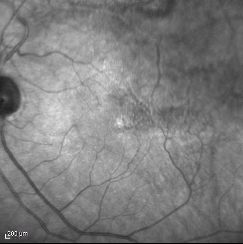
7 IR Photos OD OS
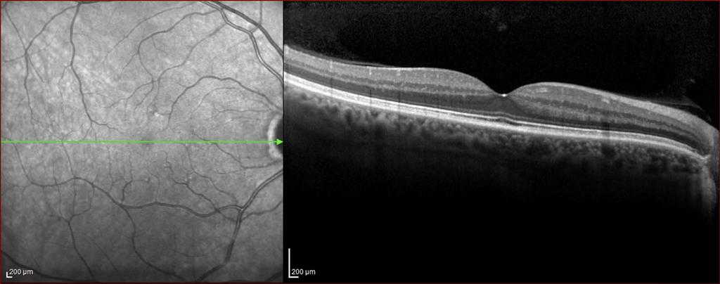
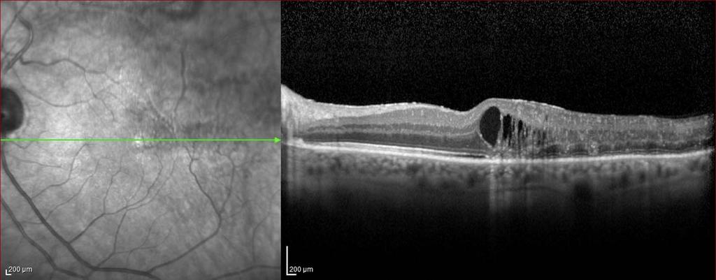
8 OCT OD OS
9 Fluorescein Angiogram Arterial phase 0:28:23 Proper filling of retinal arterioles
10 Fluorescein Angiogram Early venous phase 00:31:30 Delayed venous perfusion of superotemporal arcade

 0:40:98 Completed venous filling of superotemporal arcade")
11 Fluorescein Angiogram Venous phase Recirculation phase 0:38:10 Beginning venous filling of superotemporal arcade No significant macular ischemia (?ST FAZ) 0:40:98 Completed venous filling of superotemporal arcade with localized non-perfusion and collateralization

12 Fluorescein Angiogram Late phase 01:35:48 Widespread superotemporal non-perfusion with collateralization No macular ischemia
13 Summary 62 yo WM presents sent from optometrist for fundus appearance of BRVO 5-6 months after sudden blurred vision overnight OS. BCVA 20/30 initially, 20/25 15 days later. Represents a chronic superotemporal BRVO with retinal ischemia and vascular collaterlization OS.
14 Branch Retinal Vein Occlusion Turbulence in vein caused by atherosclerosis in crossing artery Thrombus formation at arteriovenous crossing point Varying severity of intraretinal hemorrhage, retinal edema, dilated and tortuous veins, +/- CW spots Most often superotemporal


15 Branch Retinal Vein Occlusion Photo Examples
16 Risk Factors Described by The Eye Disease Case-Control Study Group, 1993 Hypertension Cardiovascular disease Open angle glaucoma High body mass index NOT diabetes mellitus
17 2014 Almost 500,000 enrollees, 2283 with BRVOs Increased hazard of developing BRVO in pts with: HTN alone HTN and metabolic syndrome components DM, HLD DM with END-ORGAN DAMAGE Hx of CVA
18 Branch Retinal Vein Occlusion Grid & Scatter Laser Pars plana vitrectomy Intravitreal Steroids Intravitreal anti-vegf Treatment Options
19 Branch Vein Occlusion Study (BVOS) Started data 1977, published in 1984, 1986 Argon grid laser photocoagulation improved visual outcome significantly in eyes with BRVO with decreased VA to 20/40 20/200 due to macular edema not macular ischemia Scatter argon laser photocoagulation decreased risk of neovascularization from 22% to 12% in eyes with recent BRVO involving at least 5 DD of retinal non-perfusion Scatter laser also decreased risk of vitreous hemorrhage from 60% to 30% in recent BRVO eyes with neovascularization
20 Branch Vein Occlusion Study (BVOS) Vision Outcomes Eyes treated with macular laser more likely to be 20/40 or better at 3 years follow-up Mean VA improved 1.3 ETDRS lines (vs. 0.2 lines) Mean treated VA: 20/30 20/50 Mean untreated VA: 20/70
21 Branch Retinal Vein Occlusion Treatment justification for this patient: No macular ischemia with VA 20/25 Macular edema, mostly temporally with VA 20/25 No neovascularization Therefore observed at this point Any other treatment options recommended?
22 Branch Retinal Vein Occlusion Current Treatment Options Intravitreal steroids: SCORE study (2009) Intravitreal dexamethasone implant: OZURDEX GENEVA study Pars plana vitrectomy for non-resolving VH or for RD
23 Branch Retinal Vein Occlusion SCORE Study Standard Care Vs Corticosteroid for Retinal Vein Occlusion (2009) Intravitreal triamcinolone for treatment of macular edema in BRVO/CRVO BRVO arm: 1 mg vs 4 mg vs grid laser 26%, 27%, 29% respectively gained 3+ lines
24 Branch Retinal Vein Occlusion OZURDEX GENEVA Study (2010) Intravitreal dexamethasone implant for treatment of macular edema in BRVO/CRVO 0.7 mg vs 0.35 mg vs sham 26% vs 19% vs 17% respectively of 15+ letter gain
25 Branch Retinal Vein Occlusion BRAVO Study Branch Retinal Vein Occlusion Study (2010) Ranibizumab to eyes with macular edema 0.5 mg vs 0.3 mg vs sham 61.1%, 55.2%, and 28.8% respectively gained 15+ ETDRS letters at 6 months
26 Branch Retinal Vein Occlusion Prognosis Related to extent of capillary damage and retinal ischemia in macula Can try to assess with FA Integrity of parafoveal capillaries is the important prognostic factor for VA Macular edema, retinal hemorrhage, or perifoveal retinal capillary occlusion can reduce vision 50-60% of pts w/all types of BRVO maintain VA 20/40 or better after 1 year
27 2014 Retrospective chart review looked at what people are doing 885 BRVO pts treated with bevacizumab also looked at CRVO & DME pts Too few ranibizumab-treated pts for meaningful analysis 42% of BRVO pts received additional laser or IVTA therapy Patients treated with bevacizumab were monitored less frequently and received fewer injections than patients in major clinical trials of ranibizumab.
28 References 1. Argon laser scatter photocoagulation for prevention of neovascularization and vitreous hemorrhage in branch vein occlusion. A randomized clinical trial. Branch Vein Occlusion Study Group. Arch Ophthalmol. 1986(1); Campochiaro PA< Heier JS, Feiner L., et al; BRAVO Investigators. Ranibizumab for macular edema following branch retinal vein occlusion: six month primary end point results of a phase III study. Ophthalmology. 2010;117(6): Epub 2010 Apr Haller JA, Bandello F, Belfort R Jr, et al; OZURDEX GENEVA Study Group. Randomized, sham-controlled trial of dexamethasone intravitreal implant in patients with macular edema due to retinal vein occlusion. Ophthalmology. 2010; 117(6): Epub 2010 Apr Hirashima T, Chihara T, Bun T, Utsumi T, Hirose M, Oh H. Intravitreal bevacizumab alone or combined with macular laser photocoagulation for recurrent or persistent macular edema secondary to branch retinal vein occlusion. J Ophthalmol. 2014;2014: doi: /2014/ Epub 2014 Jul 7. PubMed PMID: ; PubMed Central PMCID: PMC Kiss S, Liu Y, Brown J, Holekamp NM, Almony A, Campbell J, Kowalski JW. Clinical utilization of anti-vascular endothelial growthfactor agents and patient monitoring in retinal vein occlusion and diabetic macular edema. Clin Ophthalmol Aug 26;8: doi: /OPTH.S ecollection PubMed PMID: ; PubMed Central PMCID: PMC Kolar P. Risk factors for central and branch retinal vein occlusion: a meta-analysis of published clinical data. J Ophthalmol. 2014;2014: doi: /2014/ Epub 2014 Jun 9. Review. PubMed PMID: ; PubMed Central PMCID: PMC Newman-Casey PA, Stem M, Talwar N, Musch DC, Besirli CG, Stein JD. Risk factors associated with developing branch retinal vein occlusion among enrollees in a United States managed care plan. Ophthalmology Oct;121(10): doi: /j.ophtha Epub 2014 Jun 20. PubMed PMID: Retina and Vitreous, BSCS pp Risk factors for branch retinal vein occlusion. The Eye Disease Case-Control Study Group. Am J Ophthalmol. 1993;116(3): Scott IU, Ip MS, Van Veldhuisen PC, et al; SCORE Study Research Group. A randomized trial comparing the efficacy and safety of intravitreal triamcinolone with standard care to treat vision loss associated with macular edema secondary to branch retinal vein occlusion: the Standard Care vs Corticosteroid for Retinal Vein Occlusion (SCORE) study report 6. Arch Ophthalmology. 2009;127(9):
29 THANK YOU
Vascular Disease Ocular Manifestations of Systemic Hypertension
 Vascular Disease Ocular Manifestations of Systemic Hypertension Maynard L. Pohl, OD, FAAO Pacific Cataract & Laser Institute 10500 NE 8 th Street, Suite 1650 Bellevue, WA 98004 USA 425-462-7664 Cerebrovascular
Clinical Case Presentation. Branch Retinal Vein Occlusion. Sarita M. Registered Nurse Whangarei Base Hospital
 Clinical Case Presentation on Branch Retinal Vein Occlusion Sarita M. Registered Nurse Whangarei Base Hospital Introduction Case Study Pathogenesis Clinical Features Investigations Treatment Follow-up
Macular edema (ME) is the most common
 is the most common") MANAGEMENT OF RETINAL VEIN OCCLUSIONS * Peter A. Campochiaro, MD ABSTRACT Macular edema (ME) is the most common cause of reduced vision in patients with retinal vein occlusions (RVOs). The primary cause
MANAGEMENT OF RETINAL VEIN OCCLUSIONS * Peter A. Campochiaro, MD ABSTRACT Macular edema (ME) is the most common cause of reduced vision in patients with retinal vein occlusions (RVOs). The primary cause
Diagnosis and treatment of diabetic retinopathy. Blake Cooper MD Ophthalmologist Vitreoretinal Surgeon Retina Associates Kansas City
 Diagnosis and treatment of diabetic retinopathy Blake Cooper MD Ophthalmologist Vitreoretinal Surgeon Retina Associates Kansas City Disclosures Consulted for Novo Nordisk 2017,2018. Will be discussing
Venous Occlusive Diseases
 Venous Occlusive Diseases Bruce R. Saran, MD Adjunct Assistant Clinical Professor of Medicine Scheie Eye Institute University of Pennsylvania School of Medicine Philadelphia, PA -a division of: RVO Demographics
Retinal Imaging Conference. Brooke LW Nesmith, M.D. University of Louisville Department of Ophthalmology and Visual Sciences 8/7/2014
 Retinal Imaging Conference Brooke LW Nesmith, M.D. University of Louisville Department of Ophthalmology and Visual Sciences 8/7/2014 Patient Presentation CC: Routine yearly diabetic eye exam HPI: 59yo
An Update on Branch Retinal Vein Occlusion Treatment Studies. Amiee Ho, O.D. Pacific University College of Optometry
 An Update on Branch Retinal Vein Occlusion Treatment Studies Amiee Ho, O.D. Pacific University College of Optometry Course Description This course focuses on current treatment options available for macular
OCCLUSIVE VASCULAR DISORDERS OF THE RETINA
 OCCLUSIVE VASCULAR DISORDERS OF THE RETINA Learning outcomes By the end of this lecture the students would be able to Classify occlusive vascular disorders (OVD) of the retina. Correlate the clinical features
Diabetic Retinopathy: Managing the Extremes. J. Michael Jumper, MD West Coast Retina
 Diabetic Retinopathy: Managing the Extremes J. Michael Jumper, MD West Coast Retina Case 1: EC 65 y.o. HM No vision complaints Meds: Glyburide Metformin Pioglitazone Va: 20/20 OU 20/20 Case 2: HS 68 y.o.
Retinal Vein Occlusion (RVO) Treatment pathway- Northeast England. Retinal Vein Occlusion (RVO) with Macular oedema (MO)
 Treatment pathway- Northeast England. Retinal Vein Occlusion (RVO) with Macular oedema (MO)") Retinal Vein Occlusion (RVO) Treatment pathway- Northeast England (Royal Victoria Infirmary, Sunderland Eye Infirmary, James Cook University Hospital, Darlington Memorial Hospital, University Hospital
Retinal Vein Occlusion (RVO) Treatment pathway- Northeast England (Royal Victoria Infirmary, Sunderland Eye Infirmary, James Cook University Hospital, Darlington Memorial Hospital, University Hospital
The Era of anti- - - VEGF Kirk L. Halvorson, OD
 The Era of anti- - - VEGF Kirk L. Halvorson, OD Introduction: Anti- - - Vascular Endothelial Growth Factor (Anti- - - VEGF) medication is a relatively a new line of medications used in treating a variety
The Diabetic Retinopathy Clinical Research Network. Management of DME in Eyes with PDR
 The Diabetic Retinopathy Clinical Research Network Management of DME in Eyes with PDR 1 What Has Been Learned? Diabetic Retinopathy Treatment Protocol F: Results suggest that clinically meaningful differences
Retina Conference. Janelle Fassbender, MD, PhD University of Louisville Department of Ophthalmology and Visual Sciences 09/04/2014
 Retina Conference Janelle Fassbender, MD, PhD University of Louisville Department of Ophthalmology and Visual Sciences 09/04/2014 Subjective CC/HPI: 64 year old Caucasian female referred by outside ophthalmologist
Posterior Segment Macular Edema
 Posterior Segment Macular Edema Treatment of Macular Edema following Branch Retinal Vein Occlusion Raafay Sophie, MD 1 and Peter A Campochiaro, MD 2 1. Post-doctoral Fellow; 2. Eccles Professor of Ophthalmology
EU Regulatory workshop Ophthalmology clinical development and scientific advice. Industry view on DME and macular edema secondary to RVO
 EU Regulatory workshop Ophthalmology clinical development and scientific advice. Industry view on DME and macular edema secondary to RVO Yehia Hashad, M.D. Vice President and Global Therapeutic Area Head
Clinically Significant Macular Edema (CSME)
") Clinically Significant Macular Edema (CSME) 1 Clinically Significant Macular Edema (CSME) Sadrina T. Shaw OMT I Student July 26, 2014 Advisor: Dr. Uwaydat Clinically Significant Macular Edema (CSME) 2
Clinically Significant Macular Edema (CSME) 1 Clinically Significant Macular Edema (CSME) Sadrina T. Shaw OMT I Student July 26, 2014 Advisor: Dr. Uwaydat Clinically Significant Macular Edema (CSME) 2
Marcus Gonzales, OD, FAAO Cedar Springs Eye Clinic
 Marcus Gonzales, OD, FAAO Cedar Springs Eye Clinic 25.6 million adults 11.3% of the adult population 10.9 million adults 65 years and older 26.9% of this age population 79 million people are Pre-diabetic!!
ZEISS AngioPlex OCT Angiography. Clinical Case Reports
 Clinical Case Reports Proliferative Diabetic Retinopathy (PDR) Case Report 969 PROLIFERATIVE DIABETIC RETINOPATHY 1 1-year-old diabetic female presents for follow-up of proliferative diabetic retinopathy
The Human Eye. Cornea Iris. Pupil. Lens. Retina
 The Retina Thin layer of light-sensitive tissue at the back of the eye (the film of the camera). Light rays are focused on the retina then transmitted to the brain. The macula is the very small area in
Dexamethasone Intravitreal Implant Rescue Treatment for Bevacizumab Refractory Macular Edema Secondary to Branch Retinal Vein Occlusion
 pissn: 1011-8942 eissn: 2092-9382 Korean J Ophthalmol 2017;31(2):108-114 https://doi.org/10.3341/kjo.2017.31.2.108 Original Article Dexamethasone Intravitreal Implant Rescue Treatment for Bevacizumab Refractory
EyePACS Grading System (Part 2): Detecting Presence and Severity of Background (Non-Proliferative) Diabetic Retinopathy Lesion
: Detecting Presence and Severity of Background (Non-Proliferative) Diabetic Retinopathy Lesion") EyePACS Grading System (Part 2): Detecting Presence and Severity of Background (Non-Proliferative) Diabetic Retinopathy Lesion George Bresnick MD MPA Jorge Cuadros OD PhD Anatomy of the eye: 3 Normal Retina
EyePACS Grading System (Part 2): Detecting Presence and Severity of Background (Non-Proliferative) Diabetic Retinopathy Lesion George Bresnick MD MPA Jorge Cuadros OD PhD Anatomy of the eye: 3 Normal Retina
Diabetic Retinopathy A Presentation for the Public
 Diabetic Retinopathy A Presentation for the Public Ray M. Balyeat, MD The Eye Institute Tulsa, Oklahoma The Healthy Eye Light rays enter the eye through the cornea, pupil and lens. These light rays are
 measure of your overall performance. An isolated glucose test is helpful to let you know what your sugar level is at one moment, but it doesn t tell you whether or not your diabetes is under adequate control
Retinal Vein Occlusion
 Retinal Update 2018 Retinal Vein Occlusion Case Presentations to Myself Branch Vein Occlusion What medical evaluation do you recommend for this 72 year old patient? Is there anything you ask of your medical
Facts About Diabetic Eye Disease
 Facts About Diabetic Eye Disease Points to Remember 1. Diabetic eye disease comprises a group of eye conditions that affect people with diabetes. These conditions include diabetic retinopathy, diabetic
A Patient s Guide to Diabetic Retinopathy
 Diabetic Retinopathy A Patient s Guide to Diabetic Retinopathy 840 Walnut Street, Philadelphia PA 19107 www.willseye.org Diabetic Retinopathy 1. Definition Diabetic retinopathy is a complication of diabetes
Eyes on Diabetics: How to Avoid Blindness in Diabetic Patient
 Eyes on Diabetics: How to Avoid Blindness in Diabetic Patient Rova Virgana FK Unpad Pusat Mata Nasional RS Mata Cicendo Bandung Eye Center (Hospital and Clinic) PIT IDI Jabar 2018 Keys Facts from WHO
Diabetic Retinopathy
 Diabetic Retinopathy Diabetes can be classified into type 1 diabetes mellitus and type 2 diabetes mellitus, formerly known as insulin-dependent diabetes mellitus, and non-insulin diabetes mellitus, respectively.
Applying New Data to Improve the Standard of Care in Retinal Diseases Managing Macular Edema Associated With Retinal Venous Occlusions
 Supplement to November/December 2013 CME Activity Applying New Data to Improve the Standard of Care in Retinal Diseases Managing Macular Edema Associated With Retinal Venous Occlusions By Michael Singer,
The Foundation WHAT IS THE RETINA?
 The Foundation American Society of Retina Specialists Committed to improving the quality of life of all people with retinal disease. Branch Retinal Vein Occlusion Retinal vein occlusions occur when there
Brampton Hurontario Street Brampton, ON L6Y 0P6
 Diabetic Retinopathy What is Diabetic Retinopathy Diabetic retinopathy is one of the leading causes of blindness world-wide. Diabetes damages blood vessels in many organs of the body including the eyes.
RETINAL VEIN OCCLUSIONS (RVO) PREFERRED PRACTICE PATTERNS (PPP) Philippines: 2016
 PREFERRED PRACTICE PATTERNS (PPP) Philippines: 2016") RETINAL VEIN OCCLUSIONS (RVO) PREFERRED PRACTICE PATTERNS (PPP) Philippines: 2016 The Retinal Vein Occlusions (RVO) Preferred Practice Patterns (PPP) Philippines: 2016 was prepared by the VitreoRetina
RETINAL VEIN OCCLUSIONS (RVO) PREFERRED PRACTICE PATTERNS (PPP) Philippines: 2016 The Retinal Vein Occlusions (RVO) Preferred Practice Patterns (PPP) Philippines: 2016 was prepared by the VitreoRetina
Research Article Differentiation between Good and Low-Responders to Intravitreal Ranibizumab for Macular Edema Secondary to Retinal Vein Occlusion
 Hindawi Publishing Corporation Journal of Ophthalmology Volume 2016, Article ID 9875741, 6 pages http://dx.doi.org/10.1155/2016/9875741 Research Article Differentiation between Good and Low-Responders
Diabetic Retinopatathy
 Diabetic Retinopatathy Jay M. Haynie, OD, FAAO Financial Disclosure I have received honoraria or am on the advisory board for the following companies: Carl Zeiss Meditec Arctic DX Macula Risk Advanced
OCT Angiography The Next Frontier
 Choroid Retina avascular 5/13/2017 OCT Angiography The Next Frontier Pierce Kenworthy OD, FAAO June 9, 2017 OCT Angiography (OCTA) 2016 Non-invasive, motion contrast imaging Represents erythrocyte movement
Past ocular history. DME Case 1. Patient presents blurred VA. Hemoglobin A1c 11.5% -- patient states sugars have not been in good control
 Past ocular history Patient presents blurred VA DME Case 1 Hemoglobin A1c 11.5% -- patient states sugars have not been in good control PDR with macular edema OU Rishi Singh MD Cleveland Clinic OD OS 1
Goals/Objectives. Disclosures. Risk Factors RAO and RVO. Risk Factors. Retinal Artery Occlusions Branch and Central
 Jeffrey D. Perotti, OD, MS Indiana University School of Optometry Goals/Objectives RETINAL VASCULAR OCCLUSIONS FOR THE PRIMARY CARE CLINICIAN Using cases as a framework, review current evaluation and management
What you can expect with OZURDEX
 Important Information About Macular Edema Following Branch or Central Retinal Vein Occlusion (RVO) and Treatment For patients with RVO What you can expect with OZURDEX Approved Use OZURDEX (dexamethasone
Comparison of Ranibizumab and Bevacizumab for Macular Edema Associated with Branch Retinal Vein Occlusion
 pissn: 1011-8942 eissn: 2092-9382 Korean J Ophthalmol 2017;31(3):209-216 https://doi.org/10.3341/kjo.2015.0158 Original Article Comparison of Ranibizumab and Bevacizumab for Macular Edema Associated with
4/27/2010 INTRODUCTION TO RETINAL VASCULAR DISEASE VENOUS/VENULAR CENTRAL RETINAL VEIN OBSTRUCTION / CRVO ADDITIONAL FEATURES /COMPLICATIONS
 INTRODUCTION TO RETINAL VASCULAR DISEASE VENOUS/VENULAR Leo Semes, OD Professor, UAB Optometry 2 CENTRAL RETINAL VEIN OBSTRUCTION CENTRAL RETINAL VEIN OBSTRUCTION / OCCLUSION (CRVO) obstruction of the
Chris Brown, M.D. Eye Specialty Group, PLC Continuing Education Series
 Chris Brown, M.D. Eye Specialty Group, PLC 2018 Continuing Education Series Disclaimer I have no financial interests in this lecture or any information discussed therein Objectives Fluorescein Angiogram
Scott M. Pfahler D.O. Dayton Vitreo-Retinal Associates AOCOO-HNS Palm Springs, CA 2012
 Scott M. Pfahler D.O. Dayton Vitreo-Retinal Associates AOCOO-HNS Palm Springs, CA 2012 Proliferative Diabetic Retinopathy Laser Treatments Medical Treatment Surgical Treatment Diabetic Macular Edema Laser
Diabetes & Your Eyes
 Diabetes & Your Eyes Diabetes is a disease that occurs when the pancreas does not secrete enough insulin or the body is unable to process it properly. Insulin is the hormone that regulates the level of
Applying New Data to Improve the Standard of Care in Retinal Disease. With articles by Gaurav K. Shah, MD Carl D. Regillo, MD.
 Supplement to July/August 2012 CME Activity Applying New Data to Improve the Standard of Care in Retinal Disease With articles by Gaurav K. Shah, MD Carl D. Regillo, MD Release date: August 2012. Expiration
Diabetic Retinopathy
 Diabetic Retinopathy Diabetes mellitus is one of the leading causes of irreversible blindness worldwide. In the United States, it is the most common cause of blindness in people younger than 65 years.
RVO RETINAL VEIN OCCLUSION
 RVO RETINAL VEIN OCCLUSION A guide to understanding RVO Take some time to learn about RVO - it may help you hold on to your vision Retinal vein occlusion is a common disorder of the retina and a leading
Jay M. Haynie, O.D.; F.A.A.O. Olympia Tacoma Renton Kennewick Washington
 Jay M. Haynie, O.D.; F.A.A.O. Olympia Tacoma Renton Kennewick Washington I Jay M. Haynie, OD, FAAO have received honoraria from the following companies: Reichert Technologies Notal Vision Carl Zeiss Meditec
PART 1: GENERAL RETINAL ANATOMY
 PART 1: GENERAL RETINAL ANATOMY General Anatomy At Ora Serrata At Optic Nerve Head Fundoscopic View Of Normal Retina What Is So Special About Diabetic Retinopathy? The WHO definition of blindness is
Anti VEGF Agents in Retinal Disorders Current Scenario
 Retina Anti VEGF Agents in Retinal Disorders Current Scenario Charu Gupta MS Charu Gupta MS, Cyrus M. Shroff MD Shroff Eye Centre, New Delhi T is a group of proteins involved in the regulation of angiogenesis,
THE ROLE OF anti-vegf IN DIABETIC RETINOPATHY AND AGE RELATED MACULAR DEGENERATION
 THE ROLE OF anti-vegf IN DIABETIC RETINOPATHY AND AGE RELATED MACULAR DEGENERATION MOESTIDJAB DEPARTMENT OF OPHTHALMOLOGY SCHOOL OF MEDICINE AIRLANGGA UNIVERSITY DR SOETOMO HOSPITAL SURABAYA INTRODUCTION
 Research Article http://www.alliedacademies.org/clinical-ophthalmology-and-vision-science/ A 2-year retrospective study of the treatment of retinal vein occlusion with dexamethasone 0.7 mg intravitreal
New Developments in the treatment of Diabetic Retinopathy
 New Developments in the treatment of Diabetic Retinopathy B. Jeroen Klevering University Medical Centre Nijmegen - The Netherlands Topics Management of diabetic retinopathy Interventions a. primary (prevention)
Efficacy and safety of Pro Re Nata regimen without loading dose ranibizumab injections in retinal vein occlusion
 Open Access Original Article Efficacy and safety of Pro Re Nata regimen without loading dose ranibizumab injections in retinal vein occlusion Erkan Unsal 1, Kadir Eltutar 2, Pınar Sultan 3, Hulya Gungel
Moncef Khairallah, MD
 Moncef Khairallah, MD Department of Ophthalmology, Fattouma Bourguiba University Hospital Faculty of Medicine, University of Monastir Monastir, Tunisia INTRODUCTION IU: anatomic form of uveitis involving
Grand Rounds. Eddie Apenbrinck M.D. University of Louisville School of Medicine Department of Ophthalmology & Visual Sciences 6/20/2014
 Grand Rounds Eddie Apenbrinck M.D. University of Louisville School of Medicine Department of Ophthalmology & Visual Sciences 6/20/2014 Subjective CC: sudden painless loss of vision OD HPI: 75 year old
Long term visual outcome after arteriolar constriction in patients with branch retinal vein occlusion
 Long term visual outcome after arteriolar constriction in patients with branch retinal vein occlusion Jiri Rehak a, Ladislav Dusek b, Martin Sin a, Barbora Babkova a, Zuzana Pracharova a, Matus Rehak a,c
1/25/2018. Case Management Strategies in Diabetic Retinopathy. Case Study #1: Severe DME. DDOS: 3/31/2016 Va 20/400. Disclosures
 Case Management Strategies in Diabetic Retinopathy Disclosures No financial conflict of interest Will discuss off label use of intraocular Bevacizumab (Avastin) for Diabetic Retinopathy Sundeep Dev, MD
Treatment of Retinal Vein Occlusion (RVO)
") Manchester Royal Eye Hospital Medical Retina Services Information for Patients Treatment of Retinal Vein Occlusion (RVO) What is a Retinal Vein Occlusion (RVO)? The retina is the light sensitive layer
Manchester Royal Eye Hospital Medical Retina Services Information for Patients Treatment of Retinal Vein Occlusion (RVO) What is a Retinal Vein Occlusion (RVO)? The retina is the light sensitive layer
Corporate Medical Policy
 Corporate Medical Policy Intravitreal Implant File Name: Origination: Last CAP Review: Next CAP Review: Last Review: intravitreal_implant 11/2010 6/2017 6/2018 6/2017 Description of Procedure or Service
Preventing Avoidable Vision loss from Diabetic Retinopathy in Indian Country
 Diabetes in Indian Country- 2017 Preventing Avoidable Vision loss from Diabetic Retinopathy in Indian Country Albuquerque, NM 20 September2017 Mark B. Horton, OD, MD Director, IHS/JVN Teleophthalmology
Yoshiro Minami 1*, Taiji Nagaoka 2, Akihiro Ishibazawa 1,2 and Akitoshi Yoshida 2
 Minami et al. BMC Ophthalmology (2017) 17:90 DOI 10.1186/s12886-017-0485-4 RESEARCH ARTICLE Open Access Correlation between short- and long-term effects of intravitreal ranibizumab therapy on macular edema
Recalcitrant Diabetic Macular Oedema: Therapeutic Options
 December 2007 A. Giridhar et al. - Recalcitrant DME 451 CONSULTATION S E C T I O N Recalcitrant Diabetic Macular Oedema: Therapeutic Options Dr. Cyrus M Shroff 1, Dr. N S Muralidhar 2, Dr. R Narayanan
Comparison of BRVO and CRVO management
 Comparison of BRVO and CRVO management Francesco Bandello, MD, FEBO Department of Ophthalmology University Vita-Salute Scientific Institute San Raffaele Milan, Italy 1 Financial Disclosure Advisory Board
NATIONAL INSTITUTE FOR HEALTH AND CARE EXCELLENCE. Health Technology Appraisal. Aflibercept for treating diabetic macular oedema.
 NATIONAL INSTITUTE FOR HEALTH AND CARE EXCELLENCE Health Technology Appraisal Aflibercept for treating diabetic macular oedema Final scope Final remit/appraisal objective To appraise the clinical and cost
Acknowledgements. Outline. Who were von Hippel and Lindau? Eugen von Hippel German Ophthalmologist
 Ophthalmic Therapies & Standard of Care Acknowledgements Eric Jonasch, MD & Surena Matin, MD Collaborators Franco DeMonte, MD Marcy Johnson Ian McCutcheon, MD Chaan Ng, MD Nancy Perrier, MD Dawid Schellingerhout,
Bevacizumab for Macular Edema in Central Retinal Vein Occlusion: A Prospective, Randomized, Double-Masked Clinical Study
 Bevacizumab for Macular Edema in Central Retinal Vein Occlusion: A Prospective, Randomized, Double-Masked Clinical Study David L.J. Epstein, MD, Peep V. Algvere, MD, PhD, Gunvor von Wendt, MD, PhD, Stefan
Optimal Treatment of Retinal Vein Occlusion: Canadian Expert Consensus
 Original Paper Received: December 9, 2014 Accepted: February 25, 2015 Published online: June 12, 2015 Optimal Treatment of Retinal Vein Occlusion: Canadian Expert Consensus Alan R. Berger a Alan F. Cruess
Clinical Trials in Diabetic Retinopathy. Harry W. Flynn Jr., M.D. Nidhi Relhan Batra, M.D.
 1 Clinical Trials in Diabetic Retinopathy 2018 Harry W. Flynn Jr., M.D. Nidhi Relhan Batra, M.D. Bascom Palmer Eye Institute 900 N.W. 17th Street Miami, FL 33136 Phone: (305) 326-6118 Fax: (305) 326-6417
Efficacy of combined intravitreal bevacizumab and triamcinolone for branch retinal vein occlusion
 AOP*** 1 Original Article Efficacy of combined intravitreal bevacizumab and triamcinolone for branch retinal vein occlusion Rasha I Ali1, Kapil G Kapoor 1,2, Adeel N Khan 1, Syed K Gibran 1 Purpose: To
From Outdated to Updated: A Review of Important Clinical Trials in Ocular Disease from 2014
 From Outdated to Updated: A Review of Important Clinical Trials in Ocular Disease from 2014 1. This course is designed to review the important ophthalmic literature that was released between October 2013
Diabetic Retinopathy
 Diabetic Retinopathy Introduction People with diabetes are more likely to have eye problems that can lead to blindness. Diabetic retinopathy is a disease of the eye s retina that is caused by diabetes.
Case Report Inherent Challenges in Managing Long Standing Refractory Diabetic Macular Edema
 Cronicon OPEN ACCESS EC OPHTHALMOLOGY Case Report Inherent Challenges in Managing Long Standing Refractory Diabetic Macular Edema V Swetha E Jeganathan 1,2 * and Karen Madill 3 1 Department of Ophthalmology,
Serious Eye diseases, New treatments. Mr. M. Usman Saeed MBBS, FRCS, FRCOphth Consultant Ophthalmologist
 Serious Eye diseases, New treatments Mr. M. Usman Saeed MBBS, FRCS, FRCOphth Consultant Ophthalmologist 5 major causes of loss of vision Cataracts Glaucoma Macular degeneration Retinal Vein occlusions
Neovascular Glaucoma Associated with Cilioretinal Artery Occlusion Combined with Perfused Central Retinal Vein Occlusion
 Neovascular Glaucoma Associated with Cilioretinal Artery Occlusion Combined with Perfused Central Retinal Vein Occlusion Man-Seong Seo,* Jae-Moon Woo* and Jeong-Jin Seo *Department of Ophthalmology, Chonnam
FRANZCO, MD, MBBS. Royal Darwin Hospital
 Diabetes and Eye By Dr. Nishantha Wijesinghe FRANZCO, MD, MBBS Consultant Ophthalmologist Royal Darwin Hospital 98% of Diabetics do not need to suffer from severe visual loss Yet Diabetic eye disease is
Paradigm Shift in the treatment of Diabetic Retinopathy. Haytham I. S. Salti, MD Associate Professor
 Paradigm Shift in the treatment of Diabetic Retinopathy Haytham I. S. Salti, MD Associate Professor Disclosure No financial interests related to the subject matter of this talk This presentation includes
Themes for conferences No 42
 Themes for conferences No 42 Systemic Arterial Hypertension Venous Occlusive Disease Arterial Occlusive Disease Ocular Ischemic Syndrome Med. pract. Anton R. Xavier Dr. med. Claudia Zawinka Dr. med. Stephan
Diabetic Retinopathy: Recent Advances in Treatment and Treatment Approaches
 Diabetic Retinopathy: Recent Advances in Treatment and Treatment Approaches Dr. David Wong Associate Professor Retina Specialist, Department of Ophthalmology & Vision Sciences, University of Toronto, Canada
X-Plain Diabetic Retinopathy Reference Summary
 X-Plain Diabetic Retinopathy Reference Summary Introduction Patients with diabetes are more likely to have eye problems that can lead to blindness. Diabetic retinopathy is a disease of the eye s retina
International Journal of Health Sciences and Research ISSN:
 International Journal of Health Sciences and Research www.ijhsr.org ISSN: 2249-9571 Original Research Article A Multivariate Analysis of Intravitreal Injection of Anti-VEGF Bevacizumab in the Treatment
Measures have been taken, by the Utah Department of Health, Bureau of Health Promotions, to ensure no conflict of interest in this activity.
 Measures have been taken, by the Utah Department of Health, Bureau of Health Promotions, to ensure no conflict of interest in this activity. CNE/CPE/CEU s are available for this live webinar. You must
Diabetic maculopathy 11/ An update on. Miss Vasuki Sivagnanavel
 Miss Vasuki Sivagnanavel Consultant Ophthalmologist An update on Diabetic maculopathy Despite advances in the management of diabetes, diabetic retinopathy is already the commonest cause of blindness among
BMJ Open. For peer review only -
 Comparative efficacy and safety of treatments for macular oedema secondary to branch retinal vein occlusion: a network meta-analysis Journal: Manuscript ID: bmjopen-0-00 Article Type: Research Date Submitted
Diabetic retinopathy damage to the blood vessels in the retina. Cataract clouding of the eye s lens. Cataracts develop at an earlier age in people
 Diabetic Retinopathy What is diabetic eye disease? Diabetic eye disease refers to a group of eye problems that people with diabetes may face as a complication of diabetes. All can cause severe vision loss
Optometry Student Extern Manual. Miami VA Medical Center
 Optometry Student Extern Manual Miami VA Medical Center Table of contents: Rotation Description 3-4 Clinic Schedules 5 When to check in with attending 5 Absences 5 Performance Standards 6 Examination Template
A retrospective nonrandomized study was conducted at 3
 Department of Ophthalmology, Kangbuk Samsung Hospital, Sungkyunkwan University College of Medicine 1, Seoul, Korea Hangil Eye Hospital 2, Incheon, Korea Seoul National University Bundang Hospital 3, Seongnam,
ROLE OF LASER PHOTOCOAGULATION VERSUS INTRAVITREAL TRIAMCINOLONE ACETONIDE IN ANGIOGRAPHIC MACULAR EDEMA IN DIABETES MELLITUS
 ORIGINAL ARTICLE ROLE OF LASER PHOTOCOAGULATION VERSUS INTRAVITREAL TRIAMCINOLONE ACETONIDE IN ANGIOGRAPHIC MACULAR EDEMA IN DIABETES MELLITUS Aggarwal Somesh VP 1, Shah Sonali N 2, Bharwada Rekha M 3,
Retinal vein occlusion (RVO) is a vascular disease
 is a vascular disease") INTRAVITREAL RANIBIZUMAB FOR RETINAL VEIN OCCLUSION THROUGH 1 YEAR IN CLINICAL PRACTICE TROELS BRYNSKOV, MD,* HENRIK KEMP, MD,* TORBEN L. SØRENSEN, MD, DMSC* Purpose: To evaluate the efficacy and safety
INTRAVITREAL RANIBIZUMAB FOR RETINAL VEIN OCCLUSION THROUGH 1 YEAR IN CLINICAL PRACTICE TROELS BRYNSKOV, MD,* HENRIK KEMP, MD,* TORBEN L. SØRENSEN, MD, DMSC* Purpose: To evaluate the efficacy and safety
A Systematic Approach to Diabetic Photo Reading
 A Systematic Approach to Diabetic Photo Reading Jacqueline Theis, OD, FAAO Please silence all mobile devices and remove items from chairs so others can sit. Unauthorized recording of this session is prohibited.
Diabetic Retinopathy. Barry Emara MD FRCS(C) Giovanni Caboto Club October 3, 2012
 Giovanni Caboto Club October 3, 2012") Diabetic Retinopathy Barry Emara MD FRCS(C) Giovanni Caboto Club October 3, 2012 Outline Statistics Anatomy Categories Assessment Management Risk factors What do you need to do? Objectives Summarize the
Diabetic Retinopathy Barry Emara MD FRCS(C) Giovanni Caboto Club October 3, 2012 Outline Statistics Anatomy Categories Assessment Management Risk factors What do you need to do? Objectives Summarize the
Recurrent intraocular hemorrhage secondary to cataract wound neovascularization (Swan Syndrome)
") Recurrent intraocular hemorrhage secondary to cataract wound neovascularization (Swan Syndrome) John J. Chen MD, PhD; Young H. Kwon MD, PhD August 6, 2012 Chief complaint: Recurrent vitreous hemorrhage,
Recurrent intraocular hemorrhage secondary to cataract wound neovascularization (Swan Syndrome) John J. Chen MD, PhD; Young H. Kwon MD, PhD August 6, 2012 Chief complaint: Recurrent vitreous hemorrhage,
Off-label use of intravitreal bevacizumab in non-ischemic macular edema secondary to retinal vein obstructions
 Romanian Journal of Ophthalmology, Volume 60, Issue 2, April-June 2016. pp:90-95 GENERAL ARTICLE Off-label use of intravitreal bevacizumab in non-ischemic macular edema secondary to retinal vein obstructions
Sequential pharmacological therapies in the management of macular oedema secondary to retinal vein occlusion
 Northern (NHS) Treatment Advisory Group Sequential pharmacological therapies in the management of macular oedema secondary to retinal vein occlusion Author: Paul Madill Specialty Registrar in Public Health
ADULT-ONSET FOVEOMACULAR VITELLIFORM DYSTROPHY. By: Chris Munnerlyn, OMT Student University of Arkansas for Medical Sciences
 ADULT-ONSET FOVEOMACULAR VITELLIFORM DYSTROPHY By: Chris Munnerlyn, OMT Student University of Arkansas for Medical Sciences ADULT-ONSET FOVEOMACULAR VITELLIFORM DYSTROPHY (AOFVD) AOFVD is a condition that
DIABETIC RETINOPATHY
 DIABETIC RETINOPATHY C. L. B. Canny, MD FRCSC Diabetic retinopathy is the most serious eye manifestation of diabetes and is responsible for most of the blindness caused by diabetes. Diabetic retinopathy
aflibercept 40mg/mL solution for injection (Eylea ) SMC No. (1074/15) Bayer
 SMC No. (1074/15) Bayer") aflibercept 40mg/mL solution for injection (Eylea ) SMC No. (1074/15) Bayer 07 August 2015 The Scottish Medicines Consortium (SMC) has completed its assessment of the above product and advises NHS Boards
aflibercept 40mg/mL solution for injection (Eylea ) SMC No. (1074/15) Bayer 07 August 2015 The Scottish Medicines Consortium (SMC) has completed its assessment of the above product and advises NHS Boards
Front Line Diabetic Retinopathy What Not to Miss and Why
 Front Line Diabetic Retinopathy What Not to Miss and Why David M Brown MD FACS Clinical Professor of Ophthalmology Blanton Eye Institute, Houston Methodist Hospital Baylor College of Medicine Retina Consultants
EFFICACY OF INTRAVITREAL TRIAMCINOLONE ACETONIDE FOR THE TREATMENT OF DIABETIC MACULAR EDEMA
 Basrah Journal Of Surgery EFFICACY OF INTRAVITREAL TRIAMCINOLONE ACETONIDE FOR THE TREATMENT OF DIABETIC MACULAR EDEMA Salah Zuhair Abed Al-Asadi MB,ChB, FICMS, Lecturer, Department of Surgery, College
Diabetic Retinopathy WHAT IS DIABETIC RETINOPATHY? WHAT CAUSES DIABETIC RETINOPATHY? WHAT ARE THE STAGES OF DIABETIC RETINOPATHY?
 Diabetic Retinopathy WHAT IS DIABETIC RETINOPATHY? Diabetic retinopathy affects 8 million Americans with diabetes. A leading cause of blindness in American adults, it is caused by damage to the small blood
Supplement to March Ranibizumab: Expanding Horizons in Retinal Vein Occlusion Management. Sponsored by Novartis Pharma AG
 Supplement to March 2015 Ranibizumab: Expanding Horizons in Retinal Vein Occlusion Management Sponsored by Novartis Pharma AG Ranibizumab: Expanding Horizons in Retinal Vein Occlusion Management This supplement