ophthalmoscopy seeing the eye from greek: ophthalmos - the eye skopeos to see
|
|
|
- Mae Jefferson
- 5 years ago
- Views:
Transcription
1 ophthalmoscopy seeing the eye from greek: ophthalmos - the eye skopeos to see
2 structure principles of fundus examination direct ophthalmoscopy indirect ophthalmoscopy magnification in ophthalmoscopy field of view in ophthalmoscopy

3 classic article


4
5 principles of ophthalmoscopy in the emmetropic patients, light rays emanating from a point in the fundus emerge as a parallel beam if this beam enters the pupil of the observer, the rays are focussed on his retina and an image is formed


6 principles of ophthalmoscopy so why do we generally not see the fundus when looking directly into a patient s pupil?


7 principles of ophthalmoscopy problem: fundus of patient not illuminated light source required pupillary axis of observer must be aligned with incoming light rays from light source to allow observation of the fundus limiting factor patient s pupil size


8 principles of ophthalmoscopy in large animals or patients with extremely dilated pupils, it may be possible to observe the fundus by simply aligning a bright light source with your visual axis
9 principles of ophthalmoscopy direct ophthalmoscopy indirect ophthalmoscopy key-hole view aerial image

10 direct ophthalmoscope first developed by Charles Babbage 1847 failed to prove its function when showing to eminent ophthalmic surgeon and abandoned project

11 direct ophthalmoscope clinical use introduced by Hermann von Helmholtz 1851 initially called Augenspiegel (eye mirror) within 10 years physicians using it call themselves ophthalmoscopists first fundus photograph

12 direct ophthalmoscope Helmholtz principles of direct ophthalmoscopy a source of illumination a method of reflecting the light into the eye an optical means of correcting an unsharp image of the fundus greatest improvements made were re. source of illumination candle oil burning wick development of incandescent bulbs halogen.led light source
13 direct ophthalmoscopy
14 direct ophthalmoscopy advantages real, upright image high magnification does not require full dilation cheap and mobile disadvantages closeness to patient required small field of view no stereoscopy poor penetration of cloudy media
15 direct ophthalmoscope head observer s view hole fast diopter switch diopter dial diopter indicator
16 direct ophthalmoscope head observers viewing aperture shutter light reflecting mirror slit beam setting various sizes round beam red free light

17 direct ophthalmoscopy light filters green filter (red free light) blood (vessels) appear black grid pattern facilitates lesion documentation slit beam evaluate surface topography of retina and optic nerve

18 Direct ophthalmoscope head Observers viewing aperture Shutter Light reflecting mirror Slit beam setting Various sizes round beam Red free light
19 Direct ophthalmoscope on a slit

20 Direct ophthalmoscope on a slit

21 direct ophthalmoscope if patient and observer are emmetropic, no lenses required to focus on retina why are lenses used? lenses can be used to correct known refractive error of observer
22 direct ophthalmoscope if observer and patient are emmetropic, lenses can be used to move the point of focus onto structures posterior to the retina or structures more anterior within the eye

23 direct ophthalmoscope negative lenses can be used to focus onto structures posterior to the retina colobomata, areas of scleral ectasia, cupped disc


24 direct ophthalmoscope positive lenses can be used to focus onto structures more anterior within the eye lens, iris, cornea, adnexa courtesy J Mould


25 Mirror
26 12 dioptre lens Mirror
27 15 dioptre lens Mirror


28 20 dioptre lens Mirror
29 direct ophthalmoscope diopter equivalent distance (in mm) the focal moves anterior/posterior within the eye per diopter change
30
31 0 D = focus on retina focus on ONH on + 6 D


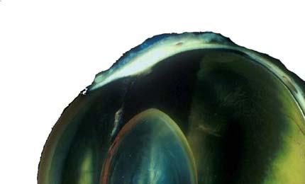
32 Diopter equivalent - clinical


33 Diopter equivalent - clinical
34 direct ophthalmoscopy how to do it align ophthalmoscope with your visual axis dim light beam to minimal intensity gain control over patient s head with your free hand pick up fundus reflex at arms length (compare fundus reflex of both eyes) following the fundus reflex, come close to the eye until the fundus becomes visible search fundus in reproducible manner
35
36

37
38

39 Tora

40


41


42 Fig. 1.12a-c Milk bottles

43
44

45


46


47 direct ophthalmoscopy apply a replicable, systematic approach to the assessment of the fundus keep landmarks in mind consider that varying species may require a varying routine!
48


49 indirect ophthalmoscopy
50
51 indirect ophthalmoscopy
52
53 indirect ophthalmoscopy dictionary.thefreedictionary.com/_/viewer.aspx?path=elmill&name=f0o-01- S2958.jpg

54


55 light options

56 all pupil setting

57

58 indirect ophthalmoscopy advantages larger field of view stereoscopic (with head mounted) penetrates opaque media better disadvantages limited magnification inverted & upside down image
59 celebrity eyes
60 condensing lenses acrylic or glass glass superior optics biconvex coated to reduce reflections variety of strenghts most commonly used 2.2 pan-retinal 20 D 30D


61 high power condensing lenses
62 image direct vs indirect
63 magnification and field of view magnification process of enlarging something only in appearance but not in physical size field of view (angular or linear or areal) extent of the observable world that is seen at any given moment
64 magnification and field of view of the fundic image depend on two main factors optical properties of the eye to be examined small eyes = high dioptric power large eyes = less dioptric power form of ophthalmoscopic technique employed direct ophthalmoscopy = high magnification, small field of view indirect ophthalmoscopy = low magnification, larger field of view


65 magnification optical properties of the eye to be examined small eyes = high dioptric power high magnification large eyes = less dioptric power low magnification
66 optical properties of the eye to be examined small eyes = high dioptric power high magnification large eyes = less dioptric power low magnification
67 ophthalmoscopy - magnification lateral magnification magnification of an area axial magnification magnification in depth

68 lateral magnification magnification across an axis perpendicular towards viewing e.g. left-right, dorsalventral with regards to fundus exemplified by action of slide projector throwing a much magnified image onto a screen

69 lateral magnification direct ophthalmoscopy the magnification M of a direct ophthalmoscope is equal to M = F e /4 where F e is the total refractive power of the eye. e.g. horse 8 dog 17 cat 20 Rat 77
70
71 lat magnification indirect ophthalmoscopy dependent on lens power and eye to be examined lat magnification of a lens = total refractive power of eye/power of lens
72 lateral magnification - indirect ophthalmoscopy power of lens inversely related to magnification high power = low magnification
73
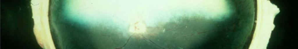
74 celebrity eyes
75 axial magnification describes magnification in depth precisely, the ratio of the distance along the optical axis between two points in image space (A B ) to the distance along the optical axis between the corresponding two points in object space (AB) this magnification is useful when considering an image in its three dimensions
76 axial magnification magnification along the axis of viewing e.g. elevation of optic nerve head, colobomata
77 axial magnification axial magnification is approximately the lateral magnification squared (axial) = (lateral) 2 e.g. lateral magnification = 2 means axial magnification = 4
78
79 indirect ophthalmoscopy in any given species, the dioptric power of lens used determines magnification field of view
80

81 axial magnification in diff. species
82

83 axial magnification in diff species


84 field of view - direct ophthalmoscopy field of view in direct ophthalmoscopy is determined by pupil size of examiner patient size of light beam
85
86 indirect ophthalmoscopy in any given species, the dioptric power of lens used determines magnification field of view
87


88 image direct vs indirect direct: 2.5 mm 20D lens: 13 mm
89 indirect ophthalmoscopy high diopter large field of view, (low magnification) low diopter small field of view (high magnification)
 virtual upright image greater working")
90 hand held PanOptic cross between direct and indirect ophthalmoscope increased field of view 25 degree vs. 5 degree increased magnification (by 26% compared to direct ophthalmoscope) virtual upright image greater working distance monocular view


91 hand held PanOptic


92


93
Year 1 MBChB Clinical Skills Session Ophthalmoscopy
 Year 1 MBChB Clinical Skills Session Ophthalmoscopy Reviewed & ratified by: Dr V Taylor-Jones, Mr M Batterbury Consultant Ophthalmologist Learning objectives o To understand the anatomy and physiology
FACING YOUR FUNDIC FEARS: EXAMINATION OF THE OCULAR FUNDUS J. Seth Eaton, VMD, DACVO Cornell University Veterinary Specialists
 FACING YOUR FUNDIC FEARS: EXAMINATION OF THE OCULAR FUNDUS J. Seth Eaton, VMD, DACVO Cornell University Veterinary Specialists The goal of a thorough fundus examination is to clinically evaluate the structures
Advanced Examination of the Retina: Scleral Indentation & Retinal 3-Mirror
 Advanced Examination of the Retina: Scleral Indentation & Retinal 3-Mirror Meredith Whiteside, OD, FAAO Nimesh Patel, OD, FAAO John Shan, OD, FAAO Please silence all mobile devices. Unauthorized recording
Direct Ophthalmoscopes Introduction
 14 Direct Ophthalmoscopes Introduction Direct Ophthalmoscopes Introduction Direct Ophthalmoscopes A combination of optical perfection, superb ergonomics and versatile features make Keeler direct ophthalmoscopes
Clinical Approach To Refractive Errors. Dr. Faizur Rahman Associate Professor Peshawar Medical College
 Clinical Approach To Refractive Errors Dr. Faizur Rahman Associate Professor Peshawar Medical College Learning objectives By the end of this lecture the students would be able to; Correlate optics with
HEINE Hand-held Ophthalmic Instruments. For precise diagnostic examination of the eye. NEW! Additional products with LED in HQ
 HEINE Hand-held Ophthalmic Instruments For precise diagnostic examination of the eye. NEW! Additional products with LED in HQ HEINE BETA 200 / BETA 200 S OPHTHALMOSCOPE For a clear, bright, glare-free
Year 2 MBChB Clinical Skills Session Ophthalmoscopy. Reviewed & ratified by: Mr M Batterbury Consultant Ophthalmologist
 Year 2 MBChB Clinical Skills Session Ophthalmoscopy Reviewed & ratified by: o Mr M Batterbury Consultant Ophthalmologist Learning objectives o To understand the anatomy and physiology of the external and
Global Health Implementation & Vision 2020 Links. Global Health Implementation & Vision 2020 Links. University of St Andrews & NHS Fife/Lothian
 ARCLIGHT - MALAWI Global Health Implementation & Vision 2020 Links University of St Andrews & NHS Fife/Lothian Global Health Implementation & Vision 2020 Links University of St Andrews & NHS Fife/Lothian
Maximizing your Retinal Exam with Slit Lamp Fundus Lenses Caroline B. Pate, OD, FAAO and Elizabeth A. Steele, OD, FAAO
 Maximizing your Retinal Exam with Slit Lamp Fundus Lenses (2 Hour Workshop) Caroline B. Pate, OD, FAAO and Elizabeth The University of Alabama at Birmingham School of Optometry 1716 University Boulevard
FUNDUS EXAMINATION. Dr Cesar Carrillo
 FUNDUS EXAMINATION Dr Cesar Carrillo October, 2014 Vien%ane/NOC **Disclaimer** The images contained in this presenta4on are not my own, they can be found on the web Normal Fundus Funduscopy Techniques/
3/16/2018. Optic Nerve Examination. Hassan Eisa Swify FRCS Ed (Ophthalmology) Air Force Hospital
 Air Force Hospital") Optic Nerve Examination Hassan Eisa Swify FRCS Ed (Ophthalmology) Air Force Hospital 1 Examination Structure ( optic disc) Function Examination of the optic disc The only cranial nerve (brain tract) which
Optic Nerve Examination Hassan Eisa Swify FRCS Ed (Ophthalmology) Air Force Hospital 1 Examination Structure ( optic disc) Function Examination of the optic disc The only cranial nerve (brain tract) which
Eye Examination Techniques in Horses
 Eye Examination Techniques in Horses Dennis E. Brooks DVM, PhD Dip ACVO University of Florida brooksd@mail.vetmed.ufl.edu Basic Instruments How to tell the potential of vision? PLRs (retina, CN 2, chiasm,
5/18/2014. Fundamentals of Gonioscopy Workshop Aaron McNulty, OD, FAAO Walt Whitley, OD, MBA, FAAO
 1 Fundamentals of Gonioscopy Workshop Aaron McNulty, OD, FAAO Walt Whitley, OD, MBA, FAAO 2 3 4 5 6 Optometry s Meeting 2014 The Most Valuable Glaucoma Tool Glaucoma Diagnosis Gonioscopy Central corneal
IRIDOLOGY THE OPHTHALMOSCOPE
 IRIDOLOGY THE OPHTHALMOSCOPE Compiled by Campbell M Gold (2008) CMG Archives http://campbellmgold.com IMPORTANT The health information contained herein is not meant as a substitute for advice from your
PRODUCTION ANIMAL AND EQUINE OPHTHALMOLOGY
 REF. NO. TITLE: C-VO.2 PRODUCTION ANIMAL AND EQUINE OPHTHALMOLOGY VALUE: 10 CREDITS NOTIONAL STUDY HOURS: 100 This module is intended to cover the theoretical knowledge of both Production Animal and Equine
RETINOSCOPY HANDBOOK FOR CLINICIANS
 RETINOSCOPY HANDBOOK FOR CLINICIANS Author: Sarah Wassnig B.Optom(OcTher), MPH New England College of Optometry created this handbook for the use of Orbis International trainees. This publication is the
Focus on Ophthalmology Inside the Eye of the Horse
 www.ivis.org Proceedings of the American Association of Equine Practitioners - Focus Meeting Focus on Ophthalmology Inside the Eye of the Horse Raleigh, NC, USA 2012 Next Focus Meetings: August 4-6, 2013
ADVANCED DIAGNOSTIC TECHNIQUES
 DIVISION OF VISION SCIENCES SESSION: 2008/2009 DIET: 1ST ADVANCED DIAGNOSTIC TECHNIQUES VISP216 LEVEL:2 MODULE LEADER: DR GUNTER LOFFLER B.Sc/B.Sc. (HONS) OPTOMETRY MAY 2009 DURATION: 2 HOURS CANDIDATES
ASSESSING THE EYES. Structures. Eyelids Extraocularmuscles Eyelashes Lacrimal glands: Lacrimal ducts Cornea Conjunctiva Sclera Pupils Iris.
 ASSESSING THE EYES Structures External Eyelids Extraocularmuscles Eyelashes Lacrimal glands: Lacrimal ducts Cornea Conjunctiva Sclera Pupils Iris 1 2 Structures Internal Optic disc Physiological cup Retinal
3/16/2018. Ultrasound Biomicroscopy in Glaucoma By Ahmed Salah Abdel Rehim. Prof. of Ophthalmology Al-Azhar University
 Ultrasound Biomicroscopy in Glaucoma By Ahmed Salah Abdel Rehim Prof. of Ophthalmology Al-Azhar University 1 Ultrasound biomicroscopy (UBM) is a recent technique to visualize anterior segment with the
Fun with the Fundus- Clinical Ophthalmoscopy
 Fun with the Fundus- Clinical Ophthalmoscopy Jed Grant, MPAS, PA-C University of the Pacific PA Program Sacramento, CA Objectives Participants in this session will learn: 1. To use effective ophthalmoscopic
Scrub In. What is the function of vitreous humor? What does the pupil do when exposed to bright light? a. Maintain eye shape and provide color vision
 Scrub In What is the function of vitreous humor? a. Maintain eye shape and provide color vision b. Maintain eye shape and refract light rays c. Provide night vision and color vision d. Provide night vision
BIO LENSES SLIT LAMP LENSES LASER LENSES GONIO LENSES SURGICAL LENSES AUTOCLAVEABLE LENSES DISPOSABLE SURGICAL LENSES
 BIO SLIT LAMP LASER GONIO SURGICAL AUTOCLAVEABLE DISPOSABLE SURGICAL BIO CLASSIC INDIRECT BIO MODEL: MACULA PLUS 5.5 Ultra High Resolution Viewing of the Posterior Pole MODEL: 14D High magnification Viewing
This module introduces students to the basic concepts of human anatomy and physiology, and correlating structures and functions.
 DIPLOMA IN OPTOMETRY (PT) DOP 101 General Anatomy, Physiology and Pathology This module introduces students to the basic concepts of human anatomy and physiology, and correlating structures and functions.
Vision Seeing is in the mind
 1 Vision Seeing is in the mind Stimulus: Light 2 Light Characteristics 1. Wavelength (hue) 2. Intensity (brightness) 3. Saturation (purity) 3 4 Hue (color): dimension of color determined by wavelength
Ocular Pathology I 6234_16385 Rm HBSB 203-E 1:00-3:00pm. Tonya G. Ketcham, OD, PhD , RM 2113
 Ocular Pathology I 6234_16385 Rm HBSB 203-E 1:00-3:00pm Tonya G. Ketcham, OD, PhD tketcham@optometry.uh.edu 3-1799, RM 2113 Course Syllabus Course Description To describe normal anomalies and pathologic
American Board of Optometry Board Certification Examination DETAILED OUTLINE
 American Board of Optometry Board Certification Examination DETAILED OUTLINE General Practice (160 items) The core of the examination is based in the following ten areas of general practice. 1. Ametropia/Ophthalmic
Non-ophthalmologist screening for retinopathy of prematurity
 130 Br J Ophthalmol 2000;84:130 134 ORIGINAL ARTICLES Clinical science N Edgar Miles Center for Pediatric Ophthalmology, Storm Eye Institute, Medical University of South Carolina, Charleston, South Carolina
Glaucoma. Cornea. Iris
 Glaucoma Introduction Glaucoma is a group of eye diseases that can lead to blindness if not treated. Openangle glaucoma, the most common form of glaucoma, affects about 3 million Americans. Half of those
VISIONCARE S IMPLANTABLE MINIATURE TELESCOPE (by Dr. Isaac Lipshitz)
") PATIENT INFORMATION BOOKLET PAGE 1 OF 32 VISIONCARE S IMPLANTABLE MINIATURE TELESCOPE (by Dr. Isaac Lipshitz) AN INTRAOCULAR TELESCOPE FOR TREATING SEVERE TO PROFOUND VISION IMPAIRMENT DUE TO BILATERAL
PATIENT INFORMATION BOOKLET PAGE 1 OF 32 VISIONCARE S IMPLANTABLE MINIATURE TELESCOPE (by Dr. Isaac Lipshitz) AN INTRAOCULAR TELESCOPE FOR TREATING SEVERE TO PROFOUND VISION IMPAIRMENT DUE TO BILATERAL
Closed Angle Glaucoma Or Narrow Angle Glaucoma. What s is a closed angle type of glaucoma,
 Closed Angle Glaucoma Or Narrow Angle Glaucoma What s is a closed angle type of glaucoma, This is where the iris is found to be blocking the drainage of the eye through the trabecular meshwork. The eye
RETINAL CONDITIONS RETINAL CONDITIONS
 GENERAL INFORMATION RETINAL CONDITIONS RETINAL CONDITIONS WHAT ARE RETINAL CONDITIONS? Retinal conditions affect the light-sensitive tissue at the back of eye known as the retina. They include diseases
_ Assessment of the anterior chamber. Review of anatomy of the angle
 Assessment of the anterior chamber Dr Simon Barnard PhD BSc FCOptom FAAO DCLP Department of Optometry & Visual Science City University London, UK Review of anatomy of the angle Figure 1. Anatomical section
3/23/2016. Diagnostic Services Taylor Pannell CRA, OCT-C. Services Available. Important info for the Tech to know. Visual Fields
 Services Available Diagnostic Services Taylor Pannell CRA, OCT-C Static and Kinetic Visual Fields Pachymetry Anterior and Posterior Segment OCT Fundus Photos FAF,FA,ICG Slit Lamp Photography Confocal HRT
GLAUCOMA SUMMARY BENCHMARKS FOR PREFERRED PRACTICE PATTERN GUIDELINES
 SUMMARY BENCHMARKS FOR PREFERRED PRACTICE PATTERN GUIDELINES Introduction These are summary benchmarks for the Academy s Preferred Practice Pattern (PPP) guidelines. The Preferred Practice Pattern series
Optic Disc: Anatomy, Variants, Unusual discs. Kathleen B. Digre, MD Professor Neurology, Ophthalmology
 Optic Disc: Anatomy, Variants, Unusual discs Kathleen B. Digre, MD Professor Neurology, Ophthalmology THE OPHTHALMOSCOPE DIRECT OPHTHALMOSCOPY Jan Purkinje 1823 Hermann von Helmholtz 1851 Hand held ophthalmoscope
Ophthalmology. Cataract
 Ophthalmology Cataract The Ophthalmology service offers the latest and most comprehensive eye care for patients. With a dedicated team of eye surgeons and consultants, we treat vision problems ranging
CHAPTER 2 THE HUMAN EYE
 7 CHAPTER 2 THE HUMAN EYE The human eye is often compared to a camera. Incoming light reflected from objects is focused on to the retina after passing through cornea, pupil and lens, which is similar to
UNIVERSITY OF ZAGREB SCHOOL OF MEDICINE. Plan of the course. Ophthalmology. Academic year 2016/2017. izv. prof. dr. sc. Smiljka Popović-Suić
 UNIVERSITY OF ZAGREB SCHOOL OF MEDICINE Plan of the course Ophthalmology Academic year 2016/2017 I. COURSE AIMS To give the students the necessary knowledge to the rational use of investigations and therapy
PREVALENCE OF GLAUCOMA AMONG FISHERMEN COMMUNITY OF MUNDRA TALUKA OF KUTCH DISTRICT- A CROSS- SECTIONAL STUDY
 ORIGINAL RESEARCH PREVALENCE OF GLAUCOMA AMONG FISHERMEN COMMUNITY OF MUNDRA TALUKA OF KUTCH DISTRICT- A CROSS- SECTIONAL STUDY Sanjay Upadhyay 1, Jayantilal Shah 2 1 Assistant Professor, 2 Associate Professor,
ID# Exam 1 PS 325, Fall 2001
 ID# Exam 1 PS 325, Fall 2001 As always, the Skidmore Honor Code is in effect, so keep your eyes foveated on your own exam. I tend to think of a point as a minute, so be sure to spend the appropriate amount
DO YOU SEE WHAT I SEE? OPHTHALMIC EXAM BASICS
 DO YOU SEE WHAT I SEE? OPHTHALMIC EXAM BASICS Shelby Reinstein, DVM, MS, DACVO Veterinary Specialty and Emergency Center 301 Veterans Highway Levittown, PA 19057 Ophthalmic History Obtaining a thorough
PRIMUS 200 from ZEISS The essential OCT
 PRIMUS 200 from ZEISS The essential OCT Seeing beyond the surface. ZEISS PRIMUS 200 // INNOVATION MADE BY ZEISS Clear Visualization. Advanced Technology. Reliability. Essential elements of your first OCT.
Tips and Tactics for Retinal Imaging
 Tips and Tactics for Retinal Imaging Retinal Imaging Fundus camera Scanning Laser Ophthalmoscope (cslo) SD-Optical Coherence Tomography Timothy J. Bennett, CRA, OCT-C, FOPS Penn State Eye Center Hershey,
MAGNITUDE OF DIABETIC EYE DISEASE IN INDIA
 Dear Doctor This booklet contains information about your role as a physician in preventing blindness in your diabetic patients. You are the first point of contact for your diabetic patients. You see them
On Different Wavelengths: The Spectrum of Retinal Imaging. On Different Wavelengths: The Spectrum of Retinal Imaging. Wavelength Specific Imaging
 On Different Wavelengths: The Spectrum of Retinal Imaging Timothy J. Bennett, CRA, FOPS, OCT-C Penn State Hershey Eye Center Hershey, PA On Different Wavelengths: The Spectrum of Retinal Imaging Wavelengths
Chapter 12. Eye. Copyright 2018, Elsevier Inc. All Rights Reserved.
 Chapter 12 Eye Chapter 12 Objective 1: Pronounce organs and anatomic structures of the eye. Anatomy of the Eye Visible Surface of the Eye Lacrimal Apparatus of the Eye Select the pronunciation of the anatomic
NEW YORK UNIVERSITY SCHOOL OF MEDICINE DEPARTMENT OF OPHTHALMOLOGY EDUCATIONAL OBJECTIVES AND GOALS
 NEW YORK UNIVERSITY SCHOOL OF MEDICINE DEPARTMENT OF OPHTHALMOLOGY EDUCATIONAL OBJECTIVES AND GOALS Revision Date: 6/30/06 Distribution Date: 7/6/06 The Department of Ophthalmology at the NYU Medical Center
VISION CARE BENEFIT LIST 2012
 NON-INSURED HEALTH BENEFITS First Nations and Inuit Health Branch VISION CARE BENEFIT LIST 2012 The Non-Insured Health Benefits (NIHB) Program provides supplementary health benefits, including vision care
Moving forward with a different perspective
 Moving forward with a different perspective The Leader In Vision Diagnostics Offers A New Perspective Marco has served the eyecare community by offering exceptional lane products and automated high tech
Neuroscience - Problem Drill 13: The Eye and Visual Processing
 Neuroscience - Problem Drill 13: The Eye and Visual Processing Question No. 1 of 10 needed, (3) Pick the answer, and (4) Review the core concept tutorial as needed. 1. Which of the following statements
Modifiers and Retransmitters (Secondary Light Sources)
") Vision and Light Vision Generators Transmitters (Light Sources) Modifiers and Retransmitters (Secondary Light Sources) Receivers Decoder Encoders Interpreter (Eyes) (Brain) Sun, Discharge lamps, fluorescent
Vision and Light Vision Generators Transmitters (Light Sources) Modifiers and Retransmitters (Secondary Light Sources) Receivers Decoder Encoders Interpreter (Eyes) (Brain) Sun, Discharge lamps, fluorescent
UNIVERSITY OF ZAGREB SCHOOL OF MEDICINE. Plan of the course. Ophthalmology. Academic year 2018/2019. izv. prof. dr. sc. Smiljka Popović-Suić
 UNIVERSITY OF ZAGREB SCHOOL OF MEDICINE Plan of the course Ophthalmology Academic year 2018/2019 I. COURSE AIMS To give the students the necessary knowledge to the rational use of investigations and therapy
Research Article A Novel Device to Exploit the Smartphone Camera for Fundus Photography
 Ophthalmology Volume 2015, Article ID 823139, 5 pages http://dx.doi.org/10.1155/2015/823139 Research Article A Novel Device to Exploit the Smartphone Camera for Fundus Photography Andrea Russo, 1 Francesco
Perspectives on Screening for Diabetic Retinopathy. Dr. Dan Samaha, Optometrist, MSc Clinical Lecturer School of Optometry, Université de Montréal
 Perspectives on Screening for Diabetic Retinopathy 1 Dr. Dan Samaha, Optometrist, MSc Clinical Lecturer School of Optometry, Université de Montréal Current standards 2 According to the Canadian Diabetes
The human eye is composed of many parts that work together. They receive visual images, focus them. So how does all this work?
 The human eye is composed of many parts that work together. They receive visual images, focus them properly, and send messages to the brain. H A N D O U T The Visual System To have vision, you must have
For details on measurement and recording of visual acuity, refer to Annex 1. VISION INTERPRETING RESULTS ABSTRACT
 management update on functional decline in older adults 2012 Unit No. 5 VISION Dr Au Eong Kah Guan, Ms Yulianti, Ms Fifiana ABSTRACT Among Singaporean adults of Chinese origin aged 40 to 79 years old,
Cataract Surgery: Patient Information
 Cataract Surgery: Patient Information How do the Eyes Work? As light enters the eye, it first passes through the cornea the clear window of the eye. Because the cornea is curved, the light rays bend (refract).
Mobile Teleophthalmology. Primary care and Screening Model
 Mobile Teleophthalmology Primary care and Screening Model SNTOP MOBILE UNIT SNTOP MOBILE UNIT Mobile Teleophthalmology PRIMARY CARE MODEL ! Primary Care " It is a mobile unit with all latest equipments
Ophthalmology. Ophthalmology Services
 Ophthalmology Ophthalmology Services The Ophthalmology service offers the latest and most comprehensive eye care for patients. With a dedicated team of eye surgeons and consultants, we treat vision problems
Katsuhiko, Fukui ; Akitoshi, Yoshida ; Hiromasa, Igarashi ; Hong-Ming, Cheng ; Hironori, Isobe
 The Journal of ophthalmic photography (1995) 17(2):54-60. Anterior Segment Fluorescein Angiography Using a Modified Photo Slit- Lamp Katsuhiko, Fukui ; Akitoshi, Yoshida ; Hiromasa, Igarashi ; Hong-Ming,
THE COMPLETE GUIDE TO. Cataract Solutions HERZIG-EYE.COM 1
 THE COMPLETE GUIDE TO Cataract Solutions HERZIG-EYE.COM 1 At the Herzig Eye Institute our commitment is to provide each patient with their best possible vision correction, superior surgical treatments,
Special Senses: Vision
 ighapmlre24pg223_230 5/12/04 2:27 PM Page 223 impos03 302:bjighapmL:ighapmLrevshts:layouts: NAME LAB TIME/DATE Special Senses: Vision REVIEW SHEET exercise 24 Anatomy of the Eye 1. Name five accessory
Ophthalmology. Cataract Surgery and IOL Implants
 Ophthalmology Cataract Surgery and IOL Implants APOLLO_Krames_Cataract surgery and IOL implants_print.indd 1 5/9/2016 5:38:55 PM What is a cataract? Sclera A cataract is an eye disease in which Ciliary
You can see vivid colours again after cataract management at Sankar Foundation Eye Hospital
 The Department of Cataract in our Sankar Foundation is equipped with state-of-the-art operation theatres, surgical microscope phacoemulsification machine and microsurgical instruments. And also the department
CATARACTS THE COMPLETE GUIDE
 CATARACTS THE COMPLETE GUIDE drsamberne.com CONTENTS 3 4 Understanding the root causes of cataracts Types of cataracts 5 How to diagnose cataracts 6 Preventing and reversing cataracts without surgery 10
MANAGING DIABETIC RETINOPATHY. <Your Hospital Name> <Your Logo>
 MANAGING DIABETIC RETINOPATHY It s difficult living with Diabetes Mellitus. Ask any diabetic... Their lives are centered around meal plans, glucose levels, and insulin
Imaging and Current/Future Technologies in Medicine & Primary Eye Care
 I. What s New in Imaging for the Primary Eye Care Practice A. Digital Refraction Analyzers B. Corneal Topography C. Optical Coherence Tomography (OCT) and Retinal Imaging D. Wide-Field Retinal Imaging,
Ophthalmology. Juliette Stenz, MD
 Ophthalmology Juliette Stenz, MD Required Slide Disclosures NO SIGNIFICANT FINANCIAL, GENERAL, OR OBLIGATION INTERESTS TO REPORT Required Slide At the end of this session, students will be able to: 1.
PRIMUS 200 from ZEISS The essential OCT
 EN 00_00I The contents of the brochure may differ from the current status of approval of the product in your country. Please contact your regional representative for more information. Subject to change
4/19/2018 FUNDUS AUTOFLUORESCENCE. Fluorescence Imaging. Fundus Autofluorescence (FAF) Fluorescence. Fluorescence
 Fluorescence. Fluorescence") I have no financial or proprietary interest in the subject matter of this presentation. FUNDUS AUTOFLUORESCENCE Timothy J. Bennett, CRA, OCT-C, FOPS Penn State Eye Center Hershey, PA Fluorescence Imaging
I have no financial or proprietary interest in the subject matter of this presentation. FUNDUS AUTOFLUORESCENCE Timothy J. Bennett, CRA, OCT-C, FOPS Penn State Eye Center Hershey, PA Fluorescence Imaging
Vision I. Steven McLoon Department of Neuroscience University of Minnesota
 Vision I Steven McLoon Department of Neuroscience University of Minnesota 1 Eye Cornea Sclera Conjunctiva 2 Eye The conjunctiva lines the inner surface of the eyelids and outer surface of the sclera. 3
Retinal Detachment PATIENT EDUCATION
 Retinal Detachment PATIENT EDUCATION What is Retinal Detachment (RD)? Retina is the light-sensitive layer at the back of the eye that converts light images into nerve impulses that are relayed to the brain
Let s Learn About. Visual Impairment
 Let s Learn About Visual Impairment Vision impairment and Blindness have many causes. Birth defects, eye disorders or injuries, and age-related diseases such as glaucoma and cataracts can lead to loss
Design of optical system for binocular fundus camera
 Computer Assisted Surgery ISSN: (Print) 2469-9322 (Online) Journal homepage: http://www.tandfonline.com/loi/icsu21 Design of optical system for binocular fundus camera Jun Wu, Shiliang Lou, Zhitao Xiao,
SOCT Copernicus REVO. * - Currently import and overlay are avaibale in manual mode only
 SOCT Copernicus REVO Easy Operation (Full auto & Auto mode) Auto alignment (Z-position, C-gate, Focus, Tomogram) Voice guide (support patient through examination) Powerful analysis tools Enhanced tomograms
Children's Eye Assessment
 Children's Eye Assessment Dr Antony Bedggood, Children s Specialist Centre Paediatric Ophthalmologist, Cataract & Strabismus Surgeon Why kids need early referral Children s eye problems are often subtle:
Surgical Anatomy Ear and Eye. Presenters: Dr. Jim Hurrell and Dr. Dennis McCurnin
 Surgical Anatomy Ear and Eye Presenters: Dr. Jim Hurrell and Dr. Dennis McCurnin A Warm Welcome from My Faculty TEAM and Me!!! 2 The Pledge of Allegiance 3 The Senses 4 Hearing 3 Layers of Ear EXTERNAL
Sense of Vision. Chapter 8. The Eye and Vision. The Eye Orbit. Eyebrows, Eyelids, Eyelashes. Accessory Organs 5/3/2016.
 Sense of Vision Chapter 8 Special Senses The Eye and Vision 70 percent of all sensory receptors are in the eyes Each eye has over 1 million nerve fibers Protection for the eye Most of the eye is enclosed
The World s fastest OCT. As simple as pressing. the start button
 The World s fastest OCT As simple as pressing the start button lution continues Optopol engineering team, designers of the first commercially available Spectral Domain OCT in the world, are proud to present
LOW VISION VISD241. MODULE LEADER: DR G WALSH B.Sc. OPHTHALMIC DISPENSING
 DIVISION OF VISION SCIENCES SESSION: 2006/2007 DIET: 1 ST LOW VISION VISD241 LEVEL: TWO MODULE LEADER: DR G WALSH B.Sc. OPHTHALMIC DISPENSING MAY 2007 DURATION: 2 HOURS CANDIDATES SHOULD ATTEMPT FOUR QUESTIONS
Scheme for Registration Assessment framework Visit 2 Supervisor training review scores and monthly summary
 Scheme for Registration Assessment framework Visit 2 Supervisor training review scores and monthly summary Trainee name This form is to be completed by the trainee and a copy given to assessor at Visit
Dr. Harvey Richman, OD, FAAO, FCOVD Diplomate American Board of Optometry Executive Committee AOA Third Party Center Founder Ask the AOA Coding
 Dr. Harvey Richman, OD, FAAO, FCOVD Diplomate American Board of Optometry Executive Committee AOA Third Party Center Founder Ask the AOA Coding Experts 92000 Codes Special Ophthalmological Services Describe
Cataract Surgery: Information for patients. Back of eye. Vitreous. Retina. Lens
 Patient information Cataract Surgery: Information for patients Front of eye Cornea Pupil Iris Back of eye Vitreous Retina Lens The anatomy of the eye is illustrated above. Your cataract is a clouding of
Ophthalmology. Caring For Your Eyes. Jurong Medical Centre
 Ophthalmology Caring For Your Eyes Jurong Medical Centre Your eyes and you At Jurong Medical Centre, we have a dedicated team of ophthalmologists that specialise in treating a wide range of acute and chronic
LABORATORY SCIENCES. The Effect of Axial Length on Laser Spot Size and Laser Irradiance
 The Effect of Axial Length on Laser Spot Size and Laser Irradiance Michael Stur, MD; Siamak Ansari-Shahrezaei, MD LABORATORY SCIENCES Objective: To determine the effect of the axial length of the eye on
CASE PRESENTATION. DR.Sravani 1 st yr PG Dept of Ophthalmology
 CASE PRESENTATION DR.Sravani 1 st yr PG Dept of Ophthalmology Name : X X X X X Age : 50yrs Sex : male Occupation : Farmer Residence : Mothkur CHIEF COMPLAINTS : - Diminision of vision in Right Eye since
Technologies and Methods for Visualizing the Retina
 Transcript Details This is a transcript of an educational program accessible on the ReachMD network. Details about the program and additional media formats for the program are accessible by visiting: https://reachmd.com/programs/revealing-retina/technologies-and-methods-for-visualizing-theretina/3663/
Texas Definition of Eye Exam. Definitions of Eye Examinations BILLING AND CODING: WHY IS THIS STUFF SO HARD? Optometry School Definition
 BILLING AND CODING: WHY IS THIS STUFF SO HARD? Craig Thomas, O.D. 3900 West Wheatland Road Dallas, Texas 75237 972-780-7199 thpckc@yahoo.com Definitions of Eye Examinations Optometry School definition
MiSight 1 day - Live Webinar Q&A
 What age does the child stop needing treatment? Our current published research tracks children up to 15 years of age and the data shows that myopia is still progressing in both MiSight and single vision
Pocket Diagnostic Instruments. Use, Care, & Maintenance
 Pocket Diagnostic Instruments Use, Care, & Maintenance Thank you for choosing an ADC Diagnostic set. We re proud of the care and quality that goes into the manufacture of each and every diagnostic instrument
Learn Connect Succeed. JCAHPO Regional Meetings 2017
 Learn Connect Succeed JCAHPO Regional Meetings 2017 Faculty Biometry and IOL Calculations ASCRS and ASOA Symposium and Congress Los Angeles, CA Daniel H. Chang, M.D. - Empire Eye and Laser Center Bakersfield,
Outline. Phase Contrast why? History. Working Principle. Dark-Field Microscopy. Polarization Microscopy. The Inverted Microscope
 Outline Phase Contrast why? History Working Principle Dark-Field Microscopy Polarization Microscopy The Inverted Microscope Phase Contrast Why? Brightfield Microscope: Detection of amplitude objects :
Cataract Surgery. Patient Information. How your care will be organised. Introduction
 Patient Information Cataract Surgery If you have any questions regarding your operation please contact Parkerswell Day Case Unit on 01392 406013. They are available between 09:00-17:30, Monday to Friday.
Mr. Silimperi Council Rock High School South Chapter 5 Sensation Sensation II
 Mr. Silimperi Council Rock High School South AP Psychology Name: Date: Chapter 5 Sensation Sensation II Psychophysics study of the relationship between physical characteristics of stimuli and our psychological
Funduscopic Interpretation Understanding the Fundus: is that normal?
 Funduscopic Interpretation Understanding the Fundus: is that normal? Gillian McLellan BVMS PhD DVOphthal DECVO DACVO MRCVS With thanks to Christine Heinrich and all who contributed images Fundus Retina
Corporate Medical Policy
 Corporate Medical Policy Glaucoma, Evaluation by Ophthalmologic Techniques File Name: Origination: Last CAP Review: Next CAP Review: Last Review: glaucoma_evaluation_by_ophthalmologic_techniques 3/2001
Identify the choice that best completes the statement or answers the question.
 Chapter 5. The Eye Multiple Choice Identify the choice that best completes the statement or answers the question. 1. The most common type of eye disorder is: A. Refractive errors B. Macular conditions