Accepted Manuscript. Punctate Inner Choroidopathy: A Review
|
|
|
- Aubrie Rodgers
- 5 years ago
- Views:
Transcription
30090-X DOI: 10.")
1 Accepted Manuscript Punctate Inner Choroidopathy: A Review Dana Ahnood, BSc MBBS, Savitha Madhusudhan, FRCOphth, Marie D. Tsaloumas, FRCOphth, Nadia K. Waheed, MD, MPH, Pearse A. Keane, MD FRCOphth, Alastair K. Denniston, PhD MRCP FRCOphth PII: S (16)30090-X DOI: /j.survophthal Reference: SOP 6666 To appear in: Survey of Ophthalmology Received Date: 31 May 2016 Revised Date: 5 October 2016 Accepted Date: 10 October 2016 Please cite this article as: Ahnood D, Madhusudhan S, Tsaloumas MD, Waheed NK, Keane PA, Denniston AK, Punctate Inner Choroidopathy: A Review, Survey of Ophthalmology (2016), doi: / j.survophthal This is a PDF file of an unedited manuscript that has been accepted for publication. As a service to our customers we are providing this early version of the manuscript. The manuscript will undergo copyediting, typesetting, and review of the resulting proof before it is published in its final form. Please note that during the production process errors may be discovered which could affect the content, and all legal disclaimers that apply to the journal pertain.
2 Title: Punctate Inner Choroidopathy: A Review Authors: Dana Ahnood 1*, BSc MBBS, Savitha Madhusudhan 2*, FRCOphth, Marie D. Tsaloumas 3,4, FRCOphth, Nadia K Waheed 5,MD, MPH, Pearse A. Keane 6, MD FRCOphth, Alastair K. Denniston 3,4,7 PhD MRCP FRCOphth Affiliations: 1 Royal Glamorgan Hospital, Cwm Taff University Health Board, United Kingdom 2 St. Paul s Eye Unit, Royal Liverpool University Hospital, Royal Liverpool and Broadgreen University Hospitals NHS Trust, United Kingdom 3 Department of Ophthalmology, University Hospitals Birmingham NHS Foundation Trust, Birmingham, United Kingdom 4 Centre for Rare Diseases, Institute of Translational Medicine, Birmingham Health Partners, United Kingdom 5 New England Eye Center, Tufts Medical Center, Tufts University School of Medicine, Boston, USA 6 NIHR Biomedical Research Centre for Ophthalmology, Moorfields Eye Hospital NHS Foundation Trust and UCL Institute of Ophthalmology, United Kingdom 7 Academic Unit of Ophthalmology, Institute of Inflammation & Ageing, University of Birmingham, United Kingdom *DA and SM contributed equally to this work and share the role of first author.
3 Corresponding Author: Alastair Denniston, PhD MRCP FRCOphth Institute of Inflammation & Ageing, University of Birmingham, United Kingdom Tel: +44 (0) Fax: +44 (0) Mob: +44 (0) Abstract Punctate Inner Choroidopathy (PIC), an idiopathic inflammatory multifocal chorioretinopathy that predominantly affects young myopic women, appears to be relatively rare, but there is limited data to support accurate estimates of prevalence, and it is likely that the condition is under-diagnosed. The etiological relationship between PIC and other conditions within the white dot syndromes group remains uncertain. We, like others, would suggest that PIC and multifocal choroiditis with panuveitis (MCP) represent a single disease process that is modified by host factors (including host immunoregulation) to cause the range of clinical phenotypes seen. The impact of PIC on the patient is highly variable, with outcome ranging from complete spontaneous recovery to bilateral severe sight-loss. Detection and monitoring has been greatly facilitated by modern scanning techniques, especially OCT and autofluorescence imaging, and may be enhanced by co-registration of sequential images to detect change over time. Depending on the course of disease and nature of complications, appropriate treatment may range from observation to systemic immunosuppression and anti-
4 angiogenic therapies. PIC is a challenging condition where treatment has to be tailored to the patient s individual circumstances, the extent of disease, and the risk of progression. Keywords Punctate inner choroidopathy, choroidal neovascularization, posterior uveitis, white dot syndromes.
5 TABLE OF CONTENTS INTRODUCTION DEFINITION HISTORICAL BACKGROUND EPIDEMIOLOGY AND DEMOGRAPHICS PATHOGENESIS CLINICAL DESCRIPTION DIAGNOSIS ANCILLIARY TESTS DIFFERENTIAL DIAGNOSES MANAGEMENT CORTICOSTEROIDS SYSTEMIC IMMUNOMODULATORS ANTI-VEGF INTRAVITREAL THERAPY PDT COMBINED PDT AND CORTICOSTEROIDS LASER PHOTOCOAGULATION SUBMACULAR SURGERY PIC IN PREGNANCY PROGNOSIS FURTHER RESEARCH AND PATIENT ENGAGEMENT CONCLUSION
6 I. Introduction Definition Punctate inner choroidopathy (PIC) is a relatively rare idiopathic inflammatory multifocal chorioretinopathy that most commonly affects young myopic women. Most of these lesions involve the posterior pole, arising at the level of the retinal pigment epithelium (RPE) and inner choroid in the absence of anterior chamber or vitreous inflammation (86). Although it may be self-limiting with a favorable outcome, inflammation or neovascularization abutting the fovea may cause permanent visual loss. Depending on the course of disease and development of complications, treatment may range from observation to systemic immunosuppression and intravitreal anti-angiogenic therapies. Historical background PIC was first described by Watzke et al. in 1984 in a series of 10 young, otherwise healthy, myopic women who presented with blurred central vision, photopsia and paracentral scotomas, and had well circumscribed yellow-grey lesions at the level of the inner choroid and retinal pigment epithelium associated with small neurosensory retinal detachments in the macula in the absence of detectable intraocular inflammation.(86) Eight out of the 10 patients presented with bilateral lesions, and six developed choroidal neovascularization (CNV). Although initially hypothesized to be secondary to myopia, the episodic nature of recurrences was subsequently acknowledged to be more suggestive of an underlying inflammatory pathogenesis.(1) Other names Punctate inner choroiditis, multifocal inner choroditis II. Epidemiology and Demographics
7 PIC is a relatively rare multifocal chorioretinopathy. It is difficult to make an accurate estimate of the incidence and prevalence of PIC for a number of reasons: first, there is a wide range of presentation and severity, such that many cases may remain unrecognized; second, there is uncertainty over its classification, notably whether it is a distinct entity from multifocal choroiditis with panuveitis (MCP) or part of the same spectrum; and third, in the absence of national registries or reporting systems, estimates are often based on data from single centers for which the size of population and completeness of coverage is uncertain. There is also variable practice between institutions as to whether patients with PIC are under of the care of uveitis subspecialists, medical retina subspecialists, or both, such that estimates drawn from a single service cohort may be an underestimate. (1, 8, 25, 35) While acknowledging these limitations, the following estimates may still be helpful. A retrospective review at the University of Iowa by Brown et al collected 16 cases of PIC over a period of 15 years (1980 to 1994). Based on an estimate of the population of Iowa of 2.8 million, this would equate to around 0.4 new cases per million population per year.(10) A study in the United Kingdom by Jones reporting the case-mix of the Manchester Uveitis Clinic from 1991 to 2013 found that PIC accounted for 2.8% of all uveitis cases referred to that service. They noted that the incidence of new cases seen from their local catchment area was 5.6/ /year for the decade (35) Based on these figures, an incidence of around 1.6 per million population per year may be estimated. This figure, however, may be an over-estimate owing to higher rates of out-of-area referrals for complex conditions such as PIC compared to more common forms of uveitis.
8 Gerstenblith et al. evaluated the demographics and clinical features of PIC by analyzing a survey questionnaire completed by 77 patients through the PIC Society.(30) Among the respondents, 90% were female, 97% were Caucasian, and 85% were myopic with a median refractive error of diopters in each eye. The majority of the participants were young, with a median age of 30 years (range, 15-55). As noted in the Gerstenblith study, PIC tends to mostly affect young women. Other series confirm this profile, with females comprising % in the USA (10, 62,86), 76-93% in the UK (25,35), and 72% in China (89). Similarly myopes predominate in all countries studied. Myopia was reported in % of series from the US, the UK and China. In their series of 136 UK patients with PIC, Essex et al. (25) reported that the mean spherical equivalent refraction was -4.6 diopters with a range of -14 to +4 diopters. Additionally Reddy et al. noted that patients with PIC had the highest level of myopia of all inflammatory chorioretinopathies. (67) There is, however, a risk of selection and reporting bias here. Since diagnosis is based on subjective assessment of a clinical pattern rather than a diagnostic test based on etiology, it is likely that clinicians will be more likely to diagnose the condition when seen in a typical patient, and thus the same clinical pattern in an older, male, hyperopic patient may be labeled with an alternative diagnosis (such as idiopathic, or multifocal choroiditis); this may be exaggerated in surveys of patient societies (such as the Gerstenblith study) where clinician bias may be compounded by patient self-reporting bias. Certainly PIC is not exclusive to the young, female, myopic population as shown in a number of series and casereports. (89, 10, 35, 30, 7) III. Pathogenesis
9 The etiology of PIC is unknown, and its pathogenesis poorly understood. In common with the other inflammatory conditions that have been lumped together as white dot syndromes, it is proposed to be an autoimmune disease that arises in the context of polygenic susceptibility triggered by an environmental stimulus, such as infection, immunization, or stress. In the context of PIC, a plausible mechanism should ideally also explain the preponderance (but not exclusivity) of the female myopic phenotype. A further challenge is to know whether PIC is truly a distinct disease with its own etiology and pathogenesis or simply a subset of a larger condition (e.g. a putative PIC/MCP spectrum ). (34) With regard to the latter hypothesis, additional questions arise. First, what are the boundaries of this condition, i.e. which other clinical white dot syndromes should be included? Second, if this spectrum is indeed a single condition arising from a common etiology and pathogenesis, do the variations in the clinical phenotype between syndromes arise due to modification of the shared disease process by modifiers such as gender and degree of myopia? Jampol and Becker suggest that, like many autoimmune inflammatory diseases, an unknown insult may trigger an autoimmune response against antigens in outer retina or inner choroid, leading to the development of PIC.(34) A genetic influence is probable, not through a strong Major Histocompatibility Complex (MHC) association such as seen in birdshot chorioretinopathy, but through the combined effect of multiple genes relevant to immunoregulation, the effect of which is likely to be to increase the risk of autoimmune diseases generally. Family history of autoimmune disease is common in PIC, reported in 26% by the Gerstenblith study. (30) Personal history of autoimmune disease varies between 3-13% (2). There has also been one case report of PIC in a mother and daughter (80).
10 Targeted genotyping in a small study of 31 PIC patients and 30 MCP patients by Atan et al. demonstrated that PIC and MCP shared an association with a specific IL10 haploptype which included two haplotypetagged small nucleotide polymorphisms (htsnps) that in a previous study were associated with a larger group of 192 non-infectious uveitis patients.(2,3) The risk-conferring alleles were IL10htSNP2A (associated with low IL-10 production) and IL10htSNP5T (functional significance uncertain). No positive associations were seen with any TNF haplotypes, although one TNF haplotype was weakly negatively associated with the PIC/MCP group. Severity of disease was not associated with any haplotypes. (2) Atan et al. also investigated possible linkage with the MHC molecules HLA-B7 and HLA-DR2 which have been linked to the clinically similar condition, POHS. HLA-B7 was not associated with MCP or PIC (55), but there was an increased frequency of the HLA-DRB1*15 allele (HLA-DR2) in PIC patients compared to controls (26% vs. 16%).(2) Histopathological studies are limited as tissue is generally restricted to excised subfoveal CNV, but analysis does support the involvement of the immune system. In a study of 6 eyes with PIC, Olsen et al showed that the CNV was usually a type 2 membrane occurring between the neurosensory retina and the RPE layer.(59) Findings included a stalk connecting the base of the neovascular membrane to the choroid, presence of endothelium-lined vascular channels, pericytes, fibrocytes, RPE cells, collagen fibrils and variable amounts of inflammatory cell aggregate, mainly lymphocytes and plasma cells. This study proposed five stages in the development of CNV in PIC developing from a focal inflammatory injury to Bruch membrane (stage 1), early neovascularization through a break in Bruch membrane into the subretinal space (stage 2), coalescence of neovascular foci (stage 3), subretinal fibrosis with contraction and bleeding (stage 4) and cicatrization resulting in a fibrotic plaque with loss of RPE and photoreceptors (stage 5).(59) In a study comparing excised CNV from six PIC vs. eight MCP
11 patients, Shimada et al. reported expression of VEGF and CD68 (macrophage/microglia marker) on CNV in both conditions, but detection of lymphocytes was limited to CD20+ cells (B cells) in three MCP CNV (no PIC CNV) and no CD3+ cells (T cells) in any CNV.(75) Recent progress in understanding the disease process,if not the etiology, comes from imaging techniques such as OCT which can catalogue the progression of inflammatory PIC lesions. Based on these studies, Zhang et al. propose the following model: the photoreceptor layer is the primary target of the disease; the elevation of the RPE seen in the early stages represents recruitment and infiltration of inflammatory cells to form an inflammatory nodule; the nodule breaks through the RPE and destroys the overlying photoreceptors.then the nodule regresses, revealing the choroidal component of the lesion and causing herniation of the retinal layers through the break in Bruch membrane, and RPE proliferation repairs the RPE break, but the photoreceptor defects remain.(90) A number of studies have shown that the choroidal thickness increases during active PIC (31,90). Additionally Hirooka et al. report that treatment in a patient with active PIC led to an increase in choroidal blood flow velocity (and decrease in choroidal thickness). This finding was replicated during a further episode of recurrence in the same patient.(31) It is not yet clear, however, where this apparent impairment of choroidal circulation during active PIC fits into the disease process. A high proportion of patients with PIC are myopic (3,25, 89), and it was originally proposed that the lesions were secondary to myopia and possibly represented a lacquer-crack type of chorioretinal scar.(86) Although the pathogenesis is now thought to be immune-mediated, the link to myopia still requires consideration. As discussed earlier all significant series demonstrate the myopic phenotype, and comparative studies suggest that the degree of myopia is higher than in
12 other white dot syndromes.(67) The advent of spectral domain optical coherence tomography (SD-OCT) has demonstrated that Bruch membrane is intact in the early phase of PIC lesion development and that disruption appears to be a consequence, rather than a cause, of the inflammatory lesions.(15,90) Although this argues against myopic cracks being a major contributor to the disease process, it is possible that myopic thinning and/or increased fragility of the Bruch membrane/rpe complex does allow greater immune trafficking and facilitation of neovascularization; ultra high-resolution OCT may in the future reveal whether this is facilitated by micro-cracks in the Bruch s membrane/rpe complex. Zhang et al offer two further possibilities: that PIC results from an infection in the photoreceptors facilitated by the myopic anatomy or that there is cross-reactivity between an infectious agent and innate retinal components associated with myopia.(90) IV. Clinical description According to the survey by Gerstenblith et al., initial symptoms in PIC are most commonly scotoma (91%), blurred vision (86%), photopsia (73%), floaters (69%), photophobia (69%), and metamorphopsia (65%). Loss of peripheral vision was reported in 26% of patients.(30) These symptoms may fluctuate in severity, with 32% reporting symptoms waxing and waning prior to commencing treatment. Visual acuity at presentation is variable, reflecting the variable location of inflammatory lesions or their complications. Watzke et al reported that 8/12 eyes had VA of 20/50 or better, but that 2/12 eyes had VA of 20/500 or worse.(86) Reddy, Brown and colleagues report on one series of 16 patients with PIC in which over 75% of 30 involved eyes had VA better than 20/40 at presentation, and this was maintained over a follow-up period of 51 months.(10,67) In a larger series Essex et al. reported that of 74 eyes with PIC lesions (but no CNV) at baseline the mean VA was 0.12
13 LogMAR at presentation falling to 0.24 LogMAR after a mean follow-up of 4.5 years (20/26 and 20/35 Snellen equivalent respectively).(23) Fundoscopic features of PIC include small ( microns), well-defined, yellow-grey spots (12-25 in number) normally limited to the posterior pole and distributed in a random (or rarely, linear) pattern. These inflammatory lesions occur at the level of the outer retina, RPE and inner choroid and may be associated with an overlying neurosensory detachment. They spare the peripapillary region (1, 25, 82, 86). In line with the original description by Watzke, typically PIC is not associated with signs of intraocular inflammation elsewhere in the eye. (86) It is perhaps the next phase that is the most critical to long-term visual prognosis. In some cases the inflammatory PIC lesions tend to resolve within a few weeks (1,23). This is the best outcome, with lesions fading and full symptomatic visual recovery. Although complete resolution of lesions is reported (particularly in older studies), our experience is that in general these lesions still cause permanent structural changes which may be detected on multimodal imaging (particularly high-resolution OCT and autofluorescence) even after complete clinical resolution. An alternative, more common, outcome is the development of atrophic chorioretinal scars at the sites of previous inflammation. Over time these scars may become increasingly well-defined ( punched out ), and may become pigmented.(1) Even in the absence of any on-going inflammation, our experience is that these scars may gradually increase in size, leading to worsening symptoms over time. This scar creep can lead to quite a profound impact on visual acuity if it arises in lesions abutting the fovea. Two further important complications may arise in a proportion of patients, both of which may significantly impact vision: choroidal neovascular
14 membranes (CNV) and subretinal fibrosis. CNV is associated with worse visual outcome. In the Brown series 5 out of 7 eyes with VA worse than 20/200 had developed CNV.(10) The risk of CNV arising in PIC varies significantly between studies. The survey by Gerstenblith et al. found that 69% percent of PIC patients reported that they had been diagnosed with CNV, most commonly within the first year.(30) Brown et al. reported CNV in 40% of eyes affected with PIC.(10) Essex et al. reported that in a cohort of 74 eyes with PIC lesions, but no CNV at baseline and a mean follow-up of 4.5 years, CNV developed in 22%.(25) More recent studies are generally reporting lower rates of CNV. This may simply reflect differences in study design, sample acquisition, and length of follow-up, but it is interesting to speculate as to whether this could be a real effect arising from higher rates of immunosuppression and disease control in this cohort. Leung et al. noted an incidence rate of 0.02/eye-year (EY) for new CNV in their cohort. (44) Subretinal fibrosis is relatively common, with 56% of patients that developed subretinal fibrosis in at least one eye in the Gerstenblith study. (30) Over time certain patterns of fibrosis may be seen including peripapillary ( napkin ring appearance) and bridging fibrosis between scars. (12) Although most patients present with unilateral symptoms, the condition is commonly bilateral. 88% of patients had bilateral disease in the Brown series and 80% in the Watzke series.(10,86) Interestingly, however, more recent studies have reported more unilateral disease. Zhang noted only 49% bilateral disease in a series from China (89), and in a series of 136 patients from Moorfields Eye Hospital (UK), 54% had bilateral disease at presentation, increasing to 58% during the period of follow-up (mean follow-up of 6.2 years)(25). The Gerstenblith survey did not specifically report on the presence of bilateral disease, but did report that around one third of those who developed either CNV or subretinal fibrosis in one eye developed the same complication in the other eye.(30)
15 V. Diagnosis Ancillary tests Investigations used in diagnosing and monitoring PIC have mainly included fluorescein angiography (FA), indocyanine green angiography (ICG) and optical coherence tomography (OCT) although newer modalities like fundus autofluorescence (FAF) imaging and OCT angiography are proving useful. (32,38, 51,79,87,90) The arteriovenous phase of FA shows punctate hyperfluorescent choroidal lesions with or without CNV. Areas of hyperfluorescence corresponding to the lesions will continue to persist through the early and late venous phase. Presence of CNV is demonstrated as a zone of hypofluorescence surrounding an area of hyperfluorescence in the early venous phase associated with leakage in the late phase.(1,46,59,82,89) ICG reveals hypofluorescent areas at the choroidal level in both early and late phases corresponding to the choroidal lesions that presumably represent localized areas of hypoperfusion.(46,82) ICG also demonstrates involvement of choriocapillaris with dilated choroidal capillaries seen around the PIC lesions (89). Hyperfluorescence of larger choroidal vessels resembling aneurysmal-like dilatations and vasculitis of the choriocapillaris have also been described (18). Channa et al evaluated the SD-OCT findings of patients with PIC and no CNV.(15) Clinically active patients demonstrated lesions with RPE elevation that fluctuated with disease activity and sub-rpe hyper-reflective signals with intact Bruch membrane. Photoreceptor-associated bands were not visible during active disease, but returned to normal visibility when lesions
16 were clinically stable. This may help in monitoring clinical activity of this condition. Zhang et al also studied the progression of PIC lesions using SD-OCT, concluding that the OCT findings vary depending on the stage of the disease activity.(90) In stage 1 there was minimal irregularity in the outer nuclear layer. In stage 2 the lesion is represented by a focal hyperreflective elevation of the RPE with corresponding disruption of the inner and outer segments of the photoreceptor interface. In stage 3 the lesions break through the RPE, forming a hump-shaped chorioretinal nodule with reflectivity beneath the outer plexiform layer (OPL). This may be associated with subsequent disruption of Bruch's membrane. During stage 4 lesions regress in a retrograde manner with tissue loss from the photoreceptor layer and inner choroid. This will result in a V-shaped hernia of the OPL and inner retina into the choroid this is sometimes seen as focal choroidal excavation. (39) In stage 5 there is loss of photoreceptors around the lesion. This is often seen as extensive attenuation of the external limiting membrane, photoreceptor ellipsoid and interdigitation zones, adjacent to the PIC lesions.(57) Spaide et al. have also observed that some of the solid conical RPE elevations appear to rupture, resulting in an outpouring of infiltrate into the outer retina.(79) They point out that, even with multimodal imaging, the differentiation between active inflammatory lesions and CNV may not be possible because both can cause infiltrative lesions with breakdown in the blood retina barrier. This is an important issue as optimal treatment depends on correct differentiation between inflammatory and neovascular PIC lesions. The use of OCT angiography to PIC lesions may enable differentiation between active inflammatory lesions and CNV. (38) Levison et al conducted OCTA in 12 patients with PIC, and noted that OCTA was able to demonstrate the presence of a CNV in 11 patients, including all those in whom FFA had been inconclusive. (45)
17 Zarranz-Ventura et al. have described enhanced depth imaging OCT (EDI- OCT) findings in clinically inactive PIC.(87) 46.6% of these lesions showed focal atrophy of the outer retina and RPE; 34.4% showed sub-rpe hyperreflective deposits while 68.5% had focal hyperreflective dots in the inner choroid; 18.8% showed localized RPE elevation with an underlying hyporeflective space. This last finding has been previously described as a sign of activity and therefore the authors suggest that it may represent subclinical PIC.(15, 84, 87) Choroidal thickness can be used to monitor the stage of disease activity.(84) Choroidal thickness increases throughout the active phase and reaches a peak during stage 3, then significantly decreases at later stages due to atrophy of outer retinal layers, reaching a minimum that was lower than the initial value at stage 1.(84,90)The use of serial quantitative assessment of retinal thickness maps on SD-OCT to detect flare-up of PIC lesions and monitor response to treatment has also been described.(51) FAF imaging in PIC has been described as showing active PIC lesions to be hypoautofluorescent spots with a hyperautofluorescent margin that fades as the lesions regress. Atrophic PIC lesions appear to be hypoautofluorescent. Subclinical lesions were hypofluorescent, but more distinctive on near infrared FAF imaging than on blue FAF imaging.(48) Photoreceptor attenuation around PIC lesions may be seen as zonal regions of hyperautofluorescence around the lesions, with corresponding visual field defects.(57) Semi-automated area-mapping software can been used in conjunction with FAF to monitor progression of lesion size, as demonstrated in a series of 22 eyes with PIC and 21 eyes of diabetic patients with photocoagulation scars, in which the PIC lesions were seen to progress at 3.7mm 2 per year vs. 0.13mm 2 per year for the laser scars.(32) Differential diagnoses
18 PIC is grouped as one of the white dot syndromes and therefore the differential diagnoses include multifocal choroiditis and panuveitis (MCP), presumed ocular histoplasmosis syndrome (POHS), progressive Subretinal Fibrosis and Uveitis Syndrome (PSFU), Acute Posterior Multifocal placoid pigment epitheliopathy (APMPPE), birdshot chorioretinopathy (BCR) and multiple evanescent white dot syndrome (MEWDS), although we would argue that at least PIC and MCP form part of a spectrum of the same condition.(24) As distinctions are frequently made between these syndromes, however, we highlight some of the classical clinical differences below. MCP resembles PIC but is characterized by the presence of significant vitritis with or without anterior chamber inflammation in association with typical fundal lesions. Clinically MCP runs a chronic and relapsing course with poorer visual prognosis, while PIC can be recurrent with intervening remissions.(37) Both conditions are known to more commonly affect young myopic women. The lesions characteristically result in yellow, punched-out atrophic scars and are associated with a high propensity for CNV. MCP can additionally be complicated by macular edema, epiretinal membrane, and optic neuropathy. CMO has been described in PIC but is rare.(2,37) We, like a number of investigators, would argue that that PIC and MCP are manifestations of the same disease, based on their overlapping clinical phenotype, similarities on multimodal imaging and even genotypic concordance.(2, 34) POHS is characterized by a triad of atrophic choroidal scars at the macula and mid-periphery, peripapillary atrophy, and CNV in the absence of aqueous or vitreous inflammation.(18,65) It is reported to be associated with HLA B7 and HLA DR2 and has a high incidence in Histoplasma-endemic regions.(18,65) While the association of Histoplasma with this distinctive phenotype would suggest true POHS (61), we would argue that many
19 superficially similar cases in non-endemic regions which are sometimes termed pseudo-pohs are better considered as part of the PIC/MCP spectrum, rather than as a separate condition. PSFU (also known as diffuse subretinal fibrosis syndrome, DSFS) also affects young myopic women and starts as multiple, small, whitish-yellow RPE or choroidal lesions in the posterior pole and midperiphery that progressively coalesce and form large areas of subretinal fibrosis.(1,36) This entity carries a significantly worse prognosis than PIC.(36) In line with the previous discussion, we would propose that this is likely to form part of the PIC/MCP spectrum, but that the unusual phenotype is the result of the condition arising in individuals primed to develop a strongly pro-fibrotic response. The remaining conditions considered here do have clear differences from the PIC/MCP spectrum, although in most cases their etiology is equally poorly understood. APMPPE can be differentiated from PIC by the level and size of the lesions. In APMPPE lesions are slightly more superficial; they often form plaque-like lesions that are larger than PIC lesions, but carry a good prognosis. On early phases of FA, APMPPE shows characteristic hypofluorescence of the lesions, whereas in PIC lesions tend to be hyperfluorescent.(77) MEWDS presents with unilateral small grey-white lesions at the level of RPE/ retinal photoreceptors at the posterior pole, in the perifoveal and peripapillary regions. Unlike PIC lesions, these usually resolve without leaving scars or leading to CNV.(4,77) Reddy et al. noted that although enlarged blind spots are a feature of PIC, MCP, DSF and MEWDS other clinical, angiographic, and electroretinographic evidence suggest that these are different entities.
20 (67)Studies have shown some overlap between acute zonal occult outer retinopathy (AZOOR) and the white dot syndromes with AZOOR being diagnosed in eyes with a previous diagnosis of PIC and MEWDS.(28) Typical abnormal electrodiagnostic findings in AZOOR, however, help to distinguish it from PIC. (67) BCR presents with midperipheral and peripheral pale yellow spots at the level of RPE/choriocapillaris. Although the aqueous is typically quiet, unlike PIC, BCR is associated with vitritis, retinal periphlebitis, vascular leakage, optic disc swelling, and macular edema and is strongly associated with HLA- A29. (56,73,74) VI. Management The management of PIC is challenging for a number of reasons. First, the variable severity of disease between patients (or even in the same patient at different times) may mean that optimal treatment may appropriately range from observation to intensive immunosuppression and/or intravitreal anti-vegf therapy. Second, any treatment strategy should distinguish between the major causes of sight loss in PIC. In pathogenetic terms, new or worsening PIC lesions are likely to reflect active inflammation, whereas a new neovascular membrane may not be a sign of active inflammation, but certainly is a sign of an active neovascular drive. Third, the decision as to whether a patient is likely to benefit from maintenance therapy is difficult because of the need to balance the variable prognosis of patients with PIC against the risks of immunosuppression. Many patients with PIC have long periods without disease activity and may go into long-term remission, but in others these quiescent periods may be punctuated by explosive, sight-threatening episodes. Fourthly, the evidence base to support any one treatment strategy is weak. Prospective clinical trials in PIC are few in number, and most evidence is derived from uncontrolled series, expert opinion,
21 and personal experience. Challenges to the design and effective delivery of studies in PIC includes the rarity of the condition, controversy over the definition and classification of the disease, difficulties in defining robust outcome measures, and the highly variable natural history of the condition. Before discussing the evidence for specific therapies, it is worth noting two general principles for which there is consensus. First, that many patients with PIC have a good visual outcome, but that there is significant variation between patients. Second, that intervention is required in the following instances: (1) to treat new or active inflammatory PIC lesions (particularly those threatening the fovea) and (2) to treat secondary CNV.(1) In addition the condition of the fellow eye needs to be considered. Poor visual acuity in the fellow eye from a previous presumed PIC episode would indicate a lower threshold to treat aggressively. Treatment options described include local and systemic corticosteroids, systemic immunomodulatory drugs, intravitreal vascular endothelial growth factor inhibitors (anti-vegf), PDT, argon laser, and submacular surgery. Additionally combination therapy has been trialed in some cases. Corticosteroids Corticosteroids have two potential roles in the management of PIC. First, they provide effective inhibition of the critical immune and inflammatory pathways that lead to the development of PIC lesions. Their rapid onset of action means that they are commonly used to control acute flares of disease. Second, they appear to have an anti-angiogenic role and may lead to regression of PIC-associated CNV, even in the absence of other treatments.(11, 26, 46, 53) Levy et al. suggest that this effect may be in part via the anti-inflammatory effect of corticosteroids reducing endothelial proliferation in active CNV.(46)
22 Administration of corticosteroids in PIC may be systemic or local (periocular or intravitreal). There is insufficient evidence to recommend one mode of steroid delivery over another or to compare different steroid formulations. In the Gerstenblith survey 60% PIC patients reported that they had received treatment with systemic corticosteroids, 22% with intraocular corticosteroids and 10% with periocular corticosteroids.(30) Evidence for the effectiveness of corticosteroids in controlling inflammation in PIC comes mainly from case-reports and uncontrolled case series. Brueggeman et al. reported that the use of oral corticosteroids was associated with reduction in the number of choroidal PIC lesions; however, visual acuity did not change probably due to subfoveal scar formation.(11) The difficulties of evaluating efficacy of intervention based on retrospective non-randomized series is nicely illustrated by the series by Essex et al.(25) In 136 patients with PIC, treatment with corticosteroids was associated with a higher rate of developing CNV in normal fellow eyes, and treatment with immunosuppression was associated with a trend towards worse outcome.(25) We would agree with the report s authors that this is almost certainly due to the selection bias inherent in these treatments being allocated to patients with more severe, active disease. Although not specific to PIC, a number of landmark trials provide evidence of the efficacy and safety of both intravitreal and systemic corticosteroids in controlling inflammation in those forms of uveitis that affect the posterior segment. The HURON study was a 26-week, prospective, multicenter, masked, randomized, sham-controlled clinical trial examined the effect of two strengths of dexamethasone intravitreal implant (both 0.35 and 0.70 mg) in posterior and intermediate uveitis and showed that there was a significant improvement in the degree of inflammation and visual acuity over a six month follow up with a single implant.(50) Retisert, a longer-lasting fluocinolone acetonide implant, has also been trialed in these groups of patients. In a multicenter randomized control study reported by Pavesio et al. the implant was compared to standard of care therapy i.e. oral corticosteroids with or without other immunosuppressive drugs in
23 noninfectious posterior uveitis.(63) They found that eyes that received the implant experienced delayed onset of observed recurrence of uveitis (P<0.01) and a lower rate of recurrence of uveitis (18% vs. 64%; P<0.01) compared with control eyes. Adverse events frequently observed in implanted eyes included elevated intraocular pressure requiring surgery (18% eyes) and cataracts (88% phakic eyes), but no non-ocular adverse events. For comparison, cataracts occurred in 26% of control subjects.(63) The implant was also evaluated in the Multicentre Uveitis Steroid Treatment (MUST) trial, which observed similar safety profiles, but did not find a statistically significant difference in efficacy between the implant and standard of care. The authors make a point that the specific advantages and disadvantages in terms of adverse events identified should dictate selection between the alternative treatments in individual patients. (38) The effectiveness of corticosteroids in controlling PIC-associated CNV was reported by Flaxel et al who noted that oral prednisolone improved or stabilized vision in 9 out of 10 eyes with PIC-associated CNV.(26) The rapidity of response is highlighted in the case-report by Levy et al in which an eye with PICassociated CNV recovered from 6/60 to 6/9 within 1 week after commencing a tapering dose of 60mg oral prednisolone.(46) They postulated that the use of corticosteroids might not alter outcome, but that it caused faster recovery of vision. B. Corticosteroid-sparing Immunosuppressants Treatment with corticosteroid-sparing immunosuppressant should be considered in patients who require maintenance therapy with corticosteroids to control their inflammation, particularly if the maintenance dose is greater than 7.5mg prednisolone per day or there are specific contra-indications for ongoing corticosteroid therapy. The evidence for these agents in PIC is very limited, but extensive safety data is available from their use in other ocular and non-ocular inflammatory conditions.
24 Mycophenolate mofetil (Cellcept ) is a commonly used anti-proliferative agent in ocular inflammatory disease.(29) Turkcuoglu et al. reported a series of 8 patients with PIC in which the frequency of recurrences was significantly lower after commencing mycophenolate mofetil based on a minimum of 12 months post-treatment follow-up.(83) Evidence of its efficacy in ocular inflammation (not specific to PIC) is reported in the SITE study in which it was successful in controlling intraocular inflammation fully in 71% of patients at 1 year follow-up.(21) Galor et al. previously found that mycophenolate mofetil was more effective than other antimetabolite treatments in achieving corticosteroid-sparing success in ocular inflammation.(29) Evidence for the use of other agents is mainly from case reports. Sirolimus (rapamycin) is a macrolide antibiotic that inhibits activation of T- and B- lymphocytes. Nussenblatt et al. report on a young female patient with PICrelated CNV who had had an adverse outcome in the first CNV-affected eye despite periocular corticosteroid injections and PDT treatment.(58) When she developed CNV in the second eye, she was treated with a 4 month course of oral rapamycin with angiographic cessation of leakage and stabilization of visual acuity over a follow-up period of 9 months. The use of interferon beta-1a is reported for a patient with multiple sclerosis (MS) and PIC-associated CNV who previously had multiple recurrences while on prednisone and methotrexate. The commencement of interferon for the patient s MS was associated with cessation of relapses of inflammation and stabilization of the CNV with recovery of vision.(17) Treatment with thalidomide failed to prevent a recurrence of CNV in a 38-year-old patient with bilateral CNV.(33) Anti-VEGF intravitreal therapy Without treatment, PIC-associated CNV tends to progress and may result in poor visual outcome; Brouzas et al. reported progression of lesion size in all cases of
25 a series of five eyes with PIC-associated CNV who did not receive treatment.(9) The success of anti-vegf treatment in stabilizing vision in patients with CNV secondary to age-related macular degeneration (AMD) is well established and is illustrated by double-blind, multi-centered, randomized controlled trials.(75) There are far fewer studies evaluating the safety and efficacy of anti-vegf therapy in CNV secondary to non-amd conditions. Mansour et al. reported on a retrospective multicenter series of intravitreal bevacizumab in 99 eyes with inflammatory CNV of which 23 had PIC. Of PIC lesions most (61%) CNV were subfoveal. The mean size was 1.1 Disc Diameters (DD). (52) Complete regression of CNV was seen in 83% cases with significant visual improvement at 6 and 12 months.zhang et al. prospectively evaluated 12 eyes with PIC-related CNV treated with intravitreal bevacizumab and found that, at 12 months of followup, all eyes had stable or improved vision, with 75% showing an improvement of visual acuity of at least two lines. (88) Cornish et al. reported on a retrospective series of 9 patients with PICassociated CNV, six of whom were treated with bevacizumab and three who were treated with ranibizumab. At final follow-up visual acuity had stabilized or improved in eight patients, with deterioration in one.(20) A series from Chan et al. included 4 eyes with PIC-related CNV in their series and after a series of three injections of bevacizumab at four-weekly intervals, there were good visual and anatomical results; FA showed absence of CNV leakage at three months and no recurrence at six months in all cases, with improvement of visual acuity of between 1 and 5 Snellen lines.(14) Rouvas et al. have reported on a retrospective case series evaluating treatment with ranibizumab for inflammatory CNV in 16 eyes of which 5 had PIC-associated CNV. The PIC subset experienced a mean gain in vision of 21 ETDRS letters using a mean 2.2 injections; with a mean follow-up of 87 weeks. All eyes (PIC and non-pic) showed regression of CNV.(71)A positive role of intravitreal ranibizumab was also found by Menezo et al in a retrospective study of 10 patients with PIC-associated CNV where 9 of the 10 eyes achieved stabilisation or improvement in their vision.(54)
26 It should be recognized, however, that the angiogenic drive is complex, and anti- VEGF treatment alone will not stabilize all cases. Pachydaki et al. have proposed that targeting platelet derived growth factor in addition to VEGF may be required in treatment-resistant cases, as well as immunomodulatory treatment to reduce choroidal lymphocytic infiltration.(60) Photodynamic therapy Verteporfin photodynamic therapy (PDT) has been used as a safe treatment modality to stabilize and improve vision in PIC-associated and other inflammatory CNV for subfoveal, extrafoveal and juxtafoveal lesions.(9,85,43,49,16,68) Some studies have reported higher recurrence rates of CNV after PDT treatment; it is not yet clear if this is a real difference or arises due to differences in follow-up duration.(78, 19) Coco et al. reported on eight patients with PIC-associated CNV (six juxatfoveal, two subfoveal) with mean follow-up of 23 months. Four of the patients had had previous or ongoing corticosteroid treatment with or without other immunosuppressants. All showed angiographic cessation of leakage. Visual acuity improved in five eyes (range +1 to +7 lines), but declined in three. One patient developed a new CNV within a few days of PDT, and late recurrences were seen in two of the three patients with extended followup.(19) Leslie et al. reported on four patients with PIC-associated CNV who had not responded to prednisolone with or without a second-line agent. Subsequent treatment with PDT resulted in visual improvement in all four cases at 10 months follow-up.(43) There are also a number of series reporting the use of PDT in inflammatory CNV, but which do not provide the sub-group data to assess the effect specifically on PIC-associated CNV.(85, 64)The most relevant of these is the series by Postelmans et al who reported on PDT treatment in 16 eyes with subfoveal classic CNV associated with PIC or
27 pseudo-pohs (i.e. POHS-like, but in a non-endemic area) and noted stabilization or improvement of VA in 13/16 cases.(64) The positive outcomes in these series and of numerous PIC-specific case-reports are generally taken as further indirect evidence to support the use of PDT at least as an adjunctive therapy in PIC.(16) PDT Combined with Corticosteroids PDT has been combined with both systemic and local corticosteroid therapy. It has been argued that the vaso-occlusive effect of PDT synergizes with the vasostatic and anti-inflammatory effect of systemic corticosteroids.(27) Fong et al. performed a prospective interventional case-series of five patients with subfoveal CNV secondary to PIC initially started on oral prednisolone (1 mg/kg body weight/day) followed by PDT treatment five days later with a mean 12 month follow-up who showed a mean improvement in vision of nine letters.(27) A nonrandomized, open-label, interventional prospective study investigated the success of combined treatment with intravitreal triamcinolone acetonide (IVTA) (4mg/0.1 ml) and PDT in PIC-associated CNV (n=4) and idiopathic CNV (n=10) (78). At one year the CNV was inactive in all cases with 13/14 eyes had stable or improved vision (mean improvement of 3.2 lines), but with one eye developing submacular fibrosis that resulted in a three line loss of VA. This patient had the largest CNVM, and it may be that PDT is less effective for larger CNVM.(13) Laser photocoagulation Brown et al. reported laser photocoagulation in two patients with extra-foveal CNV membrane who improved and retained vision to 20/40.(10) Since the advent of first PDT and then anti-vegf therapy, this treatment modality has
28 been abandoned as it can result in blinding scotomas and is thus particularly unsuitable for the predominantly centrally located macular CNV seen in PIC. Submacular surgery Several series have reported successful surgical treatment of subfoveal CNV in PIC, although this has been superseded by PDT and then anti-vegf therapy. Olsen at al. reported on six eyes with subfoveal CNV in PIC, with successful surgical excision and improvement of visual acuity in all cases, but high rates of recurrence (4/6 eyes).(59) Essex et al reported on surgical excision in 21 PICassociated subfoveal CNV as part of a series of 52 non-amd eyes. The overall frequency of recurrent CNV was 31% following excision with a median time to recurrence of 27 weeks and additional risks of lens damage and post-operative retinal detachment are described.(23) Ehlers et al. reported on macular translocation surgery in a series of 16 eyes with non-amd macular pathology of which two cases had PIC and three had POHS. Overall, the visual acuity improved by more than 3 lines in 38% of patients with a final visual acuity of better or equal to 20/50 in 31% of patients over a mean follow up period of 28 months.(22) In summary, even when initially successful, such surgery in PIC was associated with high rates of recurrence and should be avoided. VII. PIC and pregnancy Pregnancy has been associated with the development or recurrence of CNV, being reported secondary to PIC, POHS or even in the absence of pre-existing inflammation.(66, 76) It is likely that this arises from elevated levels of angiogenic factors such as VEGF and placental growth factor (PIGF). VEGF levels increase during the first trimester and then decline (47), whereas PlGF increase steadily throughout pregnancy. (42) Many immune conditions improve during pregnancy as a result of immunoregulation for the protection of the fetus; however, Rao et al. report a
29 case of exacerbation of choroiditis with development of PIC-associated CNV in the first trimester of pregnancy.(66)sim et al. described three women with PICassociated CNV arising during pregnancy and highlight specific management issues, including the avoidance of FFA. These cases received either no treatment, IVTA or sub-tenon triamcinolone acetonide (STTA). All had resolution of subretinal fluid and stabilization or recovery of vision. A number of small caseseries report on successful outcomes of the use of anti-vegf therapy in pregnancy.(76) Tarantola report on the use of bevacizumab in four patients with inflammatory CNV during pregnancy, with improvement in visual acuity in all patients and delivery of healthy infants.(81) Rosen et al. report a young pregnant female with PIC-associated CNV who was treated with PDT at 1-2 weeks gestation and a single treatment of bevacizumab at 3 months gestation. There was recovery of vision, stabilization of the CNV on OCT assessment, and delivery of a healthy infant. (69)The use of any treatment during pregnancy should be discussed in detail with the patient and with advice from the patient s obstetrician to minimize the potential risks to mother and child. VIII. Prognosis The visual outcome in PIC depends on the location of the lesions, and whether they are complicated by the formation of CNV. Many patients do well, with visual acuity of 20/40 or better; however, approximately one fifth will end up with a VA of <20/200 mainly from development of CNV and subretinal fibrosis.(10,59,86) The risk of CNV is higher in PIC than other posterior and panuveitic conditions The presence of active inflammation and previous CNV in the other eye increase the risk.(6,30, 62) The rate of CNV has been reported to be 17-75% in patients with PIC, depending on size of case series and variable follow-up, but in their large series, Essex et al. report a cohort of 74 eyes with PIC lesions, but no CNV at baseline, in which CNV developed in 22%, new PIC lesions in 12%, and no disease in 66% (mean follow-up of 4.5 years). Of those that developed CNV,
30 26% of these eyes had a final VA of less than 20/200.(25) In the Gerstenblith survey, 69% of 77 patients reported that they had experienced CNV and 56% had developed subretinal fibrosis.(30) IX. Further research and Patient Engagement Despite the advances in imaging that enable unparalleled visualization of the onset and development of PIC lesions, the etiology of PIC remains unknown, the indications for treatment are uncertain, and high-level evidence to support a treatment strategy is lacking. As discussed earlier, conducting randomized controlled trials of treatments for PIC is challenging, however there is a need for this higher level evidence. We would urge the community of clinical experts and patients to work together to undertake the studies that will address the challenges of disease stratification to decide which patients may benefit from maintenance treatment and who may safely be observed and optimal treatment (efficacy and safety). Research priorities should be informed by patient experience of their disease, such as achieved through the James Lind alliance or engagement with patient support societies. Surveys through patient support websites such as conducted by Gerstenblith are valuable, but need to be extended to assess quality of life issues.(30) Although data is lacking for patients with PIC, research into patients with similar conditions has highlighted the need to include patient reported outcome measures and quality of life measures alongside traditional clinical measures of the disease.(41) A PIC-specific patient-reported outcome measure would be ideal (as has been developed for birdshot chorioretinopathy),(5) but until such a tool is developed standard instruments such as the National Eye Institute Visual Function Questionnaire (VFQ)-25 should be used to assess impact of disease and effect of treatment. X. Conclusions
31 In summary, PIC is a rare, idiopathic inflammatory multifocal chorioretinopathy with a variable outcome ranging from complete spontaneous recovery to bilateral severe visual loss. Detection and monitoring have been greatly facilitated by modern scanning techniques, especially OCT and autofluorescence imaging, and may be enhanced by co-registration of sequential images to detect change over time. Our concern, however, is that even with these advances there are many occasions when this condition is not recognized at initial presentation, resulting in patients presenting late to a specialist center with visually significant disease. Depending on the course of disease and nature of complications, appropriate treatment may range from observation to systemic immunosuppression and antiangiogenic therapies. Extrafoveal disease is associated with better visual prognosis, and in some instances it may be appropriate to initially observe such cases. Active inflammation, particularly in the presence of sight-threatening sequelae, is an indication for immunosuppression which may range from local corticosteroids to combinations of systemic corticosteroids and second-line steroid-sparing agents. CNV may be responsive to immunosuppression alone, but is often treated effectively with anti-vegf therapy. PDT appears to be effective, but is associated with a high rate of recurrence. PIC is a challenging condition in which treatment has to be tailored to the patient s individual circumstances, the extent of disease, and the risk of progression. All parties have to accept that the prognosis is uncertain. XI. Method of Literature Search The original literature search was undertaken in January 2016, with an updated search conducted in May 2016 to identify any late-breaking articles. The following databases were searched: Medline, EMBASE, and the Cochrane Library with a date limit of 1946 to the present for Medline, 1974 to present for EMBASE and no date restriction for Cochrane. The search used the following terms: Punctate inner choroidopathy, Punctate inner choroditis, and multifocal inner choroiditis. Papers were then categorized according to their relevance to the following section headings (papers could contribute to multiple sections):
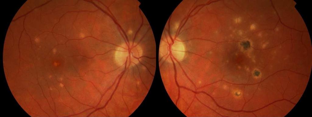
32 epidemiology, demographics, pathogenenesis, clinical description, diagnosis, ancillary tests, differential diagnoses, management, PIC in pregnanacy and prognosis. All relevant clinical studies were considered but were weighted according to their level of evidence, with well-designed randomized prospective clinical trials ranked highest and case-reports ranked lowest; case-reports were generally excluded from the final review unless they were considered to provide unique insights not evident from higher level studies. Articles which did not present primary data (such as reviews and expert opinion) were also considered and were included if they provided original insights into the condition, based on appropriate published primary data. Articles in languages other than English were also considered if they provided an English abstract which could be used for screening their relevance to decide whether translation of the full article was required. Figure Legends: Figure 1. Color fundus photography of right (A), and left (B) eyes, demonstrating the characteristic features of punctate inner choroidopathy (PIC) in a 39-year-old, white, myopic woman. This woman has a 10 year history of the disease and had presented again with new onset photopsia and scotomas. This patient explicitly and repeatedly declined all treatment so the subsequent progression reflects the natural history of her disease.
33 Figure 2. Fundus autofluorescence imaging (Blue-Peak autofluorescence, Heidelberg Engineering, Germany) of the 39 year old woman described in Figure 1. The left image (A) is taken at the time of her representation with acute symptoms and demonstrates multiple hypofluorescent punctate inner choroidopathy (PIC) lesions with surrounding zones of hyperautofluorescence. The right image (B) was taken three years previously at a time when the patient was asymptomatic and the disease was considered quiescent. Figure 3. Optical coherence tomography (OCT) imaging (Spectralis, Heidelberg Engineering, Germany) of the 39 year old woman described in Figure 1. The top (A) and middle images (B) are taken at the time of her representation with acute symptoms and demonstrates prominent choroidal thickening with choroidal hyperreflective foci, and the outer retinal involvement of multiple PIC lesions. The bottom image (C) was taken three years previously at a time when the patient was asymptomatic and the disease was considered quiescent. It demonstrates a relatively normal choroidal appearance. Figure 4. Optical coherence tomography (OCT) imaging (A) (Spectralis, Heidelberg Engineering, Germany) of a 39 year old myopic man who presented with reduced vision in his right eye and was diagnosed with punctate inner choroidopathy (PIC). OCT angiography was performed (Optovue AngioVue,
34 United States) and clearly delineated a choroidal neovascular membrane (B), without the need for fluorescein angiography. List of abbreviations Punctate inner choroidopathy (PIC) Age-related macular degeneration (AMD) Photodynamic therapy (PDT) Choroidal neovascularization (CNV) Anti- vascular endothelial growth factor (Anti-VEGF) Fluorescein angiography (FA) Indocyanine green angiography (ICG) Optical coherence tomography (OCT) Spectral domain OCT (SD-OCT) Enhanced depth imaging OCT (EDI OCT) Multifocal choroiditis and panuveitis (MCP) Acute posterior multifocal placoid pigment epitheliopathy (APMPPE) Presumed ocular histoplasmosis syndrome (POHS)
35 Diffuse subretinal fibrosis syndrome (DSFS) Birdshot chorioretinopathy (BCR) Multiple evanescent white dot syndrome (MEWDS) Intravitreal triamcinolone acetate (IVTA) Acknowledgements: None Conflicts of Interest: None Funding: PAK has received a proportion of his funding from the Department of Health s NIHR Biomedical Research Centre for Ophthalmology at Moorfields Eye Hospital and UCL Institute of Ophthalmology. The views expressed in the publication are those of the author and not necessarily those of the Department of Health. References 1. Amer R, Lois N. Punctate inner choroidopathy. Surv Ophthalmol. 2011;56(1):36-53.
36 2. Atan D, Fraser-Bell S, Plskova J, Kuffová L, Hogan A, Tufail A, Kilmartin DJ, Forrester JV, Bidwell JL, Dick AD, Churchill AJ. Punctate inner choroidopathy and multifocal choroiditis with panuveitis share haplotypic associations with IL10 and TNF loci. Invest Ophthalmol Vis Sci. 2011;52(6): Atan D, Fraser-Bell S, Plskova J, Kuffova L, Hogan A, Tufail A, Kilmartin DJ, Forrester JV, Bidwell J, Dick AD, Churchill AJ. Cytokine polymorphism in noninfectious uveitis. Invest Ophthalmol Vis Sci Aug;51(8): doi: /iovs Epub 2010 Mar 24. PubMed PMID: Barile GR, Reppucci VS, Schiff WM, Wong DT. Circumpapillary chorioretinopathy in multiple evanescent white-dot syndrome. Retina. 1997;17(1): Barry JA, Folkard A, Denniston AK, Moran E, Ayliffe W. Development and validation of quality-of-life questionnaires for birdshot chorioretinopathy. Ophthalmology. 2014;121(7): Baxter SL, Pistilli M, Pujari SS, Liesegang TL, Suhler EB, Thorne JE, Foster CS, Jabs DA, Levy-Clarke GA, Nussenblatt RB, Rosenbaum JT, Kempen JH. Risk of choroidal neovascularization among the uveitides. Am J Ophthalmol. 2013;156(3): e2. 7. Biswas J, Raman R, Bhojwani D. Unilateral Punctate inner choroidopathy with choroidal neovascular membrane in a young male. Indian J Ophthalmol. 2014;62(9):
37 8. Braley RE, Meredith TA, Aaberg TM, Koethe SM, Witkowski JA. The prevalence of HLA-B7 in presumed ocular histoplasmosis. Am J Ophthalmol. 1978;85(6): Brouzas D, Charakidas A, Rotsos T, Moschos MM, Loukianou H, Koutsandrea C, Ladas I, Baltatzis S. Choroidal neovascularization due to punctate inner choroidopathy: long-term follow-up and review of literature. Clin Ophthalmol. 2010;4: Brown J Jr, Folk JC, Reddy CV, Kimura AE. Visual prognosis of multifocal choroiditis, punctate inner choroidopathy, and the diffuse subretinal fibrosis syndrome. Ophthalmology. 1996;103(7): Brueggeman RM, Noffke AS, Jampol LM. Resolution of punctate inner choroidopathy lesions with oral prednisone therapy. Arch Ophthalmol. 2002;120(7): Buerk BM, Rabb MF, Jampol LM. Peripapillary subretinal fibrosis: a characteristic finding of multifocal choroiditis and panuveitis. Retina Feb- Mar;25(2): Chan WM, Lai TY, Lau TT, Lee VY, Liu DT, Lam DS. Combined photodynamic therapy and intravitreal triamcinolone for choroidal neovascularization secondary to punctate inner choroidopathy or of idiopathic origin: one-year results of a prospective series. Retina. 2008;28(1):71-80
38 14. Chan WM, Lai TY, Liu DT, Lam DS. Intravitreal bevacizumab (avastin) for choroidal neovascularization secondary to central serous chorioretinopathy, secondary to punctate inner choroidopathy, or of idiopathic origin. Am J Ophthalmol. 2007;143(6): Channa R, Ibrahim M, Sepah Y, Turkcuoglu P, Lee JH, Khwaja A, Hatef E, Bittencourt M, Heo J, Do DV, Nguyen QD. Characterization of macular lesions in punctate inner choroidopathy with spectral domain optical coherence tomography. J Ophthalmic Inflamm Infect. 2012;2(3): Chatterjee S, Gibson JM. Photodynamic therapy: a treatment option in choroidal neovascularization secondary to punctate inner choroidopathy. Br J Ophthalmol. 2003;87(7): Cirino AC, Mathura JR Jr, Jampol LM. Resolution of activity (choroiditis and choroidal neovascularization) of chronic recurrent punctate inner choroidopathy after treatment with interferon B-1A. Retina. 2006;26(9): Ciulla TA, Piper HC, Xiao M, Wheat LJ. Presumed ocular histoplasmosis syndrome: update on epidemiology, pathogenesis, and photodynamic, antiangiogenic, and surgical therapies. Curr Opin Ophthalmol. 2001;12(6): Coco RM, de Souza CF, Sanabria MR. Photodynamic therapy for subfoveal and juxtafoveal choroidal neovascularization associated with punctate inner choroidopathy. Ocul Immunol Inflamm. 2007; 15(1):27-9.
39 20. Cornish KS, Williams GJ, Gavin MP, Imrie FR. Visual and optical coherence tomography outcomes of intravitreal bevacizumab and ranibizumab in inflammatory choroidal neovascularization secondary to punctate inner choroidopathy. Eur J Ophthalmol Jul-Aug;21(4): Daniel E, Thorne JE, Newcomb CW, Pujari SS, Kaçmaz RO, Levy-Clarke GA, Nussenblatt RB, Rosenbaum JT, Suhler EB, Foster CS, Jabs DA, Kempen JH. Mycophenolate mofetil for ocular inflammation. Am J Ophthalmol. 2010;149(3): e Ehlers JP, Maldonado R, Sarin N, Toth CA. Treatment of non-age-related macular degeneration submacular diseases with macular translocation surgery. Retina Jul-Aug;31(7): Essex RW, Tufail A, Bunce C, Aylward GW. Twoyear results of surgical removal of choroidal neovascular membranes related to nonagerelated macular degeneration. Br J Ophthalmol. 2007;91(5): Essex RW, Wong J, Jampol LM, Dowler J, Bird AC. Idiopathic multifocal choroiditis: a comment on present and past nomenclature. Retina. 2013;33(1): Essex RW, Wong J, Fraser-Bell S, Sandbach J, Tufail A, Bird AC, Dowler J. Punctate inner choroidopathy: clinical features and outcomes. Arch Ophthalmol. 2010;128(8):982-7.
40 26. Flaxel CJ, Owens SL, Mulholland B, Schwartz SD, Gregor ZJ. The use of corticosteroids for choroidal neovascularization in young patients. Eye (Lond). 1998;12 ( Pt 2): Fong KC, Thomas D, Amin K, Inzerillo D, Horgan SE. Photodynamic therapy combined with systemic corticosteroids for choroidal neovascularization secondary to punctate inner choroidopathy. Eye (Lond). 2008;22(4): Francis PJ, Marinescu A, Fitzke FW, Bird AC, Holder GE. Acute zonal occult outer retinopathy: towards a set of diagnostic criteria. Br J Ophthalmol. 2005;89(1): Galor A, Jabs DA, Leder HA, Kedhar SR, Dunn JP, Peters GB 3rd, Thorne JE. Comparison of antimetabolite drugs as corticosteroid-sparing therapy for noninfectious ocular inflammation. Ophthalmology. 2008; 115(10): Gerstenblith AT, Thorne JE, Sobrin L, Do DV, Shah SM, Foster CS, Jabs DA, Nguyen QD. Punctate inner choroidopathy: a survey analysis of 77 persons. Ophthalmology. 2007;114(6): Hirooka K, Saito W, Hashimoto Y, Saito M, Ishida S. Increased macular choroidal blood flow velocity and decreased choroidal thickness with regression of punctate inner choroidopathy. BMC Ophthalmol. 2014;14: Hua R, Liu L, Chen L. Evaluation of the progression rate of atrophy lesions in punctate inner choroidopathy (PIC) based on autofluorescence analysis.
41 Photodiagnosis Photodyn Ther Dec;11(4): doi: /j.pdpdt Epub 2014 Jul 18. PubMed PMID: Ip M, Gorin MB. Recurrence of a choroidal neovascular membrane in a patient with punctate inner choroidopathy treated with daily doses of thalidomide. Am J Ophthalmol. 1996;122(4): Jampol LM, Becker KG. White spot syndromes of the retina: a hypothesis based on the common genetic hypothesis of autoimmune/inflammatory disease. Am J Ophthalmol. 2003; 135(3): Jones NP. The Manchester Uveitis Clinic: the first 3000 patients-- epidemiology and casemix. Ocul Immunol Inflamm. 2015;23(2): Kaiser OK, Gragoudas HY. The subretinal fibrosis and uveitis syndrome. Int Ophthalmol Clin.1996;36(1): Kedhar SR, Thorne JE, Wittenberg S, Dunn JP, Jabs DA. Multifocal choroiditis with panuveitis and punctate inner choroidopathy: comparison of clinical characteristics at presentation. Retina. 2007;27(9): Kempen JH, Altaweel MM, Holbrook JT, Jabs DA, Louis TA, Sugar EA, Thorne JE. Randomized comparison of systemic anti-inflammatory therapy versus fluocinolone acetonide implant for intermediate, posterior, and panuveitis: the multicenter uveitis steroid treatment trial. Multicenter Uveitis Steroid Treatment (MUST) Trial Research Group. Ophthalmology. 2011;118(10):
42 39. Kim H, Woo SJ, Kim YK, Lee SC, Lee CS. Focal Choroidal Excavation in Multifocal Choroiditis and Punctate Inner Choroidopathy. Ophthalmology. 2015;122(7): Klufas MA, OʼHearn T, Sarraf D. Optical coherence tomography angiography and widefield fundus autofluorescence in punctate inner choroidopathy. Retin Cases Brief Rep. 2015;9(4): Koutroumanos N, Folkard A, Mattocks R, Wright J, Xing W, Wilson-Barrett C, Bonstein K, Pavesio C, Westcott M, Moore G, Stanford M, Bunce C, Okhravi N. Bringing together patient and specialists: the first Birdshot Day. Br J Ophthalmol. 2013;97(5): Krauss T, Pauer HU, Augustin HG. Prospective analysis of placenta growth factor (PlGF) concentrations in the plasma of women with normal pregnancy and pregnancies complicated by preeclampsia. Hypertens Pregnancy. 2004;23(1): Leslie T, Lois N, Christopoulou D, Olson JA, Forrester JV. Photodynamic therapy for inflammatory choroidal neovascularization unresponsive to immunosuppression. Br J Ophthalmol Feb;89(2):147-50
43 44. Leung AK, Weisbrod DJ, Schwartz C. Intravitreal ranibizumab in the treatment of choroidal neovascular membrane secondary to punctate inner choroidopathy. Can J Ophthalmol. 2010;45(3): Levison AL, Baynes KM, Lowder CY, Kaiser PK, Srivastava SK. Choroidal neovascularisation on optical coherence tomography angiography in punctate inner choroidopathy and multifocal choroiditis. Br J Ophthalmol. 2016; bjophthalmol doi: /bjophthalmol [Epub ahead of print]. 46. Levy J, Shneck M, Klemperer I, Lifshitz T. Punctate inner choroidopathy: resolution after oral steroid treatment and review of the literature.can J Ophthalmol. 2005; 40(5): Lygnos MC, Pappa KI, Papadaki HA, Relakis C, Koumantakis E, Anagnou NP, Eliopoulos GD. Changes in maternal plasma levels of VEGF, bfgf, TGFbeta1, ET-1 and skl during uncomplicated pregnancy, hypertensive pregnancy and gestational diabetes. In Vivo. 2006;20(1): Li M, Zhang X, Wen F. The Fundus Autofluorescence Spectrum of Punctate Inner Choroidopathy. J Ophthalmol. 2015;2015:
44 49. Lim J, Flaxel C, LaBree L. Photodynamic therapy for choroidal neovascularization secondary to inflammatory chorioretinal disease. Ann Acad Med Singapore. 2006;35: Lowder C, Belfort R Jr, Lightman S, Foster CS, Robinson MR, Schiffman RM, Li XY, Cui H, Whitcup SM; Ozurdex HURON Study Group. Dexamethasone intravitreal implant for noninfectious intermediate or posterior uveitis. Arch Ophthalmol. 2011;129(5): Madhusudhan S, Keane PA, Denniston AK. Adjunctive use of systematic retinal thickness map analysis to monitor disease activity in punctate inner choroidopathy. J Ophthalmic inflamm infect. 2016;6(1): Mansour AM, Arevalo JF, Ziemssen F, Mehio-Sibai A, Mackensen F, Adan A, Chan WM, Ness T, Banker AS, Dodwell D, Chau Tran TH, Fardeau C, Lehoang P, Mahendradas P, Berrocal M, Tabbarah Z, Hrisomalos N, Hrisomalos F, Al- Salem K, Guthoff R. Long-term visual outcomes of intravitreal bevacizumab in inflammatory ocular neovascularization. Am J Ophthalmol. 2009;148(2): e Matsuda S, Gomi F, Oshima Y, Tohyama M, Tano Y. Vascular endothelial growth factor reduced and connective tissue growth factor induced by triamcinolone in ARPE19 cells under oxidative stress. Invest Ophthalmol Vis Sci. 2005;46(3):
45 (54). Menezo V, Cuthbertson F, Downes SM. Positive response to intravitreal ranibizumab in the treatment of choroidal neovascularization secondary to punctate inner choroidopathy. Retina. 2010;30(9): Meredith TA, Smith RE, Braley RE, Witkowski JA, Koethe SM. The prevalence of HLA-B7 in presumed ocular histoplasmosis in patients with peripheral atrophic scars. Am J Ophthalmol. 1978;86(3): Minos E, Barry RJ, Southworth S, Folkard A, Murray PI, Duker JS, Keane PA, Denniston AK. Birdshot chorioretinopathy: current knowledge and new concepts in pathophysiology, diagnosis, monitoring and treatment. Orphanet J Rare Dis May 12;11(1):61. doi: /s Review. PubMed PMID: ; PubMed Central PMCID: PMC Munk MR, Jung JJ, Biggee K, Tucker WR, Sen HN, Schmidt-Erfurth U, Fawzi AA, Jampol LM. Idiopathic multifocal choroiditis/punctate inner choroidopathy with acute photoreceptor loss or dysfunction out of proportion to clinically visible lesions. Retina. 2015;35(2): Nussenblatt RB, Coleman H, Jirawuthiworavong G, Davuluri G, Potapova N, Dahr SS, Ragheb JA, Levy-Clarke G. The treatment of multifocal choroiditis associated choroidal neovascularization with sirolimus (rapamycin). Acta Ophthalmol Scand. 2007;85(2): Olsen TW, Capone A Jr, Sternberg P Jr, Grossniklaus HE, Martin DF, Aaberg TM Sr. Subfoveal choroidal neovascularization in punctate inner choroidopathy.
46 Surgical management and pathologic findings. Ophthalmology. 1996;103(12): Pachydaki SI, Jakobiec FA, Bhat P, Sobrin L, Michaud NA, Seshan SV, D'Amico DJ. Surgical management and ultrastructural study of choroidal neovascularization in punctate inner choroidopathy after bevacizumab. J Ophthalmic Inflamm Infect. 2012;2(1): Parnell JR, Jampol LM, Yannuzzi LA, Gass JD, Tittl MK. Differentiation between presumed ocular histoplasmosis syndrome and multifocal choroiditis with panuveitis based on morphology of photographed fundus lesions and fluorescein angiography. Arch Ophthalmol Feb;119(2): Patel KH, Birnbaum AD, Tessler HH, Goldstein DA. Presentation and outcome of patients with punctate inner choroidopathy at a tertiary referral center. Retina. 2011; 31(7): Pavesio C, Zierhut M, Bairi K, Comstock TL, Usner DW; Fluocinolone Acetonide Study Group. Evaluation of an intravitreal fluocinolone acetonide implant versus standard systemic therapy in noninfectious posterior uveitis. Ophthalmology. 2010;117(3):567-75, Postelmans L, Pasteels B, Coquelet P, Caspers L, Verougstraete C, Leys A, Wirix M, Mauget-Faÿsse M, Quanranta M, Snyers B, Smets E. Photodynamic therapy for subfoveal classic choroidal neovascularization related to punctate
47 inner choroidopathy (PIC) or presumed ocular histoplasmosis-like syndrome (POHS-like). Ocul Immunol Inflamm. 2005;13(5): Prasad AG, Van Gelder RN. Presumed ocular histoplasmosis syndrome. Curr opin Ophthalmol. 2005;16(6): Rao VG, Rao GS, Narkhede NS. Flare up of choroiditis and choroidal neovasculazation associated with punctate inner choroidopathy during early pregnancy. Indian J Ophthalmol. 2011;59(2): Reddy CV, Brown J Jr, Folk JC, Kimura AE, Gupta S, Walker J. Enlarged blind spots in chorioretinal inflammatory disorders. Ophthalmology. 1996;103(4): Rogers AH, Duker JS, Nichols N, Baker BJ. Photodynamic therapy of idiopathic and inflammatory choroidal neovascularization in young adults. Ophthalmology. 2003;110(7): Rosen E, Rubowitz A, Ferencz JR. Exposure to verteporfin and bevacizumab therapy for choroidal neovascularization secondary to punctate inner choroidopathy during pregnancy. Eye. 2009; 23: Rosenfeld PJ, Brown DM, Heier JS, Boyer DS, Kaiser PK, Chung CY, Kim RY; MARINA Study Group. Ranibizumab for neovascular age-related macular degeneration. N Engl J Med. 2006;355(14):
48 71. Rouvas A, Petrou P, Douvali M, Ntouraki A, Vergados I, Georgalas I, Markomichelakis N. Intravitreal ranibizumab for the treatment of inflammatory choroidal neovascularization. Retina. 2011;31(5): Ryan SJ, Maumenee AE. Acute posterior multifocal placoid pigment epitheliopathy. Am J Ophthalmol. 1972;74(6): Ryan SJ, Maumenee AE. Birdshot retinochoroidopathy. Am J Ophthalmol. 1980;89(1): Shah KH, Levinson RD, Yu F, Goldhardt R, Gordon LK, Gonzales CR, Heckenlively JR, Kappel PJ, Holland GN. Birdshot chorioretinopathy. Surv Ophthalmol. 2005;50(6): Shimada H, Yuzawa M, Hirose T, Nakashizuka H, Hattori T, Kazato Y. Pathological findings of multifocal choroiditis with panuveitis and punctate inner choroidopathy. Jpn J Ophthalmol. 2008;52(4): Sim DA, Sheth HG, Kaines A, Tufail A. Punctate inner choroidopathyassociated choroidal neovascular membranes during pregnancy. Eye (Lond). 2008;22(5): Slusher MM, Weaver RG. Multiple evanescent white dot syndrome. Retina. 1988;8(2):132-5.
49 78. Spaide RF, Freund KB, Slakter J, Sorenson J, Yannuzzi LA, Fisher Y. Treatment of subfoveal choroidal neovascularization associated with multifocal choroiditis and panuveitis with photodynamic therapy. Retina. 2002;22(5): Spaide RF, Goldberg N, Freund KB. Redefining multifocal choroiditis and panuveitis and punctate inner choroidopathy through multimodal imaging. Retina. 2013;33(7): Sugawara E, Machida S, Fujiwara T, Kurosaka D, Hayakawa M. Punctate inner choroidopathy in mother and daughter. Jpn J Ophthalmol Sep;54(5): Tarantola RM, Folk JC, Boldt HC, Mahajan VB. Intravitreal bevacizumab during pregnancy. Retina. 2010;30(9): Tiffin PA, Maini R, Roxburgh ST, Ellingford A. Indocyanine green angiography in a case of punctate inner choroidopathy. Br J Ophthalmol. 1996;80(1): Turkcuoglu P, Chang PY, Rentiya ZS, Channa R, Ibrahim M, Hatef E, Sophie R, Sadaka A, Wang J, Sepah YJ, Do DV, Foster CS, Nguyen QD. Mycophenolate mofetil and fundus autofluorescence in the management of recurrent punctate inner choroidopathy. Ocul Immunol Inflamm. 2011; 19(4): Valverde Megías A, Arriola Villalobos P, Reche Frutos J, Donate López J, Calvo González C, García Feijoo J. Intravitreal ranibizumab (Lucentis ) in the
50 treatment of choroidal neovascular membrane secondary to punctate inner choroidopathy. Arch Soc Esp Oftalmol. 2010;85(4): Wachtlin J, Heimann H, Behme T, Foerster MH. Long-term results after photodynamic therapy with verteporfin for choroidal neovascularizations secondary to inflammatory chorioretinal diseases. Graefes Arch Clin Exp Ophthalmol. 2003;241(11): Watzke RC, Packer AJ, Folk JC, Benson WE, Burgess D, Ober RR. Punctate inner choroidopathy. Am J Ophthalmol. 1984;98(5): Zarranz-Ventura J, Sim DA, Keane PA, Patel PJ, Westcott MC, Lee RW, Tufail A, Pavesio CE. Characterization of punctate inner choroidopathy using enhanced depth imaging optical coherence tomography. Ophthalmology. 2014;121(9): Zhang H, Liu ZL, Sun P, Gu F. Intravitreal bevacizumab as primary treatment of choroidal neovascularization secondary to punctate inner choroidopathy: results of a 1-year prospective trial. Retina Jun;32(6): Zhang X, Wen F, Zuo C, Li M, Chen H, Huang S, Luo G. Clinical features of punctate inner choroidopathy in Chinese patients. Retina. 2011;31(8): Zhang X, Zuo C, Li M, Chen H, Huang S, Wen F. Spectral-domain optical coherence tomographic findings at each stage of punctate inner choroidopathy. Ophthalmology. 2013;120(12):
51


52 AC C EP TE D M AN U SC RI PT


53 AC C EP TE D M AN U SC RI PT

54

55

56

57


58
White-Spot Syndromes of the Retina Lee Jampol, M.D. Chicago, IL
 Objectives At the conclusion of the program, the attendees will be able to: 1. recognize the various white-spot syndromes of the retina 2. initiate appropriate diagnostic tests of patients with the white-spot
Retina Conference. Janelle Fassbender, MD, PhD University of Louisville Department of Ophthalmology and Visual Sciences 09/04/2014
 Retina Conference Janelle Fassbender, MD, PhD University of Louisville Department of Ophthalmology and Visual Sciences 09/04/2014 Subjective CC/HPI: 64 year old Caucasian female referred by outside ophthalmologist
DOME SHAPED MACULOPATHY. Ιωάννης Ν. Βαγγελόπουλος Χειρ. Οφθαλμίατρος - Βόλος
 DOME SHAPED MACULOPATHY Ιωάννης Ν. Βαγγελόπουλος Χειρ. Οφθαλμίατρος - Βόλος DOME SHAPED MACULOPATHY-DEFINITIONS The entity Dome Shaped Macula ( DSM ) was first described by Gaucher and associates in 2008
Moncef Khairallah, MD
 Moncef Khairallah, MD Department of Ophthalmology, Fattouma Bourguiba University Hospital Faculty of Medicine, University of Monastir Monastir, Tunisia INTRODUCTION IU: anatomic form of uveitis involving
ZEISS AngioPlex OCT Angiography. Clinical Case Reports
 Clinical Case Reports Proliferative Diabetic Retinopathy (PDR) Case Report 969 PROLIFERATIVE DIABETIC RETINOPATHY 1 1-year-old diabetic female presents for follow-up of proliferative diabetic retinopathy
Ophthalmology Macular Pathways
 Ophthalmology Macular Pathways Age related Macular Degeneration Diabetic Macular Oedema Macular Oedema secondary to Central Retinal Macular Oedema secondary to Branch Retinal CNV associated with pathological
White Dot Syndromes Noninfectious Chorioretinopathies Update 2019
 White Dot Syndromes Noninfectious Chorioretinopathies Update 2019 Kelly T. Mitchell, MD Retina Service TTUHSC Definition Noninfectious disease Inflammation of choroid, choriocapillaris, RPE, and Retina
OCT Angiography in Primary Eye Care
 OCT Angiography in Primary Eye Care An Image Interpretation Primer Julie Rodman, OD, MS, FAAO and Nadia Waheed, MD, MPH Table of Contents Diabetic Retinopathy 3-6 Choroidal Neovascularization 7-9 Central
CLINICALCASE PROVOST J, SEKFALI R, AMOROSO F, ZAMBROWSKI O, MIERE A
 CLINICALCASE PROVOST J, SEKFALI R, AMOROSO F, ZAMBROWSKI O, MIERE A Department of ophthalmology, Souied E. (MD,PhD) Centre Hospitalier Intercommunal de Créteil Université Paris Est HISTORY 13 years old
Spontaneous Large Serous Retinal Pigment Epithelial Tear
 This is an Open Access article licensed under the terms of the Creative Commons Attribution-NonCommercial-NoDerivs 3.0 License (www.karger.com/oa-license), applicable to the online version of the article
Diabetic maculopathy 11/ An update on. Miss Vasuki Sivagnanavel
 Miss Vasuki Sivagnanavel Consultant Ophthalmologist An update on Diabetic maculopathy Despite advances in the management of diabetes, diabetic retinopathy is already the commonest cause of blindness among
Why Is Imaging Critical in My Uveitis Practice?
 Why Is Imaging Critical in My Uveitis Practice? Dilraj S. Grewal, MD Developed in collaboration Imaging Is the Backbone of Uveitis Workup and Monitoring Treatment Response FP FAF B- scan Multimodal Imaging
Choroidal Neovascularization in Sympathetic Ophthalmia
 Choroidal Neovascularization in Sympathetic Ophthalmia Lucia Sobrin, Miguel Cordero Coma, C. Stephen Foster Case Report A 49-year-old man presented after a ruptured globe repair of his left eye status
IQ 532 Micropulse Green Laser treatment for Refractory Chronic Central Serous Retinopathy
 Cronicon OPEN ACCESS EC OPHTHALMOLOGY Case Report IQ 532 Micropulse Green Laser treatment for Refractory Chronic Central Serous Retinopathy Fawwaz Al Mamoori* Medical Retina Department, Eye Specialty Hospital,
SUMMARY. Heather Casparis, MD,* and Neil M. Bressler, MD MARINA AND ANCHOR
 The following are summaries of selected presentations and posters from the American Society of Retina Specialists and European VitreoRetinal Society Annual Meeting held September 9 13, 2006, in Cannes,
PART 1: GENERAL RETINAL ANATOMY
 PART 1: GENERAL RETINAL ANATOMY General Anatomy At Ora Serrata At Optic Nerve Head Fundoscopic View Of Normal Retina What Is So Special About Diabetic Retinopathy? The WHO definition of blindness is
Photodynamic Therapy for Choroidal Neovascularization
 Photodynamic Therapy for Choroidal Neovascularization Policy Number: 9.03.08 Last Review: 10/2014 Origination: 10/2000 Next Review: 10/2015 Policy Blue Cross and Blue Shield of Kansas City (Blue KC) will
Description. Section: Other Effective Date: July 15, Subsection: Vision Original Policy Date: December 7, 2011 Subject: Page: 1 of 23
 Last Review Status/Date: June 2015 Page: 1 of 23 Description Photodynamic therapy (PDT) is a treatment modality designed to selectively occlude ocular choroidal neovascular tissue. The therapy is a 2-step
ISPUB.COM. Photopsia post flu: A case of MEWDS. S Baisakhiya, S Dulani, S Lele INTRODUCTION CASE HISTORY
 ISPUB.COM The Internet Journal of Ophthalmology and Visual Science Volume 8 Number 1 Photopsia post flu: A case of MEWDS S Baisakhiya, S Dulani, S Lele Citation S Baisakhiya, S Dulani, S Lele. Photopsia
THE OCULAR histoplasmosis
 CLINICAL SCIENCES Reactivation of Inflammatory Lesions in Ocular Histoplasmosis David Callanan, MD; Gary E. Fish, MD, JD; Rajiv Anand, MD Background: Active inflammation has not been traditionally associated
Fluorescein Angiography
 Last revision: October 2011 by Luis Arias Fluorescein Angiography Authors: Luis Arias, MD Hospital Universitari de Bellvitge - University of Barcelona. Spain Jordi Monés, MD Institut de la Màcula i de
Although photocoagulation and photodynamic PROCEEDINGS PEGAPTANIB SODIUM FOR THE TREATMENT OF AGE-RELATED MACULAR DEGENERATION *
 PEGAPTANIB SODIUM FOR THE TREATMENT OF AGE-RELATED MACULAR DEGENERATION Evangelos S. Gragoudas, MD ABSTRACT In December 24, the US Food and Drug Administration (FDA) approved pegaptanib sodium. Pegaptanib
Optical Coherence Tomography in Diabetic Retinopathy. Mrs Samantha Mann Consultant Ophthalmologist Clinical Lead of SEL-DESP
 Optical Coherence Tomography in Diabetic Retinopathy Mrs Samantha Mann Consultant Ophthalmologist Clinical Lead of SEL-DESP Content OCT imaging Retinal layers OCT features in Diabetes Some NON DR features
Serpiginous choroidopathy
 PHILIPPINE JOURNL OF Ophthalmology Vol. 36 No. 2 July Dec em ber 2011 CSE REPORT Kristine Corpus, MD 1 ndrew ijasa, RT 1 Egidio Jose Fortuna, MD 1-2 1, 3, 4 Narciso tienza Jr., MD Serpiginous choroidopathy
Clinically Significant Macular Edema (CSME)
") Clinically Significant Macular Edema (CSME) 1 Clinically Significant Macular Edema (CSME) Sadrina T. Shaw OMT I Student July 26, 2014 Advisor: Dr. Uwaydat Clinically Significant Macular Edema (CSME) 2
Clinically Significant Macular Edema (CSME) 1 Clinically Significant Macular Edema (CSME) Sadrina T. Shaw OMT I Student July 26, 2014 Advisor: Dr. Uwaydat Clinically Significant Macular Edema (CSME) 2
FEP Medical Policy Manual
 FEP Medical Policy Manual Last Review: September 2016 Next Review: September 2017 Related Policies 9.03.20 Intraocular Radiation Therapy for Age-Related Macular Degeneration Photodynamic Therapy for Choroidal
Clinical Study Choroidal Thickness in Eyes with Unilateral Ocular Ischemic Syndrome
 Hindawi Publishing Corporation Journal of Ophthalmology Volume 215, Article ID 62372, 5 pages http://dx.doi.org/1.1155/215/62372 Clinical Study Choroidal Thickness in Eyes with Unilateral Ocular Ischemic
ISPUB.COM. An Atypical Presentation of Posterior Scleritis. A Ramanathan, A Gaur CASE REPORT
 ISPUB.COM The Internet Journal of Ophthalmology and Visual Science Volume 8 Number 2 A Ramanathan, A Gaur Citation A Ramanathan, A Gaur.. The Internet Journal of Ophthalmology and Visual Science. 2009
Misdiagnosed Vogt-Koyanagi-Harada (VKH) disease and atypical central serous chorioretinopathy (CSC)
 disease and atypical central serous chorioretinopathy (CSC)") HPTER 12 Misdiagnosed Vogt-Koyanagi-Harada (VKH) disease and atypical central serous chorioretinopathy (S) linical Features VKH disease is a bilateral granulomatous panuveitis often associated with exudative
HPTER 12 Misdiagnosed Vogt-Koyanagi-Harada (VKH) disease and atypical central serous chorioretinopathy (S) linical Features VKH disease is a bilateral granulomatous panuveitis often associated with exudative
Michael P. Blair, MD Retina Consultants, Ltd Libertyville/Des Plaines, Illinois Clinical Associate University of Chicago 17 October 2015
 Michael P. Blair, MD Retina Consultants, Ltd Libertyville/Des Plaines, Illinois Clinical Associate University of Chicago 17 October 2015 So What Parts of the Eye Retina are Affected by VHL Neural tissue
Deep Trouble. Thomas Stone, MD Retina Associates of Kentucky River City Retina Conference May 15, 2014
 Deep Trouble Thomas Stone, MD Retina Associates of Kentucky River City Retina Conference May 15, 2014 History 20 yo WM Decreased vision OU, OD>OS Sudden onset blurred central vision 12 days prior 4 days
o White dot syndromes pattern recognition o Activity and damage o Quality of life o Key points o Idiopathic o Sarcoidosis o Multiple sclerosis
 Introduction Clinical Assessment of Posterior Uveitis Philip I. Murray Centre for Translational Inflammation Research University of Birmingham Birmingham and Midland Eye Centre o Classification of uveitis
11/29/2016 MACULAR MALADIES: TYPICAL & ATYPICAL CASES
 MACULAR MALADIES: TYPICAL & ATYPICAL CASES Dawn Pewitt, OD, FAAO Triad Eye Institute, Grove, OK Dpewitt@triadeye.com Disclosure Statement: No financial disclosures COPE 51218-PS Please silence all mobile
Doc, I See a Donut in My Vision : An Optometrist s Guide to a Rare Cause of Choroidal Neovascular Membrane
 Doc, I See a Donut in My Vision : An Optometrist s Guide to a Rare Cause of Choroidal Neovascular Membrane Linda Pham, OD, Tobin Ansel, OD, Nancy Shenouda-Awad, OD, FAAO, West Haven VA Medical Center Abstract
Stabilization of visual acuity with photodynamic therapy in eyes with chorioretinal anastomoses
 Graefe s Arch Clin Exp Ophthalmol (2004) 242:368 376 CLINICAL INVESTIGATION DOI 10.1007/s00417-003-0844-0 Rufino M. Silva José R. Faria de Abreu António Travassos José G. Cunha-Vaz Stabilization of visual
Review Article A Review of the Inflammatory Chorioretinopathies: The White Dot Syndromes
 ISRN Inflammation Volume 2013, Article ID 783190, 9 pages http://dx.doi.org/10.1155/2013/783190 Review Article A Review of the Inflammatory Chorioretinopathies: The White Dot Syndromes Courtney M. Crawford
OCT Assessment of the Vitreoretinal Relationship in CSME
 December 2007 Sonia Rani John et al. - IFIS 375 ORIGINAL ARTICLE OCT Assessment of the Vitreoretinal Relationship in CSME Dr. Manoj S. DNB FRCS, Dr. Unnikrishnan Nair MS DO FRCS, Dr. Gargi Sathish MS Introduction
Department of Ophthalmology, Kangnam Sacred Heart Hospital, College of Medicine, Hallym University, Seoul, Korea
 Department of Ophthalmology, Kangnam Sacred Heart Hospital, College of Medicine, Hallym University, Seoul, Korea Purpose: To investigate the factors that affect final vision following photodynamic therapy
THE ROLE OF anti-vegf IN DIABETIC RETINOPATHY AND AGE RELATED MACULAR DEGENERATION
 THE ROLE OF anti-vegf IN DIABETIC RETINOPATHY AND AGE RELATED MACULAR DEGENERATION MOESTIDJAB DEPARTMENT OF OPHTHALMOLOGY SCHOOL OF MEDICINE AIRLANGGA UNIVERSITY DR SOETOMO HOSPITAL SURABAYA INTRODUCTION
Optical Coherence Tomography (OCT) in Uveitis Piergiorgio Neri, BMedSc, MD, PhD Head Ocular Immunology Unit
 in Uveitis Piergiorgio Neri, BMedSc, MD, PhD Head Ocular Immunology Unit") The Eye Clinic Polytechnic University of Marche Head: Prof Alfonso Giovannini November, 1991 Optical Coherence Tomography (OCT) in Uveitis Piergiorgio Neri, BMedSc, MD, PhD Head Ocular Immunology Unit
The Eye Clinic Polytechnic University of Marche Head: Prof Alfonso Giovannini November, 1991 Optical Coherence Tomography (OCT) in Uveitis Piergiorgio Neri, BMedSc, MD, PhD Head Ocular Immunology Unit
Clinical Features of Bilateral Acute Idiopathic Maculopathy
 Clinical Features of Bilateral Acute Idiopathic Maculopathy Toru Nakazawa,, Katsuhiro Yamaguchi, Masahiko Shimura, Madoka Yoshida, Yuki Yoshioka and Makoto Tamai Department of Ophthalmology, Katta General
The Human Eye. Cornea Iris. Pupil. Lens. Retina
 The Retina Thin layer of light-sensitive tissue at the back of the eye (the film of the camera). Light rays are focused on the retina then transmitted to the brain. The macula is the very small area in
Clinical Trials Related to Age Related Macular Degeneration
 Clinical Trials Related to Age Related Macular Degeneration Kirti Singh MD, DNB, FRCS Kirti Singh MD, DNB, FRCS, Pooja Jain MBBS, Nitasha Ahir MBBS, Divya Jain MD, DNB Guru Nanak Eye Centre, Maulana Azad
Diabetic Retinopathy
 Diabetic Retinopathy Diabetes can be classified into type 1 diabetes mellitus and type 2 diabetes mellitus, formerly known as insulin-dependent diabetes mellitus, and non-insulin diabetes mellitus, respectively.
Ocular imaging in acquired retinopathy with multiple myeloma
 Ocular imaging in acquired retinopathy with multiple myeloma ABDELRAHMAN GABER SALMAN MD- FRCS (GLASG)- MRCS (ED) ASSOCIATE PROFESSOR AIN SHAMS UNIVERSITY EVRS 2015 Immunogammopathies Immunogammopathies
HHS Public Access Author manuscript Ophthalmic Surg Lasers Imaging Retina. Author manuscript; available in PMC 2016 January 14.
 High-Speed Ultrahigh-Resolution OCT of Bruch s Membrane in Membranoproliferative Glomerulonephritis Type 2 Mehreen Adhi, MD, Sarah P. Read, MD, PhD, Jonathan J. Liu, PhD, James G. Fujimoto, PhD, and Jay
The Quick Guide to OCT Mastery 50 Real Cases with Expert Analysis
 OPTICAL COHERENCE TOMOGRAPHY The Quick Guide to OCT Mastery 50 Real Cases with Expert Analysis VOL 1 Sanjay Sharma, MD, FRCS, MSc (Epid), MBA Ophthalmologist, Epidemiologist Queen s University, Canada
Abbreviated Drug Evaluation: Fluocinolone acetonide intravitreal implant (Retisert )
") Copyright 2012 Oregon State University. All Rights Reserved Drug Use Research & Management Program Oregon State University, 500 Summer Street NE, E35, Salem, Oregon 97301-1079 Phone 503-947-5220 Fax 503-947-1119
Copyright 2012 Oregon State University. All Rights Reserved Drug Use Research & Management Program Oregon State University, 500 Summer Street NE, E35, Salem, Oregon 97301-1079 Phone 503-947-5220 Fax 503-947-1119
Rare Presentation of Ocular Toxoplasmosis
 Case Report Rare Presentation of Ocular Toxoplasmosis Rakhshandeh Alipanahi MD From Department of Ophthalmology, Nikookari Eye Hospital, Tabriz University of Medical Sciences, Tabriz, Iran. Correspondence:
Optical Coherence Tomography: Pearls for the Anterior Segment Surgeon Basic Science Michael Stewart, M.D.
 Optical Coherence Tomography: Pearls for the Anterior Segment Surgeon Basic Science Michael Stewart, M.D. Disclosure OCT Optical Coherence Tomography No relevant financial relationships I will refer to
Recalcitrant Diabetic Macular Oedema: Therapeutic Options
 December 2007 A. Giridhar et al. - Recalcitrant DME 451 CONSULTATION S E C T I O N Recalcitrant Diabetic Macular Oedema: Therapeutic Options Dr. Cyrus M Shroff 1, Dr. N S Muralidhar 2, Dr. R Narayanan
EPIRETINAL MEMBRANE & VITREOMACULAR TRACTION
 EPIRETINAL MEMBRANE & VITREOMACULAR TRACTION Management of ERM and VMT K.V.Chalam,MD,PhD,MBA,FACS Professor and Director of Retina Loma Linda Eye Institute Los Angeles, USA REVIEW ANATOMY The vitreous
R&M Solutions
 Mohamed Hosny El-Bradey, MD., Assistant Professor of Ophthalmology, Tanta University. Wael El Haig, MD., Professor of Ophthalmology. Zagazeeg University. 1 Myopic CNV is considered the most common vision
NATIONAL INSTITUTE FOR HEALTH AND CARE EXCELLENCE. Health Technology Appraisal. Aflibercept for treating diabetic macular oedema.
 NATIONAL INSTITUTE FOR HEALTH AND CARE EXCELLENCE Health Technology Appraisal Aflibercept for treating diabetic macular oedema Final scope Final remit/appraisal objective To appraise the clinical and cost
Intravitreal Triamcinolone Acetonide for Macular Edema in HLA-B27 Negative Ankylosing Spondylitis
 105 This is an Open Access article licensed under the terms of the Creative Commons Attribution- NonCommercial-NoDerivs 3.0 License (www.karger.com/oa-license), applicable to the online version of the
Intraocular Radiation Therapy for Age-Related Macular Degeneration
 Medical Policy Manual Medicine, Policy No. 134 Intraocular Radiation Therapy for Age-Related Macular Degeneration Next Review: April 2019 Last Review: June 2018 Effective: August 1, 2018 IMPORTANT REMINDER
Update on management of Anterior Uveitis
 Update on management of Anterior Uveitis Parthopratim Dutta Majumder Senior Consultant, Department of Uvea & Intraocular Inflammation Medical Research Foundation, Sankara Nethralaya ABCD of Treating a
Efficacy of Anti-VEGF Agents in the Treatment of Age-Related Macular Degeneration
 Efficacy of Anti-VEGF Agents in the Treatment of Age-Related Macular Degeneration Marilita M. Moschos Abstract- Purpose: To evaluate by OCT and mf-erg the macular function in eyes with CNV due to ARMD
Evaluation of efficacy of eplerenone in the management of chronic central serous choroidoretinopathy
 Original article: Evaluation of efficacy of eplerenone in the management of chronic central serous choroidoretinopathy Dr. Sushant Madaan* Department of Ophthalmology, NIMS Medical College and Hopsital,Jaipur,
Chapter 2 Indocyanine Green Angiography in Uveitis
 Chapter 2 Indocyanine Green Angiography in Uveitis Shilpa Kodati, Samuel P. Burke, and Thomas A. Albini Introduction Indocyanine green angiography (ICGA) became available in the early 1990s and has since
Yasser R. Serag, MD Tamer Wasfi, MD El- Saied El-Dessoukey, MD Magdi S. Moussa, MD Anselm Kampik, MD
 Microperimetric Evaluation of Brilliant Blue G- assisted Internal Limiting Membrane Peeling By Yasser R. Serag, MD Tamer Wasfi, MD El- Saied El-Dessoukey, MD Magdi S. Moussa, MD Anselm Kampik, MD The internal
Retinal Complications of Obstructive Sleep Apnea A Growing Concern!
 Retinal Complications of Obstructive Sleep Apnea A Growing Concern! Jay M. Haynie, OD, FAAO Financial Disclosure I have received honoraria or am on the advisory board for the following companies: Carl
Case Report Diagnosis and Monitoring of Choroidal Osteoma through Multimodal Imaging
 Case Reports in Medicine, Article ID 393804, 4 pages http://dx.doi.org/10.1155/2014/393804 Case Report Diagnosis and Monitoring of Choroidal Osteoma through Multimodal Imaging Theodoros Empeslidis, Usman
Intravitreal Corticosteroid Implants
 Intravitreal Corticosteroid Implants Policy Number: 9.03.23 Last Review: 4/2018 Origination: 07/2015 Next Review: 4/2019 Policy Blue Cross and Blue Shield of Kansas City (Blue KC) will provide coverage
CENTRAL SEROUS CHORIORETINOPATHY (CSC) IS
 IS") Association Between the Efficacy of Half-Dose Photodynamic Therapy With Indocyanine Green Angiography and Optical Coherence Tomography Findings in the Treatment of Central Serous Chorioretinopathy MASSIMO
Association Between the Efficacy of Half-Dose Photodynamic Therapy With Indocyanine Green Angiography and Optical Coherence Tomography Findings in the Treatment of Central Serous Chorioretinopathy MASSIMO
White Dot Syndromes Delhi J Ophthalmol 2015; 25 (4):
:") E-ISSN 0976-2892 White Dot Syndromes Delhi J Ophthalmol 2015; 25 (4): 223-232 DOI: http://dx.doi.org/10.7869/djo.113 * Jyotirmay Biswas, ** Sachin B Shetty, *** Swetha Palla * Vision Research Founda on,
E-ISSN 0976-2892 White Dot Syndromes Delhi J Ophthalmol 2015; 25 (4): 223-232 DOI: http://dx.doi.org/10.7869/djo.113 * Jyotirmay Biswas, ** Sachin B Shetty, *** Swetha Palla * Vision Research Founda on,
Clinical Case Presentation. Branch Retinal Vein Occlusion. Sarita M. Registered Nurse Whangarei Base Hospital
 Clinical Case Presentation on Branch Retinal Vein Occlusion Sarita M. Registered Nurse Whangarei Base Hospital Introduction Case Study Pathogenesis Clinical Features Investigations Treatment Follow-up
Fundus Autofluorescence. Jonathan A. Micieli, MD Valérie Biousse, MD
 Fundus Autofluorescence Jonathan A. Micieli, MD Valérie Biousse, MD The retinal pigment epithelium (RPE) has many important functions including phagocytosis of the photoreceptor outer segments Cone Rod
When optical coherence tomography (OCT)
") Macular Imaging: SD-OCT in nterior Segment Surgical Practice Many pathologic processes of the macula can be visualized or quantified only with this modality. y Steven G. Safran, MD When optical coherence
Macular Imaging: SD-OCT in nterior Segment Surgical Practice Many pathologic processes of the macula can be visualized or quantified only with this modality. y Steven G. Safran, MD When optical coherence
Ranibizumab and pegaptanib for the treatment of agerelated macular degeneration
 Ranibizumab and pegaptanib for the treatment of agerelated Issued: August 2008 last modified: May 2012 guidance.nice.org.uk/ta155 NHS Evidence has accredited the process used by the Centre for Health Technology
Despite our growing knowledge of the
 OVERVIEW OF AVAILABLE TREATMENTS FOR NEOVASCULAR AGE-RELATED MACULAR DEGENERATION * Carl D. Regillo, MD ABSTRACT Although potential treatments for neovascular age-related macular degeneration represent
Widefield Retinal Imaging with Auto Fluorescence Technology in the Optometric Practice
 Widefield Retinal Imaging with Auto Fluorescence Technology in the Optometric Practice This course will define ultra-widefield retinal imaging and autofluorescence for the attendee. Will show how it is
Diabetic Retinopathy A Presentation for the Public
 Diabetic Retinopathy A Presentation for the Public Ray M. Balyeat, MD The Eye Institute Tulsa, Oklahoma The Healthy Eye Light rays enter the eye through the cornea, pupil and lens. These light rays are
Choroidal neovascularization in a young, healthy eye after LASIK
 Optometry (2010) 81, 632-637 Choroidal neovascularization in a young, healthy eye after LASIK Bharti Bathija-Lala, O.D., a Sherry J. Bass, O.D., a Richard J. Madonna, O.D., a and Alan Dayan, M.D. b a State
Update on Central Serous Chorioretinopathy
 Update on Central Serous Chorioretinopathy Steve Bennett Retina Update 2017 No financial interest Off label use of Spironolactone, Eplerenone, Melatonin, Verteporfin, Rifampin 1 2 Classic Central Serous
OPTIC DISC PIT Pathogenesis and Management OPTIC DISC PIT
 OPTIC DISC PIT Pathogenesis and Management Abdel-Latif Siam Ain Shams University Cairo Egypt OPTIC DISC PIT Congenital pit is an atypical coloboma usually located on the temporal edge of the disc, associated
Intravitreal ranibizumab for choroidal neovascularization in a patient with angioid streaks and multiple evanescent white dots
 Pece et al. BMC Ophthalmology (2016) 16:122 DOI 10.1186/s12886-016-0307-0 CASE REPORT Intravitreal ranibizumab for choroidal neovascularization in a patient with angioid streaks and multiple evanescent
The College of Optometrists - Learning outcomes for the Professional Certificate in Medical Retina
 Learning outcomes for the Professional Certificate in Medical Retina, incorporating diabetic retinopathy screening and age related macular degeneration The professional certificate is a prerequisite to
Facts About Diabetic Eye Disease
 Facts About Diabetic Eye Disease Points to Remember 1. Diabetic eye disease comprises a group of eye conditions that affect people with diabetes. These conditions include diabetic retinopathy, diabetic
You can C-ME after Uveitis
 You can C-ME after Uveitis Abstract: Approximately 50% of uveitis patients will present with vision loss secondary to cystoid macular edema[1]. Two patients with uveitis present with a constant decrease
Diagnosis in AMD. Managing your AMD Patients
 Managing your AMD Patients Robert W. Dunphy, O.D., F.A.A.O. Diagnosis in AMD Have suspicion Identify relative risk Conduct surveillance Biometry Utilize technology to facilitate detection of change / stability
RETINA 2018 OBJECTIVES OCT VERY USEFUL INFORMATION SAFE AND FRIENDLY 1/11/2018 KELLY MITCHELL
 RETINA 2018 KELLY MITCHELL OBJECTIVES HIGHLIGHT NEW DIAGNOSTIC & TREATMENT OPTIONS REVIEW DIAGNOSTIC KEYS OF SELECT RETINAL DISEASES DISCUSS USE OF IMAGING AND REFERRAL RECOURSES FOR PATIENT BENEFIT OCT
What You Should Know About Acute Macular Neuroretinopathy
 What You Should Know About Acute Macular Neuroretinopathy David J. Browning MD, PhD Chong Lee BS Acute macular neuroretinopathy is a condition characterized by the sudden, painless onset of paracentral
VISUAL OUTCOME IN DIABETIC MACULAR EDEMA AFTER GRID LASER TREATMENT
 The Professional Medical Journal DOI: 10.17957/TPMJ/16.2856 ORIGINAL PROF-2856 VISUAL OUTCOME IN DIABETIC MACULAR EDEMA AFTER GRID LASER TREATMENT 1. MBBS.FCPS Assistant Professor Ophthalmology Independent
Diagnosis and treatment of diabetic retinopathy. Blake Cooper MD Ophthalmologist Vitreoretinal Surgeon Retina Associates Kansas City
 Diagnosis and treatment of diabetic retinopathy Blake Cooper MD Ophthalmologist Vitreoretinal Surgeon Retina Associates Kansas City Disclosures Consulted for Novo Nordisk 2017,2018. Will be discussing
Ophthalmology. Juliette Stenz, MD
 Ophthalmology Juliette Stenz, MD Required Slide Disclosures NO SIGNIFICANT FINANCIAL, GENERAL, OR OBLIGATION INTERESTS TO REPORT Required Slide At the end of this session, students will be able to: 1.
Audit of Macular Hole Surgery, Visual Outcome Prediction on OCT Appearance of Macular Hole
 International Journal of Ophthalmology & Visual Science 2017; 2(4): 93-97 http://www.sciencepublishinggroup.com/j/ijovs doi: 10.11648/j.ijovs.20170204.13 Audit of Macular Hole Surgery, Visual Outcome Prediction
Clinical Policy: Verteporfin (Visudyne) Reference Number: CP.PHAR.187
 Reference Number: CP.PHAR.187") Clinical Policy: (Visudyne) Reference Number: CP.PHAR.187 Effective Date: 03/16 Last Review Date: 03/17 Coding Implications Revision Log See Important Reminder at the end of this policy for important regulatory
Clinical Policy: (Visudyne) Reference Number: CP.PHAR.187 Effective Date: 03/16 Last Review Date: 03/17 Coding Implications Revision Log See Important Reminder at the end of this policy for important regulatory
Nausheen Khuddus, MD Melissa Elder, MD, PhD
 Nausheen Khuddus, MD Melissa Elder, MD, PhD Nausheen Khuddus, MD Pediatric Ophthalmologist and Strabismus Specialist Accent Physicians Gainesville, Florida What Is Uveitis? Uveitis is caused by inflammatory
Diagnosis and Treatment of Myopic Maculopathy. Tae Yokoi, MD, and Kyoko Ohno-Matsui, MD, PhD
 REVIEW ARTICLE Diagnosis and Treatment of Myopic Maculopathy Tae Yokoi, MD, and Kyoko Ohno-Matsui, MD, PhD Abstract: Visual impairment resulting from pathologic myopia is a serious issue worldwide. This
VERTEPORFIN IN PHOTODYNAMIC THERAPY STUDY GROUP
 Verteporfin Therapy of Subfoveal Choroidal Neovascularization in Age-related Macular Degeneration: Two-year Results of a Randomized Clinical Trial Including Lesions With Occult With No Classic Choroidal
ILUVIEN IN DIABETIC MACULAR ODEMA
 1 ILUVIEN IN DIABETIC MACULAR ODEMA Marie Tsaloumas Consultant Ophthalmic Surgeon Queen Elizabeth Hospital, Birmingham bars conference 2104 1 2 Declaration of interest I have sat on Advisory boards for
Neuropathy (NAION) and Avastin. Clinical Assembly of the AOCOO-HNS Foundation May 9, 2013
 and Avastin. Clinical Assembly of the AOCOO-HNS Foundation May 9, 2013") Non Arteritic Ischemic Optic Neuropathy (NAION) and Avastin Shalom Kelman, MD Clinical Assembly of the AOCOO-HNS Foundation May 9, 2013 Anterior Ischemic Optic Neuropathy Acute, painless, visual loss,
Non Arteritic Ischemic Optic Neuropathy (NAION) and Avastin Shalom Kelman, MD Clinical Assembly of the AOCOO-HNS Foundation May 9, 2013 Anterior Ischemic Optic Neuropathy Acute, painless, visual loss,
Since /01/2014 Private practice Ophthalmology, Retinal Medical Specialty and AMD, private office
 Corinne GONZALEZ Ophthalmology (MD, Ph.D ) Retinal Medical Specialist SELARL CABINET DOCTEUR GONZALEZ 27, Boulevard des Minimes 31 200 Toulouse! 05 34 40 77 30 e-mail : cabinet.dr.gonzalez@wanadoo.fr Fax
EFFICACY OF ANTI-VASCULAR ENDOTHELIAL GROWTH FACTOR AGENTS IN RETINAL DISORDER FOR BETTER VISUAL ACUITY
 EFFICACY OF ANTI-VASCULAR ENDOTHELIAL GROWTH FACTOR AGENTS IN RETINAL DISORDER FOR BETTER VISUAL ACUITY Diwakar chaudhary *1, 2, Hu shuqiong, Long Yuan and Xiong kun 1 Yangtze University, 1 Nanhuan Road
ROLE OF LASER PHOTOCOAGULATION VERSUS INTRAVITREAL TRIAMCINOLONE ACETONIDE IN ANGIOGRAPHIC MACULAR EDEMA IN DIABETES MELLITUS
 ORIGINAL ARTICLE ROLE OF LASER PHOTOCOAGULATION VERSUS INTRAVITREAL TRIAMCINOLONE ACETONIDE IN ANGIOGRAPHIC MACULAR EDEMA IN DIABETES MELLITUS Aggarwal Somesh VP 1, Shah Sonali N 2, Bharwada Rekha M 3,
The effect of a single intravitreal implantation of dexamethasone on the fellow eye in bilateral non-infectious uveitis case report
 European Review for Medical and Pharmacological Sciences The effect of a single intravitreal implantation of dexamethasone on the fellow eye in bilateral non-infectious uveitis case report J. CISZEWSKA,
Study Number CAIN457C2302 (core study) and CAIN457C2302E1 (extension study)
 and CAIN457C2302E1 (extension study)") Clinical Trial Results Database Page 1 Sponsor Novartis Generic Drug Name Secukinumab Therapeutic Area of Trial Uveitis Approved Indication Investigational Study Number CC2302 (core study) and CC2302E1
Clinical Trial Results Database Page 1 Sponsor Novartis Generic Drug Name Secukinumab Therapeutic Area of Trial Uveitis Approved Indication Investigational Study Number CC2302 (core study) and CC2302E1
Macular Hole Associated with Vogt-Koyanagi-Harada Disease at the Acute Uveitic Stage
 Published online: September 15, 2015 2015 The Author(s) Published by S. Karger AG, Basel 1663 2699/15/0063 0328$39.50/0 This article is licensed under the Creative Commons Attribution-NonCommercial 4.0
Eyes on Diabetics: How to Avoid Blindness in Diabetic Patient
 Eyes on Diabetics: How to Avoid Blindness in Diabetic Patient Rova Virgana FK Unpad Pusat Mata Nasional RS Mata Cicendo Bandung Eye Center (Hospital and Clinic) PIT IDI Jabar 2018 Keys Facts from WHO