EPIRETINAL MEMBRANE & VITREOMACULAR TRACTION
|
|
|
- Bertina Patterson
- 5 years ago
- Views:
Transcription

1 EPIRETINAL MEMBRANE & VITREOMACULAR TRACTION
2 Management of ERM and VMT K.V.Chalam,MD,PhD,MBA,FACS Professor and Director of Retina Loma Linda Eye Institute Los Angeles, USA


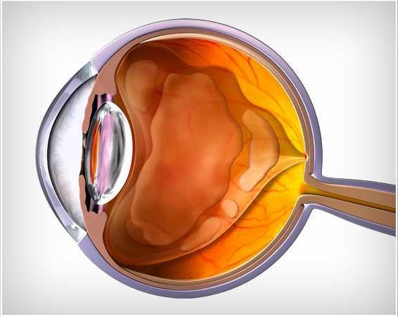
3 REVIEW ANATOMY
4 The vitreous is a transparent gel composed of water, collagen, and hyaluronic acid it occupies 80% of the volume of the eye. The vitreous body is divided into the central or core the peripheral or cortical

5


6 Macula D-E perifoveal macula fovea foveola umbo C-D parafoveal macula

7

8 RETINAL IN LAYERS

9 RETINAL IN LAYERS


10 EPIRETINAL MEMBRANE


11 Epiretinal membranes (ERMs) are sheet-like structures that develop on the inner surface of the neurosensory retina. DEFINITION

12 The macular changes that result from either ERM or VMT lead to similar symptoms: reduced visual acuity, metamorphopsia, difficulty using both eyes together, and even diplopia
13 The patient population is predominately adults. Idiopathic ERM No identifiable cause Secondary ERM after retinal breaks, tears or detachments, post-intraocular surgery, trauma, intraocular Laser proliferation of RPE or glial cells More common in DR, CRVO, BRVO


14 The prevalence of Idiopathic ERM 28.9% among Latinos in Los Angeles 2.2% and 3.4% in Beijing Handan Eye Study in rural China 7% and 8.9% 20% to 35% in the United States Australian 18.8% multi-ethnic study conducted in six communities in the United States (Multi-Ethnic Study of Atherosclerosis [MESA])
15 SD-OCT was used and documented a higher prevalence of 34.1% 20-year follow-up examinations of the Beaver Dam Eye Study population (mean age of 74.1 years)


16 cellophane maculopathy occurred more frequently than thicker//pre-retinal macular fibrosis

17 1 6.3% among Latinos in Los Angeles The prevalence of cellophane maculopathy 1.8% and 2.2% in urban and rural China 25.1% in MESA multi-ethnic study conducted in six communities in the United States (Multi-Ethnic Study of Atherosclerosis [MESA])
18 The prevalence of Pre-retinal macular fibrosis 3.8% in MESA 0.7% in urban and rural China 3.5% in Asian Indians 3.9% in Melbourne multi-ethnic study conducted in six communities in the United States (Multi-Ethnic Study of Atherosclerosis [MESA])
19 RISK FACTOR gender stroke diabetes body mass index narrow retinal arteriolar diameter Increasing age higher education smoking hypercholest erolemia refractive error RISK FACTOR
, intermediate in Hispanics (29.3%) and whites (27.")
20 prevalence of any ERM was highest in persons of Chinese ancestry (39.0%), intermediate in Hispanics (29.3%) and whites (27.5%), and lowest in blacks (26.2%),
21 pathogenesis ERMs have a variety of possible origins and causes. A longstanding hypothesis was that ERMs develop when a PVD results in microbreaks of the ILM that allow for the migration of retinal glial or RPE cells onto the anterior retinal surface, where they proliferate

22 pathogenesis Laminocytes, vitreous cells from the posterior hyaloid membrane (hyalocytes), have been shown to represent a major cellular component of idiopathic ERMs Retinal glial cells, hyalocytes transdifferentiate into fibroblasts and myofibroblasts, along with the development of extracellular matrix and fibrosis, together lead to ERM formation.

23 PATIENT OUTCOME CRITERIA Prevent vision loss and functional impairment Optimize visual function Minimize metamorphopsia and/or diplopia Maintain or improve quality of life


24 Asymptomatic > Symptomatic > Progressive loss of Visual Function Metamorphopsia Diplopia Difficulties in reading, driving, Unable to use their eyes together VA in ERM or VMT does not change dramatically in short-term follow up SYMPTOMS


25 Diagnosis The most common type of ERM appears as a thin, translucent cellophane-like membrane on the surface of the retina Epiretinal membranes can contract, leading to folds in the retina, distortion of the inner and even the outer macula, traction on retinal vessels, and even displacement of the macula or ectopia

26 Diagnosis The normal foveal depression is often absent and the macula may develop cystoid spaces, lamellar macular hole, or even a full thickness hole. Epiretinal membranes that have a thicker, white, fibrotic appearance that obscures the underlying retina, are more likely to become symptomatic and displace the macula than the thinner, more translucent ERMs
 very helpful educational tool to help patients better understand their eye problem")
27 Diagnostic tests Spectral domain OCT is a highly sensitive and routine method used to diagnose and characterize VMA, ERM, VMT (III, good quality, strong recommendation) very helpful educational tool to help patients better understand their eye problem

28


29 An ERM on OCT appears as a hyper-reflective layer on the inner surface of the retina, usually adherent across the surface of the retina. The inner retina is typically thrown into folds, with thickening of the macula and associated cystoid spaces in various retinal layers

30 Traction from the ERM leads to elevation of the normal foveal depression
 The FA may be relatively normal in eyes with early ERM.")
31 A fluorescein angiogram (FA) may be helpful to evaluate ERMs and/or VMT. ( and associated abnormality) The FA may be relatively normal in eyes with early ERM. As ERM contraction increases, the macular vessels may become tortuous near the epicenter of traction or straightened around the epicenter of traction.
 reassure that there is a very")
 Educating patients about the signs and symptoms of")
32 management Patients should be informed that the majority of ERMs will remain relatively stable and do not require therapy (good quality, strong recommendation) reassure that there is a very successful surgical procedure. (good quality, strong recommendation) Educating patients about the signs and symptoms of progression and regular monocular Amsler grid testing are both important
33 management patients should be encouraged to periodically test their central vision monocularly, such as increasing metamorphopsia or development of a small, central scotoma. (good quality, strong recommendation) Patients do not typically improve without vitrectomy surgery when the area of VMT is broad (>1500 µm), when there is a pathologic detachment of the macula, or when the presenting VA is poor.

34 ERM Using fundus photography, a populationbased study of 3654 persons with 5 years follow up 39% 29% 26% SAME PROGRESS REGRESS
35 Observation without treatment A study using SD-OCT images found that the ERM separated from the retina in only 16 of 1091 (1.5%) eyes with a pre-existing PVD in 21 of 157 (13.6%) of eyes that did not have an apparent PVD over a mean follow-up of 33 months.


36 VITREOMACULAR TRACTION

37 Definition VMA Vitreomacular adhesion is an attachment of the posterior cortical vitreous to the macula. VMT Vitreomacular traction occurs when the posterior cortical vitreous partially separates from the retina yet some tractional areas remain adherent to portions of the macula.
38


39 PVD floaters or flash or photopsia If vitreous remain adherent to the macula VMA Perimacular vitreous separate from posterior retina yet remain adherant to a region near macula center intraretinal cystoid changes, SRF, tractional detachment


40 The macular changes that result from either ERM or VMT lead to similar symptoms: reduced visual acuity, metamorphopsia, difficulty using both eyes together, and even diplopia
41 at the 20-year follow-up examinations of the Beaver Dam Eye Study population (mean age of 74.1 years), spectral domain optical coherence tomography (SD- OCT) was used and documented The same study also documented the prevalence of VMT to be 1% to 2%
42 Pathogenesis the process of a PVD may be a prolonged one and portions of the posterior cortical surface may remain adherent to the macula tractional changes
43 localized vitreomacular attachment of about 500 µm causes elevation, traction, and subsequent intraretinal cystoid spaces in the foveal neurosensory retina. A broad attachment measuring about 1500 µm (approximately one disc diameter) can cause more elevation of the macula, even to the point of a macular retinal detachment

44 Epiretinal membranes may evolve between the neurosensory retina and a vitreous attachment. Perhaps the ERMs are due to a wound-healing response. They adhere tightly to the ILM, and may play a role in VMT by binding the remaining attachment of the vitreous to the macula

45 clinical examination The macular changes in VMT are often similar to the changes of the retina that result from an ERM. In VMT, raised edges of adherent vitreous may be seen in a peripapillary distribution around the optic nerve head and is referred to as vitreopapillary traction. This condition can be confused with optic nerve disorders such as papilledema.

46 There is some recent suspicion that vitreopapillary traction might be associated with decreased vision and even ischemic optic neuropathy. Further studies are required to verify this.

47 The OCT findings of VMT are similar, except that the posterior hyaloid remains partially attached to the macula and is separated in the perimacular region. Cystoid spaces may be present in the entire macular region in VMT

48

49
50 observation VMT In eyes with VMT of 1500 µm or less, patients often have stable visual acuity, and the incidence of spontaneous release of traction from the macula occurs in approximately 30% to 40% of eyes over a follow-up of 1 to 2 years
 in 2012 The ERM/VMT in this combination group released in 8.7% subjects receiving the drug compared with 1.")
51 surgery OCRIPLASMIN a recombinant proteolytic enzyme that was approved by the FDA for intravitreal injection for the treatment of symptomatic VMA (VMT) in 2012 The ERM/VMT in this combination group released in 8.7% subjects receiving the drug compared with 1.5% in the placebo group
52 The inclusion criteria in the phase 3 studies of ocriplasmin included all eyes with vitreous traction on the macula, including a subset of eyes with stage 2 macular holes. ocriplasmin placebo 27% 10% Overall, 27% of eyes in the ocriplasmin group reached the primary end point (resolution of VMA), compared with 10% of placebo-injected eyes (P<0.001).

53 Resolution of VMA might depend on these factors 1. younger patients (<65 years), 2. eyes without an ERM 3. eyes with a full-thickness macular hole and associated VMA 4. phakic eyes 5. and eyes with a focal VMA of 1500 µm or less (III, good quality, strong recommendation)
54 Complications of Ocriplasmin A review of the adverse effects in the two phase 3 ocriplasmin studies was performed and included 465 eyes treated with ocriplasmin and 187 eyes treated with placebo. During the first week after injection, the ocriplasmin group had about a 10% risk vitreous floaters Photopsia eye pain combination of either blurred vision or decreased vision. Most of these early symptoms resolved
55 Complications of Ocriplasmin The greatest concern about potential toxicity was with acute severe vision loss, ERG abnormalities, dyschromatopsia, and disruption of the photoreceptor layers


56 Retinal tear Floater Dyschroma - topsia Decreased visual acuity possible lens subluxation OCRIPLASMIN SIDE EFFECT Visual field abnormalities ERG changes Photopsias Weakening of zonular fibers Disrupted photoreceptor layers

57 Gas Injection for VMT The injection of intravitreal perfluorocarbon gas has been reported to also induce release of VMT and is the subject of ongoing clinical trials
58 SURGERY Vitrectomy Surgery Preoperative Discussion for Vitrectomy The preoperative discussion should include the risks (e.g., cataract, retinal tears, retinal detachment, endophthalmitis) versus the benefits of vitrectomy surgery
59 SURGERY High risk of cataract progression in phakic eyes Local anesthesia is prefered VA and symptoms of distortion will probably improve Variable IOP change


60 SURGERY Epiretinal membranes and VMT are often present in the same eye. During surgery, both the VMT and ERM must be removed from the retina surface Furthermore, removal of the ILM around the macula releases the traction even more completely and reduces the rate of recurrence
61 Surgical removal of ERM/VMT is usually performed using a 23-, 25-, or, more recently, 27-gauge vitrectomy system combined with local, monitored anesthesia care The core vitreous is removed induces a detachment of the posterior hyaloid from the optic nerve and macula Triamcinolone, indocyanine green or BBG dye may be used during surgery to highlight ILM and remaining vitreous The vitreous is commonly separated from the retina using aspiration The vitreous is separated from the retinal surface at least anteriorly to the equator, and then removed the ERM and frequently the ILM are removed with intraocular forceps

62 ERM removal VS ERM+ILM removal Removal of the Internal Limiting Membrane lists seven studies that compare the results of removing the ERM alone with removing both the ERM and ILM. Five of the studies found that peeling the ILM with the ERM led to a lower incidence of recurrent ERM. Two studies showed no difference between peeling or not peeling the ILM. No article reported better results for peeling the ERM alone.
63
64 Outcome Vitrectomy surgery is often indicated in patients who are affected with a decrease in visual acuity, metamorphopsia, double vision, or difficulty using their eyes together. The visual results are excellent, however; 80% patients have more visual acuity gain, 10% to 20% of them will have unchanged vision following surgery with anatomical improvement
65 Scores on the NEI Vision Function Questionnaire, on average, improve postoperatively. Most metamorphopsia improves and may normalize Most patients are pleased with the relief from some or all of the metamorphopsia. In 50 eyes that had ERM along with cystoid macular changes, lamellar holes, or pseudoholes, the mean visual acuity improved postoperatively by two or more lines and over 70% had a visual acuity of 20/50 or better.
66 Of 30 eyes that had a lamellar macular hole secondary to an ERM, the preoperative acuity improved a mean of 3.4 lines after 17 months.
67
, was associated preoperatively with better VA after a vitrectomy for ERM using")
68 Predictors of Visual Results after Surgery IS/OS A study of 43 eyes showed that an intact inner photoreceptor and ellipsoid zone (EZ), also referred to as the inner segment/outer segment junction (IS/OS), was associated preoperatively with better VA after a vitrectomy for ERM using OCT.

69 Complications The majority of phakic patients develop a progressive nuclear cataract following vitrectomy for ERM. Retinal breaks and detachments are less common with current vitrectomy surgery
70 Follow-up Evaluation After Surgery Patients are examined on postoperative day 1 6 weeks following surgery, depending upon the development of new symptoms or new findings during early postoperative examination. (good quality, strong recommendation)
71 Follow-up Evaluation Interval history, including new symptoms Measurement of IOP Slit-lamp biomicroscopy of the anterior segment, including the wound sites and central retina, if possible Indirect binocular ophthalmoscopy of the peripheral retina Counseling on the use of postoperative medications Counseling on the signs and symptoms of retinal detachment Precautions about intraocular gas precautions, if it has been used

72
 occurs when the posterior cortical vitreous partially separates from the retina yet some tractional areas remain adherent to portions of the macula and cause retinal")
73 TAKE HOME MESSAGE Epiretinal membranes (ERMs) are sheet-like structures that develop on the inner surface of the neurosensory retina. Vitreomacular traction (VMT) occurs when the posterior cortical vitreous partially separates from the retina yet some tractional areas remain adherent to portions of the macula and cause retinal pathology. Epiretinal membranes and VMT often occur together in the same eye. Spectral domain optical coherence tomography (SD-OCT) is a highly sensitive and routine methodology used to diagnose and characterize ERM, VMT, and associated retinal changes.
, uveitis, retinal breaks, retinal vein occlusions, proliferative diabetic retinopathy, and ocular inflammatory")
74 TAKE HOME MESSAGE Increasing age and other retinal pathologies (e.g., posterior vitreous detachment (PVD), uveitis, retinal breaks, retinal vein occlusions, proliferative diabetic retinopathy, and ocular inflammatory diseases) have been identified as consistent risk factors for ERM. The prevalence of ERM appears to vary by ethnicity, but the variations are not consistent across studies.

75 TAKE HOME MESSAGE Vitrectomy surgery is often indicated in patients who are affected with a decrease in visual acuity, metamorphopsia, double vision, or difficulty using their eyes together. Vitrectomy surgery for ERM or VMT usually leads to improvement of the metamorphopsia and visual acuity. On average, approximately 80% of patients with ERM or VMT will improve by at least two lines of visual acuity following vitrectomy surgery.
, eyes without an ERM, eyes with a full-thickness macular hole and associated VMA, phakic eyes, and eyes with a focal VMA of")
76 TAKE HOME MESSAGE Ocriplasmin is a recombinant proteolytic enzyme that has been approved by the FDA for intravitreal injection for the treatment of symptomatic vitreomacular adhesion (VMA). It works best to release vitreous traction in younger patients (<65 years), eyes without an ERM, eyes with a full-thickness macular hole and associated VMA, phakic eyes, and eyes with a focal VMA of 1500 µm or less. Side effects of ocriplasmin should be kept in mind
77 THANK YOU
Vitreomacular interface disorders. Ghanbari MD 1393:10:25
 Vitreomacular interface disorders Ghanbari MD 1393:10:25 Human vitreous after dissection of the sclera, choroid, and retina. Lamellar structure of the posterior vitreous cortex (PVC) in the monkey. V =
Yasser R. Serag, MD Tamer Wasfi, MD El- Saied El-Dessoukey, MD Magdi S. Moussa, MD Anselm Kampik, MD
 Microperimetric Evaluation of Brilliant Blue G- assisted Internal Limiting Membrane Peeling By Yasser R. Serag, MD Tamer Wasfi, MD El- Saied El-Dessoukey, MD Magdi S. Moussa, MD Anselm Kampik, MD The internal
Managing the Vitreomacular Interface
 Managing the Vitreomacular Interface A Guide to VMA, VMT, Holes and ERM Anna K. Bedwell, OD, FAAO Indiana University School of Optometry Please silence all mobile devices and remove items from chairs so
VMA at the macula resulting in VMT
 Ocriplasmina for pharmacologic treatment in VMT Teresio Avitabile 1 Introduction PVD is a normal, physiologic process that occurs with aging; however, in some cases, PVD is incomplete Incomplete PVD localized
Often asymptomatic but can cause a reduction in BCVA and distortion of vision.
 Christopher Wolfe, OD, FAAO, Dipl. ABO Epiretinal Membrane (ERM) and Vitreomacular Traction (VMT) Epiretinal membrane (macular pucker, cellophane maculopathy, premacular fibrosis) consists of a layer of
PREDICTIVE FACTORS OF VISUAL OUTCOME FOR VITREOMACULAR TRACTION SYNDROME AFTER VITRECTOMY
 PREDICTIVE FACTORS OF VISUAL OUTCOME FOR VITREOMACULAR TRACTION SYNDROME AFTER VITRECTOMY Downloaded from https://journals.lww.com/retinajournal by mv7bzw+nz2blpko//cqyhwu2mokppdiwuep6ir1molueskh0dp9rbmb7dum5a2/cp6zifirtq3zbawzt+95f/m61fycawpqbpe8y2wuyzwnns2gw3+gmrxei6x11wu+s
Vitreomacular Traction: Management
 Miscellaneous Refractive Surgery Vitreomacular Traction: Management Raji K. MS, DNB Raji K. MS, DNB, A.K. Upadhyay MS, S. Waikar MS, DNB, P. Tiwari MBBS Department of Ophthalmology, Command Hospital (WC)
Audit of Macular Hole Surgery, Visual Outcome Prediction on OCT Appearance of Macular Hole
 International Journal of Ophthalmology & Visual Science 2017; 2(4): 93-97 http://www.sciencepublishinggroup.com/j/ijovs doi: 10.11648/j.ijovs.20170204.13 Audit of Macular Hole Surgery, Visual Outcome Prediction
Early surgery preserves more vision for patients with Epiretinal Membranes
 Early surgery preserves more vision for patients with Epiretinal Membranes Rahman R 1, Stephenson J 2 KEYWORDS: Epiretinal membrane, Combined phakovitrectomy, OCT. Addresses: 1 Ms Rubina Rahman*, CalderdaleRoyalHospital,
An A to Z guide on Epiretinal Membranes (ERMs) Paris Tranos PhD,ICO,FRCS OPHTHALMICA Vitreoretinal & Uveitis Department
 Paris Tranos PhD,ICO,FRCS OPHTHALMICA Vitreoretinal & Uveitis Department") An A to Z guide on Epiretinal Membranes (ERMs) Paris Tranos PhD,ICO,FRCS OPHTHALMICA Vitreoretinal & Uveitis Department Types of ERM Natural history OCT prognostic factors ERM with co-existing pathology
An A to Z guide on Epiretinal Membranes (ERMs) Paris Tranos PhD,ICO,FRCS OPHTHALMICA Vitreoretinal & Uveitis Department Types of ERM Natural history OCT prognostic factors ERM with co-existing pathology
Optical Coherence Tomography in Diabetic Retinopathy. Mrs Samantha Mann Consultant Ophthalmologist Clinical Lead of SEL-DESP
 Optical Coherence Tomography in Diabetic Retinopathy Mrs Samantha Mann Consultant Ophthalmologist Clinical Lead of SEL-DESP Content OCT imaging Retinal layers OCT features in Diabetes Some NON DR features
The Foundation WHAT IS THE RETINA? continued next page. RETINA HEALTH SERIES Facts from the ASRS
 The Foundation American Society of Retina Specialists Committed to improving the quality of life of all people with retinal disease. Vitreomacular Traction Syndrome The vitreous humor is a transparent,
OCT Assessment of the Vitreoretinal Relationship in CSME
 December 2007 Sonia Rani John et al. - IFIS 375 ORIGINAL ARTICLE OCT Assessment of the Vitreoretinal Relationship in CSME Dr. Manoj S. DNB FRCS, Dr. Unnikrishnan Nair MS DO FRCS, Dr. Gargi Sathish MS Introduction
Foveal Red Spot, Macular Microhole and Foveal Photoreceptor Defect in the Era of High-Resolution Optical Coherence Tomography
 1:15 PM Foveal Red Spot, Macular Microhole and Foveal Photoreceptor Defect in the Era of High-Resolution Optical Coherence Tomography Edward F. Hall, MD Steven J. Rose, MD Brian P. Connolly, MD Ernest
Optical Coherence Tomograpic Features in Idiopathic Retinitis, Vasculitis, Aneurysms and Neuroretinitis (IRVAN)
") Columbia International Publishing Journal of Ophthalmic Research (2014) Research Article Optical Coherence Tomograpic Features in Idiopathic Retinitis, Vasculitis, Aneurysms and Neuroretinitis (IRVAN)
Columbia International Publishing Journal of Ophthalmic Research (2014) Research Article Optical Coherence Tomograpic Features in Idiopathic Retinitis, Vasculitis, Aneurysms and Neuroretinitis (IRVAN)
8/6/17. Disclosures Aerie Pharmaceuticals Alcon BioTissue Diopsys Optovue Shire
 Nathan Lighthizer, O.D., F.A.A.O. Associate Professor Assistant Dean for Clinical Care Director of Continuing Education Chief of Specialty Care Clinics Oklahoma College of Optometry Tahlequah, OK lighthiz@nsuok.edu
Course # Getting to Know Your OCT
 Course # 140 Getting to Know Your OCT Course Title: Lecturer: Getting to Know Your OCT Brad Sutton, OD, FAAO IU School of Optometry Financial Disclosures No financial disclosures Optical Coherence Tomography-OCT
The Foundation WHAT IS THE RETINA? continued next page. RETINA HEALTH SERIES Facts from the ASRS
 The Foundation American Society of Retina Specialists Committed to improving the quality of life of all people with retinal disease. Epiretinal Membranes (ERMs), also commonly known as cellophane maculopathy
Study of clinical significance of optical coherence tomography in diagnosis & management of diabetic macular edema
 Original Research Article Study of clinical significance of optical coherence tomography in diagnosis & management of diabetic macular edema Neha Kantilal Desai 1,*, Somesh Vedprakash Aggarwal 2, Sonali
Financial Disclosures
 Financial Disclosures Consultant Genentech, Regeneron, Allergan, Thrombogenics, Optos, and ArcticDx Grant Support Regeneron, Allergan Mathew W. MacCumber, MD, PhD Professor & Assoc. Chair for Research
Early diagnosis and treatment of VMT with single Intravitreal Injection of Pharmacologic Vitreolysis. Stratos Gotzaridis MD Athens
 Early diagnosis and treatment of VMT with single Intravitreal Injection of Pharmacologic Vitreolysis Stratos Gotzaridis MD Athens The Vitreous Body Gel composed of 98-99% water 1% macromolecules Glycoproteins
Macular Hole Associated with Vogt-Koyanagi-Harada Disease at the Acute Uveitic Stage
 Published online: September 15, 2015 2015 The Author(s) Published by S. Karger AG, Basel 1663 2699/15/0063 0328$39.50/0 This article is licensed under the Creative Commons Attribution-NonCommercial 4.0
Idiopathic vitreomacular traction and macular hole: a comprehensive review of pathophysiology, diagnosis, and treatment
 OPEN (2013) 27, S1 S21 & 2013 Macmillan Publishers Limited All rights reserved 0950-222X/13 www.nature.com/eye Idiopathic vitreomacular traction and macular hole: a comprehensive review of pathophysiology,
History/principles of the OCT What does the normal retinal OCT look like Vitreal disorders Retinal/RPE disorders Choroidal disorders
 Nathan Lighthizer, O.D., F.A.A.O. Assistant Professor Assistant Dean for Clinical Care Director of Continuing Education Chief of Specialty Care Clinics Chief of Electrodiagnostics Clinic Oklahoma College
R&M Solutions
 Mohamed Hosny El-Bradey, MD., Assistant Professor of Ophthalmology, Tanta University. Wael El Haig, MD., Professor of Ophthalmology. Zagazeeg University. 1 Myopic CNV is considered the most common vision
Vitreo-retinal interface pathologies and fibrinolytic treatment approaches
 Vitreo-retinal interface pathologies and fibrinolytic treatment approaches Constantin J. Pournaras Memorial A. de Rothschild Clinical Research Group La Colline Ophthalmology Center Vitreoretinal Interface
The College of Optometrists - Learning outcomes for the Professional Certificate in Medical Retina
 Learning outcomes for the Professional Certificate in Medical Retina, incorporating diabetic retinopathy screening and age related macular degeneration The professional certificate is a prerequisite to
ZEISS AngioPlex OCT Angiography. Clinical Case Reports
 Clinical Case Reports Proliferative Diabetic Retinopathy (PDR) Case Report 969 PROLIFERATIVE DIABETIC RETINOPATHY 1 1-year-old diabetic female presents for follow-up of proliferative diabetic retinopathy
Treatment Options for VMT and Macular Holes Observation, Surgery, and Pharmacotherapy
 Treatment Options for VMT and Macular Holes Observation, Surgery, and Pharmacotherapy Andrew Moshfeghi, MD, MBA Bascom Palmer Eye Institute Palm Beach Gardens, FL Financial Disclosures Salary/Honoraria:
OCT in Diabetic Macular Edema and its Correlation with Flourescein Angiography
 Uvea OCT in Diabetic Macular Edema and its Correlation with Flourescein Angiography Kirti Jaisingh MS Kirti Jaisingh MS, Yashpal Goel* MS, Kshitij Aditya** DO * Guru Nanak Eye Centre, New Delhi ** Baba
SUMMARY BENCHMARKS FOR PREFERRED PRACTICE PATTERN GUIDELINES
 SUMMARY BENCHMARKS FOR PREFERRED PRACTICE PATTERN GUIDELINES TABLE OF CONTENTS Summary Benchmarks for Preferred Practice Pattern Guidelines Introduction... 1 Glaucoma Primary Open-Angle Glaucoma (Initial
Optical coherence tomography of the vitreoretinal interface in macular hole formation
 1092 St Thomas s Hospital, London V Tanner D S Chauhan T L Jackson T H Williamson Correspondence to: Mr V Tanner, Royal Berkshire Hospital, London Road, Reading RG1 5AN, UK tannerone@aol.com Accepted for
A retrospective nonrandomized study was conducted at 3
 Department of Ophthalmology, Kangbuk Samsung Hospital, Sungkyunkwan University College of Medicine 1, Seoul, Korea Hangil Eye Hospital 2, Incheon, Korea Seoul National University Bundang Hospital 3, Seongnam,
OPTIC DISC PIT Pathogenesis and Management OPTIC DISC PIT
 OPTIC DISC PIT Pathogenesis and Management Abdel-Latif Siam Ain Shams University Cairo Egypt OPTIC DISC PIT Congenital pit is an atypical coloboma usually located on the temporal edge of the disc, associated
evaluation of vitreoretinal adhesions in exudative AMD using optical coherence tomography
 evaluation of vitreoretinal adhesions in exudative AMD using optical coherence tomography Dr. Mahmoud Alaa Abouhusssein, FRCO Lecturer of ophthalmology, Alexandria university Dr. Amir Ramadan Gomaa, MD
Case report 12/10/2014. Delphine Lam ; Dr Mayer Srour Service d ophtalmologie Professeur E.Souied Université Paris Est
 Case report 12/10/2014 Delphine Lam ; Dr Mayer Srour Service d ophtalmologie Professeur E.Souied Medical history Man, 75 years old Complaint: Vision loss in left eye in June 2014 Past ophthalmologic history:
Outline. Outline. Vitreous Development & Anatomy OPT - 243
 2010 OPT - 243 Vitreous Disorders & Vitreoretinal Disorders of the Posterior Pole I Leo Semes, OD, FAAO 100% 0% 0% 0% 0% Which of these gives the best resolution for studying vitreoretinal disorders of
When optical coherence tomography (OCT)
") Macular Imaging: SD-OCT in nterior Segment Surgical Practice Many pathologic processes of the macula can be visualized or quantified only with this modality. y Steven G. Safran, MD When optical coherence
Macular Imaging: SD-OCT in nterior Segment Surgical Practice Many pathologic processes of the macula can be visualized or quantified only with this modality. y Steven G. Safran, MD When optical coherence
Visual and Anatomical Outcomes of Vitreous Surgery for Large Macular Holes
 March 2009 Raju K.V. et al. - Closed Globe Injuries 31 ORIGINAL ARTICLE Visual and Anatomical Outcomes of Vitreous Surgery for Large Macular Holes Dr. Mahesh G. MS DO DNB FRCSEd, Dr. A. Giridhar MS, Dr.
Ophthalmology Macular Pathways
 Ophthalmology Macular Pathways Age related Macular Degeneration Diabetic Macular Oedema Macular Oedema secondary to Central Retinal Macular Oedema secondary to Branch Retinal CNV associated with pathological
PART 1: GENERAL RETINAL ANATOMY
 PART 1: GENERAL RETINAL ANATOMY General Anatomy At Ora Serrata At Optic Nerve Head Fundoscopic View Of Normal Retina What Is So Special About Diabetic Retinopathy? The WHO definition of blindness is
The Quick Guide to OCT Mastery 50 Real Cases with Expert Analysis
 OPTICAL COHERENCE TOMOGRAPHY The Quick Guide to OCT Mastery 50 Real Cases with Expert Analysis VOL 1 Sanjay Sharma, MD, FRCS, MSc (Epid), MBA Ophthalmologist, Epidemiologist Queen s University, Canada
CORRELATION BETWEEN CENTRAL FOVEAL THICKNESS AND VISUAL ACUITY IN PATIENTS WITH IDIOPATHIC VITREOMACULAR TRACTION
 CORRELATION BETWEEN CENTRAL FOVEAL THICKNESS AND VISUAL ACUITY IN PATIENTS WITH IDIOPATHIC VITREOMACULAR TRACTION MEHMET M. UZEL, MD, MEHMET CITIRIK, MD, CAGRI ILHAN, MD, KEMAL TEKIN, MD Purpose: To evaluate
Progressive Symptomatic Retinal Detachment Complicating Retinoschisis. Initial Reporting Questionnaire
 Progressive Symptomatic Retinal Detachment Complicating Retinoschisis In association with the British Ophthalmological Surveillance Unit Ethics ref: 13/NW/0037 Initial Reporting Questionnaire Case Definition:
Ocriplasmin for the Treatment of Symptomatic Vitreomacular Adhesion/Traction. Baruch D Kuppermann, MD, PhD
 Ocriplasmin for the Treatment of Symptomatic Vitreomacular Adhesion/Traction Baruch D Kuppermann, MD, PhD Professor of Ophthalmology and Biomedical Engineering; Chief, Service; Vice-Chair, Clinical Research,
ANNEX I SUMMARY OF PRODUCT CHARACTERISTICS
 ANNEX I SUMMARY OF PRODUCT CHARACTERISTICS 1 1. NAME OF THE MEDICINAL PRODUCT JETREA 0.5 mg/0.2 ml concentrate for solution for injection 2. QUALITATIVE AND QUANTITATIVE COMPOSITION Each vial contains
RETINAL CONDITIONS RETINAL CONDITIONS
 GENERAL INFORMATION RETINAL CONDITIONS RETINAL CONDITIONS WHAT ARE RETINAL CONDITIONS? Retinal conditions affect the light-sensitive tissue at the back of eye known as the retina. They include diseases
Recalcitrant Diabetic Macular Oedema: Therapeutic Options
 December 2007 A. Giridhar et al. - Recalcitrant DME 451 CONSULTATION S E C T I O N Recalcitrant Diabetic Macular Oedema: Therapeutic Options Dr. Cyrus M Shroff 1, Dr. N S Muralidhar 2, Dr. R Narayanan
Venturi versus peristaltic pumps 33 vitrectomy dynamics 34 Fluorescein, vitreous staining 120
 Subject Index Accurus 35, 83 Aflibercept, diabetic macular edema management 167, 168 Air-forced infusion, Stellaris PC 12, 13 Alcon Constellation, see Constellation system Autoclave sterilization lens
CLINICAL COURSE OF VITREOMACULAR ADHESION MANAGED BY INITIAL OBSERVATION
 CLINICAL COURSE OF VITREOMACULAR ADHESION MANAGED BY INITIAL OBSERVATION VISHAK J. JOHN, MD,* HARRY W. FLYNN, JR., MD,* WILLIAM E. SMIDDY, MD,* ADAM CARVER, MD, ROBERT LEONARD, MD, HOMAYOUN TABANDEH, MD,
Eccentric Macular Hole after Pars Plana Vitrectomy for Epiretinal Membrane Without Internal Limiting Membrane Peeling: A Case Report
 Ophthalmol Ther (2017) 6:391 395 DOI 10.1007/s40123-017-0113-7 CASE REPORT Eccentric Macular Hole after Pars Plana Vitrectomy for Epiretinal Membrane Without Internal Limiting Membrane Peeling: A Case
Re(nal and OCT Grand Rounds
 Op#cal Coherence Tomography Op(cal: Light- based Re(nal and OCT Grand Rounds Steven Ferrucci, OD, FAAO Chief, Optometry Sepulveda VA Professor, SCCO/MBKU Coherence: property of light waves in which the
Dehiscence of detached internal limiting membrane in eyes with myopic traction maculopathy with spontaneous resolution
 Hirota et al. BMC Ophthalmology 2014, 14:39 RESEARCH ARTICLE Open Access Dehiscence of detached internal limiting membrane in eyes with myopic traction maculopathy with spontaneous resolution Kazunari
Moving forward with a different perspective
 Moving forward with a different perspective The Leader In Vision Diagnostics Offers A New Perspective Marco has served the eyecare community by offering exceptional lane products and automated high tech
Mariam Raouf Fadel M.B., B.Ch. M.Sc., Cairo University. A thesis. Submitted by. For partial fulfillment of. MD Degree in Ophthalmology
 Correlation of fundus autofluorescence and spectral domain OCT findings of the macula with visual outcome after successful repair of rhegmatogenous retinal detachment A thesis Submitted by Mariam Raouf
ATLAS OF OCT. Retinal Anatomy in Health & Pathology by Neal A. Adams, MD. Provided to you by:
 ATLAS OF OCT Retinal Anatomy in Health & Pathology by Neal A. Adams, MD Provided to you by: Atlas of OCT The OCT Atlas is written by Neal A. Adams, MD, and produced by Heidelberg Engineering, Inc. to help
Gas for Vitreomacular Traction RCT (Protocol AG) Gas for Macular Hole Single-Arm Study (Protocol AH)
 Gas for Macular Hole Single-Arm Study (Protocol AH)") Gas for Vitreomacular Traction RCT (Protocol AG) Gas for Macular Hole Single-Arm Study (Protocol AH) Protocol AG Chair: Clement Chan, MD Protocol AH Chair: Calvin Mein, MD DRCR.net Protocol AG Randomized
Gas for Vitreomacular Traction RCT (Protocol AG) Gas for Macular Hole Single-Arm Study (Protocol AH) Protocol AG Chair: Clement Chan, MD Protocol AH Chair: Calvin Mein, MD DRCR.net Protocol AG Randomized
Diabetic Retinopathy
 Diabetic Retinopathy Diabetes can be classified into type 1 diabetes mellitus and type 2 diabetes mellitus, formerly known as insulin-dependent diabetes mellitus, and non-insulin diabetes mellitus, respectively.
Vision Preference Value Scale and Patient Preferences in Choosing Therapy for Symptomatic Vitreomacular Interface Abnormality
 11:30 AM Vision Preference Value Scale and Patient Preferences in Choosing Therapy for Symptomatic Vitreomacular Interface Abnormality Adrienne W. Scott, MD Voraporn Chaikitmongkol, MD Sobha Sivaprasad,
OCT Interpretation. Financial Disclosure. Jay M. Haynie, OD, FAAO. OCT Image Layers 7/21/2014
 OCT Interpretation Jay M. Haynie, OD, FAAO Financial Disclosure I have received honoraria or am on the advisory board for the following companies: Olympia Tacoma Renton Kennewick - Washington Carl Zeiss
THE NATURAL HISTORY OF TRACTIONAL CYSTOID MACULAR EDEMA
 THE NATURAL HISTORY OF TRACTIONAL CYSTOID MACULAR EDEMA SOFIA CHARALAMPIDOU, MRCOPHTH,* JOHN NOLAN, PHD, STEPHEN BEATTY, FRCOPHTH* Background: To describe clinical outcomes in a series of patients with
 measure of your overall performance. An isolated glucose test is helpful to let you know what your sugar level is at one moment, but it doesn t tell you whether or not your diabetes is under adequate control
Clinical Case Presentation. Branch Retinal Vein Occlusion. Sarita M. Registered Nurse Whangarei Base Hospital
 Clinical Case Presentation on Branch Retinal Vein Occlusion Sarita M. Registered Nurse Whangarei Base Hospital Introduction Case Study Pathogenesis Clinical Features Investigations Treatment Follow-up
Retrospective study on outcome of macular hole surgery
 Original article Singh S, Byanju R, Pradhan S, Lamichhane G. Bharatpur Eye Hospital,Bharatpur Abstract Introduction: Macular hole is a common and treatable cause of central visual loss. Classic macular
Moncef Khairallah, MD
 Moncef Khairallah, MD Department of Ophthalmology, Fattouma Bourguiba University Hospital Faculty of Medicine, University of Monastir Monastir, Tunisia INTRODUCTION IU: anatomic form of uveitis involving
CLINICAL SCIENCES. Optical Coherence Tomography Findings in Myopic Traction Maculopathy
 Optical Coherence Tomography Findings in Myopic Traction Maculopathy Giacomo Panozzo, MD; Andrea Mercanti, MD CLINICAL SCIENCES Objective: To describe the features and incidence of epiretinal traction
Consulting Fee: Alcon Laboratories
 Consulting Fee: Alcon Laboratories Pre-Op EMM Post PPV, Forceps EMM & ILM Peeling 25/27 Gauge, Trans-Conjunctival, Sutureless PPV Inside-Out, End-Grasping Forceps Peeling w/ Alcon 25G End-Grasping DSP
Research Article Long-Term Outcome after Vitrectomy for Macular Edema with Retinal Vein Occlusion Dividing into the Occlusion Site
 Hindawi Publishing Corporation Journal of Ophthalmology Volume 2014, Article ID 198782, 6 pages http://dx.doi.org/10.1155/2014/198782 Research Article Long-Term Outcome after Vitrectomy for Macular Edema
DIABETIC VITRECTOMY INDICATIONS AND TECHNIQUES. steve charles
 DIABETIC VITRECTOMY INDICATIONS AND TECHNIQUES steve charles Traction Retinal Detachment Macula involved TRD TRD with rhegmatogenous component even if extra-macular TTRD Extra-Macular TRD should be observed
Collagens and retinal Müller cells in healthy and diseased vitreoretinal interface Bu, Shao
 University of Groningen Collagens and retinal Müller cells in healthy and diseased vitreoretinal interface Bu, Shao IMPORTANT NOTE: You are advised to consult the publisher's version (publisher's PDF)
M acular microholes have been described in patients
 189 EXTENDED REPORT Macular microholes: pathogenesis and natural history H J Zambarakji, P Schlottmann, V Tanner, A Assi, Z J Gregor... See end of article for authors affiliations... Correspondence to:
11/29/2016 MACULAR MALADIES: TYPICAL & ATYPICAL CASES
 MACULAR MALADIES: TYPICAL & ATYPICAL CASES Dawn Pewitt, OD, FAAO Triad Eye Institute, Grove, OK Dpewitt@triadeye.com Disclosure Statement: No financial disclosures COPE 51218-PS Please silence all mobile
Macular conditions. by Louise Stainer BSc(Hons)
") 4 dispensingoptics December 2013 Macular conditions by Louise Stainer BSc(Hons) CompetencIes covered: Dispensing opticians: Ocular Examination, Ocular Abnormalities, Low Vision Optometrists: Ocular Disease
4 dispensingoptics December 2013 Macular conditions by Louise Stainer BSc(Hons) CompetencIes covered: Dispensing opticians: Ocular Examination, Ocular Abnormalities, Low Vision Optometrists: Ocular Disease
RETINA 2018 OBJECTIVES OCT VERY USEFUL INFORMATION SAFE AND FRIENDLY 1/11/2018 KELLY MITCHELL
 RETINA 2018 KELLY MITCHELL OBJECTIVES HIGHLIGHT NEW DIAGNOSTIC & TREATMENT OPTIONS REVIEW DIAGNOSTIC KEYS OF SELECT RETINAL DISEASES DISCUSS USE OF IMAGING AND REFERRAL RECOURSES FOR PATIENT BENEFIT OCT
Is OCT-A Needed As An Investigative Tool During The Management Of Diabetic Macular Edema
 Is OCT-A Needed As An Investigative Tool During The Management Of Diabetic Macular Edema Ayman M Khattab MD, FRCS Professor of Ophthalmology Cairo University Diabetic Macular Edema (DME) Diabetic macular
The Human Eye. Cornea Iris. Pupil. Lens. Retina
 The Retina Thin layer of light-sensitive tissue at the back of the eye (the film of the camera). Light rays are focused on the retina then transmitted to the brain. The macula is the very small area in
VITREOMACULAR UPDATE FOR THE PRIMARY CARE OD
 VITREOMACULAR UPDATE FOR THE PRIMARY CARE OD VITREOMACULAR UPDATE FOR THE PRIMARY CARE OD 1 2 DISCLOSURE STATEMENT I have received lecture honoraria from TearScience. I have no direct financial or proprietary
Optical Coherence Tomography (OCT) in Uveitis Piergiorgio Neri, BMedSc, MD, PhD Head Ocular Immunology Unit
 in Uveitis Piergiorgio Neri, BMedSc, MD, PhD Head Ocular Immunology Unit") The Eye Clinic Polytechnic University of Marche Head: Prof Alfonso Giovannini November, 1991 Optical Coherence Tomography (OCT) in Uveitis Piergiorgio Neri, BMedSc, MD, PhD Head Ocular Immunology Unit
The Eye Clinic Polytechnic University of Marche Head: Prof Alfonso Giovannini November, 1991 Optical Coherence Tomography (OCT) in Uveitis Piergiorgio Neri, BMedSc, MD, PhD Head Ocular Immunology Unit
PROSPECTIVE THREE-DIMENSIONAL ANALYSIS OF STRUCTURE AND FUNCTION IN VITREOMACULAR ADHESION CURED BY PHARMACOLOGIC VITREOLYSIS
 PROSPECTIVE THREE-DIMENSIONAL ANALYSIS OF STRUCTURE AND FUNCTION IN VITREOMACULAR ADHESION CURED BY PHARMACOLOGIC VITREOLYSIS Kevin R. Tozer, BS,* Wolfgang Fink, PhD, ** Alfredo A. Sadun, MD, PhD, FARVO,
Ocriplasmin for Treatment of Vitreomacular Traction: An Update
 Ophthalmol Ther (2016) 5:147 159 DOI 10.1007/s40123-016-0062-6 REVIEW Ocriplasmin for Treatment of Vitreomacular Traction: An Update Mohammed Ali Khan. Julia A. Haller Received: July 22, 2016 / Published
CONTRAINDICATIONS None. (4)
") HIGHLIGHTS OF PRESCRIBING INFORMATION These highlights do not include all the information needed to use JETREA safely and effectively. See full prescribing information for JETREA JETREA (ocriplasmin) injection,
HIGHLIGHTS OF PRESCRIBING INFORMATION These highlights do not include all the information needed to use JETREA safely and effectively. See full prescribing information for JETREA JETREA (ocriplasmin) injection,
Re)nal and OCT Grand Rounds. What's new in OCT? Principles of AngioVue OCTA. Vascular Imaging No Referral Needed 3/9/18. Spectral Domain: Many Op3ons
nal and OCT Grand Rounds. What's new in OCT? Principles of AngioVue OCTA. Vascular Imaging No Referral Needed 3/9/18. Spectral Domain: Many Op3ons") Spectral Domain: Many Op3ons Re)nal and OCT Grand Rounds Steven Ferrucci, OD, FAAO Chief, Optometry Sepulveda VA Professor, SCCO/MBKU Ease of use Customer support Integra)on of other technology FAF Color
Spectral Domain: Many Op3ons Re)nal and OCT Grand Rounds Steven Ferrucci, OD, FAAO Chief, Optometry Sepulveda VA Professor, SCCO/MBKU Ease of use Customer support Integra)on of other technology FAF Color
Abstract title: Vision loss from myelinated retinal nerve fiber layer with maculopathy. Authors: Man Kin (Eric) Chow, OD Lori Vollmer, OD, FAAO
 Chow, OD Lori Vollmer, OD, FAAO") Abstract title: Vision loss from myelinated retinal nerve fiber layer with maculopathy. Authors: Man Kin (Eric) Chow, OD Lori Vollmer, OD, FAAO Joseph Sowka, OD, FAAO General Topic: Ocular Disease Primary
Abstract title: Vision loss from myelinated retinal nerve fiber layer with maculopathy. Authors: Man Kin (Eric) Chow, OD Lori Vollmer, OD, FAAO Joseph Sowka, OD, FAAO General Topic: Ocular Disease Primary
We are IntechOpen, the world s leading publisher of Open Access books Built by scientists, for scientists. International authors and editors
 We are IntechOpen, the world s leading publisher of Open Access books Built by scientists, for scientists 3,500 108,000 1.7 M Open access books available International authors and editors Downloads Our
New Developments in the treatment of Diabetic Retinopathy
 New Developments in the treatment of Diabetic Retinopathy B. Jeroen Klevering University Medical Centre Nijmegen - The Netherlands Topics Management of diabetic retinopathy Interventions a. primary (prevention)
Fixing Retinal Detachments Simple and Complex. Avoiding a Dark Day in Surgery
 Fixing Retinal Detachments Simple and Complex Avoiding a Dark Day in Surgery Phakic inferior RD with hole in lattice degeneration and a demarcation line Management? Phakic macula-on RD with lattice degeneration
Unexplained visual loss in seven easy steps
 Unexplained visual loss in seven easy steps Andrew G. Lee, MD Chair Ophthalmology, Houston Methodist Hospital, Professor, Weill Cornell MC; Adjunct Professor, Baylor COM, U Iowa, UTMB Galveston, UT MD
THESES ON INCIDENCE, CORRELATION AND NATURAL HISTORY OF EPIRETINAL MEMBRANES SURROUNDING IDIOPATHIC MACULAR HOLES
 THESES ON INCIDENCE, CORRELATION AND NATURAL HISTORY OF EPIRETINAL MEMBRANES SURROUNDING IDIOPATHIC MACULAR HOLES Duration 1YEAR AIM to evaluate the prevalence, correlation and natural history of EPIRETINAL
Royal Berkshire Hospital Dunedin Hospital. Prince Charles Eye Unit Pi Princess Margaret Hospital
 Vitreoretinal Surgery Mr Vaughan Tanner www.tanner-eyes.co.uk eyes Reading Royal Berkshire Hospital Dunedin Hospital Windsor Prince Charles Eye Unit Pi Princess Margaret Hospital Success rates VR surgery
Seokhyun Bae, Kiwon Jin, Hakyoung Kim and So Hyun Bae *
 Bae et al. BMC Ophthalmology (2015) 15:180 DOI 10.1186/s12886-015-0170-4 RESEARCH ARTICLE Open Access Clinical parameters related to metamorphopsia outcome in patients with resolved central serous chorioretinopathy
Macular Hole. Helpline
 Macular Hole The retina is a light-sensitive layer of tissue lining the back of the eye. The macula is a small area at the centre of the retina responsible for all of our central vision, most of our colour
Vitreomacular Traction
 Supplement to March 2014 Rethink Vitreomacular Traction With articles by Pravin U. Dugel, MD Anselm Kampik, MD J. Sebag, MD, FACS, FRCOphth, FARVO Ramin Tadayoni, MD, PhD Sponsored by Alcon The articles
Intravitreal triamcinolone staining observation of residual undetached cortical vitreous after posterior vitreous detachment
 (2006) 20, 423 427 & 2006 Nature Publishing Group All rights reserved 0950-222X/06 $30.00 www.nature.com/eye Intravitreal triamcinolone staining observation of residual undetached cortical vitreous after
Optical Coherence Tomography: Pearls for the Anterior Segment Surgeon Basic Science Michael Stewart, M.D.
 Optical Coherence Tomography: Pearls for the Anterior Segment Surgeon Basic Science Michael Stewart, M.D. Disclosure OCT Optical Coherence Tomography No relevant financial relationships I will refer to
Optical Coherence Tomography Findings in Highly Myopic Eyes following Cataract Surgery
 Optical Coherence Tomography Findings in Highly Myopic Eyes following Cataract Surgery Fedra Hajizadeh, MD 1 Mohammad Riazi Esfahani, MD 1,2 Hooshang Faghihi, MD 3 Mehdi Khanlari, MD 4 Abstract Purpose:
Clinically Significant Macular Edema (CSME)
") Clinically Significant Macular Edema (CSME) 1 Clinically Significant Macular Edema (CSME) Sadrina T. Shaw OMT I Student July 26, 2014 Advisor: Dr. Uwaydat Clinically Significant Macular Edema (CSME) 2
Clinically Significant Macular Edema (CSME) 1 Clinically Significant Macular Edema (CSME) Sadrina T. Shaw OMT I Student July 26, 2014 Advisor: Dr. Uwaydat Clinically Significant Macular Edema (CSME) 2
What You Should Know About Acute Macular Neuroretinopathy
 What You Should Know About Acute Macular Neuroretinopathy David J. Browning MD, PhD Chong Lee BS Acute macular neuroretinopathy is a condition characterized by the sudden, painless onset of paracentral
Assessment of macular function by multifocal electroretinogram before and after macular hole surgery
 420 Department of Ophthalmology, Gunma University School of Medicine, Japan Y-J Si S Kishi K Aoyagi Correspondence to: Ying-Jie Si, MD, Department of Ophthalmology, Gunma University School of Medicine,
Fixing Retinal Detachments Simple and Complex. Avoiding a Dark Day in Surgery
 Fixing Retinal Detachments Simple and Complex Avoiding a Dark Day in Surgery Phakic inferior RD with hole in lattice degeneration and a demarcation line Management? Phakic macula-on RD with lattice degeneration
Diagnosis and treatment of diabetic retinopathy. Blake Cooper MD Ophthalmologist Vitreoretinal Surgeon Retina Associates Kansas City
 Diagnosis and treatment of diabetic retinopathy Blake Cooper MD Ophthalmologist Vitreoretinal Surgeon Retina Associates Kansas City Disclosures Consulted for Novo Nordisk 2017,2018. Will be discussing